estudio de la producciÓn y actividad de proteÍnas tipo
TRANSCRIPT

ESTUDIO DE LA PRODUCCIÓN Y ACTIVIDAD DE PROTEÍNAS
TIPO HIDROFOBINAS Y QUITINASAS DE Lecanicillium lecanii EN
CULTIVOS EN SUSTRATO SÓLIDO Y SUMERGIDO.
TESIS
QUE PARA OBTENER EL GRADO DE
DOCTORA EN BIOTECNOLOGÍA
P R E S E N T A:
M. en B. Zaizy Rocha Pino
Directora de tesis: Dra. Concepción Keiko Shirai Matsumoto
México D.F. a 3 de Julio de 2014

“El Doctorado en Biotecnología de la Universidad Autónoma Metropolitana está incluido en el
Programa Nacional de Posgrados de Calidad (PNPC) del CONACYT, con la referencia
001466”.
Esta tesis se realizó en el Laboratorio de Biopolímeros y en la Planta Piloto 10 del
Departamento de Biotecnología de la División de Ciencias Biológicas y de la Salud,
Universidad Autónoma Metropolitana Unidad Iztapalapa, bajo la dirección de la Dra.
Concepción Keiko Shirai Matsumoto. El trabajo experimental se llevó a cabo con
financiamiento del CONACyT-SEP Básica No. 105628 y la Secretaría de Ciencia, Innovación
y Tecnología del Distrito Federal (PICSO12-152).
La transformación de Lecanicillium lecanii empleando fosfinotricina (PPT) como agente
selectivo (gen bar) se realizó en la Universidad de Brock (Ontario, Canadá), bajo la asesoría
del Prof. Dr. Michael Bidochka gracias a la beca mixta otorgada por CONACYT a Zaizy
Rocha Pino para realizar una estancia en dicha institución.

III

IV
AGRADECIMIENTOS
A la Dra. Keiko Shirai por la dirección y apoyo brindado para la realización de éste trabajo.
Gracias por ser un ejemplo a seguir, por ser una líder y por confiar en mí a lo largo de todo
este tiempo.
Al Dr. Francisco Fernández Perrino, y Dr. Gabriel Vigueras por su asesoramiento y apoyo en
la realización del trabajo y la revisión de la tesis.
A la Dra. Patricia Larralde que revisó pacientemente esta tesis y sus valiosos comentarios.
Al Dr. Israel Padilla Guerrero por su apoyo, conocimientos transmitidos y hacer aún más
grata mi estancia en Canadá.
Al Dr. M. Bidochka por haberme recibido y supervisado durante mi estancia de investigación
en Canadá y por haberme enseñado a pescar.
A mis compañeros y amigos que he logrado conocer en el laboratorio S-157 y en general en
la UAM. Gracias por compartir sus conocimientos, amistad y compañía. Gracias por hacer
que el trabajo sea más placentero y la vida aún más divertida. Sus nombres son muchos,
pero siempre vivirán en mi corazón, estarán constantemente en mi mente y en los momentos
precisos en mi boca.
En especial agradezco a mis padres y hermanos por su comprensión, apoyo y amor
incondicional. Gracias por ser lo más valioso en mi vida.
Mientras más se aleje la realidad de lo que imaginamos,
más grande será la lección...Si la deseamos aprender.

CONTENIDO
V
Contenido
Lista de figuras ..................................................................................................................... XI
Lista de tablas..................................................................................................................... XIV
RESUMEN ........................................................................................................................... XV
ABSTRACT........................................................................................................................ XVII
ESTUDIO DE LA PRODUCCIÓN Y ACTIVIDAD DE PROTEÍNAS TIPO HIDROFOBINAS Y
QUITINASAS DE Lecanicillium lecanii EN CULTIVOS EN SUSTRATO SÓLIDO Y
SUMERGIDO. ......................................................................................................................... 1
INTRODUCCIÓN GENERAL .............................................................................................. 2
JUSTIFICACIÓN ................................................................................................................. 5
HIPÓTESIS ......................................................................................................................... 5
OBJETIVOS ........................................................................................................................ 6
OBJETIVO GENERAL .................................................................................................... 6
OBJETIVOS PARTICULARES ........................................................................................ 6
CAPÍTULO 1: Fundamentos y generalidades ......................................................................... 7
1.1 Hongos entomopatógenos: Lecanicillium lecanii ........................................................... 8
1.1.1 Mecanismo de infección hongo-insecto .................................................................. 8
1.2 QUITINASAS ................................................................................................................ 9
1.2.1 Clasificación de quitinasas ................................................................................... 11
1.2.2 Producción de quitinasas de Lecanicillium lecanii ................................................ 11
1.3 HIDROFOBINAS ......................................................................................................... 14
CONTENIDO
VI
1.3.1 Actividad biológica de las hidrofobinas ................................................................. 14
1.3.2 Clasificación de hidrofobinas ................................................................................ 15
1.3.3 Ensamblaje e interacción de las Hfbs con interfases ............................................ 17
1.3.3 Producción de hidrofobinas en hongos ................................................................. 21
Referencias ....................................................................................................................... 22
CAPÍTULO 2: Production and activities of chitinases and hydrophobins from Lecanicillium
lecanii ................................................................................................................................... 29
Abstract............................................................................................................................. 31
2.1 INTRODUCTION......................................................................................................... 31
2.2 EXPERIMENTAL ........................................................................................................ 33
2.2.1 Microorganism and culture conditions .................................................................. 33
2.2.2 SmC and SSC media ........................................................................................... 33
2.2.3 Detection of chitinolytic activity on SDS–PAGE .................................................... 33
2.2.4 Determination of HexNase activity ........................................................................ 34
2.2.5 Extraction of Hfb ................................................................................................... 34
2.2.6 SDS–PAGE analysis ............................................................................................ 35
2.2.7 Evaluation of Hfb on the surface activity of Teflon ................................................ 35
2.2.8 Statistical analysis ................................................................................................ 35
2.2.9 Scanning electron microscopy .............................................................................. 35
2.3 RESULTS ................................................................................................................... 36
2.3.1 HexNase activity from SSC and SmC media ........................................................ 36

CONTENIDO
VII
2.3.2 Chitinolytic activity on SDS–PAGE ....................................................................... 37
2.3.3 Hfbs production and activity on Teflon surface ..................................................... 38
2.4 DISCUSSION .............................................................................................................. 41
2.5 CONCLUSIONS .......................................................................................................... 42
REFERENCIAS ................................................................................................................ 43
CAPÍTULO 3: The hydrophobicity of the support in solid substrate culture affected the
production of hydrophobins from Lecanicillium lecanii ........................................................... 45
Abstract............................................................................................................................. 46
3.1 INTRODUCTION......................................................................................................... 46
3.2 MATERIALS AND METHODS..................................................................................... 48
3.2.1 Microorganism and culture conditions .................................................................. 48
3.2.2 Colloidal chitin preparation and characterization................................................... 48
3.2.3 HfbLs extraction ................................................................................................... 49
3.2.4 Determination of surface activities of HfbLs by contact angle measurements ....... 49
3.2.5 Determination of HfbL surface activities by measurements of surface tension ...... 50
3.2.6 Scanning electron microscopy of L. lecanii in SSC ............................................... 50
3.2.7 Statistical analysis ................................................................................................ 50
3.3 RESULTS AND DISCUSSION .................................................................................... 51
3.3.1 Effect of chitin purity on the HfbL production from L. lecanii in SSC. ..................... 51
3.3.2 Effect of the support type on the production of class I and II HfbLs from L. lecanii 53
3.3.3 Electrophoretic analyses of classes I and II HfbLs from L. lecanii in SSC. ............ 56

CONTENIDO
VIII
3.3.4 Surface activity of class I and class II HfbLs from L. lecanii in SSC ...................... 57
3.4 CONCLUSIONS .......................................................................................................... 59
REFERENCES ................................................................................................................. 59
CAPÍTULO 4: Morphological changes, chitinolytic enzymes and hydrophobinlike proteins as
responses of Lecanicillium lecanii during growth with hydrocarbon ....................................... 63
Abstract............................................................................................................................. 65
4.1 INTRODUCTION......................................................................................................... 65
4.2 MATERIALS AND METHODS..................................................................................... 66
4.2.1 Microorganisms .................................................................................................... 66
4.2.2 Media ................................................................................................................... 67
4.2.3 Screening of fungal strains based on hydrocarbon tolerance................................ 67
4.2.4 Determination of consumption of toluene or n-hexane in microcosm experiments 67
4.2.5 Gas chromatography analysis .............................................................................. 67
4.2.6 Biomass determination from microcosms ............................................................. 68
4.2.7 Assay of chitinolytic activity .................................................................................. 68
4.2.8 Hfb extraction and determination of surface activities ........................................... 68
4.2.9 Scanning electron microscopy .............................................................................. 69
4.2.10 Statistical analysis .............................................................................................. 69
4.3 RESULTS AND DISCUSSION .................................................................................... 69
4.3.1 Selection of strains with capacity to grow with hydrocarbons ................................ 69
4.3.2 Consumption of toluene and n-hexane and chitinolytic activity for L. lecanii L460,
L157 and L2149 ............................................................................................................ 75

CONTENIDO
IX
4.3.3 Production of Hfbs-like proteins class I and II of L. lecanii L157 and their surface
activities ........................................................................................................................ 77
4.4 CONCLUSION ............................................................................................................ 79
REFERENCES ................................................................................................................. 80
CAPÍTULO 5: Phosphinothricin as a new selectable marker entomopathogenic fungus
Lecanicillium lecanii .............................................................................................................. 83
Abstract............................................................................................................................. 84
5.1 INTRODUCTION......................................................................................................... 84
5.2 MATERIAL AND METHODS ....................................................................................... 85
5.2.1 Microorganism ...................................................................................................... 85
5.2.2 Plasmid ................................................................................................................ 85
5.2.3 Transformation of L. lecanii mediated by Agrobacterium ...................................... 85
5.3 RESULTS AND DISCUSSION .................................................................................... 86
5.4 CONCLUSION ............................................................................................................ 87
REFERENCES ................................................................................................................. 87
CONCLUSIÓN GENERAL .................................................................................................... 88
PERSPECTIVAS .................................................................................................................. 90
TRABAJOS DERIVADOS DE ESTA TESIS .......................................................................... 91
PUBICACIONES EN REVISTAS INDIZADAS (ISI). .......................................................... 91
PARTICIPACIÓN EN CONGRESOS ................................................................................ 91
INTERNACIONALES .................................................................................................... 91
NACIONALES ............................................................................................................... 92

CONTENIDO
X
ANEXOS ............................................................................................................................... 94
A1. Electroburbujeo .......................................................................................................... 94
A2. Estudio de tolerancia de L. lecanii a fosfinotricina ....................................................... 94

LISTA DE FIGURAS
XI
Lista de figuras
Figura 1.1. a) Estructura química de la quitina: a) Subunidad de N-acetilglucosamina
enlazadas por uniones β (1-4) (recuadro en gris), b) α-quitina - arreglo antiparalelo de
cadenas del polímero, c) Hidrolisis de la quitina mediante enzimas quitinolíticas y proceso de
asimilación de la β-N-acetil glucosamina en hongos. ............................................................ 10
Figura 1.2. Distribución de los residuos de cisteína en la estructura primaria de las
hidrofobinas clase I y clase II. X (○); indican los aminoácidos que conforman la proteína. C
(●); indican las cisteínas contenidas en la proteína formando puentes disulfuro. .................. 16
Figura 1.3. Comparación de la estructura de las hidrofobinas clase I y clase II A:
Representación en liston de la Hfb clase I EAS y la clase II HFBI. Los blucles (loops) son
marcados por la flecha. B: Representación de superficie de EAS muestra la superficie
hidrofílica (izquierda) e hidrofóbica (derecha). ...................................................................... 19
Figure 2. 1. Time course of HexNase activities by L. lecanii in SmC with added chitin (open
triangle) or fructose (filled triangle) SSC with added chitin (open square) or fructose (filled
square) as carbon sources. ................................................................................................... 37
Figure 2. 2. SDS-PAGE with added glycol chitin and chitooligosaccharides as substrates for
the crude enzyme obtained after 144 h of SSC and SmC of L. lecanii with added chitin
mineral media. Lane M indicates the molecular weight standards. ........................................ 38
Figure 2. 3. SDS-PAGE of hydrophobin of L. lecanii extracted from solid substrate cultures. 39
Figure 2. 4. SEM micrographs of PUF after 144 h of inoculation of L. lecanii (H) with added
chitin media; producing mucilagenous coat (BP): a х 1,000, b х 2,000. ................................. 40
LISTA DE FIGURAS
XII
Figure 3. 1. Production of HfbLs associated to mycelia of L. lecanii cultured on PUF after 14
d. Class I (void), Class II (solid). YHfbL/biomass (%) with each condition is indicated. Different
letters in the same column are significantly different (α ≤ 0.05) according to Tukey-Kramer
multiple comparison test. ...................................................................................................... 52
Figure 3. 2. Scanning electron microscopy (SEM) of L.lecanii grown in SSC using as support
PUF or P. H- Hyphae, mc- Mucilage coat. ............................................................................ 55
Figure 3. 3. SDS-PAGE of class I and class II HfbLs associate to mycelium from L. lecanii in
SSC. M- Molecular weight standards; PUF as support; P as support. ................................... 57
Figure 4. 1. a) Radial growth, b) biomass, c) diameters of hypha determinations of strains of
Lecanicillium, Verticillium and B. bassiana determined in MH, MT or MTH as sole carbon
source at 25 °C and 30 days of incubation. Radial growth and biomass data are shown as the
average of six repetitions and their standard errors. The measurements of hypha diameter
were carried out on micrographs at x100 and are the average of 70–90 observations and their
standard errors. .................................................................................................................... 72
Figure 4. 2. Determination of halos of hydrolysis of chitin and b diameters of hypha of strains
of Lecanicillium, Verticillium and B. bassiana grown in MChH, MChT or MChTH. Halos of
hydrolysis data are the average of six observations and their standard errors. The
measurements of hypha diameter were carried out on micrographs at x100 and are the
average of 70–90 observations and their standard errors. .................................................... 74
Figure 4. 3. Scanning electron micrographs (x10,000) of L. lecanii L157 in microcosms with
mineral media after 6 days of incubation with added VOCs and chitin: a) MH; b) MT; c)
MChH; d) MChT. After 60 days of incubation: e) MH; f) MT; g) MChH; h) MChT. Ap-
appressorium formation, Hy- hyphae, S- spore, Mc- mucilaginous coat. ............................... 79
LISTA DE FIGURAS
XIII
Figure 5. 1. Micrography of L. lecanii grown in M-100 at 25 °C,10 days. A) Strain 313 wild
type under white light and B) L. lecanii:pBAR-GFP under fluorescent light. 40X objective,
exposure time 2s. ................................................................................................................. 86
Figura A1 1. Esquema de columna de electroburbujeo. ........................................................ 94
LISTA DE TABLAS
XIV
Lista de tablas
Tabla 1.1. Rangos de hidrofobicidad superficial de hidrofobinas y su efecto en la tensión
superficial del agua. .............................................................................................................. 20
Tabla 1.2. Ángulo de contacto reportado como actividad superficial de hidrofobinas en
superficies sólidas................................................................................................................. 21
Table 2. 1. Contact angle measurement of Teflon upon treatment with Hfbs from SSC. ....... 39
Table 3. 1. Production of classes I and II HfbLs from L. lecanii in SSC with Ch2 at 6 d. ........ 54
Table 3. 2. Surface activities of class I and class II HfbLs. .................................................... 58
Table 4. 1. Determination of biomass and chitinolytic enzymes productions as well as
consumption of toluene or n-hexane for L. lecanii L460, L157 and L2149 in microcosms at 60
days. ..................................................................................................................................... 76

RESUMEN
XV
RESUMEN
Lecanicillium lecanii es un hongo entomopatógeno usado comercialmente como
agente de control biológico en agricultura. Sin embargo, poco se sabe sobre su producción
de proteínas tipo hidrofobinas (HfbLs) y quitinasas, las cuales son requeridas para su
desarrollo y actividad patogénica. En virtud de lo anterior, en esta tesis de doctorado se
estudió diversos factores que afectan a la producción de dichas proteínas, tales como, el tipo
cultivo, de soporte y fuente de carbono. Asimismo, se determinó el efecto sobre la actividad
superficial de HfbLs.
La hidrofobicidad resultado de factores extrínsecos, como el tipo de cultivo, tal es el
caso del cultivo sumergido SmC (hidrofílico) y SSC (utilizando PUF, material hidrofóbico), la
fuente de carbono (por ejemplo, la quitina que es insoluble en agua y la fructosa que es
soluble en agua), influyeron significativamente sobre la producción de quitinasas e
hidrofobinas de clase I de L. lecanii. El crecimiento de L. lecanii en SSC y el uso de quitina
coloidal como fuente de carbono favoreció la producción de β-N-acetilhexosaminidasas e
hidrofobinas ca. 3 y 10 veces, respectivamente, en comparación con el SmC.
Interesantemente, en este estudio se observó que si bien la quitina es un inductor de
quitinasas, también tuvo efecto significativo sobre la actividad superficial de hidrofobinas. La
hidrofobina clase I obtenida de SSC con quitina mostró actividad superficial al reducir la
hidrofobicidad del teflón (ca. 50 %), lo cual no ocurrió con las proteínas producidas en SSC
adicionado con fructosa.
Las observaciones anteriores permitieron el planteamiento de la pregunta de
investigación sobre cómo la hidrofobicidad del tipo del soporte y características químicas de
la quitina empleada en SSC podrían afectar la producción y actividad de las hidrofobinas de
L. lecanii. Los resultados mostraron que L. lecanii fue capaz de producir proteínas tipo
hidrofobina clase I y clase II en cultivos en sustrato sólido (SSC) utilizando agrolita (P) o
poliuretano (PUF) como soportes inertes adicionados con quitina coloidal como fuente de
carbono. La pureza y las propiedades fisicoquímicas, así como el grado de acetilación (DA)
de la quitina, influyeron significativamente en la producción de hidrofobinas clase I de L.
lecanii en SSC sobre PUF. Además, se observó que el carácter hidrofílico de la agrolita y la
hidrofobicidad del poliuretano fueron factores significativos para la producción y actividad
superficial de las HfbLs de este hongo. La mayor producción de HfbLs clase I se obtuvo en

RESUMEN
XVI
cultivos con soporte hidrofóbico, PUF, (302.1 ± 14.8 µg HfbL mL-1), y mostraron tener
actividad superficial al reducir ca. 50 % la hidrofobicidad del teflón. Las HfbLs clase II fueron
producidas en ambos soportes, PUF y P, sin embargo su producción en cultivos con agrolita
fue ca. 3 veces mayor en comparación al PUF. Las HfbLs clase II mostraron capacidad de
reducir ca. 25 % la hidrofobicidad del teflón y ca. 50 % la tensión superficial del agua.
Se determinó que la hidrofobicidad de compuestos orgánicos volátiles (COVs)
utilizados como fuentes de carbono en cultivos sumergidos tipo microcosmos también influyó
significativamente sobre la producción de quitinasas y HfbLs. L. lecanii mostró capacidad de
crecer y consumir n-hexano y tolueno con o sin quitina coloidal como fuente de carbono en
SmC. La cepa L157 mostró el mayor consumo de n-hexano (55.6 %) y tolueno (52.9 %) al
ser utilizados como única fuente de carbono. En SmC adicionados con quitina e
hidrocarburos (MChT y MChH), la cepa L157 mostró capacidad de producir endoquitinasas y
N-acetil hexosaminidasas e incrementó hasta 10 veces la producción de HfbL clase I (548.6
± 26.3 µg Hfb mL-1 proteína) en comparación con lo observado en cultivos adicionados con
quitina como única fuente de carbono (57.4 ± 4.7 µg Hfb mL-1 proteína). La hidrofobicidad de
los COVs probados también afectó la actividad superficial de las HfbLs clase I. La mayor
reducción de la hidrofobicidad del teflón fue obtenida con las HfbLs clase I obtenidas de
MChT (ca. 48 %) en comparación con las obtenidas de MChH (ca. 10 %).
El presente estudio nos permitió observar la estrecha relación entre la producción de
las quitinasas e hidrofobinas de L. lecanii. Es destacable observar que la hidrofobicidad del
tipo de cultivo, el soporte y la fuente de carbono en el cultivo fueron elementos clave para la
producción y actividad superficial de las proteínas tipo hidrofobinas, mientras que el uso de
quitina fue esencial para la producción de HfbLs con actividad superficial. Determinar el
efecto de la hidrofobicidad sobre la producción de quitinasas y HfbLs de L. lecanii resultó
relevante para profundizar nuestro conocimiento sobre el desarrollo y patogénesis del hongo.
Además, fue posible establecer un método de transformación de L. lecanii empleando
fosfinotricina (PPT) como agente selectivo (gen bar) lo cual podría facilitar los futuros
estudios sobresu desarrollo e interacción con el ambiente. Con base en lo anterior, este
estudio podría continuar con la purificación, análisis estructural e interacción de este hongo
sobre diferentes superficies y con el estudio de la participación de las HfbLs en el desarrollo
fúngico.

ABSTRACT
XVII
ABSTRACT
Lecanicillium lecanii is an entomopathogenic fungus used commercially as a
biopesticide in agriculture and horticulture. However, there is scarcity of information about
how L. lecanii production of hydrophobins-like proteins (HfbLs) and chitinases are required for
their development and enzymes related to pathogenic activity. Therefore, this study evaluated
the effect of the hydrophobicity of the conditions culture, such as culture type, support type
and carbon source type on the chitinolytic activity, and the production and surface activity of
hydrophobins-like proteins (HfbLs) from L. lecanii.
The hydrophobicity as result of extrinsic factors such as the culture type, for example,
submerged culture, SmC (hydrophilic) and SSC (using PUF, hydrophobic), as well as the
carbon source (for example, the chitin, insoluble in water and fructose, soluble in water) had
significant effect on the chitinases and class I Hfb production of L. lecanii. SSC added with
colloidal chitin as carbon source increased the β- N- acetyl hexosaminidases and Hfbs
production ca. 3- and 10-folds, respectively, compared with the submerged culture.
Interestingly, in this study showed that the chitin as carbon source acts as inductor of
chitinases, as well as also to hydrophobins, it is due to the hydrophobins obtained from SSC-
chitin cultures showed surface activity to reduce the hydrophobicity of teflon (ca. 50 %), while
hydrophobins from SSC added with fructose showed not surface activity on Teflon.
Based on these results, the investigation moved forward on the research question of
how the hydrophobicity of support type and chemical caracteristics of chitin used in SSC
could affect the production and activity of hydrophobins of L. lecanii. This doctoral thesis
showed that L. lecanii was able to produce class I and class II HfbLs in solid substrate culture
(SSC) added with colloidal chitin as carbon source, the inert supports tested were perlite (P)
and polyurethane (PUF). The purity and physicochemical properties as the degree of
acetylation (DA) of the chitin, it had significant effect on the production of class I HfbLs of L.
lecanii in SSC with PUF. Furthermore, it was observed that the hydrophilic character of the
perlite and the hydrophobicity of the polyurethane were significant factors for the production
and surface activity of the fungal HfbLs. The class I HfbLs were produced only in cultures with
hydrophobic support, PUF, (302.1 ± 14.8 µg HfbL mL-1), and these showed surface activity to
reduced ca. 50 % the hydrophobicity of teflon. The HfbLs class II were produced in cultures

ABSTRACT
XVIII
with either both supports, PUF or P, however, HfbLs produced in SSC with P were ca. 3-folds
higher than in SSC with PUF. The class II HfbLs were able to reduce ca. 25 % the
hydrophobicity of teflon and to reduce ca. 50 % of the surface tension of water.
The hydrophobicity of volatile organic compounds (VOCs) used as carbon sources in
submerged cultures of L. lecanii also showed significantly influence on the production of
chitinases and HfbLs. L. lecanii showed be able to grow and consume n-hexane or toluene
with or without addition of colloidal chitin as carbon source in submerged culture (SmC). L157
strain showed highest consumption of n- hexane (55.6 %) and toluene (52.9 %) as sole
carbon source. In SmC cultures added with chitin and hydrocarbons (MCHT or MChH), the
strain L157 showed ability to produce endochitinases and N- acetyl hexosaminidases, also, it
increased ca. 10-folds their HfbLs class I production (548.6 ± 26.3 µg Hfb mL-1 protein)
compared with cultures added with chitin as sole carbon source (57.4 ± 4.7 µg Hfb mL -1
protein). The hydrophobicity of VOCs tested. Also, it had significant effect on the surface
activity of class I HfbLs. Class I HfbLs from MChT culture reduced ca. 48 % the
hydrophobicity of teflon, in contrast to HfbLs from MChH (ca. 10 %).
This study allowed us to observe the close relationship between the chitinases and
hydrophobins production of L. lecanii. Noteworthy, the hydrophobicity of the culture, the
support and the carbon source type were key elements to the production and surface activity
of hydrophobins-like proteins, while the chitin was essential for the production of HfbLs with
surface activity. The information obtained about the hydrofobicity effect on the chitinases and
HfbLs production, allows us to deepen our knowledge about the development and
pathogenesis of L. lecanii. Furthermore, it was possible to establish a method of
transformation of L. lecanii using phosphinothricin (PPT) as a selective agent (bar gene),
which could facilitate for studies about their development and interaction with the
environment. Based on the above, this study could continue the purification, structural
analysis and interaction of this fungus on different surfaces and with the study the
involvement of HfbLs in the fungal development.

INTRODUCCIÓN GENERAL
1
ESTUDIO DE LA PRODUCCIÓN Y ACTIVIDAD DE PROTEÍNAS TIPO
HIDROFOBINAS Y QUITINASAS DE Lecanicillium lecanii EN
CULTIVOS EN SUSTRATO SÓLIDO Y SUMERGIDO.

INTRODUCCIÓN GENERAL
2
INTRODUCCIÓN GENERAL
Lecanicillium lecanii es un hongo filamentoso entomopatógeno que se usa
comercialmente como biopesticida (Gillespie y Claydon, 1989). El proceso infectivo del
hongo sobre el insecto involucra la secreción de proteínas (hidrofobinas) que le permitan
adherirse a la superficie del huésped y, posteriormente, de enzimas hidrolíticas, tales como
las quitinasas que degradan la cutícula del insecto y permiten su invasión y lisis.
Las quitinasas son un grupo de enzimas capaces de hidrolizar la quitina y son
reguladas por productos de degradación de dicho polímero, tales como oligómeros de
diversos tamaños y su monómero, la N-acetil glucosamina. Durante el desarrollo del hongo,
estas enzimas participan en la elongación de las hifas ya que hidrolizan parte de la quitina
presente en la pared celular permitiendo el crecimiento del hongo. Durante la patogénesis,
las quitinasas actúan sobre la quitina contenida en la cutícula del insecto liberando N-acetil
glucosamina, la cual sirve como fuente de carbono para el hongo (Howard y col., 2003;
Khachatourians y Qazi, 2008). L. lecanii ha sido reportado como productor de quitinasas en
cultivo sumergido y sólido (Matsumoto y col., 2004; Quijano-Govantes y col, 2004; Marín y
col., 2008).
Por su parte, las hidrofobinas son proteínas anfipáticas caracterizadas por ser
pequeñas (ca. 100 aminoácidos) y contener ocho regiones conservadas de cisteína que
forman puentes disulfuro (Zangi y col., 2002). Las hidrofobinas forman un ensamblaje con un
arreglo en paralelo entre la interfase, ya sea de un sistema aire-líquido o aire-sólido. Este
ensamblaje depende de sus características hidropáticas y solubles, por lo que se han
dividido en dos grupos: las Hfbs clase I que forman ensamblajes llamados rodlets,
resistentes a altas temperaturas (100 °C) e insolubles en agua, y las Hfbs clase II, las cuales
forman ensambles de fácil disociación en presencia de surfactantes como el dodecil sulfato
de sodio (SDS). Los hongos pueden producir más de una hidrofobina, las cuales pueden
cumplir diferentes funciones o incluso funciones compensatorias entre unas y otras a fin de
contribuir a impartir hidrofobicidad al hongo para facilitar su desarrollo (Sevim y col., 2012).
En cultivo sumergido, los hongos secretan Hfbs que reducen la tensión superficial del medio,
permitiendo que las hifas emerjan al aire. Asimismo las hidrofobinas pueden mediar la

INTRODUCCIÓN GENERAL
3
adhesión de las hifas y esporas a superficies hidrofóbicas, ya sea en plantas o en la cutícula
de insectos, como es el caso de Schizophyllum commune (Wösten y Willey, 2000) y
Beuveria bassiana (Zhang y col., 2011). En otros casos, las hidrofobinas son componentes
estructurales que recubren a los cuerpos fructíferos, como se ha descrito en Agaricus
bisporus (Lugones y col., 1998). Tanto las hidrofobinas como las quitinasas son expresadas
en altos niveles según son requeridas para el desarrollo y proceso infectivo del hongo. Por
ello, factores extrínsecos pueden afectar a su expresión.
Se sabe poco sobre la producción de quitinasas e hidrofobinas de L. lecanii, por lo
que este trabajo propone analizar el efecto de la hidrofobicidad de las condiciones de cultivo
sobre la producción y características de las proteínas de tipo hidrofobinas y quitinasas de L.
lecanii con el fin de profundizar en el conocimiento sobre la participación de estas proteínas
durante el desarrollo del hongo y, a futuro, sobre el proceso de patogénesis del mismo.
Con la finalidad de cumplir con el objetivo principal del “Estudio de la producción y
actividad de proteínas tipo hidrofobinas y quitinasas de Lecanicillium lecanii en cultivos en
sustrato sólido y sumergido”, los resultados de la tesis se presentan en el siguiente orden:
Inicialmente se presenta una introducción general, valiosa para entender el desarrollo
de los siguientes capítulos, además de presentar la justificación, la hipótesis y los objetivos
del trabajo.
El primer capítulo resume de forma general el proceso de patogénesis de los hongos
entomopatógenos como L. lecanii, con la finalidad de conocer la interacción entre hongo-
insecto y las proteínas y enzimas que requiere secretar para lograr exitosamente la infección
del huésped. Asimismo, presenta una revisión bibliográfica sobre la producción y actividad
de quitinasas e hidrofobinas reportadas.
En el segundo capítulo se analiza el efecto del tipo de cultivo y la fuente de carbono
sobre la producción y actividad de quitinasas e hidrofobinas clase I de L. lecanii. Se
determinó la producción de quitinasas e hidrofobinas como respuesta al crecimiento de L.
lecanii en cultivos con sustrato sólido (SSC) utilizando poliuretano (PUF) como soporte inerte
y en cultivos sumergidos, así como el efecto de la fuente de carbono, utilizando quitina
coloidal o fructosa. Asimismo se evaluó el efecto de la fuente de carbono sobre la actividad
de hidrofobinas producidas por L. lecanii en SSC.
El tercer capítulo presenta la evaluación de la hidrofobicidad del soporte utilizado en
cultivos en sustrato sólido sobre la producción de proteínas tipo hidrofobinas (HfbL) de L.

INTRODUCCIÓN GENERAL
4
lecanii. En este capítulo se analiza el efecto de la pureza de la quitina utilizada como fuente
de carbono y la hidrofobicidad de el poliuretano y la agrolita utilizados como soportes inertes
para el cultivo de L. lecanii sobre la producción de proteínas tipo hidrofobinas clase I y clase
II. Además, expone el efecto de la hidrofobicidad del soporte sobre la actividad superficial de
las HfbLs; en dicha evaluación se determinó la reducción de hidrofobicidad del teflón y la
tensión superficial del agua debida a la presencia de HfbLs.
En el cuarto capítulo se estudia el efecto de la adición de hidrocarburos al cultivo de
L. lecanii sobre la producción y actividad de quitinasas y proteínas tipo hidrofobinas. La
evaluación de los cambios morfológicos del hongo, el consumo de hidrocarburos y la
actividad quitinolítica fueron realizados por Marín-Cervantes (2008). Adicionalmente en este
trabajo se evaluó la producción y actividad superficial sobre teflón de las proteínas tipo
hidrofobinas clase I y II de L. lecanii incubado en cultivo sumergido, utilizando de forma
individual o combinada el tolueno, el n-hexano y la quitina coloidal como fuentes de carbono.
En el quinto capítulo se llevó a cabo la transformación génica de L. lecanii mediante
Agrobacterium. En la transformación se determinó la eficiencia de transformación y
estabilidad de los genes de selección (resistencia a fosfinotricina) y la expresión de la
proteína verde fluorescente. El estudio se realizó con la finalidad de utilizar el hongo para
futuros estudios.
Finalmente, se presenta una conclusión general sobre los principales resultados
obtenidos de estos estudios y las perspectivas sobre la temática abordada.

JUSTIFICACIÓN E HIPÓTESIS
5
JUSTIFICACIÓN
Hoy en día existe un gran interés en investigar a fondo la producción, purificación y
caracterización de enzimas, proteínas y microorganismos con potencial aplicación industrial.
Tal es el caso de las Hfbs y quitinasas, elementos claves para el proceso de patogénesis de
L. lecanii, el cual es utilizado comercialmente como biopesticida.
Las Hfbs permiten la adhesión del hongo a superficies hidrofóbicas como la cutícula
de insectos, estas proteínas tienen potencial aplicación como biosurfactantes y para
modificar la hidropatía de materiales. Por su parte, las quitinasas son enzimas que hidrolizan
la quitina liberando mezclas de quitooligómeros y el monómero, N- acetil hexosaminidasa,
los cuales cobran interés comercial debido a su aplicación biomédica para el tratamiento de
la artritis. Se ha reportado, además, que los oligosacáridos con alto grado de acetilación
presentan actividad antitumoral.
Durante el desarrollo y proceso infectivo del hongo, las Hfbs y las quitinasas son
expresadas en niveles altos según son requeridas, de tal forma que algunos factores
extrínsecos pueden modificar su producción y actividad. Sin embargo, existen pocos reportes
sobre las condiciones de producción de las Hfbs de L. lecanii, su participación durante el
desarrollo del hongo y cómo estas condiciones afectan a la producción de quitinasas. Por
esta razón, este trabajo propone analizar la producción y características de las Hfbs y
quitinasas de L. lecanii cultivado bajo condiciones de inducción hidrofóbica.
HIPÓTESIS
La hidrofobicidad debida al tipo de cultivo, el tipo de soporte y las fuentes de carbono
afecta a la producción y actividad de quitinasas e Hfbs de L. lecanii.
OBJETIVOS
6
OBJETIVOS
OBJETIVO GENERAL
Evaluar la producción y actividad de quitinasas e Hfbs de Lecanicillium lecanii
utilizando cultivos y fuentes de carbono con diferentes niveles de hidrofobicidad.
OBJETIVOS PARTICULARES
Determinar el efecto del tipo de cultivo y la fuente de carbono sobre la producción de
quitinasas e hidrofobinas clase I de L. lecanii.
Determinar el efecto de la hidrofobicidad del soporte sobre la producción de
proteínas tipo hidrofobinas clase I y clase II de L. lecanii.
Determinar el efecto de la hidrofobicidad de la fuente de carbono (compuestos
orgánicos volátiles) sobre la producción de quitinasas e proteínas tipo hidrofobinas
clase I y II de L. lecanii cultivado en medio sumergido.

CAPÍTULO UNO
7
CAPÍTULO 1:
Fundamentos y generalidades

CAPÍTULO UNO
8
1.1 Hongos entomopatógenos: Lecanicillium lecanii
Los hongos entomopatógenos son organismos capaces de invadir y lisar diferentes
órdenes de artrópodos. Estos hongos se pueden desarrollar en ambientes hidrofóbicos,
como es el caso de la cutícula de los insectos, la cual está constituida principalmente por
ceras, proteínas y quitina (Kather y Martin, 2012).
Un representante importante de estos patógenos es Lecanicillium lecanii, hongo
filamentoso perteneciente al grupo de los ascomicetos. Este hongo tiene capacidad para
infectar insectos como la mosquita blanca, pulgones, cochinillas y trips (insectos patógenos
de plantas) y cobra importancia debido a que ha sido utilizado comercialmente como una
alternativa de biocontrol en agricultura y horticultura (Garraway y Evans, 1984; Gillespie y
Claydon, 1989; Osborne y Landa, 1992; Butt y col., 2001).
1.1.1 Mecanismo de infección hongo-insecto
El ciclo patogénico de los hongos entomopatógenos inicia con la invasión del hongo a
su huésped. Esto se lleva a cabo mediante la adhesión de las esporas, conidias o hifas del
hongo a la cutícula del insecto (Boucias y col., 1988), mediante una interacción hidrofóbica
entre ambos organismos. La hidrofobicidad del insecto es resultado de la presencia de ceras
que recubren su cutícula (Kather y Martin, 2012), mientras que la hidrofobicidad del hongo ha
sido atribuida a la presencia de proteínas llamadas hidrofobinas (Talbot y col., 1996; Tucker
y Talbot, 2001). Posteriormente, el hongo germina y forma apresorios, los cuales son
estructuras de adhesión mecánica entre el hongo y el huésped (Hajek y St. Leger, 1994).
Asimismo, secreta enzimas para hidrolizar los compuestos poliméricos que conforman la
cutícula del insecto (por ejemplo, la quitina y las proteínas) hasta compuestos simples que
puedan ser asimilados por el hongo y sirvan como nutrientes. En este grupo de enzimas se
encuentran las proteasas, que pueden ser de tipo subtilisinas, tripsinas, metaloproteasas y
peptidasas (St. Leger y col., 1986; Bidochka y Kachatourians, 1988; St. Leger y col., 1998) y
las quitinasas, que degradan la quitina (Deshpye, 1986; St. Leger y col., 1986 y 1987).

CAPÍTULO UNO
9
Una vez atravesada la barrera de la cutícula, el hongo prolifera mediante el desarrollo de sus
hifas y blastosporas, invadiendo los tejidos y órganos internos del huésped hasta completar
la lisis del mismo. Finalmente, el hongo esporula sobre el cadáver del insecto para iniciar
nuevamente su ciclo patogénico (Hajek y St. Leger, 1994).
1.2 QUITINASAS
Las quitinasas son un grupo complejo de enzimas que llevan a cabo la hidrólisis de la
quitina hasta liberar su monómero principal, la N-acetil glucosamina (2-acetamida, 2-desoxi-
β-D-glucosa) (Figura 1.1a). Los hongos entomopatógenos, en presencia de sustratos
quitinolíticos, secretan las quitinasas necesarias para la hidrolisis del polímero para así
obtener β-N-acetil glucosamina, la cual es asimilada como fuente de carbono (Figura 1.1c)
(Howard y col., 2003). Es por lo anterior que la expresión de estas enzimas se encuentra
estrechamente regulada por los productos de degradación de la quitina.

CAPÍTULO UNO
10
Endoquitinasa
Nhasa
Quitobiosa
a)
b)
c)
Figura 1.1. a) Estructura química de la quitina: a) Subunidad de N-acetilglucosamina
enlazadas por uniones β (1-4) (recuadro en gris), b) α-quitina - arreglo antiparalelo de
cadenas del polímero, c) Hidrolisis de la quitina mediante enzimas quitinolíticas y proceso de
asimilación de la β-N-acetil glucosamina en hongos.

CAPÍTULO UNO
11
1.2.1 Clasificación de quitinasas
Las quitinasas tienen afinidad por cadenas de quitina altamente acetiladas. Su alta
especificidad por el sustrato hace que estas enzimas liberen oligómeros quitinolíticos de
tamaño específico, incluyendo al monómero (la N- acetil glucosamina). Por esta razónlas
quitinasas se clasificancomo:
Endoquitinasas (EC 3.2.1.1.4), cortan aleatoriamente enlaces internos de la cadena
de quitina produciendo oligómeros de N-acetilglucosamina.
Exoquitinasas (EC 3.2.1.14), cortan enlaces no reducidos al final de la cadena de
quitina liberando diacetilquitobiosa (dímeros de N-acetilglucosamina), sin producir N-
acetilglucosamina.
Quitobiasas (EC 3.2.1.30), catalizan la liberación de, diacetilquitobiosa, mediante la
hidrolisis de los enlaces no reducidos al final de la cadena del oligómero y sin
producir monosacáridos u oligosacáridos.
N-β-acetilglucosaminidasa o N-β-acetilhexosaminidasa (EC 3.2.1.52), corta el enlace
no reducido de la quitina, aunque con preferencia utiliza como sustrato a la quitobiosa
y puede actuar sobre quitotriosa o quitotetraosa liberando N-acetil glucosamina.
1.2.2 Producción de quitinasas de Lecanicillium lecanii
Factores extrínsecos como la humedad, la actividad de agua, los gases (CO2, O2), el
pH, la concentración y tipo de nutrientes y el tipo de cultivo afectan a la fisiología y la
producción de metabolitos de los hongos.
La quitina y la cutícula de insectos han sido las principales fuentes de carbono y
nitrógeno empleadas para la inducción de quitinasas en los hongos entomopatógenos (St.
Leger y col., 1986; Barranco-Florido y col., 2002; Iglesias y col., 2002; Quijano-Govantes y
CAPÍTULO UNO
12
col., 2004). Mientras que la glucosa, e incluso la N-acetil-β-D glucosamina, pueden actuar
como represores catabólicos de las quitinasas (St. Leger y col., 1986; Bidochka y
Kachatourians, 1988; Donzelli y Harman, 2001; Barreto y col., 2004).
La aireación, en conjunto con la agitación, contribuye a incrementar la solubilidad del
oxígeno en cultivos sumergidos. Liu y col. (2003) reportaron que L. lecanii tuvo la mayor
producción de quitinasas (18.2 mU mL-1) en un reactor en lote de 5 litros al aplicar aireación
de 0.6 vvm y agitación de 150 rpm, mientras que en un reactor airlift de 30 litros la mayor
producción quitinolítica (19.9 mU mL-1) se obtuvo con aireación de 0.9 vvm.
Por otra parte, se ha reportado que los cultivos en sustrato sólido (SSC) ofrecen
ciertos beneficios para la producción de enzimas y proteínas, en comparación con los
cultivos líquidos (SmC). Entre los beneficios, se menciona que se pueden obtener procesos
con alta eficiencia biosintética (es decir, altos valores de conversión del sustrato a producto),
debido a que este sistema permite mejor difusión del oxígeno y menor susceptibilidad del
microorganismo a sufrir represión catabólica y estrés osmótico. Esto es debido a que en los
sistemas sólidos el crecimiento del microorganismo se presenta en forma de agregados
(sistema heterogéneo), formando gradientes de concentración de nutrientes y biomasa
(Viniegra y col., 2003). Entre las restricciones del SSC se puede mencionar que, al ser un
sistema heterogéneo en comparación con al cultivo SmC, es difícil controlar variables como
la temperatura y el pH (Raimbault, 1998).
Diferentes soportes han sido utilizados en SSC para la producción de quitinasas, tal
es el caso del bagazo de caña. Este material orgánico tiene la desventaja de tener exceso de
nutrientes, baja porosidad y una estructura que dificulta la difusión de oxígeno, nutrientes y la
remoción del calor. Además, dificulta la separación de la biomasa del soporte y favorece la
contaminación del producto, complicando su purificación (Matsumoto y col., 2004; Hölker y
col., 2004). Por el contrario, el uso de soportes inertes como la agrolita y la espuma de
poliuretano (PUF) ofrecen ciertas ventajas, como alta porosidad, baja densidad y
relativamente alta absorción de agua, de tal forma que dichas estructuras favorecen el
crecimiento del microorganismo, la determinación directa de la biomasa, la extracción de
productos limpios y una buena aireación y remoción del calor. El PUF presenta una
estructura en forma de nido de abeja, por lo que la superficie de intercambio gaseoso puede
ser hasta 400 veces mayor que en la interfase aire-líquido presente en un sistema SmC. Con
este tipo de soporte, el crecimiento en SSC se presenta en forma de capas delgadas de
CAPÍTULO UNO
13
agregados celulares, con una superficie de intercambio gaseoso grande, favoreciendo la
difusión de gases, sustrato y productos (Zhu y col., 1994; Auria y col., 1995; Viniegra y col.,
2003; Marin-Cervantes y col., 2008).
En los cultivos SSC, la actividad de agua (aw) es de suma importancia para el
desarrollo de los microorganismos, ya que un valor adecuado evita el alargamiento de la fase
de adaptación (fase lag) y la disminución de la velocidad de crecimiento. Barranco-Florido y
col. (2002) y Marin-Cervantes y col. (2008) han reportado que L. lecanii requiere de valores
de aw entre 0.978 y 0.997 para un adecuado desarrollo.
También se han reportado diversos estudios sobre la producción de enzimas
quitinolíticas de L. lecanii en SSC. Barranco-Florido y col. (2002) reportaron que L. lecanii
incrementó la producción de quitinasas (1.3 a 1.7 veces) en SSC en comparación con SmC,
diferencias que se atribuyen a una mejor adaptación de las cepas estudiadas al cultivo en
medio sólido. Matsumoto y col. (2004) reportaron que el crecimiento de L. lecanii en SSC,
utilizando bagazo de caña como soporte y desechos de camarón como sustrato, permitió
incrementar un 40 % la actividad Nhasa con respecto al SmC. Marin-Cervantes y col. (2008)
reportaron que la forma y tamaño de la espuma de poliuretano (PUF) afectó a la producción
quitinolítica de L. lecanii, mencionando que el crecimiento fue disperso a lo largo del PUF,
pero con agregados densos en las orillas del mismo y con una alta producción de quitinasas
(Nhasa ca. 5000 mU g-1 PUF; endoquitinasa ca. 1200 U g-1 PUF) en poliuretano cortado (ca.
0.5 x 0.5 x 0.5 cm). Por su parte, Shi y col. (2009) reportaron la optimización de la producción
de esporas de L. lecanii en SSC utilizando bagazo de caña como soporte. La producción de
esporas fue determinada después de las 72 h de cultivo (25 °C, 97 % de humedad)
obteniendo la máxima producción de 1 x 10¹⁰ esporas g de bagazo de caña seco-1. Xu y col.,
2011 reportaron el uso de soportes organicos e inorgánicos para la producción de esporas y
quitinasas de Verticillium lecanii, los soportes probados fueron bagazo de caña, mazorca de
maíz, paja de arroz, espuma de poliuretano y carbón activado. La mayor actividad
quitinolítica se obtuvo con el bagazo de caña 3.3 U mg−1 seguido por la espuma de
poliuretano 2.7 U mg−1 y con la mayor producción de esporas (1010 esporas g-1) en
cualquiera de los soportes.

CAPÍTULO UNO
14
1.3 HIDROFOBINAS
1.3.1 Actividad biológica de las hidrofobinas
Las hidrofobinas son proteínas anfipáticas que presentan actividad superficial, por lo que
actúan como biosurfactantes. Su interacción en la interfase agua-aire provoca la reducción
de la tensión superficial del agua, mientras que su interacción con superficies sólidas
produce que la superficie cambie de hidrofóbica a hidrofílica y viceversa (Wösten y Wessels,
1997; Linder y col., 2005).
Las hidrofobinas son producidas por hongos para facilitar su crecimiento y desarrollo
(Wösten, 2001; Kershaw y col., 1998). Por ejemplo, hidrofobinas clase I de Magnaporthe
oryzae se encuentran relacionadas con la conidiogénesis y formación de apresorios (Talbot y
col., 1996). Estas proteínas actúan como componentes estructurales que recubren a los
cuerpos fructíferos, como ha sido observado en Agaricus bisporus (Lugones y col., 1998).
Proporcionan hidrofobicidad a la superficie de los hongos, facilitando la dispersión de las
esporas o bien para mediar la adhesión de esporas e hifas a superficies hidrofóbicas, tal
como ocurre durante la infección de un hongo entomopatógeno a la cutícula del insecto (van
Wetter y col., 1996; Wösten y col., 1994; Talbot y col., 1996; Tucker y Talbot, 2001).
Asimismo, promueven la formación de estructuras aéreas: en cultivos sumergidos se ha
observado que las hidrofobinas interaccionan con la superficie del líquido, disminuyendo la
tensión superficial y permitiendo que las hifas aéreas emerjan del líquido (Wessels y col.,
1991). Su presencia también contribuye a evitar la desecación del hongo y a formar canales
proteicos sobre la superficie de las hifas para facilitar el intercambio gaseoso, como ocurre
con la hidrofobina SC4 de S. commune (van Wetter y col., 2000).

CAPÍTULO UNO
15
1.3.2 Clasificación de hidrofobinas
Las hidrofobinas se clasifican en dos grupos principales, de acuerdo a su hidropatía y
solubilidad (Wösten, 2001):
Hidrofobinas de clase I: son proteínas anfipáticas que pueden ser disueltas con
tratamiento de trifluoroacético o ácido fórmico, y son insolubles en dodecil sulfato
sódico (SDS) a 100 ºC (Schuren y Wessels, 1990). En interfases hidrofílica e
hidrofóbica tienen la capacidad de formar películas estables de 10 nm de espesor,
muy estables, llamadas rodlets. Estas estructuras son similares a las agrupaciones
formadas por proteínas amiloides y se forman debido a un cambio estructura α a β
(de Vocht y col., 1998; Szilvay y col., 2007). La hidrofobina clase I que ha servido
como modelo de estudio es la hidrofobina SC3 del hongo basidiomiceto
Schizophylum commune, la cual tiene la capacidad de reducir la tensión superficial
del agua de 72 a 24 mJ m-2 (Calonje y col., 2002).
Hidrofobinas de clase II: Son fácilmente solubles en 2 % de SDS o etanol al 60 %
(Linder y col., 2005). No forman rodlets, como consecuencia las películas que forman
son inestables en comparación con las de clase I. Algunos de los ejemplos más
representativos y estudiados de esta clase de hidrofobinas son: la hidrofobina cerato-
ulmin (CU) de Ophiostoma ulm (Richards y col., 1993; Bowden y col., 1994) y las
hidrofobinas HFBI y HFBII de Trichoderma reesei (Askolin y col., 2005).

CAPÍTULO UNO
16
Clase I: X26–85-C- X5–8 -C-C- X17–39 -C- X8–23 -CX5–6-C-C- X6–18 -C- X2–13
Clase II: X17–67-C-X9–10-C-CX11-C -X16-C-X6–9-C-C-X10- C-X3–7
Figura 1.2. Distribución de los residuos de cisteína en la estructura primaria de las
hidrofobinas clase I y clase II. X (○); indican los aminoácidos que conforman la proteína. C
(●); indican las cisteínas contenidas en la proteína formando puentes disulfuro.
Cabe mencionar que recientes estudios han indicado que podría haber otras clases de
hidrofobinas o proteínas tipo hidrofobinas, las cuales difieren en su secuencia de
aminoácidos y de las cuales se ha reportado muy poco (Jensen y col., 2010; Seidl-Seiboth y
col., 2011).

CAPÍTULO UNO
17
1.3.3 Ensamblaje e interacción de las Hfbs con interfases
La clasificación de las hidrofobinas (acorde a sus características estructurales e
hidropáticas) también implica diferencias en su ensamblaje e interacción en interfases.
Los rodlets de las hidrofobinas clase I se ensamblan espontáneamente en interfases
hidrofilicas-hidrofobicas, se encuentran asociados lateralmente, formando monocapas
anfipáticas resistentes a la despolimerización por ácidos (Wösten, 2000). Las monocapas
formadas por las hidrofobinas clase II son fácilmente despolimerizadas por el uso de
detergentes y calor, sin embargo, su ensamblaje no forma rodlets, sino arreglos altamente
cristalinos (Hakanpää y col., 2004a y 2004b). Ambos tipos de Hfbs son secretados por los
hongos en su forma soluble, un estado estable hasta que entra en contacto con interfases,
elemento que promueve el ensamblaje de estas proteínas (Yang y col., 2013).
Todas las Hfbs contienen un núcleo (β-barril) compuesto por cuatro cadenas β en
antiparalelo, rodeado por otros elementos de estructura secundaria y limitado por bucles
formados por cuatro puentes disulfuro, regiones relativamente flexibles y que dan estabilidad
a la estructura (Linder, 2009). Sin embargo, las Hfbs clase II, por ejemplo, HFBI y HFBII de
T. reesei exponen una estructura de núcleo barril cerrado y adicionalmente a los bucles,
contienen una secuencia corta en α-hélice. Mientras, las Hfbs clase I Dewa y EAS (de
Aspergillus nidulans y Neurospora crassa, respectivamente) tienen un núcleo relativamente
abierto, llamado también núcleo "de medio cañón" rodeado por diversas estructuras
secundarias, entre ellas estructuras β plegadas (Figura 1A). Parte de la superficie de las
hidrofobinas consiste de cadenas laterales alifáticas hidrófobas, que forman un parche. El
parche hidrofóbico se forma por dos regiones del bucle en la estructura central β- barril y
contiene únicamente los residuos alifáticos, pero no los aromáticos hidrofóbicos. Sin
embargo, representa cerca del 60 % de la superficie total accesible de la proteína, lo cual es
notable ya que en el resto de las proteínas los aminoácidos hidrofóbicos se encuentran
principalmente en el interior de la estructura, es por ello que las hidrofobinas poseen la
habilidad de formar capas anfipáticas (Sunde y col., 2008) (Figura 1B).
Las hidrofobinas clase I se autoensamblan en interfases hidrofílicas-hidrofóbicas
como una membrana anfipática, con arreglo en paralelo alrededor de las esporas o del
micelio. Se ha reportado que la cadena glicosilada de las hidrofobinas promueve la
formación de la estructura en α-hélice y esta, a su vez, es inducida durante el ensamblaje de

CAPÍTULO UNO
18
la proteína sobre un sólido hidrofóbico. Por esto se ha relacionado con la alta insolubilidad
del ensamblaje y la adhesión del hongo a sustratos hidrofóbicos, como la cutícula de
insectos. La hidrofobina SC3, por ejemplo, en presencia de una interfase aire-agua muestra
una mayor proporción de estructura β- laminar (65%) que de α-hélice (16%). Esta
configuración proporciona mayor estabilidad en comparación a su forma soluble, que
presenta un 23% de su estructura como α-hélice y un 41% β- laminar (Wösten y col., 1994;
de Vocht y col., 1998).
La formación de monocapas de las Hfbs clase II en interfases aire-agua no requiere
de cambios estructurales, como sí ocurrecon las de la clase I (Askolin y col., 2006). Sin
embargo, se ha observado que las HFBI y HFBII forman agregados en solución que al
secarse o al encontrarse en interfases muestran un empaquetamiento hexagonal y flexible a
través de la superficie (Torkkeli y col., 2002; Lumsdon y col., 2005; Cox y col., 2007; Wang y
col., 2010). Cabe mencionar que los bucles de las HFBs clase I son más largos que los de la
clase II, a lo cual se puede atribuir las diferencias de ensamblaje.

CAPÍTULO UNO
19
Figura 1.3. Comparación de la estructura de las hidrofobinas clase I y clase II A:
Representación en liston de la Hfb clase I EAS y la clase II HFBI. Los blucles (loops) son
marcados por la flecha. B: Representación de superficie de EAS muestra la superficie
hidrofílica (izquierda) e hidrofóbica (derecha).
Se ha reportado que concentraciones muy bajas de hidrofobinas, cercanas a 1 mM,
han sido suficientes para conseguir un efecto significativo en el cambio de la tensión
superficial o cambio de la hidropatía de superficies sólidas. La actividad superficial de las
Hfbs en interfases agua-aire se ha determinado mediante la prueba de tensión superficial,
mientras que el cambio de la superficie de hidrofóbica a hidrofílica y viceversa se ha
determinado mediante el ángulo de contacto (θ) de una gota de agua colocada sobre la
superficie de un sólido. Un ángulo de contacto de 90° o mayor caracteriza a una superficie

CAPÍTULO UNO
20
como no-humectable (hidrofóbica) y un ángulo menor de 90° como humectable (hidrofílica).
En la siguiente tabla se hace referencia a los valores determinados para hidrofobinas y otras
proteínas en la literatura.
Tabla 1.1. Rangos de hidrofobicidad superficial de hidrofobinas y su efecto en la tensión
superficial del agua.
Proteína/
superficie Hidrofobicidad† Hidrofilicidad†
Tensión
superficial
γ (mJ m-2)
Referencia
BSA 81.3 - - Jeffs, 1999
Soya - - 50* Scholtmeijer y col., 2001
Hfb clase I 36-59 113-117 27-37
Askolin y col., 2006
Scholtmeijer y col., 2001
Wosten y col., 1994
Lugones y col., 1999
de Votch, 1998
Hfb clase II 22-60 90-105 32-45
Askolin y col., 2006
Scholtmeijer y col., 2001
Lumsdon y col., 2005
de Votch y col., 1998
†hidrofobicidad / hidrofilicidad de la proteína
*La soya logra la reducción de tensión superficial en un tiempo de 2000 s, mientras que las
HFBII lo hacen en 200 s

CAPÍTULO UNO
21
Tabla 1.2. Ángulo de contacto reportado como actividad superficial de hidrofobinas en
superficies sólidas.
Superficie Angulo de contacto θ (°) Referencia
Sin Hfb HFBII SC3
Teflón 108 - 48 ± 10
Wosten y col., 1994 Parafilm 105 - 36 ± 3
Vidrio 15 - 23 ± 2
Teflón 108 49 32 Lumsdon y col., 2005
Vidrio 39 45 55
HFBII (clase II); SC3 (clase I)
La interacción de las hidrofobinas con gases también ha sido estudiada. Las Hfbs
clase II actúan como sitios de nucleación para la formación de burbujas de CO2, mediante la
interacción del parche hidrofóbico de la proteína y el CO2 y han mostrado mayor afinidad por
este en comparación con las hidrofobinas clase I (Linder 2009; Deckers y col., 2012). Su
afinidad con la formación de burbujas ha sido atribuida a la elasticidad del arreglo que
forman, determinándose que HFBI mostró mayor elasticidad que HFBII y mayor que caseína
y lactoglobulina (Lumsdon y col., 2005). Por su parte, Wang y col., (2005) han reportado que
la membrana formada por la hidrofobina clase I SC3 de S. commune es permeable al vapor
de agua pero no a moléculas mayores de 200 g mol-1.
1.3.3 Producción de hidrofobinas en hongos
Se ha observado que las hidrofobinas son reguladas por diversos genes y señales
ambientales. Son diferencialmente expresadas y aunque sus funciones pueden ser distintas,
estas pueden actuar de forma compensatoria entre ellas. En algunos hongos se han
encontrado más de un gen de hidrofobina, por ejemplo en S. commune se han aislado al
menos cuatro genes que codifican hidrofobinas, los cuales pueden desempeñar diferentes

CAPÍTULO UNO
22
funciones: las hidrofobinas Sc1 y Sc4 son reguladas por el tipo de apareamiento y
expresadas en los cuerpos fructíferos, mientras que Sc3 es expresada durante la formación
de las hifas aéreas (Wessels, 1992).
En Beauveria se observó que las hidrofobinas en la pared celular de conidios variaron
según las estructuras formadas con relación al tipo de cultivo en que se desarrolló el hongo.
Se determinó que hay mayor presencia de hidrofobinas en conidios aéreos (cultivo
superficial), que en blastosporas y conidios sumergidos (cultivo sumergido). Además, su
presencia modificó la adhesión de dichas estructuras a superficies polares, hidrofóbicas e
hidrofílicas, diferencias que repercuten en la especificidad de los hongos entomopatógenos a
su huésped (Holder y Keyhani, 2005). Asimismo, las hidrofobinas clase I y clase II se
encuentran involucradas con la conidiación, pigmentación, hidrofobicidad y virulencia del
hongo (Zhang y col., 2011; Sevim y col., 2012).
Entre los factores ambientales que tienen incidencia en la producción de hidrofobinas
se han reportado los siguientes. St. Leger y col. (1998) reportaron que el pH influye en la
expresión de genes que codifican proteínas como las hidrofobinas, proteasas y quitinasas.
En Metarhizium anisopliae estas proteínas fueron expresadas a pH de 5 a 8, valores que
corresponden a los del sitio de infección en la cutícula de insectos. Ying y Feng (2004)
correlacionaron la producción de hidrofobinas clase I con el tipo de sustrato y concentración,
observando que se produjeron más hidrofobinas en concentraciones menores de 4% (p/v) de
glucosa, sacarosa y almidón. Con ello mostraron que la termotolerancia de las
conidioesporas de B. bassiana está determinada por el contenido de estas hidrofobinas.
Asimismo, se han reportado diferencias entre las hidrofobinas de clase II producidas por
Rhinocladiella similis incubado sobre un biofiltro, utilizando compuestos de polaridades
opuestas. Si se utilizaba n-hexano como fuente de carbono la hidrofobina obtenida del
micelio presentó un peso molecular de 15 kDa, mientras que al utilizar etanol como fuente de
carbono la hidrofobina presentó un tamaño de 8.5 kDa (Vigueras y col., 2009).
Referencias
1. Askolin S., Penttilä M., Wösten HA., y Nakari‐Setälä T. 2005. The Trichoderma reesei
hydrophobin genes hfb1 and hfb2 have diverse functions in fungal development. FEMS
Microbiol. Lett. 253(2), 281-288.

CAPÍTULO UNO
23
2. Askolin S, Linder M, Scholtmeijer K, Tenkanen M, Penttilâ M, de Vocht ML y Wösten
HAB . 2006. Biomacromolecules., 7, 1295-1301.
3. Auria R., Hernández S., Raimbault M. y Revah S. 1995. Ion exchange resin: a model
support for solid state growth fermentation of Aspergillus niger. Biotechnol. Tech. 4: 391-396.
4. 6. Barranco-Florido JE., Alatorre R., Gutiérrez M., Viniegra G., y Saucedo G. 2002.
Criteria for the selection of strains of entomopathogenic fungi Verticillium lecanii. Enz.
Microbiol. Technol. 30: 10910-915.
5. Barreto CC., Staats CC., Schrank A. y Vainstein MH. 2004. Distribution of Chitinases in
the Entomopathogen Metarhizium anisopliae and effect of N-acetylglucosamine in protein
secretion. Curr. Microbiol. 48: 102-107.
6. Bidochka MJ. y Kachatourians G. 1988. N-acetyl-D-glucosamine mediated regulation of
extracellular protease in the entomopathogenic fungus Beauveria bassiana. Appl. Environ.
Microbiol. 54(11): 2699-2704.
7. Bowden CG., Hintz WE., Jeng R., Hubbes M. y Horgen PA. 1994. Isolation y
characterization of the cerato-ulmin toxin gene of the Dutch elm disease pathogen,
Ophiostoma ulmi. Curr. Genet. 25: 323-329.
8. Boucias DG., Pendland JC., y Latge JP. 1988. Nonspecific factors involved in attachment
of entomopathogenic deuteromycetes to host insect cuticle. Appl. Environ. Microbiol. 54:
1795-1805.
9. Butt TM., Jackson C. y Magan N. 2001. Introduction fungal biological control agents:
progress, problems and potential. In: Fungi as Biological Agents. CAB International, Nueva
York. pp. 1-8.
10. Calonje M., Bernardo D., Novaes-Ledieu M. y García MC. 2002. Properties of a
hydrophobin isolated from the mycoparasitic fungus Verticillium fungicola. Can. J. Microbiol.
48: 1030-1034.
11. Cox AR., Cagnol F., Russell AB., y Izzard MJ. 2007. Surface properties of class II
hydrophobins from Trichoderma reesei and influence on bubble stability. Langmuir, 23(15),
7995-8002.
12. Deckers SM., Venken T., Khalesi M., Gebruers K., Baggerman G., Lorgouilloux Y., ... y
de Maeyer M. 2012. Combined Modeling and Biophysical Characterisation of CO2 Interaction
with Class II Hydrophobins: New Insight into the Mechanism Underpinning Primary
Gushing. J. American Soc. Brewing Chem., 70(4), 249-256.

CAPÍTULO UNO
24
13. Deshpye MV. 1986. Enzymatic degradation of chitin and its biological Applications. J.
Sci. Ind. Res. 45: 273-281.
14. de Vocht ML., Scholtmeijer K., van der Vegte EW., de Vries OMH., Sonveaux N., Wösten
HAB., Ruysschaert JM., Hadziioannou G., Wessels JGH. y Robillard GT. 1998. Structural
characterization of the hydrophobin SC3, as a monomer and after self-assembly at
hydrophobic/hydrophilic interfaces. Biophys. J. 74(4): 2059-2068.
15. Donzelli BGG. y Harman GE. 2001. Interaction of ammonium, glucose, and chitin
regulates the expression of cell wall-degrading enzymes in Trichoderma atroviride strain P1.
Appl. Environ. Microbiol. 67: 5643-5447.
16. Garraway MO. y Evans RC. 1984. Vitamins and growth factors. In: Fungal Nutrition y
Physiology. New York: John Willey, pp.171-212.
17. Gillespie AT, y Claydon N. 1989. The uses of entomogenous fungi for pest control and
the role of toxins in pathogenesis. Pestic. Sci. 27: 203-15.
18. Hajek AE., y St. Leger RJ. 1994. Interactions between fungal pathogens and insect
hosts. Ann. Rev. Entomol. 39(1), 293-322.
19. Hakanpää J., Paananen A., Askolin S., Nakari-Setälä T., Parkkinen T., Penttilä M., Linder
MB., y Rouvinen J. (2004a). Atomic resolution structure of the HFBII hydrophobin, a self-
assembling amphiphile. J. Biol. Chem. 279, 534-539.
20. Hakanpää J., Parkkinen T., Hakulinen N., Linder M., y Rouvinen J. (2004b).
Crystallization and preliminary X-ray characterization of Trichoderma reesei hydrophobin
HFBII. Acta Crystallogr. D: Biol. Crystallogr. 60, 163–165.
21. Holder DJ. y Keyhani NO. 2005. Adhesion of the entomopathogenic fungus Beauveria
(Cordyceps) bassiana to substrata. Appl. Environ. Microbiol. 71(9): 5260-5266.
22. Hölker U., Hofer M. y Lenz J. 2004. Biotechnological advantages of laboratory-scale
solid-state fermentation with fungi. Appl. Microbiol. Biotechnol. 64: 175-186.
23. Howard MB., Ekborg NA., Weiner RM. y Hutcheson SW. 2003. Detection and
characterization of chitinases y other chitin-modifying enzymes. J. Ind. Microbiol. Biotechnol.
30: 627-635.
24. Iglesias AM., Paniagua PN., Larralde CCP. y Shirai K. 2002. “Elicitors of fungal cell wall
degrading enzymes production by Verticillium fungicola and its interaction with
phytopathogenic fungi”. VIII Simposio Latinoamericano de Polímeros, VI Congreso
CAPÍTULO UNO
25
Iberoamericano de Polímeros, II Simposio Iberoamericano de Quitina, XV Congreso Nacional
de Polímeros. Acapulco, Guerrero México. 10-15 de Noviembre 2002.
25. Jeffs LB. 1999. Physical and biochemical paoperties of entomopathogenic fungal spores
(Doctoral dissertation, University of Saskatchewan).
26. Jensen BG., Andersen MR., Pedersen MH., Frisvad JC., y Søndergaard I. 2010.
Hydrophobins from Aspergillus species cannot be clearly divided into two classes. BMC
Research Notes 3(1), 344.
27. Kather R., y Martin J. 2012. Cuticular hydrocarbon profiles as a taxonomic tool:
advantages, limitations and technical aspects. Physiol. Entomol. 37(1):25–32.
28. Kershaw MJ., Wakley G, y Talbot NJ. 1998. Complementation of the Mpg1 mutant
phenotype in Magnaporthe grisea reveals functional relationships between fungal
hydrophobins. EMBO J. 17(14), 3838-3849.
29. Khachatourians GG., y Qazi SS. 2008. Entomopathogenic fungi: biochemistry and
molecular biology. In Human and Animal Relationships. pp.33-61. Springer Berlin Heidelberg.
30. Linder MB. 2009. Hydrophobins: proteins that self assemble at interfaces. Current
Opinion in Colloid & Interface Science 14(5), 356-363.
31. Linder MB., Szilvay GR., Nakari‐Setälä T., y Penttilä ME. 2005. Hydrophobins: the
protein‐amphiphiles of filamentous fungi. FEMS Microbiol. Rev. 29(5), 877-896.
32. Liu BL., Kao PM., Tzeng YM., y Feng KC. 2003. Production of chitinase from Verticillium
lecanii F091 using submerged fermentation. Enzyme Microb. Technol. 33(4): 410-415.
33. Lugones LG., Wösten HAB., y Wessels JGH. 1998. A hydrophobin (ABH3) specifically
secreted by vegetatively growing hyphae of Agaricus bisporus (common white button
mushroom). Microbiol.-SGM. 144: 2345-2353.
34. Lugones LG., Wösten HA., Birkenkamp KU., Sjollema KA., Zagers J., y Wessels JG.
1999. Hydrophobins line air channels in fruiting bodies of Schizophyllum commune and
Agaricus bisporus. Mycological Research, 103(5), 635-640.
35. Lumsdon SO., Green J., Stieglitz B. 2005. Adsorption of hydrophobin proteins at
hydrophobic and hydrophilic interfaces. Biointerfaces. 44: 172-178.
36. Marin-Cervantes MC., Matsumoto Y., Ramírez-Coutiño L., Rocha-Pino Z., Viniegra G. y
Shirai K. 2008. Effect of moisture content in polyurethane foams as support for solid-
substrate fermentation of Lecanicillium lecanii on the production profiles of chitinases. Proc.
Biochem. 43: 24-32.

CAPÍTULO UNO
26
37. Matsumoto Y., Saucedo G., Revah S. y Shirai K. 2004. Production of beta-N-
acetylhexosaminidase of Verticillium lecanii by solid state and submerged fermentations
utilizing shrimp waste silage as substrate and inducer. Proc. Biochem. 39(6): 665-6714.
38. Osborne L. y Landa Z. 1992. Biological control of whiteflies with entomopathogenic fungi.
Florida Entomology. 75: 456-471.
39. Quijano-Govantes G., Ramírez-Coutiño L., Paniagua N., Larralde-Corona CP., y Shirai
K. 2004. Evaluation of pH of culture on the chitinolytic and proteolytic activities of Verticillium
fungicola using shrimp waste silage. Advances in Chitin Science, volume VII. Boucher I,
Jamieson K y Retnakaran A (eds.) Montreal. pp 222-225.
40. Raimbault M. 1998. General and microbiological aspects of solid substrate fermentation.
Electronic. J. Biotechnol. 1(3): 1-15.
41. Richards WC., Sticklen MB. y Sherald JL. 1993. Cerato-ulmin: a unique wilt toxin of
instrumental significance in the development of Dutch elm disease. Dutch Elm Disease
Research, cellular and molecular approaches. 89-151.
42. Scholtmeijer K., Wessels J. y Wösten H. 2001. Fungal hydrophobins in medical and
technical applications. Appl. Microbiol. Biotechnol. 56: 1-8.
43. Schuren FHJ. y Wessels JGH. 1990. Two genes specifically expressed in fruiting
dikaryons of Schizophyllum commune: homologies with a gene not regulated by mating type
genes. Gene. 90: 199-205.
44. Seidl-Seiboth V., Gruber S., Sezerman U., Schwecke T., Albayrak A., Neuhof T., y col., y
Kubicek CP. 2011. Novel hydrophobins from Trichoderma define a new hydrophobin
subclass: protein properties, evolution, regulation and processing. J. Molecular
Evolution, 72(4), 339-351.
45. Sevim A., Donzelli BG., Wu D., Demirbag Z., Gibson DM., Turgeon BG. 2012.
Hydrophobin genes of the entomopathogenic fungus, Metarhizium brunneum, are
differentially expressed and corresponding mutants are decreased in virulence. Curr. Genet.
58(2):79-92.
46. Shi Y., Xu X., y Zhu Y. 2009. Optimization of Verticillium lecanii spore production in solid-
state fermentation on sugarcane bagasse. App. Microbiol. Biotechnol. 82(5), 921-927.
47. Sunde M., Kwan AH., Templeton MD., Beever RE., y Mackay JP. 2008. Structural
analysis of hydrophobins. Micron, 39(7), 773-784.

CAPÍTULO UNO
27
48. St. Leger RJ., Cooper RM. y Charnley AK. 1986. Cuticle-degrading enzymes of
entomopathogenic fungi: regulation of production of chitinolytic enzymes. J. Gen. Microbiol.
132: 1509-1 5 171517.
49. St. Leger RJ., Charnley AK., y Cooper RM. 1987. Characterization of cuticle-degrading
proteases produced by the entomopathogen Metarhizium anisopliae. Arch. Biochem.
Biophys. 253(1): 221-232.
50. St. Leger RJ, Joshi I. y Roberts D., 1998. Ambient pH is a major determinant in the
expression of cuticle degrading enzymes and hydrophobin by Metarhizium anisopliae. Appl.
Environ. Microbiol. 64(2): 709-713.
51. Szilvay GR, Paananen A., Laurikaine K., Vuorimaa E., Lemmetyinen H., Peltonen J., y
Linder MB. 2007. Self-assembled hydrophobin protein films at the air-water interface:
structural analysis and molecular engineering. Biochem. 46(9), 2345-2354.
52. Talbot NJ., Kershaw MJ., Wakley GE., de Vries OMH., Wessels JGH. y Hamer JE. 1996.
MPG1 encodes a fungal hydrophobin involved in surface interactions during infection-related
development of Magnaporthe grisea. Plant Cell. 8: 985-999.
53. Torkkeli M., Serimaa R., Ikkala O., Linder M. 2002. Aggregation and selfassembly of
hydrophobins from Trichoderma reesei: low-resolution structural models. Biophys. J. 83,
2240–2247.
54. Tucker SL. y Talbot NJ. 2001. Surface attachment and prepenetration stage
development by plant pathogenic fungi. Annu. Rev. Phytopathol. 39:385-417.
55. van Wetter MA., Schuren FH., Schuurs TA., y Wessels JG. 1996. Targeted mutation of
the SC3 hydrophobin gene of Schizophyllum commune affects formation of aerial
hyphae. FEMS Microbiol. Lett. 140(2‐3), 265-269.
56. Vigueras G., Arriaga S., Shirai K., Morales M., y Revah S. 2009. Hydrophobic response
of the fungus Rhinocladiella similis in the biofiltration with volatile organic compounds with
different polarity. Biotechnol. Lett. 31: 1203-1209.
57. Viniegra G., Favela E., Aguilar CN., Romero-Gómez SJ., Díaz-Godínez G. y Augur C.
2003. Advantages of fungal enzyme production in solid state over liquid fermentation
systems. Biochem. Eng. J. 13: 157-167.
58. Wang X., Shi FX., Wosten HAB., Hektor H., Poolman B. y Robillard GT. 2005. The SC3
hydrophobin self-assembles into a membrane with distinct mass transfer properties. Biophys.
J. 88: 3434-3443.

CAPÍTULO UNO
28
59. Wang Z., Lienemann M., Qiau M., y Linder MB. 2010. Mechanisms of protein adhesion
on surface films of hydrophobin. Langmuir, 26(11), 8491-8496.
60. Wessels JGH. 1992. Gene expression during fruiting in Schizophyllum commune. Mycol.
Res. 96: 609-620.
61. Wessels JGH., de Vries OMH., Ásgeirsdóttir SA. y Schuren FHJ. 1991. Hydrophobin
genes involved in formation of aerial, hyphae and fruit bodies in Schizophyllum. Plant Cell
3:793-799.
62. Wösten H. 2001. Hydrophobins: multipurpose proteins. Ann. Rev. Microbiol. 55: 625-646.
63. Wösten HAB. y Wessels JGH. 1997. Hydrophobins, from molecular structure to multiple
functions in fungal development. Mycoscience. 38: 363-374.
64. Wösten HAB. y Willey J. 2000. Surface-active proteins enable microbial aerial hyphae to
grow into the air. Microbiol. 146: 767-773.
65. Wösten HAB., de Vries OMH., Van der Mei HC., Busscher HJ. y Wessels JGH. 1994.
Atomic composition of the hydrophobic and hydrophilic membrane sides of self-assembled
SC3p hydrophobin. J. Bacteriol. 176(22): 7085-7086.
66. Xu X., Yu Y., y Shi Y. 2011. Evaluation of inert and organic carriers for Verticillium lecanii
spore production in solid-state fermentation. Biotechnol. Lett. 33(4): 763-768.
67. Yang W., Ren Q., Wu YN., Morris VK., Rey A A., Braet F., Kwan AH., Sunde M. 2013.
Biopolymers 99: 84–94.
68. Ying SH. y Feng MG. 2004. Relationship between thermotolerance y hydrophobin-like
proteins in aerial conidia of Beauveria bassiana and Paecilomyces fumosoroseus as fungal
biocontrol agents. J. Appl. Microbiol. 97: 323-331.
69. Zangi R., de Vocht ML., Robillard GT., y Mark AE. 2002. Molecular dynamics study of the
folding of hydrophobin SC3 at a hydrophilic/hydrophobic interface. Biophysical J. 83(1): 112-
124.
70. Zhang S., Xia YX., Kim B., y Keyhani NO. 2011. Two hydrophobins are involved in fungal
spore coat rodlet layer assembly and each play distinct roles in surface interactions,
development and pathogenesis in the entomopathogenic fungus, Beauveria bassiana. Mol.
Microbiol. 80(3), 811-826.
71. Zhu X., Suidan MT., Pruden A., Yang C, Alonso C., Kim BJ. y Kim BR. 2004. Effect of
substrate Henry’s constant on biofilters performance. J. Air Waste Manage Assoc. 54: 409 -
418.

CAPÍTULO DOS
29
CAPÍTULO 2:
Production and activities of chitinases and
hydrophobins from Lecanicillium lecanii
Zaizy Rocha-Pino • Gabriel Vigueras • Keiko Shirai
Bioprocess Biosyst Eng (2011) 34: 681–686

CAPÍTULO DOS
30

CAPÍTULO DOS
31
Production and activities of chitinases and hydrophobins from
Lecanicillium lecanii
Zaizy Rocha-Pino • Gabriel Vigueras • Keiko Shirai
Bioprocess Biosyst Eng (2011) 34: 681–686
Abstract
The production of chitinases and hydrophobins from Lecanicillium lecanii was
influenced by the cultivation method and type of carbon source. Crude enzyme obtained from
solid-substrate culture presented activities of exochitinases (32 and 51 kDa), endochitinases
(26 kDa), β-N-acetylhexosaminidases (61, 80, 96 and 111 kDa). Additionally, submerged
cultures produced exochitinases (32 and 45 kDa), endochitinases (10 and 26 kDa) and β-N-
acetylhexosaminidases (61, 96 and 111 kDa). β-N-acetylhexosaminidases activity
determined in solid-substrate culture with added chitin was ca. threefold (7.58 ± 0.57 U mg-1)
higher than submerged culture (2.73 ± 0.57 U mg-1). Similarly, hydrophobins displayed higher
activities in solid substrate culture (627.3 ± 2 µg protein mL -1) than the submerged one (57.4
± 4.7 µg protein mL-1). Molecular weight of hydrophobins produced in solid-substrate culture
was 7.6 kDa and they displayed surface activity on Teflon.
Keywords Chitinases, Hydrophobins, Lecanicillium lecanii, Solid-substrate culture, Chitin
oligomers
2.1 INTRODUCTION
Chitin, the polysaccharide composed of β-1,4-N-acetyl-D-glucosamine repeat units
found in insect cuticles, shells of crustaceans and fungal cell walls, is a target molecule for
biological agents that might contain chitinases, chitin synthesis inhibitors or other
microorganisms. Chitinases are present in chitin-containing microorganisms, bacteria and
fungi with a diversity of roles, such as in the growing of hyphae, defence mechanisms in
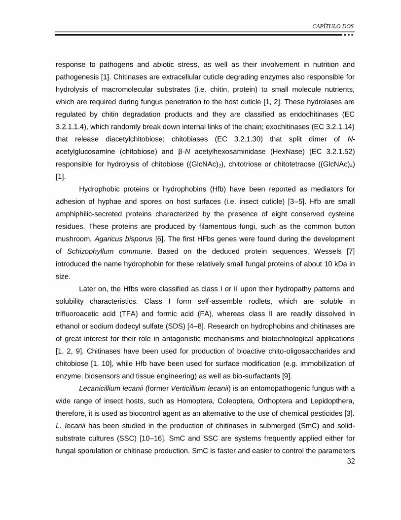
CAPÍTULO DOS
32
response to pathogens and abiotic stress, as well as their involvement in nutrition and
pathogenesis [1]. Chitinases are extracellular cuticle degrading enzymes also responsible for
hydrolysis of macromolecular substrates (i.e. chitin, protein) to small molecule nutrients,
which are required during fungus penetration to the host cuticle [1, 2]. These hydrolases are
regulated by chitin degradation products and they are classified as endochitinases (EC
3.2.1.1.4), which randomly break down internal links of the chain; exochitinases (EC 3.2.1.14)
that release diacetylchitobiose; chitobiases (EC 3.2.1.30) that split dimer of N-
acetylglucosamine (chitobiose) and β-N acetylhexosaminidase (HexNase) (EC 3.2.1.52)
responsible for hydrolysis of chitobiose ((GlcNAc)2), chitotriose or chitotetraose ((GlcNAc)4)
[1].
Hydrophobic proteins or hydrophobins (Hfb) have been reported as mediators for
adhesion of hyphae and spores on host surfaces (i.e. insect cuticle) [3–5]. Hfb are small
amphiphilic-secreted proteins characterized by the presence of eight conserved cysteine
residues. These proteins are produced by filamentous fungi, such as the common button
mushroom, Agaricus bisporus [6]. The first HFbs genes were found during the development
of Schizophyllum commune. Based on the deduced protein sequences, Wessels [7]
introduced the name hydrophobin for these relatively small fungal proteins of about 10 kDa in
size.
Later on, the Hfbs were classified as class I or II upon their hydropathy patterns and
solubility characteristics. Class I form self-assemble rodlets, which are soluble in
trifluoroacetic acid (TFA) and formic acid (FA), whereas class II are readily dissolved in
ethanol or sodium dodecyl sulfate (SDS) [4–8]. Research on hydrophobins and chitinases are
of great interest for their role in antagonistic mechanisms and biotechnological applications
[1, 2, 9]. Chitinases have been used for production of bioactive chito-oligosaccharides and
chitobiose [1, 10], while Hfb have been used for surface modification (e.g. immobilization of
enzyme, biosensors and tissue engineering) as well as bio-surfactants [9].
Lecanicillium lecanii (former Verticillium lecanii) is an entomopathogenic fungus with a
wide range of insect hosts, such as Homoptera, Coleoptera, Orthoptera and Lepidopthera,
therefore, it is used as biocontrol agent as an alternative to the use of chemical pesticides [3].
L. lecanii has been studied in the production of chitinases in submerged (SmC) and solid-
substrate cultures (SSC) [10–16]. SmC and SSC are systems frequently applied either for
fungal sporulation or chitinase production. SmC is faster and easier to control the parameters

CAPÍTULO DOS
33
(i.e. pH and temperature); however, the products are more diluted than with SSC [10–16].
Herein, we are first to report Hfb production from this fungus in SSC and SmC.
2.2 EXPERIMENTAL
2.2.1 Microorganism and culture conditions
Lecanicillium lecanii strain 2149 was obtained from the Entomopathogenic Fungal
Culture Collection (Ithaca, New York, NY, USA). L. lecanii was cultured on potato dextrose
agar at 25 °C and maintained at 4 °C until needed. Spore suspension was prepared by
agitation of cultures with a solution of 0.1 % (v v-1) of Tween 80. Spores were counted with
Neubauer chamber and the inoculum size was 5 х 107 spores per g of substrate for SmC and
SSC.
2.2.2 SmC and SSC media
Mineral medium composition (g L-1) was; K2HPO4 (2.8), MgSO4•7H2O (1.38),
CaSO4•2H2O (0.48), FeSO4•7H2O (0.22), (NH4)2SO4 (7.5) CaCl3 (0.48), NaH2PO4•7H2O
(1.67), yeast extract (1), and either colloidal chitin (30) or fructose (22) was used to SmC and
SSC [10]. Control cultures were carried out with fructose as carbon source. SmC was
conducted in flasks incubated at 25 °C in rotary shaker at 180 rpm for 144 h [12]. Biomass
was separated by centrifugation of the SmC at 4 °C and 14,000 g for 15 min (Beckman J2-MI,
USA). The supernatant was filtered and ready to use as crude enzyme for further assays, i.e.
protein determination and HexNase activity. SSC was carried out in glass columns packed
with polyurethane foam (PUF) with size ca. 0.125 cm3 within nutrient ratio of 1:15 (w v-1) and
aeration rate of 1.4 mL air min-1 per g of moist material. SSC was collected from each
column, then mixed with its same weight in water and pressurized to 1,000 psi followed by
centrifugation at 4 °C and 12,700 g for 20 min. Crude enzyme was prepared as described
above.
2.2.3 Detection of chitinolytic activity on SDS–PAGE
Gels for SDS–PAGE [17] (mini Protean II Bio-Rad, Hercules, CA, USA) were prepared
with added 0.01 % (w v-1) of either glycol chitin or chito-oligosaccharides [Chitopentaose
((GlcNAc)5), (GlcNAc)4, (GlcNAc)2] as substrates. Freeze-dried crude enzyme obtained after

CAPÍTULO DOS
34
144 h of culture was subjected to SDS–PAGE. Then, gels were treated in 0.1 M phosphate
buffer (pH 6) and 1% (v v-1) Triton X100 for 24 h at 37 °C. Further, gels were immersed for 5
min into a freshly prepared solution of Calcofluor white M2R (Sigma-Aldrich, St. Louis MO,
USA) 0.01 % (w v-1) in 0.5 M Tris–HCl buffer (pH 9). Gels were then rinsed with deionized
water for 1 h. Chitinolytic activities appeared as fluorescent zones within homogenously dark
background upon illumination using an UV transilluminator (Gel Doc Bio-Rad) [18]. Bands
were analyzed with the ImageJ 1.41o program (National Institutes of Health, USA). All
experiments were carried out by duplicate.
2.2.4 Determination of HexNase activity
HexNase activities were determined following the procedure reported by Tronsmo and
co-workers [19]. Crude enzyme (200 µL) was added to 200 µL of a citrate–phosphate buffer
(0.2 M, pH 5.6) and 200 µL of p-nitrophenyl-β-N-acetylglucosamine (2.9 mM) and then
incubated with agitation (180 rpm) at 37 °C for 1 h. Reaction was halted by the addition of
NaOH (1 mL, 20 mM). The released p-nitrophenol was monitored spectrophotometrically at k
of 400 nm in a UV–Vis JENWAY 6305 (Essex, UK). One unit of HexNase was defined as the
amount of crude enzyme required to release 1 lmol of p-nitrophenol per minute.
Determinations were carried out every 24 h until 144 h.
2.2.5 Extraction of Hfb
Hfbs class I were extracted from mycelia after 144 h of cultivation either in SSC or
SmC with the following procedure; PUF with the fungal mycelia were mixed with SDS (2 w v-1
%) in Tris–HCl (100 mM pH 9) buffer for 2 h at 25 °C and then centrifuged. Pellets were
treated with concentrated FA (J. T. Baker, Mexico), neutralized and centrifuged (4 °C, 5,000
g) for 5 min. The supernatant was precipitated by electrobubbling [20]. Extracts were
centrifuged and the pellet was solubilized with TFA and further evaporated with dry air
stream. The biomass in SmC media was separated from the culture broth by centrifugation
(14,000g) at 4 °C for 15 min.

CAPÍTULO DOS
35
2.2.6 SDS–PAGE analysis
Protein in the dried extract was determined by Bradford method [21] and subjected to
SDS–PAGE [17]. Gels were stained with silver nitrate (Bio-Rad) and analyzed with the
ImageJ 1.41o software (National Institutes of Health, USA).
2.2.7 Evaluation of Hfb on the surface activity of Teflon
Surface activities were evaluated as the ability to modify the surface hydrophobicity of
Teflon (poly(tetrafluoroethylene)) by the Hfbs by measuring the contact angle [20]. Teflon was
washed with water and ethanol and then dried. Hfb solution was prepared dissolving extract
of dried protein in deionized water at a concentration of 50 µg protein mL -1. Hfb solution was
used onto 1.32 cm2 of Teflon. Subsequently, 1 µL of deionized water was dropped on the
treated Teflon surface and observed with a side-illumination horizontal light microscope Intel
Qx3 (Intel Corporation, USA). Contact lengths (b) and heights (h) of the water droplet were
measured by ImageJ 1.41o software (National Institutes of Health). Contact angle was
calculated according to the Eq. 2.1 [22]. Analyses were conducted by quadruplicate, in three
different random locations on each surface.
1 2
2 tanh
b
(2.1)
2.2.8 Statistical analysis
A randomized design was carried out in quadruplicate with HexNase activity as
response variable and culturing condition, i.e. SSC and SmC, as source of variation. As well,
statistical analysis was carried out on surface activity of Hfbs obtained from SSC with
different carbon sources with contact angle measurements as response variable. Analysis of
variance and multiple comparisons of means by Tukey– Kramer (P ≤ 0.05) were performed
using NCSS 2000 software (NCSS Inc., USA 2001).
2.2.9 Scanning electron microscopy
Samples of SSC and SmC with added chitin media were prepared by immersion in
5% (v v-1) glutaraldehyde at 4 °C for 24 h and post-treated with OsO4 1 % (w v-1) for 2 h.
Then, samples were dehydrated in a graded alcohol series and covered with carbon and gold

CAPÍTULO DOS
36
prior to scanning electron microscopy (SEM) examinations using a JEOL JSM-5900 LV
(Japan).
2.3 RESULTS
2.3.1 HexNase activity from SSC and SmC media
Several factors have been reported as determinants in the production of chitinases in
SmC and SSC of Verticillium such as pH, addition of chitin as carbon source and inducer, as
well as culturing conditions i.e. type and size of support or agitation rate [10–16]. The
increment of moisture content of sugar cane bagasse (support) and mycelia as inoculums in
SSC of L. lecanii was reported to improve significantly the enzyme yield and to reduce the lag
phase. In fact, the system of culture has impact not only on the enzyme yield, but also in the
fungal metabolism as reported by Marin-Cervantes and co-workers [13]. Therein, catabolic
repression depended on moisture level in SSC, being stronger for lower moisture contents for
exo chitinases but milder or insignificant for endo-chitinases. In the present work, HexNases
production was significantly higher in SSC than in SmC with added chitin media, whereas,
insignificant HexNase activities were determined in SmC and SSC in the control media with
fructose as sole carbon source (Fig. 2.1).

CAPÍTULO DOS
37
b
a
a
a a
b
b
b
bb b
a
a
ca
c
cc c c
c
0
2
4
6
8
10
12
0 24 48 72 96 120 144
Hex
Nas
e (
U/m
g o
f p
rote
in)
Time (h)
Figure 2. 1. Time course of HexNase activities by L. lecanii in SmC with added chitin (open
triangle) or fructose (filled triangle) SSC with added chitin (open square) or fructose (filled
square) as carbon sources.
2.3.2 Chitinolytic activity on SDS–PAGE
Solid-substrate cultures with added chitin showed two bands in SDS–PAGE with
exochitinases activities of 32 and 51 kDa (Fig. 2.2). The endochitinases activity in this culture
displayed a band of 26 kDa upon glycol chitin and chitopentaose (GlcNAc)5. Additionally,
HexNases, which are responsible for hydrolysis of chitobiose (GlcNAc)2 and chitotetraose
(GlcNAc)4 [1] evidenced their activity with bands of 61, 80, 96 and 111 kDa. Similarly, SmC
with added chitin media produced exochitinases of 32 and 45 kDa, which released
diacetylchitobioses, further substrates for HexNases. Endochitinase activity was observed in
26 kDa, which acted on (GlcNAc)4, whereas HexNase with bands of 61, 96 and 111 kDa were
detected in the gel with (GlcNAc)2 (Fig. 2.2).
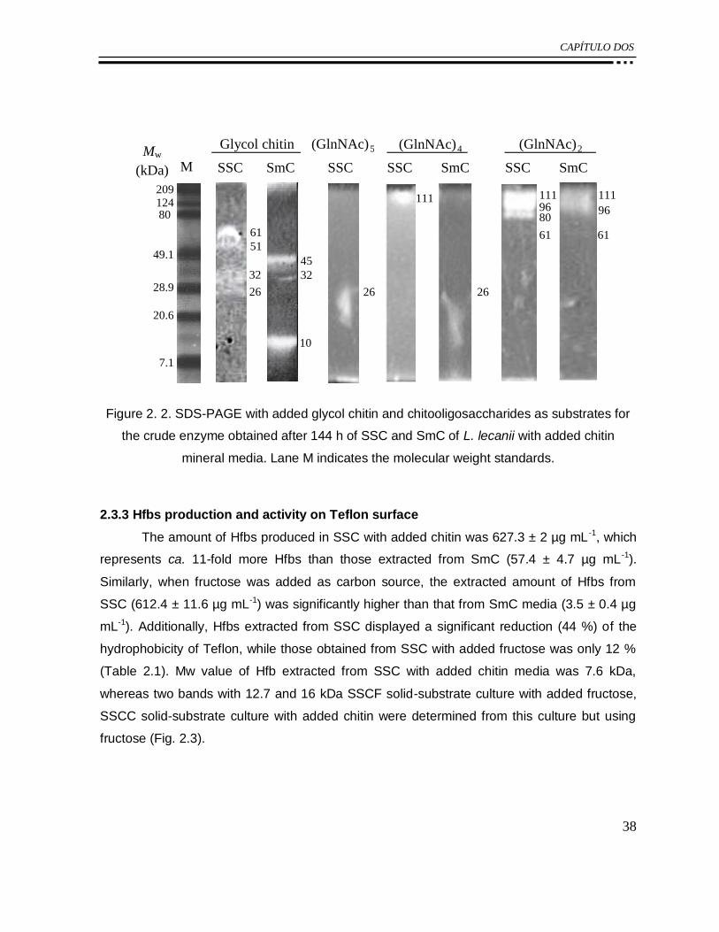
CAPÍTULO DOS
38
1
80
209 124
49.1
28.9
20.6
7.1
(GlnNAc) 5 (GlnNAc) 4 Glycol chitin (GlnNAc) 2
M SmC SSC SSC SSC SSC SmC SmC
61 51
32
26
45 32
10
26
111 111 96 80
61 61
111 96
26
Mw
(kDa)
Figure 2. 2. SDS-PAGE with added glycol chitin and chitooligosaccharides as substrates for
the crude enzyme obtained after 144 h of SSC and SmC of L. lecanii with added chitin
mineral media. Lane M indicates the molecular weight standards.
2.3.3 Hfbs production and activity on Teflon surface
The amount of Hfbs produced in SSC with added chitin was 627.3 ± 2 µg mL -1, which
represents ca. 11-fold more Hfbs than those extracted from SmC (57.4 ± 4.7 µg mL -1).
Similarly, when fructose was added as carbon source, the extracted amount of Hfbs from
SSC (612.4 ± 11.6 µg mL-1) was significantly higher than that from SmC media (3.5 ± 0.4 µg
mL-1). Additionally, Hfbs extracted from SSC displayed a significant reduction (44 %) of the
hydrophobicity of Teflon, while those obtained from SSC with added fructose was only 12 %
(Table 2.1). Mw value of Hfb extracted from SSC with added chitin media was 7.6 kDa,
whereas two bands with 12.7 and 16 kDa SSCF solid-substrate culture with added fructose,
SSCC solid-substrate culture with added chitin were determined from this culture but using
fructose (Fig. 2.3).
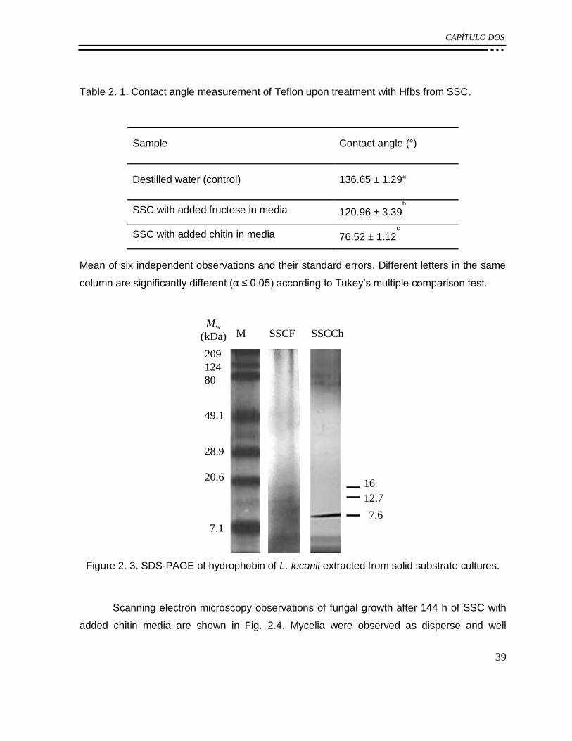
CAPÍTULO DOS
39
Table 2. 1. Contact angle measurement of Teflon upon treatment with Hfbs from SSC.
Sample Contact angle (°)
Destilled water (control) 136.65 ± 1.29a
SSC with added fructose in media 120.96 ± 3.39b
SSC with added chitin in media 76.52 ± 1.12c
Mean of six independent observations and their standard errors. Different letters in the same
column are significantly different (α ≤ 0.05) according to Tukey’s multiple comparison test.
M SSCF SSCCh
7.1
20.6
28.9
209
124
80
49.1
Mw
(kDa)
7.6
16
12.7
Figure 2. 3. SDS-PAGE of hydrophobin of L. lecanii extracted from solid substrate cultures.
Scanning electron microscopy observations of fungal growth after 144 h of SSC with
added chitin media are shown in Fig. 2.4. Mycelia were observed as disperse and well

CAPÍTULO DOS
40
distributed through the PUF (Fig. 2.4a). The biopolymers covered the fungal cells forming a
mucilaginous coat and mycelia abundantly colonized the PUF surface (Fig. 2.4b).
1
2
a
b
PUF
BP
H
H
BP
Figure 2. 4. SEM micrographs of PUF after 144 h of inoculation of L. lecanii (H) with added
chitin media; producing mucilagenous coat (BP): a х 1,000, b х 2,000.

CAPÍTULO DOS
41
2.4 DISCUSSION
Experimental evidences pointed out that SSC produced more chitinolytic enzymes
than SmC (Fig. 2.1), which might be attributed to the enhanced aeration, thus affecting the
fungal metabolism on insoluble substrates, i.e. chitin [13]. This is in agreement with previous
reports on chitinolytic enzyme production of L. lecanii in SSC using sugarcane bagasse [11,
12, 16] and PUF [13]. Barranco-Florido and co-workers [16] reported that the production of
chitinases and proteases by L. lecanii was higher in SSC than SmC using cuticle Sphenarium
purpurascens as inducer. In addition to the use of chitinous substrate as inducer and sole
carbon source, other factors such as moisture content, mycelia aggregation in the support
[13], and inoculum age have been evaluated in SSC of L. lecanii for enhancement of
HexNases production [12]. We observed that fructose, as carbon source, showed catabolic
repressive effects evidenced by negligible HexNases activities (Fig. 2.1). Similarly, it is
reported that addition of glucose to the media in SSC of L. lecanii repressed HexNases
production, despite of improved biomass production [13].
Although most of the reports dealing with chitinases detection and characterization
use SmC [14, 15, 23], our study displayed chitinases in both SSC and SmC having wide Mw
range as well as endo , exo- and HexNase activities (Fig. 2.2). Fenice and co-workers [14]
reported a chitinase of L. lecanii in SmC of culture with Mw of 45 kDa and others detected
two chitinases with Mw of 33 and 50 kDa under similar culture conditions [15]. Lu and co-
workers [23] isolated and characterized two exochitinases from submerged cultures of L.
lecanii in the presence of chitin, CHII and CHI2 with 40.93 and 45.95 kDa, respectively. In the
present work, we found an exochitinase with Mw of 45 kDa in crude enzyme from SmC of L.
lecanii (Fig. 2.2), which according to Mw was assigned to a basic chitinase also reported as
extracellular and devoted to chitin digestion [23]. Zymograms displayed exochitinases of 32
and 51 kDa in SSC with added chitin as inducer and carbon source (Fig. 2.2) and
additionally, several endochitinases and HexNases were produced both in SmC and SSC.
Regarding Hfb production, multiple genes of these proteins have been reported from
microorganisms, which can be differentially expressed as a response to different
developmental stages, thus Hfb class I is found in cell walls of Trichoderma reesei, whereas
class II is present on spore walls [9]. Environmental condition also affected the production of

CAPÍTULO DOS
42
Hfb, such as the addition of compounds of opposite polarities for production of class II Hfbs of
Rhinocladiella similis [24]. de Vocht and co-workers [25] reported that 1 mg of Hfb (class I)
extracted from S. commune was enough to reduce the contact angle by its coating over 1 m2
of a Teflon monolayer of ca. 10 nm of thickness. Herein, we focused on the determination of
Hfbs class I, considering that fungi require Hfbs to allow growth of hyphae into the air. Our
results displayed that the Hfb produced from SSC with added chitin media had significantly
higher surface activity than that obtained using fructose. It is worth to note, that in the present
work Hfb activity was influenced by the carbon source (Table 2.1). Furthermore, Hfbs
produced by L. lecanii in SSC using chitin had similar Mw (ca. 7.6 kDa) to those reported
from other sources, such as Verticillium fungicola [26].
Generally, fungal cells are relatively tolerant to stress when they growth in air by the
production of Hfbs, which are key factors for development of fungi on solid or liquid surfaces
[4, 5]. Additionally, a previous report showed that the production of mucus coat on the
hydrophilic spore of L. lecanii facilitates dispersal and adhesion onto hydrophobic host
surfaces [5].
Mucilaginous coat was also observed by SEM in our samples (Fig. 2.4) along with
remarkable capacity of the FA extracted proteins (Hfbs) to modify the hydrophobicity (Table
2.1). This suggests that the solid culture requires the presence of Hfbs to enable the
development of fungal cells attached to the support at contact to air. According to this
successful result, further work is underway on Hfb identification and purification from L.
Lecanii.
2.5 CONCLUSIONS
The production of Hfb was successfully achieved from L. lecanii. The type of culture
and carbon source influenced results significantly as SSC with added chitin and fructose
improved the Hfb production when compared with those from SmC herein and in other
reports using other these and other fungi or carbon sources. In addition, the use of chitin in
SSC media produced more chitinolytic enzymes than SmC.
Acknowledgments The authors gratefully acknowledged to CONACYT for the research
funding (Project Number 105628) and for PhD scholarship grant (ZRP).

CAPÍTULO DOS
43
REFERENCIAS
1. Shirai K (2006) Fungal chitinases. In: Guevara-González RG, Torres-Pacheco I (eds)
Advances in agricultural and food biotechnology, Research Signpost, Kerala, pp 289–304.
2. St. Leger RJ, Joshi I, Roberts D (1998) Appl Environ Microbiol 64:709–713.
3. Goettel MS, Koike M, Jun Kim J, Aiuchi D, Shinya R, Brodeur J (2008) Invertebr Pathol
98:256–261.
4. Wösten HAB, Willey JM (2000) Microbiology 146:767–773.
5. Jeffs LB, Xavier IJ, Matai RE, Khachatourians GG (1999) Can J Microbiol 45:936–948.
6. Linder MB (2009) Curr Opin Colloid Interface Sci 14:356–363.
7. Wessels JGH (1994) Annu Rev Phytopathol 32:413–437.
8. Askolin S, Nakari-Setälä T, Tenkanen M (2001) Appl Microbiol Biotechnol 57:124–130.
9. Linder M, Szilvay G, Nakari-Setälä T, Penttila M (2005) FEMS Microbiol Rev 29:877–896.
10. Ramirez-Coutiño L, Espinosa-Marquez J, Peter MG, Shirai K (2010) Bioresour Technol
101:9236–9240.
11. Barranco-Florido E, Bustamante CP, Mayorga-Reyes L, González CR, Martínez CP,
Azaola A (2009) Interciencia 34:356–360.
12. Matsumoto Y, Saucedo G, Revah S, Shirai K (2004) Proc Biochem 39:665–671
13. Marin-Cervantes MC, Matsumoto Y, Ramírez-Coutiño L, Rocha- Pino Z, Viniegra G,
Shirai K (2008) Proc Biochem 43:24–32.
14. Fenice M, Selbmann L, Di Giambattista R, Federici F (1998) Res Microbiol 149:289–300.
15. Liu BL, Kao PM, Tzeng YM, Feng KC (2003) Enzyme Microb Technol 33:410-415.
16. Barranco-Florido E, Alatorre R, Gutiérrez M, Viniegra G, Saucedo G (2002) Enzyme
Microbiol Technol 30:10910–10915.
17. Laemmli UK (1970) Nature 227:680–685.
18. Trudel J, Asselin A (1989) Anal Biochem 178:362–366.
19. Tronsmo A, Harman GE (1993) Anal Biochem 208:74–79.
20. Vigueras G, Shirai K, Martins D, Franco TT, Fleuri LF, Revah S (2008) Appl Microbiol
Biotechnol 80:147–154.
21. Bradford MM (1976) Anal Biochem 72:248–254.
22. Dynesen J, Nielsen J (2003) Biotechnol Prog 19:1049–1052.
23. Lu ZX, Laroche A, Huang HC (2005) Can J Microbiol 51:1045–1055.
CAPÍTULO DOS
44
24. Vigueras G, Arriaga S, Shirai K, Morales M, Revah S (2009) Biotechnol Lett 31:1203–
1209.
25. de Vocht ML, Wösten HAB, Wessels JGH (2001) Method of treating a surface of an
object with a hydrophobin-containing solution. WO 01/57528.
26. Calonje M, Bernardo D, Novaes LM, García M (2002) Can J Microbiol 48:1030–1034.

CAPÍTULO TRES
45
CAPÍTULO 3:
The hydrophobicity of the support in solid
substrate culture affected the production of
hydrophobins from Lecanicillium lecanii
Zaizy Rocha-Pino, Gabriel Vigueras, José D. Sepúlveda-Sánchez, Maribel Hernandez, José
Campos-Terán, Francisco J. Fernández and Keiko Shirai*
ENVIADO Y EN REVISIÓN
Process Biochemstry, Ref. No.: PRBI-D-14-00471, April 28th, 2014. Major revision received
on 17th June, 2014.

CAPÍTULO TRES
46
The hydrophobicity of the support in solid substrate culture affected
the production of hydrophobins from Lecanicillium lecanii
Zaizy Rocha-Pino,1 Gabriel Vigueras,2 José D. Sepúlveda-Sánchez,1 Maribel Hernandez,2
José Campos-Terán,2 Francisco J. Fernández1 and Keiko Shirai1*
ENVIADO Y EN REVISIÓN
Process Biochemstry, Ref. No.: PRBI-D-14-00471, April 28th, 2014. Major revision received
on 17th June, 2014
Abstract
Lecanicillium lecanii has been successfully employed to produce hydrophobin-like
proteins (HfbL) in solid substrate and submerged cultures varying the type of solid inert
support. This study show the results on the effect of the hydrophilic Perlite and hydrophobic
Polyurethane inert supports in the solid substrate cultures for production of HfbLs by L.
lecanii. Class I HfbL was only produced using the hydrophobic polyurethane foam support,
showing high surface activity that reduced ca. 50 % hydrophobicity of Teflon, whereas class II
HfbLs were produced on both polyurethane foam and perlite supports, and they reduced ca.
50 % the surface tension of water ca. 25% reduction of the hydrophobicity of Teflon.
Keywords: Hydrophobins, chitin, Lecanicillium lecanii, solid substrate culture, polyurethane
foam, perlite
3.1 INTRODUCTION
The hydrophobins (Hfbs) are amphipathic proteins produced by fungi with eight
conserved cysteine residues forming disulfide bonds [1]. These proteins are classified
according to their biophysical and hydropathy properties; Class I Hfbs are soluble in formic
acid and self-assemble like amyloid proteins forming monolayers, named rodlets during their
interaction at hydrophilic and hydrophobic interfaces. The rodlets are highly stable layers

CAPÍTULO TRES
47
which are dissolved in trifluroacetic acid and they have been observed at surfaces of aerial
structures such as conidia, conidiophores and fruiting bodies [2]. Class II Hfbs are soluble in
sodium dodecyl sulfate (SDS) or ethanol (60 %) solutions and present coat aerial structures
and yeast-like cells but they assemble as flexible layers without rodlets [3]. The amphipathic
properties and self-assemble of Hfb are related to their biological roles in fungal development
by enabling the hyphae to migrate from submerged condition to air. In addition, Hfbs have
been involved in pathogenic activity acting as toxins and allowing attachment of fungal
structures, such as conidia or hyphae, to the host surface [4,5]. Hfb layers also covers fungal
aerial structures, which confer hydrophobicity, wetting resistance and facilitating their
dispersal in air, hyphae protection against desiccation and gas exchange [1,6]. The presence
of Hfb like proteins were also reported as mediators of adhesion of on hydrophobic surfaces
[7,8]. Some examples of these are the Hfbs class II from Trichoderma reesei, which are
responsible of sporulation of the fungi in solid cultures [7]. Several authors reported class I
Hfbs from Schizophylum commune (SC3) and the class II HFBI from T. reesei, which are
secreted to liquid culture to interact at the air-water interface aiding the migration of hyphae
into the air [1,7]. In another study, Hfb-like proteins were determined only with aerial conidia
of entomopathogenic fungus Beauveria bassiana but not detected in blastospores and
submerged conidia. Another entomopathogenic fungus used as biopesticide is L. lecanii,
which is able to degrade n-hexane or toluene in submerged cultures (SmC), producing Hfbs
like proteins with surface activity at the presence of these hydrophobic solvents [9]. The
reported comparison among type of culture using this fungus displayed higher production of
Hfbs class I in solid substrate culture (SSC) with added chitin or fructose as carbon source
than in SmC with these substrates. Interestingly, the SSC-mediated Hfb reduced ca. 50 % the
hydrophobicity of Teflon unlike to those obtained from SSC with added fructose, which
showed no surface activity [10]. Despite of these reports, there are no reported studies on the
use of inert supports on production of these proteins. In this regard, Perlite (P) and
Polyurethane foam (PUF) have been employed as packed beds in SSC for several purposes
including hydrocarbon degradation, enzyme and biomass production. The P is hydrophilic
silicaceous material of volcanic origin while PUF is a synthetic polymer based on isocyanate
polymers, which consist of polar urethane groups and soft non-polar segments. Both
materials are considered as inert and did not contribute nutritionally to fungal growth,
although they can hold several times its weight in water owing to their high porosity and they

CAPÍTULO TRES
48
are neutral in water without cation exchange capacity [11-13]. This study is first to report the
use of PUF and P as solid porous matrices to support the fungal growth of L. lecanii in SSC
for Hfb production. The effect of the solid support on the type and surface activities of Hfbs is
described.
3.2 MATERIALS AND METHODS
3.2.1 Microorganism and culture conditions
Lecanicillium lecanii 2149 strain was provided by USDA-ARSEF (Agricultural
Research Service Collection of Entomopathogenic Fungi, USA) collection. L. lecanii was
grown in SSC on two inert supports: i) PUF with a particle size ca. 0.125 cm3 within nutrient
ratio of 1:15 (w v-1); ii) P with a particle diameter ca. 3.3 mm within nutrient ratio of 1:2 (w v -1).
Culture conditions were carried out according to Rocha-Pino et al. [10], the supports added
with mineral medium pH 6, colloidal chitin (30 g L-1) and inoculum of 5 x 10⁷ spores g-1 of
substrate were packed into glass columns and incubated at 25 °C. Aeration of 1.4 mL air min-
1 per g of moist material was provided during 6 and 15 days. All the materials were sterilized
at 121 °C for 15 min before inoculation.
Solids (biomass and matrices) and supernatant (soluble matter in water) were
separated by mixing the solids with water (50 wt%), then it was pressurized to 1,000 psi for
PUF SSC, while the mixture was pulverized in a mortar for P SSC and centrifuged at 12,700
g at 4 °C for 10 min. Supernatants from both supports were separated using Whatman filter
paper no. 40. 0.2 g (wet weight) of support with biomass was mixed with 5 mL of phosphoric
acid (0.15 M) and heated in a water bath for 7 min. After centrifugation (12,700 g) the
supernatant was used to determine the total protein from biomass [9]. Total soluble protein
was determined from biomass and supernatant by Bradford [14].
3.2.2 Colloidal chitin preparation and characterization
Chitin was obtained from lactic acid fermentation of shrimp wastes and then purified
by the following treatments: i) chitin was washed with distilled water (Ch1); ii) Ch1 was
treated with HCl 0.5 N and NaOH 0.4 M (Ch2). Furthermore, Ch1 and Ch2 were treated with
HCl 10 N and neutralized with water to obtain colloidal solutions. Ch1 and Ch2 were
characterized on residual protein (%) by Kjeldahl method (K-435 BÜCHI, Switzerland) [15].

CAPÍTULO TRES
49
Degree of acetylation (DA) was determined with chitin treated with DCl/D2O and analyzed by
proton nuclear magnetic resonance (1H NMR) spectroscopy (Bruker Advance III 500,
Germany) at 200 MHz using 3-(trimethylsilyl) propionic acid as internal reference. DA was
calculated from integration of assigned signals on the NMR spectra [16].
3.2.3 HfbLs extraction
The samples used for this analysis were class I and class II HfbLs fractions obtained
from the mycelia of L. lecanii after 6 d of SSC. HfbLs were extracted from the biomass and
the supernatant of L. lecanii in SSC. Class II HfbLs were extracted from mycelia following the
methodology reported by Askolin et al. [3]. Supports with mycelia were washed with distilled
water and incubated with SDS 2 % (w v-1) in 100 mM Tris-HCl buffer, pH 9.0 for 2 h in cold
water bath and soft stirring agitation. Then, mycelia and support were separated by
compressing or centrifugation for PUFSSC and PSSC, respectively. Remaining SDS extract
was precipitated with KCl (2 M) and centrifuged (12,700 g) at 6 °C for 20 min. Subsequently,
supports were washed with water and Class I HfbLs were extracted with formic acid, followed
by electrobubbling at 300 mA during 3 h. The foam obtained was centrifuged and the pellet
solubilized with TFA and evaporated with dry air stream [10]. Class I HfbLs from supernatant
were extracted by the following procedure: protein from supernatant was precipitated with
trichloroacetic acid (TCA) 5 % (w v-1) at 4 °C for 2 h and centrifuged (12,700 g) at 4 °C for 20
min. Pellet was washed with acetone and solubilized with performic acid at 4 °C h during 4 h,
subsequently the acid was evaporated with air flow [17]. Protein precipitated (without addition
of performic acid) was treated as class II HfbLs [17]. For each step of the HfbLs extraction,
the protein was determined by Bradford [14]. Protein yield with respect to biomass
(YHfbL/biomass) was calculated considering protein concentration of each step of purification and
the total protein of either biomass or supernatant. Classes I and II HfbLs fractions were
analyzed by denaturing electrophoresis SDS-PAGE according to Laemmli [18] using
resolving gel of polyacrylamide in concentrations of 17 % and 15 % for class I and class II
HfbLs, respectively. Gels were stained with coomassie blue or silver nitrate (Bio-Rad, USA)
and analyzed with the image processing software (ImageJ 1.41o National Institutes of Health,
USA).
3.2.4 Determination of surface activities of HfbLs by contact angle measurements

CAPÍTULO TRES
50
Surface activity of HfbLs at air-solid hydrophobic interface was measured by contact
angle () of a water drop (1 µL) on Teflon (poly (tetrafluoroethylene)) surface previously
treated with an HfbL solution of 50 µg mL-1 [10]. Teflon treated with class I HfbL was washed
with 2% SDS at 100 °C and then with deionized water, while class II HfbLs were washed only
with deionized water at room temperature. In addition, class II HfbLs were subjected to a
molecular weight cut off membrane of 30 kDa (Amicon Millipore, USA). Digital images of the
water droplets were obtained in a horizontal light microscope Qx3 Intel with image processor
(Intel Corporation, USA). Contact angles measurements were carried out by duplicate,
randomly determined in six areas per sample and images analyzed with ImageJ 1.41o
software.
3.2.5 Determination of HfbL surface activities by measurements of surface tension
Surface activity of HfbLs was determined at air-water interface by measuring the
symmetry of a pendant drop of deionized water as reference and HfbLs solutions (50 µg mL-
1). Analysis was carried out using a Theta KSV optical tensiometer system (KSV Instruments,
Finland) calibrated with a 4.00025 mm, grade 25 and tungsten carbide sphere. Images were
analyzed through a Young-Laplace model employing Attention Theta software V 4.1.0 (Biolin
Scientific, Finland). Surface tension determinations were carried out by duplicate with six
determinations per sample.
3.2.6 Scanning electron microscopy of L. lecanii in SSC
L. lecanii grown on PUF or P and colloidal chitin were immersed in glutaraldehyde 5 %
(v v-1) at 4 °C for 24 h and subsequently treated with OsO4 1 % (w v-1) for 2 h. Then,
dehydrated with alcohol and covered with carbon and gold for scanning electron microscope
JEOL JSM-5900 LV (Japan).
3.2.7 Statistical analysis
A randomized design was applied using yield, contact angle and surface tension of
HfbLs as response variables and chitin purity and support type as independent variables.

CAPÍTULO TRES
51
Analysis of variance (ANOVA) and multiple comparisons of means by Tukey-Kramer test (p ≤
0.05) were performed using NCSS 2000 software (NCSS Inc., USA 2001).
3.3 RESULTS AND DISCUSSION
3.3.1 Effect of chitin purity on the HfbL production from L. lecanii in SSC.
Insoluble biopolymers, such as starch, cellulose, pectin, lignin or chitin have been
used as substrates for SSC. Chitin has been studied as support and sole carbon source, as
well as enzyme inducer for chitinases production [15, 16, 19-21]. The physicochemical
properties of chitinous substrates might have influence when used for microbial growth and
production of chitinolytic enzymes therefore the purity of this biopolymer on the class I and II
Hfbs production from L. lecanii in SSC using PUF as support is prompted in this study. The
chitins tested presented 8.6 ± 0.2 % w w-1 and 5.7 ± 0.3 % w w-1 of residual protein and DAs
of 100 % and 89.8 % for Ch1 and Ch2, respectively. Ch2 used as carbon source increased
ca. 14 % the class I HfbLs production from L. lecanii grown on PUF compared to Ch1 (Figure
3.1). However, the difference in purity of the chitin shows no significant effect (p ≤ 0.05) on
the class II HfbLs production, which might be ascribed to the residual protein bonded to chitin.
This residual protein might form a physical interference between fungus-substrate
interactions. Furthermore, the residual protein induces fungal proteases [21], which could
reduce chitinases and the Hfbs production required to the adhesion on chitin thereby its
hydrolysis.

CAPÍTULO TRES
52
YHfbL/biomass
5.2 0.3 % a
YHfbL/biomass
19.1 0.8 % c
YHfbL/biomass
16 1.3 % b,c
YHfbL/biomass
17.6 1.1 % c
0
50
100
150
200
250
300
350
Ch1 Ch2
Hfb
L (
µg m
L-1
)
Chitin
Figure 3. 1. Production of HfbLs associated to mycelia of L. lecanii cultured on PUF after 14
d. Class I (void), Class II (solid). YHfbL/biomass (%) with each condition is indicated. Different
letters in the same column are significantly different (α ≤ 0.05) according to Tukey-Kramer
multiple comparison test.
On the other hand, Ch2 showed lower DA (89.8 %) than Ch1 (100 %) which might
affect the interaction between fungus and biopolymer. According to Holder and Keyhani [8],
Hfbs from Beauveria bassiana were adhered to specific surfaces upon their hydrophobicity. In
this regard, the hydrophobic character in chitin is associated to the acetyl groups responsible
for their interactions among chains, including the hydrogen bonding, electrostatic repulsion
owing to positive charged amino group and crystallinity [22]. Therefore, the crystallinity
reduction and the acetyl moieties increase the interaction among chitin and enzymes
produced by L. lecanii during its hydrolysis [21]. It has been reported that Hfbs class II show
high affinity to polar and cationic surfaces but not to anionic surfaces [23] and thus, positively
charged amino groups as a consequence of deacetylation might influence the Hfbs
production from L. lecanii.
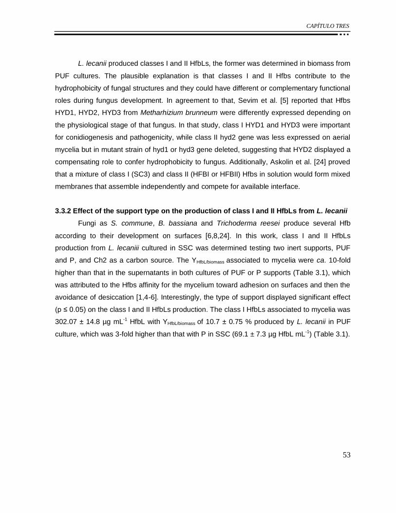
CAPÍTULO TRES
53
L. lecanii produced classes I and II HfbLs, the former was determined in biomass from
PUF cultures. The plausible explanation is that classes I and II Hfbs contribute to the
hydrophobicity of fungal structures and they could have different or complementary functional
roles during fungus development. In agreement to that, Sevim et al. [5] reported that Hfbs
HYD1, HYD2, HYD3 from Metharhizium brunneum were differently expressed depending on
the physiological stage of that fungus. In that study, class I HYD1 and HYD3 were important
for conidiogenesis and pathogenicity, while class II hyd2 gene was less expressed on aerial
mycelia but in mutant strain of hyd1 or hyd3 gene deleted, suggesting that HYD2 displayed a
compensating role to confer hydrophobicity to fungus. Additionally, Askolin et al. [24] proved
that a mixture of class I (SC3) and class II (HFBI or HFBII) Hfbs in solution would form mixed
membranes that assemble independently and compete for available interface.
3.3.2 Effect of the support type on the production of class I and II HfbLs from L. lecanii
Fungi as S. commune, B. bassiana and Trichoderma reesei produce several Hfb
according to their development on surfaces [6,8,24]. In this work, class I and II HfbLs
production from L. lecaniii cultured in SSC was determined testing two inert supports, PUF
and P, and Ch2 as a carbon source. The YHfbL/biomass associated to mycelia were ca. 10-fold
higher than that in the supernatants in both cultures of PUF or P supports (Table 3.1), which
was attributed to the Hfbs affinity for the mycelium toward adhesion on surfaces and then the
avoidance of desiccation [1,4-6]. Interestingly, the type of support displayed significant effect
(p ≤ 0.05) on the class I and II HfbLs production. The class I HfbLs associated to mycelia was
302.07 ± 14.8 µg mL-1 HfbL with YHfbL/biomass of 10.7 ± 0.75 % produced by L. lecanii in PUF
culture, which was 3-fold higher than that with P in SSC (69.1 ± 7.3 µg HfbL mL-1) (Table 3.1).
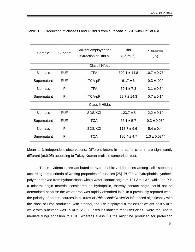
CAPÍTULO TRES
54
Table 3. 1. Production of classes I and II HfbLs from L. lecanii in SSC with Ch2 at 6 d.
Sample Support Solvent employed for
extraction of HfbLs
HfbL
(µg mL-1)
YHfbL/biomass
(%)
Class I HfbLs
Biomass PUF TFA 302.1 ± 14.8 10.7 ± 0.75c
Supernatant PUF TCA-pF 61.7 ± 6 0.3 ± .02a
Biomass P TFA 69.1 ± 7.3 3.1 ± 0.3b
Supernatant P TCA-pF 98.7 ± 14.3 0.7 ± 0.1a
Class II HfbLs
Biomass PUF SDS/KCl 123.7 ± 8 2.2 ± 0.1b
Supernatant PUF TCA 66.1 ± 5.7 0.3 ± 0.03a
Biomass P SDS/KCl 118.7 ± 9.6 5.4 ± 0.4c
Supernatant P TCA 180.4 ± 4.7 1.3 ± 0.03ª,b
Mean of 3 independent observations. Different letters in the same column are significantly
different (α≤0.05) according to Tukey-Kramer multiple comparison test.
These evidences are attributed to hydrophobicity differences among solid supports,
according to the criteria of wetting properties of surfaces [25]. PUF is a hydrophobic synthetic
polymer derived from hydrocarbons with a water contact angle of 121.3 ± 1.3 °, while the P is
a mineral origin material considered as hydrophilic, thereby contact angle could not be
determined because the water drop was rapidly absorbed in P. In a previously reported work,
the polarity of carbon sources in cultures of Rhinocladiella similis influenced significantly with
the class of Hfbs produced, with ethanol, the Hfb displayed a molecular weight of 8.5 kDa
while with n-hexane was 15 kDa [26]. Our results indicate that Hfbs class I were required to
mediate fungi adhesion to PUF, whereas Class II Hfbs might be produced for protection
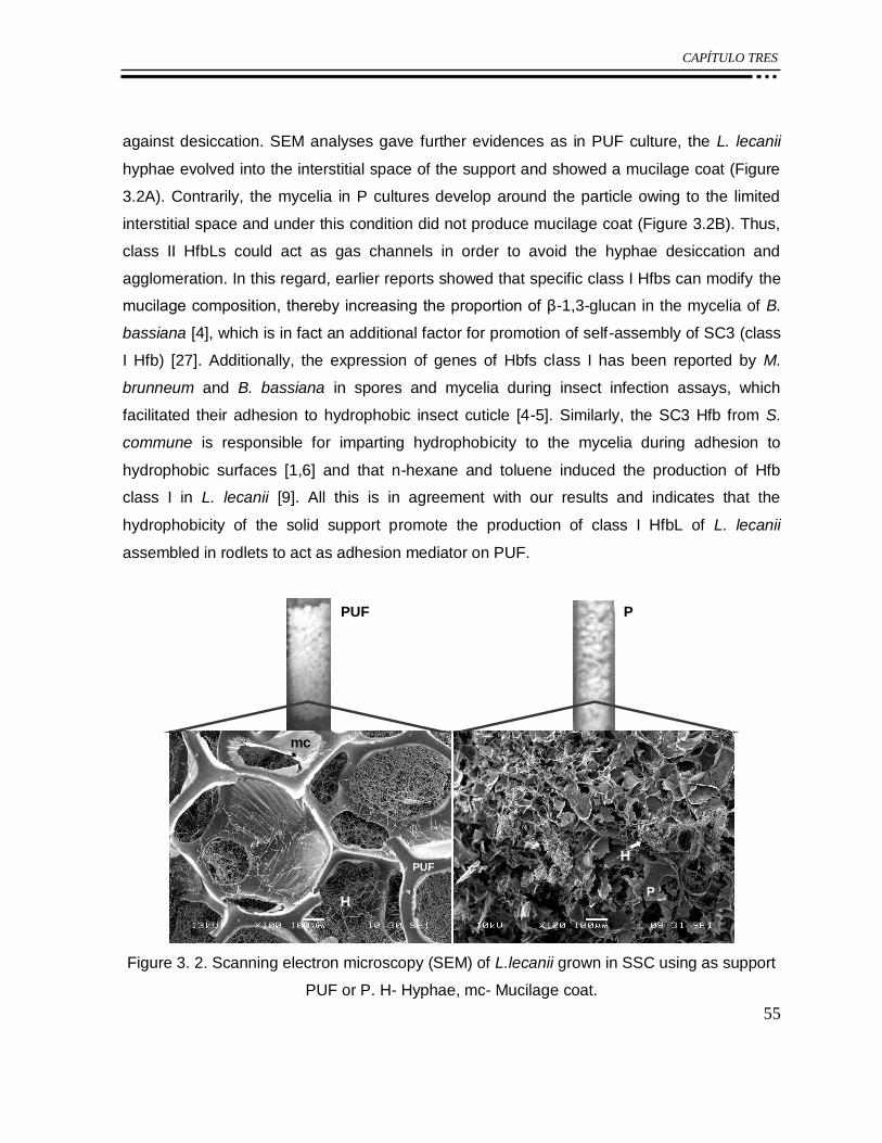
CAPÍTULO TRES
55
against desiccation. SEM analyses gave further evidences as in PUF culture, the L. lecanii
hyphae evolved into the interstitial space of the support and showed a mucilage coat (Figure
3.2A). Contrarily, the mycelia in P cultures develop around the particle owing to the limited
interstitial space and under this condition did not produce mucilage coat (Figure 3.2B). Thus,
class II HfbLs could act as gas channels in order to avoid the hyphae desiccation and
agglomeration. In this regard, earlier reports showed that specific class I Hfbs can modify the
mucilage composition, thereby increasing the proportion of β-1,3-glucan in the mycelia of B.
bassiana [4], which is in fact an additional factor for promotion of self-assembly of SC3 (class
I Hfb) [27]. Additionally, the expression of genes of Hbfs class I has been reported by M.
brunneum and B. bassiana in spores and mycelia during insect infection assays, which
facilitated their adhesion to hydrophobic insect cuticle [4-5]. Similarly, the SC3 Hfb from S.
commune is responsible for imparting hydrophobicity to the mycelia during adhesion to
hydrophobic surfaces [1,6] and that n-hexane and toluene induced the production of Hfb
class I in L. lecanii [9]. All this is in agreement with our results and indicates that the
hydrophobicity of the solid support promote the production of class I HfbL of L. lecanii
assembled in rodlets to act as adhesion mediator on PUF.
PUF P
mc
H
PUFH
P
Figure 3. 2. Scanning electron microscopy (SEM) of L.lecanii grown in SSC using as support
PUF or P. H- Hyphae, mc- Mucilage coat.

CAPÍTULO TRES
56
The concentration of class II HfbLs associated to mycelium of L. lecanii cultured on P
and PUF were not significantly different with 118.73 ± 9.6 µg mL-1 HfbL and 123.7 ± 8 µg HfbL
mL-1, respectively. Although, the YHfbL/biomass in P SSC was 5.4 %, that is, 2-fold higher than
that determined with PUF (YHfbL/biomass= 2.2 %). This point out that the production of class II
HfbLs is enhanced on P in SSC owing to the hydrophilicity of this support, and thereby the
proteins could contribute to fungal development on the surface of P by means of formation of
hydrophobic coat layer. This result is in agreement with the spores of T. reesei having the
expression of class II Hfbs gene of hfbII [7].
3.3.3 Electrophoretic analyses of classes I and II HfbLs from L. lecanii in SSC.
The HfbLs from biomass at steady state, 6 d of culture, were subjected to
electrophoretic analysis. Sevim et al. [5] have reported that Hfb expression from M. brunneum
depends on growth stage, such as the formation of aerial mycelia and conidia. These authors
observed that on solid glucose medium, 3 d-old colonies of strain with hfb1 delete were
fluffier than colonies of wild type strain, however, after 7 d of growth, no morphological
differences between strain mutant (hfb1) and wild type were detected, which was ascribed to
the expression of other Hfb which could complement the hfb1 function.
The HfbL obtained from P were extracted only by the protocol of class II Hfb, and
there were no protein bands assigned to Hfb class I (Figure 3.3). The class II HfbL obtained
from both, PUF and P SSC, showed more than one protein band and agree with previous
reported data, with Hfbs molecular masses in the range of 22, 11 and 7 kDa [1]. L. lecanii on
PUF favored the production of a protein band of 22 kDa but using P SSC promotes small
proteins of 11 kDa (Figure 3.3).

CAPÍTULO TRES
57
PUFM
7.1
20.6
49.1
34.8
28.9
80209
12 kDa
Class I HfbL
7.1
20.6
49.1
34.8
28.9
80
209
Class II HfbL
22 kDa
11 kDa
124
PM PUF
7 kDa
Figure 3. 3. SDS-PAGE of class I and class II HfbLs associate to mycelium from L. lecanii in
SSC. M- Molecular weight standards; PUF as support; P as support.
3.3.4 Surface activity of class I and class II HfbLs from L. lecanii in SSC
In this study, the class I HfbL reduced the hydrophobicity of Teflon ca. 50 %, while,
class II HfbLs showed only 25 % of reduction (Table 3.2). It has been reported that class I
Hfbs are self-assembling as rodlet layers as result of exposure of its glycosylated zone, which
promotes a structural change of the protein from α-helix to β-sheet, this configuration can be
induced by hydrophobic environments, such as high concentrations of protein or the
presence of β-1,3-glucan [2,27].
Nonetheless, class II HfbLs reduced the surface tension of water ca. 50 %, that is 10-
fold higher than class I HfbL, which decreased the surface tension of water only 5 %. It has
been reported that class II HFBI and HFBII from Trichoderma can reduce the surface tension
of water in a few minutes as opposed to SC3 which needs several hours and this has been

CAPÍTULO TRES
58
explained for the structural change experienced by class I during its exposure to the air, while
the class II only showed changes of orientation of their side chain in contact with the water-air
without changes above its absorption or oligomers assembly [24].
Table 3. 2. Surface activities of class I and class II HfbLs.
Surface tension Contact angle
Treatment γ (mN m-1) *Reduction of γ of
water (%) (°)
+ Reduction of
compared with water on
Teflon (%)
Class I HfbLs
PUF 69.0 ± 0.2e 4.6 56.1 ± 5.15a 51.32
Class II HfbLs
PUF total extract 36.9 ± 0.1a 48.9 119.5 ± 1.2b,c 15.6
PUF > 30 kDa 40.5 ± 0.3b 43.9 137.8 ± 2.7d 1.7
PUF < 30 kDa 37.6 ± 0.1a 48.0 106.9 ± 3.9b 25.1
P total extract 37.1 ± 0.4a 48.7 108.0 ± 2.5b 24.33
P > 30 kDa 66.3 ± 0.9d 8.2 121.7 ± 2.8c 13.9
P < 30 kDa 57.7 ± 0.1c 20.2 116.1 ± 2.5b,c 18.1
HfbLs extracts at 50 µg mL-1. (*) γwater = 72.3 ± 0.2 mN m-1. (+) Contact angle of drop of water
on Teflon is 131.9 ± 0.6°. Different letters in the same column are significantly different
(α≤0.05) according to Tukey-Kramer multiple comparison test.
The class II HfbLs showed proteins bands at 22, 11 and 7 kDa, these were favored
according to the support used to L. lecanii culture, thus, the class II HfbLs were subjected to a
molecular weight cut off at 30 kDa prior to surface tension measurements. The fraction class
II HfbLs < 30 kDa showed greater surface activity, in both contact angle and tension surface
than that of fractions > 30 kDa. The fraction < 30 kDa obtained from PUF SSC decreased
from 72.3 ± 0.2 to 37.6 ± 0.1 mN m-1 the surface tension of water (ca. 48 % reduction)
indicating higher surface activity compared to P SSC < 30 kDa fraction from 72.3 ± 0.2 to

CAPÍTULO TRES
59
57.7 mN m-1 (ca. 20.2 % reduction). According to previous reports on Trichoderma, the two
reported proteins, HFBII and HFBI, showed significant differences in their surface activity
because HFBI has a glycine at position 59 (gly59) in the amino acid sequence of the protein,
thus, the steric and hydrophobic contribution of gly explains its high affinity to hydrophobic
surfaces, compared to its association in solution. On the other hand, the HFBII showed
Asp59, which reduces the contact area between the hydrophobic surface and Hfb [28]. It is
worth noticing that the values of surface tension of water obtained in this work are within the
values reported for other Hfbs class II [24].
3.4 CONCLUSIONS
The hydrophobicity of the supports used in SSC influenced the yield and surface
activities of the class I and class II produced by L. lecanii. This highlights the high level of
specificity and importance of the HfbLs and its biological function for the fungal development
by surface adhesion with a direct implication in pathogenesis. Furthermore, the purity of the
chitin can increase the HfbL production from L. lecanii in SSC representing a very important
factor of process control.
Acknowledgements The authors would like to thank to Secretaría de Ciencia, Tecnología e
Innovación del Distrito Federal (SECITI) (Project No. PICSO12-152) and Consejo Nacional
de Ciencia y Tecnología (CONACyT) for scholarship (ZRP).
REFERENCES
[1] Wösten HA. Hydrophobins: multipurpose proteins. Annu Rev Microbiol 2001;55(1):625-
646.
[2] de Vocht ML, Scholtmeijer K, van der Vegte EW, de Vries OMH, Sonveaux N, Wösten
HAB, Ruysschaert JM, Hadziioannou G, Wessels JGH, Robillard GT. Structural
characterization of the hydrophobin SC3, as a monomer and after self-assembly at
hydrophobic/hydrophilic interfaces. Biophys J 1998;74(4):2059-2068.
[3] Askolin S, Nakari-Setälä T, Tenkanen M. Overproduction, purification, and
characterization of the Trichoderma reesei hydrophobin HFBI. Appl Microbiol Biotechnol
2001; 57:124-130.

CAPÍTULO TRES
60
[4] Zhang S, Xia YX, Kim B, Keyhani NO. Two hydrophobins are involved in fungal spore coat
rodlet layer assembly and each play distinct roles in surface interactions, development and
pathogenesis in the entomopathogenic fungus, Beauveria bassiana. Mol Microbiol 2011;
80(3):811-826.
[5] Sevim A, Donzelli BG, Wu D, Demirbag Z, Gibson DM, Turgeon BG. Hydrophobin genes
of the entomopathogenic fungus, Metarhizium brunneum, are differentially expressed and
corresponding mutants are decreased in virulence. Curr Genet 2012; 58(2):79-92.
[6] van Wetter MA, Wösten HA, Wessels JG. SC3 and SC4 hydrophobins have distinct roles
in formation of aerial structures in dikaryons of Schizophyllum commune. Mol Microbiol 2000;
36(1):201-210.
[7] Askolin S, Penttilä M, Wösten HA, Nakari-Setälä T. The Trichoderma reesei hydrophobin
genes hfb1 and hfb2 have diverse functions in fungal development. FEMS Microbiol Lett
2005; 253(2):281-288.
[8] Holder DJ, Keyhani NO. Adhesion of the entomopathogenic fungus Beauveria
(Cordyceps) bassiana to substrata. Appl Environ Microbiol 2005; 71(9):5260-5266.
[9] Rocha-Pino Z, Marín-Cervantes MC, Martínez-Archundia M, Soriano-Blancas E, Revah S,
Shirai K. Morphological changes, chitinolytic enzymes and hydrophobin-like proteins as
responses of Lecanicillium lecanii during growth with hydrocarbon. Bioproc Biosyst Eng 2013;
36: 531-539.
[10] Rocha-Pino Z, Vigueras G, Shirai K. Production and activities of chitinases and
hydrophobins from Lecanicillium lecanii. Bioproc Biosyst Eng 2011; 34:681- 686.
[11] Kerem Z, Hadar Y. Chemically defined solid-state fermentation of Pleurotus ostreatus.
Enzyme Microb Technol 1993; 15:785-790.
[12] Prenafeta-Boldú FX, Illa J, van Groenestijn JW, Flotats X. Influence of synthetic packing
materials on the gas dispersion and biodegradation kinetics in fungal air biofilters. Appl
Microbiol Biotechnol 2008;79:319–327.
[13] Meincken M, Klash A, Seboa S, Sanderson RD. Influence of the viscosity and the
substrate on the surface hydrophobicity of polyurethane coatings. Appl Surf Sci
2006;253(2):805-809.
[14] Bradford MM. A rapid y sensitive method for the quantitation of microgram quantities of
protein utilizing the principle of protein-dye binding. Anal Biochem 1976;72:248-254.
CAPÍTULO TRES
61
[15] Cira LA, Huerta S, Hall GM, Shirai K. Pilot scale lactic acid fermentation of shrimp wastes
for chitin recovery. Proc Biochem 2002;37(12):1359-1366.
[16] Percot A, Viton C, Domard A. Optimization of chitin extraction from shrimp shells.
Biomacromolecules 2003;4:12-18.
[17] de Vries OM, Fekkes M, Wösten HA, Wessels JG. Insoluble hydrophobin complexes in
the walls of Schizophyllum commune and other filamentous fungi. Arch Microbiol
1993;159(4):330-335.
[18] Laemmli UK. Cleavage of structural proteins during the assembly of the head of
bacteriophage T4. Nature 1970;227:680-685.
[19] Fenice M, Gooday GW. Mycoparasitic actions against fungi and oomycetes by a strain
(CCFEE 5003) of the fungus Lecanicillium muscarium isolated in Continental Antarctica. Ann
Microbiol 2006;56(1):1-6.
[20] Marín-Cervantes MDC, Matsumoto Y, Ramírez-Coutiño L, Rocha-Pino Z, Viniegra G,
Shirai K. Effect of moisture content in polyurethane foams as support for solid-substrate
fermentation of Lecanicillium lecanii on the production profiles of chitinases. Proc Biochem
2008;43(1):24-32.
[21] Villa-Lerma G, González-Márquez H, Gimeno M, López-Luna A, Bárzana E, Shirai K.
Ultrasonication and steam-explosion as chitin pretreatments for chitin oligosaccharide
production by chitinases of Lecanicillium lecanii. Bioresour Technol 2013;146:794-798.
[22] Rinaudo M. Chitin and chitosan: properties and applications. Prog Polym Sci
2006;31(7):603-632.
2 Grunér MS, Szilvay GR, Berglin M, Lienemann M, Laaksonen P, Linder MB. Self-
assembly of Class II Hydrophobins on Polar Surfaces. Langmuir 2012; 28(9):4293-4300.
[24] Askolin S, Linder M, Scholtmeijer K, Tenkanen M, Penttilä M, de Vocht ML, Wösten HA.
Interaction and comparison of a class I hydrophobin from Schizophyllum commune and class
II hydrophobins from Trichoderma reesei. Biomacromolecules 2006;7(4):1295-1301.
[25] Bico J, Thiele U, Quéré D. Wetting of textured surfaces. Colloids Surf A Physicochem
Eng Asp 2002;206(1):41-46.
[26] Vigueras G, Arriaga S, Shirai K, Morales M, Revah S. Hydrophobic response of the
fungus Rhinocladiella similis in the biofiltration with volatile organic compounds with different
polarity. Biotechnol lett 2009;31(8):1203-1209.

CAPÍTULO TRES
62
[27] Scholtmeijer K, de Vocht ML, Rink R, Robillard GT, Wösten HA. Assembly of the fungal
SC3 hydrophobin into functional amyloid fibrils depends on its concentration and is promoted
by cell wall polysaccharides. J Biol Chem 2009;284(39):26309-26314.
28 Moldovan C, Thompson D. Molecular dynamics of the “hydrophobic patch” that
immobilizes hydrophobin protein HFBII on silicon. J Mol Model 2011;17(9):2227-2235.

CAPÍTULO CUATRO
63
CAPÍTULO 4:
Morphological changes, chitinolytic enzymes
and hydrophobin like proteins as responses of
Lecanicillium lecanii during growth with
hydrocarbon
Zaizy Rocha-Pino • María del Carmen Marín-Cervantes • Marlet Martínez-Archundia •
Elizabeth Soriano-Blancas • Sergio Revah • Keiko Shirai
Bioprocess Biosyst Eng (2013) 36:531–539

CAPÍTULO CUATRO
64

CAPÍTULO CUATRO
65
Morphological changes, chitinolytic enzymes and hydrophobinlike
proteins as responses of Lecanicillium lecanii during growth with
hydrocarbon
Zaizy Rocha-Pino • María del Carmen Marín-Cervantes • Marlet Martínez-Archundia •
Elizabeth Soriano-Blancas • Sergio Revah • Keiko Shirai
Bioprocess Biosyst Eng (2013) 36:531–539
Abstract
Lecanicillium lecanii, Verticillium chlamydosporium, V. fungicola var flavidum and
Beauveria bassiana were evaluated on their growth with pure n-hexane, toluene and n-
hexane:toluene 17:83 (v:v) mixture. Another set of treatments were conducted with colloidal
chitin as additional carbon source. All the strains of Lecanicillium were able to grow using
hydrocarbons with or without the addition of chitin, although the presence of hydrocarbons
showed significant inhibition evidenced by measured biomass, radial growth and microscopic
analyses. Degradation of n-hexane ranged within 43 and 62 % and it was higher than that
with toluene. The strains L460, L157 and L2149, which presented the highest growth, were
further selected for determinations of hydrocarbon consumptions in microcosms. Strain L157
showed the highest consumption of n-hexane (55.6 %) and toluene (52.9 %) as sole carbon
source and it also displayed activities of endochitinases, N-acetylhexosaminidase and
production of hydrophobins class I and II.
Keywords Entomopathogenic fungi • Hydrocarbons • Chitinases • Hydrophobins
4.1 INTRODUCTION
Entomopathogenic fungi such as Lecanicillium lecanii are able to produce
hydrophobins (Hfbs) [1], which are small proteins capable to self-assemble in amphipathic
membranes.Their role in nature consists in preventing desiccation of the hyphae or spores
CAPÍTULO CUATRO
66
and decreasing the surface tension of water allowing the hyphae emerge from the liquid
medium to in-air environment [2–5]. Hfbs are required for the successful development of
filamentous fungi by the adhesion of hyphae in the hydrophobic cuticles of insects, which are
covered by waxes and hydrocarbons [6], and subsequently, excreted cell wall degrading
enzymes, chitinases, proteases and lipases, thus allowing host invasion [7].
On the other hand, filamentous fungi utilize hydrocarbons as sole carbon source.
Isolates of Paecilomyces sp., Verticillium sp., Beauveria sp., and Penicillium sp. were tested
for their ability to metabolize a variety of n-alkylbenzenes [8]. Fungi used in biological control,
B. bassiana and Metarhizium anisopliae, can degrade n-Pentacosane, 3,11-
dimethylnonacosane and n-hexadecane [9, 10]. Interestingly, the increase of
entomopathogenic activity of Beauveria bassiana on the bean weevil was observed under
cultivation with n-hexadecane as sole carbon source [9, 11]. With regard to the
entomopathogenic fungi Lecanicillium, Krivobok et al. [12] showed 10 % degradation of
anthracene, besides, Vroumsia et al. [13] claimed the degradation of 22 % of 2,4-dichloride
phenol acid, but despite these reports, there is no information, at least to the best of our
knowledge, on the tolerance of Lecanicillium to toluene or n-hexane and their effect on the
production of Hfb and enzymes involved in the pathogenesis. Therefore, the aim of this work
was to evaluate several strains of Lecanicillium, Verticillium and B. bassiana on tolerance,
growth, morphological changes and production of chitinolytic enzymes and Hfbs with volatile
organic compounds (VOCs).
4.2 MATERIALS AND METHODS
4.2.1 Microorganisms
Twenty strains of Lecanicillium lecanii were provided by the American type culture
collection (ATCC26854 and ATCC46578), USDA-ARSEF collection of entomopathogenic
fungal cultures (L157, L974, L991, L1029, L2009, L2149, L2460, L2832, L2858, L2916,
L3909, L5129 and L5153) and from culture collection of the Basic Mycology Laboratory of the
Department of Microbiology and Parasitology, Faculty of Medicine, Universidad Nacional
Autonoma de Mexico (L348, L457, L458, L459 and L460). Verticillium chlamydosporium
strain V2218, V. fungicola var flavidum strain V4519 and Beauveria bassiana (Bb) were
propagated on potato dextrose agar slants at 25 °C and stored at 4 °C.

CAPÍTULO CUATRO
67
4.2.2 Media
Mineral media (g L-1): K2HPO4 (2.80), MgSO4•7H2O (1.38), CaCl2 (0.48), FeSO4•7H2O
(0.22), (NH4)2SO4 (7.50), (NH4)Cl (4.10), NaH2PO4•7H2O (1.67) at pH 6.0. 15 g L-1 of
bacteriological agar was added to mineral media for screening fungal strains. Carbon sources
added to the mineral media were (g L-1): glucose (25.68) as control; n-hexane (MH); toluene
(MT); mixture of both solvents (17:83 molar ratio) (MTH) and chitin (10) combined with n-
hexane (MChH); toluene (MChT) and mixture of both solvents (17:83 molar ratio) (MChTH).
4.2.3 Screening of fungal strains based on hydrocarbon tolerance
107 spores mL-1 of the fungal strains was inoculated in Petri dishes placed in hermetic
containers. Each container was equipped with a tube of activated coal impregnated with the
hydrocarbons, then, 1 mL of hydrocarbons was added throughout to the tube every 2 days.
Each container was incubated at 25 °C during 30 days. Radial growth, hyphae diameters and
halos of colloidal chitin hydrolysis were determined at the end of the incubation period. Agar
was melted and filtered and fungal biomass determined by gravimetry.
4.2.4 Determination of consumption of toluene or n-hexane in microcosm experiments
107 spores mL-1 of L. lecanii strains L460, L157 and L2149 were placed in 125 mL
flask containing 10 mL of mineral medium with added toluene (4.35 mg mL-1) or n-hexane
(3.3 mg mL-1) as carbon sources. Flasks were sealed with inert Teflon valves (VICI Precision
Sampling) and toluene or n-hexane was added by a microsyringe. All samples were
incubated at 25 °C and 180 rpm for 60 days. Hydrocarbon concentrations were monitored by
gas chromatography (GC) from samples withdrawn from headspaces. The experiments were
performed by duplicate. VOCs consumptions were calculated considering the initial
hydrocarbon concentration (µg mL-1) and the concentration at time t.
4.2.5 Gas chromatography analysis
100 µL of each sample was injected (VICI Precision Sampling) into a gas
chromatographer (Agilent Technologies 6890N) equipped with a flame ionization detector and
an AT-WAX (Alltech Heliflex 25 m) column. GC settings were injector at 200 °C, oven 65 °C,
detector at 250 °C and carrier (helium) flow rate of 0.6 mL min-1. CO2 concentration was
determined by thermal conductivity detector with a Poropak 55-m column with operating
CAPÍTULO CUATRO
68
conditions: injector at 110 °C, oven 65 °C, detector at 110 °C and helium flow rate of 4.4 mL
min-1 [14]. Measurements were carried out by duplicate.
4.2.6 Biomass determination from microcosms
Biomass was determined measuring the total protein by Bradford [15]. Culture
medium was centrifuged (11,500 rpm) at 4 °C for 20 min. Then, 0.2 g of pellet was mixed with
5 mL of phosphoric acid (0.15 M) and heated in a water bath for 7 min and afterward
centrifuged at 12,000 rpm. The protein from the pellet and supernatant were determined by
Bradford. Bovine serum albumin was used as standard.
4.2.7 Assay of chitinolytic activity
N-acetylhexosaminidase (HexNase) and endochitinases (Endo) activities from the
crude enzyme (supernatant after centrifugation) were assayed according to the methodology
reported by Tronsmo and Harman [16]. Yield coefficients, YHexNase/x and YEndo/x, were
determined considering maxima enzyme activities on total biomass produced [17].
4.2.8 Hfb extraction and determination of surface activities
Hfb class I were extracted using formic acid following the procedure reported by
Vigueras et al. [18] from biomass of the microcosm cultures. Hfb class II was carried out
using SDS 1 % (p v-1) as reported by Askolin et al. [19]. Surface activity was evaluated as the
ability to modify the surface hydrophobicity of Teflon [poly(tetrafluoroethylene)] by the Hfbs by
measuring the contact angle of a drop of water [18]. Teflon was washed with water and
ethanol and then dried. Hfb solution was prepared dissolving extract of dried protein in
deionized water at a concentration of 50 µg protein-1. Hfb solution was used onto 1.32 cm2 of
Teflon. Subsequently, 1 µL of deionized water was dropped on the treated Teflon surface and
observed with a side-illumination horizontal light microscope Intel Qx3 (Intel Corporation,
Santa Clara, CA, USA). Contact lengths (b) and heights (h) of the water droplet were
measured by ImageJ 1.41o software (National Institutes of Health, USA). Contact angle was
calculated according to the Eq. (4.1) [1]. Analyses were conducted by quadruplicate, in three
different random locations on each surface. The reduction of the hydrophobicity of Teflon was
calculated as percentage considering the contact angles of water and the extracted Hfb.
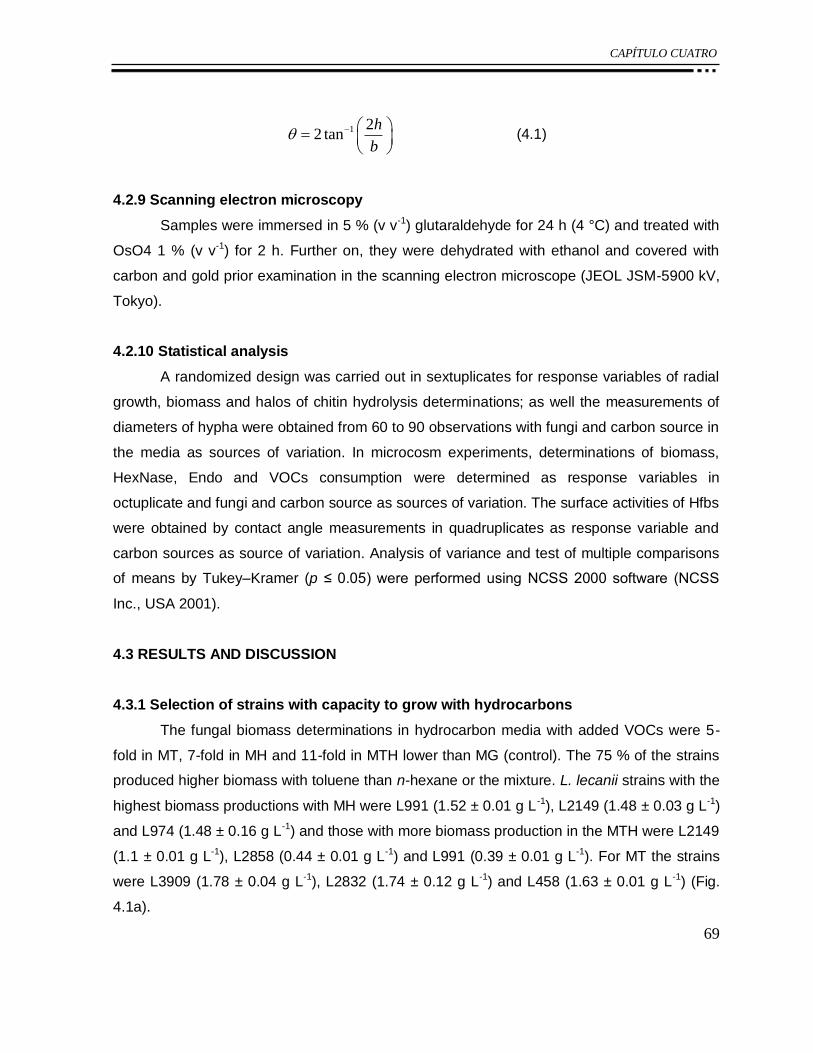
CAPÍTULO CUATRO
69
1 22 tan
h
b
(4.1)
4.2.9 Scanning electron microscopy
Samples were immersed in 5 % (v v-1) glutaraldehyde for 24 h (4 °C) and treated with
OsO4 1 % (v v-1) for 2 h. Further on, they were dehydrated with ethanol and covered with
carbon and gold prior examination in the scanning electron microscope (JEOL JSM-5900 kV,
Tokyo).
4.2.10 Statistical analysis
A randomized design was carried out in sextuplicates for response variables of radial
growth, biomass and halos of chitin hydrolysis determinations; as well the measurements of
diameters of hypha were obtained from 60 to 90 observations with fungi and carbon source in
the media as sources of variation. In microcosm experiments, determinations of biomass,
HexNase, Endo and VOCs consumption were determined as response variables in
octuplicate and fungi and carbon source as sources of variation. The surface activities of Hfbs
were obtained by contact angle measurements in quadruplicates as response variable and
carbon sources as source of variation. Analysis of variance and test of multiple comparisons
of means by Tukey–Kramer (p ≤ 0.05) were performed using NCSS 2000 software (NCSS
Inc., USA 2001).
4.3 RESULTS AND DISCUSSION
4.3.1 Selection of strains with capacity to grow with hydrocarbons
The fungal biomass determinations in hydrocarbon media with added VOCs were 5-
fold in MT, 7-fold in MH and 11-fold in MTH lower than MG (control). The 75 % of the strains
produced higher biomass with toluene than n-hexane or the mixture. L. lecanii strains with the
highest biomass productions with MH were L991 (1.52 ± 0.01 g L -1), L2149 (1.48 ± 0.03 g L-1)
and L974 (1.48 ± 0.16 g L-1) and those with more biomass production in the MTH were L2149
(1.1 ± 0.01 g L-1), L2858 (0.44 ± 0.01 g L-1) and L991 (0.39 ± 0.01 g L-1). For MT the strains
were L3909 (1.78 ± 0.04 g L-1), L2832 (1.74 ± 0.12 g L-1) and L458 (1.63 ± 0.01 g L-1) (Fig.
4.1a).

CAPÍTULO CUATRO
70
The radial growth was not related with the fungal biomass production because fungi
grew in aggregates or dispersed mycelia without significant difference (p ≤ 0.05) in the
biomass production among hydrocarbons (Fig. 4.1a, b). Agglomerated and dense mycelia
were observed with toluene or n-hexane, where 70 % of the strains showed from 3- to 35-fold
lower radial growth than the control. On the other hand, similar percentage of strains with the
mixture displayed dispersed and spread mycelia, which were scarce and ramified covering
completely the plate surface (Fig. 4.1b). This effect might be attributed to the hydrocarbon
toxicity, as reported by Krivobok et al. [12], where L. lecanii was cultivated on agar medium
using several anthracene concentrations as carbon source. In that report, the increase in the
toxicity decreased the growth along with the observation of sterile and more disperse mycelia.
According to the microscopic observations, n-hexane was rather toxic than toluene or
the mixture, since the 61 % of the strains showed decreased diameters of hypha, while the
media added with the mixture or pure toluene, displayed only 30 and 8 % of the strains,
respectively (Fig. 4.1c). The low solubility of the n-hexane in medium might explain the low
fungal growth. Other toxic effect was the decrease in the diameter of hyphae, evidenced by
morphological damages, such as the reduction of hypha diameter, which pointed to a fungal
response to increase the surface area for gaseous substrate exchange. For instance, when
hydrocarbons were used as sole carbon source, hypha diameters were significantly smaller
(p ≤ 0.05) (0.7 ± 0.19) than that of the strains in the control (1.6 ± 0.04 µm). The strains with
the highest hyphae diameter in toluene were the strain ATCC26854, L157 and L2149 (1.1 ±
0.01 µm); in n-hexane was L460 (1.2 ± 0.01 µm) and with hydrocarbon mixture was L2009
(1.0 ± 0.03 µm) (Fig. 4.1c).

CAPÍTULO CUATRO
71
a)
0
0.2
0.4
0.6
0.8
1
1.2
1.4
1.6
1.8
2
Bb
AT
CC
26854
AT
CC
46578
157
974
991
1029
2009
2149
2218
2460
2832
2858
2916
3909
4519
5129
5153
EH
-348
EH
-457
EH
-458
EH
-459
EH
-460
Bio
mass (g L
-1)
Strains
MT MH MTH
b)
0
10
20
30
40
50
60
Bb
AT
CC
26
85
4
AT
CC
46
57
8
15
7
97
4
99
1
10
29
20
09
21
49
22
18
24
60
28
32
28
58
29
16
39
09
45
19
51
29
51
53
EH
-34
8
EH
-45
7
EH
-45
8
EH
-45
9
EH
-46
0
Dia
me
ter
(mm
)
Strains
MT MH MTH

CAPÍTULO CUATRO
72
c)
0.4
0.5
0.6
0.7
0.8
0.9
1
1.1
1.2
1.3
Bb
AT
CC
268
54
AT
CC
465
78
15
79
74
99
11
02
92
00
92
14
92
21
82
46
02
83
22
85
82
91
63
90
94
51
95
12
95
15
3E
H-3
48
EH
-45
7E
H-4
58
EH
-45
9E
H-4
60
Dia
me
ter (
µm
)
Strains
MT MH MTH
Figure 4. 1. a) Radial growth, b) biomass, c) diameters of hypha determinations of strains of
Lecanicillium, Verticillium and B. bassiana determined in MH, MT or MTH as sole carbon
source at 25 °C and 30 days of incubation. Radial growth and biomass data are shown as the
average of six repetitions and their standard errors. The measurements of hypha diameter
were carried out on micrographs at x100 and are the average of 70–90 observations and their
standard errors.
In a previous report by Ramírez-Coutiño et al. [20], Verticillium and Lecanicillium
strains were able to produce chitinolytic enzymes in media amended with colloidal chitin.
Herein, the tested strains also displayed extracellular chitinolytic enzymes production by the
formation of halos of hydrolysis in the VOCs and colloidal chitin agar media. Interestingly, all
strains displayed activity in toluene media, and the largest halos of hydrolysis were
determined with L2009 and L458. On the other hand, the strains L157 and L5153 presented
halos of hydrolysis in MChH, whereas for the mixture of hydrocarbons only the strain L2009
presented activity (Fig. 4.2a). This evidence suggests that L2009 possesses higher tolerance
CAPÍTULO CUATRO
73
as it presented growth with the mixture of VOCs as sole carbon source (Fig. 4.1a) without
significant diameter changes compared to the control (Figs. 4.1c, 2b).
The diameter of the hypha in the culture media with colloidal chitin and hydrocarbons
was less damaged than those in the media with VOCs as sole carbon source (Fig. 4.2b).
Indeed, the average of hypha diameter of medium with chitin and hydrocarbon varied from
1.4 to 1.1 µm, whereas in the medium with the hydrocarbon were 1.08 and 0.8 lm for toluene
and n-hexane, respectively. The effect of toluene in the MChT medium was not remarkable in
the structure of the hypha, neither in the ssimilation of the chitin, as the diameters were
similar to those found in the control (0.9–1.2 µm). The n-hexane addition (MChH), however,
reduced significantly the hypha diameter (0.7–0.8 µm) compared to control or MChT (Figs.
4.1c, 4.2b). The strains with the highest hyphae diameter with MChT were ATCC26854 (1.13
± 0.01 µm), ATCC46578 (1.12 ± 0.02 µm) and L1029 (1.10 ± 0.01 µm); those with MChH
were L460 (1.42 ± 0.02 µm), L2460 (1.39 ± 0.03 µm) and L2832 (0.84 ± 0.02 µm); and with
MChTH were L974 (0.90 ± 0.02 µm), L2832 (0.82 ± 0.02 µm) and ATCC46578 (0.76 ± 0.02
µm) (Fig. 4.2b).

CAPÍTULO CUATRO
74
a)
0
2
4
6
8
10
12
14
16
18
Bb
AT
CC
26854
AT
CC
46578
157
974
991
1029
2009
2149
2218
2460
2832
2858
2916
3909
4519
5129
5153
EH
-348
EH
-457
EH
-458
EH
-459
EH
-460
Dia
mete
r (m
m)
Strains
MChT MChH MChTH
b)
0.0
0.2
0.4
0.6
0.8
1.0
1.2
1.4
1.6
B.b
AT
CC
26854
AT
CC
46578
157
974
991
1029
2009
2149
2218
2460
2832
2858
2916
3909
4519
5129
5153
EH
-348
EH
-457
EH
-458
EH
-459
EH
-460
Dia
mete
r (µ
m)
Strains
MChT MChH MChTH
Figure 4. 2. Determination of halos of hydrolysis of chitin and b diameters of hypha of strains
of Lecanicillium, Verticillium and B. bassiana grown in MChH, MChT or MChTH. Halos of
hydrolysis data are the average of six observations and their standard errors. The
measurements of hypha diameter were carried out on micrographs at x100 and are the
average of 70–90 observations and their standard errors.

CAPÍTULO CUATRO
75
4.3.2 Consumption of toluene and n-hexane and chitinolytic activity for L. lecanii L460,
L157 and L2149
In the degradation experiments, only pure hydrocarbon treatments were analyzed
because the mixture showed lower biomass production than in the control (11-fold), and most
of the strains did not show halos of hydrolysis of colloidal chitin. The strains of L. lecanii were
chosen due to the highest hyphae diameters and growth (Fig. 4.1). The strain L460 displayed
both highest radial growth (3.83 ± 0.31 mm) and diameter hypha (1.15 ± 0.01 µm) with n-
hexane. In toluene, the strain L157 showed the highest radial growth (6.0 ± 0.01 mm) and
hyphae diameter (1.08 ± 0.01 µm). However, the strain L2149 presented the highest biomass
(1.48 ± 0.03 g L-1) with n-hexane and the highest hypha diameter (1.09 ± 0.01 µm) with
toluene.
The total biomass produced, chitinolytic enzymes activities and consumption of VOCs
were determined during 60 days of incubation (Table 4.1). The protein measurements from
the media with VOCs alone or in combination with colloidal chitin were conducted as an
indirect estimation of biomass. L. lecanii L460, L157 and L2149 presented degrading
activities of n-hexane and toluene with that of hydrocarbons from 43 to 62 % (Table 4.1). L.
lecanii L157 showed the highest consumption of toluene (52.9 ± 0.11 %) and n-hexane (55.6
± 0.30 %) used as sole carbon source. The strains L157 and L2149 degraded more n-hexane
than toluene with ca. 2 and 5 %, respectively, whereas in the media with chitin showed ca. 18
and 17 %, respectively. The strain L460 did not show significant differences in the VOCs
consumption (Table 4.1). These biodegradation results can be related to hyphae diameters
(Fig. 4.1c), which decreased 5 % in the presence of n-hexane compared to the control.
The strains L460 and L157 had a positive correlation between biomass production
and VOCs consumption (p ≤ 0.01) in media with chitin owing to tenfold increase in biomass
production, as well as higher degradation of n-hexane (6–19 %) and toluene (2.7–12.3 %)
than in the media with hydrocarbons as sole carbon source. The exception was the strain
2149 in the medium MChT where consumption decreased 3.4 % compared to MT medium.
The three strains tested showed N-acetylhexosaminidase (HexNase) and
endochitinase (Endo) activities in the media with or without chitin. The highest yield of
HexNase was determined for L460 in hydrocarbon media with or without chitin with values
between 0.26 and 0.8 mU µg protein-1, while strains L2149 and L157 did not show significant

CAPÍTULO CUATRO
76
differences in production. Certainly, the addition of chitin to the medium increased the yield of
HexNase and production of biomass compared to the media with only hydrocarbons.
However, the hydrophobicity of hydrocarbon affected the Endo activity, which was
higher in MT than MH, this difference might be explained in terms of solubility as toluene is
more soluble, 0.534 g L-1, [21] than n-hexane, 0.0095 g L-1 [22]. The strain L157 had the
highest yields of Endo in MT (17.1 U µg protein-1), MH (5.12 U µg protein-1) and MChT (1.63
U µg protein-1). Ooki et al. [23] reported that the presence of polycyclic aromatic
hydrocarbons (PAHs) competitively inhibit the activity of chitinases of Aeromonas hydrophila
subsp. anaerogenes. These authors reported that inhibition is greater as the number of
aromatic rings increase. Herein, there were no significant differences on chitinolytic activities
among aromatic and aliphatic VOCs (Table 4.1).
Table 4. 1. Determination of biomass and chitinolytic enzymes productions as well as
consumption of toluene or n-hexane for L. lecanii L460, L157 and L2149 in microcosms at 60
days.
Medium Strain Biomass
(μg protein mL-1
)
N-acetyl
hexosaminidase
Ye/x (mU gproteina-1
)
Endochitinases
Ye/x (U gproteina-1
)
VOCs (%)
consumption
MH
EH-460 0.29 ± 0.01a,b,c
0.26 ± 0.03a,b
4.75 ± 0.9b 45.4 ± 0.54
a
157 0.59 ± 0.06a,b,c
0.04 ± 0.01a,b
5.12 ± 1.11b 55.6 ± 0.30
d
2149 0.83 ± 0.02c 0.09 ± 0.01
a,b 1.82 ± 0.32
a 43.0 ± 0.78
a
MChH
EH-460 4.09 ± 0.17f 0.64 ± 0.06
c 0.73 ± 0.09
a 53.24 ± 0.30
c,d
157 5.40 ± 0.03g 0.13 ± 0.01
a,b 0.53 ± 0.13
a 62.28 ± 0.47
e
2149 4.29 ± 0.14f 0.20 ± 0.01
a,b 1.55 ± 0.15
a 62.21 ± 0.21
e
MT
EH-460 0.05 ± 0.02a 0.48 ± 0.09
a,b 20.24 ± 1.18
d 43.0 ± 0.17
a
157 0.16 ± 0.07a,b
0.07 ± 0.02a 17.07 ± 1.47
c 52.9 ± 0.11
c
2149 0.69 ± 0.06b,c
0.11 ± 0.01a,b
2.33 ± 0.37a,b
48.6 ± 0.70b
MChT
EH-460 5.05 ± 0.24g 0.80 ± 0.03
c 0.51 ± 0.09
a 55.26 ± 0.92
c,d
157 2.50 ± 0.04d 0.24 ± 0.02
a,b 1.63 ± 0.48
a 44.27 ± 0.62
a
2149 3.09 ± 0.10e 0.28 ± 0.01
b 0.70 ± 0.20
a 45.42 ± 0.29
a
Values with different superscript letters in the same column showed significant differences (p
≤ 0.05) according to Tukey–Kramer multiple comparison test.

CAPÍTULO CUATRO
77
4.3.3 Production of Hfbs-like proteins class I and II of L. lecanii L157 and their surface
activities
Rocha-Pino et al. [1] reported recently the use of chitin as a carbon source to promote
the production of Hfb-I in mycelia of L. lecanii cultured for 6 days. The production of Hfbs is
regulated by environmental factors [2, 24] and depends on growth stage, such as the
formation of aerial mycelia and conidia. Mulder and Wessels [25] reported that the expression
of Hfbs of Schizophyllum commune reached the maximum at 4 days of culture on agar.
Herein, the production of proteins with Hfb activity was determined from 6-day culture
of L. lecanii L157; this strain was selected owing to its VOCs consumption and its less
inhibited chitinolytic activity.
L. lecanii produced 57.4 ± 4.7 µg protein-1 of Hfb class I in liquid media with chitin as
sole carbon source [1]; however, in this work, the production of Hfb was 88.3 ± 4.6 µg protein -
1 and 548.6 ± 26.3 µg protein-1 in media MChT and MChH, respectively. These results
indicate that the addition of hydrophobic compound increased the Hfb production. In addition,
the extracts of Hfb class I of the media MChT and MChH reduced the Teflon hydrophobicity
to 47.8 ± 3.5 % and 10.1 ± 1.5 %, respectively. The differences on surface activity indicate
that L. lecanii might be able to produce more than one Hfb depending on the hydrophobicity
of media. It has been reported that S. commune expressed Hfbs SC3 depending on the
biological function, for instance, SC3 covered the aerial hyphae reducing the surface tension
and mediated the adhesion to hydrophobic surfaces, while Hfbs SC4 coated the fungal fruit
bodies forming channels for gas transfer [26].
The media MChH significantly favored the production of proteins corresponding to Hfb
class II (194.1 ± 11.6 µg Hfb mL-1) and remarkably decreased the hydrophobicity of Teflon
27.2 ± 2.1 %, whereas in MChT, the Hfb class II was 69.9 ± 3.4 µg Hfb mL-1 with a surface
activity on Teflon of 26.3 ± 2.6 %.
Figure 4.3 shows SEM micrographs obtained from strains cultivated in n-hexane and
toluene compared to the media supplemented with chitin and VOCs. The micrographs
showed the toxic effect of VOCs causing morphological changes in spores and hyphae. The
damages were more evident at longer period of culture with VOCs (60 days) (Fig. 4.3a, b, e,
f) compared to the strain cultivated in media with added chitin (Fig. 4.3c, d, g, h). Figure 4.3e
depicts the strain cultivated with n-hexane with appreciable damage in the hyphae, having
irregular shape with roughness instead of those flat and circular from the strain after 6 days of

CAPÍTULO CUATRO
78
incubation (Fig. 4.3g), besides higher number of non-germinated spores than those in toluene
(Fig. 4.3f). The reduction of average diameter of spores was 0.57 ± 0.01 µm with n-hexane
whereas that with toluene was 0.7 ± 0.02 µm (Fig. 4.3e, f). It has been reported that Hfbs
interact with hydrophobic surfaces and triggered the appressorium formation [3] and that
agrees with our findings as shown in Fig. 4.3c and d.
Crespo et al. [9] reported that the variation in the morphology is an indication of fungal
cell wall composition changes in the presence of hydrocarbon as sole carbon source,
specifically on the lipid fraction. Also according to Funtikova and Mysyakina [27], these
modifications reduce the ability for the spore to germinate and that the change in the lipid
proportion has some effect on the carbohydrates content on the surface, which has a positive
correlation with the hydrophobicity. In such conditions, the mycelia surface is roughish, while
under hydrophilic condition, it appears as flat and smooth [28]. In this work, similar effect was
observed on the mycelia morphologies when fungi were cultivated within chitin and VOCs or
pure hydrocarbons (Fig. 4.3e–h).
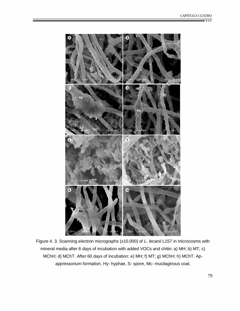
CAPÍTULO CUATRO
79
C D
A B
D
E F
HG
Ap
Ap
Hy
Mc
Figure 4. 3. Scanning electron micrographs (x10,000) of L. lecanii L157 in microcosms with
mineral media after 6 days of incubation with added VOCs and chitin: a) MH; b) MT; c)
MChH; d) MChT. After 60 days of incubation: e) MH; f) MT; g) MChH; h) MChT. Ap-
appressorium formation, Hy- hyphae, S- spore, Mc- mucilaginous coat.

CAPÍTULO CUATRO
80
4.4 CONCLUSION
Lecanicillium and Beauveria bassiana cultivated in liquid cultures and agar that
contained n-hexane, toluene, or the mixture of both hydrocarbons, presented agglomerates of
spores as well as fine, scarce and ramified mycelia, showing their ability to grow in media
with VOCs. Generally, the inhibition with n-hexane was higher than toluene or mixture of
hydrocarbons n-hexane:toluene 17:83 (v:v). The strains of Lecanicillium L460, L157 and
L2149 degraded from 43 to 55 % of n-hexane or toluene in liquid medium and they produced
chitinolytic enzymes and Hfbs-like proteins with surface activity as response of the presence
of hydrophobic substrates.
Acknowledgments The authors would like to thank SEP-CONACYT (No. 105628) for
research funding and scholarship grants to Miss Rocha-Pino and Miss Marı´n-Cervantes. Dr.
Jose´ Sepulveda is greatly acknowledged for his invaluable assistance in the SEM studies.
REFERENCES
1. Rocha-Pino Z, Vigueras G, Shirai K (2011) Production and activities of chitinases and
hydrophobins from Lecanicillium lecanii. Bioprocess Biosyst Eng 34:681–686.
2. St. Leger RJ, Joshi I, Roberts D (1998) Ambient pH is a major determinant in the
expression of cuticle degrading enzymes and hydrophobin by Metarhizium anisopliae. Appl
Environ Microbiol 64(2):709–713.
3. Talbot NJ, Kershaw MJ, Wakley GE, De Vries O, Wessels J, Hamer JE (1996) MPGI
encodes a fungal hydrophobin involved in surface interactions during infection-related
development of Magnaporthe grisea. Plant Cell 8:985–999.
4. Wösten HAB, De Vries OMH, Wessels JGH (1993) Interfacial self-assembly of a fungal
hydrophobin into a hydrophobic rodlet layer. Plant Cell 5:1567–1574.
5. Wösten HAB, Schuren FHJ, Wessels JGH (1994) Interfacial selfassembly of a hydrophobin
into an amphipathic protein membrane mediates fungal attachment to hydrophobic surfaces.
EMBO J 13(24):5848–5854.
6. Kather R, Martin J (2012) Cuticular hydrocarbon profiles as a taxonomic tool: advantages,
limitations and technical aspects. Physiol Entomol 37(1):25–32.

CAPÍTULO CUATRO
81
7. Fenice M, Gooday GW (2006) Mycoparasitic actions against fungi and oomycetes by a
strain (CCFEE 5003) of the fungus Lecanicillium muscarium isolated in Continental
Antarctica. Ann Microbiol 56(1):1–6.
8. Fedorak PM, Westlake DWS (1986) Fungal metabolism of n-alkylbenzenes. Appl Environ
Microbiol 51:435–437.
9. Crespo R, Juarez MP, Dal Bello GM, Padín S, Calderón G, Pedrini N (2002) Increased
mortality of Acanthoscelides obtectus by alkane-grown Beauveria bassiana. Biocontrol
47:685–698.
10. Napolitano R, Juárez MP (1997) Entomopathogenous fungi degrade epicuticular
hydrocarbons of Triatoma infestans. Arch Biochem Biophys 344:208–214.
11. Crespo R, Juárez MP, Cafferatta LFR (2000) Biochemistry of the interaction between
entomopathogenous fungi and their insect host-like hydrocarbons. Mycologia 92:528–536.
12. Krivobok S, Miriouchkine E, Seigle-Murandi F, Benoit-Guyod JL (1998) Biodegradation of
anthracene by soil fungi. Chemosphere 37(3):523–530.
13. Vroumsia T, Steiman R, Seigle-Murandi F, Benoit-Guyod JL (1999) Effects of culture
parameters on the degradation of 2,4-dichlorophenoxyacetic acid (2,4-D) and 2,4-
Dichlorophenol (2,4-DCP) by selected fungi. Chemosphere 39(9):1397–1405.
14. Matsumoto Y, Saucedo G, Revah S, Shirai K (2004) Production of β-N-
acetylhexosaminidase of Verticillium lecanii by solid state and submerged fermentation
utilizing shrimp waste silage as substrate and inducer. Process Biochem 39(6):665–671.
15. Bradford MM (1976) A rapid and sensitive method for the quantitation of microgram
quantities of protein utilizing the principle of protein-dye binding. Anal Biochem 72:248–254.
16. Tronsmo A, Harman GE (1993) Detection and quantification of N-acetyl β-D
glucosaminidase, chitobioside, and endochitinse in solutions and on gels. Anal Biochem
208:74–79.
17. Marín-Cervantes MC, Matsumoto Y, Ramírez-Coutiño L, Rocha-Pino Z, Viniegra G, Shirai
K (2008) Effect of moisture content in polyurethane foams as support for solid-substrate
fermentation of Lecanicillium lecanii on the production profiles of chitinases. Process
Biochem 43:24–32.
18. Vigueras G, Shirai K, Martins D, Franco TT, Fleuri LF, Revah S (2008) Toluene gas
phase biofiltration by Paecilomyces lilacinus and isolation and identification of a hydrophobin
protein produced thereof. Appl Microbiol Biotechnol 80:147–154.

CAPÍTULO CUATRO
82
19. Askolin S, Nakari-Setälä T, Tenkanen M (2001) Overproduction, purification, and
characterization of the Trichoderma reesei hydrophobin HFBI. Appl Microbiol Biotechnol
57:124–130.
20. Ramírez-Coutiño L, Marín-Cervantes M, Huerta S, Revah S, Shirai K (2006) Enzymatic
hydrolysis of chitin for production of N-acetylglucosamine using Lecanicillium fungicola
chitinases. Process Biochem 41:1106–1110.
21. Howard PH (1990) Handbook of environmental fate and exposure data for organic
chemicals. Vol. II. Solvents. Lewis Publishers, Inc, Chelsea, p 576.
22. Chiou CT, Kile DE, Malcolm RL (1988) Sorption of vapors of some organic liquids on soil
humic acid and its relation to partitioning of organic compounds in soil organic matter. Environ
Sci Technol 22(3):298–303.
23. Ooki Y, Kumemura M, Itoh M, Korenaga T (2007) Inhibitory analysis of the effect of
polycyclic aromatic hydrocarbons on the activity of chitinase by means of liquid
chromatography-mass spectrometry of chitin oligosaccharides. Anal Bioanal Chem
387:2641–2644.
24. Ying SH, Feng MG (2004) Relationship between thermotolerance and hydrophobin-like
proteins in aerial conidia of Beauveria bassiana and Paecilomyces fumosoroseus as fungal
biocontrol agents. J Appl Microbiol 97:323–331.
25. Mulder GH, Wessels JGH (1986) Molecular cloning of RNA’s differentially expressed in
monokaryons and dikaryons of Schizophyllum commune in relation to fruiting. Exp Mycol
10:214–227.
26. Van Wetter MA, Wösten HA, Wessels JG (2000) SC3 and SC4 hydrophobins have
distinct roles in formation of aerial structures in dikaryons of Schizophyllum commune. Mol
Microbiol 36(1):201–210.
27. Funtikova NS, Mysyakina IS (2003) Sporangiospores of the fungus Mucor lusitanicus
12M: correlation between lipid composition, viability, and morphology of growth upon
germination. Microbiology 72(6):686–689.
28. Jeffs LB, Xavier IJ, Matai RE, Khachatourians GG (1999) Relationships between fungal
spore morphologies and surface properties for entomopathogenic members of the genera
Beauveria, Metarhizium, Paecilomyces, Tolypocladium and Verticillium. Can J Microbiol
45:936–948.

CAPÍTULO CINCO
83
CAPÍTULO 5:
Phosphinothricin as a new selectable marker
entomopathogenic fungus Lecanicillium lecanii
Zaizy Rocha-Pino, Israel Padilla-Guerrero, Keiko Shirai, Michael Bidochka
En preparación para su publicación
CAPÍTULO CINCO
84
Phosphinothricin as a new selectable marker entomopathogenic
fungus Lecanicillium lecanii
Zaizy Rocha-Pino, Israel Padilla-Guerrero, Keiko Shirai, Michael Bidochka
EN PREPARACIÓN PARA SU PUBLICACIÓN
Abstract
Lecanicillium lecanii is an entomopathogenic fungus used commercially as a biological
pesticide. In this work Agrobacterium tumefaciens-mediated transformation method was
applied to L. lecanii, using phosphinothricin (PPT) as a selectable marker (bar gene), and
green fluorescent protein. The transformation showed an efficiency of 17 ± 2 transformants
per 5 x 10⁻⁵ target conidia of L. lecanii and stability of green fluorescent proteins. This
transformation method is a useful tool to study L. lecanii, its development and interaction with
the environment.
Keywords: Lecanicillium lecanii, Agrobacterium, Glufosinate ammonium, GFP
5.1 INTRODUCTION
The biological pesticides used commercially are object of great interest due to their
potential for development of environmentally friendly technologies, for this reason is important
to know more about impact on the ecosystem where they are applied. An example of this is
Lecanicillium lecanii, entomopathogenic fungus, able to infect a wide variety of insects
(whiteflies, aphids and others insects) which are causing economic losses in agriculture and
horticulture (Butt et al., 2001; Goettel et al., 2008).
In the other hand, the transformation technique is a useful tool for studying genes to
obtain strains with increased benefits to development more efficient insect control agents
(Moon et al., 2008; Donzelli et al., 2010; Tseng, Chung and Tzean 2011). Thus, Hasan et al.,

CAPÍTULO CINCO
85
2011, has report the genetic transformation of protoplast of L. lecanii using nitrate reductase
gene.
Currently, there is no much information about the effects caused by the L. lecanii
when it is applied as biopesticide in an open field. For this reason, we require the tools to
study and improve the morphological and biochemical interaction of the fungus with the
environment. The aim of this study was to apply a method to transforming L. lecanii via
Agrobacterium-mediated that serve as a simple tool for following and analysis of its
development and interaction with the environmental as well as for genetic analysis in future
studies.
5.2 MATERIAL AND METHODS
5.2.1 Microorganism
Lecanicicillium lecanii strain 313 was provided by United States Department of
Agriculture Collection of Entomopathogenic Fungal Cultures (USDA-ARSEF), Ithaca, NY. It
was maintained on potato dextrose agar at 27 °C for 10 days to obtain conidia suspension.
Agrobacterium tumefaciens AGL-1 was used for L. lecanii transformation according to Fang,
Pei and Bidochka (2006).
5.2.2 Plasmid
We used a casset containing phosphinothricin resistance gen (BAR) and green
fluorescent protein gen (eGFP) constructed for Padilla-Guerrero (unpublished).
5.2.3 Transformation of L. lecanii mediated by Agrobacterium
The transformation of L. lecanii via A. tumefaciens AGL-1 was performed according to
the procedure described by Fang, Pei and Bidochka (2006) with the following modifications:
one milliliter of conidia suspension (5 x 10⁵ conidia mL-1) of L. lecanii was centrifuged at 5000
rpm during 5 min, the pellet obtained was mixed with one milliliter of culture of A. tumefaciens
(AGL1:pBAR-GFP) previously grown and induced with 200 µM acetosyringone. One hundred
microlliter of the mixture was spread on a black filter paper (Ahlstrom, 0.18 mm thickness)
deposited on IMAS agar plate and incubated at 27 °C in darkness conditions during 2 days.
After, the black filter was transferred onto M-100 agar (1.5 % w w-1) plates added with 100 µg

CAPÍTULO CINCO
86
mL-1 cloramphenicol to kill Agrobacterium remaining and 250 µg mL-1 PPT (previous
concentration tested) as marker selection; these plates were incubated during 1 day at 27 °C,
after the black filter was overlaid with M-100 agar (1 %) plates containing 100 µg mL-1
cloramphenicol and 250 µg mL-1 PPT and incubated at 27 °C for 10 days. The transformant
method was carried out by triplicate. After, the transformants L. lecanii were cultured on non
selective medium M-100 by 3 generations. The transformants were confirmed by resistence
to glufosinate and with a fluorescent microscope to observe the green fluorescent protein
expression.
5.3 RESULTS AND DISCUSSION
In this work we transformed Lecanicillium lecanii using the cassett BAR-GFP as a new
selectable marker. The tansformant efficiency was 17 ± 2 colonies for 5 x10⁻⁵ target conidia.
The transformants of L. lecanii were visualized after 5 to 6 days and these were collected
after 10 days of incubation and subculture 3 times on selective medium (250 PPT) for mitotic
stability. The transformants were stable after 3 generations of subculture on non-selective
media. All L. lecanii transformants obtained were available to express gfp (Figure 5.1), this
may be suitable to simultaneously detect of different proteins for multiple labeling in vivo
studies of L. lecanii.
A B
Figure 5. 1. Micrography of L. lecanii grown in M-100 at 25 °C,10 days. A) Strain 313 wild
type under white light and B) L. lecanii:pBAR-GFP under fluorescent light. 40X objective,
exposure time 2s.

CAPÍTULO CINCO
87
5.4 CONCLUSION
We observed that Agrobacterium-mediated transformant system may be applied to L
lecanii successfully. The dual property of the selectable marker, PPT resistance and
florescent proteins (GFP) of this system is a useful tool to future applications and studies of
distribution and dynamics interaction with the environmental of L. lecanii.
Acknowledgement The research was supported by NSERC Discovery Grant to MJB. The
authors acknowledge the research funding provided by CONACYT (No. 105628) and the PhD
scholarship of ZRP.
REFERENCES
1. Butt TM., Jackson C. y Magan N. 2001. Introduction fungal biological control agents:
progress, problems and potential. In: Fungi as Biological Agents. CAB International, Nueva
York. pp. 1-8.
2. Goettel MS, Koike M, Jun Kim J, Aiuchi D, Shinya R, Brodeur J. 2008. Potential of
Lecanicillium spp. for management of insects, nematodes and plant diseases. J Invertebr
Pathol 98:256–26.
3. Moon YS, Donzelli BG, Krasnoff SB, McLane H, Griggs MH, Cooke P, Vandenberg JD,
Gibson DM, Churchill AC. 2008. Agrobacterium-mediated disruption of a nonribosomal
peptide synthetase gene in the invertebrate pathogen Metarhizium anisopliae reveals a
peptide spore factor. Appl Environ Microbiol. 74(14): 4366-80.
4. Donzelli BG, Krasnoff SB, Churchill AC, Vandenberg JD, Gibson DM. 2010. Identification
of a hybrid PKS-NRPS required for the biosynthesis of NG-391 in Metarhizium robertsii. Curr
Genet. 56(2): 151-62.
5. Tseng MN, Chung PC, Tzean SS. 2011. Enhancing the stress tolerance and virulence of
an entomopathogen by metabolic engineering of dihydroxynaphthalene melanin biosynthesis
genes. Appl Environ Microbiol. 77(13): 4508-19.
6. Hasan S., Singh RI., Singh SS. 2011. Development of transformation system of
Verticillium lecanii (Lecanicillium spp.) (Deuteromycotina: Hyphomycetes) based on nitrate
reductase gene of Aspergillus nidulans. Indian J Microbiol 51(3): 390-395.
7. Fang W, Pei Y, Bidochka MJ. 2006. Transformation of Metarhizium anisopliae mediated by
Agrobacterium tumefaciens. Can J Microbiol. 52(7): 623-6.

CONCLUSIÓN GENERAL
88
CONCLUSIÓN GENERAL
Estos son los primeros estudios sobre la producción y la actividad superficial de
proteínas tipo Hfbs de L. lecanii, lo que resulta importante debido a las aplicaciones
biotecnológicas que tienen las Hfbs y quitinasas. Asimismo, los resultados de este estudio
permiten profundizar en el conocimiento sobre la función biológica de las Hfbs y su estrecha
relación con las quitinasas, así como las implicaciones que esto puede tener en el desarrollo
y patogénesis del hongo. Finalmente, todo lo mencionado anteriormente abre la posibilidad
de mejorar las formulaciones para la aplicación de L. lecanii como biopesticida.
Production and activities of chitinases and hydrophobins from Lecanicillium lecanii
El uso de quitina coloidal como fuente de carbono en cultivos en substrato sólido,
SSC, incrementó ca. 10 veces la producción de quitinasas y HfbL clase I de L. lecanii, en
comparación con cultivos sumergidos y el uso de fructosa. Se determinó que solo las Hfbs
clase I producidas por L. lecanii en SSC sobre PUF con quitina mostraron actividad
superficial sobre el teflón, a diferencia de las Hfbs obtenidas de los cultivos en PUF con
fructosa, las cuales no mostraron tener actividad superficial. Esto sugiere una estrecha
relación entre la producción de quitinasas e hidrofobinas con actividad superficial de L.
lecanii.
The hydrophobicity of the support in solid substrate culture affected the
production of hydrophobins from Lecanicillium lecanii
Continuando con el análisis sobre la producción de porteínas tipo hidrofobinas de L.
lecanii en cultivos con sustrato sólido se determinó que este hongo tiene capacidad de
producir HfbLs clase I y clase II. Se observó que la hidrofobicidad del tipo de soporte, la
agrolita y el PUF, utilizados como soporte inerte en SSC e incluso se determinó que las
características fisicoquímicas de la quitina empleada como fuente de carbono influyeron
significativamente sobre la producción de HfbLs y su actividad superficial. Esto evidenció la
importancia de las hidrofobinas para la interacción de L. lecanii con las superficies en que se
desarrolla, incluyendo su sustrato el cual es naturalmente la quitina obtenida de insectos.

CONCLUSIÓN GENERAL
89
Morphological changes, chitinolytic enzymes and hydrophobin-like proteins as
responses of Lecanicillium lecanii during growth with hydrocarbon
Por su parte, la hidrofobicidad debida a la presencia de compuestos orgánicos
volátiles como el tolueno y el n-hexano, promovieron la producción de las HfbLs de L. lecanii
en cultivos sumergido tipo microcosmos. La diferencia de hidrofobicidad entre los
hidrocarburos probados influyó significativamente sobre la actividad superficial de las HfbLs.
Un compuesto de alta hidrofobicidad como el n-hexano favoreció la producción de HfbLs, sin
embargo, las HfbLs obtenidas de los cultivos con el tolueno (menos hidrofóbico) mostraron
mayor actividad superficial que las anteriores, lo que sugiere una alta especificidad de las
Hfbs de L. lacanii para favorecer el desarrollo del hongo.
Phosphinothricin as a new selectable marker entomopathogenic fungus
Lecanicillium lecanii
Actualmente, el estudio de hongos entomopatogenos como L. lecanii sigue siendo de
gran relevancia debido a su uso como biopesticida, el entender mejor su desarrollo e
interacción con el medio nos permitirá utilizarlo de forma cada vez más eficaz. Es por esta
razón que surge la importancia de contar con herramientas de transformación génica que
faciliten su estudio. Con el método de transformación empleado en este trabajo se obtuvo
una cepa de L. lecanii que expresa el gen bar el cual otorga resistencia a fosfinotircina,
principio activo de algunos pesticidas, y con la expresión de la proteína verde fluorescente,
GFP. La expresión de GFP podría ser de gran ayuda para determinar su interacción con el
ambiente, siendo esta una herramienta de facíl monitoreo.

TRABAJOS DERIVADOS DE LA TESIS
90
PERSPECTIVAS
La importancia de las Hfbs y quitinasas en el desarrollo de hongos entomopatógenos
has sido ampliamente investigadas, sin embargo, como se ha observado en este trabajo las
características y funciones biológicas de las Hfbs muestran tener un alto grado de
especificidad, lo cual tendría implicaciones significativas para la patogénesis del hongo y que
deberían ser consideras para la aplicación de L. lecanii como biopesticida. Incluso, las
propiedades de estas HfbLs podrían ser aprovechadas en aplicaciones biotecnológicas como
la modificación de materiales o su aplicación como biosurfactante.
Es por esto que se sugiere profundizar en el estudio de las Hfbs de L. lecanii. El
análisis genómico y proteómico de estas proteínas nos permitiría determinar su regulación
génica, estructura tridimensional y mecanismo de ensamblaje para entender mejor su
mecanismo específico de interacción con diferentes moléculas y superficies. Lo anterior nos
permitiría determinar las funciones biológicas que llevan a cabo HfbLs de L. lecanii y a
entender la interacción del hongo en ambientes complejos como ocurre durante su aplicación
como biopesticida en campo.

TRABAJOS DERIVADOS DE LA TESIS
91
TRABAJOS DERIVADOS DE ESTA TESIS
PUBICACIONES EN REVISTAS INDIZADAS (ISI).
1. Z. Rocha-Pino, MC. Marín-Cervantes, M. Martínez-Archundia, E. Soriano-Blancas, S.
Revah, K. Shirai. 2013. Morphological changes, chitinolytic enzymes and hydrophobin-
like proteins as responses of Lecanicillium lecanii during growth with hydrocarbon.
Bioprocess and Biosystems Engineering. 36(5): 531-539.
2. Z. Rocha-Pino, G. Vigueras and K. Shirai. 2011. Production and activities of chitinases
and hydrophobins from Lecanicillium lecanii. Bioprocess and Biosystems Engineering.
34: 681-686.
3. Z. Rocha-Pino, G. Vigueras, JD. Sepúlveda-Sánchez, M. Hernandez, J. Campos-Terán,
FJ. Fernández and K Shirai. 2014. The hydrophobicity of the support in solid substrate
culture affected the production of hydrophobins from Lecanicillium lecanii. Process
Biochemstry, Ref. No.: PRBI-D-14-00471 (En revisión).
4. Zaizy Rocha-Pino, Israel Padilla-Guerrero, Keiko Shirai, Michael Bidochka.
Phosphinothricin as a new selectable marker entomopathogenic fungus Lecanicillium
lecanii. (En preparación)
PARTICIPACIÓN EN CONGRESOS
INTERNACIONALES
1. Israel Enrique Padilla-Guerrero, Zaizy Rocha-Pino, Keiko Shirai and Michael J Bidochka.
Overlapping gene functions in the endophytic insect-pathogenic fungus Metarhizium.
46th Annual Meeting of the Society for Invertebrate Pathology Conference on
Invertebrate Pathology and Microbial Control & NEMASYM RCN Nematode-Bacterium
Symbioses Research Coordination Network 5th NEMASYM Meeting. 11 to 15 August,
2013. Pittsburgh, Pennsylvania, USA.

TRABAJOS DERIVADOS DE LA TESIS
92
2. Zaizy Rocha-Pino, Israel Padilla-Guerrero, Keiko Shirai, Michael J. Bidochka.
Phosphinothricin as a new selectable marker entomopathogenic fungus Lecanicillium
lecanii. 12th International Symposium on the Genetics of Industrial Microorganisms (GIM-
2013). June 23 to 28, 2013, in Cancun QR, México.
3. Z. Rocha-Pino, G. Vigueras, M. Hernandez, J. Campos-Terán, S. Revah, F. Fernández,
K. Shirai. PRODUCTION OF CLASS I AND II HYDROPHOBINS OF Lecanicillium lecanii
IN SOLID SUBSTRATE CULTURE USING DIFERENT CHITINS AS CARBON
SOURCE. VI Simposio Iberoamericano de Quitina, Sociedad Iberoamericana de Quitina
y 12da Conferencia Internacional de Quitina y Quitosano. Fortaleza, Brasil, 2 al 5 de
Septiembre, 2012.
4. Z. Rocha-Pino, MC. Marín-Cervantes, M. Martínez-Archundia, E. Soriano-Blancas, S.
Revah, K. Shirai. CHITINASES AND HYDROPHOBIN-LIKE PROTEINS FROM
Lecanicillium lecanii BY HYDROCARBON DEGRADATION. VI Simposio Iberoamericano
de Quitina, Sociedad Iberoamericana de Quitina y 12da Conferencia Internacional de
Quitina y Quitosano. Fortaleza, Brasil, 2 al 5 de Septiembre, 2012.
5. Zaizy Rocha y Keiko Shirai. Producción y actividad de quitinasas e Hfbs obtenidas de
cultivos sólidos y líquidos de Lecanicillium lecanii. V Simposio Iberoamericano de
Quitina, Sociedad Iberoamericana de Quitina. Santiago de Chile, Chile, 6 al 9 de Junio,
2010.
NACIONALES
1. Zaizy Rocha‐Pino, Gabriel Vigueras, Maribel Hernandez, José Campos‐Terán, Sergio
Revah, Francisco J. Fernández y Keiko Shirai. Producción de hidrofobinas de clase I y II
de Lecanicillium lecanii cultivado en sólido utilizando diferentes quitinas como fuente de
carbono. 1er Simposio: Perspectivas en Biotecnología. Distrito Federal, México, 13 de
abril de 2012.

TRABAJOS DERIVADOS DE LA TESIS
93
2. Zaizy Rocha-Pino, Gabriel Vigueras, Maribel Hernández, José Campos-Terán, Sergio
Revah, Francisco J. Fernández y Keiko Shirai. Determinación de hidrofobinas clase I y II
de Lecanicillium lecanii producidas en cultivo en medio sólido utilizando dos tipos de
soportes inertes. XIV Congreso Nacional de Biotecnología y Bioingeniería. Querétaro,
Qro., México, 19 al 24 de Junio, 2011.

ANEXOS
94
ANEXOS
A1. Electroburbujeo
El electroburbujeo fue llevado a cabo en comlunmas de vidrio equipada con alambre
de platino a la cual se le hizo pasar una corriente de 300 mA (Figura A.1). Las hidrofobinas
se ensamblan alrededor de los iones de hidrógeno que se generan através de este sistema y
suben a la superfie del líquido en forma de espuma, donde son colectadas para su análisis.
Cátodo +
Ánodo -
Agitación suave
Figura A1 1. Esquema de columna de electroburbujeo.
A2. Estudio de tolerancia de L. lecanii a fosfinotricina (PPT)
La tolerancia de L. lecanii 313 a PPT fue determinada mediante la observación del
crecimiento del hongo cultivado en medio M-100 con agar y adicionado con PPT en
concentraciones de 50, 100, 150, 200, 250, 300 y 400 µg mL-1.
La concentración elegída fue de 250 ug mL-1 PPT donde no se observó crecimiento
de L. lecanii 313 después de 15 dias de incubación a 27 °C.
