tesis doctoral estudio de las toxinas dsp producidas por
TRANSCRIPT

UNIVERSIDAD DE SANTIAGO DE COMPOSTELA
INSTITUTO DE ACUICULTURA
TESIS DOCTORAL
Estudio de las toxinas DSP producidas por los dinoflagelados de
las Rías Gallegas y de sus transformaciones en los moluscos.
Gestión y mitigación de los episodios tóxicos.
Memoria presentada por
Mª Luisa Fernández Cañamero
para optar al grado de Doctora en Química
2007

PORTADA: Arrastre de fitoplancton dominado por Dinophysis acuta (Ría de Pontevedra). Fotografía cedida por gentileza de Yolanda Pazos del INTECMAR.

El doctor Juan Blanco Pérez, investigador del Centro de Investigacións Mariñas de la
Consellería de Pesca e Asuntos Marítimos (Xunta de Galicia),
CERTIFICA
Que el trabajo titulado “Estudio de las toxinas DSP producidas por los dinoflagelados de las
Rías Gallegas y de sus transformaciones en los moluscos. Gestión y mitigación de los
episodios toxicos”, que presenta Dª Maria Luisa Fernández Cañamero para optar al grado de
Doctora en Química, ha sido realizado bajo su dirección y, considerándolo concluido, autoriza
su presentación a fin de que sea juzgado por el tribunal correspondiente.
Vilanova de Arousa, 25 de Abril de 2007
Fdo. Dr. Juan Blanco Pérez Fdo. Dr. Oscar García Martín
Director de la tesis Tutor de la tesis
CIMA USC
La doctoranda
Fdo. Maria Luisa Fernández Cañamero

Este trabajo fue desarrollado en el Laboratorio de Sanidad Exterior de Vigo del Ministerio de
Administraciones Públicas, y dio lugar a las siguientes publicaciones:
1) From Dinophysis spp toxicity to DSP outbreaks: a preliminary model of toxin
accumulation in mussels. 1995. Blanco, J., Fernández, M.L., Mariño, J., Reguera, B.,
Míguez, A., Maneiro, J., Cacho, E. and Martínez, A. In: P. Lassus, G. Arzul, E. Erard, P.
Gentien and C. Marcaillou (eds). Harmful Marine Algal Blooms, Lavoisier, Intercept Ltd,
Paris, pp. 777-82.
2) Toxin content of Dinophysis acuminata, D. acuta and D. caudata from the Galician Rías
Bajas. 2001. Fernández, M.L., Reguera, B., Ramilo, I. and Martinez, A. In: G.M.
Hallegraeff, S.I. Blackburn, C.J. Bolch and R.J. Lewis (eds). Harmful Algal Blooms, IOC of
UNESCO, pp. 360-3.
3) Toxin composition of the toxic dinoflagellate Prorocentrum lima isolated from different
locations along the Galician coast (NW Spain). 2001. Bravo, I., Fernández, M.L., Ramilo, I.
and Marínez, A. Toxicon, 39: 1537-45.
4) First report of PTX-2 in phytoplankton net hauls and mussels from the Galician Rías Bajas
during proliferations of Dinophysis acuta and D. caudata. 2003. Fernández, M.L., Míguez,
A., Martínez, A., Moroño, A., Arévalo, F., Pazos, Y., Salgado, C., Correa, J., Blanco, J.,
González-Gil, S. and Reguera, B. In: A. Villalba, B. Reguera, J.L. Romalde and R. Beiras
(eds). Molluscan Shellfish Safety, Xunta de Galicia and IOC of UNESCO, pp. 75-83.
5) Pectenotoxin 2 in single cells isolates of Dinophysis caudata and Dinophysis acuta from
the Galician Rias (NW Spain). 2006. Fernández, M.L., Reguera, B., Gonzalez-Gil, S. and
Míguez, A. Toxicon, 48: 477-490.
6) Okadaic acid depuration in the mussels Mytilus galloprovincialis: one and two
compartment models and the effect of environmental conditions. 1999. Blanco, J., Fernández
M.L., Míguez, A. and Moroño, A. Mar. Ecol. Prog. Ser., 176: 153-63.
7) Detection of okadaic acid esters in the hexane extracts of Spanish mussels. 1996.
Fernández, M.L., Miguez, A., Cacho, E. and Martinez, A. Toxicon, 34: 381-7
8) Detoxification of low polarity toxins (DTX3) from mussels Mytilus galloprovincialis
in Spain. 1998. Fernández, M.L., Míguez, A., Moroño, A., Cacho, E., Martínez, A. and
Blanco, J. In: B., Reguera, J., Blanco, M.L., Fernández and T. Wyatt (eds). Harmful
Algae, Xunta de Galicia and IOC of UNESCO, Santiago de Compostela, pp. 449-52
9) Accumulation and transformation of DSP toxins in mussels Mytilus galloprovincialis
during a toxic episode caused by Dinophysis acuminata. 2003. Moroño, A., Arévalo, F.,
Fernández, M.L., Maneiro, J., Pazos, Y., Salgado, C. and Blanco, J. Aquatic
Toxicology, 62: 269-80
10) In vivo assays for phycotoxins. 2003. Fernández, M.L., Richard, D.J.A. and
Cembella, A. D. In: Hallegraeff, G.M., Cembella, A.D. and Anderson, D.M., (eds.).
Manual on Harmful Marine Microalgae. Paris. UNESCO Publishing, pp. 347-380
11) Management of shellfish resources. 2003. Fernández, M.L., Shumway, S. and
Blanco, J. In: Hallegraeff, G.M., Cembella, A.D. and Anderson, D.M., (eds.). Manual
on Harmful Marine Microalgae. Paris.UNESCO Publishing, pp 657-692.

A mi familia

AGRADECIMIENTOS
Esta tesis culmina una etapa de mi vida que fue fundamental en mi formación y crecimiento
profesional y personal. Distanciada ahora de las batas blancas, del ruido de los rota-vapores y
de los aromas del laboratorio y embarcada en la frenética “gestión”, he echado de menos en
muchas ocasiones aquel mundo conocido y familiar ligado a las toxinas marinas que, si bien
estuvo lleno de esfuerzos, desafíos y alguna que otra desilusión, también lo estuvo de
descubrimientos, alegrías, viajes exóticos y, sobre todo, de grandes amigos y profundos
afectos, algunos de los cuales, afortunadamente, todavía están muy presentes en mi vida.
Aunque dejar el Laboratorio Comunitario de Referencia de Biotoxinas Marinas fue un acto
voluntario, resultó un proceso difícil y hasta doloroso, tanto por lo que abandonaba o perdía,
como por lo desconocido y difícil que se me revelaba mi nuevo trabajo en el Centro
Tecnológico del Mar. Por suerte, CETMAR ha traído a mi vida nuevos amigos, ilusiones y
retos e, instalada y ya más familiarizada en el “control y gestión del medio y de los recursos
marinos”, se ha ido atenuando la nostalgia, de manera que esta tesis me reconcilia con ese
pasado y me refuerza en mi actual carrera profesional.
Llegado el momento de las menciones y agradecimientos, tengo que hablar de mis
compañeros de “Sanidad” y antes de nadie, de Aurea Míguez (Auri), mi gran amiga-hermana
con la que compartí tanto en aquellos años y con la que ahora me gustaría compartir esta tesis
que es también suya, por el esfuerzo, inteligencia, coraje y alegría que aportó en aquel tiempo
para realizar juntas una gran parte de estos trabajos.
Del laboratorio de Sanidad me gustaría mencionar con mucho cariño a Ana Martínez con la
que sigo vinculada por proyectos profesionales y estrechos lazos familiares y a Emiliano
Cacho. Con ellos compartí también muchos trabajos, esfuerzos y alegrías. También a Justa y a
Viki, dos magníficas personas y buenas amigas, a Leo, a Mati, a Javier y a Leandro.
Del IEO, la lista de buenos amigos es también grande: Beatriz Reguera, que me mantuvo
cerca de las toxinas marinas en estos últimos años, me animó en los momentos bajos con

estimulantes y valiosos comentarios y con la que realicé muchos de estos trabajos; Pepe
Franco, siempre tan generoso con sus muchos conocimientos y experiencia y siempre
dispuesto a echar una mano, Isabel Bravo con la que pude descubrir la generosa producción
de toxinas por parte de sus cuidadas cepas de Prorocentrum lima, Santi Fraga y Sonsoles
González-Gil.
Del antiguo Centro de Control de Calidad del Medio Marino (hoy INTECMAR) a Covadonga
Salgado, Yolanda Pazos, Fabiola Arévalo, Juan Maneiro y Angeles Moroño, ejemplares en su
ilusión y dedicación y con los que también he compartido trabajos.
Del CIMA, Antonio Villalba, con el que tuve la suerte y el placer de compartir la
organización de la Conferencia Internacional de Salubridad de los Moluscos en Santiago de
Compostela en el 2002 (risas y lágrimas incluidas) y, por supuesto, Juan Blanco, mi apreciado
director de tesis, con el espero embarcarme en unas cuantas aventuras más.
De la Universidad de la Laguna, Manolo Norte, tan querido también e inspirador de trabajos y
esfuerzos y Javier Fernández, con los que he tenido la alegría de seguir trabajando en otros
proyectos posteriores.
De CETMAR, a todos mis compañeros por su apoyo y a Gonzalo Borrás por su valiosa ayuda
en la edición de la tesis.
Fuera de España, fueron muchos los maestros y amigos que me inspiraron. Mi más sincero
agradecimiento al Profesor Takeshi Yasumoto, que me ayudó en mi andadura internacional y
que fue siempre tan generoso y cercano; mi recuerdo a los entrañables colegas de la red de
laboratorios europeos de referencia y a una lista enorme de amigos de muchas partes del
mundo que llenaron aquellos años de color, alegría y descubrimientos.
Y ahora llega el tiempo de hablar de los más cercanos e inevitablemente más “damnificados”
por los esfuerzos. De Nano, mi marido, que siempre me ha dado fuerza con su energía y
entusiasmo natural y que fue el innovador autor de los fondos de mis primeras presentaciones
en power point en foros internacionales, y de mis hijos, Santi, Luis y Silvia, que han tenido
que soportar fín de semana tras fin de semana, en esta largas travesías por el desierto en las

que a veces se convierten las tesis, la imagen de su madre echando humo delante del
ordenador, haciendo a menudo caso omiso a sus demandas de tiempo, atención, juegos, y
meriendas. Quizás el tiempo no dedicado a los más queridos es lo que nunca compensa y por
supuesto no se recupera, pero somos así de ciegos los que nos apasionamos con el trabajo.
Por último, me gustaría dedicar esta tesis a mi padre, que se fue tan pronto, y que fue durante
mis primeros 16 años de vida mi estímulo para los esfuerzos académicos, a mis hermanos y a
mi magnífica madre, siempre tan combativa, positiva, generosa y estimulante.

ABREVIATURAS
ADAM 9-Antril-diazometano
AMP Adenosina monofosfato
AO Acido okadaico
APCI Ionización química a presión atmosférica
API Ionización a presión atmosférica
ASP Intoxicación amnésica por bivalvos
BAP Bromoacetil-pireno
DSP Intoxicación diarreica por bivalvos
DTXs Dinofisistoxinas
DTX1 Dinofisistoxina-1
DTX2 Dinofisistoxina-2
DTX3 Dinofisistoxina-3
DTX4 Dinofisistoxina-4
DTX5 Dinofisistoxina-5
ELISA Inmunoensayos enzimáticos en fase sólida
ESI Electro spray
FDP Fluoresceína difosfato
FIA Inmunoensayos fluorescentes
FMUP Metilumbeliferona fosfato
HPLC-FD Cromatografía Líquida de Alta Eficacia acoplada a Detección Fluorimétrica
HPLC-MS Cromatografía Líquida de Alta Eficacia acoplada a Espectrometría de Masas
KB Carcinoma epidérmico humano
LOAEL Nivel más bajo con efecto adverso observado
MTT Colorante de tetrazolio
NOAEL Nivel sin efecto adverso observado
NSP Intoxicación neurotóxica por bivalvos
PDE: Fosfodiesterasas
PP2A Protein fosfatasas
PSP Intoxicación paralizante por bivalvos
PTX2SA Seco-ácido de la pectenotoxina-2
PTX2 Pectenotoxina 2
PTXs Pectenotoxinas

RIA Radioinmunoensayos
YTXs Yesotoxinas

Índice - 1 -
ÍNDICE

Índice - 3 -
ÍNDICE
• Introducción…………………………………………………………………………...5
o Consideraciones generales…………………………………………………….7
o Toxinas y especies fitoplanctónicas asociadas al fenómeno DSP…………...10
o Epidemiologia y toxicologia de las toxinas DSP…………………………….18
o Métodos de detección de las toxinas DSP…………………………………...22
o Acumulación, transformación y eliminación de las toxinas DSP…………....35
o Gestión y mitigación de los episodios tóxicos………………………………50
• Objetivos……………………………………………………………………………..61
• Desarrollo de la tesis………………………………………………………………....65
• Capítulo I: Toxinas y dinoflagelados responsables de los episodios de toxicidad
DSP en las Rias Gallegas…………………………………………………………....71
• Capítulo II: Acumulación, transformación y eliminación de las toxinas DSP en
los moluscos…………………………………………………………………...........155
• Capítulo III: Bioensayos para la detección de toxinas marinas…………………….225
• Capítulo IV: Gestión y mitigación de los episodios tóxicos………………………..273
• Conclusiones generales…………………………………………………………......321
• Perspectivas…………………………………………………………………………327
• Referencias………………………………………………………………………….329


Introducción - 5 -
INTRODUCCIÓN

Introducción - 7 -
1. CONSIDERACIONES GENERALES La presencia de biotoxinas en alimentos de origen marino, principalmente en moluscos,
representa un grave problema sanitario y económico de extensa incidencia geográfica, y que
afecta tanto a países industrializados como a países en desarrollo. La transferencia y
acumulación de estas toxinas, producidas por un reducido número de especies de
dinoflagelados y diatomeas, tiene lugar a través de las redes tróficas. Los principales vectores
de transmisión al hombre son los moluscos bivalvos y, en el caso de la ciguatera, algunas
especies de peces de zonas intertropicales. Asimismo, diferentes especies de gasterópodos y
crustáceos pueden actuar como transvectores.
Las biotoxinas marinas comprenden un amplio espectro de sustancias de estructura
molecular, mecanismo de acción y actividad biológica muy diversa y pueden clasificarse de
diversas formas, aunque la clasificación más extendida se basa en sus diferentes efectos
toxicológicos. Basándose en la sintomatología de la intoxicación, se han definido diversos
síndromes relacionados con las siguientes toxinas:
· Toxinas paralizantes de los mariscos, asociadas al síndrome “Paralytic Shellfish
Poisoning” (PSP);
· Toxinas diarreicas de los mariscos, asociadas al síndrome “Diarrhetic Shellfish Poisoning”
(DSP);
· Toxinas amnésicas de los mariscos, asociadas al síndrome “Amnesic Shellfish Poisoning”
(ASP);
· Toxinas neurotóxicas de los mariscos, asociadas al síndrome “Neurotoxic Shellfish
Poisoning ” (NSP);
· Toxinas ciguatéricas, asociadas al síndrome “Ciguatera Fish Poisoning” (CFP);
. Azáspirácidos, asociados al síndrome “Azaspiracid Poisoning” (AZP), que se manifiesta
con síntomas muy similares al envenenamiento diarreico (DSP).
Dentro de los síndromes mencionados anteriormente, el tradicionalmente denominado
“DSP” se manifiesta como una intoxicación intestinal producida por la ingestión de moluscos
contaminados con toxinas de naturaleza polietérea, producidas por dinoflagelados
planctónicos pertenecientes al género Dinophysis o por ciertos dinoflagelados bentónicos del
género Prorocentrum. Tras la ingestión de los moluscos contaminados, y después de un
periodo de incubación que oscila entre treinta minutos y varias horas, aparece la
sintomatología típica, caracterizada por diarrea, nauseas, dolor abdominal y vómitos. En la

Introducción - 8 -
mayor parte de los casos, las víctimas se recuperan en un periodo no superior a tres días, no
existiendo registros de víctimas mortales del síndrome.
Los primeros registros de intoxicaciones gastrointestinales asociadas al consumo de
moluscos expuestos a dinoflagelados proceden de Holanda y se remontan a los años 60 (Kat
1979). En 1976 y 1977, se produjeron en Japón intoxicaciones similares asociadas al consumo
de mejillones y vieiras. Los estudios japoneses (Yasumoto et al., 1978, 1979, 1980a)
revelaron una estrecha correlación entre el dinoflagelado Dinophysis fortii y las
intoxicaciones, de manera que la toxina se denominó dinophysistoxina (DTX) y el síndrome
se denominó “Diarrhetic Shellfish Poisoning” (DSP) (Yasumoto et al., 1980ª, 1984). Estudios
posteriores demostraron la responsabilidad de otras especies del mismo género como son D.
acuminata, D. acuta y D. norvegica en este tipo de episodios así como la posible implicación
del género Prorocentrum (Yasumoto et al., 1980b, Lee et al., 1989a).
Aunque con posterioridad a las primeras intoxicaciones en Holanda y Japón, se han
registrado episodios de la misma naturaleza en numerosas partes del mundo, Europa y Japón
son sin duda las zonas más afectadas por este tipo de toxicidad algal que, si bien no es letal,
da lugar a prolongados periodos de prohibición de captura o recogida de los moluscos en las
zonas de producción, con el consecuente daño económico para los sectores relacionados con
el recurso.
En las Rías Gallegas, que son zonas de una elevada producción marisquera, estos
episodios tóxicos constituyen un fenómeno relativamente frecuente que interfiere y altera
notablemente las distintas fases de las estrategia de explotación empleada por el sector
marisquero y acuicultor, fundamentalmente debido a las prohibiciones de extracción que es
preciso establecer cuando los niveles de toxinas acumuladas en los moluscos sobrepasan los
límites legalmente establecidos. La repercusión de estos episodios es clara si se tiene en
cuenta que el sector de la acuicultura de moluscos, especialmente el cultivo de mejillón, es
estratégico en Galicia, donde esta especie constituye el primer producto del mar por volumen
de desembarcos, con unas importantes implicaciones económicas y sociales. El número de
unidades de producción (bateas) instalado es de 3537 y, aunque el número y longitud de
cuerdas de cultivo por vivero está limitado, la producción anual se sitúa en torno a las 260000
t. El empleo directo generado por el cultivo oscila entre las 9000 y las 13000 personas según
la época del año. En total, contemplando los puestos de trabajo generados en industrias
derivadas de este cultivo tales como conserveras, depuradoras, astilleros, transporte,
Introducción - 9 -
cocederos, industrias auxiliares, etc, el empleo total del sector mejillonero en Galicia
sobrepasa las 20.000 personas.
La prevención de los riesgos para la salud pública y del impacto sobre la explotación
de los recursos marinos ha llevado a la implantación de programas de control y manejo de los
episodios tóxicos. Las áreas cubiertas en dichos programas varían dependiendo de las
regiones ó países y, de manera general, incluyen como objetivos la vigilancia y el seguimiento
de las microalgas potencialmente tóxicas en las aguas de producción, el control de la
presencia de biotoxinas en los diferentes vectores de transmisión y la comparación de los
niveles detectados con los límites establecidos en la legislación con el objeto de tomar
decisiones en cuanto a la extracción y puesta en el mercado de los productos marinos.
Adicionalmente, y ligado a la importancia económica del recurso en el área de estudio,
algunos programas contemplan estrategias encaminadas a la protección del recurso de cara a
minimizar los graves impactos ocasionados por estos episodios en los sectores productores.
Para optimizar el cumplimiento de los objetivos mencionados anteriormente, además
de conocer las especies tóxicas y las toxinas producidas por las mismas, resulta de crucial
importancia adquirir capacidad de predicción de la iniciación, duración y desaparición de los
episodios tóxicos. Para ello, además de monitorizar parámetros oceanográficos (temperatura,
salinidad, nutrientes, pigmentos) y condiciones meteorológicas (vientos, aportes fluviales) que
permitan establecer una red de alerta temprana de los episodios tóxicos, es importante adquirir
conocimiento sobre los procesos de transformación que sufren las toxinas en los moluscos así
como la cinética de depuración o eliminación de las mismas. Todo ello encaminado a predecir
la duración de los episodios y por tanto al establecimiento de las estrategias más adecuadas de
producción y comercialización.
En lo que se refiere a la gestión de los episodios tóxicos, además del abordaje de los
aspectos de monitorización y control, es de vital relevancia el acometer estudios de mitigación
de sus efectos, mediante la identificación y propuesta de posibles sistemas de manejo que
permitan minimizar la acumulación de toxinas o propicien su eliminación.
En el caso de las toxinas asociadas históricamente al síndrome diarreico DSP y que
son el objeto principal de esta tesis doctoral, el establecimiento de un programa que aborde
los objetivos mencionados anteriormente no es una tarea sencilla. El desarrollo de
conocimiento asociado a las mejoras en la instrumentación analítica ha desvelado la enorme
complejidad de la tradicionalmente denominada toxicidad diarreica “DSP” que en la
Introducción - 10 -
actualidad integra un elevado número de toxinas, congéneres y derivados que sufren
complejas transformaciones e inter-conversiones en su transmisión desde el fitoplancton a los
moluscos y posteriormente durante su permanencia en los mismos hasta su eliminación.
Asimismo, sustancias bioactivas históricamente asociadas a este síndrome y tradicionalmente
reguladas dentro del grupo “DSP” han revelado con el tiempo que su distinta naturaleza
química y mecanismo de acción las hace merecedoras de una clasificación y regulación al
margen del síndrome DSP. Así, el control y manejo de este complejo grupo de toxinas
lipofílicas constituye un desafío desde los puntos de vista analítico, toxicológico, legislativo y
sanitario.
2. TOXINAS Y ESPECIES FITOPLANCTÓNICAS ASOCIADAS AL FENÓMENO
DSP. Históricamente, tres grupos de sustancias han sido incluidas en el grupo DSP: el ácido
okadaico (AO) y las dinofisistoxinas (DTX1, DTX2, DTX3, etc), las pectenotoxinas (PTXs) y
las yesotoxinas (YTXs). Todas ellas son poliéteres de elevado peso molecular, gran lipofilia y
que comparten propiedades de solubilidad de tal forma que pueden ser co-extraidas utilizando
los protocolos genéricos de extracción del ácido okadaico. Aunque presentan analogías, y en
concreto los dos primeros grupos son producidos por las mismas especies de dinoflagelados
(Dinophysis spp), su actividad biológica es muy diferente. Esto ha dado lugar a que en los
últimos años se hayan realizado considerables esfuerzos dirigidos a elucidar su mecanismo de
acción y potencia tóxica con el objeto de clasificarlas y regularlas de forma separada sobre la
base de su verdadera trascendencia desde el punto de vista de la salud pública y de su
compatibilización con la explotación de los recursos. Si bien estos aspectos se discutirán más
adelante en profundidad, a lo largo de esta tesis doctoral se utilizará la denominación de
toxinas “DSP” para el grupo del AO y las pectenotoxinas siguiendo el criterio marcado
actualmente por la legislación europea (Decisión 225/2002), a sabiendas de que tan sólo el
AO y algunos de sus derivados han mostrado un claro efecto diarreico en humanos y de la
posibilidad de que en un futuro próximo las pectenotoxinas sean reguladas como un grupo de
toxinas independiente del AO y derivados.
2.1. Ácido okadaico y derivados
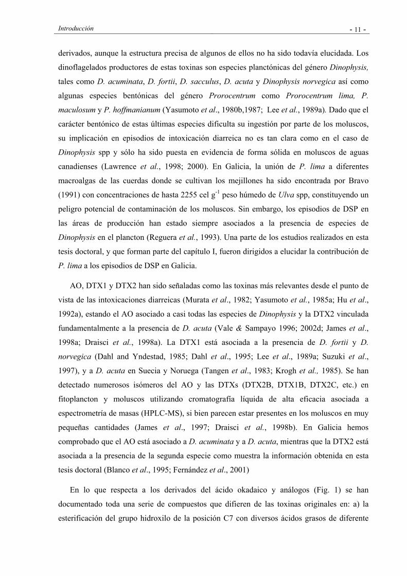
Las principales toxinas de este grupo, el AO, la DTX1 y la DTX2, son sustancias polietéreas
constituidas por una cadena carbonada lineal con un grupo carboxílico libre y que difieren en
difieren en la presencia de grupos metilo en los carbonos C31 y C35 (Fig. 1). El peso
molecular del AO es 804, y se han detectado un amplio número de isómeros, análogos y
Introducción - 11 -
derivados, aunque la estructura precisa de algunos de ellos no ha sido todavía elucidada. Los
dinoflagelados productores de estas toxinas son especies planctónicas del género Dinophysis,
tales como D. acuminata, D. fortii, D. sacculus, D. acuta y Dinophysis norvegica así como
algunas especies bentónicas del género Prorocentrum como Prorocentrum lima, P.
maculosum y P. hoffmanianum (Yasumoto et al., 1980b,1987; Lee et al., 1989a). Dado que el
carácter bentónico de estas últimas especies dificulta su ingestión por parte de los moluscos,
su implicación en episodios de intoxicación diarreica no es tan clara como en el caso de
Dinophysis spp y sólo ha sido puesta en evidencia de forma sólida en moluscos de aguas
canadienses (Lawrence et al., 1998; 2000). En Galicia, la unión de P. lima a diferentes
macroalgas de las cuerdas donde se cultivan los mejillones ha sido encontrada por Bravo
(1991) con concentraciones de hasta 2255 cel g-1 peso húmedo de Ulva spp, constituyendo un
peligro potencial de contaminación de los moluscos. Sin embargo, los episodios de DSP en
las áreas de producción han estado siempre asociados a la presencia de especies de
Dinophysis en el plancton (Reguera et al., 1993). Una parte de los estudios realizados en esta
tesis doctoral, y que forman parte del capítulo I, fueron dirigidos a elucidar la contribución de
P. lima a los episodios de DSP en Galicia.
AO, DTX1 y DTX2 han sido señaladas como las toxinas más relevantes desde el punto de
vista de las intoxicaciones diarreicas (Murata et al., 1982; Yasumoto et al., 1985a; Hu et al.,
1992a), estando el AO asociado a casi todas las especies de Dinophysis y la DTX2 vinculada
fundamentalmente a la presencia de D. acuta (Vale & Sampayo 1996; 2002d; James et al.,
1998a; Draisci et al., 1998a). La DTX1 está asociada a la presencia de D. fortii y D.
norvegica (Dahl and Yndestad, 1985; Dahl et al., 1995; Lee et al., 1989a; Suzuki et al.,
1997), y a D. acuta en Suecia y Noruega (Tangen et al., 1983; Krogh et al., 1985). Se han
detectado numerosos isómeros del AO y las DTXs (DTX2B, DTX1B, DTX2C, etc.) en
fitoplancton y moluscos utilizando cromatografía líquida de alta eficacia asociada a
espectrometría de masas (HPLC-MS), si bien parecen estar presentes en los moluscos en muy
pequeñas cantidades (James et al., 1997; Draisci et al., 1998b). En Galicia hemos
comprobado que el AO está asociado a D. acuminata y a D. acuta, mientras que la DTX2 está
asociada a la presencia de la segunda especie como muestra la información obtenida en esta
tesis doctoral (Blanco et al., 1995; Fernández et al., 2001)
En lo que respecta a los derivados del ácido okadaico y análogos (Fig. 1) se han
documentado toda una serie de compuestos que difieren de las toxinas originales en: a) la
esterificación del grupo hidroxilo de la posición C7 con diversos ácidos grasos de diferente
Introducción - 12 -
longitud (usualmente entre 14 y 17 carbonos) y nº de insaturaciones, y que da lugar a una
mezcla compleja de derivados acilados agrupados comúnmente bajo el término “DTX3” b)
formación de diol ésteres con dioles insaturados de entre 7 y 9 carbonos, esterificando el
grupo carboxilo c) oxidación del fragmento diol de las moléculas de diol ésteres d)
esterificación de los diol ésteres con cadenas sulfatadas que pueden incluir o no una función
amida en la parte polar de la cadena para dar lugar a derivados hidrosolubles del tipo DTX4 ó
DTX5 respectivamente y e) la carencia de grupo hidroxilo en la posición C2 o en la posición
C7 .
La “DTX3”, no ha sido detectada en fitoplancton y su aparición en los moluscos es
producto de la acilación metabólica del AO, DTX1 o DTX2 en los tejidos de los moluscos
(Suzuki et al., 1999). Su presencia ha sido demostrada en moluscos de Japón, Irlanda y
Portugal (Yasumoto et al., 1989; Hu et al., 1993, Vale and Sampayo, 1999). Los trabajos que
forman parte de esta tesis doctoral (Fernández et al., 1996; 1998), y que se muestran en los
artículos 7 y 8 del capítulo II, permitieron entre otros resultados, la primera detección de estos
compuestos en moluscos de las Rías Gallegas y alertaron de la pérdida de los mismos que se
producía durante la aplicación de ciertos protocolos de purificación de las muestras de
moluscos destinada al análisis de toxinas DSP. La presencia de DTX3 en almejas
(Scrobicularia plana) y navajas (Solen marginatus) de aguas portuguesas ha sido relacionada
con una reciente intoxicación diarreica en Portugal (Vale and Sampayo, 2002c).
En lo que respecta a los diol ésteres y a los derivados hidrosolubles sulfatados, DTX4 y
DTX5, inicialmente se asumió que estos compuestos tenían una presencia minoritaria y,
además, restringida a los dinoflagelados bentónicos del género Prorocentrum, lo que los hacía
relativamente poco relevantes desde el punto de vista de la salubridad de los moluscos.
Recientemente se ha demostrado que diferentes especies del género Dinophysis pueden
producirlos (Suzuki et al., 2004; Mackenzie et al., 2005).
La posibilidad de que estos compuestos puedan ser relevantes en Dinophysis de las Rías
Gallegas se discute en dos contribuciones a esta tesis. En el artículo 5 del capítulo I, una de
las contribuciones más recientes (Fernández et al., 2006), se observó que las células de D.
acuta sometidas a inhibición enzimática no presentaban concentraciones detectables de AO y
DTX2, apareciendo la PTX2 como la toxina principal y sugiriendo la presencia del AO y la
DTX2, ya evidenciada en trabajos anteriores, en forma de derivados conjugados (ésteres o
compuestos sulfatados).
Introducción - 13 -
Figura 1.- Estructuras del AO, dinophysistoxinas y algunos de sus derivados. Los diol esteres del ácido okadaico se desginan como OA-D#x, donde # es el nº de átomos de carbono de la cadena diol y x es una asignación arbitraria. DTX6 es un diol ester que ha recibido esta designación específica. “Oxidized OA-Dx” son algunos derivados producidos por la oxidación de los diol esteres. Norokadanona es un posible precursor del AO encontrado en Prorocentrum lima
Introducción - 14 -
En el artículo 8 del capítulo II (Moroño et al., 2003), los resultados obtenidos sugieren que
estos compuestos pueden llegar a ser el componente principal del perfil de toxinas de las
poblaciones de Dinophysis.
2.2. Pectenotoxinas
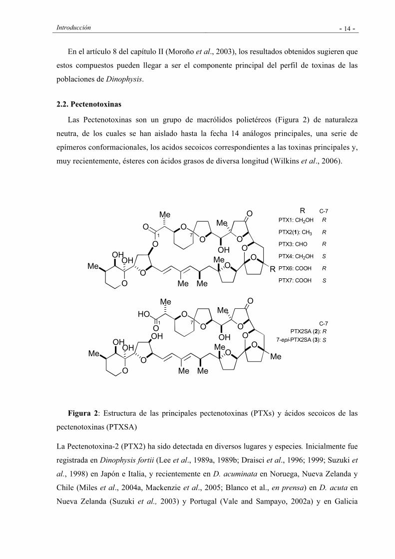
Las Pectenotoxinas son un grupo de macrólidos polietéreos (Figura 2) de naturaleza
neutra, de los cuales se han aislado hasta la fecha 14 análogos principales, una serie de
epímeros conformacionales, los acidos secoicos correspondientes a las toxinas principales y,
muy recientemente, ésteres con ácidos grasos de diversa longitud (Wilkins et al., 2006).
Figura 2: Estructura de las principales pectenotoxinas (PTXs) y ácidos secoicos de las
pectenotoxinas (PTXSA)
La Pectenotoxina-2 (PTX2) ha sido detectada en diversos lugares y especies. Inicialmente fue
registrada en Dinophysis fortii (Lee et al., 1989a, 1989b; Draisci et al., 1996; 1999; Suzuki et
al., 1998) en Japón e Italia, y recientemente en D. acuminata en Noruega, Nueva Zelanda y
Chile (Miles et al., 2004a, Mackenzie et al., 2005; Blanco et al., en prensa) en D. acuta en
Nueva Zelanda (Suzuki et al., 2003) y Portugal (Vale and Sampayo, 2002a) y en Galicia
OO
Me
OHO
MeO
OO
O
MeOH
OH Me
MeMe
O
O
OO
R
OO
Me
OHO
MeO
OO
O
MeOH
OH Me
MeMe
O
HO
OO
Me
OHC-7R
7
S
R
R
1
7-epi-PTX2SA (3):
PTX2SA (2):
C-7
PTX1: CH2OH
PTX2(1): CH3
PTX3: CHO
PTX4: CH2OH
PTX6: COOH
PTX7: COOH
7
S
S
R
R
R1

Introducción - 15 -
(Fernández et al, 2003 a; Fernández et al 2006) así como también en D. caudata (Fernández
et al., 2006), como se muestra en los Artículos 4 y 5 del capítulo I, que forman parte de la
presente tesis doctoral.
Se han detectado otras pectenotoxinas también en células de Dinophysis, como es el caso
de la PTX11, PTX13 y PTX14 en D. acuta y la PTX12 en D. acuta, D. acuminata, D.
rotundata y D. norvegica (Miles et al., 2004 b; Miles et al., 2006). La mayor parte de las
restantes PTXs, incluidos los ácidos secoicos (PTX2SA) son probablemente, en su inmensa
mayoría, productos de transformaciones metabólicas oxidativas de las pectenotoxinas
principales en los moluscos o en el plancton. Estos últimos compuestos se han encontrado en
moluscos de diferentes localizaciones geográficas (Daiguji et al., 1998; James et al., 1999b) y
también en moluscos de Galicia, como se muestra en el Artículo 4 del capítulo I de la presente
tesis (Fernández et al., 2003).
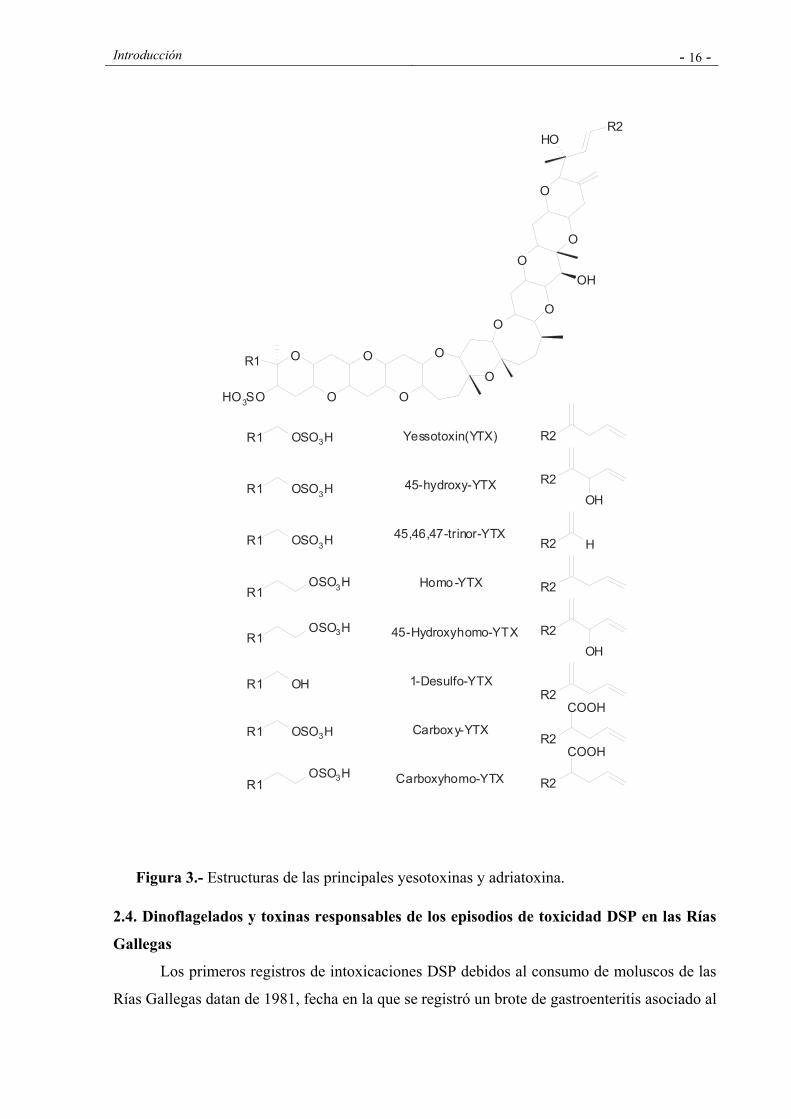
2.3. Yesotoxinas
La Yesotoxina (YTX) es una sustancia polietérea disulfatada, cuya estructura recuerda a
las brevetoxinas. La presencia de YTX ha sido confirmada en moluscos de Japón (Murata et
al., 1987), Noruega (Lee et al., 1989), Chile (Zhao et al., 1993), Italia (Ciminiello et al., 1997,
1999) y Nueva Zelanda (Yasumoto & Takizawa, 1997; MacKenzie et al., 1998). En los
últimos años, se ha confirmado la ocurrencia de un amplio número de análogos: 45-hidroxy
YTX, 45, 46, 47 trinor YTX, homo YTX, adriatoxina, carboxi-YTX y 1-desulfo-YTX
(Ciminiello et al., 1998, 1999; Daiguji et al., 1998; Satake et al., 1997, 1999; Tubaro et al.,
1998) (Fig. 3). Protoceratium reticulatum ha sido identificado como la principal especie
productora de YTX en Nueva Zelanda (MacKenzie et al., 1998) y de YTX y 45, 46, 47 trinor
YTX en Japón (Satake et al., 1999). La Homo-YTX fue detectada en una muestra de red en la
que la especie predominante era Lingulodinium polyedrum. Si bien cultivos de laboratorio de
esta misma especie procedentes de Nueva Zelanda, Estados Unidos y Japón no produjeron
yesotoxinas, la producción de YTX por cultivos de L. polyedrum procedentes de aguas de
Andalucía fue confirmada por HPLC-MS (Paz et al., 2004). En Galicia se han registrado
eventos ligados a P. reticulatum y Yesotoxinas en las Rías Altas (Arévalo et al.; 2006). Tanto
Protoceratium reticulatum como Lingulodinium polyedrum, son especies ubicuas, por lo que
la contaminación de moluscos con YTXs se presenta como un fenómeno generalizado.
Introducción - 16 -
Figura 3.- Estructuras de las principales yesotoxinas y adriatoxina.
2.4. Dinoflagelados y toxinas responsables de los episodios de toxicidad DSP en las Rías
Gallegas
Los primeros registros de intoxicaciones DSP debidos al consumo de moluscos de las
Rías Gallegas datan de 1981, fecha en la que se registró un brote de gastroenteritis asociado al
O
O
O
O
O
O
OO
O
O
O
R2OH
R1
HO3SO
OH
R2
R2OH
R2
R2OH
R2 H
R2
R2
COOH
R2
COOH
R1 OSO3H
R1OSO3H
R1 OH
R1 OSO3H
R1 OSO3H
R1OSO3H
R1OSO3H
R1 OSO3H
Yessotoxin(YTX)
45-hydroxy-YTX
45,46,47-trinor-YTX
Homo-YTX
45-Hydroxyhomo-YTX
1-Desulfo-YTX
Carboxy-YTX
Carboxyhomo-YTX
Introducción - 17 -
consumo de mejillones que afectó a 5000 personas en España (Campos et al., 1982). El
conocimiento de la “Intoxicación diarreica por bivalvos” (DSP) y la identificación de
Dinophysis fortii como el agente de estos episodios en Japón sugirió que el episodio tóxico de
1981, así como otros brotes asociados al consumo de mejillones depurados en 1978 y 1979,
podían tratarse de episodios de DSP asociados a la presencia de Dinophysis spp (Campos et
al., 1982; Fraga et al., 1984).
A partir de 1982, las autoridades sanitarias gallegas incorporaron el bioensayo en ratón
para la detección de toxicidad DSP al programa de control sanitario ya existente para la
detección de PSP desde el año 1977, y en el que el control de fitoplancton era desarrollado
por el Instituto Español de Oceanografía. A partir de 1992, el seguimiento de las especies
fitoplanctónicas nocivas y de la toxicidad de origen microalgal en moluscos fue asumido por
el Centro Galego para el Control da Calidade do Medio Mariño (Conselleria de Pesca, Xunta
de Galicia), en la actualidad INTECMAR.
Los episodios de DSP en las Rías Gallegas están correlacionados principalmente con
proliferaciones de D. acuminata en la Ría de Ares y de D. acuminata y D. acuta en las Rías
Bajas. Si bien D. acuminata y D. acuta son componentes regulares del fitoplancton y
constituyen las especies de Dinophysis más abundantes, otras especies como D. rotundata, D.
caudata, D. tripos y D. hastata, están también presentes en ocasiones, aunque en número
considerablemente más bajo. Los primeros estudios sobre el perfil de toxinas de los moluscos
de las Rías Gallegas habían demostrado la presencia de AO en mejillones causantes de
episodios DSP (Kumagai et al., 1986; Rodríguez Vázquez et al., 1989; Gago et al., 1993) y
los estudios del contenido celular de toxinas habían demostrado la presencia de AO en D.
acuta de la Ría de Vigo (Lee et al., 1989a).
Estudios posteriores, algunos de los cuales forman parte de esta tesis, revelaron un
perfil de toxinas en los moluscos y en el fitoplancton considerablemente más complejo.
Además del AO, se identificó la presencia de DTX2 en arrastres fitoplanctónicos (Blanco et
al., 1995), en mejillones (Gago-Martínez et al., 1996; Fernández et al., 1998) y en células de
D. acuta aisladas por micromanipulación (Fernández et al., 2001) y la existencia en los
moluscos de derivados acilados de ambas toxinas (Fernández et al., 1996; Fernández et al.,
1998). Más recientemente, se demostró la presencia de PTX2, en ocasiones como toxinas
dominantes en arrastres fitoplanctónicos ricos en D.acuta y D. caudata (Fernández et al.,
2003a) y en células aisladas de estas dos mismas especies (Fernández et al., 2006). Asimismo,
se detectó la presencia en mejillones de ácidos secoicos de la PTX2 (Fernández et al., 2003a).
Introducción - 18 -
En lo que se refiere a P. lima, su presencia en las costas de Galicia es conocida desde
1985, fecha en la que se realizaron cultivos de esta especie a partir de muestras de la laguna
de las Islas Cíes y de muestras tomadas en las cuerdas de mejillón en las rías de Vigo y
Pontevedra (Bravo et al., 1997). Los análisis de toxinas a partir de cinco clones de P. lima
aislados de la costa gallega realizados por Lee et al. (1989a) mostraron la presencia de AO y
DTX1 y diferencias en las cantidades de AO y DTX1 entre las distintas cepas. En un estudio
posterior, dos diol ésteres del AO fueron aislados e identificados mediante métodos
espectroscópicos en cepas de la Ría de Vigo (Norte et al., 1994). Los estudios realizados en
esta tesis doctoral y que se presentan en el capítulo I fueron dirigidos a estudiar el perfil de
toxinas de 19 cepas de P. lima aisladas en la Ría de Vigo y en la Ría de Pontevedra, y a
elucidar la contribución de este dinoflagelado bentónico a los episodios de DSP en Galicia.
3. EPIDEMIOLOGIA Y TOXICOLOGIA DE LAS TOXINAS DSP
Los distintos grupos de toxinas incluidas históricamente en la amplia denominación de
toxinas “DSP” presentan diferentes efectos en los humanos. Existen datos epidemiológicos de
intoxicaciones producidas por el ácido okadaico y derivados que muestran que la intoxicación
comienza entre tres y doce horas después de la ingestión de los moluscos y cursa con
trastornos gastrointestinales tales como diarrea, nauseas y vómitos (Yasumoto et al., 1978,
1980; Kat et al., 1979, 1983), produciéndose una recuperación completa, en la mayor parte de
los individuos afectados, en el plazo de tres días, y no habiéndose registrado hasta la fecha
casos de mortalidad. La toxicidad depende de la toxina específica y parece estar asociada a la
presencia de la función carboxílica en forma libre. El nivel de AO necesario para inducir
diarrea en adultos está entre 40 y 50 µg. En una intoxicación ocurrida en Noruega, alrededor
de 70 personas fueron hospitalizadas con los síntomas usuales de DSP tras el consumo de
mejillones. Los niveles encontrados en mejillones no consumidos fueron del orden de 55-56
µg eq. AO/100 g vianda de mejillón (Aune, 2001). Los derivados en los que el grupo
carboxilo está esterificado muestran una toxicidad considerablemente menor (Hu et al.,
1992b, 1995). La esterificación del hidroxilo en la posición C7, sin embargo, no parece que
reduzca sustancialmente la toxicidad y se han documentado intoxicaciones producidas por
bivalvos que contenían principalmente este tipo de ésteres del ácido okadaico (Vale and
Sampayo, 2002c).
Estudios experimentales realizados en ratones muestran que la dosis letal del AO por
administración intraperitoneal (LD50) es de 200 µg kg-1. Los valores correspondientes para
Introducción - 19 -
DTX1 y “DTX3” son 160 y 500 µg kg-1 respectivamente (Yasumoto et al., 1989), aunque
observaciones recientes (Ito et al., 2006) indican que la DTX3 presenta una toxicidad similar
a la del AO si se asegura su solubilidad en el líquido inyectado a los ratones. Cuando el AO se
administra oralmente a ratas, se observan daño intestinal, diarrea y muerte, pero no se
observan efectos en el hígado. Sin embargo, la administración intraperitoneal no produce
apenas efecto en la función intestinal pero afecta al hígado de manera considerable (Berven et
al., 2001).
Aparte de los efectos agudos, diferentes trabajos han puesto de manifiesto que este grupo
de toxinas podrían producir importantes efectos crónicos. Así, se ha demostrado que estos
compuestos son promotores de crecimiento tumoral (Fujiki et al., 1988) y se ha sugerido
recientemente que podrían actuar como inductores tumorales (Ten-Hage et al., 2000; Creppy
et al., 2002). Aunque las observaciones disponibles sobre poblaciones humanas expuestas no
son concluyentes, existen ciertas evidencias de asociación con cáncer localizado en diferentes
partes del aparato digestivo (cáncer de esófago, estómago, colon e hígado en varones y de
estómago y páncreas en mujeres) (Cordier et al., 2000). En la tortuga, Chelonia midas, la
aparición de una neoplasia, fibropapilomatosis, ha sido relacionada con la abundancia de
Prorocentrum lima, productor de toxinas DSP del grupo del ácido okadaico (Landsberg et al.,
1999).
Con respecto al mecanismo de acción, el AO y análogos son potentes inhibidores de las
protein-fosfatasas (serina y treonina), enzimas que son responsables de la defosforilación de
ciertas proteinas y que desempeñan un papel crítico en el control de procesos biológicos tales
como la división y el crecimiento celular, la organización estructural, el metabolismo, el
control hormonal, etc (Bialojan and Takai, 1988; Cohen, 1989). Este mecanismo es
probablemente el principal responsable de la inducción de diarrea, que estaría asociada a la
hiperfosforilación de los canales iónicos de las células epiteliales del intestino que alterarían
el equilibrio hídrico (Cohen et al., 1990) y de la promoción tumoral observada en estudios in
vitro (Suganuma et al., 1988).
En lo que concierne a las Pectenotoxinas y Yesotoxinas no existen datos epidemiológicos
que asocien inequívocamente la presencia de estas sustancias en los moluscos con
intoxicaciones diarreicas. En Australia se registraron dos episodios de intoxicación en
humanos coincidentes con la presencia de PTXs en las tellinas ó “pipis” (Donax deltoides).
En diciembre de 1997, 100 personas resultaron intoxicadas tras el consumo de este molusco,
Introducción - 20 -
de las cuales al menos 56 casos requirieron hopitalización. Los principales síntomas de la
intoxicación fueron nauseas, vómitos y diarrea. En marzo de 2000, se registró otra
intoxicación con la misma especie. La principal pectenotoxina detectada en ambos casos fue
el ácido secoico de la PTX2. La dosis ingerida por el individuo que resultó más afectado fue
de aproximadamente 150 µg de PTX2SA, lo que representa una dosis de 2 µg de toxinas por
kg de peso corporal (Burguess and Shaw, 2001). Sin embargo, al menos en el primero de los
episodios, se detectó también la presencia de ésteres del ácido okadaico en los moluscos
consumidos (Quilliam et al., 2000), lo que pone en cuestión el origen real de la intoxicación
en humanos y por tanto la toxicidad real de las Pectenotoxinas.
Estudios con animales de experimentación muestran que la toxicidad aguda intraperitoneal
de las PTXs oscila entre 230 µg kg-1en el caso de la PTX2 y 770 µg kg-1en el caso de la
PTX4. Las pectenotoxinas no causan diarrea por administración intraperitoneal y no se
observa toxicidad oral a dosis de 5000 µg de PTX2 y PTX2SA (Miles et al., 2004). Las PTXs
no inhiben las protein-fosfatasas, pero exhiben una potente citotoxicidad de manera que dosis
comprendidas entre 500 y 1000 µg kg-1 inducen necrosis en el hígado a las pocas horas de la
inoculación intraperitoneal (Terao et al., 1986, 1993). La inducción de apoptosis ha sido
documentada por Fladmark et al. (1998). Se ha demostrado también que concentraciones
micromolares de PTX6 inducen la despolarización de la F-actina en células de neuroblastoma
(Leira et al. 2002). Estos mismos autores han sugerido que la disrupción del citoesqueleto
puede ser un mecanismo clave en la toxicidad de esta toxina, y muy probablemente del resto
de las pectenotoxinas.
Con respecto a las Yesotoxinas, no se han registrado hasta la fecha casos de intoxicación
en humanos. La toxicidad intraperitoneal de estas toxinas es muy alta. Las dosis letales en
ratón para los diferentes análogos por administración intraperitoneal oscilan entre 100 y 400
µg kg-1siendo en este caso el corazón el órgano diana (Murata et al., 1987). Si bien el
derivado desulfatado (desulfo-YTX) no presenta cardiotoxicidad, hígado y páncreas se ven
afectados a partir de dosis intraperitoneales de 300 µg kg-1 (Terao et al., 1990). La toxicidad
oral es inferior a la toxicidad intraperitoneal en más de un orden de magnitud (Ogino et al.,
1997; Aune et al., 2001, 2002; Tubaro et al., 2004 a, 2004b), probablemente debido a la
presencia de los grupos sulfato en la molécula que hace que estos compuestos no sean
absorbidos o metabolizados en el tracto digestivo cuando se administran oralmente. No se
observan efectos diarreicos tras su administración y la LD 50 es muy alta (Tabla 1).
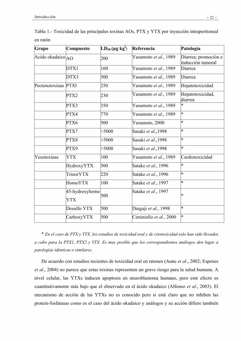
Introducción - 21 -
Tabla 1.- Toxicidad de las principales toxinas AOs, PTX y YTX por inyección intraperitoneal
en ratón
Grupo Compuesto LD50 (µg kgl) Referencia Patología
Acido okadaico AO 200 Yasumoto et al., 1989 Diarrea; promoción e inducción tumoral
DTX1 160 Yasumoto et al., 1989 Diarrea
DTX3 500 Yasumoto et al., 1989 Diarrea
Pectenotoxinas PTXl 250 Yasumoto et al., 1989 Hepatotoxicidad
PTX2 230 Yasumoto et al., 1989 Hepatotoxicidad, diarrea
PTX3 350 Yasumoto et al., 1989 *
PTX4 770 Yasumoto et al., 1989 *
PTX6 500 Yasumoto, 2000 *
PTX7 >5000 Sasaki et al.,1998 *
PTX8 >5000 Sasaki et al.,1998 *
PTX9 >5000 Sasaki et al.,1998 *
Yesotoxinas YTX 100 Yasumoto et al., 1989 Cardiotoxicidad
HydroxyYTX 500 Satake et al., 1996 *
TrinorYTX 220 Satake et al., 1996 *
HomoYTX 100 Satake et al., 1997 *
45-hydroxyhomo
YTX 500
Satake et al., 1997 *
Desulfo YTX 500 Daiguji et al., 1998 *
CarboxyYTX 500 Ciminiello et al., 2000 *
* En el caso de PTX y YTX, los estudios de toxicidad oral y de citotoxicidad solo han sido llevados
a cabo para la PTX1, PTX2 y YTX. Es muy posible que los correspondientes análogos den lugar a
patologías idénticas o similares,
De acuerdo con estudios recientes de toxicidad oral en ratones (Aune et al., 2002; Espenes
et al., 2004) no parece que estas toxinas representen un grave riesgo para la salud humana. A
nivel celular, las YTXs inducen apoptosis en neuroblastoma humano, pero este efecto es
cuantitativamente más bajo que el observado en el ácido okadaico (Alfonso et al., 2003). El
mecanismo de acción de las YTXs no es conocido pero si está claro que no inhiben las
protein-fosfatasas como es el caso del ácido okadaico y análogos y su acción difiere también
Introducción - 22 -
de la de los otros dos grupos de toxinas estructuralmente similares como son las ciguatoxinas
y las brevetoxina. Estudios con linfocitos humanos sugieren que estas toxinas actúan sobre el
AMP cíclico, incrementando los niveles citosólicos de calcio (Rosa et al., 2001). Estudios
recientes señalan que un aumento en la actividad de las fosfodiesterasas (PDE) es el principal
mecanismo de acción, si bien tanto el tipo de fosfodiesterasas implicadas como la ruta precisa
de interacción requieren estudios adicionales (Alfonso et al., 2003). En el caso de que la diana
de estas toxinas fuera la PDE III, muy abundante en el músculo cardíaco, esto explicaría
fácilmente la cardiotoxicidad.
La mayor parte de los datos toxicológicos disponibles en relación a las PTXs y YTXs
proceden de estudios realizados mediante administración intraperitoneal de las toxinas a los
animales de experimentación, de manera que la extrapolación de estos resultados para
determinar los efectos en el hombre y por tanto el establecimiento de los límites de tolerancia
es difícil, siendo necesario completar los mismos con estudios robustos de toxicidad oral que
han estado dificultados hasta la fecha por la escasez o carencia de toxinas puras. La carencia
de datos epidemiológicos añade dificultades a la realización de estudios formales de análisis
de riesgo que permitan el establecimiento de límites de tolerancia, y por tanto faciliten el
establecimiento de regulaciones que, salvaguardando la salud pública, no produzcan perdidas
económicas innecesarias a los sectores de producción.
A pesar de las evidentes limitaciones de los datos disponibles, en los últimos años se han
establecido en las legislaciones niveles para los diferentes grupos de toxinas lipofílicas que
serán discutidos en el apartado relativo a las regulaciones.
4. MÉTODOS DE DETECCIÓN DE LAS TOXINAS DSP.
4.1. Generalidades
Con el fin de prevenir que puedan llegar al consumidor moluscos tóxicos, se han
desarrollado diferentes métodos para la detección de toxinas marinas, que en muchos casos,
en un tiempo reducido, permiten detectar su presencia en los moluscos a niveles muy por
debajo de lo que se considera un riesgo; de ahí que en la última década se hayan reducido
notablemente el número de intoxicaciones. Dichos métodos han de abordar dificultades tales
como la presencia de las toxinas en matrices biológicas complejas y la presencia de mezclas
de congéneres relacionados estructuralmente y con potencias tóxicas que pueden llegar a
diferir hasta en tres órdenes de magnitud, como en el caso de las toxinas paralizantes PSP
Introducción - 23 -
(Oshima et al., 1993). Hay que añadir las dificultades ocasionadas por la ausencia en la
mayoría de estas sustancias de grupos cromóforos que faciliten su detección por las técnicas
convencionales de espectrofotometría UV-Visible o de fluorescencia.
La aparición de toxinas de diferentes grupos en un mismo producto marino no es un
hecho infrecuente y representa una complicación adicional a efectos de determinar la
toxicidad de los mariscos. Es digna de mención la ocurrencia simultánea en las aguas costeras
de especies fitoplanctónicas productoras de toxinas diarreicas y aquellas productoras de
toxinas paralizantes, y como consecuencia de este hecho, la co-ocurrencia de DSP y PSP en
los moluscos (Gago-Martínez et al., 1996; Amzil et al., 1999). Las toxinas paralizantes,
incluso cuando se encuentran en niveles por debajo del límite de detección del bioensayo en
ratón utilizado habitualmente para su determinación (AOAC, 1995), dan lugar a interferencias
en uno de los métodos más extendidos de detección de toxinas DSP, el bioensayo en ratón
con extracción acetónica (Yasumoto et al., 1978), que de no ser eliminadas no permitirían la
determinación de la presencia de toxinas diarreicas.
Hay que mencionar la posibilidad que se discutirá en capítulos posteriores de esta tesis
doctoral de biotransformaciones de las toxinas a lo largo de su transmisión a través de la
cadena trófica, mediante diferentes procesos metabólicos, de tal forma que la composición
tóxica de los transvectores, puede diferir notablemente de aquella de las microalgas
productoras de las toxinas. Una última dificultad viene dada por la labilidad química de
algunas de estas sustancias que hace que puedan sufrir transformaciones durante los procesos
de extracción, purificación y análisis.
De manera general, y atendiendo a la naturaleza de la información obtenida a partir de
los métodos de determinación, éstos pueden clasificarse en métodos de ensayo y métodos
analíticos (Sullivan et al., 1993).
En el área de la determinación de toxinas, el término ensayo, se refiere a aquellos
métodos que proporcionan un valor del contenido total de toxinas o de actividad tóxica
basado en la medición de una única respuesta, biológica o bioquímica, que engloba a todos los
congéneres presentes en la muestra. La determinación de la toxicidad se lleva a cabo en
función de una curva dosis-respuesta que se realiza usualmente con una de las toxinas
representativas del grupo objeto de estudio, expresándose finalmente la toxicidad total en
equivalentes de dicha toxina. Dentro de este primer grupo se encuadrarían los bioensayos in
Introducción - 24 -
vivo, los ensayos de inhibición enzimática, los ensayos celulares, los ensayos de receptor, los
inmunoensayos y los ensayos electrofisiológicos.
El término análisis se refiere a aquellos métodos de detección en los que se realiza una
separación, identificación y posterior cuantificación individual de las toxinas en función de
una respuesta instrumental que es proporcional a la concentración de cada una de las toxinas
presentes en la muestra. Dicha cuantificación requiere la calibración previa del equipo
instrumental con patrones de concentración conocida de cada una de las toxinas objeto de
determinación. La respuesta instrumental debe ser convertida posteriormente a valores de
toxicidad en función de factores de conversión específicos para cada toxina, de manera que la
toxicidad global se determina como un sumatorio de las toxicidades individuales. En este
segundo grupo se incluyen los métodos químicos basados en la separación de las toxinas por
cromatografía líquida de alta eficacia y detección colorimétrica, fluorimétrica o por
espectrometría de masas, así como la electroforesis capilar.
Dejando a un lado valoraciones de carácter económico, la elección de unos u otros
métodos se realiza en función de los objetivos del estudio a realizar. Los métodos químicos
instrumentales se utilizan generalmente en programas de investigación que requieran la
identificación y cuantificación de cada una de las toxinas presentes. En los programas de
monitorización o control sanitario, en aras de proteger la salud pública, se da relevancia al
conocimiento de la toxicidad global potencial por lo que se han utilizado preferentemente
ensayos.
4.2. Ensayos in vivo
Hasta la fecha, la mayor parte de los programas de control de toxinas en los alimentos
han estado basados en ensayos in vivo, esto es, ensayos con animales, que si bien han
demostrado su eficacia en la protección de la salud pública, al poner en evidencia la toxicidad
de alimentos no aptos para el consumo, están lejos de la consideración de “ideales”. El
principio general de los ensayos in vivo para determinación de ficotoxinas se basa en la
administración al animal de la muestra a analizar o de un extracto de la misma, ya sea por vía
oral o, lo que es más usual, mediante inyección intraperitoneal, seguida de la observación de
los síntomas y/o de la determinación del tiempo de supervivencia, parámetro que se utiliza
frecuentemente para la cuantificación a través de curvas dosis-respuesta previamente
establecidas. Los bioensayos en ratón son poco selectivos, de baja especificidad y
Introducción - 25 -
sensibilidad, pueden producir falsos positivos debido a interferencias de las matrices
biológicas y la variabilidad entre laboratorios es alta (alrededor de un 20%). Sin embargo, la
baja especificidad es de gran utilidad desde el punto de vista de la estimación del riesgo para
la salud pública, puesto que permite la detección de nuevas toxinas o congéneres,
proporcionando de esta forma un alto grado de protección al consumidor.
A diferencia de la situación del control sanitario de las toxinas PSP, en el caso de las
toxinas históricamente incluidas en el grupo DSP no ha existido un protocolo de bioensayo en
ratón acordado y reconocido internacionalmente y que haya sido sometido a un proceso
formal de estandarización y validación. Si bien es cierto que la mayor parte de los países que
realizan el control de estas toxinas en los moluscos han venido utilizando ensayos in vivo,
existen diferencias en cuanto al animal usado (rata o ratón albino), en cuanto al modo de
administración de la toxinas (ingestión oral en el caso de la rata, o inyección intraperitoneal
en el caso del ratón) y en cuanto a la naturaleza y proporción de los solventes utilizados para
la extracción de las toxinas. Esto se ha traducido en una diferente especificidad, selectividad y
recuperabilidad de los bioensayos y por tanto en una falta de equivalencia de los mismos, de
tal manera que algunos protocolos pueden detectar todo el rango de sustancias lipofílicas
bioactivas potencialmente presentes en los mariscos contaminados, mientras que la mayor
especificidad de otros protocolos limita el espectro de toxinas detectables por los mismos.
Existen igualmente discrepancias en cuanto a los criterios de positividad del ensayo.
Esta situación no es gratuita sino que se ha producido como consecuencia de la
escasez de estudios epidemiológicos y toxicológicos que permitieran establecer los riesgos
derivados de la exposición aguda y crónica a las sustancias bioactivas actualmente incluidas
en el grupo DSP, y por tanto los niveles de tolerancia. A esto hay que añadir la controversia
histórica que ha existido en relación a la idoneidad de la clasificación de algunas de estas
sustancias polietéreas liposolubles dentro del grupo DSP.
En el trabajo que constituye el capítulo III de esta tesis doctoral y que fue publicado
con el título “In vivo assays for phycotoxins” como capítulo del libro “Manual on Harmful
Marine Microalgae” (Fernández et al., 2003b) se describen y valoran los diferentes
bioensayos con mamíferos utilizados para la detección de toxinas marinas.

Introducción - 26 -
4.3. Ensayos in vitro
Con el fin de reducir en el futuro el uso de animales en los programas de control
alimentario y en aras de lograr una mayor sensibilidad y especificidad, se han desarrollado en
los últimos años diferentes ensayos in vitro, que pueden ser clasificados en dos grupos:
ensayos funcionales y ensayos estructurales. En los ensayos funcionales la respuesta que se
mide está directamente relacionada con el mecanismo de acción responsable de la toxicidad
de la sustancia que se determina, ya sea la inhibición de la actividad enzimática ó la
activación o el bloqueo de canales iónicos. El grado de correlación entre la respuesta
observada y la toxicidad real es esperable que sea muy alto con independencia de los perfiles
tóxicos, es decir, de los tipos y cantidades de toxinas presentes en la muestra analizada. En el
caso de los ensayos estructurales (ej. inmunoensayos) la respuesta está basada en el
reconocimiento por parte de los anticuerpos de fragmentos de las estructuras moleculares de
las toxinas que no están necesariamente ligados al mecanismo de acción de las mismas y por
tanto a su actividad biológica. En este caso no siempre existe una buena correlación entre la
respuesta inmunológica y la toxicidad real, de manera que los anticuerpos no identificarán a
todos los congéneres activos (falsos negativos) y podrían reconocer congéneres inactivos
(falsos positivos).
En general, la sensibilidad de los ensayos in vitro es varios órdenes de magnitud
superior a la de los ensayos in vivo, aspecto relevante no sólo desde el punto de vista sanitario
sino también desde el punto de vista de posibilitar una detección precoz de los episodios
tóxicos que permita mitigar el impacto negativo de los mismos. Gran parte de los ensayos in
vitro han sido desarrollados para su aplicación en placas de pocillos múltiples (ej. 96
pocillos), utilizando lectores con detección colorimétrica o fluorimétrica, o mediante
contadores de centelleo, lo que permite la realización simultánea en un tiempo reducido de
curvas de calibrado y el análisis de un alto número de muestras a través de diferentes
diluciones. En contrapartida, los ensayos in vivo presentan la ventaja difícilmente superable
de su potencial para la detección no solo de las toxinas ya conocidas, para las que han sido
estandarizados, sino de otros compuestos bioactivos no conocidos de antemano que puedan
representar un riesgo para la salud pública, y que no podrían ser detectados en el caso de
utilizar métodos más específicos.
La aplicación de ensayos in vitro en laboratorios de control sanitario con fines de
control oficial está todavía dificultada por el hecho de que no se ha realizado una validación

Introducción - 27 -
formal de los mismos a través de protocolos internacionalmente reconocidos (AOAC, ISO,
etc.). Dichos protocolos establecen básicamente la realización de estudios intercomparativos y
colaborativos entre un número suficiente de laboratorios de tal forma que se establezcan las
características del método en términos de exactitud y precisión (repetibilidad y
reproducibilidad) y que se verifique la aplicabilidad del ensayo para la determinación de
diferentes toxinas de un mismo grupo en las diferentes matrices biológicas que puedan ser
objeto de análisis. Tanto en los procesos de desarrollo de los ensayos como en lo que se
refiere a la validación e implantación de los mismos, es necesario disponer de las toxinas
puras y de materiales tóxicos de concentración certificada que permitan la puesta a punto del
ensayo y la realización de controles de calidad de los análisis. Si bien la disponibilidad y
distribución de ciertas toxinas ha mejorado en los últimos años, existen todavía graves
carencias en esta área que están retrasando el desarrollo, validación e implantación de estas
nuevas metodologías. En lo que respecta a la detección de toxinas DSP se han desarrollado
los siguientes ensayos in vitro:
Ensayos de inhibición enzimática
Como ya se mencionó en apartados anteriores, el AO y algunos de sus análogos
(DTX1, DTX2) ejercen su actividad biológica a través de mecanismos de inhibición de las
protein-fosfatasas PP1 y PP2A, dos importantes enzimas que intervienen en el control de
procesos tales como la división y crecimiento celular, organización estructural, metabolismo,
control hormonal, etc. Teniendo en cuenta que la magnitud de la inhibición de fosfatasas es
proporcional a la concentración de toxinas, éste mecanismo puede ser utilizado para medir el
contenido de estas sustancias en muestras de moluscos o fitoplancton.
Basándose en estos hallazgos se han desarrollado diferentes ensayos cuyo principal
interés es que el parámetro que se evalúa, esto es la inhibición de las fosfatasas, está
directamente relacionado con el mecanismo de acción de estas toxinas y por tanto con su
acción biológica y toxicidad. Son por tanto ensayos funcionales. La magnitud de la inhibición
enzimática puede ser medida utilizando diversos sustratos naturales o artificiales de las
enzimas. Las fosfatasas actúan sobre los diferentes sustratos causando su desforilación,
proceso que es fuertemente inhibido en presencia de estas toxinas. Así, se han desarrollado
ensayos basados en sustratos radiactivos (Holmes et al, 1991; Chen et al., 1993; Honkanen et
al., 1996), ensayos colorimétricos (Takai and Mieskes 1991; Simon and Vernoux, 1994;
Tubaro et al., 1996) y ensayos fluorimétricos (Vieytes et al., 1997). El ensayo fluorimétrico

Introducción - 28 -
utiliza PP2A comercial y diferentes sustratos fluorescente como son: 4-metil-lumbeliferona,
fluorescein difosfato (FDP), 6,8, difluoro-7-hidroxi-4-metilumbeliferona fosfato (Di FMUP),
9H- (1,3-dicloro-9,9-dimetilacridin-2-one-7-yl) fosfato, sal diamonio (DDAO fosfato) y 4
metilumbeliferona fosfato (4-MUP). El ensayo puede realizarse con cualquiera de los
sustratos mencionados en placas de pocillos múltiples, siendo necesario en este caso un lector
de fluorescencia. Conlleva una extracción sencilla de las toxinas con metanol 80%, no
requiere una purificación posterior de las muestras, y permite la realización del análisis
simultáneo de un número elevado de muestras en un tiempo reducido. Es muy sencillo, de
ejecución rápida y gran sensibilidad, con un límite de detección de 3.2 pg AO ml-1. Los
mismos autores han aplicado con éxito este tipo de ensayo a la determinación de microcistinas
(Fontal et al., 1999). Recientes estudios comparando el ensayo colorimétrico y el
fluorimétrico (Mounfort et al. 1999) han mostrado que el ensayo fluorimétrico es más preciso
y sensible y produce menos falsos positivos que el que utiliza los sustratos cromogénicos.
La metodología de los ensayos de inhibición enzimática hacen que estos sean muy
simples, sensibles, rápidos y selectivos, y tienen capacidad para detectar otras sustancias
bioactivas potencialmente tóxicas a través de los mismos mecanismos de inhibición
enzimática. El ensayo es adecuado para la detección del AO, DTX1, DTX2 y cualquier
derivado activo de estas toxinas. Asi en los casos de la DTX3, los dioles-ésteres y la DTX4
que no presentan actividad inhibitoria su presencia mediante estos ensayos solo puede ser
detectada si se someten a una hidrólisis previa. (Mounfort et al., 2001).
A pesar de todo lo anterior, la utilización de estos ensayos con fines de control de la
presencia de ácido okadaico y análogos en los moluscos, esta todavía limitada por la falta de
estudios robustos de validación ya que los intentos realizados no han dado buenos resultados
debido a problemas relacionados con la falta de estabilidad de los enzimas. Recientemente,
Sekiguchi et al. (2006) han propuesto un ensayo similar con fosfatasas obtenidas a partir del
gasterópodo marino Neptuna arthritica que presenta una estabilidad de un año. Ésta y otras
iniciativas importantes dirigidas a la validación, hacen previsible que en un futuro próximo
los ensayos de inhibición enzimática sean utilizados como herramientas de control sanitario.
Como las pectenotoxinas no son inhibidores de las protein-fosfatasas, los ensayos de
inhibición enzimática con fines de control sanitario tendrán que ser complementados con
otros ensayos o análisis que detecten estos compuestos.
Introducción - 29 -
Inmunoensayos
Existen numerosas variantes de inmunoensayos que utilizan diferentes estrategias de
acoplamiento antígeno-anticuerpo como reacciones directas o indirectas, reacciones de
competencia, etc. La magnitud de la reacción inmunológica puede ser detectada de diversas
maneras, ya sea mediante el uso de marcadores radiactivos (radioinmunoensayos, RIA),
marcadores fluorescentes (FIA) o mediante acoplamiento de una reacción enzimática (EIA).
Los inmunoensayos más frecuentes para la determinación de ficotoxinas son los denominados
ELISAs (Enzyme-Linked Immunosorbent Assay) que pueden ser configurados en una gran
variedad de formatos y en los que básicamente, se mide la interacción toxina-anticuerpo
mediante la reacción de una enzima ligada al anticuerpo con un sustrato específico de la
misma, reacción que da lugar a un producto coloreado o fluorescente.
Los inmunoensayos tienen un grado variable de reactividad con las diferentes toxinas
pertenecientes a un mismo grupo y la sustitución de determinados grupos funcionales afecta a
la reactividad cruzada de las toxinas con los anticuerpos. Los inmunoensayos son ensayos
estructurales de tal forma que los anticuerpos reconocen moléculas de similar estructura, que
podrían no ser tóxicas, produciéndose en este caso falsos positivos, y no reaccionan con
moléculas diferentes que presenten actividad biológica similar produciéndose en este caso
falsos negativos. La correlación entre la respuesta inmunológica y la toxicidad real depende
por tanto de los perfiles tóxicos de las muestras de fitoplancton o de mariscos.
En relación a la detección de toxinas DSP, se han desarrollado varios métodos
configurados como RIA o ELISA que utilizan anticuerpos preparados contra el AO. El
radioinmunoensayo desarrollado por Levine y colaboradores (Levine et al., 1988) se basa en
la inhibición competitiva de la reacción entre AO tritiado y el anticuerpo del AO producido
por el ácido okadaico de las muestras. La magnitud de la inhibición se determina mediante
contador de centelleo y permite la detección de hasta 0.2 ng AO ml-1. El método es sin
embargo muy laborioso y complejo para su uso rutinario.
Se han desarrollado dos ELISAs en forma de kits para la determinación de AO, el
“DSP-Check” (Sceti, Tokio, Japón) y el “Okadaic acid kit” de Rougier Bio-Tech (Montreal),
si bien solo el primero está disponible comercialmente. El “DSP-check” utiliza anticuerpos
monoclonales previamente desarrollados por Usagawa y colaboradores (Usagawa et al.,
1989). Los anticuerpos monoclonales marcados con una enzima, reaccionan

Introducción - 30 -
competitivamente con una cantidad constante de ácido okadaico fijada en los pocillos y con el
ácido okadaico procedente de la muestra objeto de análisis. La cantidad de enzima es
directamente proporcional a la cantidad de anticuerpo fijado, que será tanto mayor cuanto
menor sea la cantidad de AO en las muestras, y que es medida colorimétricamente, de manera
que el contenido de AO de las muestras es inversamente proporcional a la absorbancia. La
sensibilidad del ensayo es de 20 ng g-1. Los anticuerpos utilizados en el kit presentan una
reactividad cruzada de aproximadamente 40 % con la DTX2 (Carmody et al., 1995 b) y del
70 % con la DTX1 (Usagawa et al., 1989) y no reaccionan con la DTX3.
El kit de Rougier Bio-Tech, está basado en el uso de un anticuerpo monoclonal
específico del AO (denominado 6/50) y un anticuerpo específico del anticuerpo monoclonal
(1/59) (Shestowsky et al., 1992) que está unido a la fase sólida y que compite con el ácido
okadaico de las muestras por el anticuerpo 6/50. La cantidad de anticuerpo fijado es
inversamente proporcional a la cantidad de AO y se mide mediante una reacción enzimática.
La afinidad que presenta el anticuerpo 6/50 por la DTX1 y la DTX2 es entre 10 y 20 veces
menor que la correspondiente al AO (Chin et al., 1995), y la DTX3 no es detectada. Sin
embargo, algunos derivados del AO como son ésteres o diol ésteres, DTX4 y DTX5 presentan
una afinidad similar a la del AO (Shewstowsky et al., 1993).
El principal inconveniente para adoptar los inmunoensayos en el control sanitario de
ficotoxinas es la falta de respuesta de algunas toxinas, el desconocimiento de las reacciones
cruzadas y la dificultad de obtener una correlación satisfactoria entre la reacción
inmunológica y la toxicidad real de las muestras. En contrapartida, la gran sensibilidad de los
ensayos inmunológicos permite importantes aplicaciones como por ejemplo la localización de
las toxinas en diferentes compartimentos de las células o de los tejidos así como el análisis de
muestras clínicas para el diagnóstico de intoxicaciones. Además, la posibilidad de diseñar y
desarrollar algunos de estos ensayos en formatos muy sencillos de fácil aplicación, por
ejemplo en la forma de bastoncillos (“sticks”) (Laycock et al., 2006), hace que puedan ser
fácilmente utilizados para hacer análisis cualitativos o semi-cuantitativos en el campo, sin
necesidad de trasladar las muestras al laboratorio.
Tanto las pectenotoxinas como las yesotoxinas no reaccionan con ninguno de los
anticuerpos anteriormente mencionados y por tanto no son detectadas mediante estos
inmunoensayos. La detección de la DTX3, en cualquiera de las versiones del inmunoensayo
requiere una hidrólisis previa antes de su determinación.

Introducción - 31 -
Ensayos celulares
La detección y cuantificación de toxinas marinas en extractos obtenidos a partir de
microalgas o de alimentos puede realizarse mediante ensayos celulares que permiten detectar,
cualitativa o cuantitativamente, una respuesta tóxica en células mantenidas en cultivo y
expuestas a las toxinas. La respuesta celular, se basa en las propiedades bioquímicas o
fisiológicas de las toxinas que pueden ser comunes a toxinas que presenten diferentes
estructuras moleculares, de ahí que a menudo estas respuestas sean inespecíficas.
Se han desarrollado diversos ensayos para detectar una respuesta celular tóxica al
ácido okadaico, utilizando observaciones microscópicas y/o cuantificaciones colorimétricas.
Aune et al. (1991) utilizaron hepatocitos de rata para la evaluación de la respuesta citotóxica
al AO por microscopía óptica y electrónica de barrido. La observación de alteraciones
morfológicas diferenciales del AO respecto a otras toxinas fue un paso hacia la especificidad
parcial del ensayo. Diferentes modelos celulares fueron propuestos con posterioridad. Amzil
et al. (1992) obtuvieron una correlación positiva entre los resultados del ensayo celular y la
cuantificación del AO por HPLC, si bien no se contempló la especificidad del ensayo. Fessard
et al. (1994) describieron con un ensayo de fibroblastos una nueva respuesta morfológica al
AO, que fue más tarde completada con un ensayo más específico basado en el análisis de las
alteraciones del citoesqueleto de actina (Diogene et al., 1995). La especificidad de la
respuesta al AO puede ser mejorada si durante la etapa de purificación de la muestra se
utilizan protocolos que excluyan otro tipo de toxinas. Algunos de los ensayos anteriormente
descritos para el AO han sido utilizados para la detección de otras toxinas. Este es el caso de
los trabajos de Aune et al. (1991) con la DTX1, la PTX1 y la Yesotoxina y de Diogène et al.
(1994) con la maitotoxina (MTX). Así, Aune ha descrito diferencias entre las actividades o
alteraciones morfológicas provocadas por el AO, DTX1, PTX y YTX, evidentes en
microscopio electrónico de barrido pero más confusas con microscopio óptico.
Tubaro et al. (1996) desarrollaron un ensayo cuantitativo de citotoxicidad para la
determinación de AO en mejillones basado en la utilización de células KB (carcinoma
epidérmico humano) con un límite de detección de 50 µg AO Kg-1. El ensayo está basado en
la conversión metabólica del colorante MTT (tetrazolium) dando lugar a un producto de color
azul cuya absorbancia puede ser medida con un espectrofotómetro de “scanning” de
microplacas.
Introducción - 32 -
Los ensayos celulares han demostrado su utilidad en investigación y resultan
fundamentales para elucidar el mecanismo de acción de moléculas con actividad biológica, y
en particular de toxinas, siendo utilizados rutinariamente en toxicología como etapas
obligadas en el largo proceso de validación de fármacos para poner en evidencia actividades
biológicas de interés y efectos secundarios no deseables. A esta gran ventaja cabe añadir
propiedades suplementarias tales como una alta sensibilidad, la posibilidad de multiplicar
fácilmente el número de muestras examinadas, una relativa sencillez y la posibilidad de
reducir el número de animales empleados en los bioensayos con animales. Uno de los
principales inconvenientes proviene de las dificultades para replicar los resultados en distintos
laboratorios. De todas formas, es de esperar que dichos modelos puedan ser utilizados en un
futuro para la detección y cuantificación de toxinas marinas en programas de control sanitario
en la medida en que se realicen estudios rigurosos de validación..
4.4. Métodos químicos
Cromatografía líquida de alta eficacia asociada a detección fluorimétrica
Dentro de los métodos químicos de detección de toxinas, la cromatografía líquida de
alta eficacia acoplada a la detección fluorimétrica (HPLC-FD) ha sido ampliamente utilizada
durante muchos años para la determinación de los diferentes grupos de toxinas lipofílicas.
Debido a la falta de cromóforos en la molécula, la mayor parte de los métodos se han basado
en la derivatización química precolumna de las toxinas por reacción de los extractos con
reactivos fluorescentes que, tras su posterior separación en la columna cromatográfica, son
detectados como derivados fluorescentes de las toxinas.
Se han descrito un amplio número de reactivos fluoróforos que pueden reaccionar con
el grupo carboxilo del ácido okadaico y el de sus análogos (DTX1, DTX2). Entre ellos, el 9-
antril-diazometano (ADAM) (Lee et al., 1987), ha sido el más ampliamente utilizado debido a
su selectividad y sensibilidad. El límite de cuantificación está alrededor de 100 ng g-1 de tejido
o 10-20 ng g-1 en el caso de que se utilice la glándula digestiva como muestra para el análisis.
Este método se aplicó con éxito en la determinación de AO y DTXs en mejillones, arrastres
fitoplanctónicos y en células aisladas en varias contribuciones de esta tesis doctoral
Los derivados acilados de este grupo de toxinas necesitan de una hidrólisis química o
enzimática de los grupos ésteres previa a su detección fluorimétrica. En el caso de las toxinas
tipo “DTX3” la dificultad de separación cromatográfica en el método de Lee (1987) es
consecuencia de sus elevados pesos moleculares y alta lipofilicidad, por lo que su presencia
Introducción - 33 -
solo puede ser investigada mediante su conversión previa a la toxina de origen mediante la
hidrólisis alcalina de los extractos. Este procedimiento fue utilizado en uno de los trabajos que
forman parte de esta tesis doctoral (Fernández et al., 1996) y permitió la primera detección de
este tipo de derivados acilados del AO en mejillones de las Rías Gallegas así como la
posterior investigación, en otra de las contribuciones de esta tesis doctoral, de la presencia de
derivados acilados del AO y DTX2 en mejillones, posibilitando además el estudio de la
dinámica de formación y eliminación de estas toxinas (Fernández et al., 1998).
En el caso de las toxinas DSP esteríficadas en el grupo carboxilo, tales como los diol
ésteres o compuestos del tipo DTX4 y DTX5, éstas resultan opacas en la detección directa al
no estar el grupo carboxilo, necesario para la derivatización fluorimétrica, libre. Por este
motivo se requiere también una hidrólisis previa ya sea de tipo químico como la
anteriormente mencionada o enzimática. Quilliam et al. (1996) han mostrado que cuando las
células se rompen durante extracciones simples con metanol acuoso, tiene lugar una hidrólisis
rápida catalizada enzimáticamente de la DTX4 a los diol ésteres que, a su vez son más
lentamente hidrolizados a AO, dando lugar a resultados erráticos en la determinación de las
concentraciones de toxinas. Para evitar esto, los autores proponen realizar un procedimiento
de extracción con una ebullición previa que restringe la hidrólisis enzimática y permite la
detección del contenido original celular de AO, DTX1 y DTX2. Los mismos autores han
propuesto un método de extracción mediante congelación-descongelación-hidrólisis que
promueve la conversion de las toxinas esterificadas a AO, DTX1 o DTX2 y permite la
valoración de manera indirecta de la contribucion de dichos derivados al contenido de toxinas
de tipo DSP. Estos procedimientos fueron aplicados con éxito en el estudio de los perfiles de
toxinas de diferentes cepas de P. lima (Bravo et al., 2001) y de diferentes especies de
Dinophysis (Fernández et al; 2001, 2003a, 2006) como se muestra en el capítulo I.
El método de ADAM ha sido ampliamente investigado y con el tiempo se han
introducido una serie de modificaciones destinadas a mejorar la robustez del mismo (Quilliam
et al., 1995; 1998) y, aunque la inestabilidad del reactivo de ADAM y la posibilidad de
pérdidas de toxinas durante los procesos de purificación sólido-líquido de las muestras han
sido descritos como los puntos débiles de este procedimiento, un estudio en profundidad de
los métodos disponibles de HPLC-FD mostró que era el más robusto (Ramstad et al., 2001a,
b). Entre los reactivos alternativos investigados en el estudio comparativo se encuentran el 1-
Bromoacetilpireno (BAP) (Dickey et al., 1993) (que mostró una mayor estabilidad pero
menor sensibilidad y selectividad); varios derivados de cumarina (Shen et al., 1991, Luckas et
al., 1992, Marr et al. 1994); el 2-(antraceno-2,3-dicarboximida etil-trifluorometanesulfonato
Introducción - 34 -
(AE-Otf) (Akasaka et al., 1996b) (también utilizado para la determinación de “DTX3”
(Akasaka et al., 1996 a) y la luminarina-3 (James et al., 1998b).
En lo que respecta a las pectenotoxinas, aquellas que poseen un grupo carboxílico libre
tales como la PTX6 y la PTX7 pueden ser determinadas utilizando ADAM (Yasumoto et al.,
1995) en tanto que para la determinación de PTX1 y PTX4 se ha propuesto el uso de
antrilcarbocianida (Lee et al., 1989b). Para las yesotoxinas se ha desarrollado un
procedimiento alternativo que utiliza el reactivo dienófilo DMEQ-TAD (4-[2-(6,7-dimetoxi-
4-metil-3-oxo-3,4-dihidroquinoxalinil) etil]-1, 2, 4-triazolino-3,5-diona) como reactivo
fluoróforo (Yasumoto y Takizawa, 1997). Este mismo reactivo fue también propuesto para la
determinación de PTX2 (Sasaki et al., 1999).
Cromatografía líquida de alta eficacia asociada a espectrometría de masas
El desarrollo y la implementación de la cromatografía líquida de alta eficacia acoplada
con la espectrometría de masas (HPLC-MS y HPLC-MS/MS) para la detección y
determinación de toxinas marinas en fitoplancton y moluscos ha sido crucial en el avance del
conocimiento en este campo. Además de su elevada sensibilidad, selectividad y especificidad,
esta técnica permite evadir los laboriosos procesos de derivatización asociados a los métodos
de HPLC-FD, al tiempo que proporciona una información estructural muy valiosa para la
identificación de las toxinas conocidas o para la detección de toxinas desconocidas. En efecto,
aunque al igual que en los otros métodos se requiere la disponibilidad de estándares a efectos
de calibración y cuantificación, la cromatografía líquida asociada a la espectrometría de masas
nos proporciona una información estructural relevante que permite establecer la presencia de
congéneres o compuestos relacionados aunque solo se disponga de patrones de la toxina
cabeza de serie.
Existen diferentes tipos de espectrómetros de masas disponibles comercialmente
(cuadrupolo sencillo, triple cuadrupolo, trampa iónica, etc) que difieren principalmente en la
sensibilidad y en la capacidad para fragmentar los iones, proporcionando información
estructural y una detección selectiva de las toxinas. La denominada ionización con
electrospray (ESI) es la fuente de ionización a presión atmosférica (API) más utilizada para la
detección de toxinas marinas, si bien otras fuentes tales como la ionización química a presión
atmosférica (APCI) han sido también utilizadas para la determinación de este tipo de
moléculas. Se han desarrollado diferentes métodos que difieren en la fase móvil, tipo de
tampón, pH, fuerza iónica, fase estacionaria y modo de utilización de la fuente ESI (positivo ó
negativo) para la detección del ácido okadaico, DTX1 y DTX2, (Pleasance el al., 1990, 1992;
Introducción - 35 -
Quilliam et al., 1995; Draisci et al., 1998b; Suzuki and Yasumoto, 2000) “DTX3” y diol
ésteres (Hu et al., 1992b; Marr et al., 1992; Suzuki et al., 2004), pectenotoxinas (Suzuki et al.,
1998; Suzuki and Yasumoto, 2000), Yesotoxinas (Draisci et al., 1998b) y azaspirácidos (Ofuji
et al., 1999; Draisci et al., 2000). El método de Ofuji (1999) ha sido aplicado con éxito en
varios trabajos que forman parte de esta tésis doctoral para la determinación de
pectenotoxinas, ácido okadaico y DTX2 en arrastres fitoplanctónicos, en células aisladas por
micromanipulación, de diferentes especies de Dinophysis y en extractos de hepatopáncreas de
mejillón (Fernández et al., 2003a, 2006).
Un aspecto importante que debe ser considerado en la aplicación de estas técnicas es
que la eficiencia de la ionización de los analitos puede verse afectada de manera significativa
debido a diferentes componentes de la matriz que pueden coeluir con las sustancias de interés,
bien por una inadecuada separación cromatográfica, bien por ser acumulados en la columna
cromatográfica después de sucesivas inyecciones. La inclusión de procesos intermedios de
limpieza y purificación de las muestras mediante extracción en fase sólida se ha utilizado para
para eliminar el efecto de la matriz (Ofuji et al., 1999; Suzuki and Yasumoto, 2000). La
magnitud de las interferencias puede variar en función de las diferentes matrices. Este efecto
ha sido descrito por Ito and Tsukada (2001), que concluyeron que la supresión de la eficiencia
de la ionización originada por sustancias co-eluyentes podía causar variaciones significativas
en la respuesta del detector, dificultando la cuantificación mediante el simple uso de
soluciones puras de estándares. Se ha sugerido el método de adición para la cuantificación de
las toxinas DSP (Stobo et al., 2005).
La cromatografía líquida de alta eficacia acoplada con la espectrometría de masas está
empezando a ser utilizada en los programas de monitorización de toxinas lipofílicas en países
como Nueva Zelanda para la gestión de las zonas de producción de bivalvos (McNabb and
Holland, 2003) y como complemento del bioensayo en ratón en algunos países de la UE (Hess
et al., 2003).
5. ACUMULACION, TRANSFORMACIÓN Y ELIMINACIÓN DE LAS TOXINAS
DSP
El objetivo principal de un programa de control de toxinas en los moluscos en las aguas de
producción es la protección de la salud pública y de los recursos explotables. Adicionalmente,
es importante adquirir capacidad de predicción que permita a las autoridades competentes y a
los sectores productivos conocer con cierta anticipación el inicio de los episodios tóxicos y
Introducción - 36 -
predecir su severidad y duración, para de esta manera establecer las estrategias más adecuadas
de cara a la producción y la protección de los consumidores.
Para ello, además de determinar las especies potencialmente productoras de toxinas y
controlar tanto su presencia en el plancton como el perfil y contenido celular de las mismas,
es necesario conocer los procesos de acumulación, transformación y eliminación que sufren
estos compuestos una vez que son ingeridos por los bivalvos. Ello es de vital importancia para
predecir el desarrollo y evolución de los episodios tóxicos y para minimizar sus
consecuencias.
5.1. Contenido de toxinas por célula
El tipo y contenido de toxina por célula de dinoflagelado depende de la especie y
dentro de ella, se observan grandes diferencias en función de las poblaciones y de las
condiciones fisiológicas (Lee et al., 1989a; Subba Rao et al., 1993; Andersen et al., 1996;
Johansson et al., 1996; Sato et al., 1996; Suzuki et al., 1997; Fernández et al., 2001). Existen
abundantes referencias en la literatura sobre estimaciones de contenido de toxina por célula de
Dinophysis spp. Al no existir cultivos establecidos de éste género de dinoflagelados, las
estimaciones se han hecho a partir de arrastres de red de poblaciones naturales, de arrastres
mantenidos en laboratorio un corto período de tiempo y, en casos excepcionales, a partir de
laboriosos aislamientos de células individuales mediante micromanipulación de poblaciones
naturales al microscopio óptico. En estudios muy recientes, Parker et al. (2006) han
conseguido cultivos densos, de pequeña escala, de la especie mixótrofa Dinophysis acuminata
alimentada con el ciliado Mesodinium rubrum (= Myrionecta rubra). El ciliado a su vez
requiere aportes de microalgas criptofíceas del género Teleaulax. Se trata de cultivos
incipientes, mucho más complejos de mantener que los cultivos masivos de especies
autótrofas con medios de cultivo estándares. No obstante, el hallazgo de Park et al. (2006) ha
supuesto un enorme avance que abre avenidas para futuros estudios sobre la fisiología y
producción de toxinas de Dinophysis spp.
Los primeros intentos de correlacionar el número de células de Dinophysis y la
toxicidad en moluscos revelaron una gran variabilidad inter e intra específicas en el contenido
de toxinas de las especies fitoplanctónicas (Haamer et al., 1990; Dahl et al., 1995). Se han
observado diferencias de uno o dos órdenes de magnitud para una misma especie dentro de la
misma área (Lee et al., 1989 a; Subba Rao et al., 1993; Blanco et al., 1995; Johansson et al.,
1996; Andersen et al., 1996; Sato et al., 1996; Suzuki et al., 1997). Estas variaciones han sido
Introducción - 37 -
también resaltadas a lo largo de estudios que forman parte del capítulo I de esta tesis doctoral
(Fernández et al., 2001; Fernández et al., 2006).
Las diferencias geográficas entre poblaciones de la misma especie son muy grandes.
Así, D. acuminata, responsable de cierres frecuentes y prolongados en zonas costeras
europeas produce problemas muy leves en Japón, por su bajo contenido tóxico (Lee et al.,
1989 a; Yasumoto et al., 1990). Dejando aparte consideraciones genéticas y fisiológicas, el
contenido en toxinas parece estar afectado por otros factores todavía no bien definidos, entre
los que no debe ser obviado el que algunas discrepancias notables en la cuantificación del
contenido de toxina de células de Dinophysis estén relacionadas con el tratamiento de las
muestras previo al análisis. Como ejemplos representativos, Masselin et al. (1992)
compararon la toxicidad celular de D. sacculus estimada a partir de arrastres multiespecíficos
y a partir de aislamientos de células individuales, observando que los datos obtenidos de los
arrastres eran hasta de un orden de magnitud inferiores a los obtenidos de células aisladas por
micromanipulación. Asimismo, en un estudio que forma parte de esta tesis doctoral (Blanco et
al., 1995) se estimó el contenido de toxina por célula y especie de Dinophysis mediante
regresiones múltiples de los resultados de toxicidad y el contaje de células de las distintas
especies de Dinophysis en arrastres de red multiespecíficos. Estos estudios mostraron que la
toxicidad estimada por célula era tanto mayor cuanto más se diluían los arrastres
fitoplanctónicos excesivamente densos antes del análisis por HPLC.
De forma general se pueden citar como posibles fuentes de imprecisión las siguientes:
• Perturbaciones del material acompañante, que dan lugar a cromatogramas con mucho
“ruido químico” que dificulta la cuantificación;
• Posible pérdida de toxinas, ya sea por roturas de las células en los arrastres
fitoplanctónicos densos o por liberación de toxinas al agua por parte de las células, en
cuyo caso pasan de filtrado. En estos casos obtendríamos una subestimación de la
toxicidad original.
• Incremento del contenido en toxinas debido al stress celular causado por el aislamiento
(principalmente debido a la hidrólisis de compuestos esterificados debido a procesos
autolíticos asociados al stress celular químico o mecánico).
Esta segunda hipótesis ha sido estudiada recientemente (Johansen and Rundberget,
2006), aunque en el caso de las células de Dinophysis ya se había observado con carácter
general que cualquier proceso que ocasione estrés celular o, simplemente, su mantenimiento
Introducción - 38 -
en condiciones de laboratorio puede causar un incremento en el contenido celular de toxinas
(Fernandez et al., 2001).
Resultados previos sobre la estimación de toxicidad de células de Dinophysis spp han
puesto de manifiesto que existe una gran variabilidad en los valores obtenidos para cada
especie en distintas zonas del mundo o incluso en la misma localidad. Al no disponerse hasta
muy recientemente de cultivos de estas especies, resulta difícil determinar si esta variabilidad
está genéticamente determinada; si se debe a que estamos trabajando con cepas distintas de
toxicidad muy diversa; si son las condiciones ambientales las que influyen a través de
cambios en los procesos metabólicos o si simplemente es la tasa de crecimiento o el
biovolumen los factores que producen las diferencias detectadas. En el caso de D. acuminata,
el problema se complica por la gran variabilidad morfológica que presenta esta especie. Es
necesario, pues, obtener repetidas estimaciones de toxicidad por célula en series temporales
durante distintas estaciones del año y relacionar estos valores con el tamaño/forma de las
células, y aún mejor, acompañar estos estudios de otros que determinen la variabilidad
genética de las células procedentes de distintos episodios.
5.2. Toxinas producidas
El perfil de toxinas es también muy variable, pero la mayor parte de los estudios realizados
deben ser interpretados con cierta precaución. Además de las principales toxinas DSP (AO,
DTX1 y DTX2) en las que el grupo carboxilo está libre, se han detectado en los
dinoflagelados bentónicos Prorocentrum lima y Prorocentrum maculosum varios derivados
conjugados tales como los diol ésteres y los compuestos sulfatados solubles en agua del tipo
DTX4 y DTX5 (Yasumoto et al., 1989; Hu et al., 1992b; Hu et al., 1993; Hu et al., 1995a,
1995b). Más recientemente y en parte por los trabajos que forman parte de esta tesis doctoral,
se ha sugerido su presencia en poblaciones de plancton conteniendo Dinophysis (Fernández et
al., 1998; Moroño et al., 2003; Fernández et al., 2006). Su presencia ha sido demostrada en
D. acuta (Suzuki et al., 2004). La relevancia de este tipo de compuestos había sido ya puesta
de manifiesto por Quilliam et al. (1996), que mostraron que la DTX4 podía ser la toxina
principal en un cultivo de P. lima. Estos compuestos se detectaron sometiendo las células a un
tratamiento de ebullición para prevenir su hidrólisis enzimática mediante la desnaturalización
de los enzimas responsables por acción del calor. Los mismos autores observaron que la
extracción mediante un tratamiento de congelación-descongelación promovía una rápida
conversión enzimática de la DTX4 a diol ésteres que eran posteriormente hidrolizados a las
correspondientes toxinas carboxílicas (AO, DTX1 o DTX2) a velocidad más lenta. Así pues,
Introducción - 39 -
la DTX4 y el enzima responsable de su hidrólisis estarían almacenadas en compartimentos
celulares distintos (Quilliam et al., 1996). Un proceso similar, pero no bajo condiciones
controladas, puede ocurrir cuando se rompen las células en los diferentes procesos de
extracción. La DTX4, se hidroliza enzimáticamente al diol éster, y éste último se hidroliza a
su vez convirtiéndose en AO. Teniendo en cuenta estos procesos, es muy posible que una
parte de los resultados obtenidos hasta la fecha en relación al perfil de toxinas sean fruto de la
hidrólisis parcial o total de las toxinas presentes en formas conjugadas en las células de los
dinoflagelados y por tanto no reflejen de manera precisa el perfil original de las células.
Siempre teniendo presente esta consideración, los resultados obtenidos hasta la fecha
han mostrado que tanto las diferentes especies del género Dinophysis como las especies
bentónicas de Prorocentrum contienen AO como toxina principal, ya sea en forma libre o
conjugada. Sin embargo existen excepciones tales como el caso de la prevalencia de DTX1 y
PTX2 en D. fortii en Japón (Lee et al., 1989a; Suzuki et al., 1997, Suzuki et al., 1998; Suzuki
and Mitsuya, 2001), de DTX2 en D. acuta en Irlanda (James et al., 1997, 1998a, 1999a) de
PTX12 en Dinophysis en Noruega (Miles et al., 2004) ó PTX2 en D. acuminata en Chile
(Blanco et al., en prensa). En Prorocentrum hoffmannianum, P. belizeanum y P. arenarium
sólo se ha detectado AO (Aikman et al., 1993; Morton et al., 1994, 1998; Ten-Hage et al.,
2000), mientras que en P. lima, se ha detectado AO en forma libre o conjugada como la
principal toxina, DTX1 en forma libre representando un porcentaje importante del contenido
total de toxinas y, en algunas cepas, una pequeña cantidad de DTX2. Uno de los trabajos que
forma parte de esta tesis doctoral (Bravo et al., 2001) muestra diferencias considerables en los
perfiles tóxicos de diferentes cepas de P. lima aisladas en un área relativamente pequeña (Rías
Baixas, NW Spain).
Como ya se comentó, la DTX4 y los diol ésteres son opacos a la detección
flurimétrica, ya que al no presentar el grupo carboxilo libre no pueden ser derivatizados con
fluoróforos como el 9-Antrildiazometano (ADAM). Por ello su detección requiere la
aplicación de espectrometría de masas acoplada a HPLC. No obstante, se puede hacer una
estimación de la presencia de estos compuestos calculando la diferencia de niveles de AO
entre dos muestras de células de la misma población, una sometida a un proceso de
inactivación enzimática, y otra en la que se ha favorecido la hidrólisis enzimática de estos
ésteres. La diferencia de AO entre ambas extracciones será un índice de la cantidad de toxina
que se encontraba en forma de DTX4 y/o diol-ésteres. Este procedimiento fue utilizado en los

Introducción - 40 -
estudios de toxicidad de células de arrastres fitoplanctónicos (Moroño et al., 2003) y de
diferentes cepas de P. lima (Bravo et al, 2001) que forman parte de esta tesis doctoral.
5.3. Procesos de acumulación, transformación y eliminación de las toxinas
Los moluscos acumulan toxinas principalmente por filtración directa de las células
productoras de las mismas o alimentándose de organismos contaminados, como es el caso de
algunos moluscos gasterópodos. En los bivalvos, la alimentación implica varios procesos.
Primero, la actividad de las branquias fuerza el flujo del agua a través de ellas de tal manera
que la mayor parte de las partículas son retenidas. Seguidamente éstas son transportadas a la
boca tras un proceso de selección (selección pre-ingestiva) en el que aquellas que el sistema
no es capaz de retener o que son rechazadas por sus características físicas (como la densidad ó
el sabor) pueden ser excluidas.
Una vez que las toxinas han sido ingeridas, pueden ser absorbidas y acumuladas como tal
o transformadas. Los trabajos que forman parte de esta tesis doctoral muestran cómo estas
transformaciones pueden dar lugar a un espectro complejo de compuestos de distintas
polaridades y con diversas eficiencias de absorción y velocidad de eliminación,
probablemente en función de su diferente capacidad para atravesar las membranas celulares.
Con frecuencia, los compuestos generados poseen una toxicidad diferente a la de la toxina
original, modificándose de esta manera la toxicidad del organismo que produce o acumula las
toxinas sin que tenga lugar ningún proceso de adquisición o pérdida de la misma. El hecho de
que la polaridad de las toxinas transformadas pueda ser diferente de la de las toxinas
originales ocasiona que haya que utilizar diferentes protocolos de extracción a efectos de su
detección.
5.3.1. Transformaciones en el grupo del AO y derivados
Hasta la fecha se han documentado 3 tipos de transformaciones para las toxinas del
grupo del ácido okadaico: a) Hidrólisis de las formas conjugadas: compuestos tipo DTX4,
DTX5 y diol ésteres, b) oxidación de diol-ésteres y c) acilación de las toxinas con el ácido
carboxílico libre.
a) Hidrólisis de las formas conjugadas
El estrés celular inherente a la retención, ingestión y disrupción mecánica implicado
en la adquisición y digestión de nutrientes por parte de los moluscos puede desencadenar
procesos autolíticos de las especies productoras de toxinas y/o liberar sustancias de otras
Introducción - 41 -
especies del fitoplancton ingerido capaces de hidrolizar las toxinas más probables en el
fitoplancton, esto es DTX4, DTX5 y compuestos relacionados, a diol-esteres y otros
derivados (Windust et al. 1997). Las esterasas que intervienen en la digestión efectúan muy
probablemente el mismo proceso hidrolítico sobre las toxinas que sobre los alimentos, para
dar lugar, al menos en parte, a las toxinas con el grupo carboxílico libre: AO, DTX2, etc.
Se ha sugerido que la DTX4 y la DTX5 son productos de novo mayoritarios de
Prorocentrum lima y que el AO y los diol-esteres se obtendrían únicamente como productos
de la hidrólisis de las toxinas sulfatadas (Hu et al., 1995 a; 1995b; Bauder et al., 1996;
Windust et al., 2000). DTX4 y DTX5 son compuestos dotados de cierta polaridad debido a
los grupos sulfatos que contienen y son sólo parcialmente solubles en cloroformo (Hu et al.,
1995a) y hexano. Son rápidamente hidrolizados a diol-esteres (en segundos) y a AO (horas)
por la acción de enzimas autolíticas de las células que las contienen (Bauder et al., 1996;
Windust et al., 2000) y probablemente también por la actividad de las enzimas del tracto
digestivo de las organismos que se alimentan de dinoflagelados tóxicos. Todas estas
circunstancias han sido probablemente la causa de que estas toxinas hayan pasado
inadvertidas en muchos estudios en los que la hidrólisis y la partición líquido-líquido no
estaban especialmente diseñadas para su detección. Asímismo, los procesos asociados a la
manipulación y tratamiento de las muestras han podido en muchos estudios desencadenar
procesos de hidrólisis que han dado lugar a la falsa interpretación de que las toxinas
mayoritariamente presentes en fitoplancton son aquellas con el grupo del ácido carboxílico
libre. Estudios más recientes, entre los que se incluyen contribuciones de la presente tesis
doctoral (Moroño et al., 2003; Fernández et al., 2006) y que no presentan estas limitaciones
técnicas, sugieren que este tipo de compuestos sulfatados son también importantes en el caso
de Dinophysis (Vale and Sampayo, 1999, Mackenzie et al., 2002; Vale and Sampayo, 2002 b,
Moroño et al., 2003) y que también son metabolizados de diversas maneras por diferentes
organismos invertebrados (Vale and Sampayo, 2002b).
Los diol-ésteres producidos por la hidrólisis de DTX4 y DTX5 son menos polares,
capaces de traspasar muy fácilmente las membranas celulares (Windust et al. 1996; 2000) y
susceptibles de ser oxidados. Los diolésteres y sus derivados oxidados pueden ser también
hidrolizados dando lugar a las principales toxinas tanto por la acción de las enzimas presentes
en las células cuando se produce su ruptura (Windust et al., 2000) como por la de esterasas
inespecíficas durante el proceso de digestión en los bivalvos (Hu et al., 1999). Diferentes
estudios han mostrado que la capacidad hidrolítica depende de la especie de bivalvo. En la
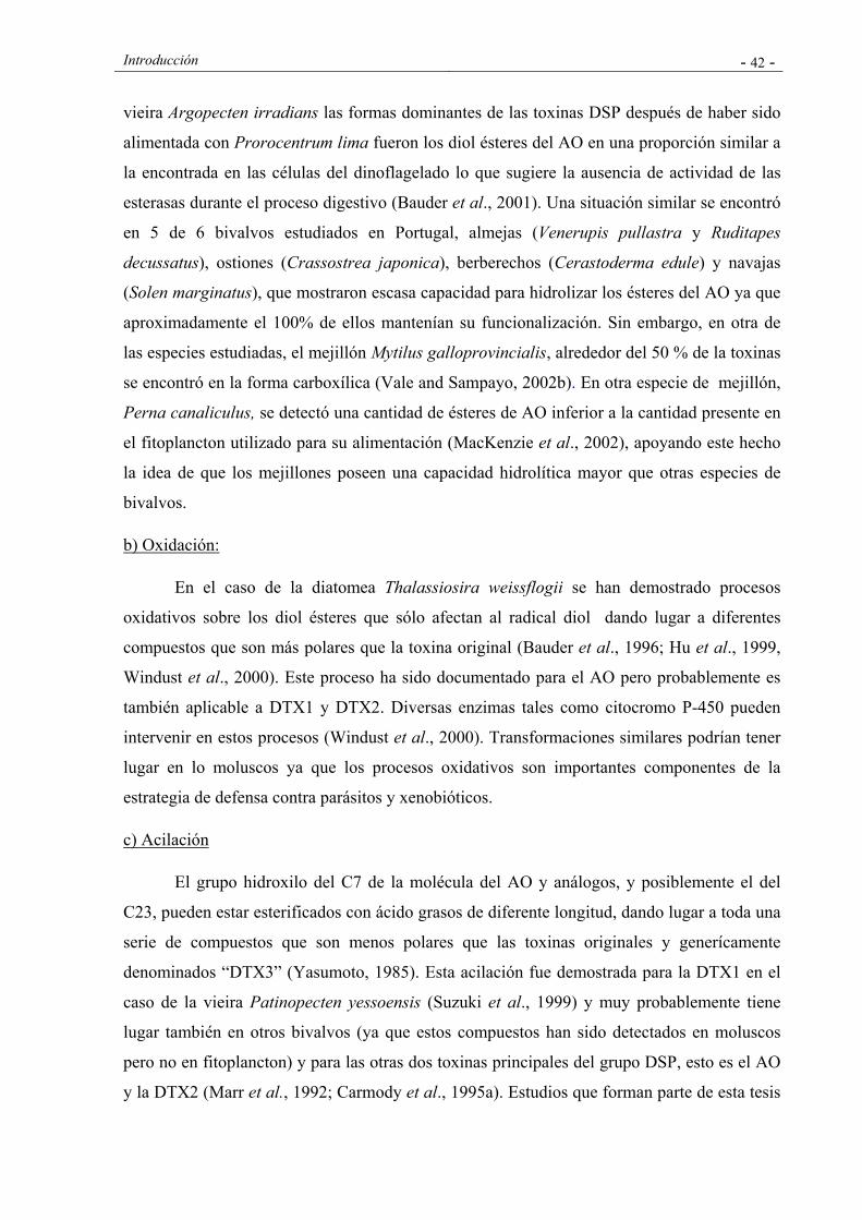
Introducción - 42 -
vieira Argopecten irradians las formas dominantes de las toxinas DSP después de haber sido
alimentada con Prorocentrum lima fueron los diol ésteres del AO en una proporción similar a
la encontrada en las células del dinoflagelado lo que sugiere la ausencia de actividad de las
esterasas durante el proceso digestivo (Bauder et al., 2001). Una situación similar se encontró
en 5 de 6 bivalvos estudiados en Portugal, almejas (Venerupis pullastra y Ruditapes
decussatus), ostiones (Crassostrea japonica), berberechos (Cerastoderma edule) y navajas
(Solen marginatus), que mostraron escasa capacidad para hidrolizar los ésteres del AO ya que
aproximadamente el 100% de ellos mantenían su funcionalización. Sin embargo, en otra de
las especies estudiadas, el mejillón Mytilus galloprovincialis, alrededor del 50 % de la toxinas
se encontró en la forma carboxílica (Vale and Sampayo, 2002b). En otra especie de mejillón,
Perna canaliculus, se detectó una cantidad de ésteres de AO inferior a la cantidad presente en
el fitoplancton utilizado para su alimentación (MacKenzie et al., 2002), apoyando este hecho
la idea de que los mejillones poseen una capacidad hidrolítica mayor que otras especies de
bivalvos.
b) Oxidación:
En el caso de la diatomea Thalassiosira weissflogii se han demostrado procesos
oxidativos sobre los diol ésteres que sólo afectan al radical diol dando lugar a diferentes
compuestos que son más polares que la toxina original (Bauder et al., 1996; Hu et al., 1999,
Windust et al., 2000). Este proceso ha sido documentado para el AO pero probablemente es
también aplicable a DTX1 y DTX2. Diversas enzimas tales como citocromo P-450 pueden
intervenir en estos procesos (Windust et al., 2000). Transformaciones similares podrían tener
lugar en lo moluscos ya que los procesos oxidativos son importantes componentes de la
estrategia de defensa contra parásitos y xenobióticos.
c) Acilación
El grupo hidroxilo del C7 de la molécula del AO y análogos, y posiblemente el del
C23, pueden estar esterificados con ácido grasos de diferente longitud, dando lugar a toda una
serie de compuestos que son menos polares que las toxinas originales y generícamente
denominados “DTX3” (Yasumoto, 1985). Esta acilación fue demostrada para la DTX1 en el
caso de la vieira Patinopecten yessoensis (Suzuki et al., 1999) y muy probablemente tiene
lugar también en otros bivalvos (ya que estos compuestos han sido detectados en moluscos
pero no en fitoplancton) y para las otras dos toxinas principales del grupo DSP, esto es el AO
y la DTX2 (Marr et al., 1992; Carmody et al., 1995a). Estudios que forman parte de esta tesis
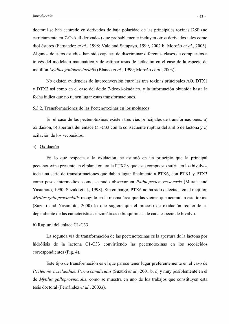
Introducción - 43 -
doctoral se han centrado en derivados de baja polaridad de las principales toxinas DSP (no
estrictamente en 7-O-Acil derivados) que probablemente incluyen otros derivados tales como
diol ésteres (Fernandez et al., 1998; Vale and Sampayo, 1999, 2002 b; Moroño et al., 2003).
Algunos de estos estudios han sido capaces de discriminar diferentes clases de compuestos a
través del modelado matemático y de estimar tasas de acilación en el caso de la especie de
mejillón Mytilus galloprovincialis (Blanco et al., 1999; Moroño et al., 2003).
No existen evidencias de interconversión entre las tres toxinas principales AO, DTX1
y DTX2 así como en el caso del ácido 7-deoxi-okadaico, y la información obtenida hasta la
fecha indica que no tienen lugar estas transformaciones.
5.3.2. Transformaciones de las Pectenotoxinas en los moluscos
En el caso de las pectenotoxinas existen tres vías principales de transformaciones: a)
oxidación, b) apertura del enlace C1-C33 con la consecuente ruptura del anillo de lactona y c)
acilación de los secoácidos.
a) Oxidación
En lo que respecta a la oxidación, se asumió en un principio que la principal
pectenotoxina presente en el plancton era la PTX2 y que este compuesto sufría en los bivalvos
toda una serie de transformaciones que daban lugar finalmente a PTX6, con PTX1 y PTX3
como pasos intermedios, como se pudo observar en Patinopecten yessoensis (Murata and
Yasumoto, 1990; Suzuki et al., 1998). Sin embargo, PTX6 no ha sido detectada en el mejillón
Mytilus galloprovincialis recogido en la misma área que las vieiras que acumulan esta toxina
(Suzuki and Yasumoto, 2000) lo que sugiere que el proceso de oxidación requerido es
dependiente de las características enzimáticas o bioquímicas de cada especie de bivalvo.
b) Ruptura del enlace C1-C33
La segunda vía de transformación de las pectenotoxinas es la apertura de la lactona por
hidrólisis de la lactona C1-C33 convirtiendo las pectenotoxinas en los secoácidos
correspondientes (Fig. 4).
Este tipo de transformación es el que parece tener lugar preferentemente en el caso de
Pecten novaezelandiae, Perna canaliculus (Suzuki et al., 2001 b, c) y muy posiblemente en el
de Mytilus galloprovincialis, como se muestra en uno de los trabajos que constituyen esta
tesis doctoral (Fernández et al., 2003a).
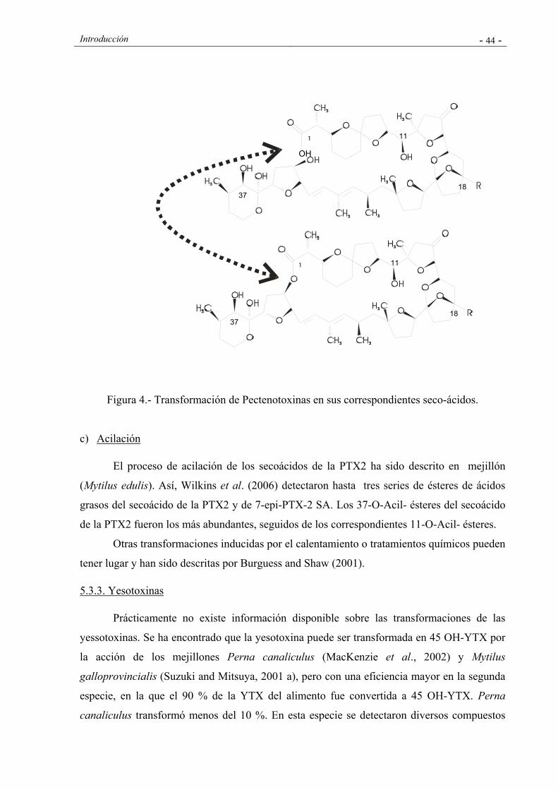
Introducción - 44 -
Figura 4.- Transformación de Pectenotoxinas en sus correspondientes seco-ácidos.
c) Acilación
El proceso de acilación de los secoácidos de la PTX2 ha sido descrito en mejillón
(Mytilus edulis). Así, Wilkins et al. (2006) detectaron hasta tres series de ésteres de ácidos
grasos del secoácido de la PTX2 y de 7-epi-PTX-2 SA. Los 37-O-Acil- ésteres del secoácido
de la PTX2 fueron los más abundantes, seguidos de los correspondientes 11-O-Acil- ésteres.
Otras transformaciones inducidas por el calentamiento o tratamientos químicos pueden
tener lugar y han sido descritas por Burguess and Shaw (2001).
5.3.3. Yesotoxinas
Prácticamente no existe información disponible sobre las transformaciones de las
yessotoxinas. Se ha encontrado que la yesotoxina puede ser transformada en 45 OH-YTX por
la acción de los mejillones Perna canaliculus (MacKenzie et al., 2002) y Mytilus
galloprovincialis (Suzuki and Mitsuya, 2001 a), pero con una eficiencia mayor en la segunda
especie, en la que el 90 % de la YTX del alimento fue convertida a 45 OH-YTX. Perna
canaliculus transformó menos del 10 %. En esta especie se detectaron diversos compuestos
OH
18
11
37
18 37
11
Introducción - 45 -
relacionados, probablemente metabolitos de YTX no identificados hasta la fecha (MacKenzie
et al., 2002).
5.4. Compartimentalización
La distribución de las toxinas DSP en los diferentes órganos y tejidos de los moluscos
ha sido estudiada de forma esporádica. Los primeros estudios coinciden en señalar que este
tipo de toxinas se acumulan principalmente en las glándulas digestivas (Yasumoto et al.,
1978; 1979), lo que parece ser la situación más frecuente como se ha observado en mejillones
(Mytilus edulis) (Pillet et al., 1995; Stabell et al., 1992) y en los pectínidos Argopecten
irradians (Bauder et al., 2001) y Pecten maximus (Hess et al., 2003). Sin embargo, esta
conclusión queda matizada por los resultados de Bauder et al (2001) que mostraron que las
vísceras (principalmente las glándulas digestivas) de Argopecten irradians depuraban las
toxinas DSP con mucha más lentitud que el resto de los tejidos, por lo que en ellas había
mayor acumulación de toxina. De hecho en el caso de A. irradians, el porcentaje de toxina
aumentó hasta un 95% tras dos días de depuración.
5.5. Eliminación
Los moluscos no retienen las toxinas indefinidamente, de manera que, transcurrido
cierto tiempo desde la interrupción de la ingestión de células tóxicas, el contenido de toxinas
se hace indetectable. Dado que la mayor parte de los programas de control usan o han usado
hasta el pasado muy reciente el bioensayo en ratón para la detección de las toxinas, las tasas
de depuración han sido estudiadas desde la perspectiva de la eliminación de la toxicidad más
que desde el punto de vista de la eliminación de las toxinas propiamente dichas. En
situaciones simples, en las que sólo aparecen una o varias toxinas con tasas de depuración y
potencia tóxica similar, la eliminación de toxicidad transcurre en paralelo con la eliminación
de toxinas. Sin embargo, en casos más complejos, la eliminación de la toxicidad tiene una
correlación pobre con la eliminación de cada una de las toxinas específicas. Las diferentes
especies de moluscos depuran la toxicidad DSP, y concretamente el AO (la más estudiada de
este grupo de toxinas), con velocidades de depuración diferentes.
Si bien existe una variabilidad notable entre especies, no parece que las diferencias sean
tan grandes como las observadas en otros grupos de biotoxinas marinas. No existen registros
de especies con un tiempo de retención tan largo como el de Saxidomus giganteus para las
toxinas PSP (Kitts et al., 1992), ó como Pecten maximus (Blanco et al., 2002a) y Siliqua
patula (Horner et al., 1993) para las toxinas ASP. De manera similar, no se han encontrado
Introducción - 46 -
especies que depuren este tipo de toxinas tan rápido como lo hacen los mitílidos con el ácido
domoico, como es el caso de Perna canaliculus ,1.97día-1 (MacKenzie et al., 1993), Mytilus
californianus, 0.34-0.55 día-1, (Whyte et al., 1995), M. edulis ,0.49-0.99 día-1, (Novaczek et
al., 1992), 2.01-10.59 día-1 en experimentos de 1 ó 2 horas (Novaczek et al., 1991), 10.59 y
2.2 dia-1 en experimentos de 2 y 24 horas respectivamente (Wohlgeschaffen et al., 1992) y M.
galloprovincialis, 0.40-0.58 dia-1 (Blanco et al., 2002b).
Las diferentes toxinas son eliminadas a diferentes velocidades. La DTX2 y sus
derivados de baja polaridad “DTX3” son depuradas en Mytilus galloprovincialis más
lentamente que el AO y sus derivados y, a su vez, cada grupo de derivados de baja polaridad
presenta una tasa de depuración más baja que las toxinas de las que provienen, como se
muestra en estudios que forman parte de esta tesis doctoral (Fernández et al., 1998). El primer
hecho sugiere que los episodios tóxicos en los que la DTX2 esté implicada se prolongarán
más en el tiempo que aquéllos en los que la toxina principal sea el AO. El segundo hecho
sugiere que cuando los derivados acilados se produzcan en grandes cantidades, el proceso de
detoxificación será más lento.
Existe muy poca información en lo que se refiere a la depuración de DTX4, DTX5 y
diol ésteres. Su desaparición en los bivalvos procede más probablemente de un proceso de
hidrólisis que da lugar a las toxinas carboxílicas, que de un proceso de eliminación. La
combinación de transformación y depuración de las toxinas pudiera dar lugar a cinéticas
complejas y de aparente incoherencia para cada una de las toxinas cuando se analiza
independientemente y a dificultades de interpretación si las cinéticas de las toxinas no son
modelizadas.
5.6. Regulación de la depuración
El proceso de depuración de las toxinas está potencialmente afectado por factores
ambientales tales como la temperatura y disponibilidad de alimento entre otros. Sin embargo
ninguno de estos factores ha sido inequívocamente vinculado a la velocidad de depuración.
En general, se asume que las bajas temperaturas dan lugar a bajas tasas de depuración
(Shumway and Cembella, 2003) pero no existen evidencias sólidas que sustenten este hecho.
Las tasas de depuración de mejillón en áreas de bajas temperaturas tales como Suecia o
Noruega no parecen ser más bajas que las correspondientes a mejillones de España, Francia o
Italia. Los intentos realizados para establecer estimaciones precisas del efecto de la
temperatura y cuyos resultados forman parte de esta tesis doctoral (Blanco et al., 1999) no
Introducción - 47 -
dieron lugar a conclusiones claras a este respecto por lo que podría concluirse que si la
temperatura incide en la tasa de depuración lo hace de forma poco relevante. La salinidad
tampoco parece afectar a la depuración.
Se han documentado diferencias en las tasas de depuración entre mejillones
mantenidos en tanques y los que permanecieron en el mar (Marcaillou-Le Baut et al., 1993)
que fueron atribuidas a diferencias en la alimentación. Observaciones realizadas por Sampayo
et al. (1990) durante varios episodios de DSP de las costas portuguesa sugieren que la tasa de
depuración se incrementa con la concentración de fitoplancton, que es el alimento consumido
por los bivalvos. La misma conclusión fue alcanzada en esta tesis doctoral (Blanco et al.,
1999) con mejillones gallegos, si bien el efecto registrado fue pequeño. Recientemente
Svensson (2003) llegó a la conclusión opuesta, encontrando que los mejillones que no
recibían alimento depuraban el AO más rápidamente que aquellos que eran alimentados. Sin
embargo, el patrón de depuración de los mejillones de este experimento no siguió la típica
cinética de disminución exponencial y experimentó un aumento al comienzo del experimento
que sugiere que otros procesos además de la depuración podrían estar implicados
(posiblemente transformaciones de derivados esterificados).
El cambio de metabolismo aeróbico a anaeróbico inducido por la emersión de los
mejillones varias horas al día no produjo alteraciones en la depuración del AO y de la DTX2
(Moroño et al., 1998), lo que implica que algunos de los principales procesos metabólicos de
estos moluscos no afectan sustancialmente la tasa de depuración.
Como se muestra en varias contribuciones a esta tesis doctoral, parte de esta
incertidumbre en relación a los verdaderos efectos de factores ambientales en la tasa de
depuración radica en el hecho de que una correcta estimación requiere un sólido conocimiento
conceptual y matemático de la cinética de las diferentes toxinas implicadas (Moroño et al.,
1998; Fernández et al., 1998; Blanco et al., 1999) y este conocimiento ha empezado a
emerger en años recientes con el descubrimiento y la demostración de la contribución real de
muy diversos compuestos al contenido total de toxinas en los bivalvos (Vale and Sampayo,
2002 b; MacKenzie et al., 2001; 2002; Moroño et al., 2003) y con el desarrollo de modelos
matemáticos de acumulación.
5. 7. Predicción de la acumulación de toxinas DSP: Modelizado
La predicción de la acumulación de toxinas en los moluscos requiere la aplicación de
modelos matemáticos. Dichos modelos están compuestos de al menos dos términos: un

Introducción - 48 -
término de ganancia que describe la toxina adquirida por el organismo, comenzando desde la
toxina que está disponible en el medio y un término de pérdida que normalmente integra todas
las vías posibles de eliminación en el término genérico “depuración”. El término de ganancia
incluye todos los procesos que están implicados en la adquisición de toxinas. En los moluscos
bivalvos la ganancia es usualmente reducida a pocos términos que describen la adquisición de
alimento, ya que las toxinas son principalmente ingeridas con el fitoplancton tóxico que las
produce y como consecuencia, otros posibles mecanismos de adquisición, tales como una
absorción directa desde el agua a través de las branquias o el manto, no son tenidos en cuenta.
En los modelos que se han implementado hasta la fecha, el proceso de adquisición de toxinas
se describe con dos parámetros: la tasa de filtración y la eficiencia de absorción de toxinas.
Los procesos de captura de alimentos, principalmente el bombeo de agua a través de las
branquias, la retención en ellas, y la selección pre-ingestión son integrados en un único
parámetro: la tasa de filtración, que describe la tasa con la que los bivalvos ingieren el
fitoplancton. Una vez que el alimento que contiene las toxinas es ingerido, es sometido a
procesos de selección pre-ingestión y digestión con el resultado de que sólo una porción de las
toxinas son incorporadas al organismo. Esto se describe como la eficiencia de absorción de las
toxinas. Matemáticamente, esta parte del modelo puede ser expresada de la siguiente forma:
dtox/dt= TA• FA • TF • EA
TA es la tasa de aclaramiento, FA la concentración de fitoplancton tóxico en el agua, TF el
contenido de toxinas por célula de fitoplancton, y el producto de estos tres factores, la
cantidad de toxina retenida por el molusco. EA es la eficiencia de absorción de las toxinas, y
su producto por las toxinas retenidas da lugar a la toxina absorbida por el molusco.
La cinética de depuración de varias especies de moluscos previamente afectados por
episodios tóxicos de DSP ha sido estudiada por diversos autores. En algunos de los estudios
(Marcaillou-Le-Baut et al., 1993; Shumway and Cembella 1993) se estableció una curva de
depuración en dos fases, con tasas altas de depuración durante los primeros días, seguidas
posteriormente de unas tasas de depuración mucho más lentas. En otros casos (Croci et al.,
1994) se ha demostrado una única tasa de depuración para todo el proceso. Como en el caso
de las toxinas PSP, estas dos respuestas pueden ser descritas por modelos de dos y un
compartimento respectivamente (Silvert and Cembella, 1995, Blanco et al., 1997).
En el proceso de depuración de un organismo, están implicados varios órganos y tejidos,
cada uno de los cuales posee su propia tasa de depuración, de manera que un modelo ideal de
Introducción - 49 -
cinética utilizaría un compartimento para cada uno de los órganos/tejidos (definido por la
cantidad inicial de toxinas que contiene, su tasa de depuración y las tasas de transferencia
desde/hacia otros compartimentos). Estos modelos complejos son difíciles de implementar en
situaciones reales debido al gran número de parámetros que han de ser estimados, lo que en
muchos casos resulta imposible. En situaciones relativamente simples, cuando la mayor parte
de las toxinas se acumulan en un único órgano, o cuando todos los órganos tienen tasas de
depuración similares (caso poco probable), un compartimento puede ser suficiente para
modelizar de forma satisfactoria la cinética, que en tal caso se conoce como monofásica.
Una situación más complicada, en la que los toxinas se distribuyen entre diferentes
órganos presentando diferentes tasas de depuración, puede ser modelizada usualmente con
dos compartimentos, dando lugar a cinéticas conocidas como bifásicas (incluso cuando en
realidad son el resultado de dos procesos actuando simultáneamente y no un proceso actuando
de manera diferente en dos fases distintas).
Teniendo en cuenta las consideraciones previas, el término de pérdida puede ser expresado
de la siguiente manera:
Modelo de un compartimento:
dtox/dt= -K•tox
“tox” es la concentración de toxina y K la tasa de depuración.
Modelo de dos compartimentos:
dtox1/dt= -K1•tox1
dtox2/dt= -K2•tox2-T12•tox1
Los subíndices indican el compartimento y T12 la tasa de transferencia entre el primero y
el segundo compartimento.
Uno de los trabajos incluidos en el capítulo I de esta tésis doctoral (Blanco et al., 1995),
ha mostrado que un modelo muy simple de un compartimento puede describir de manera
razonable algunos episodios de DSP en Galicia.
Cuando varias toxinas, que pueden ser transformadas unas en otras, están implicadas, los
modelos resultan mucho más complejos. Sin embargo, ésta podría ser la única vía de estimar
las verdaderas tasas de depuración, puesto que estudiar las toxinas como si fueran

Introducción - 50 -
independientes daría lugar a una infra-estimación de la velocidad de depuración de las toxinas
que son el producto final de las transformaciones, como se puede observar en el caso de las
toxinas DSP en dos trabajos que forman parte de esta tesis doctoral (Blanco et al., 1999;
Moroño et al., 2003) y en el caso de las toxinas PSP (Silvert et al., 1998).
6. GESTIÓN Y MITIGACIÓN DE LOS EPISODIOS TOXICOS
6.1. Consideraciones generales
La prevención de los riesgos de las toxinas marinas para la salud pública y para el sector
productor conlleva, además del conocimiento de los organismos, de los compuestos tóxicos, y
de los procesos de acumulación, transformación y eliminación de las toxinas en los moluscos,
el desarrollo e implantación de una legislación que regule los requisitos básicos de los
programas de monitorización y control, los niveles máximos admisibles de toxinas en los
mariscos y los criterios de aceptabilidad de los métodos de ensayo y análisis para su detección
y cuantificación.
Los aspectos cubiertos por los programas de monitorización y control dependen mucho del
país y área geográfica considerada. En la mayor parte de los casos, los programas incluyen la
vigilancia de la presencia de especies fitoplanctónicas potencialmente tóxicas (identificación
y cuantificación) y la determinación del contenido en toxinas de los moluscos.
Adicionalmente algunos países o regiones complementan estos datos con la monitorización de
parámetros físicos, químicos y biológicos (temperatura del agua, salinidad, nutrientes,
clorofila, corrientes, estratificación de la columna de agua, etc) y con el conocimiento de las
condiciones oceanográficas y meteorológicas. Todo esto constituye una importante
herramienta de predicción. Los distintos aspectos y características de los diferentes programas
de control sanitario de toxinas se describen en el capítulo IV de la presente tesis doctoral
(Fernández et al., 2003c).
En lo que respecta a las regulaciones relativas al control de las toxinas históricamente
incluidas en el grupo DSP, han sido objeto de gran controversia, existiendo discrepancias en
lo que se refiere a qué toxinas deben ser incluidas dentro de esta clasificación y cuáles deben
ser objeto de monitorización y control, así como en lo que respecta a los niveles máximos
admisibles y a los métodos más adecuados para su detección y cuantificación. Como ya se ha
comentado anteriormente, tradicionalmente se han incluido dentro de este grupo tres familias
de toxinas -ácido okadaico y derivados, pectenotoxinas y yesotoxinas- que presentan diferente
Introducción - 51 -
actividad biológica. Ha existido un consenso general en la pertinencia de clasificar el ácido
okadaico y derivados dentro del grupo de toxinas DSP, por sus documentados efectos
diarreicos, pero ha existido un debate en la comunidad científica en lo que se refiere a la
idoneidad de la inclusión de las pectenotoxinas y yesotoxinas en este grupo y en lo que
respecta al riesgo que representan para la salud humana y por tanto a la necesidad o no de
regulación de las mismas.
El análisis de riesgo (“risk assessment”) es la metodología fundamental para el
establecimiento de los niveles máximos admisibles para este tipo de compuestos, e incluye las
siguientes etapas: 1) identificación del tóxico, 2) caracterización del tóxico, 3) valoración de
la exposición al mismo y 4) caracterización del riesgo (FAO/WHO, 1995). La primera etapa,
conlleva la identificación de la sustancia química presente en el alimento capaz de producir
efectos adversos en la salud humana. En la mayor parte de los casos, esta etapa está
restringida a estudios epidemiológicos. La segunda etapa, la caracterización, consiste en la
evaluación cualitativa y cuantitativa de los efectos adversos para la salud asociados a la
presencia de los tóxicos en los alimentos. El conocimiento de la cinética asociada a las toxinas
(procesos de absorción, distribución, metabolismo, acumulación y excreción de las toxinas y
sus metabolitos) juega un papel fundamental en esta etapa. El conocimiento del mecanismo de
acción y de las relaciones dosis – repuesta son también importantes a la hora de la
caracterización y del establecimiento de los márgenes de protección.
La valoración de la exposición es el tercer factor a tener en cuenta en la valoración del
riesgo. Esto se refiere a la evaluación cualitativa y cuantitativa de la ingesta más probable de
compuestos con propiedades tóxicas a través de los alimentos. Uno de los principales
problemas del uso cuantitativo de datos epidemiológicos para estimar los niveles seguros de
exposición humana es que las estimaciones de exposición procedentes de dichos estudios son
a menudo muy imprecisas. Se necesitan datos sobre las concentraciones de los diferentes
compuestos tóxicos en los diferentes alimentos y el patrón de consumo. La exposición de los
niños es en muchos casos objeto de particular atención y preocupación, debido a la
relativamente alta ingesta de alimentos y a la vez a la elevada susceptibilidad en muchos
aspectos.
El paso final es la caracterización del riesgo. Esto implica estimaciones cuantitativas,
incluyendo incertidumbres, de la probabilidad de ocurrencia de efectos adversos para la salud
en una población dada. La caracterización del riesgo debe incluir descripciones del origen de

Introducción - 52 -
las incertidumbres. Para compuestos no genotóxicos, la valoración del riesgo se basa en la
determinación del llamado “NOAEL”, esto es, la dosis umbral por debajo de la cual no se
observan valores adversos. Esta dosis se divide usualmente por un factor basado en la
incertidumbre, para obtener el nivel seguro de exposición humana. Normalmente se utiliza un
factor de 100 en el caso de que el NOAEL provenga de estudios con animales. Esta cifra
resulta de la consideración de un factor de 10 para compensar diferencias entre animales y los
humanos representativos y de otro factor de 10 para compensar las diferencias entre humanos
representativos y grupos más sensibles. En el caso de que estos datos se obtengan de la
extrapolación del denominado LOAEL (dosis en la que se observa el efecto adverso más leve)
es posible que se aplique otro factor adicional de 10. En el caso de que estén disponibles datos
robustos de intoxicación en humanos, se utiliza normalmente un factor de incertidumbre de
10.
En el caso de las toxinas marinas incluidas históricamente en el grupo DSP, no ha sido
siempre posible utilizar la metodología anteriormente mencionada. Los estudios toxicológicos
son escasos y han estado limitados por la escasez de toxinas puras en suficiente cantidad. Los
datos sobre exposición e ingesta son también escasos o imprecisos. Esto ha dado lugar a que
los límites hayan sido establecidos en un primer momento en base a los límites de detección
de los métodos disponibles y posteriormente en base a los escasos datos epidemiológicos. La
falta de toxinas puras y material certificado de referencia ha dificultado igualmente el
desarrollo y validación de métodos adecuados de determinación. Todo esto ha ocasionado
discrepancias en los métodos de detección y cuantificación utilizados y en los criterios de
positividad.
A pesar de las dificultades y de las carencias y limitaciones de los datos disponibles,
en los últimos años se han realizado esfuerzos considerables para racionalizar y mejorar la
legislación sanitaria relativa a estas familias de toxinas, llevándose a cabo iniciativas en
diferentes marcos y ámbitos que dieron lugar, primeramente a cambios significativos en la
legislación europea y posteriormente a una nueva propuesta de niveles y criterios de
aceptabilidad de métodos que, muy posiblemente, dará lugar a nuevas modificaciones a nivel
comunitario e internacional.
6.2. Regulación de mé todos de detección y de niveles máximos de toxinas DSP
Los bioensayos han sido utilizados en la mayor parte de los países para la determinación de
toxinas DSP con fines de control sanitario, si bien con notables diferencias en lo que se refiere
Introducción - 53 -
a aspectos tales como el animal de experimentación utilizado (rata o ratón), los
procedimientos de extracción y purificación, la parte del molusco utilizada para el análisis
(cuerpo entero, glándula digestiva) y el criterio de positividad (muerte de los ratones antes de
5 horas o antes de 24 horas) que han dado lugar a la falta de equivalencia entre los mismos.
Los diferentes bioensayos utilizados para la determinación de este grupo de toxinas han sido
descritos y analizados de manera exhaustiva en uno de los trabajos que forma parte de esta
tesis doctoral (capítulo III).
En relación a los niveles máximos admisibles, la mayor parte de los países los
establecieron en un principio en función de los límites de detección del método utilizado,
normalmente el bioensayo. En lo que respecta a la Unión Europea (UE), las Directivas
91/492/EEC y 91/493/EEC establecían las normas aplicables a la producción y
comercialización de organismos marinos vivos (moluscos, crustáceos y tunicados) y de
productos de la pesca respectivamente. Dichas regulaciones contemplaban el control de las
áreas de producción y de las diferentes actividades de recolección, transporte, depuración,
almacenamiento, procesado y comercialización, así como algunos de los requisitos
microbiológicos, químicos y aquellos relativos al contenido de toxinas marinas.
Con respecto al control de toxinas DSP se establecía lo siguiente:
- Los métodos habituales de análisis biológico no deben dar reacción positiva respecto
de la presencia de DSP en las partes comestibles de los moluscos (cuerpo entero o cualquier
parte consumible por separado).
La Directiva era vaga, puesto que no se especificaban los métodos o los criterios de
aceptabilidad de los mismos, y el límite regulatorio quedaba a expensas del límite de
detección del método seleccionado. Esto dio lugar a discrepancias en los Estados Miembros
en su interpretación y por tanto en los métodos de detección y en los criterios de positividad
utlizados con fines de control sanitario.
En aras de conseguir la armonización de las regulaciones y establecer criterios
equivalentes en lo que concierne a los métodos y límites aplicados al control de toxinas
marinas, la Comisión Europea estableció una red de Laboratorios Nacionales de Referencia de
Toxinas Marinas (EU-NRLs), coordinados por un Laboratorio Comunitario de Referencia,
cuya nominación recayó en el Laboratorio de Sanidad Exterior de Vigo.
Las tareas asignadas a los Laboratorios Nacionales de Referencia son las siguientes:
Introducción - 54 -
- Coordinar las actividades de los laboratorios nacionales encargados de los análisis de
toxinas en el Estado Miembro de que se trate;
- Ayudar a la autoridad competente del Estado Miembro en la organización del sistema
de control de toxinas marinas;
- Organizar pruebas comparativas en los distintos laboratorios nacionales encargados de
los análisis de toxinas marinas;
- Comunicar la información proporcionada por el Laboratorio Comunitario de
Referencia a la autoridad competente del Estado Miembro y a los laboratorios adicionales
encargados de los análisis de toxinas marinas;
Las Tareas del Laboratorio Comunitario de Referencia son las siguientes.
- Proporcionar información a los laboratorios nacionales de referencia sobre los
métodos de análisis y las pruebas comparativas;
- Coordinar la aplicación de los métodos por los laboratorios nacionales de referencia
mediante la organización de pruebas intercomparativas;
- Coordinar la investigación de nuevos métodos de análisis e informar a los laboratorios
nacionales de referencia de los avances en este sector;
- Organizar cursos de formación y perfeccionamiento para el personal de los
Laboratorios Nacionales de Referencia;
- Colaborar con los laboratorios encargados de los análisis de biotoxinas marinas en los
terceros países;
- Proporcionar asistencia técnica y científica a la Comisión, especialmente en caso de
desacuerdo entre los Estados Miembros sobre los resultados de los análisis.
Gran parte de la actividad de la red de laboratorios europeos de referencia de
biotoxinas marinas se centró en la realización de estudios intercomparativos y en la
consecución de acuerdos en cuanto a métodos de análisis de toxinas marinas y criterios de
toxicidad que permitieran una interpretación común de la Directiva 91/492 y que al mismo
tiempo fueran el germen de futuras regulaciones. Con el propósito de mejorar la interacción
entre los laboratorios de la red, se crearon diferentes grupos de trabajo integrados por
expertos y miembros de dichos laboratorios para avanzar en diversas áreas en las que se

Introducción - 55 -
identificaron especiales carencias. Como consecuencia de estos grupos de trabajo, que
fueron auspiciados por la Comisión Europea y coordinados por el Laboratorio Comunitario
de Referencia de Biotoxinas Marinas, y sobre la base de sus informes técnicos, la Unión
Europea adoptó la Decisión 2002/225/CE en el año 2002 que establece las condiciones
específicas para la implantación de la Directiva 91/492 en lo que se refiere a los niveles
máximos de ácido okadaico y dinofisistoxinas, yesotoxinas, pectenotoxinas y azaspirácidos,
así como a los métodos de análisis que deben emplearse para su detección. Se aplica a los
moluscos bivalvos, los equinodermos, los tunicados y los gasterópodos marinos destinados
al consumo humano directo o a una transformación previa al consumo.
En lo respecta a los niveles máximos, la Decisión contempla lo siguiente:
El nivel máximo total de ácido okadaico, dinofisistoxinas y pectenotoxinas será de
160 µg de equivalentes de ácido okadaico/kg (el cuerpo entero o cualquier parte
comestible por separado).
El nivel máximo de yesotoxinas será de 1 mg de equivalente de yesotoxina/kg (el
cuerpo entero o cualquier parte comestible por separado).
El nivel máximo de azaspirácidos será de 160 µg de equivalentes de azaspirácido/kg
(el cuerpo entero o cualquier parte comestible por separado).
Esta Decisión es pionera en cuanto al establecimiento de niveles de tolerancia para
las diferentes familias de biotoxinas marinas polietéreas, a la vez que armoniza su control
en los Países Miembros uniformizando los criterios de positividad del bioensayo en ratón,
aspecto que ha sido muy problemático en el pasado tanto desde el punto de vista de
protección de la salud pública, como desde el punto de vista de agravios comparativos para
el sector productor. La Decisión abre una puerta a la posibilidad de utilizar métodos
alternativos al bioensayo en ratón, tales como la cromatografía líquida de alta resolución
con detección fluorimétrica, la cromatografía líquida acoplada a espectometría de masas o
ensayos inmunológicos y funcionales como el de inhibición de fosfatasas, a condición de
que, por separado o combinados, puedan detectar, como mínimo, los análogos siguientes:
ácido okadaico y dinofisistoxinas (para detectar la presencia de DTX3 puede ser necesaria
una fase de hidrólisis), pectenotoxinas (PTX1 y PTX2) yesotoxinas (YTX, 45 OH YTX,
Homo YTX y 45 OH Homo YTX) y azaspirácidos (AZA1, AZA2 y AZA3). Establece
asímismo que en el caso de que se descubran nuevos análogos importantes en relación con
la salud pública, deberán incluirse en los análisis. Será necesario disponer de patrones para
Introducción - 56 -
poder realizar el control mediante análisis químicos y la toxicidad total se calculará
mediante factores de conversión basados en los datos sobre la toxicidad de cada toxina. Las
características de funcionamiento de estos métodos deberán definirse tras ser validados
conforme a un protocolo acordado a nivel internacional. Cuando los resultados de los
análisis efectuados demuestren que hay discrepancias entre los diferentes métodos, el
bioensayo en ratones deberá considerarse el método de referencia.
Las Directivas 91/492/EEC y 91/493/EEC son dos de las 17 Directivas verticales de
Seguridad Alimentaria que se desarrollaron desde el año 1964 en la Unión Europea. Como
consecuencia de la publicación en el año 2000 del Libro Blanco Europeo de la Seguridad
Alimentaria, se llevó a cabo una revision de todas estas Directivas que dio lugar a su
integración en un paquete legislativo de Higiene Alimentaria que entró en vigor en enero de
2006 y que contiene cinco reglamentos de aplicación en los Estados Miembros y legalmente
vinculantes, de los cuales tres están relacionados con la producción de moluscos bivalvos:
-Reglamento EC No 852/2004 de higiene de los alimentos
-Reglamento EC No 853/2004 que establece las reglas específicas de higiene para los
alimentos de origen animal.
-Reglamento EC No 854/2004 que establece las reglas específicas para la organización de
los controles oficiales para los productos de origen animal destinados al consumo humano.
El Reglamento 853/2004 en su seccion VII del Anexo establece las condiciones
específicas para la producción de moluscos bivalvos, no existiendo cambios significativos en
lo que respecta a las condiciones establecidas en la Directiva 91/492.
La UE contempla en la actualidad una política de eliminación de los bioensayos con
animales en tanto en cuanto estén disponibles métodos alternativos validados siguiendo
estudios colaborativos internacionalmente reconocidos. El Reglamento de la Comisión (EC)
2074/2005 establece que “se establecerán acciones para la sustitución de los bioensayos con
animales tan pronto como sea posible”. En lo que se refiere a las toxinas lipofílicas, existe un
acuerdo general en eliminar los bioensayos en ratón en la medida en que se puedan utilizar
métodos alternativos que hayan sido previamente validados.
Las regulaciones existentes en otras áreas geográficas y económicas, así como otras
iniciativas a nivel comunitario o internacional orientadas a la estandarización y/o
Introducción - 57 -
armonización de los programas de control se describen y analizan en detalle en el capítulo IV
de la presente tesis doctoral.
6.3. Redes de control de los episodios tóxicos de origen fitoplanctónico en España
El establecimiento y desarrollo de los programas de control en las áreas de producción de
moluscos en España, así como el control en mercado es una competencia transferida a las
Comunidades Autónomas. La competencia sobre el control de toxinas en bivalvos y
productos marinos de importación procedentes de Países Terceros, recae sobre los
laboratorios de Sanidad Exterior que dependen orgánicamente del Ministerio de
Administraciones Públicas (MAP) y funcionalmente del Ministerio de Sanidad y Consumo y
que ejercen su control a través de los Puntos de Inspección Fronterizos.
El Laboratorio de la Dependencia de Sanidad en Vigo, perteneciente a la red del MAP fue
nominado, en 1993, Laboratorio Comunitario de Referencia de Biotoxinas Marinas (LCR), y
como tal ejerció las labores de coordinación de la red de Laboratorios Europeos Nacionales de
referencia de biotoxinas marinas. En la actualidad, y como consecuencia de un cambio de
titularidad, el LCR está vinculado a la Agencia Española de Seguridad Alimentaria del
Ministerio de Sanidad y Consumo.
En lo que respecta a la Comunidad Autónoma Gallega, el organismo oficial encargado del
control de la presencia de plancton tóxico en las aguas de producción y biotoxinas en los
moluscos es el INTECMAR, dependiente de la Consellería de Pesca y Asuntos Marítimos de
la Xunta de Galicia que actúa como Laboratorio Oficial de la Comunidad Autónoma en la
materia. El control en mercado interior es competencia de la Consellería de Sanidad.
El sistema de control de las aguas de producción de moluscos establecido en Galicia tiene
como objetivo fundamental salvaguardar la salud pública y al mismo tiempo, minimizar los
efectos negativos que provocan las prohibiciones de extracción en el sector productivo. El
problema se aborda desde dos aspectos distintos:
a) Predicción de episodios tóxicos: la predicción de un episodio tóxico con suficiente
antelación permite al sector la adopción de decisiones encaminadas a modificar su estrategia
de explotación de forma que se minimicen en lo posible los efectos negativos.
b) Minimización de sus efectos: una vez desarrollado el episodio, y dado que en la
actualidad no se conocen técnicas que permitan reducir o eliminar la acumulación de toxinas
Introducción - 58 -
en los moluscos, las medidas a tomar para la reducción de sus efectos pasan por evitar
mantener las prohibiciones de extracción más tiempo del imprescindiblemente necesario.
Con el fin de cumplimentar estos aspectos, el sistema de control tiene las siguientes
características:
1. Control intensivo de las poblaciones fitoplanctónicas, y de las condiciones fisico-
químicas que condicionan su desarrollo (climatología, parámetros físico-químicos de las
aguas, condiciones de estratificación, etc). Este control permite a corto plazo una predicción
de las variaciones de toxicidad acumulada en los moluscos, y a largo plazo, mediante la
combinación de toda la información disponible, la predicción de la aparición, desarrollo y
desaparición de los episodios tóxicos.
2. Control intensivo del contenido de biotoxinas en moluscos que permite tener un
conocimiento pormenorizado de la situación tanto de las distintas zonas de producción como
de las distintas especies cultivadas. En cada zona de producción, la periodicidad del muestreo
varía, en función de la situación, desde un mínimo de una vez por semana hasta diaria.
3. Actualización individualizada sobre pequeñas unidades geográficas (zonas de
producción, bancos marisqueros, polígonos de bateas, etc) y frecuencia de muestreo muy
intensa. De esta manera es posible garantizar que cualquier producto apto para el consumo no
se verá retenido en la zona de producción más tiempo del necesario, y a la vez disminuir la
variabilidad entre lotes en los mariscos comercializados, ya que se elimina de la variabilidad
el componente de la localización.
4. Actualización de los Planes de Actuación que mediante una información generada por el
Departamento de Condiciones Oceanográficas y Fitoplancton y el de Biotoxinas permiten
disponer de información predictiva a corto plazo sobre la evolución de la toxicidad de cada
una de las zonas de producción.
6.4. Estrategias para evitar o eliminar la contaminación de los moluscos
Una gran parte de las acciones descritas anteriormente (monitorización del
fitoplancton tóxico y de las toxinas y establecimiento de regulaciones) están dirigidas a la
mitigación de los impactos negativos de los episodios tóxicos mediante la reducción del
riesgo para la salud pública. Además del establecimiento de programas de monitorización y
control, se han realizado intentos de desarrollar estrategias dirigidas a evitar la contaminación
de los moluscos o a eliminar las toxinas de los mismos una vez que se ha producido la
Introducción - 59 -
contaminación. En lo que respecta a las primeras, una de las estrategias consiste en intentar la
reducción de la exposición de los moluscos a las poblaciones de fitoplancton tóxico. Esto
puede realizarse mediante dos procedimientos: 1) reducir la proliferación del fitoplancton ó 2)
cambiar la ubicación de los moluscos de manera que se evite su contaminación. En lo que
respecta al primer procedimiento, el único método que ha demostrado hasta la fecha su
eficacia es el realizado en Corea, que consiste en la dispersión de arcilla adsorbentes del
plancton para evitar la proliferación de Cochlodinium polykikroides (Kim et al., 1998), pero
que presenta el inconveniente del evidente impacto medioambiental. Estrategias tales como el
uso de ultrasonidos o cloro para eliminar las algas tóxicas o la aplicación de virus, bacterias,
hongos o protozoos que originen la ruptura de las células tóxicas han sido experimentadas
pero sólo a nivel de laboratorio y nunca en proliferaciones reales de fitoplancton. El segundo
procedimiento, consiste en la relocalización de los moluscos (especialmente en el caso de
cultivos) o la restricción de los cultivos a áreas o profundidades en las cuales la concentración
de fitoplancton sea mínima (Desbiens and Cembella, 1993; Lodeiros et al., 1998; Franzosini
et al., 2000).
Otra estrategia para paliar el impacto de los episodios tóxicos es la eliminación de las
toxinas de los moluscos. Esto puede ser abordado mediante dos vías: la aplicación de técnicas
tales como tratamientos con ozono y cloro, cambios fuertes de temperatura y salinidad, etc,
que aceleren la pérdida de las toxinas y permitan al mismo tiempo obtener moluscos vivos no
tóxicos o bien la aplicación de técnicas específicas de procesado de los moluscos que
permitan obtener productos libres de toxinas. Las diferentes estrategias y procedimientos han
sido estudiadas y valoradas en una de las contribuciones de esta tesis doctoral (“Management
of shellfish resources”, capítulo IV).
En lo que respecta a las toxinas DSP, dicha valoración ha mostrado que el traslado de
los moluscos a zonas libres de fitoplancton tóxico y con condiciones ambientales que
aumenten la tasa de depuración es la estrategia más efectiva. En lo que se refiere a la
eliminación de las toxinas, la evisceración de los moluscos, concretamente la eliminación de
las glándulas digestivas, puede ser efectiva, pero sólo resulta rentable en aquellas especies de
moluscos cuyo tamaño permita una disección fácil y cuyo valor comercial compense los
costes del proceso de evisceración. La extracción de las toxinas mediante técnicas de
extracción de punto crítico (González et al., 2002) aparece cómo una técnica interesante para
eliminar toxinas de estos grupos u otras de carácter lipofílico. La técnica requiere la
deshidratación del producto por lo que sólo será útil en aquellos productos que requieran o al
menos toleren dicho proceso.


Objetivos - 61 -
OBJETIVOS

Objetivos - 63 -
OBJETIVOS
Los trabajos que constituyen esta tesis doctoral se desarrollaron estrechamente vinculados a
mi actividad profesional en el Laboratorio Comunitario de Referencia de Biotoxinas Marinas
(Sanidad Exterior- Ministerio de Administraciones Públicas) y tienen por tanto un marcado
carácter sanitario, abarcando tanto aspectos químicos y analíticos como aspectos de
predicción, regulación y control orientados a mitigar el impacto de las toxinas en la salud
pública y en las actividades económicas relacionadas con la explotación de los moluscos.
El objetivo global de esta tesis es contribuir a minimizar los riesgos derivados de las toxinas
“DSP” para la salud pública y para las actividades económicas vinculadas al recurso mediante
la consecución de los siguientes objetivos específicos:
OBJETIVO 1): Mejorar la capacidad de control de los episodios tóxicos DSP por medio del
conocimiento de los agentes y procesos implicados: las toxinas polietéreas responsables de los
mismos y las especies fitoplanctónicas que las originan.
OBJETIVO 2): Desarrollar capacidad de predicción, utilizando el conocimiento sobre las
especies tóxicas y su contenido en toxinas y mediante el estudio de las capacidades de los
moluscos para acumular, transformar y eliminar las toxinas que ingieren.
OBJETIVO 3): Mejorar la capacidad de monitorización y control mediante el análisis y
valoración de los diferentes bioensayos para la detección de las toxinas en los bivalvos.
OBJETIVO 4) Contribuir a una mejor gestión y mitigación del impacto de las toxinas marinas
en la salud pública y en la explotación de los moluscos mediante la identificación, revisión y
valoración de diferentes estrategias, iniciativas y regulaciones desarrolladas en el ámbito
comunitario e internacional.


Desarrollo de la tesis - 65 -
DESARROLLO DE LA TESIS


Desarrollo de la tesis - 67 -
DESARROLLO DE LA TESIS
En el capítulo I, que responde al objetivo 1, se presentan los trabajos experimentales
asociados a la identificación de las especies fitoplanctónicas productoras de toxinas DSP en
las Rías Gallegas y a la determinación de las toxinas producidas por dichos organismos, que
dieron lugar a las siguientes publicaciones.
1) From Dinophysis spp toxicity to DSP outbreaks: a preliminary model of toxin
accumulation in mussels. (1995). Blanco, J., Fernández, M., Mariño, J., Reguera, B., Míguez,
A., Maneiro, J., Cacho, E. and Martínez, A. (1995). In: P. Lassus, G. Arzul, E. Erard, P.
Gentien and C. Marcaillou (eds.). Harmful Marine Algal Blooms, Lavoisier, Intercept Ltd,
Paris, pp. 777-82.
2) Toxin content of Dinophysis acuminata, D. acuta and D. caudata from the Galician Rías
Bajas. Fernández, M.L., Reguera, B., Ramilo, I. and Martinez, A. (2001). Toxin content of
Dinophysis acuminata, D. acuta and D. caudata from the Galician Rias Baixas. In: G.M.
Hallegraeff, S.I. Blackburn, C.J. Bolch and R.J. Lewis (eds). Harmful Algal Blooms 2000,
IOC of UNESCO, pp. 360-3.
3) Toxin composition of the toxic dinoflagellate Prorocentrum lima isolated from different
locations along the Galician coast (NW Spain). Bravo, I., Fernández, M.L., Ramilo, I. and
Marínez, A. (2001). Toxicon, 39: 1537-45.
4) First report of PTX-2 in phytoplankton net hauls and mussels from the Galician Rías Bajas
during proliferations of Dinophysis acuta and D.caudata. (2003). Fernández, M.L., Míguez,
A., Martínez, A., Moroño, A., Arévalo, F., Pazos, Y., Salgado, C., Correa, J., Blanco, J.,
González-Gil, S. and Reguera, B. In: A. Villalba, B. Reguera, J.L. Romalde and R. Beiras
(eds). Molluscan Shellfish Safety, Xunta de Galicia and IOC of UNESCO, pp. 75-83.
5) Pectenotoxin 2 in single cells isolates of Dinophysis caudata and Dinophysis acuta from
the Galician Rias (NW Spain). (2006). Fernández, M.L., Reguera, B., Gonzalez-Gil, S. and
Míguez, A. 2006. Toxicon, 48: 477-490.
Desarrollo de la tesis - 68 -
En el capítulo II, que responde al objetivo 2, se presentan los trabajos experimentales
asociados al estudio de las capacidades de los moluscos para acumular, transformar y eliminar
las toxinas y los resultados de los mismos que dieron lugar a las siguientes publicaciones:
6) Okadaic acid depuration in the mussels Mytilus galloprovincialis: one and two
compartment models and the effect of environmental conditions (1999). Blanco, J.,
Fernández, M.L., Míguez, A. and Moroño, A. Mar. Ecol. Prog. Ser., 176: 153-63.
7) Detection of okadaic acid esters in the hexane extracts of Spanish mussels.(1996).
Fernández, M.L., Miguez, A., Cacho, E. and Martinez, A. Toxicon, 34: 381-7.
8) Detoxification of low polarity toxins (DTX3) from mussels Mytilus galloprovincialis in
Spain. (1998). Fernández, M.L., Míguez, A., Moroño, A., Cacho, E., Martínez, A. and
Blanco, J. In: B., Reguera, J., Blanco, M.L., Fernández and T. Wyatt (eds). Harmful Algae,
Xunta de Galicia and IOC of UNESCO, Santiago de Compostela, pp. 449-52.
9) Accumulation and transformation of DSP toxins in mussels Mytilus galloprovincialis
during a toxic episode caused by Dinophysis acuminata (2003). Moroño, A., Arévalo, F.,
Fernández, M.L., Maneiro, J., Pazos, Y., Salgado, C. and Blanco, J. (2003). Aquatic
Toxicology, 62: 269-80.
En el capítulo III, que responde al objetivo 3 se presenta el análisis y valoración de los
diferentes bioensayos para la detección de las toxinas en los bivalvos que dio lugar a la
publicación:
10) In vivo assays for phycotoxins.(2003). Fernández, M.L., Richard, D.J.A. and Cembella,
A. D. In: Hallegraeff, G.M., Cembella, A.D. and Anderson, D.M., (eds.). Manual on Harmful
Marine Microalgae. Paris. UNESCO Publishing. pp. 347-380
Por último, en el capítulo IV, que responde al objetivo IV se presenta el análisis, revisión y
valoración de diferentes estrategias, iniciativas y regulaciones para el control y gestión de los
episodios tóxicos, que dio lugar a la siguiente publicación:

Desarrollo de la tesis - 69 -
11) Management of shellfish resources. (2003). Fernández, M.L., Shumway, S. and Blanco, J.
In: Hallegraeff, G.M., Cembella, A.D. and Anderson, D.M., (eds.). Manual on Harmful
Marine Microalgae. Paris. UNESCO Publishing. pp.657-692.
En la sección final se presentan las conclusiones generales de la tesis y las perspectivas.

Capítulo I - 71 -
CAPITULO I
TOXINAS Y DINOFLAGELADOS
RESPONSABLES DE LOS EPISODIOS DE
TOXICIDAD DSP EN LAS RIAS GALLEGAS

Capítulo I - Introducción - 73 -
INTRODUCCIÓN
El objetivo de los trabajos que constituyen este capítulo fue mejorar la capacidad de
control de los episodios de toxicidad DSP por medio del conocimiento del perfil de toxinas
DSP y el contenido de toxina por célula de las distintas especies de Dinophysis y de
Prorocentrum lima de las Rías Gallegas, elucidando la contribución de las diferentes especies
a los episodios tóxicos.
Con la excepción del primer estudio, que se centró en la investigación de la presencia de
AO y DTX2, en el resto de los trabajos realizados sobre Dinophysis y Prorocentrum se
persiguió no sólo investigar la presencia de las toxinas con el grupo carboxílico libre (OA,
DTX1 y DTX2) sino también investigar la presencia de formas conjugadas del AO y de las
dinophysistoxinas (diol ésteres y compuestos tipo DTX4 y DTX5). La presencia de este tipo
de sustancias no había sido investigada hasta entonces ni en los dinoflagelados ni en los
moluscos de las Rías Gallegas.
En lo que se refiere a las pectenotoxinas, no existían estudios que hubieran abordado su
contribución a los episodios de toxicidad DSP en las Rías Gallegas. La puesta a punto de las
técnicas de cromatografía líquida de alta eficacia acoplada a espectrometría de masas (HPLC-
MS) nos permitió la investigación de la presencia de estas sustancias en diferentes especies de
Dinophysis y en los moluscos.
En el artículo 1 “From Dinophysis spp toxicity to DSP outbreaks: a preliminary model
of toxin accumulation in mussels” se analizaron por cromatografía líquida de alta eficacia
acoplada a detección fluorimétrica (HPLC-FD) arrastres multiespecíficos ricos en Dinophysis
de la Ría de Pontevedra y se estimaron, mediante un análisis de regresión multiple, los
contenidos de ácido okadaico y análogos de las diferentes especies de Dinophysis presentes en
dichos arrastres por medio de la relación entre la abundancia de las mismas en el plancton y
las concentraciones detectadas de dichas toxinas. Asimismo, se desarrolló un modelo
preliminar de acumulación de toxinas en los mejillones para predecir el impacto de
Dinophysis en la toxicidad de los mismos.
En el artículo 2 “Toxin content of Dinophysis acuminata, D .acuta and D. caudata from
the Galician Rías Bajas” se analizó por HPLC-FD el contenido en toxinas DSP de células
de D. acuminata, D. acuta y D. caudata aisladas de poblaciones naturales de las Rías
Gallegas durante episodios otoñales de DSP. Adicionalmente, en las muestras de D.
Capítulo I - Introducción - 74 -
acuminata, se hicieron estudios preliminares sobre estimación indirecta de los diol ésteres y
DTX4 en células aisladas individualmente, calculando la diferencia de niveles de AO entre
células sometidas a un proceso de inactivación enzimática y células de la misma población en
las que si se permitió la hidrólisis enzimática.
En el artículo 3 “Toxin composition of the toxic dinoflagellate Prorocentrum lima
isolated from different locations along the Galician coast (NW Spain)” se analizaron
mediante HPLC-FD 19 cepas de Prorocentrum lima procedentes de diferentes localidades
dentro de las Rías Gallegas, determinándose tanto su contenido en toxinas con el grupo
carboxilo libre como el correspondiente a sus derivados esterificados. El procedimiento de
ebullición/congelación/ descongelación /hidrólisis que permite la detección de AO, DTX1 y
DTX2 y la determinación indirecta de sus derivados esterificados fue aplicado durante la
extracción de las toxinas.
En el artículo 4 “First report of PTX-2 in phytoplankton net hauls and mussels from
the Galician Rías Bajas during proliferations of Dinophysis acuta and D. caudata” se
investigó mediante cromatografía líquida de alta eficacia acoplada a espectrometría de masas
(HPLC-MS) la presencia de pectenotoxinas, AO y derivados en arrastres fitoplanctónicos y en
mejillones de las Rías de Pontevedra y Vigo.
Finalmente, en el artículo 5 “Pectenotoxin 2 in single cells isolates of Dinophysis
caudata and Dinophysis acuta from the Galician Rías (NW Spain)” se investigó mediante
cromatografía líquida de alta eficacia acoplada a espectrometría de masas (HPLC-MS) la
presencia de pectenotoxinas, AO y derivados en células aisladas de D. acuta y D. caudata.
Al final del presente capítulo se presenta un resumen de los resultados y discusión de los
diferentes artículos que lo componen así como las conclusiones más relevantes de los mismos.
Capítulo I – Artículo 1 - 75 -
ARTÍCULO 1
From Dinophysis spp toxicity to DSP outbreaks: a preliminary
model of toxin accumulation in mussels.
Blanco, J., Fernández, M.L., Mariño, J., Reguera, B., Míguez, A., Maneiro, J.,
Cacho, E. and Martínez, A. 1995. In: P. Lassus, G. Arzul, E. Erard, P. Gentien,
C. Marcaillou (eds), Harmful Marine Algal Blooms. Technique et
Documentation – Lavoisier, Intercept Ltd, pp. 777-782
.

Capítulo I – Artículo 1 Resumen - 77 -
From Dinophysis spp toxicity to DSP outbreaks: a preliminary model of
toxin accumulation in mussels.
RESUMEN
Entre los años 1990 y 1993 se llevaron a cabo análisis periódicos de toxinas DSP por HPLC-
FD en arrastres de fitoplancton obtenidos en una estación de la Ría de Pontevedra. Si bien el
AO era la única toxina cuya presencia había sido confirmada en estudios previos de
fitoplancton realizados en esta región, un segundo pico cromatográfico próximo al
correspondiente al AO y con un comportamiento cromatográfico similar a la DTX2 fue
detectado en un 57 % de las muestras analizadas. Se ponen de manifiesto posibles
interferencias analíticas que explicarían las discrepancias observadas en los resultados cuando
las muestras de plancton son diluidas. Se aplicó un análisis de regresión múltiple para estimar
el contenido en toxinas y sus fluctuaciones en las diferentes especies de Dinophysis y para
relacionar el contenido total de toxinas por volumen de agua marina con los cierres y
aperturas de la extracción de moluscos realizadas a través de un programa de control basado
en el bioensayo en ratón. Se presenta un modelo preliminar basado en la fisiología nutricional
de los mejillones, en las concentraciones de las especies potencialmente tóxicas y en la
concentración y composición del seston con el objeto de predecir el impacto de Dinophysis
spp en la toxicidad del mejillón.

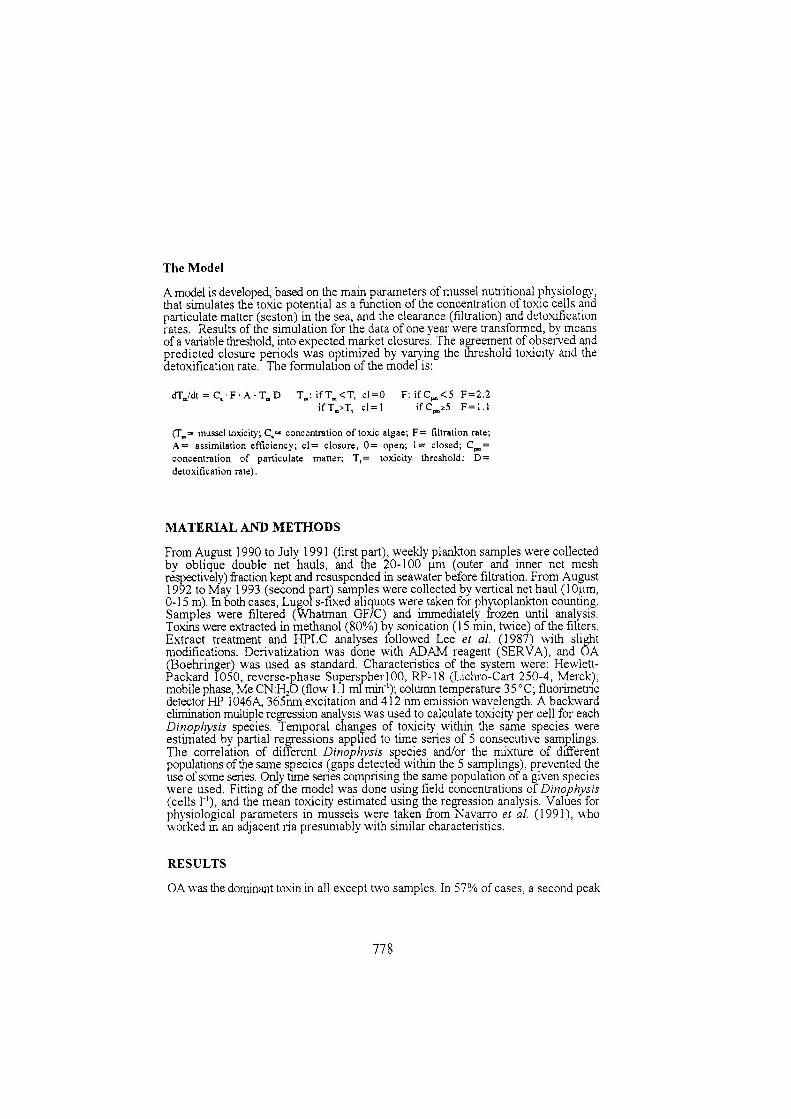
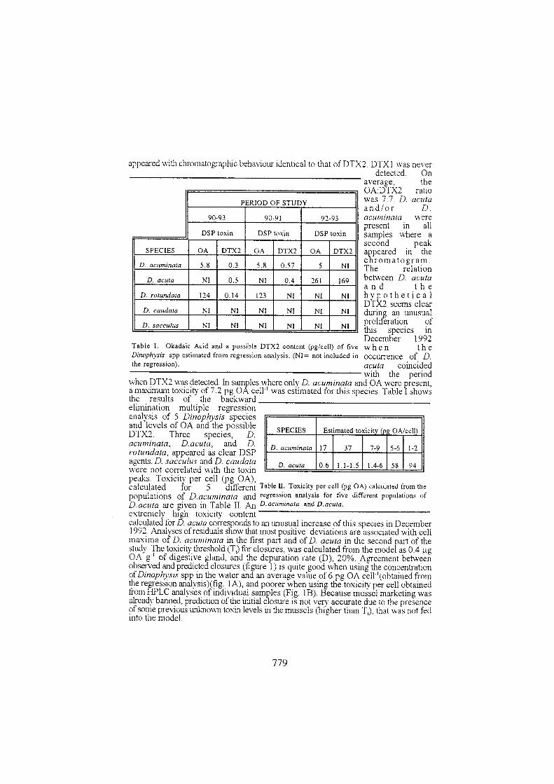


Capítulo I – Artículo 2 - 85 -
ARTÍCULO 2
Toxin content of Dinophysis acuminata, D. acuta and D.
caudata from the Galician Rías Bajas.
Fernández M.L., Reguera, B., Ramilo, I., Martinez, A. 2001. In: Hallegraeff,
G.M., Blackburn, S.I., Bolch, C.J., Lewis, R.J. (Eds), Harmful Algal Blooms,
Intergovernmental Oceanographic Commission of UNESCO, Paris, pp. 360-
363.

Capítulo I – Artículo 2 Resumen - 87 -
Toxin content of Dinophysis acuminata, D. acuta and D. caudata from the
Galician Rías Bajas.
RESUMEN
Dinophysis acuminata, D. acuta y D. caudata co-ocurren durante los episodios tóxicos de
DSP de principios de otoño en las Rías Bajas Gallegas, siendo necesario establecer su
contribución a la toxicidad de los moluscos. Los análisis de HPLC-FD de células aisladas en
diferentes episodios confirmaron que D.acuta, con una razón OA/DTX2 de 3:2, es la fuente
de la DTX2 detectada en los mejillones de Galicia y que tanto D. acuminata como D. acuta
pueden ser los principales contribuyentes a los episodios tóxicos de otoño. Nuestros
resultados sugieren que D. caudata presenta cantidades traza de AO (‹ 1 pg .cel-1) y confirman
la gran variabilidad en el contenido de toxinas de D. acuminata y D. acuta durante diferentes
episodios estacionales, o incluso durante el mismo episodio, después de que las poblaciones
naturales se mantuvieran en el laboratorio entre 5 y 9 días. Se discute el riesgo para la salud
pública derivado de utilizar el “índice de Dinophysis” en sustitución de la información del
bioensayo en ratón para la detección temprana de toxicidad DSP.

Harmful Algal Blooms Hallegraeff, G., Blackburn, S., Lewis, R. & Bolch, C. (eds.) Intergovernmental Oceanographic Commission of UNESCO 2001, pp. 360-363.
TOXIN CONTENT OF DINOPHYSIS ACUMINATA, D. ACUTA AND D. CAUDATA FROM THE GALICIAN RIAS BAJAS. M.Luisa Fernández*, Beatriz Reguera**, Isabel Ramilo** and Ana Martínez* * European Union Reference Laboratory on Marine Biotoxins. Aptdo. 90, 36271 Vigo, Spain **Instituto Español de Oceanografía. Centro Oceanográfico de Vigo. Aptdo 1552, 36280 Vigo, Spain. ABSTRACT
Dinophysis acuminata, D. acuta, and D. caudata co-occur during early autumn DSP outbreaks in the Galician Rías Bajas, and their contribution to shellfish toxicity needs to be established. HPLC–FD analyses of single cell isolates during different events confirmed that D. acuta, with an OA:DTX2 ratio of 3:2, is the source of DTX2 toxin detected in Galician mussels, and that either D. acuta or D. acuminata can be the main contributors to autumn toxic outbreaks. Our results suggest that D. caudata has trace amounts of OA (<1 pg · cell–1) and confirm the large variability in toxin content in D. acuta and D. acuminata during different seasonal outbreaks, or even during the same outbreak after natural populations were maintained in the laboratory for 5-9 days. The risk to human health of using a “Dinophysis index” as substitute information for the standard mouse bioassay in the early detection of diarrhetic shellfish toxicity is discussed. INTRODUCTION
Species of Dinophysis cause recurrent DSP outbreaks in Galicia and long closures of bivalve marketing in a region with an annual production over 2 · 105 t of mussels [1-3]. Dinophysis acuminata is always present in the Galician Rías Bajas, and may cause DSP outbreaks from May to October or even the whole year. Dinophysis acuta and Dinophysis caudata have a marked seasonality and are associated (if any) with DSP outbreaks from September to November. D. acuminata and D. acuta can reach levels of 1-2 · 104 cell · l-1, whereas D. caudata occurs in moderate concentrations (200 cell · l-1), rarely exceeding 103 cell · l-1. Large variations in bivalve toxicity related to Dinophysis spp concentrations are attributed partly to interspecific and intraspecific variations in toxin content of Dinophysis cells (Table I). The main objectives of this study were: • To obtain the toxin profile and the toxin content per
cell in different species of Dinophysis causing DSP outbreaks in Galicia.
• To investigate the presence of conjugated forms of OA (diol-esters and DTX4).
• To evaluate the contribution of each species during toxic outbreaks associated with multispecific populations of Dinophysis.
• To find possible relations between toxin content per cell and different morphotypes of D. acuminata occurring throughout the year.
METHODS
Vertical and oblique plankton net-hauls (20-100-µm fraction) were made during different DSP events in the Galician Rías. Single cell isolation of D.acuminata, D.acuta and D.caudata was made by microcapillary manipulation under an inverted microscope (25 X and 100 X). Cells were transfered several times through drops of filter-sterilized seawater (Millipore, 0.22 µm), and finally filtered on preheated (400ºC, 6h) Whatman
20,000,00 5,00 10,00 15,00 20,00
AODTX2 DTX2
a)
min
Fluo
resc
ence
20,00
45,00
0,00 5,00 10,00 15,00 20,00
AO
b)
min
Fluo
resc
ence
20,00
45,00
0,00 5,00 10,00 15,00 20,00
min
Fluo
resc
ence
c)AO
Figure 1 HPLC chromatograms obtained from extracts of a) D. acuta; b) D. acuminata; c) OA standard.
Harmful Algal Blooms Hallegraeff, G., Blackburn, S., Lewis, R. & Bolch, C. (eds.) Intergovernmental Oceanographic Commission of UNESCO 2001, pp. 360-363.
glass fibre filters (1.2 µm, 2cm). For analyses of whole population extracts, net-hauls were filtered on board and inmediately kept in the deep freeze. An aliquot was taken and fixed with neutral formaldehyde for species quantification.
Table II shows the number of cells isolated for each analysis. In the case of D. acuta, cells were isolated on the day of collection, and 5 and 9 days after being maintained in a culture chamber (L:D 14:10; 15º+ 1ºC). Most samples were analyzed following the procedure of Lee et al. [4] with slight modifications. A double extraction of each filter was performed by sonification in 2 ml of methanol (80%). After addition of half a volume of water to each volume of aliquot of the previous extract, a double extraction with 1.5 volumes of chloroform followed. In the samples of D. acuminata from June’98 the boiling and the freeze/thaw procedures developed by Quilliam et al. [5] for Prorocentrum lima cultures were applied: the first method, to destroy the enzymes of Dinophysis cells and prevent hydrolysis of potentially present conjugated forms of OA; the second method, to allow the enzymatic hydrolysis of OA conjugated forms. Derivatization was performed with ADAM reagent (SERVA). Characteristics of the system were: Hewlett-Packard 1050, Merck Superspher 100-RP18 (4 mi, 250 x 4 mm); mobile phase MeCN:H2O (80:20); 1.1 ml min-1 flow; column temperature 35ºC; fluorescence detector HP 1046 A with excitation and emission waves of 365 and 412 nm respectively. Standards used were OA (NRC, Canada) and DTX2 kindly provided by K.James (Cork, Ireland). The coefficient of variation (C.V) of the analyses was lower than 10%.
RESULTS
Figure 1 shows specimen chromatograms obtained from the Dinophysis extracts and from the standards. Table II show the main results on toxin content per cell and species.
Dinophysis acuminata: OA was found in all samples. Significant differences, indicating the presence of conjugated forms of OA in the cells were not found in the samples from June’98 (9.9 and 7.9 pg OA · cell -1 respectively) subjected to the two extraction protocols. A very small peak with retention time corresponding to DTX2 was found. Cells from September showed a toxin content more than double (21.7 pg OA· cell-1) that found in the June cells.
Dinophysis acuta: OA and DTX2 were found in the three samples. The OA:DTX2 ratio was 3:2 except in the samples from specimen that were maintained longer in the culture chamber. Important quantitative differences were found in the cells isolated as soon as they were collected at sea (10.7 pg · cell –1) and those which were maintained in the laboratory (55.1 and 24.1 pg · cell –1). In extracts from net-haul populations where D. acuta was
the overwhelmingly dominant Dinophysis species, OA and DTX2 were found in similar proportion, but total toxin content in cells from that year’s episode were much lower (0.6 – 4 pg · cell –1) [2].
Dinophysis caudata: The sample chromatogram exhibited a small peak with the same retention time as OA that can be interpreted as traces of OA (0.7 pg OA · cell –1).
DISCUSSION
These results show for the first time that Galician populations of D. acuta contain DTX2, and that this is the main species associated with the presence of this toxin in Galician mussels [6]. OA and DTX2 have also been found in D.acuta populations in Ireland [7] and in Portuguese mussels associated with the occurrence of D. acuta [8]. An OA:DTX2 ratio of 3:2 has been found in plankton net-hauls where the dominant species of Dinophysis was D. acuta, and in isolated cells, except in those stressed after several days maintained in the laboratory. The lowest toxin content per cell found in 1990, may be explained partly by a dilution effect of the toxicity in populations with a higher growth rate during a year with unsual high concentrations of this species, and partly because of the lower toxin content per cell estimated from net-haul extracts due to the matrix and other effects [9]. Regarding D.acuminata, although esterified toxins have not been found in the sample tested in this study by the boiling and the freeze/thaw procedures, the possibility of minor concentrations of OA being present in Dinophysis cells as diol-esters or DTX4-type compounds should not be discarded, because esterified OA has been found in Galicia in net-haul extracts where D.acuminata was the dominant dinoflagellate [10]. For the first time, isolates of Dinophysis caudata have been analyzed, showing trace amounts of OA. Blanco et al. [11] obtained a negative correlation coefficient for D. caudata from multiple regression of HPLC analyses of net-hauls containing a mixture of Dinophysis spp and other microplanktonic organisms [11]. D. caudata seems to have a minor rôle in autumn DSP toxicity in Galicia, but further tests need to be done to confirm these preliminary results.
Differences in Dinophysis cell numbers of at least one order of magnitude have been found in samples collected at the same station within 24 h [12], and toxicity concentrated by the bivalves is largely determined by the proportion that toxic microalgae represent from the total food available [2].These new data on variability in toxin content per cell of Dinophysis together with the problems mentioned above, and the well known sampling problem of planktonic species which are not very abundant and distributed in a patchy fashion, further weaken the value of a “Dinophysis index” as a substitute for the compulsory mouse bioassay in the detection of diarrhetic shellfish toxins associated with Dinophysis blooms.
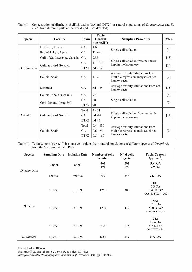
Harmful Algal Blooms Hallegraeff, G., Blackburn, S., Lewis, R. & Bolch, C. (eds.) Intergovernmental Oceanographic Commission of UNESCO 2001, pp. 360-363.
Table I. Concentration of diarrhetic shellfish toxins (OA and DTXs) in natural populations of D. acuminata and D. acuta from different parts of the world (nd = not detected).
Species
Locality
ToxinToxin
Content (pg · cell-1)
Sampling Procedure
Refer.
Le Havre, France. OA 1.6 Bay of Tokyo, Japan OA Traces
Single cell isolation [4]
Gulf of St. Lawrence, Canada OA 25.5 [13]
Gulmar Fjord, Sweden OA DTX1
1.1- 23.2 nd - 0.2
Single cell isolation from net-hauls kept in the laboratory [14]
Galicia, Spain OA 1- 37 Average toxicity estimations from multiple regression analyses of net-haul extracts.
[2]
D. acuminata
Denmark OA nd - 40 Average toxicity estimations from net- haul extracts [15]
Galicia , Spain (Oct. 87) OA 9.4 [4]
Cork, Ireland (Aug. 96) OA DTX2
58 78
Single cell isolation [7]
Gulmar Fjord, Sweden Total OA DTX1
4 - 21 nd -14 nd - 7
Single cell isolation from net-hauls kept in the laboratory [14] D. acuta
Galicia, Spain Total OA DTX2
0.4 - 430 0.6 - 94 0.5 - 169
Average toxicity estimations from multiple regression analyses of net-haul extracts
[2]
Table II. Toxin content (pg · cel-1) in single cell isolates from natural populations of different species of Dinophysis
from the Galician Southern Rías.
Species
Sampling Date
Isolation Date
Number of cells isolated
Nº of cells injected
Toxin Content
(pg · cel-1)
18.06.98 06.98 461 491
201 199
9.9 OA 7.9 OA
D. acuminata 8.09.98 9.09.98 857 246 21.7 OA
9.10.97 10.10.97 1250 308
10.7 6.3 OA
1.4 DTX2 OA: DTX2 = 3:2
9.10.97 14.10.97 1214 412
55.1 33.1 OA
22.0 DTX2 OA: DTX2 = 3:2
D. acuta
9.10.97 16.10.97 534 175
24.1 18.4 OA
5.7 DTX2 OA:DTX2 = 3:1
D. caudata 9.10.97 10.10.97 1388 342 0.73 OA

Harmful Algal Blooms Hallegraeff, G., Blackburn, S., Lewis, R. & Bolch, C. (eds.) Intergovernmental Oceanographic Commission of UNESCO 2001, pp. 360-363.
Table III. Estimated toxin content (pg · cel-1) per cell of Dinophysis from net-hauls where D. acuta was the overwhelmingly dominant species.
Dinophysis cells in the filters
Sampling Date
Toxin Content (pg · cel-1)
D. acuta (507934); D. acuminata (75044); D. caudata (5300)
28.08.90
2.6
OA:DTX = 1.6
D. acuta (26928); D. caudata (528)
2.10.90
4.0
OA: DTX2 = 1.5
D. acuta (45425); D. caudata (7820); D. rotundata (115)
22.10.90
0.6
OA: DTX2 = 1.5
ACKNOWLEDGEMENTS We thank the “Centro de Control da Calidade do Medio Mariño” (Xunta de Galicia) for monitoring information; Elena Ávila for her technical support; and the crew of R.V. “J.M. Navaz” for their helpful cooperation. This work was supported with funds from projects MAR98-0978 and MAR99-0224 (CICYT, Spain). REFERENCES
1. B. Reguera, I. Bravo, C. Marcaillou-Le Baut, P.
Masselin, M.L. Fernández, A. Míguez and A. Martínez, in: Smayda T.J., and Shimizu Y. (Eds.) Toxic Phytoplankton Blooms in the Sea. Elsevier, Amsterdam, pp. 553-558 (1993).
2. J. Blanco, M.L. Fernández, J. Mariño, B. Reguera, A. Míguez, J. Maneiro, E. Cacho and A. Martínez, in: Harmful Marine Algal Blooms, P. Lassus, G. Arzul, E. Erard-Le Denn, P. Gentien and C. Marcaillou-Le Baut C., eds., Lavoisier, París, pp. 777-782 (1995).
3. J. Blanco, A. Moroño, Y. Pazos, J. Maneiro and J. Mariño, in: B. Reguera, J. Blanco, M.L. Fernández and T. Wyatt, eds., Harmful Algae, Xunta de Galicia and IOC of UNESCO Publishers, pp. 204-207 (1998).
4. J-S. Lee, T.T. Igarashi, S. Fraga, E. Dahl, P. Hovgaard and T. Yasumoto, J. Appl. Phycol., 1, 147-152 (1989).
5. M.A.Quilliam, W.R. Hardstaff, N. Ishida, J.L. McLachlan, A.R. Reeves, N.W. Ross, and A.J. Windust, in: T. Yasumoto, Y. Oshima and Y. Fukuyo, eds., Harmful and Toxic Algal Blooms, IOC of UNESCO Publishers, pp. 289-292 (1996).
6. A. Gago-Martínez, J.A. Rodríguez-Vázquez, P. Thibault and M.A. Quilliam, Nat. Toxins, 4 (2), 72-79 (1996).
7. K.J. James, A.G. Bishop, B.M. Healy, C. Roden, I.R. Sherlock, M. Twohig, R. Draisci, L. Giannetti and L. Lucentini, Toxicon. 37(2): 343-357 (1999).
8. P. Vale, M.A.M.Sampayo and M. Quilliam, in: B. Reguera, J. Blanco, M.L. Fernández and T. Wyatt, eds., Harmful Algae, Xunta de Galicia and IOC of UNESCO Publishers, pp. 503-506 (1998).
9. P. Masselin, P. Lassus and M. Bardouil, J. Appl. Phycol. 4: 385-389 (1992)
10. A. Moroño, F. Arévalo, M.L. Fernández, J. Maneiro, Y. Pazos, C. Salgado and J. Blanco, in: Abstracts from the 3rd International Conference on Molluscan Shellfish Safety, Long Island, USA, 19-23 June, p. 58 (2000)
11. J. Blanco, Y. Pazos, F. Arévalo, C. Salgado, M. Bermúdez de la Puente, J. Maneiro y A. Moroño, in: Junta de Andalucía, Proceedings of the VI Iberian Workshop on Toxic Phytoplankton and Phytotoxins, Publicaciones de la Consejería de Agricultura y Pesca, pp. 127-138 (1999).
12. B. Reguera, I. Bravo, H. MacCall and M.I.Reyero, in: T. Yasumoto, O. Oshima & Y. Fukuyo, eds., Harmful and Toxic Algal Blooms, IOC of UNESCO Publishers, pp. 257-260 (1996).
13. A.D. Cembella, Occurrence of okadaic acid, a major diarrhetic shellfish toxin, in natural populations of Dinophysis spp from the Eastern coast of North America. J.Appl. Phycol. 1: 307-310 (1989).
14. N. Johansson, E. Granéli, T. Yasumoto, P. Carlsson and C. Legrand, in: T. Yasumoto, Y. Oshima & Y. Fukuyo, eds., Harmful and Toxic Algal Blooms, IOC of UNESCO Publishers, pp. 277-280 (1996).
15. P. Andersen, B. Hald and H. Emsholm, in: T. Yasumoto et al., Y. Oshima & Y. Fukuyo, eds., Harmful and Toxic Algal Blooms. IOC of UNESCO Publishers, pp. 281-284 (1996).
Capítulo I – Artículo 3 - 93 -
ARTÍCULO 3:
Toxin composition of the toxic dinoflagellate Prorocentrum
lima isolated from different locations along the Galician coast
(NW Spain).
Bravo, I., Fernández, M.L., Ramilo, I. and Marínez, A. 2001. Toxicon 39, 1537-
1545.


Capítulo I – Artículo 3 Resumen - 95 -
Toxin composition of the toxic dinoflagellate Prorocentrum lima isolated
from different locations along the Galician coast (NW Spain).
RESUMEN
La composición de toxinas DSP de 19 cepas de Prorocentrum lima de diferentes
localizaciones de las Rías Gallegas (Vigo y Pontevedra) fue investigada por cromatografía
líquida de alta eficacia acoplada a detección fluorimétrica. El procedimiento de ebullición-
congelación-descongelación-hidrólisis fue aplicado durante la extracción para la detección de
AO, DTX1, DTX2 y sus derivados esterificados. El AO y la DTX2 fueron detectadas en
forma libre y esterificada, ésta última siempre en cantidades considerablemente menores,
mientras que la DTX1 estuvo siempre presente en forma libre. Este resultado sugiere que la
hipótesis de que las formas esterificadas representarían un mecanismo de autoprotección para
el almacenamiento de las toxinas en una forma menos activa no sería aplicable a la DTX1. Se
detectó un ligero incremento en la concentración de toxina por célula durante el crecimiento,
si bien la composición de toxinas no varió de manera apreciable. La producción y el perfil de
toxinas variaron significativamente en función de la cepa. El análisis de cluster permitió la
diferenciación de 4 grupos de P.lima de acuerdo con su composición de toxinas. Es de
destacar que uno de los clusters comprendió todas las cepas recogidas en una localización
caracterizada por su aislamiento geográfico, mientras que el resto de los clusters
comprendieron cepas de distintas localizaciones. Las diferencias encontradas entre el perfil de
toxinas de las diferentes cepas de P. lima y el correspondiente a los moluscos contaminados
con toxinas DSP, junto con la muy buena correlación entre la ocurrencia de Dinophysis spp y
la toxicidad DSP en los moluscos corroboran que estas especies planctónicas son los
principales agentes responsables de los eventos tóxicos en Galicia.


Toxin composition of the toxic dino¯agellate Prorocentrum limaisolated from different locations along the Galician coast
(NW Spain)
I. Bravoa,*, M.L. FernaÂndezb, I. Ramiloa, A. MartõÂnezb
aInstituto EspanÄol de OceanografõÂa. Centro Oceanogra®co de Vigo. Aptdo 1552, 86200 Vigo, SpainbLaboratorio de Sanidad Exterior de Vigo. European Community Reference Laboratory on Marine Biotoxins. EstacioÂn MarõÂtima de Vigo,
s/n, 36271 Vigo, Spain
Received 27 November 2000; accepted 31 March 2001
Abstract
The DSP toxin composition of 19 Prorocentrum lima isolates from different locations of the Galician rias (Vigo and
Pontevedra) was investigated by high performance liquid chromatography coupled with ¯uorimetric detection. Boiling and
freeze/thaw/hydrolyse methodology were applied during extraction to detect OA, DTX1, DTX2 and their esteri®ed derivatives.
OA and DTX2 were detected in both free and esteri®ed form, the latter always in very low amounts, whilst DTX1 was always
present in the free form. This indicate that the hypothesized self-protection mechanism of toxin storage in the less active
esteri®ed forms does not seem to apply to DTX1. A slight increase in the toxin concentration per cell was found during growth,
although toxin composition did not vary appreciably. Toxin production and toxin pro®le varied signi®cantly depending on the
isolate. Four groups of P. lima were differentiated by cluster analysis according to their toxin composition. It is noteworthy that
one of the clusters comprised all the strains collected from one location characterised by its geographical isolation, whereas the
other clusters consisted of isolates from different locations. The differences in the toxin pro®le from P. lima strains and from the
DSP contaminated shell®sh, together with the very good correlation between Dinophysis spp occurrence and DSP toxicity in
shell®sh, support that these planktonic species are the main agents responsible for DSP events in Galicia. q 2001 Elsevier
Science Ltd. All rights reserved.
Keywords: Prorocentrum lima; Diarrhetic shell®sh poisoning; DSP toxins; Toxic dino¯agellates; Galician rias
1. Introduction
Prorocentrum lima (Ehrenberg) Dodge is a benthic dino-
¯agellate usually found attached or associated to macro-
phytes, ¯oating detritus, debris or other substrates and less
commonly found in plankton. Although this is a ubiquitous
species, it appears to be most abundant in tropical and
subtropical waters. Together with other benthic species
associated with coral reefs, such as G. toxicus, P. concavum,
and O. tamarensis, it has been linked to the toxic syndrome
known as Ciguatera (Yasumoto et al., 1980; Bomber et al.,
1988). The severity of this complex syndrome has led to a
number of studies in the epiphytic environment of tropical
and subtropical areas, whereas little is known on benthic
dino¯agellates in general and P. lima in particular from
temperate waters.
P. lima and Dinophysis spp are known to produce a suite
of polyether metabolites, some of which, when present in
shell®sh, are the cause of diarrhetic shell®sh poisoning
(DSP), a severe gastrointestinal disease. The implication of
P. lima in DSP episodes is questioned because its benthic
character makes its uptake by bivalve molluscs very unli-
kely. The attachment of P. lima to different macroalgae foul-
ing mussel rafts from the Galician rias (NW Spain) has been
reported by Bravo (1991) in concentrations of up to 2255
cells/g fresh weight of Ulva spp. However, DSP events in
these production areas have always been associated with the
presence of Dinophysis species in plankton (Reguera et al.,
1993; Blanco et al., 1999). DSP toxins present in shell®sh
from other European countries and Japan, New Zealand,
Toxicon 39 (2001) 1537±1545
0041-0101/01/$ - see front matter q 2001 Elsevier Science Ltd. All rights reserved.
PII: S0041-0101(01)00126-X
www.elsevier.com/locate/toxicon
* Corresponding author.
Singapore, Chile, South Africa and Australia, have also been
linked to Dinophysis blooms (Dahl and Yndestad, 1985; Kat,
1985; Lassus et al., 1985; Kumagai et al., 1986; Hallegraeff,
1987; MacKenzie, 1991; Lembeye et al., 1993; Pitcher et al.,
1993; Tubaro et al., 1995; Carmody et al., 1996; Suzuki et
al., 1997; Holmes et al., 1999). However, not all the DSP
incidents clearly correlate with Dinophysis blooms, as in the
case of the coasts of Nova Scotia (Marr et al., 1992; Lawr-
ence et al., 1998), where P. lima cells found primarily within
the ®laments of the phaeophyte, Pilayella littoralis, have
been put forward as the most likely source of DSP toxicity
(Lawrence et al., 1998).
Due to the great dif®culties in obtaining pure laboratory
cultures of Dinophysis spp, P. lima cultures have been
widely used to study the kinetics of DSP toxin production,
since Murakami et al. (1982) identi®ed okadaic acid as one
of the basic components produced by this epiphytic species.
P. lima has also been shown to be a reliable source of
okadaic acid (OA) and dinophysitoxin-1 (DTX1) to produce
toxin standards and reference material.
OA and its methyl homologue, DTX1 were for many
years thought to be the major toxins produced by P. lima
species (Lee et al., 1989). Dinophysitoxin-2 (DTX2), an
isomer of OA ®rst isolated from Irish mussels (Hu et al.,
1992), has also been found in P. lima cultures, but only in
trace amounts. OA, DTX1 and DTX2 are powerful inhibi-
tors of protein phosphatases-1 and -2A and potent tumour-
promoting agents in vitro. A number of esters of OA were
described by Yasumoto et al. (1989) and two diol esters of
OA were isolated and identi®ed on the basis of spectro-
scopic methods in P. lima strains from Vigo (Norte et al.,
1994). Pharmacological experiments showed that diol esters
are as active as the free acid, although the period of latency
is signi®cantly higher for the esters.
The development and implementation of liquid chroma-
tography±mass spectrometry (LC±MS) and LC±MS/MS
techniques has been crucial in improving the knowledge
of DSP toxin production, and additional derivatives such
as new diol esters by P. lima (Hu and Marr, 1992). The
water soluble dinophysitoxin-4 (DTX4), a tri-sulphate
ester derivative of OA (Hu et al., 1995b), has been isolated
and identi®ed. Moreover, two new sulphated water soluble
DSP toxin derivatives, DTX5a and DTX5b were found in
cultures of Prorocentrum maculosum (Hu et al., 1995a).
Nevertheless, although LC±MS equipment has become
less expensive in the last few years, it is not available in
most laboratories involved in marine biotoxin monitoring
and research. High performance liquid chromatography
coupled with ¯uorimetric detection (HPLC±FD) techniques
are still valuable tools in the study of DSP toxin production,
in spite of their limitations.
HPLC±FD techniques permit the detection of those DSP
toxins which possess a free carboxylic group in the molecule
(OA, DTX1, DTX2). Lee's method (Lee et al., 1987) invol-
ving the use of 9-anthryldiazomethane (ADAM) as ¯uoro-
phore, is the most widely used method for detection of OA
and its homologues in microalgae and shell®sh. Detection of
esteri®ed DSP toxins such as diol esters or DTX4 by this
method requires previous chemical or enzymatic hydrolysis.
Quilliam et al. (1996) have shown that a rapid enzymati-
cally-catalysed hydrolysis of DTX4 to the diol esters, which
are more slowly hydrolysed to OA, occurs when cells are
broken during simple extractions with aqueous methanol,
leading to erratic results in the determination of toxin
concentrations. To determine OA, DTX1 and DTX2, a boil-
ing procedure that restricts enzymatic hydrolysis has been
developed by the aforementioned authors. They have also
proposed a freeze/thaw/hydrolyse extraction method which
promotes the conversion of the esteri®ed toxins to OA,
DTX1 or DTX2 and permits the assessment of the contribu-
tion of these derivatives to total DSP toxicity.
In the present study both the boiling and the freeze/thaw/
hydrolyse extraction method have been used to investigate
the toxin composition of 19 P. lima isolates collected from
the Galician rias (Pontevedra and Vigo). The authors also
discuss the signi®cance of this benthic species in DSP
contamination of shell®sh.
2. Material and methods
2.1. P. lima strains, isolation and culture conditions
P. lima strains were isolated from macroalgae samples
collected at different locations in the rias of Vigo and Ponte-
vedra, NW Iberian Peninsula (Fig. 1). Table 1 shows the P.
lima strains used in the experiment as well as dates of isola-
tion, type of substrate and location. All the sampling stations
(beaches and mussel rafts) are located inside the rias with
the exception of the lagoon of the Cies Islands which
connects with the open sea [Fig. 1(b)]. Macroalgae from
beaches and mussel rafts were attached to rocks and ropes
respectively. The substrate collected in the lagoon of the
Cies Islands was clearly different, being a ®lamentous
algae located in a sandy environment.
Macroalgae were thoroughly shaken with seawater from
the same site to dislodge epiphytic microalgae, which were
separated through 150 and 20 mm sieves. P. lima cells were
isolated from the material collected in the 20 mm sieve after
washing with 0.2 mm-®ltered seawater and placed in F/2
(strains isolated before 1988) or K medium (Keller and
Guillard, 1985). Strains were kept in F/2 (before 1988) or
K medium until the experiment and in the same light and
temperature conditions as those described below.
Cultures of P. lima were grown in plastic bags with 1 L of
K medium at 19 ^ 18C on a 14:10 h light:dark cycle at a
photon ¯ux density of 60±70 mE s21 m22. Duplicate 10±
20 mL aliquots of culture were harvested for toxin analyses
on the 27th day of culture for all strains. For the growth
experiment, PL2V, PL6V, PL11V and PL27V strains were
used, and samples were also collected on the 13th and 41st
day of culture. A cell count was also carried out for each
I. Bravo et al. / Toxicon 39 (2001) 1537±15451538
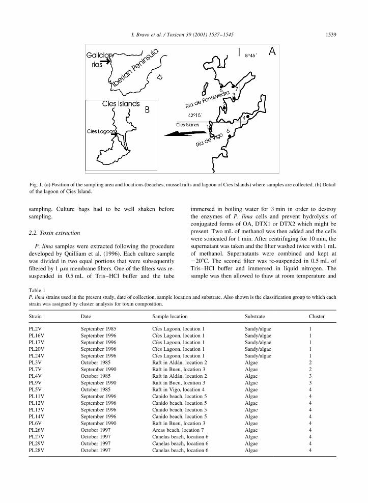
sampling. Culture bags had to be well shaken before
sampling.
2.2. Toxin extraction
P. lima samples were extracted following the procedure
developed by Quilliam et al. (1996). Each culture sample
was divided in two equal portions that were subsequently
®ltered by 1 mm membrane ®lters. One of the ®lters was re-
suspended in 0.5 mL of Tris±HCl buffer and the tube
immersed in boiling water for 3 min in order to destroy
the enzymes of P. lima cells and prevent hydrolysis of
conjugated forms of OA, DTX1 or DTX2 which might be
present. Two mL of methanol was then added and the cells
were sonicated for 1 min. After centrifuging for 10 min, the
supernatant was taken and the ®lter washed twice with 1 mL
of methanol. Supernatants were combined and kept at
2208C. The second ®lter was re-suspended in 0.5 mL of
Tris±HCl buffer and immersed in liquid nitrogen. The
sample was then allowed to thaw at room temperature and
I. Bravo et al. / Toxicon 39 (2001) 1537±1545 1539
Fig. 1. (a) Position of the sampling area and locations (beaches, mussel rafts and lagoon of Cies Islands) where samples are collected. (b) Detail
of the lagoon of Cies Island.
Table 1
P. lima strains used in the present study, date of collection, sample location and substrate. Also shown is the classi®cation group to which each
strain was assigned by cluster analysis for toxin composition.
Strain Date Sample location Substrate Cluster
PL2V September 1985 CõÂes Lagoon, location 1 Sandy/algae 1
PL16V September 1996 CõÂes Lagoon, location 1 Sandy/algae 1
PL17V September 1996 CõÂes Lagoon, location 1 Sandy/algae 1
PL20V September 1996 CõÂes Lagoon, location 1 Sandy/algae 1
PL24V September 1996 CõÂes Lagoon, location 1 Sandy/algae 1
PL3V October 1985 Raft in AldaÂn, location 2 Algae 2
PL7V September 1990 Raft in Bueu, location 3 Algae 2
PL4V October 1985 Raft in AldaÂn, location 2 Algae 3
PL9V September 1990 Raft in Bueu, location 3 Algae 3
PL5V October 1985 Raft in Vigo, location 4 Algae 4
PL11V September 1996 Canido beach, location 5 Algae 4
PL12V September 1996 Canido beach, location 5 Algae 4
PL13V September 1996 Canido beach, location 5 Algae 4
PL14V September 1996 Canido beach, location 5 Algae 4
PL6V September 1990 Raft in Bueu, location 3 Algae 4
PL26V October 1997 Areas beach, location 7 Algae 4
PL27V October 1997 Canelas beach, location 6 Algae 4
PL29V October 1997 Canelas beach, location 6 Algae 4
PL28V October 1997 Canelas beach, location 6 Algae 4
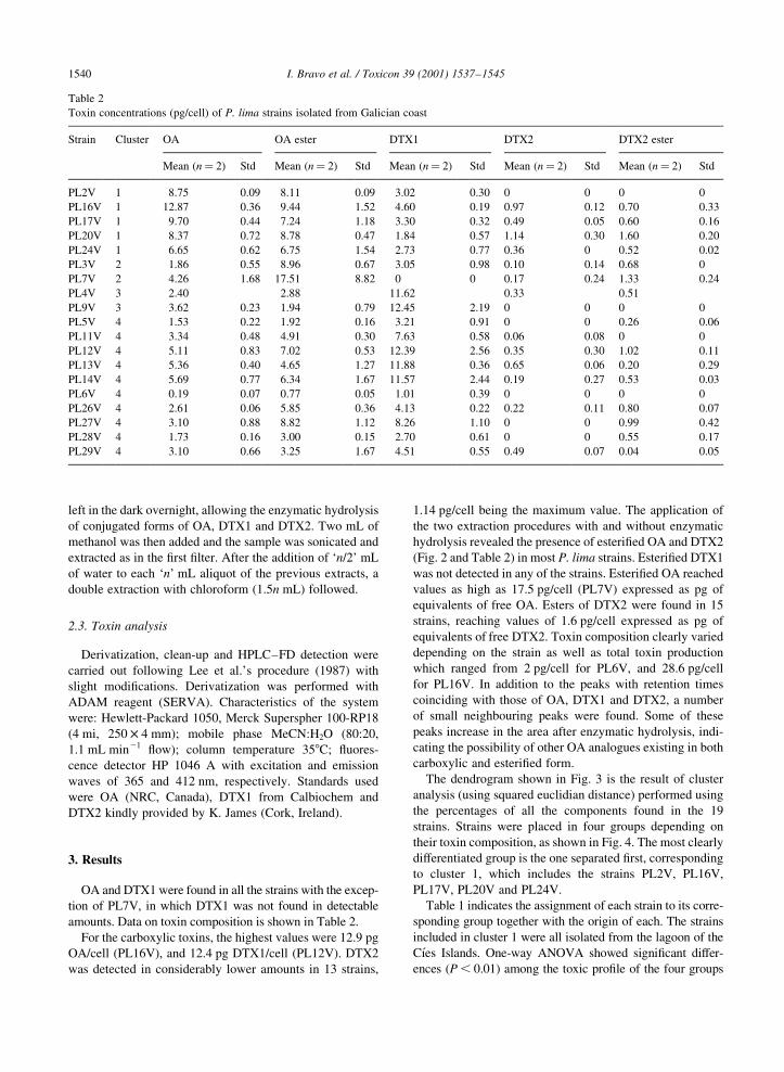
left in the dark overnight, allowing the enzymatic hydrolysis
of conjugated forms of OA, DTX1 and DTX2. Two mL of
methanol was then added and the sample was sonicated and
extracted as in the ®rst ®lter. After the addition of `n/2' mL
of water to each `n' mL aliquot of the previous extracts, a
double extraction with chloroform (1.5n mL) followed.
2.3. Toxin analysis
Derivatization, clean-up and HPLC±FD detection were
carried out following Lee et al.'s procedure (1987) with
slight modi®cations. Derivatization was performed with
ADAM reagent (SERVA). Characteristics of the system
were: Hewlett-Packard 1050, Merck Superspher 100-RP18
(4 mi, 250 £ 4 mm); mobile phase MeCN:H2O (80:20,
1.1 mL min21 ¯ow); column temperature 358C; ¯uores-
cence detector HP 1046 A with excitation and emission
waves of 365 and 412 nm, respectively. Standards used
were OA (NRC, Canada), DTX1 from Calbiochem and
DTX2 kindly provided by K. James (Cork, Ireland).
3. Results
OA and DTX1 were found in all the strains with the excep-
tion of PL7V, in which DTX1 was not found in detectable
amounts. Data on toxin composition is shown in Table 2.
For the carboxylic toxins, the highest values were 12.9 pg
OA/cell (PL16V), and 12.4 pg DTX1/cell (PL12V). DTX2
was detected in considerably lower amounts in 13 strains,
1.14 pg/cell being the maximum value. The application of
the two extraction procedures with and without enzymatic
hydrolysis revealed the presence of esteri®ed OA and DTX2
(Fig. 2 and Table 2) in most P. lima strains. Esteri®ed DTX1
was not detected in any of the strains. Esteri®ed OA reached
values as high as 17.5 pg/cell (PL7V) expressed as pg of
equivalents of free OA. Esters of DTX2 were found in 15
strains, reaching values of 1.6 pg/cell expressed as pg of
equivalents of free DTX2. Toxin composition clearly varied
depending on the strain as well as total toxin production
which ranged from 2 pg/cell for PL6V, and 28.6 pg/cell
for PL16V. In addition to the peaks with retention times
coinciding with those of OA, DTX1 and DTX2, a number
of small neighbouring peaks were found. Some of these
peaks increase in the area after enzymatic hydrolysis, indi-
cating the possibility of other OA analogues existing in both
carboxylic and esteri®ed form.
The dendrogram shown in Fig. 3 is the result of cluster
analysis (using squared euclidian distance) performed using
the percentages of all the components found in the 19
strains. Strains were placed in four groups depending on
their toxin composition, as shown in Fig. 4. The most clearly
differentiated group is the one separated ®rst, corresponding
to cluster 1, which includes the strains PL2V, PL16V,
PL17V, PL20V and PL24V.
Table 1 indicates the assignment of each strain to its corre-
sponding group together with the origin of each. The strains
included in cluster 1 were all isolated from the lagoon of the
CõÂes Islands. One-way ANOVA showed signi®cant differ-
ences (P , 0.01) among the toxic pro®le of the four groups
I. Bravo et al. / Toxicon 39 (2001) 1537±15451540
Table 2
Toxin concentrations (pg/cell) of P. lima strains isolated from Galician coast
Strain Cluster OA OA ester DTX1 DTX2 DTX2 ester
Mean (n� 2) Std Mean (n� 2) Std Mean (n� 2) Std Mean (n� 2) Std Mean (n� 2) Std
PL2V 1 8.75 0.09 8.11 0.09 3.02 0.30 0 0 0 0
PL16V 1 12.87 0.36 9.44 1.52 4.60 0.19 0.97 0.12 0.70 0.33
PL17V 1 9.70 0.44 7.24 1.18 3.30 0.32 0.49 0.05 0.60 0.16
PL20V 1 8.37 0.72 8.78 0.47 1.84 0.57 1.14 0.30 1.60 0.20
PL24V 1 6.65 0.62 6.75 1.54 2.73 0.77 0.36 0 0.52 0.02
PL3V 2 1.86 0.55 8.96 0.67 3.05 0.98 0.10 0.14 0.68 0
PL7V 2 4.26 1.68 17.51 8.82 0 0 0.17 0.24 1.33 0.24
PL4V 3 2.40 2.88 11.62 0.33 0.51
PL9V 3 3.62 0.23 1.94 0.79 12.45 2.19 0 0 0 0
PL5V 4 1.53 0.22 1.92 0.16 3.21 0.91 0 0 0.26 0.06
PL11V 4 3.34 0.48 4.91 0.30 7.63 0.58 0.06 0.08 0 0
PL12V 4 5.11 0.83 7.02 0.53 12.39 2.56 0.35 0.30 1.02 0.11
PL13V 4 5.36 0.40 4.65 1.27 11.88 0.36 0.65 0.06 0.20 0.29
PL14V 4 5.69 0.77 6.34 1.67 11.57 2.44 0.19 0.27 0.53 0.03
PL6V 4 0.19 0.07 0.77 0.05 1.01 0.39 0 0 0 0
PL26V 4 2.61 0.06 5.85 0.36 4.13 0.22 0.22 0.11 0.80 0.07
PL27V 4 3.10 0.88 8.82 1.12 8.26 1.10 0 0 0.99 0.42
PL28V 4 1.73 0.16 3.00 0.15 2.70 0.61 0 0 0.55 0.17
PL29V 4 3.10 0.66 3.25 1.67 4.51 0.55 0.49 0.07 0.04 0.05
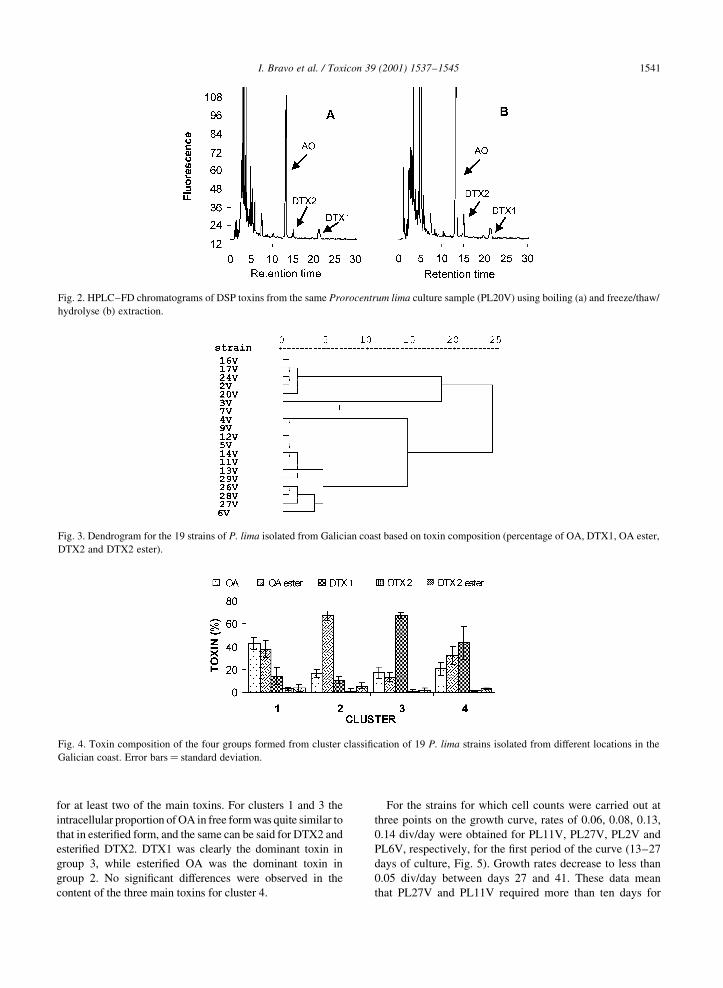
for at least two of the main toxins. For clusters 1 and 3 the
intracellular proportion of OA in free form was quite similar to
that in esteri®ed form, and the same can be said for DTX2 and
esteri®ed DTX2. DTX1 was clearly the dominant toxin in
group 3, while esteri®ed OA was the dominant toxin in
group 2. No signi®cant differences were observed in the
content of the three main toxins for cluster 4.
For the strains for which cell counts were carried out at
three points on the growth curve, rates of 0.06, 0.08, 0.13,
0.14 div/day were obtained for PL11V, PL27V, PL2V and
PL6V, respectively, for the ®rst period of the curve (13±27
days of culture, Fig. 5). Growth rates decrease to less than
0.05 div/day between days 27 and 41. These data mean
that PL27V and PL11V required more than ten days for
I. Bravo et al. / Toxicon 39 (2001) 1537±1545 1541
Fig. 3. Dendrogram for the 19 strains of P. lima isolated from Galician coast based on toxin composition (percentage of OA, DTX1, OA ester,
DTX2 and DTX2 ester).
Fig. 4. Toxin composition of the four groups formed from cluster classi®cation of 19 P. lima strains isolated from different locations in the
Galician coast. Error bars� standard deviation.
Fig. 2. HPLC±FD chromatograms of DSP toxins from the same Prorocentrum lima culture sample (PL20V) using boiling (a) and freeze/thaw/
hydrolyse (b) extraction.
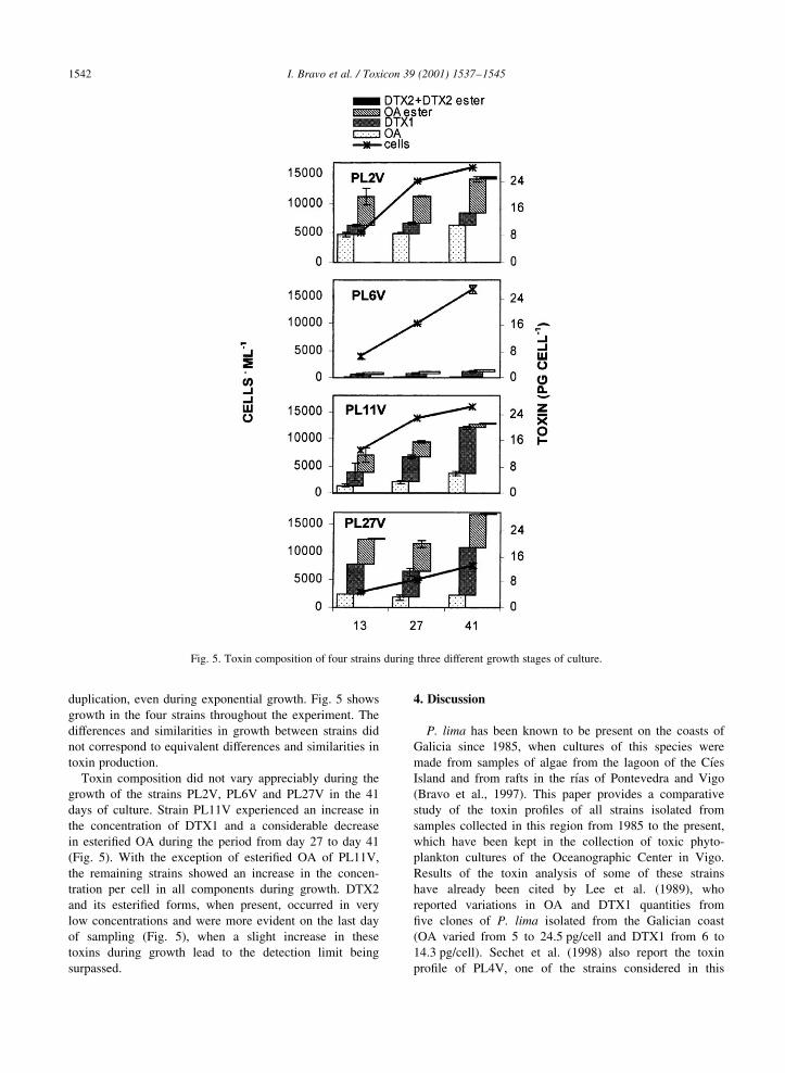
duplication, even during exponential growth. Fig. 5 shows
growth in the four strains throughout the experiment. The
differences and similarities in growth between strains did
not correspond to equivalent differences and similarities in
toxin production.
Toxin composition did not vary appreciably during the
growth of the strains PL2V, PL6V and PL27V in the 41
days of culture. Strain PL11V experienced an increase in
the concentration of DTX1 and a considerable decrease
in esteri®ed OA during the period from day 27 to day 41
(Fig. 5). With the exception of esteri®ed OA of PL11V,
the remaining strains showed an increase in the concen-
tration per cell in all components during growth. DTX2
and its esteri®ed forms, when present, occurred in very
low concentrations and were more evident on the last day
of sampling (Fig. 5), when a slight increase in these
toxins during growth lead to the detection limit being
surpassed.
4. Discussion
P. lima has been known to be present on the coasts of
Galicia since 1985, when cultures of this species were
made from samples of algae from the lagoon of the CõÂes
Island and from rafts in the rõÂas of Pontevedra and Vigo
(Bravo et al., 1997). This paper provides a comparative
study of the toxin pro®les of all strains isolated from
samples collected in this region from 1985 to the present,
which have been kept in the collection of toxic phyto-
plankton cultures of the Oceanographic Center in Vigo.
Results of the toxin analysis of some of these strains
have already been cited by Lee et al. (1989), who
reported variations in OA and DTX1 quantities from
®ve clones of P. lima isolated from the Galician coast
(OA varied from 5 to 24.5 pg/cell and DTX1 from 6 to
14.3 pg/cell). Sechet et al. (1998) also report the toxin
pro®le of PL4V, one of the strains considered in this
I. Bravo et al. / Toxicon 39 (2001) 1537±15451542
Fig. 5. Toxin composition of four strains during three different growth stages of culture.

paper. Coinciding with the data presented here, these
authors report DTX1 as the major toxin component in
their cultures. In addition, using the boiling and freeze/
thaw methods of extraction for quantitative analysis by
liquid chromatography-mass spectrometry (LC±MS), they
were able to detect an oxidized derivative of OA in the
strain from the rõÂa of Vigo and a very complex mixture
of DTX4-related compounds, which were probably
detected as esteri®ed OA in our HPLC±FD analyses.
The freeze/thaw/hydrolyse method, which promotes the
conversion of the esteri®ed toxins to OA, DTX1 or
DTX2, while not discriminating between diol-esters and
DTX4-type compounds, permits the assessment of the
contribution of these derivatives to total DSP toxicity.
The application of this procedure is clearly indicated
when assessing toxic potential of dino¯agellate samples,
especially when taking into account the important contri-
bution of esteri®ed derivatives to total toxicity. This
should also be extended to the analysis of the toxicity
of Dinophysis samples, as shown by MoronÄo et al. (in
preparation).
OA, DTX1 and DTX2 are strong inhibitors of protein
phosphatases PP1 and PP2A (Haystead et al., 1989) and it
has been suggested that they have an allelopathic effect on
other microalgal growth (Windust et al., 1996). Hu et al.
(1995a) have suggested that intracellular storage of the
toxins as the less active sulphates could be an effective
self-protection mechanism for the DSP-producing cells
and even a means of excreting the acidic toxins from the
cell to the aqueous medium. However, from the results
obtained in this study, there does not seem to be a clear
pattern of storage of DSP toxins inside the cells, although
for some of the strains esteri®ed forms were clearly domi-
nant (cluster 2); for other strains, the acidic form (e.g. DTX1
in cluster 3) was present in considerably greater amounts.
This hypothetical self-protection mechanism does not seem
to be applicable in the case of DTX1 which, although
reported to be more slightly toxic than OA, has not been
clearly detected in esteri®ed form in signi®cant amounts in
our experiment.
Few studies have reported on variability in toxin produc-
tion of different P. lima strains. Morton and Tindall (1995)
describe a signi®cant difference in biochemical features
between 17 clones from three separate sites of Herbon
Island (Great Barrier Reef of Australia). Based on the differ-
ences detected in reproduction rates, pigments, toxins (OA
and DTX1) and macromolecular compounds (total protein,
lipid and carbohydrate), these authors suggest the existence
of two genetically different populations in this area. From
the results of the present paper, the differentiation observed
in the cluster analysis of the strains corresponding to the
region of the lagoon of the Cies Islands must be emphasised.
All the strains from this area were separated into a distinct
group according to their toxin compositions (cluster 1 in Fig.
4), whereas the other groups were not separated by de®ned
locations (Table 1). It must be mentioned that the lagoon of
the Cies Islands is characterised by its geographical isola-
tion relative to the ria of Vigo and Pontevedra, since its
connection with the outside is orientated towards the open
sea [Fig. 1(b)], which would support the hypothesis of an
isolation of the population in this area.
In relation to toxin production during growth, Fig. 5 illus-
trates a progressive increase in total toxicity per cell. Maxi-
mum toxin concentrations in the stationary phase of P. lima
cultures has also been cited in cultures of this species
coming from other regions (MacLachlan et al., 1994; Quil-
liam et al., 1996; Sechet et al., 1998). Also, in our experi-
ment an increase in all toxin components was detected
during growth with the exception of the esteri®ed compo-
nents of OA of the strain PL11V, which fell signi®cantly
throughout the culture period. The possible excretion of
some component into the medium was not considered in
our experiment, although this does not seem likely to be
the cause, since Quilliam et al. (1996) describe the accumu-
lation of OA and DTX1 in the medium, but only at trace
levels of DTX4 and OA diol ester.
As for the growth rates of P. lima cultures described in the
present study, the low values of divisions/day must be
stressed. Previous results from the cultures of some strains
used in this study cite maximum values of up to 0.40 ^ 0.09
div/day in 250 mL of K medium in tissue culture plate (Bravo
et al., 1997). However, these authors obtain maximum values
of 0.15 div/day in Erlenmeyers with 150 mL of the same
medium, ascribing it to the progressive mortality occurring
when the cells detach from the culture ¯asks. On using plas-
tic bags to which cells do not adhere to such an extent, this
inconvenience was avoided in our experiment, but higher
growth rates were not obtained. An effect not evaluated in
the P. lima growth experiment in bags was that the plastic
may bring about the transmission of light radiation to the
cultures. A reduced light intensity or a change in the light
spectrum may have led to a fall in the rates of cell division.
Even if this were the case, it would not explain the differ-
ences found in the growth of the four strains used for this
experiment. On the other hand, the differences in growth
rates do not seem to be related to differences in toxin compo-
sition nor to total toxin content, since the groups of strains
differentiated by growth do not correspond to those differen-
tiated by toxin composition in the cluster analysis. From the
data described by Morton et al. (1995), differences in growth
rates of the strains supposedly belonging to the same popula-
tion group can also be deduced.
The presence of P. lima on the rafts where all Galician
mussel production takes place, and its known capacity to
produce diarrhetic toxins, led to the association of this
species, at ®rst, with the appearance of DSP toxins in shell-
®sh from the Galician rias (Marr et al., 1992). Nevertheless,
there is now enough knowledge of toxins produced by
species of the genus Dinophysis present in the area, together
with the good correlation between the occurrence of
these species and DSP incidents, to clearly indicate that
Dinophysis spp is the causative agent of DSP episodes
I. Bravo et al. / Toxicon 39 (2001) 1537±1545 1543
detected in the region. In addition, the main toxins found in
mussels from Galician rias in the last few years are OA,
DTX2 and DTX3, the latter in low quantities, while
DTX1, which is present in most P. lima strains, has never
been found in shell®sh from this region at signi®cant levels
(Fernandez et al., 1996, 1998). Therefore, regarding the
causative species, it seems that the DSP events from Galicia
are not similar to those of eastern Canada, where P. lima is
strongly suspected of producing the DSP toxins found in
mussels (Jackson et al., 1993; Lawrence et al., 1998). Unlike
the case of Galician mussels, DTX1 is the major toxin
component in DSP-contaminated shell®sh in Nova Scotia,
Canada (Quilliam et al., 1993), a fact which ®ts in well with
the toxin pro®le detected by LC±MS analysis of epiphytic
microalgal material collected from a mussel culture site with
P. lima as the only dino¯agellate present in the sample
(Lawrence et al., 2000). Morton et al. (1999) have also
identi®ed the presence of DTX1 in epiphytic samples rich
in P. lima from Maine (United States), although these
authors did not detect OA or DTX1 by LC±MS/MS in the
shell®sh from these sampling sites, but only low levels of
OA-like activity by protein phosphatase inhibition assay.
In the same way as has been documented on the Atlantic
coast of Canada (Lawrence et al., 2000), the mussel rafts of
the Galician rias provide a habitat which favours the colo-
nisation and growth of macroalgae which support the micro-
organism communities among which epiphytes such as P.
lima are found. Bravo (1991) mentions maximum values of
2255 P. lima cells/g fresh weight on Ulva spp and 1600
cells/g fresh weight on Enteromorpha sp 1 Giffordia sp
from a mussel raft on 9 September 1987, quantities which
may be similar to the maximum concentrations of P. lima
cited by Lawrence et al. (2000) (15,000 cells/g dry weight)
in Nova Scotia. From the study by Bravo (1991) it is
deduced that P. lima populations on the rafts of the Galician
rias are governed by a seasonality and a speci®city of
substrate which make them very limited both in time and
location. For this reason, while the ability of these popula-
tions to detach from the substrate and enter the mussel
through the trophic chain is unknown, it cannot be dismissed
that this may occur on a lesser scale than that used in the
detection of toxic phytoplankton on the Galician coast.
Acknowledgements
We thank Amelia Fernandez-VillamarõÂn for her technical
assistance and the crew of B/O Jose M. Navaz for collecting
samples. Thanks also are extended to J. James for a sample
of DTX2. This research was supported by projects MAR95-
1849 and PGIDT99MAR40201.
References
Blanco, J., Fernandez, M.L., Miguez, A., MoronÄo, A., 1999.
Okadaic acid depuration in the mussel Mytilus galloprovincia-
lis: One- and two-compartment models and the effect of envir-
onmental conditions. Mar. Ecol. Prog. Ser. 176, 153±163.
Bomber, J.W., Morton, S.L., Babinchack, J.A., Norris, D.R.,
Morton, J.G., 1988. Epiphytic dino¯agellates of drift algae-
another toxigenic community in the ciguatera food chain.
Bull. Mar. Sci. 43, 204±214.
Bravo, I., 1991. Results of preliminary studies in the ®eld and in
culture of the toxic dino¯agellate Prorocentrum lima (Ehren-
berg) Dodge. Revue Internationale d'Oceanographie Medicale
101±104, 189±196.
Bravo, I., Ramilo, I., Franco, J.M., MaronÄo, E., Fraga, S., 1997.
Coleccion de cultivos de microalgas nocivas del Centro Ocea-
nogra®co de Vigo. Resultados experimentales de algunas espe-
cies. Inf. Tec. Inst. Esp. Oceanogr. 168, 32 pp.
Carmody, E.P., James, K.J., Kelly, S.S., 1996. Dinophysitoxin-2:
The predominant diarrhetic shell®sh toxin in Ireland. Toxicon
34 (3), 351±359.
Dahl, E., Yndestad, M., 1985. Diarrhetic shell®sh poisoning (DSP)
in Norway in the autumn 1984 related to the occurrence of
Dinophysis spp. In: Anderson, D.M., White, A.W., Baden,
D.G. (Eds.). Toxic Dino¯agellates. Elsevier, New York,
pp. 495±500.
Fernandez, M.L., Miguez, A., Cacho, E., Martinez, A., 1996. Detec-
tion of okadaic acid esters in the hexane extracts of spanish
mussels. Toxicon 34 (3), 381±387.
Fernandez, M.L., Miguez, A., MoronÄo, A., Cacho, E., Martinez, A.,
Blanco, J., 1998. Detoxi®cation of low polarity toxins (DTX3)
from mussels Mytilus galloprovincialis in Spain. In: Reguera,
B., Blanco, J., Fernandez, M.L., Wyatt, T. (Eds.). Harmful
Microalgae. Xunta de Galicia and UNESCO, Santiago de
Compostela, Spain, pp. 449±452.
Hallegraeff, G.M., 1987. Red tides in the Australasian region. Rep.
CSIRO Mar. Lab. Hobart, 187, 20 pp.
Haystead, T.A.J., Sim, A.T.R., Carling, D., Honnor, R.C., Tsukitani,
Y., Cohen, P., Hardie, D.G., 1989. Effects of the tumour promo-
ter okadaic acid on intracelular protein phosphorylation and
metabolism. Nature 337, 78±81.
Holmes, M.J., Teo, S.L.M., Lee, F.C., Khoo, H.W., 1999. Persistent
low concentrations of diarrhetic shell®sh toxins in green
mussels Perna viridis from the Johor Strait, Singapore: First
record of diarrhetic shell®sh toxins from South-East Asia.
Mar. Ecol. Prog. Ser. 181, 257±268.
Hu, T., Marr, J., 1992. New diol esters isolated from cultures of the
dino¯agellates Prorocentrum lima and Prorocentrum conca-
vum. Journal of Natural Products 55 (11), 1631±1637.
Hu, T., Doyle, J., Jackson, D., Marr, J., Nixon, E., Pleasance, S.,
Quilliam, M.A., Walter, J.A., Wright, J.L.C., 1992. Isolation of
a new Diarrhetic Shell®sh Poison from Irish mussels. J. Chem.
Soc. Chem. Commun., 39±41.
Hu, T., Curtis, J.M., Walter, J.A., MacLachlan, J.L., Wright, J.L.C.,
1995. Two new water-soluble DSP toxin derivatives from the
Dino¯agellate Prorocentrum maculosum: Possible storage and
excretion products. Tetrahedron Letters 36 (51), 9273±9276.
Hu, T., Curtis, J.M., Walter, J.A., Wright, L.C., 1995. Identi®cation
of DTX-4, a new water-soluble phosphatase inhibitor from the
toxic dino¯agelate Prorocentrum lima. J. Chem. Soc. Chem.
Commun., 597±599.
Jackson, A.E., Marr, J.C., McLachlan, J.L., 1993. The production of
Diarrhetic Shell®sh Toxins by an isolate of Prorocentrum lima
from Nova Scotia, Canada. In: Smayda, T.J., Shimizu, Y. (Eds.).
I. Bravo et al. / Toxicon 39 (2001) 1537±15451544

Toxic Phytoplankton Blooms in the Sea. Elsevier, New York,
pp. 513±518.
Kat, M., 1985. Dinophysis acuminata blooms, the distinct cause of
Dutch mussel poisoning. In: Anderson, D.M., White, A.W.,
Baden, D.G. (Eds.). Toxic Dino¯agellates. Elsevier, New
York, pp. 73±77.
Keller, M.D., Guillard, R.R.L., 1985. Factors signi®cant to marine
dino¯agellate culture. In: Anderson, D.M., White, A.W., Baden,
D.G. (Eds.). Toxic Dino¯agelates. Elsevier, New York, pp.
113±116.
Kumagai, M., Yanagi, T., Murata, M., Yasumoto, T., Kat, M.,
Lassus, P., Rodriguez-Vazquez, J.A., 1986. Okadaic acid as
the causative toxin of diarrhetic shell®sh poisoning in Europe.
Agric. Biol. Chem. 50 (11), 2853±2857.
Lassus, P., Bardouil, M., Truquet, I., Truquet, P., Le Baut, C.,
Pierre, M., 1985. Dinophysis acuminata distribution and toxicity
along the southern Brittany coast (France): Correlation with
hydrological parameters. In: Anderson, D.M., White, A.W.,
Baden, D.G. (Eds.). Toxic Dino¯agellates. Elsevier, New
York, pp. 159±164.
Lawrence, J.E., Bauder, A.G., Quilliam, M.A., Cembella, A.D.,
1998. Prorocentrum lima: A putative link to diarrhetic shell®sh
poisoning in Nova Scotia, Canada. In: Reguera, B., Blanco, J.,
Fernandez, M.L., Wyatt, T. (Eds.). Harmful Microalgae. Xunta
de Galicia and UNESCO, Santiago de Compostela, Spain, pp.
78±79.
Lawrence, J.E., Grant, J., Quilliam, M.A., Bauder, A.G., Cembella,
A.D., 2000. Colonization and growth of the toxic dino¯agellate
Prorocentrum lima and associated fouling macroalgae on
mussels in suspended culture. Mar. Ecol. Progr. Ser. 201,
147±154.
Lee, J-S., Yanagi, T., Kenna, R., Yasumoto, T., 1987. Fluorometric
determination of diarrhetic shell®sh toxins by high-performance
liquid chromatography. Agric. Biol. Chem. 51, 877±881.
Lee, J-S., Igarashi, T., Fraga, S., Dahl, E., Hovgaard, P., Yasumoto,
T., 1989. Determination of diarrhetic shell®sh toxins in various
dino¯agellates species. J. Appl. Phycol. 1, 147±152.
Lembeye, G., Yasumoto, T., Zhao, J., Fernandez, R., 1993. DSP
outbreak in Chilean ®ords. In: Anderson, D.M., White, A.W.,
Baden, D.G. (Eds.). Toxic Phytoplankton Blooms in the Sea.
Elsevier, Amsterdam, pp. 525±530.
MacKenzie, L., 1991. Toxic and noxious phytoplankton in Big
Glory Bay, Stewart Island, New Zealand. J. Appl. Phycol. 3
(1), 19±34.
MacLachlan, J.L., Marr, J.C., Colon-Kelly, A., Adamson, A., 1994.
Effect of nitrogen concentrations and cold temperatures on DSP-
toxin concentrations in the dino¯agellate Prorocentrum lima
(Prorocentrales, Dinophyceae). Natural Toxins 2 (5), 263±270.
Marr, J.C., Jackson, A.E., McLachlan, J.L., 1992. Ocurrence of
Prorocentrum lima, a DSP toxin-producing species from the
Atlantic coast of Canada. J. Appl. Phycol. 4, 17±24.
Morton, S.L., Tindall, D.R., 1995. Morphological and biochemical
variability of the toxic dino¯agellate Prorocentrum lima
isolated from three locations at Heron Island. Australia. J.
Phycol. 31, 914±921.
Morton, S.L., Leigh®eld, T.A., Haynes, B.L., Petitpain, D.L.,
Busman, M.A., Moeller, P.D.R., Bean, L., McGowan, J.,
Hurst, J.W., Van Dolah, J.R., F, M., 1999. Evidence of Diarrhe-
tic Shell®sh Poisoning along the coast of Maine. J. Shell. Res.
18 (2), 681±686.
Murakami, Y., Oshima, Y., Yasumoto, T., 1982. Identi®cation of
okadaic acid as a toxic component of a marine dino¯agellate
Prorocentrum lima. Bull. Japan Soc. Sci. Fish. 48, 69±72.
Norte, M., Padilla, A., Fernandez, J.J., Souto, M.L., 1994. Structural
determination and biosynthetic origin of two ester derivatives of
okadaic acid isolated from Prorocentrum lima. Tetrahedron 50
(30), 9175±9180.
Pitcher, G.C., Horstman, D.A., Calder, D., De Bruyn, J.H., Post,
B.J., 1993. The ®rst record of diarrhetic shell®sh poisoning on
the South African coast. S. Afr. J. Sci. 89 (10), 512±514.
Quilliam, M.A., Gilgan, M.W., Pleasance, S., Defreitas, A.S.W.,
Douglas, D., Fritz, L., Hu, T., Marr, J.C., Smyth, C., Wright,
J.L.C., 1993. Con®rmation of an incident of Diarrhetic Shell®sh
Poisoning in eastern Canada. In: Smayda, T.J., Shimizu, Y.
(Eds.). Toxic Phytoplankton Blooms in the Sea. Elsevier, New
York, pp. 547±552.
Quilliam, M.A., Hardstaff, W.R., Ishida, N., McLachlan, J.L.,
Reeves, A.R., Rose, N.W., Windust, A.J., 1996. Production od
Diarrhetic Shell®sh Poisoning (DSP) toxins by Prorocentrum
lima in culture and development of analytical methods. In:
Yasumoto, T., Oshima, Y., Fukuyo, Y. (Eds.). Harmful and
Toxic Algal Blooms. IOC of UNESCO, Senday, pp. 289±292.
Reguera, B., MarinÄo, J., Campos, M.J., Bravo, I., Fraga, S., Carbo-
nell, A., 1993. Trends in the occurrence of Dinophysis spp. in
Galician waters. In: Smayda, T.J., Shimizu, Y. (Eds.). Toxic
Phytoplankton Blooms in the Sea. Elsevier, New York, pp.
559±564.
Sechet, V., Quilliam, M.A., Rocher, G., 1998. Diarrhetic Shell®sh
Poisoning (DSP) toxins in Prorocentrum lima in axenic and
non-axenic batch culture: detection of new compounds and
kinetics of production. In: Reguera, B., Blanco, J., Fernandez,
M.L., Wyatt, T. (Eds.). Harmful Microalgae. Xunta de Galicia
and UNESCO, Santiago de Compostela, pp. 485±488.
Suzuki, T., Mitsuya, T., Imai, M., Yamasaki, M., 1997. DSP toxin
contents in Dinophysis fortii and scallops collected at Mutsu
Bay, Japan. J. Appl. Phycol. 8 (6), 509±515.
Tubaro, A., Sosa, S., Bussani, D., Sidari, L., Honsell, G., Della
Loggia, R., 1995. Diarrhetic toxicity induction in mussels of
the Gulf of Trieste. In: Lassus, P., Arzul, G., Erard-Le Denn,
E., Gentien, P., Marcaillou-Le Baut, C. (Eds.). Harmful Marine
Algal Blooms. Lavoisier, Paris, pp. 249±254.
Windust, A.J., Wright, J.L.C., McLachlan, J.L., 1996. The effects of
the diarrhetic shell®sh poisoning toxins, okadaic acid and dino-
physistoxin-1, on the growth of microalgae. Marine Biology
126, 19±25.
Yasumoto, T., Oshima, Y., Murakami, Y., Nakajima, I., Bagnis, R.,
Fukuyo, Y., 1980. Toxicity of benthic dino¯agellates found in
coral reef. Bull. Japan Soc. Sci. Fish. 46 (3), 327±331.
Yasumoto, T., Murata, T., Lee, J.S., Torigoe, K., 1989. Polyeter
toxins produced by dino¯agellates. In: Natori, S., Hashimoto,
K., Ueno, Y. (Eds.). Mycotoxins and Phycotoxins. Elsevier,
Amsterdam, pp. 375±382.
I. Bravo et al. / Toxicon 39 (2001) 1537±1545 1545

Capítulo I – Artículo 4 - 107 -
ARTÍCULO 4:
First report of PTX-2 in phytoplankton net hauls and mussels
from the Galician Rías Bajas during proliferations of
Dinophysis acuta and D. caudata.
Fernández, M.L., Míguez, A., Martínez, A., Moroño, A., Arévalo, F., Pazos, Y.,
Salgado, C., Correa, J., Blanco, J., González-Gil, S. and Reguera, B. In:
Villalba, A., Reguera B., Romalde, J., Beiras, R. (eds), Consellería de Pesca y
Asuntos Marítimos, Xunta de Galicia and Intergovernmental Oceanographic
Commission of Unesco. 2003. pp 75-83.

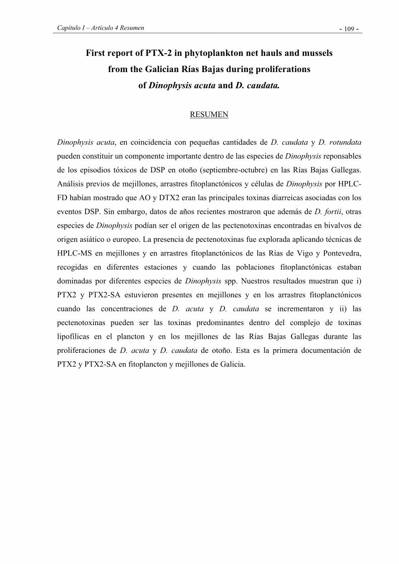
Capítulo I – Artículo 4 Resumen - 109 -
First report of PTX-2 in phytoplankton net hauls and mussels
from the Galician Rías Bajas during proliferations
of Dinophysis acuta and D. caudata.
RESUMEN
Dinophysis acuta, en coincidencia con pequeñas cantidades de D. caudata y D. rotundata
pueden constituir un componente importante dentro de las especies de Dinophysis reponsables
de los episodios tóxicos de DSP en otoño (septiembre-octubre) en las Rías Bajas Gallegas.
Análisis previos de mejillones, arrastres fitoplanctónicos y células de Dinophysis por HPLC-
FD habían mostrado que AO y DTX2 eran las principales toxinas diarreicas asociadas con los
eventos DSP. Sin embargo, datos de años recientes mostraron que además de D. fortii, otras
especies de Dinophysis podían ser el origen de las pectenotoxinas encontradas en bivalvos de
origen asiático o europeo. La presencia de pectenotoxinas fue explorada aplicando técnicas de
HPLC-MS en mejillones y en arrastres fitoplanctónicos de las Rías de Vigo y Pontevedra,
recogidas en diferentes estaciones y cuando las poblaciones fitoplanctónicas estaban
dominadas por diferentes especies de Dinophysis spp. Nuestros resultados muestran que i)
PTX2 y PTX2-SA estuvieron presentes en mejillones y en los arrastres fitoplanctónicos
cuando las concentraciones de D. acuta y D. caudata se incrementaron y ii) las
pectenotoxinas pueden ser las toxinas predominantes dentro del complejo de toxinas
lipofílicas en el plancton y en los mejillones de las Rías Bajas Gallegas durante las
proliferaciones de D. acuta y D. caudata de otoño. Esta es la primera documentación de
PTX2 y PTX2-SA en fitoplancton y mejillones de Galicia.

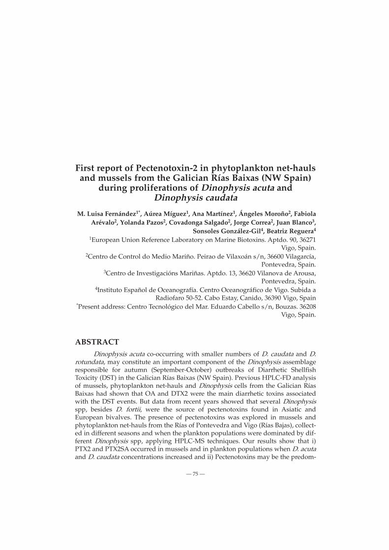
������������������ ��� ���� �������� �� � ��������� ������������������������ ����������������� ��� !
���� "���������� ���#� ������������ � �#� ���������������
$%�&�������� ' ��()*+�,-����$�"��()+�, ��$���� �()+�. "�����$�/�+���0���,�12���+�3�� �����(��+�42�� "�� ��"���+�5�"��4�����+�5�� ���� �6+
������ ('��(����7+�������(���"����7����������� ������������������������������������������� !�
"�#��$���� %������%����&�����������'��(�������"�&���)�*������"�&�#���+��
(���,�����$�����%������-,����#���.�����'��������������� �"�&��,����������
(���,�����$����/-�����������'�&��0����#���+��%����0����#�)������"�#��$������
��������1�21 �%���������%����������"�#��$���3(������������4%����5���&.#�����&�����������%���&&��*����6����� �7
"�#��$����
,� 8�,48������������ ��2�������#8��9�:�&&���:����������� ����
�� ��� �:�������������:��������:�������9���������������:�&�#���������&� ��� ����: ;$����:���20������< �������=� �� >����9���� $9�&&���95�������;>$5<��9�?�&���� +��������;@A$���<�(��,����B(�%2C>��&������:����&���9����&�=����29��&���������������&&� ���: �9�?�&���� +��������9�� �9�8 �9��0� ��>5D 8��� �9�:�������9���� ���������������8��9 �9�>$5�,���� ������� ���:�����������9�8�� �9����,���&�������������� ������� �� ��� ��� 8��� �9� ������ �� ����������� ���� � ������� ��������� ��,�&,��� 59� ������� �� ����������� 8�� ���&���� �:����&� ���9����&�=����29��&����:�9� +����(���,������"�#�; +����E��<���&&���2�������������������89��9��&�=������&�����8�����:����������2��������������� ���� ���&��# B(�%2�$ ���9�F���� 0�� ����&�� �9�8 �9�� �<(5D ��(5D $� ���������:����&�����&�=������&�����89����� ������� �����������������������<(����������:�����9������:2
G!1 G
��� ����� �� �9� &����9�&��2����2��:�&�� � �&�=�� �� :����&� ���: �9�?�&���� +������������#����:���&��������������� ������� �59�����9��������������(5D ��(5D $� ��9����&�=����:����&����:?�&�����
9:3��;�# <�>����9�����9�&&���9������#;>$(<���������������������=�2���� ���� ;0�<� ����9��������� ;>5D�<� ����������� ;(5D�<� &�F��� �9��:���2#���9�2:����������:����;�%2�$<�
=�8�;#>48=;�(��&��������� �� ���������� ���� ���������� 8��9 �������� �� >����9����
$9�&&���9(�����# ;>$(< ����� ���,�&,�����,���#�&����� &�,�&� ;>$5�,���<�����������9��������&�:�9����������9�������9�&&���9����������9�?�&���� +��;@A$���<; �#�������&�������&������&����1����7<�������������,���&��,���� ��:����&� ;�� ���� ���������������< � �&����# ����� ; 1� H ��� � �������<�>$5�,������:��������������9�I +��������J;����9��%���C��������<���������&&���9�����9��:��� +����"�#���(���,����;C�#��<�59������2�#��&��=�������� ��8���9���������/�($( ��&&�8����>$( ����&�,�&����,���#�&�����&�:���&�����&��������:����&������������:��:�������� +���(���,��������/18��=�;��K,�&����&�����<���������������� ��8��9�������9�:���#����>$5�,����?�&������������������&��������,�����������������&������&&����9������������������,���9�89�&����������@�,���9�&����������&����������9�������������� ��2�������#8��9�:�&&���:����������� ���� �����:���,����&&�����������9���:�����:2�������9���������������:�&�#���������&��������:;$����:���20������<>$5�,���������&��� ��=���������;0�<8���9��&������9��������=�8�� ������ ��,�&,�� �?�&���� ;��� �� �&� ��7�L ���+#��62")6F��6 �� �&� ��7�<� ������������������8��:���������������0� ����������:�����B(�%&��������������9������������9���������:�����;��K,�&����&����/<�59�&��=����::�����&�������������������:0������9�&�����&,��9����������������F���=&�����������������9���������������>5D �-���9:����&���9��:���2#���9�����=�8��9���������:����:�&�����9�����>5D 8���������9���2�&�=����29��&���:����&��:�&�����8����������� ;�&������&����1<�>5D 8�� ���&&� ��������� �?�&����:����&����%2�$ ;?�#� �� �&� ����<����� � �9�:�� ���������� ��>5D � -���9:����&��8�� ��������� �� �&�� �9���:���&����(����#������?�&�����9�&&���9�-���������0� ��>5D ����&����,���,�����9�&����������8������������:����&�;C��)��6���&�����<�
(��,����B(�%2C>��&������������������ ������� ��&&� ���:�9�?�&���� +��������� ���&������������:����:����&������9�8���9��0� ��>5D 8�������������� ��&&����4 ��������9������� 9����������0� ;C��)��6���&� ���<�@�,���9�&�����������:�����������9�8���9�� ��,���&���������� ���� ��������� ��� ��� 8��� �9� ������ �� �������������������������������,�&,��;>��#�E����&����7�>���������&������$�6�=����&����7<�� ����# �9�� �������:������� �9�:������#�� �9��� ����� �?�&�����9�&&���9����#���������� �&��:��(��&�:��������&�������������9����B(�%2�$������&������4 �<�9����&�=����29��&��:�&�� ���:����������������9� +����(���,������"�#�����9����� ������ ��*����
G!� G
��� ?-@?�� -@�(BM%050D-@$�@>�@��M5-%����5B0>$
����� ���&&���������#����#;N��<������:;0������<>$5�,��������<:����&���&&��������:��������� +���(���,�����0���������/�����!�59�����9��������������(5D ��9����&�=������&�������(5D $� ��9�2���&�=����:����&����:�9�?�&���� +���������
$,8:�=,&�,�#�$:8?;#
(9����&�=����29��&���:����&��:�&��8�����&&������������������2�������: +���(���,������ +���"�#�;C�#��<�
C�#������������ +���(���,������ +���"�#���9��8�����9��:������9�����?�&���� +��������;@A$���<�
������� �� � ������$�:�&��1����!���:����; +���(���,����<8������������,������&
�9����&�=����29��&� ;��O::��9��6�<�� ��:�&��� �9���29��&��������8����:�,���������8��9&�#�&�������9��F��������,���&������������������&&����:� ����:���� ��9�:�������������� �9�����:P9&:��9���59���:�����������:������#���,�&�:��8���������������������#�����59���&2&��8���������������1:� ��5���2B%&���������::���������&�#8�����������,��� �9� �6�:�� �� ��������� �� Q��&&��: �� �&� ;����<� C���9��� :� ��:��9��&8���������������������&������1:�������=�9���&&��C�&&�8�#�������#������9����������8����=���=������6����&�%2�$��&�����
59���9���9����&�=����:�&������:�����������������9� +����"�#���(���,�����8�����&&���������&�F��9��&����8�������:������9����&�=2���������O:2:��9�9�������� �O:2:��9�9��������8��9�9���&&��2�������9� �2���O:��6��������8����������59���29��&,�&�:�8��:������������ ��:�,�# � �&�F���� �� ��&����� � ����� �9���#9 ���2��:������ ;/�� R%����#�9<A9��:�?C*C��&�����59���&����8�����&����������������::�2
G!! G
C��)��6���&�4(������������9����&�=����29��&���:����&����:?�&����
�����&����6����&��:�����&��������������� ���������������8�������:��2�����: 2���������:�2�����:�&���$��#�8��=2 ������9�:�����5���������2���8��������:���������������9���&���8��97�S:��9��&���1:��������������#����� �9� ���������8�� ��=� �� =��� ���6����& �%2�$ ��&�����$�:�&�7���:��:��; +���"�#�<8�������������9���9����29��&������2�#�&�����&�#�&&�����������#��:���������� �8��=����&�,�����&2�����9�:������9�&��������������,���&��������������������
$������ ������������&��:�&��8�����&&����������; +���(���,����<�0���������/
�� 0������ ���!� >�#����,� #&��� 8��� 9�:�#��6�� �� � #2������� 8������������8��9/:� ��7�S:��9��&�������������#�������&�F������9������2����8����&������9���#9��/1O:�������9���%2�$�E�����������&����:�9����8���:�&��9���������������9����&�=������&�������::�&������,�����9������9���8�&&�#������8��9��������&�������������:��������:2������&��#�����&�#�&&����;:��&����������� ����<���:�&&�&�#�&&�����59�:������������������� �������8���������� ������ � ��&&�8�����:�&&���:����������� ���� ����� ����/���������� ����!�
&�@����4�����"������A�$���� �����������, ������<�%2�$ ��&����8��� ������:�����# � C��#���5(/��� F��������
��:����C��#��$����������:�&��� ����&��8��9� �� �����%Q2>�0����$������:�����F������8��9���:���9�������������$-���������
%9��:���#���9�� ��������4 %9��:���#���9�� ���������8�� ������� ���� �D5�����$%7� �1�::� ��:: ���� ��&�:���=��8��9 1O:������&������9��&4A�����!�4��������#���S������������9���:�:���&��9������9������������0��E����&�;����<����6�����������8����������&�8�������� :�*:��59��E�����,�&�:�8��1O��
���� �������:���� ��������4 59� ��:�������� �� �9� 9����� ����&&��8�� 1�RT%���9�����&&���,�&��#��1�11"�B�#9����������#�8�������59��9���9#���������#��������8��1�����9�����9�����&����#�� ������C���&&��:�&�����������9�9��:���#���9������8�����,�������&&���������,����$-�$�����&&���������,��$*�$8���������:����:�&������&��C�&&����������8�����&&��������: ����� ��:*6�-�:������#8��������:����9������:�������9�����&������(5D ���&&���:������������9�����#U�V@�WV ��:*677��1�%�&&�����������$2�$8��������:����9��������U�V@�WV �����#1�S ��&&���� ���#�� 59� �����:��� ���#:�� 8�� :*6 7�!�1� - ������� ��(5D � (5D $� ���&� ��:������� ��:*6 7���1� 0� ��>5D � 89� ����������&����������������9����,�����������:*67 !�1�C���&&�9���&������9�U�V@�WV ������8���9��������&����������8��9,���&�8�����������&���#2�&����9������=�����������#��U�VBWV�
5��� ��������4 (5D ������� 8�� � #������ #��� ���: (�������� 5�M���:��� �� >5D ���: >� X� N�:��� 0� 8�� ������� ���: �9� @�����& ������9%����&;%����<�
G!7 G
��� ?-@?�� -@�(BM%050D-@$�@>�@��M5-%����5B0>$
�: >&8 �,�#�#= 4> =;�
5��&���9�8��9�������������9���:�&����&�6���@���9��(5D���>5D ����������&�&�,�&�8��������,�����9����&�=����29��&;��:�&�Y�<���9 ���������������� ���: &��� ����# ;N��< �9�� �9�8��� ���������&���:�������0��59������#���:��8��9���,��������&�����������B(�%2C>��9���9�8��0� ����:��������E�#�������:����9�:��������������#&���&&���&������������� ;C��)��6���&� ���<����9����&�=����29��&�89���������� 8���9���:����������;����'����&� ���<�59���9���9����&�=�� ��29��&� ���: ���&� ����: ;$����:���20������< ���9 � &��#�������������������9������ ������ ���� ����� �9�8���9��(5D �������������:�&��������������9�0� �������&�#������,����9���:2��������C�#� �9�8��9��%2�$���%2�$*�$�9��:���#��:�����������#���9������������&�=����29��&;��:�&�Y7<��&&������0���������!���:��; +���"�#�<�:��&���:����������������� ����8��9���� ���9���:��������������&&���������9���:�&��������9����������&���:�������0��>5D ��(5D �
5��&� �� 5��� ����� �����:��� �� �%2�$ � �9����&�=�� ��29��&� ;�#*��&& ������������<���:����&�;O#*#<���:�9�?�&���� +����(���,����;(<��"�#�;"<�C�����9��29��&��:�&���9�����&�:�������&&������������� ��������������9������������������� ���������9���&�����#�,��;�Z����������<�
%����&�������8���9�����&�:��������������� ��&&����9�#&���&���������������������:�9����&�=����29��&�8��,��������59�������&���9�8�9���9�������������������� &����9�&�� �����;����0� ����,���,����������2
G!� G
C��)��6���&�4(������������9����&�=����29��&���:����&����:?�&����
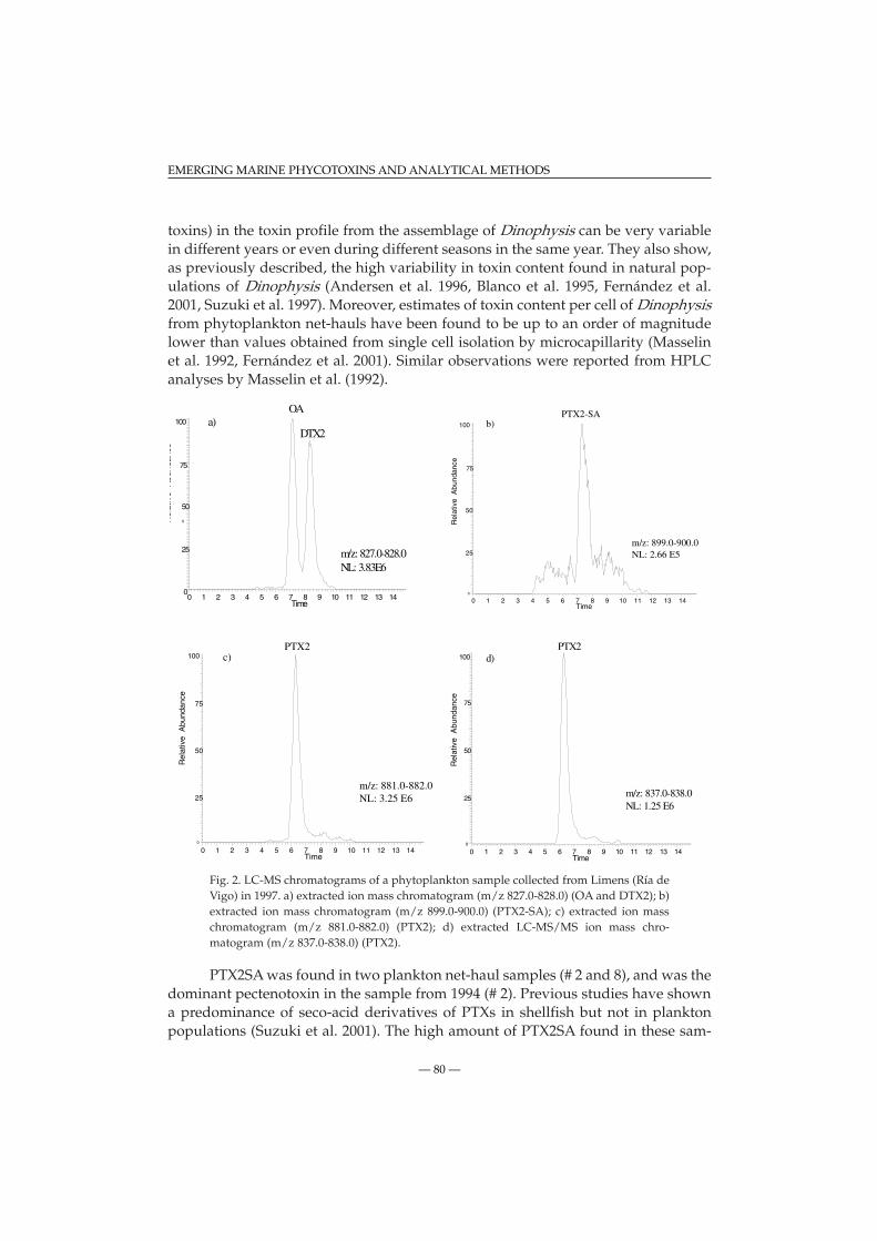
�����<��9����������&����:�9�����:�&�#������������� ����,���,�����&������������������,�����#����������������9���:������59���&���9�8������,����&������������9�9�#9,������&�����������������������&���2�&����������������� ;������ �� �&� ������&��� �� �&� ���1� C��)��6 �� �&� ����$�6�=����&����!<������,�������:��������������������&&���������������:�9����&�=����29��&�9�,������������������������:�#�����&�8���9�,�&�������������:��#&���&&���&������:��������&&�����;�����&����&���� �C��)��6���&� ���<�$�:�&�������,�����8��������������:B(�%��&�����������&����&�;��� <�
C�#� ��%2�$�9��:���#��:�����9����&�=����:�&���&&��������:��:��; +���"�#�<����!��<�����������:����9��:���#��:;:*67 !��27 7��<;0� ��>5D <L�<��������� ��:��� �9��:���#��: ;:*67����2�����< ;(5D 2$�<L �< ��������� ��:����9��:���#��: ;:*6 77���277 ��< ;(5D <L �< ��������� �%2�$*�$ �� :��� �9��2:���#��:;:*67�!��27�7��<;(5D <�
(5D $�8��������8��&�=����29��&��:�&��;Y ��7<���8���9���:���������������9���:�&����:���/;Y <�(��,�����������9�,��9�8������:������ ����2��������,���,����(5D� � �9�&&���9����� ��&�=������&�����;$�6�=����&� ���<�59�9�#9�:�����(5D $� ������9�����:2
0 1 2 3 4 5 6 7 8 9 10 11 12 13 140
25
0
50
75
100
Rel
ativ
eA
bund
ance
OA
DTX2
Time
a)
m/z: 827.0-828.0NL: 3.83E6
0 1 2 3 4 5 6 7 8 9 10 11 12 13 14Time
0
25
50
75
100
Rel
ativ
e A
bund
ance
PTX2-SA
m/z: 899.0-900.0NL: 2.66 E5
b)
0 1 2 3 4 5 6 7 8 9 10 11 12 13 14Time
0
25
50
75
100
Rel
ativ
e A
bund
ance
PTX2
m/z: 881.0-882.0NL: 3.25 E6
c)
0 1 2 3 4 5 6 7 8 9 10 11 12 13 14Time
0
25
50
75
100
Rel
ativ
e A
bund
ance
PTX2
m/z: 837.0-838.0NL: 1.25 E6
d)
G7� G
��� ?-@?�� -@�(BM%050D-@$�@>�@��M5-%����5B0>$

�&��8���� ��������&� �������������������� ����� ��:��9�,� ������2���������9��:�:������ �9�:�����&�=�� �9�� ��������������� �� ���9������&������:�6�:������,�������(5D ������9���29��&:������&8����&������� ������� 5� �,��� �9� �6�:���� ��,������ ��8��&� 9�,� ��� ��F����� ������2����6��9���:�&��::������&���:��9&�8����:����������9��9������:�9��:�&&��:���������6�������������(5D $������������&�&�8���:�����9�(5D $��8���&�������������9��9��&�=����29��&����9�&&���9��:2�&��89��,�� �9� &����� ����8��������������������� �� ����� ���:���&�=�&� �9�:�E���������������(5D�����:����#�9�&&���9��9�����:�����9�������������9������������9�������������������:�9���#������9���� ����� ������ ��� ��������&�8�����������������,������������������ �������� �9� &��=��(5D���9���8��:�&����&�6��������#�&��9��������������������������������������
-:����&��������������:����&��������9����&�=���89��������:�8�����������&&������������&�#�&&����8�����:����������������� ����;������� ����� ������� <�(5D $�8���9���:����������������(5D &�,�&�8������������&�&�8���9��9������9�������2��������,���,��;5��&�����:�&�������<�C�#���9�8��9��%2�$�9��:���#��:���:�:����&��:�&���&&����������; +���(���,����<�0���������!�-��9�(5D $� �������:��� ����&�=����29��&�8��� �9� ����&� �� �������:���� ���:���� 9��&�#�����&����������9�����������������9�&&���9�����#���:��8��9���,������������9����������9��(5D ����,�������(5D $� ������6�:��������������9�&&���9�������;$�6�=����&� ���<��&�9��#9�9�&��=��(5D $� ��������������&&�8���F�������������������&����##����9����9�&&���9��������9������:��������������:�&�����,�9�#9��������������9��9�����0� ��>5D �
C�#� �� �%2�$ �9��:���#��:� ���: �� :����& ��:�&� ��&&����� � ���� ; +� ��(���,����<�0���������!��<�����������:����9��:���#��:;:*67 !��27 7��<;0���>5D <L �< ��������� ��:��� �9��:���#��: ;:*67����2�����< ;(5D 2$� �����(5D 2$�<�
0����,���������������9��������9�8�9��(5D �����:�E����:��2�����9�&����9�&��2����2��:�&��2�����&�������9����&�=����(5D $�
0 1 2 3 4 5 6 7 8 9 10 11 12 13 14 15 16 17Time
0
25
50
75
100
Rel
ativ
e A
bund
ance
OA
DTX2
a)
m/z: 827.0-828.0NL: 8.37 E5
0 1 2 3 4 5 6 7 8 9 10 11 12 13 14 15 16 17Time
0
25
50
75
100
Rel
ativ
e A
bund
ance
PTX2-SA
epi PTX2-SA
b)
m/z: 899.0-900.0NL: 3.46 E6
G7� G
C��)��6���&�4(������������9����&�=����29��&���:����&����:?�&����
��9�&&���9���:�9�?�&���� +���������������:���&��������������� ������� �
59����������&��������:�����������������9������:��������������9�&&���9�&&�8��9�����������0�����,���,�����:�(5D�����9��&����9�&�����2����������#���#��::��������&�����#�����:��9�������9��B(�%2C>���6�:���� ������ �9�� ����:���0� �� ��� ����,���,��� ��� ����F���� �� ����������&�� 9��&�9 � ����� 89��� �9��� �� � ���= �� ��������� �� (5D� �� ��9��&����9�&��������$�����������9���9��������������::�����&&��,��&��&����&����8��#�&�������������������9�:����9����8�����%2�$:�&��2������&���� :�� �� � #��� �&������,� �� �9� :���� ����� 89� ���� ������������:��,��&��&��
59���9�������&���#�&�����:���:�:&�,�&���(5D��������&��9���9�����9�:���&:��9�����������(5D���(5D �9��&���:�������;���:��� �� <� %�������# �9� �:������ ���������� �� (5D $� �� �9� ���� �����&������,���:����&����:?�&��������9�����������#�������9������9���9��������������������:�������������������������������&�E�����;>��#�E����&����7����:��� ���<������&�#���&������������#��&���������&��������9����=������&��9��&�9����������8��9�9� �#�����������2��������,���,�� ��9�&&���9�
,49�;�&:#�:$:�8
59��8��=8��������&&������8��9���E������ �72��!7���� ��2� /�
&=8:�,8>�:�4=8:#������� (�� �� B�&� [ B� �:�9�&:� ����� 5������� ������������ � >���9 ������& 8������ -4 5�
M���:����M�0�9�:�[M�C�=������������B��:��&��5�����&#�&�&��:��-���#�,��:���&0����#���9��%�::��������@�$%0�$�����N������� 7�2 7/�
���:���� ���� ��������9�8��=�##�����������&�#��������9�����9�&&���9������#;>$(<���6����������9�&&���9������#;�\(<�%�::��������9��������%�::�������������&�� �2 ���� ���� /���
���:���� �� �%�::�����>������ �� * 1*�%���:�1����9 �� �&���#��8�����&����#�2&���������9����&��������%����&>������,���*/� *��%������������������ ���� �!14� 2�/�
��K,�&��C��N���"�&�)���&���>�:���[��N�")6F��6����/����������:������,���&������&�������������������������B(�%�&������:����.�����������>$(�>�������&�:������-4N�����'�[N�%�����������������������&�--- ���.-�K���������C����&����5.��������������� %���&&��+� �� (���� �����F��� � ������&����� D��� �� ?�&����� $����#� ��%�:�����&��$�������7�2���
��K,�&�� C�� �� ���:]��6 �� &� (����� �� >�:���� �� (K��6� %� $�&#���� �� N� ")6F��6 [ N� ��"�&�)���&���������������($( �>$( �&�� +��?�&&�#��4%����&��,�&���.������&���'������ � ���/� -4 ������:����[��>�&#���� �������� -" ���. -�K���� ����� C����&����5.����������������������&� ���.�?����&������%���&���������&���$���������21��
�&����N������C��)��6�N�����'���� �#��������+#��6�N�%����������%��9����[������+�6����1�C��:���������� ��������������>$( �������=�4� ���&�:����:���&����������:�&�2����:����&��-4(��������?���6�&��������2��>��(�?����[%�������&&��2���������������B��:��&������&#�&�&��:����,������(��&��9�#�����(��+�����!!!2!7 �
�&����N��������'��M�(�6���N�������[N�����'��N����7�5������,�����������9����������:��($( ��>$( �������#���������9�?�&���� +��4�,���:���&�����&�#���&��&�2�����-4�� �#�����N��&��������C��)��6[5�A�������������B��:��&�&#���D�����
G7 G
��� ?-@?�� -@�(BM%050D-@$�@>�@��M5-%����5B0>$
?�&����[-���#�,��:���&0����#���9��%�::��������@�$%0�$����#���%�:�����&��$������� �/2 �!�
>��#�E�������$���=��X�N�N�:��������9��������X�6���B�@��=�[5�M���:�������7�$������������8������������&�#������������2 ����������!2���2����������2 ������������&�������:�����&�#�&&�����#����9�&&:����&��������� �!4�1�2�1/�
>������� ��������������?�������(������[ �(�&����������C����������������������2 ;(5D2 <��&#��;������������� ��<��&�������������������#��������������� �/4� �2��1�
C��)��6���������+#��6���%��9�[������+�6������>����������=�����������������9�9���2�����������$����9:����&��������� �/4�7�2�7!�
C��)��6�������� �#�����-� �:�&�[������+�6� ����5�������������������� ����� ����� ������� ���:�9�?�&���� +����E���-4?���B�&&�#������$�-��&��=����%�N���&�9 [ � N� ��8��� �������� B��:��& �&#�& �&��:�� -���#�,��:���& 0����#���9��%�::��������@�$%0�(�����C�����������2����
C��)��6��� ��� �� �#����� (� ����K�[���+#��6� �� � C���� ������ �� ����������� � ���&�������������� ��� ��&&� �����:��� �� ��F��� %9��:���#���9� 2 ���� $������:����� -4����������� �9�D-��������&%��������B��:��&�&#�&�&��:��$��(��������#�C&�������$��0������ �� ������
?�#����� N��� ���+#��62")6F��6�(�59����&�[��Q��&&��:������$�:�&���������������������2�9����������&�����9�&&���9������#������$����9:����&������� �����/4! 2!��
���� N2$��5� -#����9��$�C��#����>�9&�(�B�,#����[5�M���:������7��>����:������������9�����9�&&���9������,����������&�#�&&�������������!����"������ �4�/!2�1 �
�����&��(��(�������[���������&���� �B�#9������:���&�F����9��:���#���9���&����������2�9�������������������� �������:�9�C���9��������!����"������/4�712�7��
����'�����C���K,�&������C��)��6�N��������M�(�6���%�$�&#���[N��&���� ��������:�&������ �������:������>$( ����� �:����&��� ������������������� ����#� ����������������������������������� �!#� ���������� 4 ��2 7��
0��E��X����$���=��M�0�9�:��5�����9��X�N�N�:��[5�M���:���������� ������,����������������:�����:��9������6������������������&��������6���������������#���&�F����9��2:���#���9�*:����������:����� ����� !4 /!2 1��
Q��&&��:������A� �B���������@�-�9����N��������9&���� � ��,���@�A� ���[��N�A�����������(���������������9�����9�&&���9������#;>$(<�������"������� ������ ���&������ ��,�&��:�� �� ��&�����& :��9���� -4 5� M���:���� M� 0�9�:�� [ M� C�=���� ��������B��:��&��5�����&#�&�&��:��-���#�,��:���&0����#���9��%�::��������@�$%0�$�����N������� 7�2 � �
���+#��62")6F��6�N������?�#��M�0�9�:��X�$�#���N�$����[5�M���:������7����&���������2���:����&����:�9���&�����������$����-4$�@������X�B��9�:���[M������������������������(9��������^77��&��,�����:������:������!2�!1�
$�6�=��5��5�����������-:��[��M�:���=�����!�>$( ������������������������ �� �����&&�����&&���������������� ��!����"������741��21�1�
$�6�=��5��5���������B����������[��M�:���=�����7�>����:�����������������2 �������&��2�9��� ��������� ���: ���8���� �� ���: �9� ����&�#�&&������������� ��� �� �� &�F��� �9��2:���#���9�8��9�&����������:����������:�������&���,��&������������,������������2���������������2 ������������2�����&&���������� ���! 7�14�112����
$�6�=��5�������=�6���>�$���&�#[N����:��� ����(���������2 ��������4�������,��������:����������2 ���9�@�8\��&��?����9�&&:����&��"�������������������� ��41�!21�/�
G7� G
C��)��6���&�4(������������9����&�=����29��&���:����&����:?�&����

Capítulo I – Artículo 5 - 121 -
ARTÍCULO 5:
Pectenotoxin 2 in single cells isolates of
Dinophysis caudata and Dinophysis acuta from the Galician
Rías (NW Spain).
Fernández, M.L., Reguera, B., Gonzalez-Gil, S. and Míguez, A. 2006.Toxicon
48, 477-490.

Capítulo I – Artículo 5 Resumen - 123 -
Pectenotoxin 2 in single cells isolates of Dinophysis caudata and Dinophysis
acuta from the Galician Rías (NW Spain
RESUMEN
Dinophysis acuta y D. caudata son componentes estacionales de la comunidad de
dinoflagelados de las Rías Bajas Gallegas. Estas especies pueden ser los principales
contribuyentes a la ocurrencia de Toxinas Lipofílicas de los Moluscos en septiembre-octubre,
dando lugar a la prohibición de la recolección en un área de intensa actividad de maricultura
(250 x 10 3 t de mejillones cultivados y 60 x 10 3 t de otros moluscos de bancos naturales, por
año). Análisis previos por HPLC de AO y derivados en estas dos especies revelaron
cantidades significativas de AO y DTX2 en D. acuta, pero solo cantidades traza de AO en
D. caudata, y llevaron a la errónea conclusión de que la contribución de esta última especie
era irrelevante. Análisis recientes por LC-MS/MS de células de D. acuta y D. caudata
aisladas individualmente han mostrado que ambas especies pueden tener niveles elevados de
PTX2 (hasta 30 pg y 130 pg cel -1 respectivamente) y que esta toxina puede ser la toxina
dominante durante los episodios tóxicos asociadas a Dinophysis spp. Aunque la rápida
conversión de PTX2 a PTX2SA que tiene lugar en los mejillones pudiera reducir el riesgo de
estas toxinas para la salud humana, estos resultados tienen implicaciones importantes para los
programas de monitorización. Los complejos perfiles tóxicos de los moluscos expuestos a
poblaciones de Dinophysis spp deben ser tenidos en cuenta a la hora de tomar decisiones en
cuanto a los métodos de control de toxinas que deben ser utilizados como base de dichos
programas. Se requieren estudios toxicológicos concluyentes para elucidar la relevancia desde
el punto de vista de la salud pública de las diferentes PTXs para proporcionar la base
científica de las regulaciones.

ARTICLE IN PRESS
0041-0101/$ - see
doi:10.1016/j.tox
�Correspondifax: +34986 29
E-mail addre
(M. Luisa Ferna
Toxicon 48 (2006) 477–490
www.elsevier.com/locate/toxicon
Pectenotoxin-2 in single-cell isolates of Dinophysis caudata andDinophysis acuta from the Galician Rıas (NW Spain)
M. Luisa Fernandeza,�, Beatriz Reguerab, Sonsoles Gonzalez-Gilb, Aurea Mıguezc
aCentro Tecnologico del Mar, Fundacion CETMAR, Eduardo Cabello s/n, 36208 Vigo, SpainbInstituto Espanol de Oceanografıa, Centro Oceanografico de Vigo, Cabo Estay, Canido, Aptdo. 1552, 36200 Vigo, Spain
cSanidad Exterior, Estacion Marıtima s/n, 36271 Vigo, Spain
Received 23 December 2005; accepted 30 May 2006
Available online 28 June 2006
Abstract
Dinophysis acuta and Dinophysis caudata are seasonal components of the dinoflagellate community in the Galician Rıas
Bajas (NW Spain). These species can be the main contributors to the occurrence of Lipophilic Shellfish Toxins (LST) in
September–October, leading to prohibition of harvesting in an area of intensive mariculture (250� 103 t of cultured
mussels, 60� 103 t of other shellfish in natural banks, per year). Previous analyses of okadaic acid (OA) and related toxins
in these two species by HPLC revealed significant amounts of OA and DTX2 in D. acuta, but only trace amounts of OA in
D. caudata cells, and led to the erroneous conclusion that the contribution of the latter species to autumn LST events was
negligible. Recent analyses by LC–MS/MS of individually picked cells of D. acuta and D. caudata have shown that both
species may have high levels of PTX2 (up to 30 pg and 130 pg cell�1, respectively) and that this toxin can be the dominant
toxin during toxic outbreaks associated with Dinophysis spp. Although the quick conversion of PTX2 to PTX-2SA in
mussels may reduce the risks for human health, these results have important implications for monitoring programmes. The
complex toxin profile of shellfish exposed to Dinophysis spp. populations should be taken into account when making
decisions on the toxin control methods to be used as the basis of the programme. Conclusive toxicological studies are
required to elucidate the public health relevance of the different PTXs derivatives and to provide the scientific basis for
regulations.
r 2006 Elsevier Ltd. All rights reserved.
Keywords: Dinophysis caudata; Dinophysis acuta; Dinophysis-toxins; Pectenotoxin-2; Liquid chromatography–tandem mass spectrometry;
Galicia
1. Introduction
The dinoflagellate Dinophysis caudata occurs intropical, subtropical and warm-temperate seas.
front matter r 2006 Elsevier Ltd. All rights reserved
icon.2006.05.016
ng author. Tel.: +34986 24 70 47;
60 19.
´ ndez).
Proliferations of this species have been associatedwith diarrhetic shellfish intoxications (DSP) in theAdriatic Sea (Poletti et al., 1998), Black Sea(Vershinin and Kamnev, 2001), Northeast Atlanticwaters in Southern Europe (Fernandez et al., 2001)and Morocco (Tahri-Joutei, 1998), Pacific coasts ofMexico (Flores et al., 2002), South western Atlantic(Mendez and Ferrari, 2002), Indian Ocean (Kar-unasagar et al., 1989), Southeast Asia (Fukuyo,
.
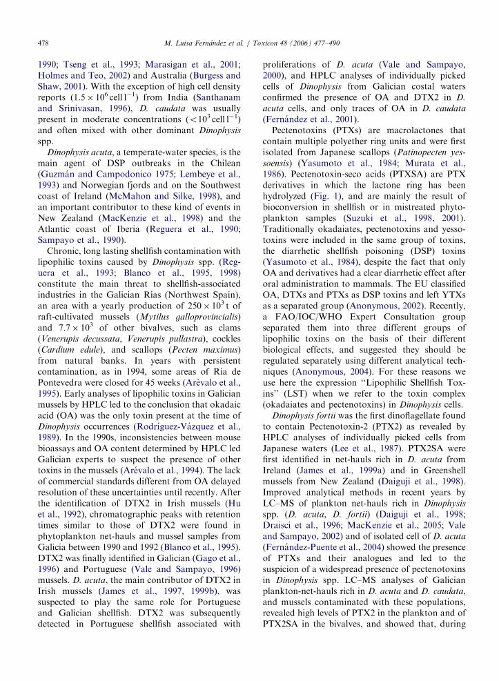
ARTICLE IN PRESSM. Luisa Fernandez et al. / Toxicon 48 (2006) 477–490478
1990; Tseng et al., 1993; Marasigan et al., 2001;Holmes and Teo, 2002) and Australia (Burgess andShaw, 2001). With the exception of high cell densityreports (1.5� 106 cell l�1) from India (Santhanamand Srinivasan, 1996), D. caudata was usuallypresent in moderate concentrations (o103 cell l�1)and often mixed with other dominant Dinophysis
spp.Dinophysis acuta, a temperate-water species, is the
main agent of DSP outbreaks in the Chilean(Guzman and Campodonico 1975; Lembeye et al.,1993) and Norwegian fjords and on the Southwestcoast of Ireland (McMahon and Silke, 1998), andan important contributor to these kind of events inNew Zealand (MacKenzie et al., 1998) and theAtlantic coast of Iberia (Reguera et al., 1990;Sampayo et al., 1990).
Chronic, long lasting shellfish contamination withlipophilic toxins caused by Dinophysis spp. (Reg-uera et al., 1993; Blanco et al., 1995, 1998)constitute the main threat to shellfish-associatedindustries in the Galician Rıas (Northwest Spain),an area with a yearly production of 250� 103 t ofraft-cultivated mussels (Mytilus galloprovincialis)and 7.7� 103 of other bivalves, such as clams(Venerupis decussata, Venerupis pullastra), cockles(Cardium edule), and scallops (Pecten maximus)from natural banks. In years with persistentcontamination, as in 1994, some areas of Rıa dePontevedra were closed for 45 weeks (Arevalo et al.,1995). Early analyses of lipophilic toxins in Galicianmussels by HPLC led to the conclusion that okadaicacid (OA) was the only toxin present at the time ofDinophysis occurrences (Rodrıguez-Vazquez et al.,1989). In the 1990s, inconsistencies between mousebioassays and OA content determined by HPLC ledGalician experts to suspect the presence of othertoxins in the mussels (Arevalo et al., 1994). The lackof commercial standards different from OA delayedresolution of these uncertainties until recently. Afterthe identification of DTX2 in Irish mussels (Huet al., 1992), chromatographic peaks with retentiontimes similar to those of DTX2 were found inphytoplankton net-hauls and mussel samples fromGalicia between 1990 and 1992 (Blanco et al., 1995).DTX2 was finally identified in Galician (Gago et al.,1996) and Portuguese (Vale and Sampayo, 1996)mussels. D. acuta, the main contributor of DTX2 inIrish mussels (James et al., 1997, 1999b), wassuspected to play the same role for Portugueseand Galician shellfish. DTX2 was subsequentlydetected in Portuguese shellfish associated with
proliferations of D. acuta (Vale and Sampayo,2000), and HPLC analyses of individually pickedcells of Dinophysis from Galician costal watersconfirmed the presence of OA and DTX2 in D.
acuta cells, and only traces of OA in D. caudata
(Fernandez et al., 2001).Pectenotoxins (PTXs) are macrolactones that
contain multiple polyether ring units and were firstisolated from Japanese scallops (Patinopecten yes-
soensis) (Yasumoto et al., 1984; Murata et al.,1986). Pectenotoxin-seco acids (PTXSA) are PTXderivatives in which the lactone ring has beenhydrolyzed (Fig. 1), and are mainly the result ofbioconversion in shellfish or in mistreated phyto-plankton samples (Suzuki et al., 1998, 2001).Traditionally okadaiates, pectenotoxins and yesso-toxins were included in the same group of toxins,the diarrhetic shellfish poisoning (DSP) toxins(Yasumoto et al., 1984), despite the fact that onlyOA and derivatives had a clear diarrhetic effect afteroral administration to mammals. The EU classifiedOA, DTXs and PTXs as DSP toxins and left YTXsas a separated group (Anonymous, 2002). Recently,a FAO/IOC/WHO Expert Consultation groupseparated them into three different groups oflipophilic toxins on the basis of their differentbiological effects, and suggested they should beregulated separately using different analytical tech-niques (Anonymous, 2004). For these reasons weuse here the expression ‘‘Lipophilic Shellfish Tox-ins’’ (LST) when we refer to the toxin complex(okadaiates and pectenotoxins) in Dinophysis cells.
Dinophysis fortii was the first dinoflagellate foundto contain Pectenotoxin-2 (PTX2) as revealed byHPLC analyses of individually picked cells fromJapanese waters (Lee et al., 1987). PTX2SA werefirst identified in net-hauls rich in D. acuta fromIreland (James et al., 1999a) and in Greenshellmussels from New Zealand (Daiguji et al., 1998).Improved analytical methods in recent years byLC–MS of plankton net-hauls rich in Dinophysis
spp. (D. acuta, D. fortii) (Daiguji et al., 1998;Draisci et al., 1996; MacKenzie et al., 2005; Valeand Sampayo, 2002) and of isolated cell of D. acuta
(Fernandez-Puente et al., 2004) showed the presenceof PTXs and their analogues and led to thesuspicion of a widespread presence of pectenotoxinsin Dinophysis spp. LC–MS analyses of Galicianplankton-net-hauls rich in D. acuta and D. caudata,and mussels contaminated with these populations,revealed high levels of PTX2 in the plankton and ofPTX2SA in the bivalves, and showed that, during
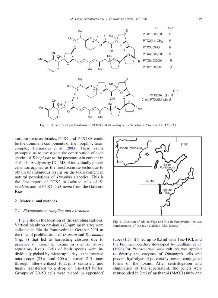
ARTICLE IN PRESS
O
O
Me
OH
O
Me
O
OO
O
Me
OHOH Me
MeMe
O
O
O
O
R
O
O
Me
OH
O
Me
O
OO
O
Me
OHOH Me
MeMe
O
HO
O
O
Me
OH
C-771
7-epi-PTX2SA (3): SPTX2SA (2): R
PTX1: CH2OH R
PTX2(1): CH3 R
PTX3: CHO R
PTX4: CH2OH S
PTX6: COOH R
PTX7: COOH S
7
R C-7
1
Fig. 1. Structures of pectenotoxin 2 (PTX2) and its analogue, pectenotoxin 2 seco acid (PTX2SA).
Ría de Vigo
Ría de Pontevedra
8°45´
42°15´
Iberian Peninsula
Fig. 2. Location of Rıa de Vigo and Rıa de Pontevedra, the two
southernmost of the four Galician Rıas Baixas.
M. Luisa Fernandez et al. / Toxicon 48 (2006) 477–490 479
autumn toxic outbreaks, PTX2 and PTX2SA couldbe the dominant components of the lipophilic toxincomplex (Fernandez et al., 2003). These resultsprompted us to investigate the contribution of eachspecies of Dinophysis to the pectenotoxin content inshellfish. Analyses by LC–MS of individually pickedcells was applied as the most accurate technique toobtain unambiguous results on the toxin content innatural populations of Dinophysis species. This isthe first report of PTX2 in isolated cells of D.
caudata, and of PTX2 in D. acuta from the GalicianRıas.
2. Material and methods
2.1. Phytoplankton sampling and extraction
Fig. 2 shows the location of the sampling stations.Vertical plankton net-hauls (20-mm mesh size) werecollected in Rıa de Pontevedra in October 2001 atthe time of proliferations of D. acuta and D. caudata
(Fig. 3) that led to harvesting closures due topresence of lipophilic toxins in shellfish aboveregulatory levels. Cells of both species were in-dividually picked by microcapillarity at the invertedmicroscope (25� and 100� ), rinsed 2–3 timesthrough filter-sterilized (0.22 mm) seawater, andfinally transferred to a drop of Tris–HCl buffer.Groups of 20–50 cells were placed in eppendorf
tubes (1.5ml) filled up to 0.5ml with Tris–HCl, andthe boiling procedure developed by Quilliam et al.(1996) for Prorocentrum lima cultures was appliedto destroy the enzymes of Dinophysis cells andprevent hydrolysis of potentially present conjugatedforms of the toxins. After centrifugation andelimination of the supernatant, the pellets wereresuspended in 2ml of methanol (MeOH) 80% and
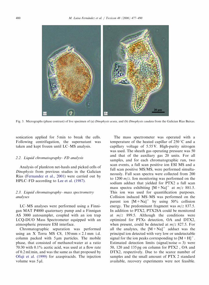
ARTICLE IN PRESS
20 µm(b)(a)
Fig. 3. Micrographs (phase contrast) of live specimen of (a) Dinophysis acuta, and (b) Dinophysis caudata from the Galician Rıas Baixas.
M. Luisa Fernandez et al. / Toxicon 48 (2006) 477–490480
sonication applied for 5min to break the cells.Following centrifugation, the supernatant wastaken and kept frozen until LC–MS analysis.
2.2. Liquid chromatography– FD analysis
Analysis of plankton net-hauls and picked cells ofDinophysis from previous studies in the GalicianRias (Fernandez et al., 2001) were carried out byHPLC–FD according to Lee et al. (1987).
2.3. Liquid chromatography– mass spectrometry
analyses
LC–MS analyses were performed using a Finni-gan MAT P4000 quaternary pump and a FinniganAS 3000 autosampler, coupled with an ion trapLCQ-DUO Mass Spectrometer equipped with anatmospheric pressure ESI interface.
Chromatographic separation was performedusing an X Terra MS C8, 150mm� 2.1mm i.d.column packed with 5 mm particles. The mobilephase, that consisted of methanol:water at a ratio70:30 with 0.1% acetic acid, was used at a flow rateof 0.2ml/min, and was the same as that proposed byOfuji et al. (1999) for azaspiracids. The injectionvolume was 5 ml.
The mass spectrometer was operated with atemperature of the heated capillar of 250 1C and acapillary voltage of 5.55V. High-purity nitrogenwas used. The sheath gas operating pressure was 50and that of the auxiliary gas 20 units. For allsamples, and for each chromatographic run, twoscan events, a full scan positive ion ESI MS and afull scan positive MS/MS, were performed simulta-neously. Full scan spectra were collected from 200to 1200 m/z. Ion monitoring was performed on thesodium adduct that yielded for PTX2 a full scanmass spectra exhibiting [M+Na]+ at m/z 881.5.This ion was used for quantification purposes.Collision induced MS–MS was performed on theparent ion [M+Na]+ by using 50% collisionenergy. The predominant fragment was m/z 837.5.In addition to PTX2, PTX2SA could be monitoredat m/z 899.5. Although the conditions wereoptimized for PTXs detection, OA and DTX2,when present, could be detected at m/z 827.5. Forall the analytes, the [M+Na]+ adduct was theprincipal ion detected with very low or undetectablesignal for the ion peaks corresponding to [M+H]+.Estimated detection limits (signal/noise ¼ 3) were50, 120 and 135 pg on column for PTX2 , OA andDTX2, respectively. Due to the scarce number ofsamples and the small amount of PTX 2 standardavailable, recovery experiments were not feasible.
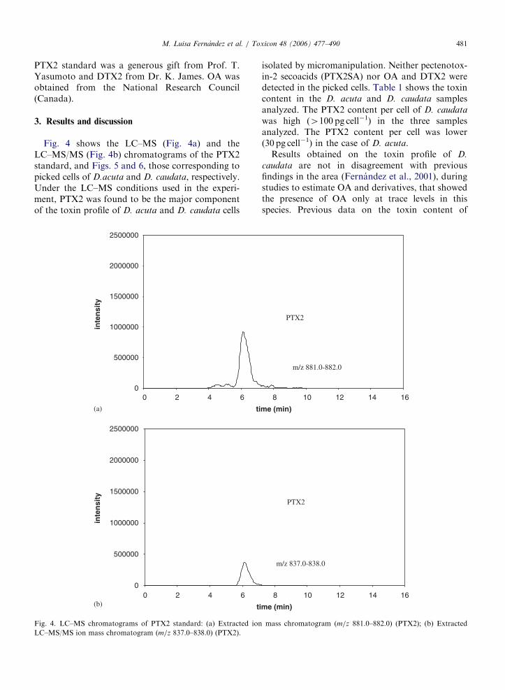
ARTICLE IN PRESSM. Luisa Fernandez et al. / Toxicon 48 (2006) 477–490 481
PTX2 standard was a generous gift from Prof. T.Yasumoto and DTX2 from Dr. K. James. OA wasobtained from the National Research Council(Canada).
3. Results and discussion
Fig. 4 shows the LC–MS (Fig. 4a) and theLC–MS/MS (Fig. 4b) chromatograms of the PTX2standard, and Figs. 5 and 6, those corresponding topicked cells of D.acuta and D. caudata, respectively.Under the LC–MS conditions used in the experi-ment, PTX2 was found to be the major componentof the toxin profile of D. acuta and D. caudata cells
0
500000
1000000
1500000
2000000
2500000
0 2 4 6
ti
0 2 4 6
ti
inte
nsi
ty
0
500000
1000000
1500000
2000000
2500000
inte
nsi
ty
(a)
(b)
Fig. 4. LC–MS chromatograms of PTX2 standard: (a) Extracted ion
LC–MS/MS ion mass chromatogram (m/z 837.0–838.0) (PTX2).
isolated by micromanipulation. Neither pectenotox-in-2 secoacids (PTX2SA) nor OA and DTX2 weredetected in the picked cells. Table 1 shows the toxincontent in the D. acuta and D. caudata samplesanalyzed. The PTX2 content per cell of D. caudata
was high (4100 pg cell�1) in the three samplesanalyzed. The PTX2 content per cell was lower(30 pg cell�1) in the case of D. acuta.
Results obtained on the toxin profile of D.
caudata are not in disagreement with previousfindings in the area (Fernandez et al., 2001), duringstudies to estimate OA and derivatives, that showedthe presence of OA only at trace levels in thisspecies. Previous data on the toxin content of
8 10 12 14 16
me (min)
8 10 12 14 16
me (min)
m/z 837.0-838.0
m/z 881.0-882.0
PTX2
PTX2
mass chromatogram (m/z 881.0–882.0) (PTX2); (b) Extracted
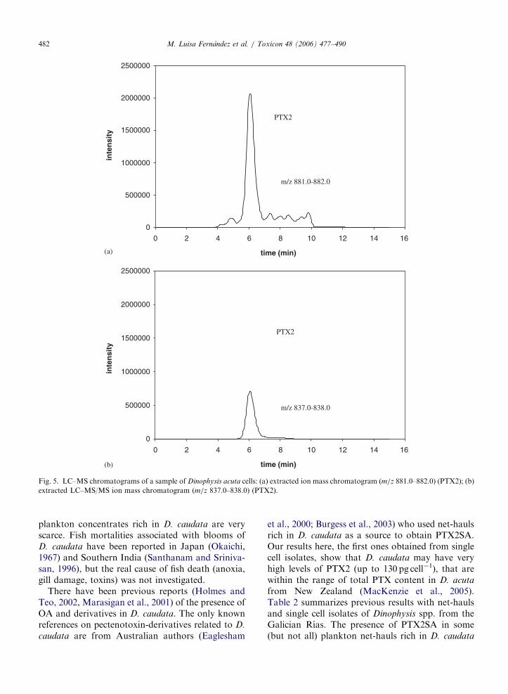
ARTICLE IN PRESS
0
500000
1000000
1500000
2000000
2500000
0 2 4 6 8 10 12 14 16
time (min)
inte
nsi
ty
0
500000
1000000
1500000
2000000
2500000
inte
nsi
ty
m/z 837.0-838.0
m/z 881.0-882.0
PTX2
PTX2
0 2 4 6 8 10 12 14 16
time (min)
(a)
(b)
Fig. 5. LC–MS chromatograms of a sample of Dinophysis acuta cells: (a) extracted ion mass chromatogram (m/z 881.0–882.0) (PTX2); (b)
extracted LC–MS/MS ion mass chromatogram (m/z 837.0–838.0) (PTX2).
M. Luisa Fernandez et al. / Toxicon 48 (2006) 477–490482
plankton concentrates rich in D. caudata are veryscarce. Fish mortalities associated with blooms ofD. caudata have been reported in Japan (Okaichi,1967) and Southern India (Santhanam and Sriniva-san, 1996), but the real cause of fish death (anoxia,gill damage, toxins) was not investigated.
There have been previous reports (Holmes andTeo, 2002, Marasigan et al., 2001) of the presence ofOA and derivatives in D. caudata. The only knownreferences on pectenotoxin-derivatives related to D.
caudata are from Australian authors (Eaglesham
et al., 2000; Burgess et al., 2003) who used net-haulsrich in D. caudata as a source to obtain PTX2SA.Our results here, the first ones obtained from singlecell isolates, show that D. caudata may have veryhigh levels of PTX2 (up to 130 pg cell�1), that arewithin the range of total PTX content in D. acuta
from New Zealand (MacKenzie et al., 2005).Table 2 summarizes previous results with net-haulsand single cell isolates of Dinophysis spp. from theGalician Rıas. The presence of PTX2SA in some(but not all) plankton net-hauls rich in D. caudata
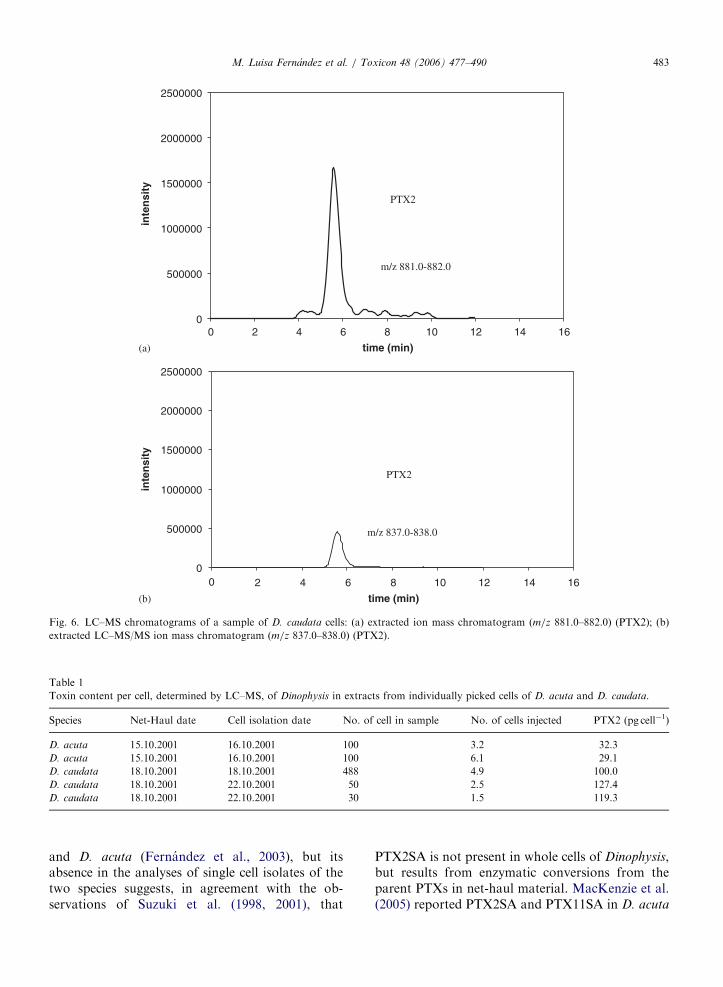
ARTICLE IN PRESS
0
500000
1000000
1500000
2000000
2500000
0 8 10 12 14 16
time (min)
inte
nsi
ty
0
500000
1000000
1500000
2000000
2500000
inte
nsi
ty
PTX2
m/z 837.0-838.0
PTX2
m/z 881.0-882.0
2 4 6
0 8 10 12 14 16
time (min)
2 4 6
(a)
(b)
Fig. 6. LC–MS chromatograms of a sample of D. caudata cells: (a) extracted ion mass chromatogram (m/z 881.0–882.0) (PTX2); (b)
extracted LC–MS/MS ion mass chromatogram (m/z 837.0–838.0) (PTX2).
Table 1
Toxin content per cell, determined by LC–MS, of Dinophysis in extracts from individually picked cells of D. acuta and D. caudata.
Species Net-Haul date Cell isolation date No. of cell in sample No. of cells injected PTX2 (pg cell�1)
D. acuta 15.10.2001 16.10.2001 100 3.2 32.3
D. acuta 15.10.2001 16.10.2001 100 6.1 29.1
D. caudata 18.10.2001 18.10.2001 488 4.9 100.0
D. caudata 18.10.2001 22.10.2001 50 2.5 127.4
D. caudata 18.10.2001 22.10.2001 30 1.5 119.3
M. Luisa Fernandez et al. / Toxicon 48 (2006) 477–490 483
and D. acuta (Fernandez et al., 2003), but itsabsence in the analyses of single cell isolates of thetwo species suggests, in agreement with the ob-servations of Suzuki et al. (1998, 2001), that
PTX2SA is not present in whole cells of Dinophysis,but results from enzymatic conversions from theparent PTXs in net-haul material. MacKenzie et al.(2005) reported PTX2SA and PTX11SA in D. acuta

ARTICLE IN PRESS
Table 2
Summary of previous results on toxin content determined by either HPLC–FD (Fernandez et al., 2001) or LC/MS (Fernandez et al., 2003)
in phytoplankton net-hauls rich in D. acuta and D. caudata and in picked cells from the Galician Rıas of Vigo and Pontevedra.
Species (cells) Date location PTX2
(pg cell�1)
OA
(pg cell�1)
DTX2 (pg cell�1) PTX2SA/PTX2
(area ratio)
PTX2SA/OA
(area ratio)
D. acuminata (75044) 08/1990a ne 1.6 1.1
D. acuta (507934) Pontevedra
D. caudata (5300)
D. acuta (26928) 10/1990a ne 2.4 1.6
D. caudata (528) Pontevedra
D. acuta (45425) ne 0.4 0.25
D. caudata (7820) 10/1990a
D. rotundata (115) Pontevedra
D. acuta 10/1997a ne 6.3 4.4
(picked cells) Vigo 33.1 22.0
18.4 5.7
D. caudata 10/1997a ne 0.73 nd
(picked cells) Vigo
D. acuminata (263750) 06/1991b nd 9.2 nd
Dinophysis spp.(8250) Vigo
D. acuminata (112423) 0.24 1.0 0.24 5.97 1.32
D. acuta (24125) 10/1994b
D. caudata (22678) Vigo
Dinophysis spp (29433)
D. acuminata (1338) 2.9 8.3 7.9 0.27 0.09
D. acuta (28809) 10.1997b
D. caudata (2328) Vigo
D. fortii (58)
D. acuminata (1300) 1.54 3.1 nd
D. acuta (550) 10/1997b
D. caudata (1450) Pontevedra
Dinophysis spp. (900)
D. acuminata (21695) 0.90 nd nd
D. acuta (348637) 10/2001b
D. caudata (245879) Pontevedra
D. fortii (3699)
D. acuminata (16313) 0.40 nd nd
D. acuta (202489) 10/2001b
D. caudata (159266) Pontevedra
D. fortii (2018)
D. acuminata (5549) 2.50 nd nd
D. acuta (124453) 10/2001b
D. caudata (96030) Pontevedra
D. fortii (1009)
ne ¼ not explored; nd ¼ not detected.aHPLC–FD.bLC–MS.
M. Luisa Fernandez et al. / Toxicon 48 (2006) 477–490484
concentrates that had much higher levels of PTX2and PTX11 than the Galician specimens. Cells fromnet-hauls may suffer rupture from handling, and
enzymatic conversions may take place very quicklyif the samples are not treated for enzymaticinhibition, as was the case in the New Zealand
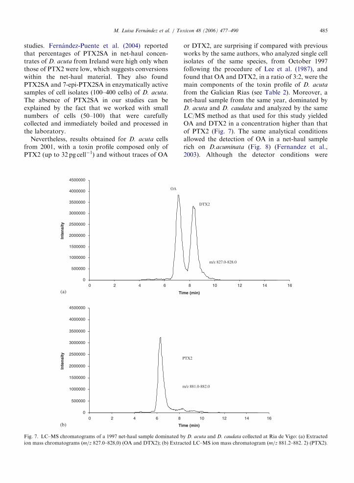
ARTICLE IN PRESSM. Luisa Fernandez et al. / Toxicon 48 (2006) 477–490 485
studies. Fernandez-Puente et al. (2004) reportedthat percentages of PTX2SA in net-haul concen-trates of D. acuta from Ireland were high only whenthose of PTX2 were low, which suggests conversionswithin the net-haul material. They also foundPTX2SA and 7-epi-PTX2SA in enzymatically activesamples of cell isolates (100–400 cells) of D. acuta.The absence of PTX2SA in our studies can beexplained by the fact that we worked with smallnumbers of cells (50–100) that were carefullycollected and immediately boiled and processed inthe laboratory.
Nevertheless, results obtained for D. acuta cellsfrom 2001, with a toxin profile composed only ofPTX2 (up to 32 pg cell�1) and without traces of OA
0
500000
1000000
1500000
2000000
2500000
3000000
3500000
4000000
4500000
0 2 4 6
Ti
0 2 4 6 8
Ti
0
500000
1000000
1500000
2000000
2500000
3000000
3500000
4000000
4500000
Inte
nsi
tyIn
ten
sity
OA
(a)
(b)
Fig. 7. LC–MS chromatograms of a 1997 net-haul sample dominated b
ion mass chromatograms (m/z 827.0–828,0) (OA and DTX2); (b) Extra
or DTX2, are surprising if compared with previousworks by the same authors, who analyzed single cellisolates of the same species, from October 1997following the procedure of Lee et al. (1987), andfound that OA and DTX2, in a ratio of 3:2, were themain components of the toxin profile of D. acuta
from the Galician Rias (see Table 2). Moreover, anet-haul sample from the same year, dominated byD. acuta and D. caudata and analyzed by the sameLC/MS method as that used for this study yieldedOA and DTX2 in a concentration higher than thatof PTX2 (Fig. 7). The same analytical conditionsallowed the detection of OA in a net-haul samplerich on D.acuminata (Fig. 8) (Fernandez et al.,2003). Although the detector conditions were
8 10 12 14 16
me (min)
10 12 14 16
me (min)
m/z 827.0-828.0
DTX2
PTX2
m/z 881.0-882.0
y D. acuta and D. caudata collected at Ria de Vigo: (a) Extracted
cted LC–MS ion mass chromatogram (m/z 881.2–882. 2) (PTX2).
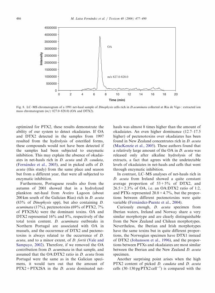
ARTICLE IN PRESS
0
500000
1000000
1500000
2000000
2500000
3000000
3500000
4000000
4500000
0 2 4 6 8 10 12 14 16 18 20
Time (min)
Inte
nsi
ty
OA
m/z 827.0-828.0
Fig. 8. LC–MS chromatogram of a 1991 net-haul sample of Dinophysis cells rich in D.acuminata collected at Ria de Vigo : extracted ion
mass chromatogram (m/z 827.0–828.0) (OA and DTX2).
M. Luisa Fernandez et al. / Toxicon 48 (2006) 477–490486
optimized for PTX2, these results demonstrate theability of our system to detect okadaiates. If OAand DTX2 detected in the samples from 1997resulted from the hydrolysis of esterified forms,these compounds would not have been detected ifthe samples had been subjected to enzymaticinhibition. This may explain the absence of okadai-ates in net-hauls rich in D. acuta and D. caudata,(Fernandez et al., 2003), and in picked cells of D.
acuta (this study) from the same place and seasonbut from a different year, that were all subjected toenzymatic inhibition.
Furthermore, Portuguese results also from theautumn of 2001 showed that in a hydrolyzedplankton net-haul from Aveiro Lagoon (about200 km south of the Galician Rıas) rich in D. acuta
(83% of Dinophysis spp), but also containing D.
acuminata (17%), pectenotoxins (69% of PTX2, 7%of PTX2SA) were the dominant toxins. OA andDTX2 represented 16% and 8%, respectively of thetotal toxin content. D. acuminata outbreaks inNorthern Portugal are associated with OA inmussels, and the occurrence of DTX2 and pecteno-toxins is always related to the occurrence of D.
acuta, and to a minor extent, of D. fortii (Vale andSampayo, 2002). Therefore, if we removed the OAcontribution from D. acuminata in that sample, andassumed that the OA:DTX2 ratio in D. acuta fromPortugal were the same as in the Galician speci-mens, it would turn out that the amount ofPTX2+PTX2SA in the D. acuta dominated net-
hauls was almost 8 times higher than the amount ofokadaiates. An even higher dominance (12.7–17.5higher) of pectenotoxins over okadaiates has beenfound in New Zealand concentrates rich in D. acuta
(MacKenzie et al., 2005). These authors found thata relatively large amount of the OA in D. acuta wasreleased only after alkaline hydrolysis of theextracts, a fact that agrees with the undetectablelevels of okadaiates in net-hauls and cells that wentthrough enzymatic inhibition.
In contrast, LC–MS analyses of net-hauls rich inD. acuta from Ireland showed a quite constantaverage proportion of 53+5% of DTX2, and26.5+2.3% of OA, i.e. an OA:DTX2 ratio of 1:2,and PTXs represented 20.8+4.7%, but the propor-tions between different pectenotoxins were quitevariable (Fernandez-Puente et al., 2004).
Curiously enough, D. acuta specimen fromIberian waters, Ireland and Norway share a verysimilar morphotype and are clearly distinguishablefrom the New Zealand and Chilean morphotypes.Nevertheless, the Iberian and Irish morphotypeshave the same toxins but in quite different propor-tions, the Norwegian specimen have DTX1 insteadof DTX2 (Johansson et al., 1996), and the propor-tions between PTXs and okadaiates are most similarbetween the Iberian and the New Zealand D. acuta
specimen.Another surprising point arises when the high
PTX2 content of picked D. caudata and D. acuta
cells (30–130 pg PTX2 cell�1) is compared with the

ARTICLE IN PRESSM. Luisa Fernandez et al. / Toxicon 48 (2006) 477–490 487
average content of toxin per cell of Dinophysis
(2.5 pg PTX2 cell�1) estimated from analyses of net-haul samples during the same season in October2001. This decrease in the signal may be explainedby matrix effects on the LC–MS response due toinfluences of coextractives on the electrosprayionization of the analytes. Large differences in toxincontent per cell between picked cells and averageestimates per cell of Dinophysis in net-haul sampleshave been already reported by the same authors(Blanco et al. 1995, Fernandez et al. 2001).
In any case, and considering that extra-cellularenzymatic activity can induce considerable changesin toxin profiles, extreme care should be takenduring dinoflagellate cell handling to obtain accu-rate information on their toxin composition. In thelight of current knowledge, previous results on toxinprofiles of Dinophysis spp. should be reviewed andaccepted with caution. Recovery experimentsshould be undertaken, when analyses with thesekind of matrices are performed, to reduce uncer-tainties in the data.
Our results show that PTX2 can be a majorcomponent of the lipophilic-toxin-complex-profilein Galician phytoplankton populations in theautumn when D. acuta and D. caudata are thedominant Dinophysis spp. and diatoms are practi-cally absent. But in mussels exposed to theseautumn populations, PTX2SA was found to bethe dominant pectenotoxin, and PTX2 levels wereconsiderably lower than those of their seco-acidderivatives (Fernandez et al., 2003). Similar resultswere found in Portuguese shellfish exposed to thesame species of Dinophysis (Vale and Sampayo,2002). This is in agreement with previous reportsthat support the conclusion that PTX2 is convertedto PTX2SA after enzymatic oxidation in shellfishtissues (Suzuki et al., 2001). Therefore, PTX2 can bea major component of the lipophilic-toxin-complex-profile in phytoplankton and PTX2SA the principalPTX of the toxin complex in shellfish of theGalician Rıas exposed to autumn proliferations ofD. acuta and D. caudata. The production of thesetoxins by other Dinophysis spp. from the region,such as D. tripos and D. rotundata, present in lowerconcentrations, or even by D. acuminata, despite thefact that PTXs were not found in the few samples sofar analyzed, that contained only this species ofDinophysis, can not be discarded.
These results have important implications forphycotoxin monitoring programmes. Decision2002/225/EC (Anonymous, 2002) regulates a max-
imum level for OA derivatives and pectenotoxinstogether of 160 mg eq.OAkg�1 edible part, andestablishes that if chemical methods are used,PTX1 and PTX2 should be monitored. PTX2SA isnot mentioned in the European Directive, thatstates that ‘‘if new toxin analogues of public healthsignificance are discovered, they should be includedin the regulation analyses’’ (Anonymous,2002).Human toxicity data on PTX2SA is still scarceand uncertain. High levels of PTX2SA found inAustralian ‘‘pipis’’ (Donax deltoides) in 1997 weresuspected to be the cause of a severe human DSPintoxication (Burguess and Shaw, 2001), but laterstudies showed that the samples that caused thistoxic outbreak were contaminated with esters ofokadaic acid, and the latter were considered to beresponsible for the gastrointestinal damage ob-served in mice (Burguess and Shaw, 2003).
Recent studies by Miles et al. (2004) confirmedthe toxicity of PTX2 to mice, given by intraper-itoneal injection, but previously reported toxicityafter oral administration (Ogino et al., 1997) wasnot confirmed. The same authors suggested thattheir results possibly reflected a poor absorption ofPTX2 in the gastrointestinal tract, or its conversioninto a less toxic compound, such as PTX2SA; in theguts. PTX2SA was of low toxicity to mice, bothorally and intraperitoneally, and neither PTX2 norits secoacid derivatives had diarrhetic effects. Theseresults led Miles et al. (2004) to conclude that thesevere diarrhea in mice attributed to PTX2 by Ishigeet al. (1998) was a result of contamination of thesamples with okadaic acid derivatives. Uncertaintiesof this kind, on the toxic effects of differentlipophilic toxins in mammals, may have been morefrequent than reported, as now we know pecteno-toxins and okadaic acid derivatives have a commonsource, i.e. dinoflagellate cells of the genus Dino-
physis, and are likely to co-occur in shellfish exposedto blooms of these microalgae.
In relation with control methods, if mousebioassay based on intraperitoneal injection is thestandard procedure in use for regulation of lipo-philic toxins, injection of shellfish extract gives anon-specific result for the combination of okadai-ates and pectenotoxins, and PTX2SA is not detected(Daiguji et al., 1998). Considering that regulationson pectenotoxins may be modified in the future,when more toxicological data become available, itwould be desirable to complement the mousebioassay methods or even substitute them, ifpossible, to distinguish in a reliable manner the
ARTICLE IN PRESSM. Luisa Fernandez et al. / Toxicon 48 (2006) 477–490488
different groups of lipophilic toxins that have quitedifferent effects on human health.
Finally, results presented here show once morethat toxin content of the same dinoflagellate speciescan vary considerably even within specimenscollected in the same locality. Authors from NewZealand (MacKenzie et al., 2005) and Ireland(Fernandez-Puente et al., 2004) claimed that toxinprofiles of the same species in the same locality werevery stable, and that toxin content varied withinnarrow limits. But in our opinion, these conclusionshave been reached on the basis of very limitednumbers of samples, most of them collected at thetime of the species annual peak in cell density. It isnecessary to carry out more systematic studies onthe toxin profiles and toxin contents of each speciesof Dinophysis throughout the whole growth seasonof the species. This task, that is one of our presentobjectives, is perfectly attainable with the latesttechnological advances that allow analyses of verysmall samples (composed of a few cells), by liquidchromatography coupled to mass spectrometry.
Acknowledgements
We thank Patricia Loures for technical assistance,the crew of R.V. ‘‘Jose M. Navaz’’ for continuoushelp in collecting samples and the Instituto Tecno-logico del Mar (INTECMAR, Xunta de Galicia) forweekly information on phytoplankton monitoringdata. This research was funded by Spanish NationalProjects MAR98-0978 and REN2002-02486.
References
Anonymous, 2002. Commission Decision 2002/225/EC from 15
March 2002, laying down detailed regulations for the
application of Council Directive 91/492/EEC. Off. J. Eur.
Communities L 075, 62–64.
Anonymous, 2004. Report of the Joint FAO/IOC/WHO ad hoc
Expert Consultation on Biotoxins in Bivalve Molluscs, Oslo,
Norway, September 26–30, 2004, 31pp.
Arevalo, F., Valcarcel, J.A., Domonte, M., Vazquez, M.J., 1994.
Estudio comparativo de los resultados obtenidos por bioen-
sayo y HPLC en la determinacion de biotoxinas DSP. Datos
preliminares. In: Marino, J., Maneiro, J. (Eds.), Actas de la
III Reunion Iberica sobre Fitoplancton Toxico y Biotoxinas,
Ediciones de la Consellerıa de Pesca Marisqueo e Acuicultura.
Xunta de Galicia, pp. 89–93.
Arevalo, F., Bermudez de la Puente, M., Domonte, M., Perez,
M., Salgado, C., Vazquez, M.J., Valcarcel, J.A., 1995.
Biotoxinas PSP y DSP en las Rıas Gallegas: Control y
evolucion durante los anos 1993 y 1994. In: Matamoros, E.,
Delgado, M. (Eds.), IV Reunion Iberica sobre Fitoplancton
Toxico y Biotoxinas. Actas de la Reunion. Generalitat de
Catalunya, Barcelona, pp. 39–51.
Blanco, J., Fernandez, M.L., Marino, J., Reguera, B., Mıguez,
A., Maneiro, J., Cacho, E., Martınez, A., 1995. From
Dinophysis spp toxicity to DSP outbreaks: a preliminary
model of toxin accumulation in mussels. In: Lassus, P., Arzul,
G., Erard-Le Denn, E., Gentien, P., Marcaillou-Le Baut, C.
(Eds.), Harmful Marine Algal Blooms. Lavoisier, Parıs,
pp. 777–782.
Blanco, J., Morono, A., Pazos, Y., Maneiro, J., Marino, J., 1998.
Trends and variations of the abundance of main PSP and
DSP producing species in the Galician Rıas: environmental
and biological influences. In: Reguera, B., Blanco, J.,
Fernandez, M.L., Wyatt, T. (Eds.), Harmful Algae. Xunta
de Galicia and Intergovernmental Oceanographic Commis-
sion of UNESCO, Santiago de Compostela, New York,
pp. 204–207.
Burgess, V., Shaw, G., 2001. Pectenotoxins—an issue for public
health. A review of their comparative toxicology and
metabolism. Environ. Int. 27, 275–283.
Burgess, V., Shaw, G., 2003. Investigations into the toxicology of
pectenotoxin-2-seco acid and 7-epi pectenotoxin 2-secoa cid
to aid in a health risk assessment for the consumption of
shellfish contaminated with these shellfish toxins in Australia.
Report of Project No. 2001/258. National Research Centre
for Environmental Toxicology, Archerfield, Queensland,
Australia, 50pp.
Daiguji, M., Satake, M., James, K.J., Bishop, A., MacKenzie, L.,
Naoki, H., Yasumoto, T., 1998. Structures of new pecteno-
toxin analogs, pectenotoxin-2 seco acid and 7-epi-pectenotox-
in-2 seco acid, isolated from a dinoflagellate and greenshell
mussels. Chem. Lett. 7, 653–654.
Draisci, R., Lucentini, L., Giannetti, L., Boria, P., Poletti, R.,
1996. First report of pectenotoxin-2 (PTX-2) in algae
(Dinophysis fortii) related to seafood poisoning in Europe.
Toxicon 34, 923–935.
Eaglesham, G.K., Brett, S.J., Davis, B.C., Holling, N., 2000.
Detection of Pectenotoxins-2 and Pectenotoxins-2 Seco Acid
in phytoplankton and shellfish from the Ballina region of New
South Wales, Australia. In: Abstracts from the X Interna-
tional IUPAC symposium on Mycotoxins and Phycotoxins,
Brazil, May 2000, p. 40.
Fernandez, M.L., Reguera, B., Ramilo, I., Martınez, A., 2001.
Toxin content of Dinophysis acuminata, D. acuta and D.
caudata from the Galician Rıas Bajas. In: Hallegraeff, G.M.,
Blackburn, S.I., Bolch, C.J., Lewis, R.J. (Eds.), Harmful
Algal Blooms. Intergovernmental Oceanographic Commis-
sion of UNESCO, Paris, pp. 360–363.
Fernandez, M.L., Miguez, A., Morono, A., Arevalo, F., Pazos,
Y., Salgado, C., Correa, J., Blanco, J., Gonzalez-Gil, S.,
Reguera, B., 2003. First report of pectenotoxin-2 in phyto-
plankton net-hauls and mussels from the Galician Rıas Baixas
during proliferations of Dinophysis acuta and D. caudata. In:
Villalba, A., Reguera, B., Romalde, J., Beiras, R. (Eds.),
Molluscan Shellfish Safety. Consellerıa de Pesca e Asuntos
Marıtimos da Xunta de Galicia and Intergovernmental
Oceanographic Commission of UNESCO. Santiago de
Compostela, Spain, pp. 75–83.
Fernandez-Puente, P., Fidalgo-Saez, M.J., Hamilton, B., Furey,
A., James, K.J., 2004. Studies of polyether toxins in the
marine phytoplankton, Dinophysis acuta, in Ireland using
multiple tandem mass spectrometry. Toxicon 44, 919–926.
ARTICLE IN PRESSM. Luisa Fernandez et al. / Toxicon 48 (2006) 477–490 489
Flores, M., Franco, J.M., Lluch-Cota, S.E., Lluch-Cota, D.B.,
Cortes-Altamirano, R., Sierra-Beltran, A.P., 2002. Recent
species shifts on the HAB occurrences in Acapulco Bay,
Mexico. Abstracts of the Xth International Conference on
Harmful Algae, St Pete Beach, FL, p. 96.
Fukuyo, Y., Takano, H., Chihara, M., Matsuoka, K., 1990. Red
Tide Organisms in Japan—An Illustrated Taxonomic Guide.
Uchida Rokakuho, Tokyo, 430pp.
Gago, A., Rodrıguez-Vazquez, J.A., Thibault, P., Quilliam, M.,
1996. Simultaneous occurrence of diarrhetic and paralytic
shellfish poisoning toxins in Spanish mussels in 1993. Nat.
Toxins 4, 72–79.
Guzman, L., Campodonico, I., 1975. Marea roja en la Region de
Magallanes. Publ. Inst. Pat. Ser. Mon. 9, 44.
Holmes, M.J., Teo, S.L.M., 2002. Toxic marine dinoflagellates in
Singapore waters that cause seafood poisonings. Clin. Exp.
Pharmacol. Physiol. 29, 829–836.
Hu, T., Doyle, J., Jackson, D., Marr, J., Nixon, E., Pleasance, S.,
Quilliam, M.A., Walter, J.A., Wright, J.L.C., 1992. Isolation
of a new diarrhetic shellfish poison from Irish mussels. J.
Chem. Soc. Chem. Commun. 39–41.
James, K.J., Bishop, A.G., Gillman, M., Kelly, S.S., Roden, C.,
Draisci, R., Lucentini, L., Giannetti, L., Boria, P., 1997.
Liquid chromatography with fluorimetric, mass spectrometric
and tandem mass spectrometric detectionfor investigation of
the sea-food toxin producing phytoplankton Dinophysis
acuta. J. Chromatogr. A 777, 213–221.
James, K.J., Bishop, A.G., Draisci, R., Palleschi, L., Marchia-
fava, C., Ferretti, E., Satake, M., Yasumoto, T., 1999a.
Liquid chromatographic methods for the isolation and
identification of new pectenotoxin-2 analogues from marine
phytoplankton and shellfish. J. Chromatogr. A 844, 53–65.
James, K.J., Bishop, A.G., Healy, B.M., Roden, C., Sherlock,
I.R., Twohig, M., Draisci, R., Giannetti, L., Lucentini, L.,
1999b. Efficient isolation of the rare diarrhoeic shellfish toxin,
Dinophysistoxin-2, from marine phytoplankton. Toxicon 37
(2), 343–357.
Johansson, N., Graneli, E., Yasumoto, T., Carlsson, P., Legrand,
C., 1996. Toxin production by Dinophysis acuminata and D.
acuta cells grown under nutrient sufficient and deficient
conditions. In: Yasumoto, T., Oshima, Y., Fukuyo, Y. (Eds.),
Harmful and Toxic Algal Blooms. Intergovernmental Ocea-
nographic Commission of UNESCO. Sendai, Japan,
pp. 277–280.
Karunasagar, I., Segar, K., Karunasagar, I., 1989. Potentially
toxic dinoflagellates in shellfish harvesting areas along the
coast of Karnataka State (India). In: Okaichi, T., Anderson,
D.M., Nemoto, T. (Eds.), Red Tide: Biology, Environmental
Science, and Toxicology. Elsevier, New York, pp. 65–68.
Lee, J.-S., Yanagi, T., Kenma, R., Yasumoto, T., 1987.
Fluorometric determination of diarrhetic shellfish toxins by
high performance liquid chromatography. Agric. Biol. Chem.
51, 877–881.
Lembeye, G., Yasumoto, T., Zhao, J., Fernandez, R., 1993. DSP
outbreak in Chilean fiords. In: Smayda, T.J., Shimizu, Y.
(Eds.), Toxic Phytoplankton Blooms in the Sea. Elsevier,
Amsterdam, pp. 525–529.
MacKenzie, L., Truman, P., Satake, M., Yasumoto, T.,
Adamson, J., Mountfort, D., White, D., 1998. Dinoflagellate
blooms and associated DSP toxicity in shellfish in New
Zealand. In: Reguera, B., Blanco, J., Fernadez, M.L., Wyatt,
T. (Eds.), Harmful Algae. Xunta de Galicia and Intergovern-
mental Oceanographic Commission of UNESCO,, Santiago
de Compostela, pp. 74–77.
MacKenzie, L., Beuzenberg, V., Holland, P., McNabb, P.,
Suzuki, T., Selwood, A., 2005. Pectenotoxin and okadaic
acid-based toxin profiles in Dinophysis acuta and Dinophysis
acuminata from New Zealand. Harmful Algae 4, 75–85.
Marasigan, A.N., Sato, S., Fukuyo, Y., Kodama, M., 2001.
Accumulation of a high level of diarrhetic shellfish toxins in
the green mussel Perna viridis during a bloom of Dinophysis
caudata and Dinophysis miles in Sapian Bay, Panay Island, the
Philippines. Fish. Sci. 67, 994–996.
McMahon, T., Silke, J., 1998. Algal blooms and shellfish toxicity
in Irish Coastal Waters. In: Wilson, J.G. (Ed.), Eutrophica-
tion in Irish Coastal Waters. Royal Irish Academy, Dublin,
pp. 106–114.
Mendez, S., Ferrari, G., 2002. Floraciones algales nocivas en
Uruguay: Antecedentes, proyectos en curso y revision de
resultados. In: Sar, E.A., Ferrario, M.E., Reguera, B. (Eds.),
Floraciones algales nocivas en el Cono Sur Americano.
Instituto Espanol de Oceanografıa, Madrid, pp. 269–288.
Miles, C.O., Wilkins, A.L., Munday, R., Dines, M.H., Hawkes,
A.D., Briggs, L.R., Sandvik, M., Jensen, D.J., Cooney, J.M.,
Holland, P.T., Quilliam, M.A., MacKenzie, A.L., Beuzen-
berg, V., Towers, N.R., 2004. Isolation of pectenotoxin-2
from Dinophysis acuta and its conversion to pectenotoxin-2
seco acid, and preliminary assessment of their acute toxicities.
Toxicon 43 (1), 1–9.
Murata, M., Masaki, S., Iwashita, T., Naoki, H., Yasumoto, T.,
1986. The structure of pectenotoxin-3, a new constituent of
diarrhetic shellfish toxins. Agric. Biol. Chem. 50, 2693–2695.
Ofuji, K., Satake, M., Oshima, Y., McMahon, T., James, K.J.,
Yasumoto, T., 1999. A sensitive and specific determination
method for azaspiracids by Liquid Chromatography Mass
Spectrometry. Nat. Toxins 7, 247–250.
Ogino, H., Kumagai, M., Yasumoto, T., 1997. Toxicologic
evaluation of yessotoxin. Nat. Toxins 5 (6), 255–259.
Okaichi, T., 1967. Red tides found in and around the Seto Inland
Sea in 1965. Tech. Bull. Fac. Agricul. Kagawa Univ. 15,
181–185.
Poletti, R., Cettul, K., Bovo, F., Milandri, A., Pompei, M., Frate,
R., 1998. Distribution of toxic dinoflagellates and their
impact on shellfish along the northwest Adriatic coast. In:
Reguera, B., Blanco, J., Fernadez, M.L., Wyatt, T. (Eds.),
Harmful Algae. Xunta de Galicia and Intergovernmental
Oceanographic Comision of UNESCO, Santiago de Com-
postela, pp. 88–90.
Quilliam, M.A., Hardstaff, W.R., Ishida, N., McLachlan, J.L.,
Reeves, A.R., Ross, N.W., Windust, A.J., 1996. Production
of diarrhetic shellfish poisoning (DSP) toxins by Prorocen-
trum lima in culture and development of analytical methods.
In: Yasumoto, T., Oshima, Y., Fukuyo, Y. (Eds.), Harmful
and Toxic Algal Blooms. Intergovernmental Oceanographic
Commission of UNESCO, Sendai, Japan, pp. 289–292.
Reguera, B., Bravo, I., Fraga, S., 1990. Distribution of
Dinophysis acuta at the time of a DSP outbreak in the
rıas of Pontevedra and Vigo (Galicia, NW Spain). Interna-
tional Council for the Exploration of the Sea, C.M. 1990/
L:14, 8.
Reguera, B., Bravo, I., Marcaillou-Le Baut, C., Masselin, P.,
Fernandez, M.L., Mıguez, A., Martınez, A., 1993. Monitor-
ing of Dinophysis spp and vertical distribution of okadaic acid
on mussel rafts from Rıa de Pontevedra (NW Spain). In:
ARTICLE IN PRESSM. Luisa Fernandez et al. / Toxicon 48 (2006) 477–490490
Smayda, T.J., Shimizu, Y. (Eds.), Toxic Phytoplankton
Blooms in the Sea. Elsevier, Amsterdam, pp. 553–555.
Rodrıguez-Vazquez, J.A., Gago, A., Oshima, Y., Sugino, K.,
Lee, J.S., Yasumoto, T., 1989. Analyses of toxins in mussels
from the Atlantic coast of Spain. In: Natori, S., Hashimoto,
K., Ueno, Y. (Eds.), Mycotoxins and Phycotoxins’88. Else-
vier, Amsterdam, pp. 367–375.
Sampayo, M.A.M., Alvito, P., Franca, S., Sousa, I., 1990.
Dinophysis spp. toxicity and relation to accompanying species.
In: Graneli, E., Sundstrom, B., Edler, L., Anderson, D.M.
(Eds.), Toxic Marine Phytoplankton. Elsevier, New York, pp.
215–220.
Santhanam, R., Srinivasan, A., 1996. Impact of dinoflagellate
Dinophysis caudata bloom on the hydrography and fishery
potentials of Tuticorin Bay, South India. In: Yasumoto, T.,
Oshima, Y., Fukuyo, Y. (Eds.), Harmful and Toxic Algal
Blooms. Intergovernmental Oceanographic Commission of
UNESCO, Sendai, Japan, pp. 41–44.
Suzuki, T., Mitsuya, T., Matsubara, H., Yamasaki, M., 1998.
Determination of pectenotoxin-2 after solid-phase extraction
from seawater and from the dinoflagellate Dinophysis fortii by
liquid chromatography with electrospray mass spectrometry
and ultraviolet detection. Evidence of oxidation of pecteno-
toxin-2 to pectenotoxin-6 in scallop. J. Chromatogr. A. 815,
155–160.
Suzuki, T., MacKenzie, L., Stirling, D., Adamson, J., 2001.
Pectenotoxin-2 seco acid: a toxin converted from pectenotox-
in-2 by the New Zealand Greenshell mussels, Perna canali-
culus. Toxicon 39, 507–514.
Tahri-Joutei, L., 1998. Gymnodinium catenatum Graham blooms
on Moroccan waters. In: Reguera, B., Blanco, J., Fernandez,
M.L., Wyatt, T. (Eds.), Harmful Algae. Xunta de Galicia and
Intergovernmental Oceanographic Commission of UNESCO,
Santiago de Compostela, pp. 66–67.
Tseng, C.K., Zhou, M.J., Zou, J.Z., 1993. Toxic phytoplankton
studies in China. In: Smayda, T.J., Shimizu, Y. (Eds.), Toxic
Phytoplankton Blooms in the Sea. Elsevier, Amsterdam,
pp. 347–352.
Vale, P., Sampayo, M.A., 1996. DTX-2 in Portuguese shellfish.
In: Yasumoto, T., Oshima, Y., Fukuyo, Y. (Eds.), Harmful
and Toxic Algal Blooms. Intergovernmental Oceanographic
Commission of UNESCO, Sendai, Japan, pp. 539–542.
Vale, P., Sampayo, M.A.M., 2000. Dinophysistoxin-2: a rare
diarrhoeic toxin aasociated with Dinophysis acuta. Toxicon
38, 1599–1606.
Vale, P., Sampayo, M.A.M., 2002. Pectenotoxin-2 seco acid, 7-
epi-pectenotoxin-2 seco acid and pectenotoxin-2 in shellfish
and plankton from Portugal. Toxicon 40, 979–987.
Vershnin, A., Kamnev, A., 2001. Harmful algae in Russian
European coastal waters. In: Hallegraeff, G.M., Blackburn,
S.I., Bolch, C.J., Lewis, R.J. (Eds.), Harmful Algal Blooms.
Intergovernmental Oceanographic Commission of UNESCO,
Paris, pp. 112–114.
Yasumoto, T., Murata, M., Oshima, Y., Matsumoto, G.K.,
Clardy, J., 1984. Diarrhetic shellfish poisoning. In:
Ragelis, E.P. (Ed.), Seafood Toxins. American Chemical
Society, Symposium Series No. 262, Washington, DC,
pp. 207–216.
Capítulo I – Resumen de resultados y discusión - 139 -
RESUMEN DE RESULTADOS Y DISCUSIÓN
ARTÍCULO 1: “From Dinophysis spp toxicity to DSP outbreaks: a preliminary model of
toxin accumulation in mussels.”
En el trabajo que se muestra en el artículo 1 se analizaron mediante HPLC-FD
arrastres multiespecíficos de la Ría de Pontevedra ricos en Dinophysis y se estimaron,
mediante un análisis de regresión multiple, los contenidos de ácido okadaico y análogos de las
diferentes especies de Dinophysis presentes en dichos arrastres por medio de la relación entre
la abundancia de las mismas en el plancton y las concentraciones detectadas de dichas
toxinas. Asimismo, se desarrolló un modelo preliminar de acumulación de toxinas en los
mejillones para predecir el impacto de Dinophysis en la toxicidad de los mismos. El modelo
se basó en los principales parámetros de la fisiología nutricional de los mejillones, simulando
el potencial tóxico como una función de las células tóxicas, de la materia particulada (seston)
en el mar y de las tasas de filtración y detoxificación.
Resultados y discusión:
Los resultados del análisis cromatográfico mostraron que el ácido okadaico fue la
toxina predominante en la mayoría de las muestras. En más de la mitad de los arrastres
fitoplanctónicos se observó un segundo pico próximo al correspondiente al AO, que se asignó
a la dinophysistoxina 2 (DTX2) - previamente identificada en mejillón irlandés- al
comprobarse que presentaba un comportamiento cromatográfico idéntico al encontrado en los
extractos del citado mejillón de Irlanda. No se detectó DTX1 en ninguno de los arrastres.
Dinophysis acuta y D. acuminata estaban presentes en todas las muestras en las que
apareció el ácido okadaico y el pico asignable a la DTX2. La especial relación entre D. acuta
y esta última toxina se hizo evidente durante una proliferación inusual de esta especie en
diciembre de 1992, sugiriendo que D. acuta es la principal productora de DTX2 en el área y
que puede alcanzar concentraciones considerables en el plancton.
Los trabajos realizados hasta la fecha en relación a la toxicidad DSP de los mejillones
y de las células de Dinophysis de las Rías Gallegas habían confirmado la presencia de AO
(Kumagai et al. 1986, Rodríguez Vázquez et al., 1989, Gago et al., 1993, Lee et al., 1989a).
Sin embargo, los resultados de este trabajo muestran la prácticamente segura presencia en
Dinophysis de una segunda toxina, la DTX2. Adicionalmente se mostró que la DTX1,
frecuentemente asociada a Dinophysis en otras áreas (Dahl and Yndestad, 1985; Dahl et al.,
Capítulo I – Resumen de resultados y discusión - 140 -
1995; Lee et al., 1989a; Suzuki et al., 1997, Tangen et al., 1983; Krogh et al., 1985), no lo
está en Galicia y que su presencia en el plancton resulta despreciable.
En las muestras en las que únicamente D. acuminata y el AO estaban presentes, se
estimó un máximo de contenido en toxina de 7.2 pg cel-1. Los resultados del análisis de
regresión múltiple mostraron a D. acuminata, D. acuta y D. rotundata como los principales
agentes productores de toxinas DSP, en tanto que no se obtuvo correlación entre la presencia
de AO y DTX2 con la presencia de D. caudata y D. sacculus en el plancton.
La estimación del contenido medio de AO por célula de D. acuminata, 6 pg AO cel-1,
fue próximo al obtenido por Lee et al. (1989) con células obtenidas por micromanipulación.
En el caso de D. acuta, se estimaron dos valores mucho más altos que los documentados
previamente (Lee et al., 1989 a) en células correspondientes a una ocurrencia inusual de esta
especie en diciembre. Estos valores podrían explicarse considerando que esta proliferación
fuera debida a una introducción tardía de aguas de la plataforma en las rías (fenómeno común
a principios de otoño), y suponiendo que esta población tardía de D. acuta estuviera formada
por células adultas de crecimiento lento, en el último periodo de la fase exponencial o ya en
fase estacionaria, fases en las que para las que en el caso de Prorocentrum lima, otra especie
de dinoflagelado productora de toxinas DSP se han documentado los contenidos máximos de
toxinas.
Los resultados obtenidos para D. rotundata difieren de los encontrados por Cembella
et al. (1989) y Masselin et al. (1992) que consideraron esta especie como no tóxica y de los de
Lee et al., (1989) que, en contraste, mostraron un contenido de 101 pg cel-1 de DTX1 para
esta especie.
La acumulación de toxicidad puede predecirse adecuadamente por medio de un
modelo sencillo de acumulación que utiliza como entradas la concentración de células de
Dinophysis en el mar y el contenido en toxinas de sus células. La concordancia entre los
cierres del mercado reales y los obtenidos a partir del modelo fue buena cuando se utilizó la
concentración de Dinophysis en el agua y el valor medio de contenido de toxinas obtenido a
partir del análisis de regresión, aunque, sorprendentemente, resultó más pobre cuando se
utilizó el contenido en toxinas por célula obtenida del análisis por HPLC de las muestras de
los arrastres.
Si bien el comportamiento y la eficiencia nutricional de los mejillones parecen estar
afectados por varias variables medioambientales, entre las que la concentración y el contenido
en materia orgánica del seston son consideradas las más importante (Bayne et al., 1983;
1987), el modelo preliminar presentado en este trabajo, a pesar de su simplificación, encaja
Capítulo I – Resumen de resultados y discusión - 141 -
razonablemente bien con los datos reales. Éste es el primer modelo de acumulación de toxinas
DSP y apoya la idea de que la incorporación de parámetros fisiológicos de los bivalvos es
esencial a la hora de predecir y modelar el impacto del fitoplancton tóxico en la toxicidad de
los mismos.
ARTICULO 2: “Toxin content of Dinophysis acuminata, D. acuta and D. caudata from
the Galician Rías Bajas”.
En este artículo se presentan los resultados relativos al perfil y contenido de toxinas por
célula de D. acuminata, D. acuta y D. caudata aisladas por micromanipulación a partir de
arrastres de red de poblaciones naturales fitoplanctónicas multiespecíficas recogidas durante
episodios de DSP en las Rías de Pontevedra y Vigo en la primavera y otoño de 1997 y 1998.
Adicionalmente, en las muestras de D. acuminata de junio de 1998, se realizaron estudios
preliminares para la estimación indirecta de los diol ésteres y DTX4 en células aisladas
individualmente, calculando la diferencia de niveles de AO entre células sometidas a un
proceso de inactivación enzimática y células de la misma población en las que sí se permitió
la hidrólisis.
Resultados y discusión:
Se detectó AO en todas las muestras de Dinophysis acuminata. En las muestras de junio
de 1998, sometidas a dos tipos de extracción, no se observaron diferencias significativas en
los resultados que pudieran indicar la presencia mayoritaria de formas conjugadas del AO en
las células analizadas (9.9 pg cel-1 en la muestra sometida a hidrólisis enzimática y 7.9 pg cel-1
en la muestra en la que se evitaron transformaciones enzimáticas). Se observa un minúsculo
pico con un tiempo de retención similar al de la DTX2.
En lo que respecta a Dinophysis acuta, se detectó AO y DTX2 en las tres muestras
analizadas. En las dos primeras muestras la proporción AO: DTX2 fue aproximadamente 3:2.
En el caso de Dinophysis caudata, se observó en el cromatograma un pico muy pequeño
con el mismo tiempo de retención que el AO que podría interpretarse como trazas de AO (~
0.7 pg cel-1).
En estos resultados preliminares se establece y confirma por primera vez la presencia de
DTX2 en D. acuta de las Rías Bajas Gallegas, ya observada en los trabajos mostrados en el
artículo 1 (Blanco et al., 1995), en los que se detectaron picos con comportamiento
cromatográfico idéntico a la DTX2 en los análisis de arrastres fitoplanctónicos que sugirieron
que la aparición de este compuesto estaba asociada con la ocurrencia de D. acuta.

Capítulo I – Resumen de resultados y discusión - 142 -
Posteriormente, Gago et al. (1996) y Vale y Sampayo (1996) confirmaron la presencia de
DTX2 en mejillón de Galicia y Portugal respectivamente. Asimismo, Vale y Sampayo (1998)
correlacionaron la aparición de DTX2 en bivalvos portugueses con la ocurrencia de D. acuta.
James et al (1998) identificaron mediante HPLC-MS que la DTX2 es la toxina predominante
en D. acuta en Irlanda en una proporción de AO: DTX2 de aproximadamente 3:2 que,
curiosamente, coincidía con la observada por Blanco et al. (1995) en los análisis de arrastres
de poblaciones naturales multiespecíficas entre las que se incluía D. acuta. De igual forma,
los resultados obtenidos en este trabajo confirmaron las estimaciones previas de Blanco et al.
(1995) en relación a que la DTX2 puede constituir hasta un 40% del total de toxinas.
Todo lo anterior no tiene trascendencia en lo que respecta al control sanitario realizado
mediante bioensayo en ratón, que estima el efecto biológico del conjunto de toxinas
diarreicas, pero si podría tenerla en el caso de la utilización de métodos alternativos que no
fueran muy sensibles a la detección de esta toxina.
La presencia de cantidades traza de AO en una única muestra analizada de D. caudata de
septiembre de 1997, sugiere su capacidad para biosintetizar toxinas aunque al tratarse de una
sola muestra se requiere confirmación de este resultado en futuros análisis. No obstante, en el
trabajo que se muestra en el artículo 1 (Blanco et al., 1995) en el que se aplicó análisis de
regresión múltiple a los resultados de concentración de toxinas de arrastres fitoplanctónicos se
sugiere ya que esta especie, debía contribuir poco o nada a la toxicidad global obtenida.
El contenido de toxinas por célula parece estar influenciado tanto por la cepa que
desarrolla mayoritariamente el episodio como por las condiciones en las que se encuentran las
células en la proliferación. En relación con el primer aspecto, cabe destacar que, en
Dinophysis acuminata, las muestras de septiembre presentan un contenido celular en toxinas
dos veces superior a la de junio (21.7 pg cel-1). En relación con el segundo aspecto, se observó
que Dinophysis acuta presentó importantes diferencias cuantitativas entre las muestras de
arrastres mantenidos en el laboratorio 5 y 9 días (55.1 y 24.1 pg cel-1) y la muestras resultantes
de los aislamientos de células realizados el mismo día de recogida del arrastre (10.7 pg cel-1).
El contenido en toxinas de las células aumentó durante 5 días de mantenimiento en el
laboratorio, estimándose una tasa de producción de toxina por célula en 0.33 dia-1. Tras 7 días,
sin embargo, se produjo un descenso. Una posible explicación sería que la tasa de división
celular fue muy reducida (quizás por las manipulaciones del arrastre, o debido a condiciones
subóptimas en las cámaras de cultivo), mientras que continuaba la síntesis de toxinas DSP. El
Capítulo I – Resumen de resultados y discusión - 143 -
descenso de toxina por célula obtenido tras 7 días de mantenimiento artificial revelaría el
principio del decaimiento del arrastre.
Los resultados previos sobre la estimación de contenido en toxinas de células de
Dinophysis spp habían puesto de manifiesto una gran variabilidad en los valores obtenidos
para cada especie en distintas zonas del mundo o incluso en la misma localidad (Lee et al.,
1989a; Subba Rao et al., 1993; Andersen et al., 1996; Johansson et al., 1996; Sato et al.,
1996; Suzuki et al., 1997). Al no disponerse de cultivos de estas especies, resulta difícil
determinar si esta variabilidad se debe a que estamos trabajando con cepas distintas de
toxicidad muy diversa o si se trata de las mismas cepas respondiendo a distintas condiciones
ambientales o poblaciones con distinta tasa de crecimiento y biovolumen. En el caso del
complejo D. acuminata, el problema se complica por la gran variabilidad morfológica, o
plasticidad, que presenta esta especie. Es necesario, pues, obtener repetidas estimaciones de
toxicidad por célula en series temporales durante distintas estaciones del año, y relacionar
estos valores con el tamaño/forma de las células, y aún mejor, acompañar estos estudios de
otros que determinen la variabilidad genética de las células procedentes de distintos episodios.
ARTICULO 3: “Toxin composition of the toxic dinoflagellate Prorocentrum lima
isolated from different locations along the Galician coast (NW Spain)”.
El objetivo de los trabajos mostrados en el artículo 3 fue conocer el contenido y el
perfil de toxinas DSP de diferentes cepas de Prorocentrum lima de las Rías Gallegas, así
como elucidar su contribución a los episodios de toxicidad DSP en los moluscos. Para ello se
realizó el análisis por HPLC-FD de la composición de toxinas DSP de 19 cepas de
Prorocentrum lima obtenidas en diferentes localizaciones en las Rías Gallegas (Vigo y
Pontevedra). El procedimiento de ebullición/congelación/ descongelación /hidrólisis que
permite la detección de AO, DTX1 y DTX2 y la determinación indirecta de sus derivados
esterificados fue aplicado durante la extracción de las toxinas.
Resultados y discusión:
Todas las cepas analizadas contenían AO y DTX1, con la excepción de una de ellas, en la
que no se detectó DTX1 en cantidades apreciables.
Con respecto a las toxinas carboxílicas (AO, DTX1 y DTX2) los valores más altos fueron
12.9 pg AO cel-1 y 12.4 pg DTX1 cel-1. La DTX2 fue detectada en cantidades
considerablemente menores en 13 cepas, siendo 1.14 pg cel-1 el máximo valor.

Capítulo I – Resumen de resultados y discusión - 144 -
La aplicación de los dos procedimientos de extracción, con y sin hidrólisis enzimática
reveló la presencia de AO y DTX2 en forma esterificada en la mayor parte de las cepas de P.
lima. La DTX1 no fue detectada en forma esterificada en ninguna de las cepas. El AO
esterificado alcanzó valores de hasta 17.5 pg cel-1 expresados como pg de equivalentes de AO
libre. Se detectaron ésteres de DTX2 en 15 cepas, alcanzándose valores de hasta 1.6 pg cel-1
expresados como pg de equivalentes de DTX2 libre. La composición de toxinas varió
claramente dependiendo de la cepa en cuestión así como la producción total de toxina que
varió entre 2 pg cel-1 y 28.6 pg cel-1. Además de los picos con tiempos de retención
coincidentes con los de AO, DTX1 y DTX2, se encontraron varios picos próximos, algunos
de los cuales aumentaron su área tras la hidrólisis enzimática, indicando la posibilidad de
otros análogos del AO presentes en forma carboxílica y en forma esterificada.
El análisis de cluster llevado a cabo usando los porcentajes de todas las toxinas
encontradas en las 19 cepas las situó en 4 grupos distintos dependiendo de su composición de
toxinas. El grupo más claramente diferenciado es el que se separa en primer lugar. Las cepas
incluidas en este cluster fueron todas aisladas de la laguna de las Islas Cíes. El análisis
ANOVA mostró diferencias entre el perfil de toxinas de los cuatro grupos para al menos dos
de las principales toxinas. Para los clusters 1 y 3 la proporción intracelular de AO en forma
libre fue bastante similar a aquella en forma esterificada, y lo mismo puede decirse de DTX2
y su forma esterificada. La DTX1 fue claramente la toxina predominante en el grupo 3,
mientras que el AO esterificado fue la toxina dominante en el grupo 2. Para el cluster 4, no se
observaron diferencias significativas en el contenido de las tres principales toxinas.
La composición de toxinas no varió apreciablemente durante el crecimiento de las cepas y
en su lugar se observó un incremento en la concentración por célula de todos los componentes
durante el crecimiento.
El procedimiento de congelación/descongelación/hidrólisis, que promueve la conversión
de las toxinas esteríficadas a AO, DTX1 o DTX2, aunque no discrimina entre diol esteres y
compuestos del tipo DTX4 permite la valoración de la contribución de estos derivados a la
toxicidad total DSP. La aplicación de este procedimiento está claramente indicada para la
valoración de la toxicidad potencial de los dinoflagelados, especialmente teniendo en cuenta
la importante contribución de los derivados esterificados a la toxicidad total.
AO, DTX1 y DTX2 son potentes inhibidores de las proteínas fosfatasas PP1 y PP2A
(Haystead et al., 1989) habiendose sugerido que estas sustancias ejercen un efecto alelopático
Capítulo I – Resumen de resultados y discusión - 145 -
en el crecimiento de otras microalgas (Windust et al., 1996). Hu et al. (1995) han sugerido
que el almacenamiento intracelular de las toxinas en forma de sulfatos, menos activos, podría
ser un mecanismo de auto-protección para las células productoras de DSP, e incluso un medio
de excretar las toxinas carboxílicas desde la célula al medio acuoso. Sin embargo, de los
resultados obtenidos en este estudio no parece claro que exista un patrón común de
almacenamiento de las toxinas DSP en el interior de las células. En efecto, aunque para
algunas de las cepas las formas esterificadas fueron claramente dominantes (cluster 2), para
otras lo fueron las formas carboxílicas (ej: DTX1 en cluster 3). Este hipotético mecanismo de
autoprotección no parece ser aplicable al caso de la DTX1 que, aunque es ligeramente más
tóxica que el AO, no fue detectada en forma esterificada en cantidades significativas en
nuestro experimento.
La variabilidad en la producción de toxinas de diferentes cepas de P. lima ha sido
mostrada en varios estudios. De los resultados de nuestro estudio, es digna de mención la
diferenciación observada a través del análisis de cluster de las cepas correspondientes a la
laguna de las Islas Cíes. Todas las cepas de esta área fueron separadas hacia un grupo distinto
en función de la composición de toxinas (cluster 1 en figura 4), mientras que los otros grupos
no se separaron en función de localidades definidas. Es importante mencionar que la laguna
de las Cíes se caracteriza por su aislamiento geográfico en relación con las rías de Vigo o
Pontevedra, y que su conexión con el exterior se orienta hacia el mar abierto, lo que apoyaría
la hipótesis de un aislamiento de las poblaciones en esa área. Este hecho muestra que para
analizar los riesgos asociados a esta especie y quizás a otras bentónicas, hay que realizar
estudios detallados de la distribución geográfica y de las características de las posibles cepas
implicadas.
En relación a la producción de toxinas durante el crecimiento, se observa un progresivo
incremento en la toxicidad total por célula. La concentración máxima de toxinas en la fase
estacionaria de los cultivos de P. lima ha sido también citada en cultivos de especies
provenientes de otras regiones (McLachlan et al., 1994; Quilliam et al., 1996; Sechet et al.,
1998).
La presencia de P. lima en las cuerdas de las bateas donde tiene lugar la producción de
mejillón en Galicia, así como el conocimiento de que esta especie era productora de toxinas
DSP llevó en un primer momento a asociar esta especie con la aparición de toxinas DSP en
moluscos de las rías gallegas. Sin embargo, los conocimientos actuales sobre las toxinas

Capítulo I – Resumen de resultados y discusión - 146 -
producidas por las especies del género Dinophysis presentes en el área (Blanco et al., 1995,
Fernández et al.,2001), junto con la buena correlación entre la ocurrencia de estas especies y
los episodios de DSP claramente indican que Dinophysis spp son los principales agentes
responsables de los eventos DSP en la region. Además, las principales toxinas encontradas en
mejillones de las rías Gallegas en los últimos años son AO, DTX2 y en cantidades
relativamente bajas sus correspondientes ésteres mientras que la DTX1, que está presente en
la mayoría de las cepas de P. lima, no ha sido encontrada en moluscos de esta región
(Fernández et al., 1998). Por tanto, en relación a las especies causantes, parece que los
episodios de DSP en Galicia no son similares a los de la costa este de Canadá, en los que se
sospecha que P. lima es el origen de las toxinas DSP encontradas en los mejillones (Jackson
et al., 1993, Lawrence et al., 1998).
Al igual que se ha documentado en la costa atlántica de Canadá (Lawrence et al., 2000),
las bateas de mejillón de Galicia proporcionan un habitat que favorece la colonización y el
crecimiento de macroalgas que soportan comunidades de microorganismos entre los cuales se
encuentran epifitos tales como P. lima. A partir del estudio de Bravo (1991) se deduce que las
poblaciones de P. lima en las bateas de las Rías Gallegas están gobernadas por una
estacionalidad y una especificidad de sustrato que las hace muy limitadas en el tiempo y en la
localización. Por esta razón, si bien la capacidad de estas poblaciones para desprenderse del
sustrato e introducirse en el mejillón a través de la cadena trófica es desconocida, no puede
descartarse que este hecho pudiera ocurrir en una escala menor que la usada para la detección
del fitoplancton en las costas gallegas.
ARTICULO 4: “First report of PTX-2 in phytoplankton net hauls and mussels from the
Galician Rías Bajas during proliferations of Dinophysis acuta and D. caudata”.
El objetivo de los trabajos que se muestran en el artículo 4 fue investigar la presencia de
pectenotoxinas, ácido okadaico y derivados en arrastres fitoplanctónicos y en mejillones. Para
ello, se analizaron por HPLC-MS muestras de arrastres fitoplanctónicos ricas en D. acuta, D.
caudata y/o D. acuminata recogidas en primavera y otoño durante episodios de toxicidad
DSP y muestras de mejillón recogidas en una estación de la Ría de Pontevedra en los meses
de octubre de 1994 y 1997.
Capítulo I – Resumen de resultados y discusión - 147 -
Resultados y discusión:
El AO fue la toxina predominante en un arrastre fitoplanctónico rico en D. acuminata,
no detectándose la presencia de DTX2 ni de pectenotoxinas. Este resultado es acorde con
resultados previos obtenidos por HPLC-FD que mostraron el AO como la principal toxina
detectada en células aisladas de D. acuminata obtenidas por micromanipulación (Fernández et
al., 2001) y en arrastres fitoplanctónicos en los que D. acuminata fue la especie dominante
(Moroño et al., 2003). Los resultados obtenidos con los arrastres fitoplanctónicos de
principios de otoño (septiembre-octubre) ricos en D. acuta, D. caudata y D. tripos mostraron
que la PTX2 puede estar presente en concentraciones similares al AO y derivados o incluso
ser la toxina mayoritaria. Los resultados correspondientes a uno de los arrastres obtenido en
octubre de 1997 en Limens (Ría de Vigo) rico en D. acuta ilustran la complejidad de los
perfiles de toxinas en los que pueden coexistir AO, DTX2 y PTX2.
La correlación entre el número total de células de Dinophysis y el contenido global de
toxinas en los extractos de los arrastres fitoplanctónicos fue pobre. La proporción de las
diferentes toxinas de carácter lipofílico (ej: derivados del AO y pectenotoxinas) en los perfiles
de toxinas de Dinophysis puede ser muy variable en años diferentes e incluso en diferentes
estaciones a lo largo de un mismo año. Se pone igualmente de manifiesto, como ya se
describió en trabajos previos, la elevada variabilidad en el contenido de toxinas hallado en
poblaciones naturales de Dinophysis (Andersen et al., 1996; Blanco et al., 1995; Fernández et
al., 2001; Suzuki et al., 1997).
El seco ácido de la PTX2 (PTX2SA) fue detectado en dos muestras de arrastres y
llegó a ser la toxina predominante en una muestra de 1994. Estudios previos mostraron la
predominancia de los seco ácidos de la PTX2 en moluscos pero no en el plancton (Suzuki et
al., 2001). La elevada cantidad de PTX2SA hallada en estas muestras no tendría que ser
necesariamente producida por las células de Dinophysis y podría haber sido producida por
otros componentes de los arrastres fitoplanctónicos que se alimentan de Dinophysis o bien el
resultado de la conversión enzimática de PTX2 producida después de la filtración y
almacenamiento de los arrastres. Para evitar las conversiones enzimáticas, las muestras
tendrían que haber sido congeladas inmediatamente después de su obtención. En todas las
muestras de plancton y mejillón en las que se detectó PTX2-SA, se encontró epi PTX2-SA,
aunque en cantidades considerablemente menores.
Capítulo I – Resumen de resultados y discusión - 148 -
Si bien los resultados obtenidos muestran a D. acuta y D. caudata como los
principales agentes productores de la contaminación de los moluscos con pectenotoxinas en
otoño, no se debe descartar la producción de estas toxinas por parte de D. tripos y D.
rotundata, presentes en más bajas concentraciones, e incluso su producción por parte de D.
acuminata, a pesar de la ausencia de PTXs en las muestras analizadas en este estudio.
El seco ácido de la PTX2 fue la pectenotoxina dominante en el perfil de toxinas de los
mejillones expuestos a las poblaciones de fitoplancton de otoño, en los que las diatomeas
están prácticamente ausentes, y las poblaciones de dinoflagelados están dominadas por
Dinophysis (D. acuminata, D. acuta y D. caudata), siendo los niveles de PTX2 presentes en
las muestras considerablemente menores que los del seco ácido correspondiente.
Considerando que los niveles de seco ácido de la PTX2 encontrado en plancton fueran el
resultado de transformaciones enzimáticas producidas durante la manipulación de las
muestras, los resultados obtenidos en relación a la presencia de PTX2SA en mejillones están
de acuerdo con trabajos previos que establecen que la PTX2 se convierte en PTX2SA tras la
oxidación enzimática que tiene lugar en los tejidos del mejillón (Suzuki et al., 2001). Aunque
la carencia de estándar de PTX2SA no permitió su cuantificación en los mejillones, nuestros
resultados sugieren que esta toxina puede estar presente en los tejidos del mejillón en
concentraciones similares e incluso superiores al AO y la DTX2.
Las observaciones de este trabajo muestran que la PTX2 puede ser en ocasiones el
componente mayoritario dentro del perfil de toxinas DSP del fitoplancton dominado por D.
acuta y D. caudata, mientras que la PTX2SA puede ser el componente mayoritario en los
mejillones.
La aplicación en rutina del bioensayo en ratón para la determinación de las toxinas en
los moluscos permite la detección del AO y sus derivados, algunas pectenotoxinas y otras
toxinas de carácter lipofílico. Los programas de monitorización basados en métodos de
determinación de mayor especificidad que solo detecten el AO y sus derivados pudieran no
ser adecuados desde el punto de vista de protección de la salud pública en áreas en las que
exista riesgo de ocurrencia de PTXs y otras toxinas lipofílicas. Las principales dificultades
para la aplicación de métodos químicos de carácter más específico provienen de las
dificultades para obtener patrones de muchas de estas toxinas, que no están disponibles
comercialmente y que son producidas por un número reducido de laboratorios para su uso
particular. De todas formas, el análisis “multi-toxinas” por HPLC-MS se presenta como una
Capítulo I – Resumen de resultados y discusión - 149 -
buena alternativa al bioensayo en la medida en que en un futuro próximo dichos estándares
estén disponibles comercialmente.
La UE ha regulado recientemente un nivel máximo para las PTXs* (Decisión
2002/225/CE) y ha establecido que en el caso de que se utilicen métodos químicos, la PTX1 y
la PTX2 deben ser monitorizadas. Teniendo en cuenta la importante contribución de la
PTX2SA al perfil de toxinas de los mejillones de Galicia y otras regiones europeas, y el hecho
de que esta toxina no es detectada por el bioensayo en ratón, parece urgente el desarrollo de
estudios toxicológicos para elucidar el riesgo para la salud pública asociado a la ingestión de
PTX2SA presente en los moluscos.
(* “El nivel máximo total de ácido okadaico, dinofisistoxinas y pectenotoxinas será de 160 µg de
equivalentes de ácido okadaico/kg - el cuerpo entero o cualquier parte comestible por separado)”.
ARTICULO 5: “Pectenotoxin 2 in single cells isolates of Dinophysis caudata and
Dinophysis acuta from the Galician Rías (NW Spain)”
El objetivo de los trabajos presentados en el artículo 5 fue investigar mediante
cromatografía líquida de alta eficacia acoplada a espectrometría de masas (HPLC-MS) la
presencia de pectenotoxinas, AO y derivados en células aisladas de D. acuta y D. caudata.
Resultados y discusión:
En las condiciones de LC-MS utilizadas, la PTX2 fue el componente mayoritario del
perfil de toxinas de las células de D. acuta y D. caudata aisladas por micromanipulación. No
se detectó AO ni DTX2 en forma libre ni PTX2SA. El contenido celular de PTX2 fue muy
elevado en todas las muestras de D. caudata analizadas (> 100 pg cel-1) y resultó más bajo en
el caso de D. acuta (30 pg cel-1).
Los resultados obtenidos sobre el perfil de toxinas de D. caudata están de acuerdo con
nuestros hallazgos anteriores (Fernández et al., 2001) que mostraron la presencia de AO a
niveles traza. Sin embargo, las únicas referencias previas de la presencia de derivados de
PTXs en D. caudata provienen de Australia (Eaglesham et al., 2000; Burgess et al., 2003)
dónde se utilizaron arrastres ricos en esta especie como fuente de obtención de PTX2SA.
Nuestros resultados son los primeros obtenidos a partir de células aisladas y muestran valores
muy elevados (hasta 130 pg cel-1) que están dentro del rango del contenido total de PTX2 en
D. acuta de Nueva Zelanda (MacKenzie et al., 2005). La presencia de PTX2SA en sólo
alguno de los arrastres ricos en D. acuta y D. caudata (Fernández et al., 2003) y su ausencia
Capítulo I – Resumen de resultados y discusión - 150 -
en las células aisladas de estas dos especies sugieren, de acuerdo con las observaciones de
Suzuki et al. (1998; 2001) que la PTX2SA no está presente como tal en las células de
Dinophysis, sino que es el resultado de conversiones enzimáticas. La ausencia de PTX2SA en
nuestros estudios puede ser explicada por el hecho de que se trabajó con un número reducido
de células que fueron aisladas cuidadosamente y que se sometieron inmediatamente a
inactivación enzimática en el laboratorio.
Sin embargo, los resultados obtenidos con células de D. acuta de 2001, con un perfil
tóxico únicamente compuesto de PTX2 (hasta 32 pg cel-1) y sin trazas de AO ó DTX2 son
sorprendentes si los comparamos con nuestros trabajos previos, en los que analizando por
HPLC-FD (Lee et al., 1987) células aisladas de la misma especie, recogidas en octubre de
1997, encontramos AO y DTX2 en un relación 3:2 como los principales componentes de D.
acuta. Un arrastre del mismo año, dominado por D. caudata y D. acuta y analizado por el
mismo método de LC-MS utilizado en el presente estudio mostró concentraciones de AO y
DTX2 superiores a las de la PTX2. Si el AO y la DTX2 detectadas en las muestras de 1997
hubieran resultado de la hidrólisis enzimática de formas esterificadas, estos compuestos no
habrían sido detectados en el caso de que las muestras hubieran sido sometidas previamente a
una inhibición enzimática. Esto podría explicar la ausencia de AO y derivados en los arrastres
ricos en D. caudata y D. acuta (Fernández et al., 2003) y en células aisladas de D. acuta (de
este estudio) del mismo lugar y estación, pero de diferentes años, y que si fueron sometidas a
inhibición enzimática.
Otro aspecto sorprendente resulta de la comparación entre el elevado contenido de
toxina encontrado en las células de D. acuta y D. caudata aisladas por micromanipulación
(30-130 pg PTX2 cel-1) y el contenido medio de toxinas por célula de Dinophysis (2.5 pg cel-1)
estimado a partir del análisis de arrastres obtenidos durante el mismo periodo (octubre 2001).
Esta disminución en la señal podría ser explicada por un efecto matriz sobre la respuesta del
detector debido a la influencia de sustancias coextraídas. Grandes diferencias entre el
contenido de toxina obtenido a partir de células aisladas de Dinophysis y el obtenido a partir
de arrastres han sido también documentadas en nuestros trabajos previos (Blanco et al., 1995;
Fernández et al., 2001).
De cualquier forma, considerando que la actividad enzimática extracelular puede
inducir a cambios considerables en los perfiles de toxinas originales, se debe tener un cuidado
extremo durante la manipulación de las células de dinoflagelados, con el objeto de obtener
Capítulo I – Resumen de resultados y discusión - 151 -
una información exacta sobre la composición original de toxinas. A la vista de los
conocimientos actuales, los resultados previos sobre los perfiles de toxinas de Dinophysis spp
deben ser considerados con cierta precaución. Parece también aconsejable la realización de
ensayos de recuperabilidad cuando se realizan determinaciones sobre este tipo de matrices
con la finalidad de reducir la incertidumbre de los resultados.
La PTX2 podría ser, por tanto, el componente mayoritario del perfil de toxinas del
fitoplancton y la PTX2SA la principal pectenotoxina del perfil de los moluscos de las Rías
Gallegas expuestos a las proliferaciones otoñales de D. acuta y D. caudata. La producción de
estas toxinas por parte de otras especies de Dinophysis de la región tales como D. tripos y D.
rotundata, presentes en menores concentraciones, o incluso por parte de D. acuminata, a
pesar de que no fueran detectadas pectenotoxinas en las muestras analizadas, no debe ser
descartada.
Los resultados obtenidos tienen importantes implicaciones en los programas de
monitorización de toxinas. La Decisión 2002/225/EC establece un nivel máximo de AO y
derivados y pectenotoxinas de 160 µg eq. AO kg -1 de parte comestible y establece que en el
caso de que se utilice un método químico, se deben monitorizar la PTX1 y la PTX2. No se
menciona la PTX2SA, pero se establece que en caso de que se descubran nuevos análogos
significativos desde el punto de vista de la salud pública, deberán ser incluidos en los
programas de monitorización. Los datos sobre la toxicidad en humanos de la PTX2SA son
todavía escasos e inciertos. Aunque niveles elevados de PTX2SA hallados en moluscos de
Australia (Donax deltoides) en 1997 fueron considerados en un principio la causa de una
severa intoxicación diarreica en humanos (Burguess and Shaw, 2001), estudios posteriores
mostraron que los moluscos causantes de dicha intoxicación estaban contaminados con ésteres
del AO.
Estudios recientes (Miles et al., 2004) confirmaron la toxicidad en ratones de la PTX2
por administración intraperitoneal, pero la toxicidad oral que había sido establecida
previamente por Ogino et al. (1997) no se confirmó en los experimentos. Estos mismos
autores sugirieron que sus resultados eran posiblemente el reflejo de una absorción pobre de
la PTX2 en el tracto gastrointestinal o de su conversión en un compuesto de menor toxicidad
tal como PTX2SA. Este último compuesto presentó una toxicidad oral e intraperitoneal
menor. Ninguna de las dos toxinas produjo efectos diarreicos. Estos resultados llevaron a sus
autores a concluir que la diarrea severa en ratones atribuida por Ishige et al. (1998) fue el
Capítulo I – Resumen de resultados y discusión - 152 -
resultado de una contaminación de las muestras con derivados del AO. Incertidumbres de esta
índole en lo que respecta a los efectos tóxicos de las diferentes toxinas lipofílicas en
mamíferos, pudieran haber sido más frecuentes de lo documentado, a la vista del
conocimiento actual de que las pectenotoxinas y el ácido okadaico y sus derivados tienen un
origen común, dinoflagelados del género Dinophysis, y que muy probablemente co-ocurren en
moluscos expuestos a proliferaciones de estas microalgas.
En relación a los métodos de control, en el caso de que se utilice el bioensayo en ratón
basado en la inyección intraperitoneal, la inyección del extracto daría lugar a una respuesta
resultante de la combinación del AO y derivados y la pectenotoxinas, con la excepción de
PTX2SA que no sería detectada (Daiguji et al., 1998). Considerando que las regulaciones
sobre las pectenotoxinas podrían ser modificadas en un futuro próximo, en la medida en que
estén disponibles más datos toxicológicos, sería deseable que el bioensayo en ratón fuera
complementado o incluso sustituido por ensayos o métodos que permitan discriminar entre las
diferentes toxinas lipofílicas con distintos efectos sobre la salud pública.
Capítulo I – Conclusiones - 153 -
CONCLUSIONES
El perfil de toxinas de Dinophysis acuminata está compuesto mayoritariamente por AO.
No se detecta la presencia de DTX1, DTX2 ni de pectenotoxinas. Es posible que una parte
importante del AO se encuentre en las células en forma de diol ésteres o de compuestos tipo
DTX4 /DTX5.
El perfil de toxinas de Dinophysis acuta está compuesto por PTX2, AO y DTX2, esta
última toxina siempre presente en menor cantidad que el AO. Al igual que en el caso de D.
acuminata, existen indicios sólidos de que una parte significativa del AO y de la DTX2 se
encuentran en las células en forma de diol ésteres o de compuestos tipo DTX4 /DTX5.
El perfil de toxinas de D. caudata está dominado por PTX2, observándose
concentraciones de toxina por célula muy elevadas (del orden de 100 pg cel-1.).
En lo que se refiere al contenido de toxina por célula, se observa una gran variabilidad
temporal y espacial entre las distintas especies y dentro de una misma especie.
Todas las cepas de Prorocentrum lima aisladas en Galicia son productoras de toxinas
DSP. Las toxinas principales que contienen las diferentes cepas de Prorocentrum lima son
AO, DTX1 y DTX2. AO y DTX2 aparecen en forma libre y esterificada mientras que DTX1
lo hace siempre en forma libre. Existe una notable heterogeneidad en contenido y perfil de
toxinas entre las diferentes cepas.


Capítulo II - 155 -
CAPITULO II
ACUMULACIÓN, TRANSFORMACIÓN Y
ELIMINACIÓN DE LAS TOXINAS DSP EN
LOS MOLUSCOS

Capítulo II – Introducción - 157 -
INTRODUCCIÓN
El objetivo de los trabajos que constituyen este capítulo fue desarrollar la capacidad de
predicción de la evolución de un episodio tóxico de tipo DSP en los moluscos, utilizando el
conocimiento sobre las especies tóxicas y su capacidad de producción de toxinas adquirido a
partir de los trabajos que constituyen el capítulo 1 y mediante el estudio de los procesos de
acumulación, transformación y eliminación de las toxinas que tienen lugar en los moluscos.
Los estudios presentados en esta tesis doctoral han sido la primera aproximación al
conocimiento de los procesos de acumulación, transformación y eliminación de las toxinas
DSP en los moluscos, ya sea mediante un modelo de asimilación-depuración como el
presentado en el artículo (Blanco et al., 1995) o mediante un modelo de depuración-
transformación como el presentado en (Fernández et al., 1998).
En el artículo 6 “Okadaic acid depuration in the mussels Mytilus galloprovincialis: one
and two compartment models and the effect of environmental conditions” se estudió la
cinética de detoxificación del AO y el efecto sobre la misma de las condiciones ambientales,
mediante la transferencia de mejillones contaminados a distintas zonas libres de toxinas y con
diferentes condiciones ambientales. Se ensayaron cuatros modelos diferentes para describir la
cinética de la depuración.
En el artículo 7 “Detection of okadaic acid esters in the hexane extracts of Spanish
mussels” se estudió mediante HPLC-FD el contenido en toxinas del grupo del AO de
mejillones y utilizando una hidrólisis alcalina, se investigó la presencia de derivados acilados
de baja polaridad “DTX3” en la fracción de hexano proveniente del proceso de purificación
del extracto de mejillón.
En el artículo 8 “Detoxification of low polarity toxins (DTX3) from mussels Mytilus
galloprovincialis in Spain” se estudió la cinética de detoxificación del AO, DTX2 y DTX3
mediante la transferencia de mejillones contaminados a una zona libre de toxinas y el análisis
por HPLC-FD de los niveles de toxinas durante el periodo de depuración.
En el artículo 9 “Accumulation and transformation of DSP toxins in mussels Mytilus
galloprovincialis during a toxic episode caused by Dinophysis acuminata” se midió la
concentración de toxinas DSP en plancton y en mejillones durante varios episodios de DSP
causados por D. acuminata y se implementaron una serie de modelos que fueron ensayados

Capítulo II – Introducción - 158 -
con los datos obtenidos, con el propósito de estimar la importancia de los diferentes procesos
implicados en la asimilación, transformación y eliminación de las toxinas.
Al final del presente capítulo se expone un resumen de los resultados y discusión de los
diferentes artículos, así como las conclusiones más relevantes de los mismos.
Capítulo II – Artículo 6 - 159 -
ARTÍCULO 6
Okadaic acid depuration in the mussels Mytilus
galloprovincialis: one and two compartment models and the
effect of environmental conditions.
Blanco, J., Fernández M.L., Míguez, A. and Moroño, A. 1999. Marine Ecology
Progress Series 176, 153-163.


Capítulo II – Artículo 6 Resumen - 161 -
Okadaic acid depuration in the mussels Mytilus galloprovincialis: one and
two compartment models and the effect of environmental conditions.
RESUMEN
A continuación de un episodio de DSP en mejillones (Mytilus galloprovincialis) de la Ría de
Pontevedra (Galicia, NO España) producido por organismos del género Dinophysis, un lote de
mejillones contaminados fue transplantado a diferentes zonas no contaminadas, con diferentes
condiciones ambientales. La cinética de depuración del AO (la toxina predominante en el
episodio tóxico) fue monitorizada en cada lote experimental de mejillones durante un periodo
de 70 días así como la salinidad, temperatura, fluorescencia, transmisión de luz y peso del
cuerpo del mejillón. El efecto de las condiciones ambientales y del peso del cuerpo del
mejillón en las tasas de depuración fue estudiado mediante un análisis de componentes
principales y de un análisis de regresión múltiple. Aunque la mayor parte de las variables
estudiadas mostraron un alto grado de covariación, la fluorescencia y la transmisión de luz,
que se utilizan para medir el fitoplancton y la disponibilidad de sólidos para los mejillones,
fueron los parámetros que tuvieron un efecto más prominente sobre la depuración. Con el
propósito de describir la cinética de depuración, se ensayaron cuatro modelos: modelos de 1 y
2 compartimentos, cada uno de ellos en dos versiones, incluyendo y sin incluir los efectos de
las 4 variables ambientales y el peso del cuerpo. El modelo de 1 compartimento sin la
inclusión de las variables ambientales no dio lugar a una descripción apropiada de las
cinéticas, pero cuando dichas variables fueron incluidas, el encaje general fue bueno y se
identificó un patrón para explicar las desviaciones más importantes. El más simple de los
modelos de dos compartimentos produjo resultados que describieron adecuadamente las
cinéticas observadas. Sin embargo, los intentos de estimar los parámetros de los modelos de 2
compartimentos que incluyeron las variables complementarias desembocaron en un modelo
de 1 compartimento. El modelo escogido para describir la cinética de la depuración tiene una
influencia sustancial en la estimación del efecto de las variables ambientales.

Vol. 176: 153-163.1999 1 MARINE ECOLOGY PROGRESS SERIES Mar Ecol Prog Ser
Published January 18
Okadaic acid depuration in the mussel Mytilus galloprovincialis: one- and two-compartment
models and the effect of environmental conditions
Juan Blancol~*, Maria Luisa Fernandez2, Aurea ~ i g u e z ~ , Angeles ~ o r o n o '
'Centro de Investigacions Marinas, Conselleria de Pesca, Marisqueo e Acuicultura. Xunta de Galicia, Pedras de Coron s/n, Aptdo. 13, E-36620 Vilanova de Arousa, Spain
'Laboratorio de Sanidad Exterior de Vigo, European Community Reference Laboratory on Marine Biotoxins, Estacion Maritima s/n. E-36271 Vigo. Spain
ABSTRACT: Following an episode of Diarrhetic Shellfish Poisoning (DSP) in mussels Mytilus gallo- provincialis from the Ria de Pontevedra (Galicia, NW Spain), produced by organisms of the genus Dinophysis (dinoflagellate), a batch of contaminated mussels was transplanted to several uncontami- nated sites having different environmental conditions. The depuration kinetics of okadaic acid (the dominant toxin involved in the episode) in each experimental mussel batch was monitored during a 70 d period, as were the corresponding salinity, temperature, fluorescence, light transmission and mus- sel body weight. Environmental conditions and body weight were shown to have an effect on depura- tion rates by principal component and multiple regression analysis. Although most of the variables studied showed a high degree of covariation, fluorescence and light transmission, which serve to mea- sure the availability of solids and phytoplankton for mussels, appeared to have the most prominent effect on depuration. Four different models were tested in order to describe depuration kinetics: 1- and 2-compartment models, each in 2 versions, with and without the inclusion of the effects of the 4 envi- ronmental variables studied and body weight. The 1-compartment model without the complementary variables did not give an appropriate description of the kinetics, but when these variables were included, the general fitting was good and a pattern was found in the most important deviations. The simplest of the 2-compartment models fitted also produced an output that adequately described the kinetics observed. However, attempts to estimate the parameters of 2-compartment models including the complementary variables consistently produced a 1-compartment model. The model chosen to describe depuration kinetics substantially influences the estimation of the effect of the environmental variables.
KEY WORDS: Okadaic acid . Depuration . Models . Mussel . Salinity . Temperature . Body weight . Fluorescence . Light transmission
INTRODUCTION
Marine phycotoxins are becoming one of the major threats to aquaculture and particularly to bivalve cul- tures (Shumway & Cembella 1993). There is a wide diversity of phycotoxins that differ in chemical struc- ture and properties as well as in their biological activ- ity. Their repercussions on aquaculture vary both geo-
'E-mail: [email protected]
graphically and temporally. Among the biologically active compounds, DSP (Diarrhetic Shellfish Poison- ing) toxins are one of the most important types, because they are widely distributed and their episodes are usually long-lasting. The main toxins in this group are okadaic acid, DTX-1 (Murata et al. 1982) and DTX- 2 (Hu et al. 1992, Blanco et al. 1995, Gago et al. 1996) and their acyl-derivatives, formerly described as DTX-3 (Yasumoto et al. 1985, Marr et al. 1992, Fernan- dez et al. 1996). These toxins have diarrheagenic effects (Yasumoto et al. 1978) and are tumor promoters
0 Inter-Research 1999 Resale of full article not permitted
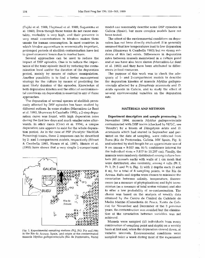
154 Mar Ecol Prog Ser 176: 153-163, 1999
(Fujiki et al. 1988, Haystead et al. 1989, Suganuma et al. 1989). Even though these toxins do not cause mor- tality, morbidity is very high, and their presence in very small concentrations in bivalves makes them unsafe for human consumption. In most countries in which bivalve aquaculture is economically important, prolonged periods of shellfish contamination have led to great economic losses due to market bans.
Several approaches can be used to reduce the impact of DSP episodes. One is to reduce the impor- tance of the toxic episode itself by reducing the conta- mination level and/or the duration of the depuration period, mainly by means of culture manipulation. Another possibility is to find a better management strategy for the culture by means of predicting the most likely duration of the episodes. Knowledge of both depuration kinetics and the effect of environmen- tal conditions on depuration is essential to any of these approaches.
The depuration of several species of shellfish previ- ously affected by DSP episodes has been studied by different authors. In some studies (Marcaillou-Le Baut et al. 1993, Shumway & Cembella 1993), a 2-step depu- ration curve was found, with high depuration rates during the first few days and much smaller rates after- wards. In other cases (Croci et al. 1994), a unique depuration rate appears to exist for the whole depura- tion period. As in the case of PSP (Paralytic Shellfish Poisoning) toxins, these 2 responses can be described by 2- and 1-compartment models, respectively (Silvert & Cembella 1995, Blanco et al. 1997). Blanco et al. (1995) have shown that a very simple 1-compartment
Fig. 1. Experimental sampling stations (Pt2, Pt3, Pt5 and Pt6) in the Ria de Arousa, Spain, and origin of the contaminated mussels Mytilus galloprovincialis (Ria de Pontevedra, Bueu)
model can reasonably describe some DSP episodes in Galicia (Spain), but more complex models have not been tested.
The effect of the environmental conditions on depu- ration has not been directly evaluated. It is generally assumed that low temperatures lead to low depuration rates (Shumway & Cembella 1993) but no strong evi- dence of this fact exists. Differences in depuration rates between mussels maintained in a culture pond and at sea have also been shown (Marcaillou-Le Baut et al. 1993) and they have been attributed to differ- ences in food resources.
The purpose of this work was to check the ade- quacy of 1- and 2-compartment models to describe the depuration kinetics of mussels Mytilus gallopro- vincialis affected by a Dinophysis acuminata and D. acuta episode in Galicia, and to study the effect of several environmental variables on the depuration rate.
MATERIALS AND METHODS
Experiment description and sample processing. In November 1994, mussels Mytilus galloprovincialis contaminated with DSP toxins (checked by HPLC, see 'Results') by a bloom of Dinophysis acuta and D. acuminata which had started in September and per- sisted on the date of sampling, were collected from Bueu (Ria de Pontevedra, Galicia, NW Spain; Fig. 1) and selected by shell length for an approximate size of 9 cm (mean = 9.057 cm; 95% confidence interval for the individual sizes = 7.817 to 10.297 cm). Finally, the mussels were randomly distributed among culture bas- kets (60 mussels each) with walls of 1 cm mesh that were distributed, also randomly, among 4 rafts (Pt 2, Pt 3, Pt 5 and Pt 6; Fig. 1) with 2 depths each (3 and 8 m), for a total of 8 sampling points, in the Ria de Arousa. Rafts and depths were chosen to minimize the covariation between salinity, temperature, fluores- cence (as a measure of phytoplankton) and light trans- mission (as a measure of total seston volume) and also to offer a low probability of re-contamination. The choice was based on the analysis of weekly data obtained by the Centro de Control da Calidade do Medio Marino (Conselleria de Pesca, Xunta de Gali- cia) for November and December of the 3 previous years. Re-contamination was avoided but the elimina- tion of the covariation between variables was not achieved.
Mussels were sampled (15 individuals from every combination of sampling point and depth) on a weekly basis at first and, when the depuration slowed down, at variable intervals. Environmental conditions were sampled twice a week during most of the experiment

Blanco et al.: Okadaic acid depuration in mussels 155
1 7 ENVlRONMENl
With linear control of the environment: rate variable with
I rate detox.
Fig. 2. Conceptual representation of the 1-compartment and 2-compartment models. Solid arrows represent toxin flows and shaded arrows represent the environmental control of
the rates
with a CTD Sea-Bird 25 equipped with a Seatech Flu- orometer and an integrated transmissometer .
Once in the laboratory, mussels were measured and weighed, and the 15 digestive glands pooled and homogenized. The DSP toxins contained in the homogenate were extracted and analyzed following the procedure given by Lee et al. (1987).
The effect of the environmental conditions and body weight on the depuration rate was identified by Princi- pal Component Analysis (PCA) and quantified by multiple regression with forward inclusion using the Minitab 11 Statistical Package. Fluorescence data were logarithmically transformed. Two types of regres- sions and PCAs were carried out: (1) general, using all data of the experiment (all depuration periods and all sampling points and depths) and (2) per depuration period, using the 8 sampling points from each individ- ual sampling date. Unless otherwise noted all signifi- cant observations or comparisons had a significance level of 95 % (p I 0.05).
Models. One- and two-compartment models, which have previously been used to describe PSP depuration kinetics (Blanco et al. 1997), were also used in this study (Fig. 2). In the 1-compartment model it was assumed that all toxicity in the digestive glands of the mussels has the same depuration kinetics. Two ver-
sions of this basic model were used: one in which the depuration rate was independent from the environ- mental variables and body weight (it was therefore a constant), and another in which depuration rate was considered to be a linear combination of the environ- mental variables considered in this study and of the body weight (it was therefore calculated by a regres- sion equation). The 2 versions of the 1-compartment model are formally described as follows:
Version 1 -constant depuration rate DR = K d(Tox)/dt = - DR.Tox
Version 2-depuration rate linearly related with envi- ronmental variables and body weight D R = a + b . S + c . T + d . F + e . T r + f.Bw d(Tox)/dt = - DR.Tox
where DR = depuration rate (d-I), a to f are constants, Tox = okadaic acid concentration in the digestive gland (expressed as pg g-I), S = salinity (psu), T = tem- perature ("C), F = fluorescence (volts), Tr = transmit- tance (percentage of light transmitted) and Bw = body weight (g).
In the 2-compartment model it is also assumed that toxins are distributed in 2 compartments, the first hav- ing a fast and the second a slow depuration rate, and that a transfer from the first to the second compartment exists. The same 2 versions as in the 1-compartment model (constant or dependent on the environment depuration rates) were used. The formulation of these 2-compartment models is as follows:
Version 1- constant depuration rate DR, = Kl DR2 = K2 d(Tox1)/dt = - DR1.Toxl - CR.TOX, d(Toxz)/dt = - DRz.Tox2 + CR.Toxl d(Tox)/dt = d(Toxl)/dt + d(Tox2)/dt
Version 2-depuration rate linearly related with en- vironmental variables and body weight DR1 = al + bl.S+ c l .T+ d l .F+ el.Tr+ fl.Bw DR, = a, + b2.S+ c2.T+ d2.F+ e2.Tr+ f2.Bw d(Toxl)/dt = -DR1.Toxl - CR.Toxl d(T0~2)ldt = -DR2.Tox2 + CR.Toxl d(Tox)/dt = d(Toxl)/dt + d(Tox2)/dt
where CR is the net transport rate from the first to the second compartment, numerical subscripts indicate compartment, and the remaining variables are as in the 1-compartment model.
The models which did not include environmental variables were fitted to the data by minimizing the sum of squares of the residuals by varying iteratively the depuration rate in the case of the 1-compartment model, or the depuration rates corresponding to each compartment, the transfer rate between compartments
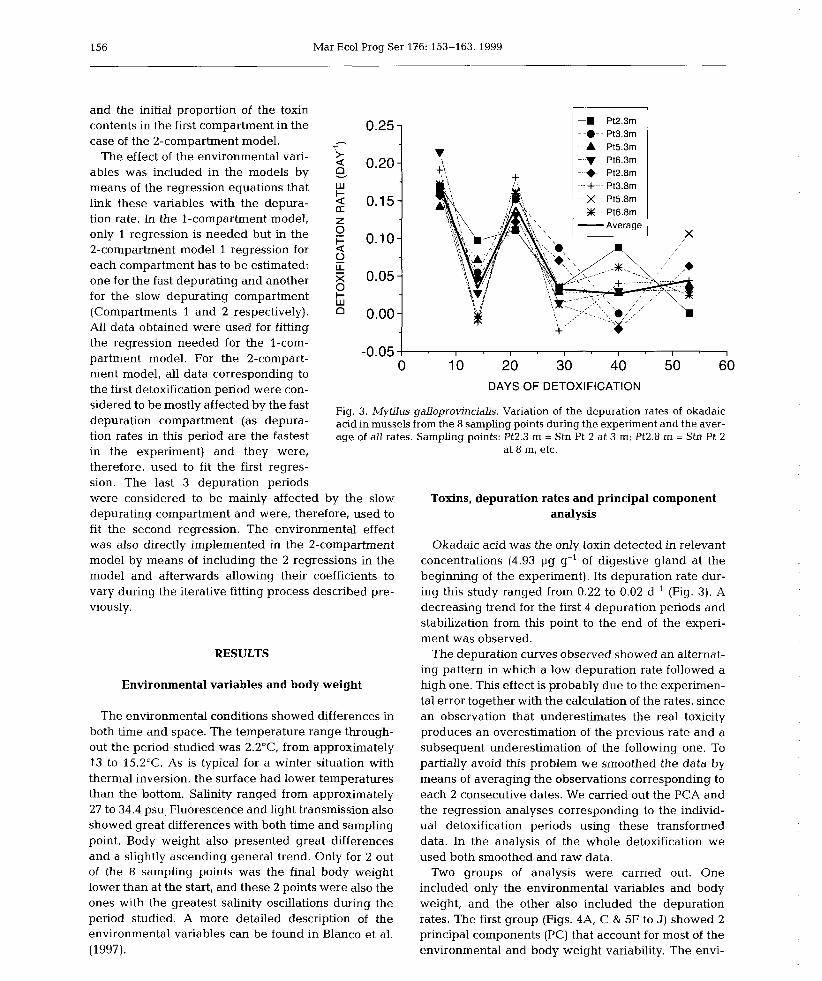
156 Mar Ecol Prog Ser 176: 153-163, 1999
and the initial proportion of the toxin contents in the first compartment in the case of the 2-compartment model.
The effect of the environmental vari- ables was included in the models by means of the regression equations that link these variables with the depura- tion rate. In the 1-compartment model, only 1 regression is needed but in the 2-compartment model 1 regression for each compartment has to be estimated: one for the fast depurating and another for the slow depurating compartment (Compartments 1 and 2 respectively). All data obtained were used for fitting the regression needed for the l-com- partment model. For the 2-compart- ment model, all data corresponding to the first detoxification period were con- sidered to be mostly affected by the fast depuration compartment (as depura- tion rates in this period are the fastest in the experiment) and they were, therefore, used to fit the first regres- sion. The last 3 depuration periods
-.- Pt2 3rn - a- Pt3 3m A Pt5 3rn
- v- Pt6 3rn - +- Pt2 8rn
+ Pt3 8rn X Pt5 8rn X-- Pt6 8rn - Average
-0.05 1 I I I I I I
0 10 20 30 40 50 60 DAYS OF DETOXIFICATION
Fig. 3. Mytilus galloprovincialis. Variation of the depuration rates of okadaic acid in mussels from the 8 sampling points during the experiment and the aver- age of all rates. Sampling points: Pt2.3 m = Stn Pt 2 at 3 m; Pt2.8 rn = Stn Pt 2
at 8 m, etc.
were considered to be mainly affected by the slow depurating compartment and were, therefore, used to fit the second regression. The environmental effect was also directly implemented in the 2-compartment model by means of including the 2 regressions in the model and afterwards allowing their coefficients to vary during the iterative fitting process described pre- viously.
RESULTS
Environmental variables and body weight
The environmental conditions showed differences in both time and space. The temperature range through- out the period studied was 2.2"C, from approximately 13 to 15.2"C. As is typical for a winter situation with thermal inversion, the surface had lower temperatures than the bottom. Salinity ranged from approximately 27 to 34.4 psu, Fluorescence and light transmission also showed great differences with both time and sampling point. Body weight also presented great differences and a slightly ascending general trend. Only for 2 out of the 8 sampling points was the final body weight lower than at the start, and these 2 points were also the ones with the greatest salinity oscillations during the period studied. A more detailed description of the environmental variables can be found in Blanco et al. (1997).
Toxins, depuration rates and principal component analysis
Okadaic acid was the only toxin detected in relevant concentrations (4.93 pg g-' of digestive gland at the beginning of the experiment). Its depuration rate dur- ing this study ranged from 0.22 to 0.02 d-' (Fig. 3). A decreasing trend for the first 4 depuration periods and stabilization from this point to the end of the experi- ment was observed.
The depuration curves observed showed an alternat- ing pattern in which a low depuration rate followed a high one. This effect is probably due to the experimen- tal error together with the calculation of the rates, since an observation that underestimates the real toxicity produces an overestimation of the previous rate and a subsequent underestimation of the following one. To partially avoid this problem we smoothed the data by means of averaging the observations corresponding to each 2 consecutive dates. We carried out the PCA and the regression analyses corresponding to the individ- ual detoxification periods using these transformed data. In the analysis of the whole detoxification we used both smoothed and raw data.
Two groups of analysis were carried out. One included only the environmental variables and body weight, and the other also included the depuration rates. The first group (Figs. 4A, C & 5F to J) showed 2 principal components (PC) that account for most of the environmental and body weight variability. The envi-
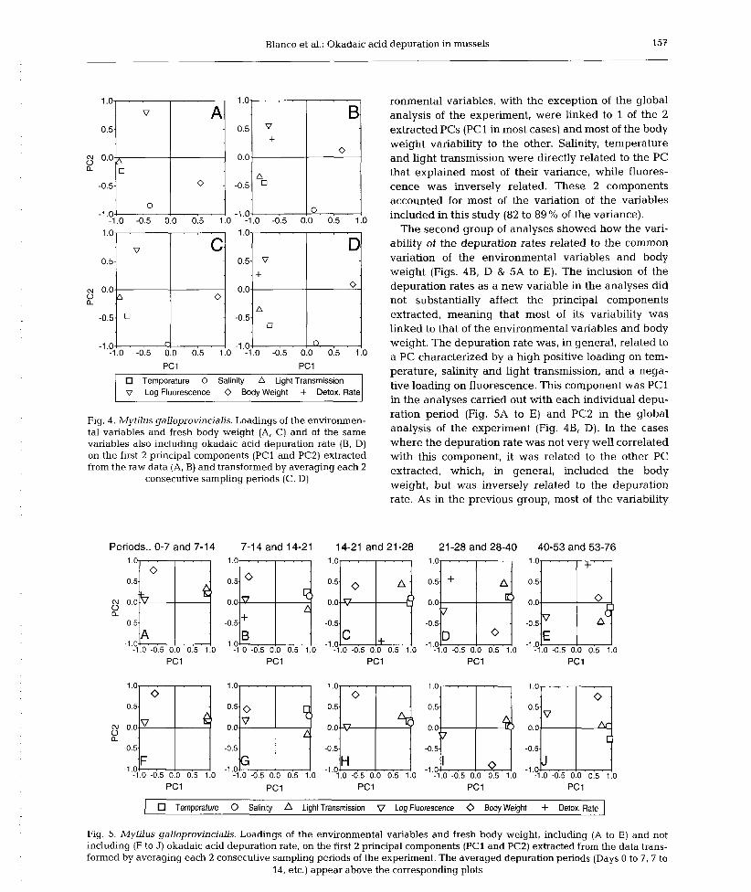
Blanco et al.: Okadaic acid depuration in mussels 157
PC1 PC1
Temperature 0 Salinity A Light Transmission o Log Fluorescence 0 Body Weight + Detox. Rate
Fig. 4. Mytilus galloprovincialis. Loadings of the environmen- tal variables and fresh body weight (A, C) and of the same variables also including okadaic acid depuration rate (B, D) on the first 2 principal components (PC1 and PC2) extracted from the raw data (A, B) and transformed by averaging each 2
consecutive sampling periods (C, D)
Periods.. 0-7 and 7-14 7-14 and 14-21
ronmental variables, with the exception of the global analysis of the experiment, were linked to 1 of the 2 extracted PCs (PC1 in most cases) and most of the body weight variability to the other. Salinity, temperature and light transmission were directly related to the PC that explained most of their variance, while fluores- cence was inversely related. These 2 components accounted for most of the variation of the variables included in this study (82 to 89 % of the variance).
The second group of analyses showed how the vari- ability of the depuration rates related to the common variation of the environmental variables and body weight (Figs. 4B, D & 5A to E). The inclusion of the depuration rates as a new variable in the analyses did not substantially affect the principal components extracted, meaning that most of its variability was linked to that of the environmental variables and body weight. The depuration rate was, in general, related to a PC characterized by a high positive loading on tem- perature, salinity and light transmission, and a nega- tive loading on fluorescence. This component was PC1 in the analyses carried out with each individual depu- ration period (Fig. 5A to E) and PC2 in the global analysis of the experiment (Fig. 4B, D). In the cases where the depuration rate was not very well correlated with this component, it was related to the other PC extracted, which, in general, included the body weight, but was inversely related to the depuration rate. As in the previous group, most of the variability
14-21 and 21 -28 21 -28 and 28-40 40-53 and 53-76
1 Temperature 0 Salinity A Light Transmission V Log Fluorescence 0 Body Weight + Detox. Rate 1 Fig. 5. Mytilus galloprovincialis. Loadings of the environmental variables and fresh body weight, including (A to E) and not including (F to J) okadaic acid depuration rate, on the first 2 principal components (PC1 and PC2) extracted from the data trans- formed by averaging each 2 consecutive sampling periods of the experiment. The averaged depuration periods (Days 0 to ?, ? to
14, etc.) appear above the corresponding plots
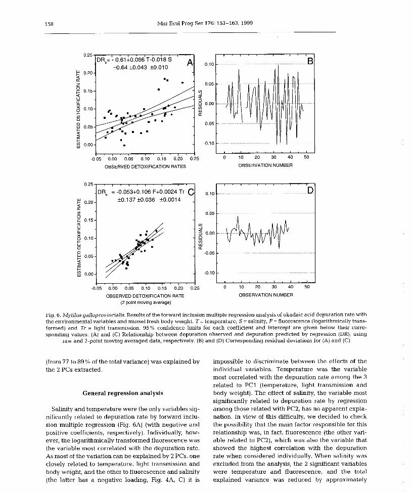
158 Mar Ecol Prog Ser 176: 153-163, 1999
OBSERVED DETOXIFICATION RATES OBSERVATION NUMBER
Fig. 6. Mytilus galloprovincialis. Results of the forward inclusion multiple regression analysis of okadaic acid depuration rate with the environmental variables and mussel fresh body weight. T = temperature, S = salinity, F = fluorescence (logarithmically trans- formed) and Tr = light transmission. 95% confidence limits for each coefficient and intercept are given below their corre- sponding values. (A) and (C) Relationship between depuration observed and depuration predicted by regression (DR), using
raw and 2-point moving averaged data, respectively. (B) and (D) Corresponding residual deviations for (A) and (C)
(from 77 to 89 % of the total variance) was explained by the 2 PCs extracted.
0.25
.
? 0.20- a [r
Z 0 F 0.15- 6 II lL :: 0.10- + W n 0 ? 0.05 a
E 3 0.00-
General regression analysis
OBSERVED DETOXIFICATION RATE OBSERVATION NUMBER (2 point moving average)
o.lo
0.05
Y 5 0.00 P m W a:
-0.05
-0.10-
. , . , . , . I . I .
D% = -0.053+0.106 F+0.0024 Tr C k0.137 *0.036 +0.0014
:I; ; .
-
I
-
, . , . , ' , . I .
-0.05 0.00 0.05 0.10 0.15 0.20
Salinity and temperature were the only variables sig- nificantly related to depuration rate by forward inclu- sion multiple regression (Fig. 6A) (with negative and positive coefficients, respectively). Individually, how- ever, the logarithmically transformed fluorescence was the variable most correlated with the depuration rate. As most of the variation can be explained by 2 PCs, one closely related to temperature, light transmission and body weight, and the other to fluorescence and salinity (the latter has a negative loading, Fig. 4A, C) it is
I . I . I . I ~ I ' I
-. . . . . . . . . . . . . . . . . . . . . . . . . . . . . . . . . . . . . . . . . . D
....... . . . . . . . . . . . . . . . . . . --
. . . . . . . . . . : . . . . . . . . . . . . . . . . . . . . . . . . . . . . . . . . . . . . . . . . . . . . . . . . . -
. . . . . . . . . . . . . . . . . . . . . . . . . . . . . . . .
I . I . I . 1 . I . I
impossible to discriminate between the effects of the individual variables. Temperature was the variable most correlated with the depuration rate among the 3 related to PC1 (temperature, light transmission and body weight). The effect of salinity, the variable most significantly related to depuration rate by regression among those related with PC2, has no apparent expla- nation. In view of this difficulty, we decided to check the possibility that the main factor responsible for this relationship was, in fact, fluorescence (the other vari- able related to PC2), which was also the variable that showed the highest correlation with the depuration rate when considered individually. When salinity was excluded from the analysis, the 2 significant variables were temperature and fluorescence, and the total explained variance was reduced by approximately
0.25 0 10 20 30 40 50
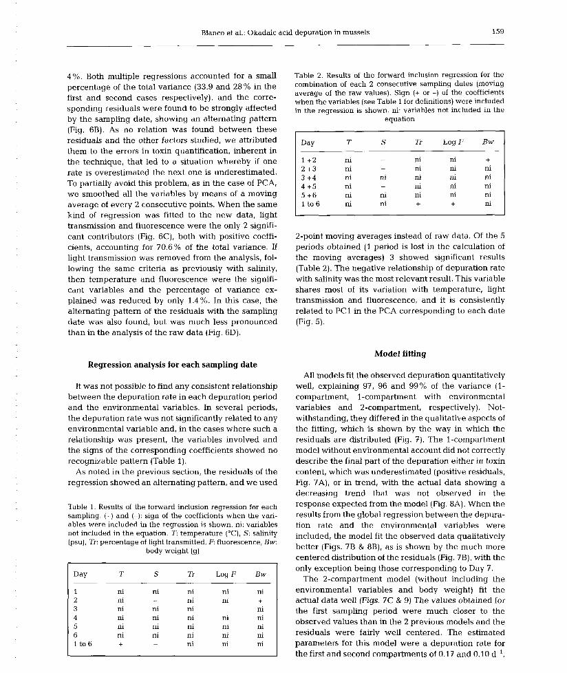
Blanco et al.: Okadaic acid depuration in mussels 159
4%. Both multiple regressions accounted for a small percentage of the total variance (33.9 and 28% in the first and second cases respectively), and the corre- sponding residuals were found to be strongly affected by the sampling date, showing an alternating pattern (Fig. 6B). As no relation was found between these residuals and the other factors studied, we attributed them to the errors in toxin quantification, inherent in the technique, that led to a situation whereby if one rate is overestimated the next one is underestimated. To partially avoid this problem, as in the case of PCA, we smoothed all the variables by means of a moving average of every 2 consecutive points. When the same kind of regression was fitted to the new data, light transmission and fluorescence were the only 2 signifi- cant contributors (Fig. 6C), both with positive coeffi- cients, accounting for 70.6% of the total variance. If light transmission was removed from the analysis, fol- lowing the same criteria as previously with salinity, then temperature and fluorescence were the signifi- cant variables and the percentage of variance ex- plained was reduced by only 1.4%. In this case, the alternating pattern of the residuals with the sampling date was also found, but was much less pronounced than in the analysis of the raw data (Fig. 6D).
Table 2. Results of the forward inclusion regression for the combination of each 2 consecutive sampling dates (moving average of the raw values). Sign (+ or -) of the coefficients when the variables (see Table 1 for definitions) were included in the regression is shown, ni: variables not included in the
equation
Day T S Tr Log F Bw
2-point moving averages instead of raw data. Of the 5 periods obtained (1 period is lost in the calculation of the moving averages) 3 showed significant results (Table 2). The negative relationship of depuration rate with salinity was the most relevant result. This variable shares most of its variation with temperature, light transmission and fluorescence, and it is consistently related to PC1 in the PCA corresponding to each date (Fig. 5).
Model fitting Regression analysis for each sampling date
It was not possible to find any consistent relationship between the depuration rate in each depuration period and the environmental variables. In several periods, the depuration rate was not significantly related to any environmental variable and, in the cases where such a relationship was present, the variables involved and the signs of the corresponding coefficients showed no recognizable pattern (Table 1).
As noted in the previous section, the residuals of the regression showed an alternating pattern, and we used
Table 1. Results of the forward inclusion regression for each sampling. (+) and (-): sign of the coefficients when the vari- ables were included in the regression is shown. ni: variables not included in the equation. T: temperature ("C), S: salinity (psu), Tr: percentage of light transmitted, F: fluorescence, Bw:
body weight (g)
Day T S Tr Log F Bw
All models fit the observed depuration quantitatively well, explaining 97, 96 and 99% of the variance (1- compartment, 1-compartment with environmental variables and 2-compartment, respectively). Not- withstanding, they differed in the qualitative aspects of the fitting, which is shown by the way in which the residuals are distributed (Fig. 7). The 1-compartment model without environmental account did not correctly describe the final part of the depuration either in toxin content, which was underestimated (positive residuals, Fig. ?A), or in trend, with the actual data showing a decreasing trend that was not observed in the response expected from the model (Fig. 8A). When the results from the global regression between the depura- tion rate and the environmental variables were included, the model fit the observed data qualitatively better (Figs. 7B & 8B), as is shown by the much more centered distribution of the residuals (Fig. 7B), with the only exception being those corresponding to Day 7.
The 2-compartment model (without including the environmental variables and body weight) fit the actual data well (Figs. 7C & 9) The values obtained for the first sampling period were much closer to the observed values than in the 2 previous models and the residuals were fairly well centered. The estimated parameters for this model were a depuration rate for the first and second compartments of 0.17 and 0.10 d-',

Mar Ecol Prog Ser 176: 153-163,1999
respectively, a transfer rate between Compartments 1 and 2 of 0.31 d-' and an initial contribution of toxins in the first compartment to the total of 99.9%.
As mentioned in 'Materials and methods', we fol- lowed 2 different strategies to implement the effect of the environmental variables on the 2-compartment model. In both cases, after optimization, the parame- ters obtained caused the initial model to revert to a 1- compartment model because 100 % of the initial toxic- ity was put in the first compartment and the transfer rate from Compartment 1 to Compartment 2 was esti- mated to be zero. This indicates that all the depuration takes place in Compartment 1 and, therefore, that the model is a 1-compartment model.
DISCUSSION
The depuration rates found in this study are very close to our previous estimates in the same area, although they were obtained by optimizing the fit of a contamination-depuration model (Blanco et al. 1995). They were slightly lower than those found in other studies on Mytilus galloprovincialis such as those by Poletti et al. (1996) and Croci et al. (1994) for depura- tion using ozone (from nearly 0 to 0.27 d-l). Marcaillou- Le Baut et al. (1993) also found lower depuration rates, 0.07 and 0.08 d-l, for Mediterranean and Breton mus- sels respectively (in the latter case during the first 15 d and subsequently decreasing).
Days of experiment
A One-compartment, independent of environment 0 One-compartment, rates depending on environment 0 Two-compartment, independent of environment
Fig. 7. Mytilus galloprovincialis. Residual deviations corre- sponding to the 3 depuration models fitted to the observed data. (A) 1-compartment model independent of the environ- ment, (B) 1-compartment model including the effect of envi- ronment, and (C) 2-compartment model independent of the
environment
I . .
0 28 56 0 28 56 0 28 56 0 28 56 0 28 56 0 28 56 0 28 56 0 28 56
Days of detoxification Fig. 8. Mytilus galloprovincialis. Depuration kinetics and fitted 1-compartment models for each sampling point (Pt2-3 m: Stn Pt 2 at 3 m; Pt2-8 m: Stn Pt 2 at 8 m; etc.) (A) Fitted fixed rate models (without environmental effect) and (B) corresponding variable rate models (environmentally determined rates). Toxin concentration is expressed as pg of total toxins per gram of digestive gland
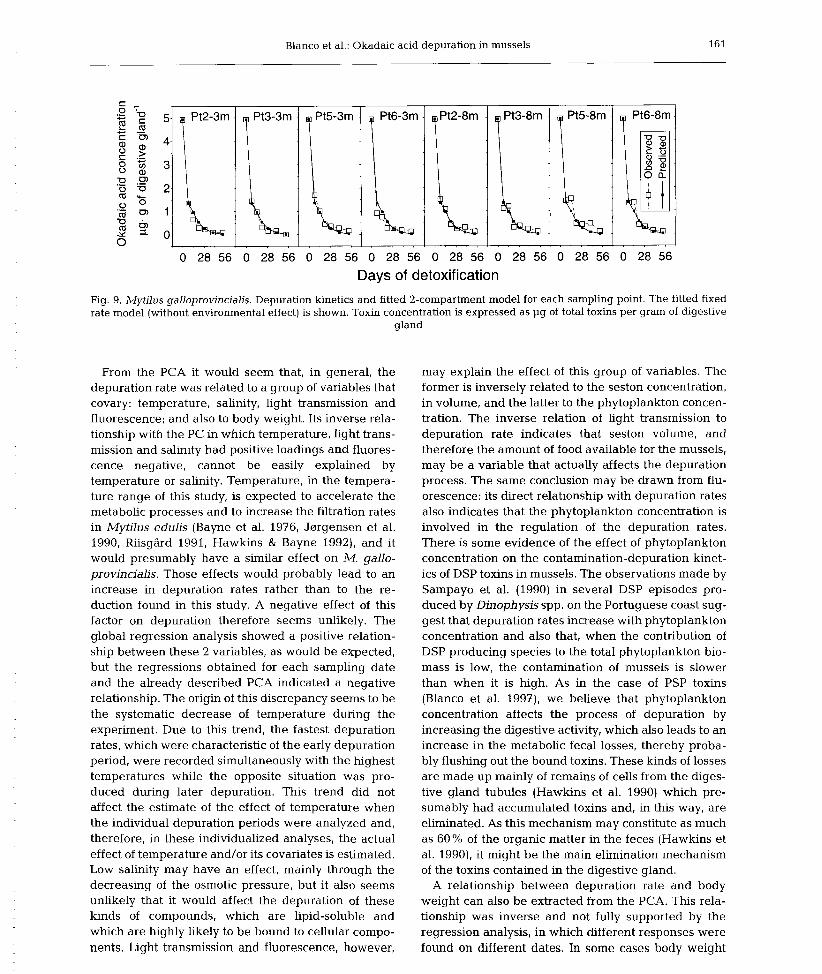
Blanco et al.: Okadaic acid depuration in mussels 161
0 28 56 0 28 56 0 28 56 0 28 56 0 28 56 0 28 56 0 28 56 0 28 56
Days of detoxification
Fig. 9. Mytilus galloprovincialis. Depuration kinetics and fitted 2-compartment model for each sampling point. The fitted fixed rate model (without environmental effect) is shown. Toxin concentration is expressed as pg of total toxins per gram of digestive
gland
From the PCA it would seem that, in general, the depuration rate was related to a group of variables that covary: temperature, salinity, light transmission and fluorescence; and also to body weight. Its inverse rela- tionship with the PC in which temperature, light trans- mission and salinity had positive loadings and fluores- cence negative, cannot be easily explained by temperature or salinity. Temperature, in the tempera- ture range of this study, is expected to accelerate the metabolic processes and to increase the filtration rates in Mytilus edulis (Bayne et al. 1976, Jmgensen et al. 1990, RiisgGrd 1991, Hawkins & Bayne 1992), and it would presumably have a similar effect on M. gallo- provincialis. Those effects would probably lead to an increase in depuration rates rather than to the re- duction found in this study. A negative effect of this factor on depuration therefore seems unlikely. The global regression analysis showed a positive relation- ship between these 2 variables, as would be expected, but the regressions obtained for each sampling date and the already described PCA indicated a negative relationship. The origin of this discrepancy seems to be the systematic decrease of temperature during the experiment. Due to this trend, the fastest depuration rates, which were characteristic of the early depuration period, were recorded simultaneously with the highest temperatures while the opposite situation was pro- duced during later depuration. This trend did not affect the estimate of the effect of temperature when the individual depuration periods were analyzed and, therefore, in these individualized analyses, the actual effect of temperature and/or its covariates is estimated. Low salinity may have an effect, mainly through the decreasing of the osmotic pressure, but it also seems unlikely that it would affect the depuration of these kinds of compounds, which are lipid-soluble and which are highly likely to be bound to cellular compo- nents. Light transmission and fluorescence, however,
may explain the effect of this group of variables. The former is inversely related to the seston concentration, in volume, and the latter to the phytoplankton concen- tration. The inverse relation of light transmission to depuration rate indicates that seston volume, and therefore the amount of food available for the mussels, may be a variable that actually affects the depuration process. The same conclusion may be drawn from flu- orescence: its direct relationship with depuration rates also indicates that the phytoplankton concentration is involved in the regulation of the depuration rates. There is some evidence of the effect of phytoplankton concentration on the contamination-depuration kinet- ics of DSP toxins in mussels. The observations made by Sampayo et al. (1990) in several DSP episodes pro- duced by Dinophysis spp. on the Portuguese coast sug- gest that depuration rates increase with phytoplankton concentration and also that, when the contribution of DSP producing species to the total phytoplankton bio- mass is low, the contamination of mussels is slower than when it is high. As in the case of PSP toxins (Blanco et al. 1997), we believe that phytoplankton concentration affects the process of depuration by increasing the digestive activity, which also leads to an increase in the metabolic fecal losses, thereby proba- bly flushing out the bound toxins. These kinds of losses are made up mainly of remains of cells from the diges- tive gland tubules (Hawkins et al. 1990) which pre- sumably had accumulated toxins and, in this way, are eliminated. As this mechanism may constitute as much as 60 % of the organic matter in the feces (Hawkins et al. 1990), it might be the main elimination mechanism of the toxins contained in the digestive gland.
A relationship between depuration rate and body weight can also be extracted from the PCA. This rela- tionship was inverse and not fully supported by the regression analysis, in which different responses were found on different dates. In some cases body weight
seems to have a positive effect on depuration. These cases may be explained by the toxin dilution effect demonstrated for PSP toxins by Bricelj et al. (1990), and which is derived from the fact that, with constant toxin content, if the body weight of the bivalves increases, then the toxin concentration per unit of weight de- creases and also by the fact that larger mussels have larger metabolic fecal losses (Hawkins et al. 1990). Nonetheless, in most of the cases in this experiment, there was an inverse relation between depuration and body weight, and therefore it cannot be explained as described previously. In our opinion, this relation was produced by the higher depuration capability of small mussels as compared to larger specimens, as was found for domoic acid (Wohlgeschaffen et al. 1992). The variation in mussel size due to the random sam- pling will probably allow for detection of this effect.
From our results it cannot be clearly concluded whether the DSP depuration in this species evolves following 1- or 2-compartment kinetics because the benefit obtained by fitting a 2-compartment model is partially overlapped by the effect of including the environmental variables in the model. It is obvious that the 1-compartment model with a fixed depuration rate did not fit the actual data as well as the other 2 models, but the differences between the latter 2 models are not clear, with each having its strengths and its weak- nesses. The 1-compartment model with environmental variables included fit the peculiarities of each data set better. The 2-compartment model, on the other hand, fit the first depuration point in each data set better. In our opinion the 1-compartment model that includes the environmental variables and body weight is more real- istic than the 2-compartment model because its main drawback, the poor fitting of the first point of each data set, (1) coincides with an atypically low okadaic acid concentration for these points followed by similar or higher toxin contents on the next sampling date, and (2) seems to be derived from one or several unknown factors which were not monitored during the experi- ment, as it showed a pattern in which the deviation of the model was larger in the bottom (8 m) than in the surface samples (3 m) and followed the same station order at each depth, and also because DSP analyses are carried out on the digestive gland, and therefore the second compartment does not involve other organs and, thus, might comprise a smaller fraction of the ana- lyzed biomass than in the case of PSP (Blanco et al. 1997).
Our inability to fit the 2-compartment model includ- ing environmental conditions may also be affected by the precision of the toxin quantification of the first depuration point, because very small errors might make impossible the discrimination between 2 models that fit so well. If this point has been significantly
Mar Ecol Prog Ser 176: 153-163. 1999
affected by random sampling or analytical errors, our estimation of the effect on these conditions on the first compartment would be inadequate and the model might be incorrectly fitted.
If we consider the 2-compartment model to be the most realistic, we must conclude that the environmen- tal variables have little or no importance in regulating the depuration, which is in contrast with the small dis- tortion that the inclusion of the depuration rate induced on the components extracted by PCA. In this case, the 2 compartments would have depuration rates which are very different from each other and a very high transfer rate (0.3 d-I).
In conclusion, it would appear that environmental conditions, mainly through the amount of available food, affect the depuration kinetics of okadaic acid, but the real influence of these variables cannot be quanti- fied if a depuration model is not previously assumed.
Acknowledgements. This work was funded by the Con- selleria de Pesca, Marisqueo e Acuicultura, and CICYT (Pro- ject ALI-95-1012-C05-02). We thank M. C. Marino and M. Miranda for their technical assistence and the department of biotoxins of the Centro de Control de Calidade do Medio Marino for obtaining the experimental mussels and their help in the preparation of the experiment.
LITERATURE CITED
Bayne BL, Thompson RJ, Widdows J (1976) Physiology I. In: Bayne BL (ed) Marine mussels: their ecology and physiol- ogy. Cambridge University Press, Cambridge, p 121-206
Blanco J, Fernandez M, Mar50 J, Reguera B, Miguez A, Maneiro J, Cacho E, Martinez A (1995) From Dinophysis spp. toxicity to DSP outbreaks: a preliminary model of toxin accumulation in mussels. In: Lassus P, Arzul G , Erard E, Gentien P, Marcaillou C (eds) Harmful marine algal blooms. Lavoisier, Intercept Ltd, Paris, p 777-782
Blanco J, Morono A, Franco J, Reyero MI (1997) PSP depura- tion kinetics in the mussel Mytilus galloprovincialis. One- and two-compartment models and the effect of some envi- ronmental variables. Mar Ecol Prog Ser 158:165-175
Bricelj VM, Lee JH, Cembella AD, Anderson DM (1990) Uptake and kinetics of paralytic shellfish toxins from the dinoflagellate Alexandrium fundyense in the mussel Mytilus edulis. Mar Ecol Prog Ser 63:177-188
Croci L, Toti L, De Medici D, Cozzi L (1994) Diarrhetic shell- fish poison in mussels: comparison of methods of detection and determination of the effectiveness of depuration. Food Microbial 24:337-342
Fernandez ML, Miguez A, Cacho E, Martinez A (1996) Okadaic acid esters in Spanish mussels. Toxicon 34: 381-387
Fujiki H, Suganuma M, Suguri H, Yoshizawa S, Takagi K, Uda N, Wakamatsu K, Yamada K, Murata M (1988) Diar- rhetic shellfish toxin dinophysistoxin-1 is a potent tumour promoter on mouse skin. Jpn J Cancer Res 79:1089-1093
Gago A, Rodriguez Vazquez JA, Leao JM, Comesana M, Quilliam MA, Thibault P (1996) 1985-1995. A decade of studies on PSP and DSP toxins in Spanish mussels by chemical methods. In: Yasumoto T, Oshirna Y, Fukuyo Y
Blanco et al.: Okadaic acid depuration in mussels 163
(eds) Harmful and toxic algal blooms. IOC of UNESCO, Paris, p 7-10
Hawkins AJS, Bayne BL (1992) Physiological interrelations, and the regulation of production. In: Gossling E (ed) The mussel Mytilus: ecology, physiology, genetics and culture. Elsevier, Amsterdam, p 171-222
Hawkins AJS, Navarro E, Iglesias JIP (1990) Comparative allometries of gut content, gut passage time and metabolic faecal loss in Mytilus edulis and Cerastoderma edule. Mar Biol 105:197- 204
Haystead TAJ, Sim ATR, Carling D, Honnor RC, Tsukitani Y, Cohen P, Hardie DG (1989) Effect of the tumour promoter okadaic acid on intracellular protein phosphorylation and metabolism. Nature 337:78-81
Hu T, Doyke J , Jackson D, Marr J , Nixon E, Pleasance S, Quil- liam MA, Walter JA, Wright JLC (1992) Isolation of a new diarrhetic shellfish poison from Irish mussels. Chem Com- mun 30:39-41
Jerrgensen CB, Larsen PS, Riisgdrd HU (1990) Effects of tem- perature on the mussel pump. Mar Ecol Prog Ser 64:89-97
Lee JS, Yanagi T, Kenma R, Yasumoto T (1987) Fluorimetric detection of diarrhetic shellfish toxins by high-perfor- mance liquid chromatograpy. Agric Biol Chem 51: 877-881
Marcaillou-Le Baut C, Bardin B, Bardouil M, Bohec M, Le Dean L, Masselin P, Truquet P (1993) DSP depuration rates of mussels reared in a laboratory and an aqua- culture pond. In: Smayda TJ, Shimizu Y (eds) Toxic phytoplankton blooms in the sea. Elsevier, Amsterdam, p 531-535
Marr JC, Hu T, Pleasance S, Quilliam M, Wright J (1992) Detection of new 7-0-acyl-derivatives of diarrhetic shell- fish poisoning toxins by liquid chromatography-mass spectrometry. Toxicon 30:381-387
Murata M, Shimatani M, Sugitani H, Oshima Y, Yasumoto T (1982) Isolation and structural elucidation of the causative
Editorial responsibility: Otto Kinne (Editor), Oldendorf/Luhe, Germany
toxin of diarrhetic shellfish poisoning. Bull Jpn Soc Sci Fish 43:549-552
Poletti R, Viviani R, Casadei C, Lucentini L, Giannetti L, Funari E, Draisci R (1996) Decontamination dynamics of mussels naturally contaminated with diarrhetic toxins relocated to a basin of the Adriatic Sea. In: Yasumoto T, Oshima Y, Fukuyo Y (eds) Harmful and toxic algal blooms. IOC of UNESCO, Paris, p 429-432
Riisgdrd HU (1991) Filtration rate and growth in the blue mus- sel, Mytilus edulis Linnaeus, 1758: dependence on algal concentration. J Shellfish Res 10:29-35
Sampayo MA, Alvito P, Franca S, Sousa I (1990) Dinophysis spp. toxicity and relation to accompanying species. In: Graneli E, Sundstrom B, Edler L, Anderson DMM (eds) Toxic marine phytoplankton. Elsevier, New York, p 215-220
Silvert W, Cembella, AD (1995) Dynamic modelling of phyco- toxin kinetics in the blue mussel, Mytilus edulis, with implications for other marine invertebrates. Can J Fish Aquat Sci 52:521-521
Shumway SE, Cembella AD (1993) The impact of toxic algae on scallop culture and fisheries. Rev Fish Aquat Sci 1: 121-150
Suganuma M, Suttajit M, Suguri H, Ojika M, Yamada K, Fujiki H (1989) Specific binding of okadaic acid, a new tumour promoter in mouse skin. FEBS Lett 250:615-618
Wohlgeschaffen GD, Mann KH, Subba Rao DV, Pocklington R (1992) Dynamics of the phycotoxin domoic acid: accumu- lation and excretion in two commercially important bivalves. J Appl Phycol4:297-310
Yasumoto T, Oshima Y, Yamaguchi M (1978) Occurrence of a new type of toxic shellfish in Japan and chemical proper- ties of the toxin. Bull Jpn Soc Sci Fish 44:1249-1255
Yasumoto T, Murata M, Oshima Y, Sano M, Matsumoto, GK Clardy J (1985) Diarrhetic shellfish toxins. Tetrahedron 41: 1019-1025
Submitted: February 27,1998; Accepted: September 24, 1997 Proofs received from author(s): January 11, 1999

Capítulo II – Artículo 7 - 175 -
ARTÍCULO 7
Detection of okadaic acid esters in the hexane extracts of
Spanish mussels.
Fernández, M.L., Miguez, A., Cacho, E. and Martinez, A. 1996. Toxicon, 3,
381-387.


Capítulo II – Artículo 7 Resumen - 177 -
Detection of okadaic acid esters in the hexane extracts of Spanish mussels
RESUMEN
Se han documentado dos tipos de derivados de baja polaridad del AO en moluscos o en
fitoplancton: 7-O- acil- ésteres con un ácido graso esterificando el grupo OH en posición 7 y
los diol ésteres en los que el grupo carboxílico de las toxinas ha sido esterificado. Estos
compuestos no pueden ser detectados directamente por cromatografía líquida y detección
fluorimétrica de los derivados del 9-antril-diazometano, debido en el caso del primer grupo a
su baja polaridad y elevado peso molecular y en el caso del segundo grupo debido a la
presencia del grupo carboxilo esterificado. Todos ellos deben ser hidrolizados antes de la
derivatización, de manera que puedan ser detectados como derivados de ADAM de las
correspondientes toxinas no aciladas o no esterificadas. En el procedimiento de Lee (1987),
después de la extracción de las glándulas digestivas con metanol al 80%, se realiza una
partición líquido-líquido con un disolvente no polar tal como el hexano con el objetivo de
eliminar los lípidos no polares. La presencia de toxinas no polares fue investigada en
mejillones españoles y confirmada en la capa de hexano, usualmente desechada en los
procedimientos convencionales de extracción, mediante el análisis de los productos de la
hidrólisis alcalina. Se demuestra la solubilización preferente de estas toxinas en un solvente
no polar tal como el hexano. Se sugiere la inclusión en el procedimiento general de una etapa
de hidrólisis del extracto de hexano con la finalidad de monitorizar la contribución de las
toxinas DSP no polares a la toxicidad DSP total de los moluscos. Esta es la primera
documentación de otras toxinas DSP diferentes al OA y a la DTX2 en mejillones españoles.

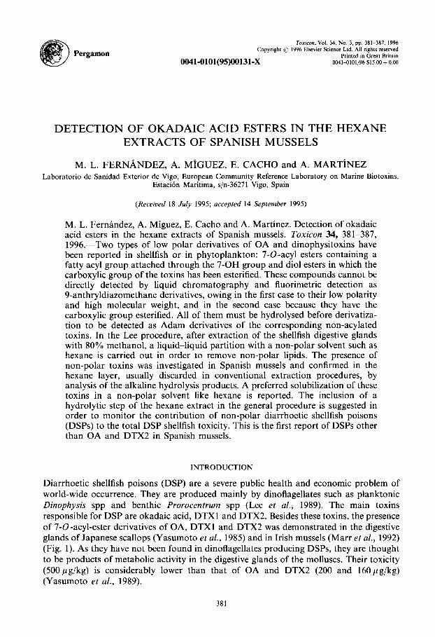
Pergamon
Toxicon, Vol. 34, No. 3, pp. 381-387, 1996
Copyright 0 1996 Elsevier Science Ltd. All rights reserved Printed in Great Britain
0041-0101(95)00131-x 00414101/96 $15.00 +O.OO
DETECTION OF OKADAIC ACID ESTERS IN THE HEXANE EXTRACTS OF SPANISH MUSSELS
M. L. FERNANDEZ, A. MiGUEZ, E. CACHO and A. MARTINEZ Laboratorio de Sanidad Exterior de Vigo, European Community Reference Laboratory on Marine Biotoxins,
Estaci6n Maritima, s/n-36271 Vigo, Spain
(Received 18 July 1995; accepted 14 September 1995)
M. L. Fernandez, A. Miguez, E. Cache and A. Martinez. Detection of okadaic acid esters in the hexane extracts of Spanish mussels. Toxicon 34, 381-387, 1996.-Two types of low polar derivatives of OA and dinophysitoxins have been reported in shellfish or in phytoplankton: 7-0-acyl esters containing a fatty acyl group attached through the 7-OH group and diol esters in which the carboxylic group of the toxins has been esterified. These compounds cannot be directly detected by liquid chromatography and fluorimetric detection as 9-anthryldiazomethane derivatives, owing in the first case to their low polarity and high molecular weight, and in the second case because they have the carboxylic group esterified. All of them must be hydrolysed before derivatiza- tion to be detected as Adam derivatives of the corresponding non-acylated toxins. In the Lee procedure, after extraction of the shellfish digestive glands with 80% methanol, a liquid-liquid partition with a non-polar solvent such as hexane is carried out in order to remove non-polar lipids. The presence of non-polar toxins was investigated in Spanish mussels and confirmed in the hexane layer, usually discarded in conventional extraction procedures, by analysis of the alkaline hydrolysis products. A preferred solubilization of these toxins in a non-polar solvent like hexane is reported. The inclusion of a hydrolytic step of the hexane extract in the general procedure is suggested in order to monitor the contribution of non-polar diarrhoetic shellfish poisons (DSPs) to the total DSP shellfish toxicity. This is the first report of DSPs other than OA and DTX2 in Spanish mussels.
INTRODUCTION
Diarrhoetic shellfish poisons (DSP) are a severe public health and economic problem of world-wide occurrence. They are produced mainly by dinoflagellates such as planktonic Dinophysis spp and benthic Prorocentrum spp (Lee et al., 1989). The main toxins responsible for DSP are okadaic acid, DTXl and DTX2. Besides these toxins, the presence of 7-O-acyl-ester derivatives of OA, DTXl and DTX2 was demonstrated in the digestive glands of Japanese scallops (Yasumoto et al., 1985) and in Irish mussels (Marr et al., 1992) (Fig. 1). As they have not been found in dinoflagellates producing DSPs, they are thought to be products of metabolic activity in the digestive glands of the molluscs. Their toxicity (500 pgg/kg) is considerably lower than that of OA and DTX2 (200 and 160 pg/kg) (Yasumoto et al., 1989).
381
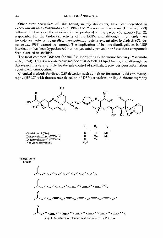
382 M. L. FERNbiNDEZ et al.
Other ester derivatives of DSP toxins, mainly diol-esters, have been described in Prorocentrum lima (Yasumoto et al., 1987) and Prorocentrum concavum (Hu et al., 1993) cultures. In this case the esterification is produced at the carboxylic group (Fig. 2) responsible for the biological activity of the DSPs, and although in principle their toxicological activity is cancelled, their potential toxicity evident after hydrolysis (Cande- nas et al., 1994) cannot be ignored. The implication of benthic dinoflagellates in DSP intoxication has been hypothesized but not yet totally proved, nor have these compounds been detected in shellfish.
The most common DSP test for shellfish monitoring is the mouse bioassay (Yasumoto et al., 1978). This is a non-selective method that detects all lipid toxins, and although for this reason it is very suitable for the safe control of shellfish, it provides poor information about toxin composition.
Chemical methods for direct DSP detection such as high-performance liquid chromatog- raphy (HPLC) with fluorescence detection of DSP derivatives, or liquid chromatography
Me
Okadaic acid (OA) H H Me
Dinophysistoxin-1 (DTX-I) H Me Me
Dinophysistoxin-2 (DTX-2) H Me H
7-0-Acyl derivatives acyl
Typical Acyl groups
0
Fig. 1. Structures of okadaic acid and related DSP toxins.
Okadaic Acid Esters in Spanish Mussels 383
Okadaic acid
Diol esters
II OH H
HO-0
Fig. 2. Okadaic acid and its diol esters.
coupled with mass spectrometry (LC-MS) have been shown to be valuable analytical tools in characterizing toxin profiles in phytoplankton and shellfish. Although LCMS is without doubt the most powerful technique for confirming the identities of toxins and identifying new toxins, the equipment required is very sophisticated and expensive, and not available in most of the laboratories involved in marine biotoxin analysis. For this reason, HPLC with fluorimetric detection, and in particular Lee’s procedure (1987) which uses 9-anthryldiazomethane as the fluorophore, is still the most widely used for DSP detection, for both research and confirmatory purposes. With this procedure, only OA, DTXl and DTX2 are directly detected. Ester-derivatives of the latter toxins are not monitored by the procedure, owing to their less polar character or to the esterification of the carboxylic group, and must be hydrolysed prior to derivatization.
In the Lee procedure (1987) after the extraction of the shellfish digestive glands with 80% methanol, a liquid-liquid partition with hexane ip carried out in order to remove non-polar lipids. This partition is also included in some mouse bioassay procedures in order to avoid false positive results from possible free fatty acids present eventually in shellfish samples, which are toxic to mice but not to humans (Takagi et al., 1984). We found that low polar DSPs are preferentially solubilized in this solvent. Direct analysis of the hexane shellfish extracts did not reveal the presence of DSPs, but after alkaline hydrolysis of the hexane extract, and depending on the mussel samples, different amounts of OA were found.
MATERIALS AND METHODS
Materials
Esters of OA from P. lima used for the optimization of the hydrolysis conditions were kindly provided by Dr Norte from La Laguna University (Spain). Mussel samples from different production areas of the Galician Rias were provided by the Centro Galego para o Control da Calidade do Medio Marifio. Okadaic acid standard, supplied by Boehringer Mannhein (Barcelona, Spain), was used for standard solution preparations. Adam reagent used for derivatization was supplied by Serva (Comercial Anger, Barcelona, Spain). HPLC-grade solvents were purchased from Merck (Igoda S.A., Barcelona, Spain). Silica cartridges used for clean-up were obtained from Waters (ref. 51900).
384 M. L. FERNANDEZ et al.
DSP sample extraction The DSP sample extraction was done following the Lee procedure (1987) with slight modifications. For each
mussel sample, 1 g of homogenized hepatopancreas was extracted with 4ml of methanol/water 80:20. After centrifugation, 2.5 ml of the supernatant was extracted twice with 2.5 ml of hexane. The hexane extracts were kept for detection of OA esters after hydrolysis. One millilitre of water was added to the methanolic extract and this layer was extracted twice with 4 ml of chloroform. The final chloroform extract was made up to 10 ml, and an aliquot of 0.5 ml evaporated to dryness and reserved for derivatization with Adam.
Hydrolysis of DSP esters Hexane extracts were evaporated to dryness with nitrogen, and 0.5 ml of 0.5 N NaOH solution in 90%
methanol was added to each extract. The mixture was kept at room temperature, protected against the light, and agitated continuously for 1 hr. After concentrating, 2 ml of 0.125 N HCl was added. The final pH was about 2.5. The solution was extracted three times with 2 ml of chloroform and dried with disodium sulphate. An aliquot of the latter solution was dried under nitrogen and reserved for Adam derivatization.
Adam derivatization Two-hundred microlitres of 0.2% 9-Adam methanolic solution was added to the dried mussel extracts. The
mixture was shaken for 2 min and kept for 1 hr at 40°C protected against the light. The Adam solution was prepared daily from small portions of the reagent kept at - 30°C by adding some drops of acetone to facilitate the further solution in methanol, and was then filtered through 0.45 pm.
Clean -up The clean-up step was identical to that described in the Lee procedure (1987).
HPLC analysis HPLC analysis followed Lee’s method (1987) with slight modifications. The fluorescent esters were separated
using a Hewlett-Packard 1050 liquid chromatograph in a Merck Superspher lOO-RP18 (4pm, 250 x 4mm) column and detected in a Hewlett-Packard 1046 fluorescence detector. The mobile phase was acetonitrile:water 80: 20, the column temperature was 35°C and the excitation and emission wavelengths were 365 nm and 412 nm, respectively. The flow rate was 1.1 ml/min.
RESULTS
OA was the major toxin found in the mussels. All the samples contaminated with OA contained other less polar DSPs in higher or lower amounts that were only evident after hydrolysis of the hexane extracts (Table 1). We hypothesize that they belong to the acyl-derivatives group. The highest low-polar DSP concentrations corresponded to the samples containing the highest OA concentrations. In no case did the concentration of the less polar toxins exceed the OA concentration.
Analysis of the mussel extracts that had previously been washed with hexane yielded the same OA concentration before and after the hydrolysis step, showing a preferential and total solubilization of the low-polar DSPs in hexane. No traces of OA were found in the hexane extracts without carrying out the alkaline hydrolysis step (Fig. 3). To determine the best hydrolysis conditions, we tried different times, pH and solvents with OA esters of P. lima and subsequently with contaminated mussels. One hour was long enough to reach the highest yield. Different pH values were assayed, and the best yields were obtained by adding 2 ml of 0.125 N HCl to the concentrated alkaline mixture, the final pH being 2.5. Similar tests were obtained using diethylether and chloroform as extraction solvents, so we decided to use the latter, for its better handling properties.
DISCUSSION
The presence of low-polar DSPs has been demonstrated in Spanish mussels. Two different low-polar toxin derivatives that yield the corresponding polar toxins after
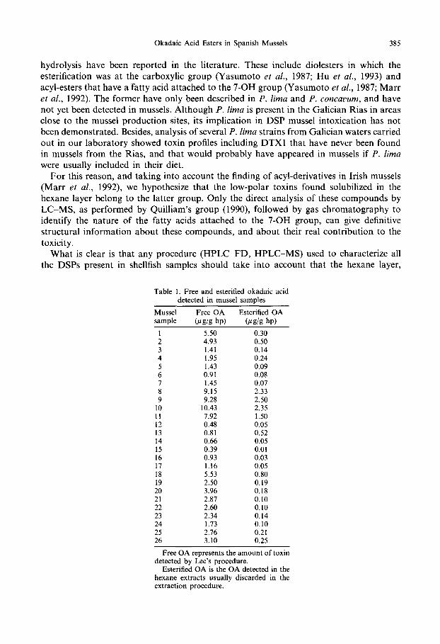
Okadaic Acid Esters in Spanish Mussels 385
hydrolysis have been reported in the literature. These include diolesters in which the esterification was at the carboxylic group (Yasumoto et al., 1987; Hu et al., 1993) and acyl-esters that have a fatty acid attached to the 7-OH group (Yasumoto et al., 1987; Marr et al., 1992). The former have only been described in P. lima and P. concavum, and have not yet been detected in mussels. Although P. lima is present in the Galician Rias in areas close to the mussel production sites, its implication in DSP mussel intoxication has not been demonstrated. Besides, analysis of several P. lima strains from Galician waters carried out in our laboratory showed toxin profiles including DTXl that have never been found in mussels from the Rias, and that would probably have appeared in mussels if P. lima were usually included in their diet.
For this reason, and taking into account the finding of acyl-derivatives in Irish mussels (Marr et al., 1992) we hypothesize that the low-polar toxins found solubilized in the hexane layer belong to the latter group. Only the direct analysis of these compounds by LC-MS, as performed by Quilliam’s group (1990) followed by gas chromatography to identify the nature of the fatty acids attached to the 7-OH group, can give definitive structural information about these compounds, and about their real contribution to the toxicity.
What is clear is that any procedure (HPLC-FD, HPLC-MS) used to characterize all the DSPs present in shellfish samples should take into account that the hexane layer,
Table 1. Free and esterified okadaic acid detected in mussel samples
Mussel Free OA Esterified OA sample @S/S hp) ‘&S/S hp)
1 5.50 0.30 2 4.93 0.50 3 1.41 0.14 4 1.95 0.24 5 1.43 0.09 6 0.91 0.08 7 1.45 0.07 8 9.15 2.33 9 9.28 2.50
10 10.43 2.35 11 7.92 1.50 12 0.48 0.05 13 0.81 0.52 14 0.66 0.05 15 0.39 0.01 16 0.93 0.03 17 1.16 0.05 18 5.53 0.80 19 2.50 0.19 20 3.96 0.18 21 2.87 0.10 22 2.60 0.10 23 2.34 0.14 24 1.73 0.10 25 2.76 0.21 26 3.10 0.25
Free OA represents the amount of toxin detected by Lee’s procedure.
Esterified OA is the OA detected in the hexane extracts usually discarded in the extraction procedure.

L_
M. L. FERNANDEZ et al.
OA
J&7 _..
+---T-T I: 20 2: 0 Time (minutes)
r I I I I I 0 5 IO 15 20 25
Time (minutes)
OA
0
(d
IO 15 20 25 Time (minutes)
r I I I I I 0 5 IO 15 20 25
Time (minutes)
Fig. 3. (a) Chromatogram for OA standard; (b) chromatogram for mussel sample 8 (Table 1) obtained after extraction, derivatization with Adam and clean-up, following Lee’s procedure (V. inj = 10 ~1, corresponding to 2.5 pg hp); (c) chromatogram for hexane extract (sample 8) usually discarded in the Lee extraction procedure, after derivatization with Adam (no traces of DSP toxins); (d) chromatogram for the hexane extract (sample 8) after alkaline hydrolysis and
derivatization with Adam (V. inj = 10 ~1, corresponding to 25 pg hp).

Okadaic Acid Esters in Spanish Mussels 387
usually discarded, can be very rich in low-polar DSPs. It is worth mentioning that some bioassay procedures used for shellfish monitoring involve hexane washing steps, to avoid
false positive results when free fatty acids are present in shellfish samples. Although in the case of the mussel samples analysed in this experiment, the concentration of these compounds only reached high values when they were already highly contaminated with non-acylated DSP toxins, the advantages of the hexane washing step should be reconsid- ered, taking into account the possible losses of toxins. This step must be avoided when analysing samples of unknown origin and with unknown DSP profiles. It is worth pointing out that acyl-derivatives of DTXl, also called DTX3, have been reported as the major toxins in Japanese scallops; therefore, depending on the shellfish species, their origin, and on different factors affecting metabolic activities, the contribution of acyl-derivatives to DSP toxicity may be more important. It must therefore be taken into account in shellfish monitoring procedures. Some cases of positive results of bioassays showing no traces of DSPs in the chromatograms obtained by the normal procedures could be due to the presence of these compounds, which are not detectable by conventional protocols.
It is suggested that alkaline hydrolysis of the hexane layer obtained after liquid-liquid partition should be included in the HP-FD procedures.
Acknowledgemenrs-We wish to express our gratitude to J. M. France, M. Norte and B. Reguera for valuable comments and suggestions, and to T. Wyatt for the English correction of the manuscript. J. Iturbe and L. Fernandez provided technical assistance in the experiments, M. I. Reyero and E. Maroiio prepared the figures and J. Fernandez typed the manuscript.
REFERENCES
Candenas, M. L., Norte, M., Arteche, E. and Martin, J. D. (1994) El Bcido okadaico coma inhibidor selective de proteinfosfatasas. Estudios a nivel de mirsculo liso. In: Actas de la Tercera Reunibn Iberico sobre Fitoplancton T6xico y Biotoxinas, pp. 64-68 (Marifio, J. and Maneiro, J. C., Eds). Santiago de Compostela: Xunta de Galicia.
Hu, T., de Freitas, A., Doyle, J., Jackson, D., Marr, J., Nixon, E., Pleasance, S., Quilliam, M., Walter, J. and Wright, J. L. C. (1993) New DSP derivatives isolated from toxic mussels and the dinoflagellates Prorocentrum lima and Prorocentrum concawm. In: Toxic Phytoplankton Blooms in the Sea, pp. 507-512 (Smayda, T. and Shimizu, Y., Eds). Amsterdam: Elsevier.
Lee, J. S., Yanagi, T., Kenma, R. and Yasumoto, T. (1987) Fluorimetric determination of diarrhetic shellfish toxins by high-performance liquid chromatography. Agric. Biol. Chem. 51, 8777881.
Lee, J. S., Igarashi, T., Fraga, S., Dahl, E., Hovgaard, P. and Yasumoto, T. (1989) Determination of diarrhetic shellfish toxins in various dinoflagellate species. J. appl. Phycol. 1, 1477152.
Marr, J. C., Hu, T., Pleasance, S., Quilliam, M. and Wright, J. (1992) Detection of a new 7-O-acyl derivatives of diarrethic shellfish poisoning toxins by liquid chromatography-mass spectrometry. Toxicon 30, 1621-1630.
Pleasance, S., Quilliam, M. A., de Freitas, A. S. W., Marr, J. C. and Cembella, A. D. (1990) Ion-spray mass spectrometry of marine toxins II. Analysis of diarrhetic shellfish toxins in plankton by liquid chromato- graphy/mass spectrometry. Rapid Commun. Mass Spectrom. 4, 206-213.
Takagi, T., Hayashi, K. and Itabashi, Y. (1984) Toxic effect of free unsaturated fatty acids in the mouse assay of diarrhetic shellfish toxin by intraperitoneal injection, Bull Jpn. Sot. Scient. Fish. 50, 1413-1418.
Yasumoto, T., Oshima, Y. and Yamaguchi, M. (1978) Occurrence of a new type of toxic shellfish in Japan and chemical properties of the toxin. BuU. Jpn. Sot. Scient. Fish. 44, 1249-1255.
Yasumoto, T.. Murata, M., Oshima, Y., Sano, M., Matsumoto, G. and Clardy, J. (1985) Diarrhetic shellfish toxins. Tetrahedron 41, 1019~1025.
Yasumoto, T., Seino N., Murakami, Y. and Murata, M. (1987) Toxins produced by benthic dinoflagellates. Biol. BUN. 172, 128-131.
Yasumoto, T., Murata, M., Lee, J. S. and Torigoe, K. (1989) Polyether toxins produced by dinoflagellates. In: Mycofoxins and Phycotoxins, pp. 375-382 (Natori, S., Hashimoto, K. and Ueno, T., Eds). Amsterdam: Elsevier.


Capítulo II – Artículo 8 - 187 -
ARTÍCULO 8
Detoxification of low polarity toxins (DTX3) from mussels
Mytilus galloprovincialis in Spain
Fernández, M.L., Míguez, A., Moroño, A., Cacho, E., Martínez, A. and
Blanco, J. 1998. In: “Harmful Algae”. B.Reguera, J. Blanco, M.L. Fernandez, T.
Wyatt. (Eds.).Xunta de Galicia-IOC of Unesco. Santiago de Compostela. pp
449-452.


Capítulo II – Artículo 8 Resumen - 189 -
Detoxification of low polarity toxins (DTX3) from mussels Mytilus
galloprovincialis in Spain
RESUMEN
Aunque el AO y la DTX2 son las principales toxinas en los mejillones españoles, la
ocurrencia de derivados acilados (ej: DTX3) de baja polaridad ha sido documentada
recientemente. Con el objeto de estudiar la cinética de detoxificación de DTX3 en
comparación con la de las toxinas precursoras, se recolectaron mejillones en la Ría de
Pontevedra inmediatamente después de una proliferación de Dinophysis. Los mejillones
fueron transferidos a un área libre de células tóxicas, dónde se mantuvieron durante 38 días.
Los niveles de AO y DTX2 durante el periodo de detoxificación fueron evaluados por HPLC-
FD. La variación temporal de DTX3 fue monitorizada mediante la hidrólisis alcalina de los
extractos de hexano de los mejillones y posterior determinación por HPLC-FD de las
correspondientes toxinas precursoras obtenidas tras la hidrólisis. La cinética de detoxificación
de la DTX3 fue diferente a la correspondiente a las toxinas precursoras. La detoxificación de
AO y DTX2 encajó bien cuantitativamente en un modelo de detoxificación de dos
compartimentos. En el caso de los derivados acilados, el ajuste fue pobre, poniendo de
manifiesto que algunos factores adicionales podrían estar implicados en el proceso. Se
muestran el contenido total de DSP, la variación en el perfil de toxinas y las tasas de
detoxificación a lo largo del periodo de detoxificación.

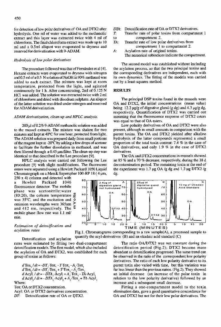


Capítulo II – Artículo 9 - 195 -
ARTÍCULO 9
Accumulation and transformation of DSP toxins in mussels
Mytilus galloprovincialis during a toxic episode caused by
Dinophysis acuminata.
Moroño, A., Arévalo, F., Fernández, M.L., Maneiro, J., Pazos, Y., Salgado, C.
and Blanco, J. 2003. Aquatic Toxicology 62, 269-280

Capítulo II – Artículo 9 Resumen - 197 -
Accumulation and transformation of DSP toxins in mussels Mytilus
galloprovincialis during a toxic episode caused by Dinophysis acuminata
RESUMEN En el presente trabajo se estudió la evolución de varios episodios del organismo productor
de DSP, Dinophysis acuminata y la correspondiente cinética de acumulación y pérdida de
toxinas en mejillones Mytilus galloprovincialis. Para ello se recogieron muestras de
mejillones y agua de mar en una batea de la Ría de Vigo con una frecuencia de 2-3 veces por
semana. El contenido en toxinas DSP en los mejillones y en el agua fue analizado por HPLC-
FD y el fitoplancton, en muestras sedimentadas, fue cuantificado mediante microscopia
inversa. En las muestras de plancton, que estuvieron constituidas principalmente por células
de D. acuminata, se detectaron AO y derivados conjugados del mismo (AO, CF) que fueron
determinados por hidrólisis enzimática. La toxina acumulada en los mejillones estuvo
mayoritariamente presente en forma de AO, alcanzándose un máximo de 10.1 µg AO g-1. en
la glándula digestiva en 16 días, y reduciéndose por debajo del nivel máximo permitido
(aproximadamente 2 µg AO g-1) en 45 días. Las formas conjugadas de baja polaridad (LPCF)
estimadas mediante su extracción con hexano, representaron el 6.2 % del contenido total en
toxinas de los mejillones. Para cuantificar las tasas de los procesos implicados en la
acumulación, transformación y pérdida de las toxinas, se diseñaron e implantaron dos
modelos dinámicos, uno de ellos de un compartimento y otro de dos compartimentos,
incluyendo AO y sus formas conjugadas como variables. El modelo de un compartimento
proporcionó un buen ajuste con los datos reales de AO y sus formas conjugadas (r2 = 0.92 y r2
= 0.94 respectivamente). El modelo de dos compartimentos no ajustó los datos de manera
significativamente mejor que el modelo de un compartimento (r2 = 0.93 y r2 = 0.95
respectivamente). Se estimaron tasas de hidrólisis elevadas para la mayor parte de los
derivados conjugados del AO, lo que significa que la mayor parte de estas formas provienen
del plancton ingerido. Las bajas tasas de acilación estimadas apoyan la hipótesis previa y
sugieren que la formación de LPFC por acilación directa del AO es de pequeña importancia
en M. galloprovincialis. Sólo en aquellos casos en los que el periodo de intoxicación es muy
largo, los derivados acilados pueden llegar a ser importantes, dado que parece que se
acumulan por largo tiempo en los mejillones, como sugieren las bajas tasas de hidrólisis y
depuración estimadas por el modelo.

Accumulation and transformation of DSP toxins in musselsMytilus galloprovincialis during a toxic episode caused by
Dinophysis acuminata
A. Morono a,*, F. Arevalo a, M.L. Fernandez b, J. Maneiro a, Y. Pazos a,C. Salgado a, J. Blanco c
a Centro de Control do Medio Marino (Xunta de Galicia), Peirao de Vilaxoan s/n, Vilagarcıa de Arousa 36611, Pontevedra, Spainb Laboratorio de Sanidad Exterior de Vigo, European Community Reference Laboratory on Marine Biotoxins, Estacion Marıtima s/n,
36271 Vigo, Spainc Centro de Investigacions Marinas (Xunta de Galicia), Apto. 13, 36620 Vilanova de Arousa, Pontevedra, Spain
Received 29 November 2001; received in revised form 21 June 2002; accepted 3 July 2002
Abstract
The time course of several outbreaks of the diarrhetic shellfish poisoning (DSP) producer Dinophysis acuminata and
the consequent kinetic of accumulation and loss of toxins in mussels Mytilus galloprovincialis feeding on them was
studied. Samples of mussels and seawater were frequently (2�/3 times a week) collected from a raft in the Rıa de Vigo.
DSP toxins content of mussels and water was analyzed by HPLC-FD and phytoplankton was quantified in an inverted
light microscope. Only okadaic acid (OA) and some of its conjugated forms (OA CF), estimated by enzymatic
hydrolysis, were found in the plankton samples obtained, comprised mainly of D. acuminata cells. The main
accumulated form in mussels was OA reaching a maximum of 10.1 mg OA g�1 in the digestive gland (d.g.) in 16 days,
falling below the quarantine level (ca. 2 mg OA g�1 d.g.) by 45 days. The low polarity conjugated forms (LPCF),
estimated by hexane extraction, accounted for 6.2% of the total toxin burden of the mussels. To quantify the rates of the
processes involved in the accumulation, transformation and loss of the toxins, two dynamic models, a one-compartment
and a two-compartment, including OA and its conjugated forms as variables were designed and implemented. The one-
compartment model provided a good fit to the OA and LPCF actual data (r2�/0.92 and r2�/0.94, respectively). The
two-compartment model did not fit the data markedly better than its one-compartment counterpart (r2�/0.93 and r2�/
0.95, for OA and LPCF, respectively). High hydrolysis rates were estimated for most of the OA CF, which means that
these forms came largely from the ingested plankton. The low estimated acylation rates support the previous point and
suggest that the formation of LPCF by direct acylation of the OA is of little importance in M. galloprovincialis . Only in
cases where the intoxication period is very long, can the formed acyl-derivatives be important, because they seem to
accumulate for a long time in the mussels, as suggested by the low hydrolysis and depuration rates estimated from
model fitting.
# 2002 Elsevier Science B.V. All rights reserved.
* Corresponding author. Tel.: �/34-986-512-320; fax: �/34-986-512-300
E-mail address: [email protected] (A. Morono).
Aquatic Toxicology 62 (2003) 269�/280
www.elsevier.com/locate/aquatox
0166-445X/02/$ - see front matter # 2002 Elsevier Science B.V. All rights reserved.
PII: S 0 1 6 6 - 4 4 5 X ( 0 2 ) 0 0 1 0 5 - 4
Keywords: Accumulation; Dinophysis acuminata ; DSP toxins; Models; Mytilus galloprovincialis ; Transformation
1. Introduction
The accumulation of phycotoxins is one of the
most critical problems in bivalve aquaculture. To
date paralytic shellfish poisoning (PSP) toxins,
amnesic shellfish poisoning (ASP) toxins and
diarrhetic shellfish poisoning (DSP) toxins are
considered to be the main phycotoxin groups. Of
these, DSP includes some of the toxins with the
lowest acute effect, okadaic acid (OA) and its
derivatives, as they only seem to produce severe
gastrointestinal disorders. However, in many bi-
valve-producing areas, they have a far-reaching
economic impact as their appearance leads to the
imposition of long bans on the culture harvest
(Masselin et al., 1992; Reguera et al., 1993;
Carmody et al., 1995; McMahon et al., 1998).
The phytoplankton cells that produce and contain
the toxins are ingested by bivalves and the toxins
are accumulated, transformed and eliminated. A
knowledge of all these processes is of the utmost
importance for being able to predict the time
course of a toxic episode and therefore, to mini-
mize its consequences.
The toxins produced and the cellular toxins
content are species-specific. They also have a
noticeable intra-specific variation, depending on
strains and physiological conditions (Lee et al.,
1989; Subba Rao et al., 1993; Andersen et al.,
1996; Johansson et al., 1996; Sato et al., 1996;
Suzuki et al., 1997; Fernandez et al., 2001). Most
of the species belonging to the genus Dinophysis ,
the most important DSP toxins producer, contain
mainly OA and its derivatives, dinophysistoxin-1
(DTX1) and dinophysistoxin-2 (DTX2) (Cem-
bella, 1989; Lee et al., 1989; Andersen et al.,
1996; Johansson et al., 1996; Suzuki et al., 1997;
James et al., 1998; Fernandez et al., 2001). A
number of conjugated forms of OA, DTX1 and
DTX2 have been discovered and a wide variety
appear to exist, mainly because the original toxins
may combine with a great number of substances.
Several derivatives such as diol-esters or DTX4-
type compounds have been detected in Prorocen-
trum lima and P. maculosum , two benthic dino-
flagellates that produce DSP toxins (Yasumoto et
al., 1989; Hu et al., 1992, 1993; Quilliam et al.,
1996), but they have not been found in bivalves or
in pelagic plankton. Acyl-derivatives of the DTX3-
type, in which saturated or unsaturated fatty acids
are attached through the 7-OH group, have been
reported in the digestive glands (d.g.) of toxic
bivalves (Yasumoto et al., 1985; Marr et al., 1992;
Fernandez et al., 1996; Suzuki et al., 1999; Vale
and Sampayo, 1999). They have never been found
in phytoplankton and, based on current knowl-
edge, they would seem to originate from the
transformation of OA and analogues in the
bivalves (Yasumoto et al., 1989; Lee et al., 1989;
Suzuki et al., 1999; Suzuki and Mitsuya, 2001).
The polarity of DTX4 type compounds is con-
siderably higher than that of OA. Diol-esters, on
the other hand, are slightly less polar than OA and
the acyl-derivatives usually have low polarity and,
for the most part, can be separated from the free-
acid forms by extraction with hexane (Suzuki et
al., 1999; Suzuki and Mitsuya, 2001; Vale, 1999).
Once the toxins are ingested, they can be
accumulated or transformed. To date only three
possible transformations have been considered:
hydrolysis and oxidation of conjugated forms
and acylation of the free-acid forms. Acylation
has been demonstrated in bivalves by Suzuki et al.
(1999), Suzuki and Mitsuya (2001). Hydrolysis has
been suggested in bivalves by Fernandez et al.
(1998) and demonstrated in homogenates of the
DSP-producer dinoflagellate P. lima (Quilliam et
al., 1996; Windust et al., 1997, 2000). Oxidation of
OA diol-ester has been observed by Windust et al.
(1997) in Thalassiosira weissflogii but only when
the diatom cells are intact (Windust et al., 2000).
A. Morono et al. / Aquatic Toxicology 62 (2003) 269�/280270
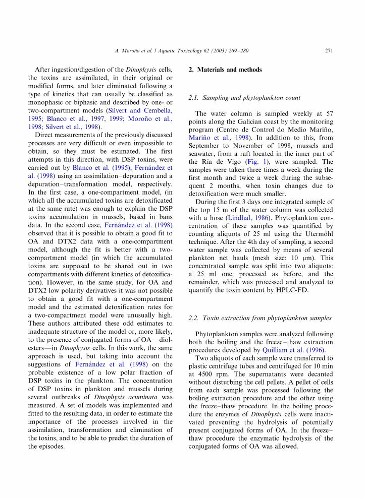
After ingestion/digestion of the Dinophysis cells,
the toxins are assimilated, in their original or
modified forms, and later eliminated following a
type of kinetics that can usually be classified as
monophasic or biphasic and described by one- or
two-compartment models (Silvert and Cembella,
1995; Blanco et al., 1997, 1999; Morono et al.,
1998; Silvert et al., 1998).Direct measurements of the previously discussed
processes are very difficult or even impossible to
obtain, so they must be estimated. The first
attempts in this direction, with DSP toxins, were
carried out by Blanco et al. (1995), Fernandez et
al. (1998) using an assimilation�/depuration and a
depuration�/transformation model, respectively.
In the first case, a one-compartment model, (in
which all the accumulated toxins are detoxificated
at the same rate) was enough to explain the DSP
toxins accumulation in mussels, based in bans
data. In the second case, Fernandez et al. (1998)
observed that it is possible to obtain a good fit to
OA and DTX2 data with a one-compartment
model, although the fit is better with a two-
compartment model (in which the accumulated
toxins are supposed to be shared out in two
compartments with different kinetics of detoxifica-
tion). However, in the same study, for OA and
DTX2 low polarity derivatives it was not possible
to obtain a good fit with a one-compartment
model and the estimated detoxification rates for
a two-compartment model were unusually high.
These authors attributed these odd estimates to
inadequate structure of the model or, more likely,
to the presence of conjugated forms of OA*/diol-
esters*/in Dinophysis cells. In this work, the same
approach is used, but taking into account the
suggestions of Fernandez et al. (1998) on the
probable existence of a low polar fraction of
DSP toxins in the plankton. The concentration
of DSP toxins in plankton and mussels during
several outbreaks of Dinophysis acuminata was
measured. A set of models was implemented and
fitted to the resulting data, in order to estimate the
importance of the processes involved in the
assimilation, transformation and elimination of
the toxins, and to be able to predict the duration of
the episodes.
2. Materials and methods
2.1. Sampling and phytoplankton count
The water column is sampled weekly at 57
points along the Galician coast by the monitoring
program (Centro de Control do Medio Marino,
Marino et al., 1998). In addition to this, from
September to November of 1998, mussels and
seawater, from a raft located in the inner part of
the Rıa de Vigo (Fig. 1), were sampled. The
samples were taken three times a week during thefirst month and twice a week during the subse-
quent 2 months, when toxin changes due to
detoxification were much smaller.
During the first 3 days one integrated sample of
the top 15 m of the water column was collected
with a hose (Lindhal, 1986). Phytoplankton con-
centration of these samples was quantified by
counting aliquots of 25 ml using the Utermohltechnique. After the 4th day of sampling, a second
water sample was collected by means of several
plankton net hauls (mesh size: 10 mm). This
concentrated sample was split into two aliquots:
a 25 ml one, processed as before, and the
remainder, which was processed and analyzed to
quantify the toxin content by HPLC-FD.
2.2. Toxin extraction from phytoplankton samples
Phytoplankton samples were analyzed following
both the boiling and the freeze�/thaw extraction
procedures developed by Quilliam et al. (1996).
Two aliquots of each sample were transferred to
plastic centrifuge tubes and centrifuged for 10 min
at 4500 rpm. The supernatants were decanted
without disturbing the cell pellets. A pellet of cells
from each sample was processed following theboiling extraction procedure and the other using
the freeze�/thaw procedure. In the boiling proce-
dure the enzymes of Dinophysis cells were inacti-
vated preventing the hydrolysis of potentially
present conjugated forms of OA. In the freeze�/
thaw procedure the enzymatic hydrolysis of the
conjugated forms of OA was allowed.
A. Morono et al. / Aquatic Toxicology 62 (2003) 269�/280 271
2.3. Toxin extraction from mussel samples
The DSP toxin extraction from mussel samples,
ADAM derivatization and clean-up were done
following the Lee et al. (1987) procedure. Hydro-
lysis was carried out following the procedure
developed by Fernandez et al. (1996).
2.4. HPLC analysis
HPLC analysis of phytoplankton and mussels
were performed following the method described by
Lee et al. (1987) with slight modifications. The
fluorescent esters were separated using a Waters
600-E liquid chromatographic system with a
Fig. 1. Map of the Rıa de Vigo (NW, Spain). The black arrow shows the location of the raft where mussels and seawater were
periodically sampled throughout a D. acuminata event. The blank geometric figures are groups of rafts.
A. Morono et al. / Aquatic Toxicology 62 (2003) 269�/280272

Merck LiChrosphere100-RP18 (5 mm, 125�/4mm) column and detected in a Waters 474 scan-
ning fluorescence detector. The mobile phase was
acetonitrile:water (80:20), the column temperature
was 25 8C and the excitation and emission wave-
lengths were 365 and 412 nm, respectively. Flow
rate was 0.8 ml min�1.
The OA standard was supplied by NRC-Ca-
nada. The 9-anthryldiazomethane (ADAM) re-agent used for derivatization was supplied by
Boehringer Ingelheim. The HPLC grade solvents
were purchased from Merck. The SPE Silica
cartridges used for clean-up were obtained from
Waters.
2.5. Models
The models were implemented using Matlab and
Matlab Simulink. The optimum fit to the data was
obtained minimizing chi-square (x2) by changing
the parameters of the different models using the
Matlab Optimization Toolbox.
Filtration rate and assimilation efficiency were
considered to be constant and their numerical
values were taken from the results obtained in theRıa de Arousa for Mytilus galloprovincialis (Na-
varro et al., 1991). The ingestion rate was not
included in the model because it is considered to be
equivalent to the filtration rate in Galician Rıas,
due to the fact that the concentration of seston is
usually below the level of pseudofaeces production
(5 mg l�1, Widdows et al., 1979; Navarro et al.,
1991). The detoxification rates (OA and OA CF),acylation rates, hydrolysis rate and transfer rates
were estimated by fitting the model to the real DSP
toxins data. In addition, the toxin content per D.
acuminata cell, for the first day of D. acuminata
detection and for the three subsequent sampling
days, were estimated by the fit of the model.
3. Results
3.1. The Dinophysis acuminata bloom
In late August 1998, a D. acuminata bloom built
up in the Galician Rıas with strong southerly
winds, which generated an oceanographic situa-
tion of downwelling. This bloom was first detectedon August 31st at most of the 57 stations sampled
by the monitoring program of the Galician coast,
with a concentration of 1320 cells l�1 in the
station closest to the selected mussel raft. Three
days later, a maximum of 5240 cells l�1 was
detected on the same sampling station, decreasing
afterwards to 40 cells l�1 in 14 days (Fig. 2). D.
acuminata persisted at this low concentration for 3months with a weak increase to 240 cells l�1 in
early October and to 480 cells l�1 in early
November (Fig. 2).
3.2. DSP toxins in mussels and plankton
OA was the main toxin found in the mussels
(from 0.18 to 10.1 mg g�1of d.g.). In addition to
OA, low polarity (hexane-soluble) conjugated
forms (LPCF)*/very likely DTX3 type
compounds*/were also found, accounting for a
maximum of 6.2% of all accumulated toxins
(between undetected and 0.31 mg g�1 d.g.). The
ratio LPCF/OA varied with time (Fig. 3), showingan initial decrease followed by a subsequent
increase to levels higher than those detected at
the beginning.
Part of the OA in the plankton was present as
conjugated forms (very likely diol-esters or DTX4-
type compounds), as revealed by the increase in
the OA yield after allowing the enzymatic hydro-
lysis (freeze�/thaw procedure) of the planktonsamples. The OA toxin content per D. acuminata
cell varied between not detected and 11.3 pg cell�1
and its conjugated forms between not detected and
Fig. 2. Temporal evolution of the concentration of D. acumi-
nata sampled on the experimental raft (Rıa de Vigo) in 1998.
A. Morono et al. / Aquatic Toxicology 62 (2003) 269�/280 273
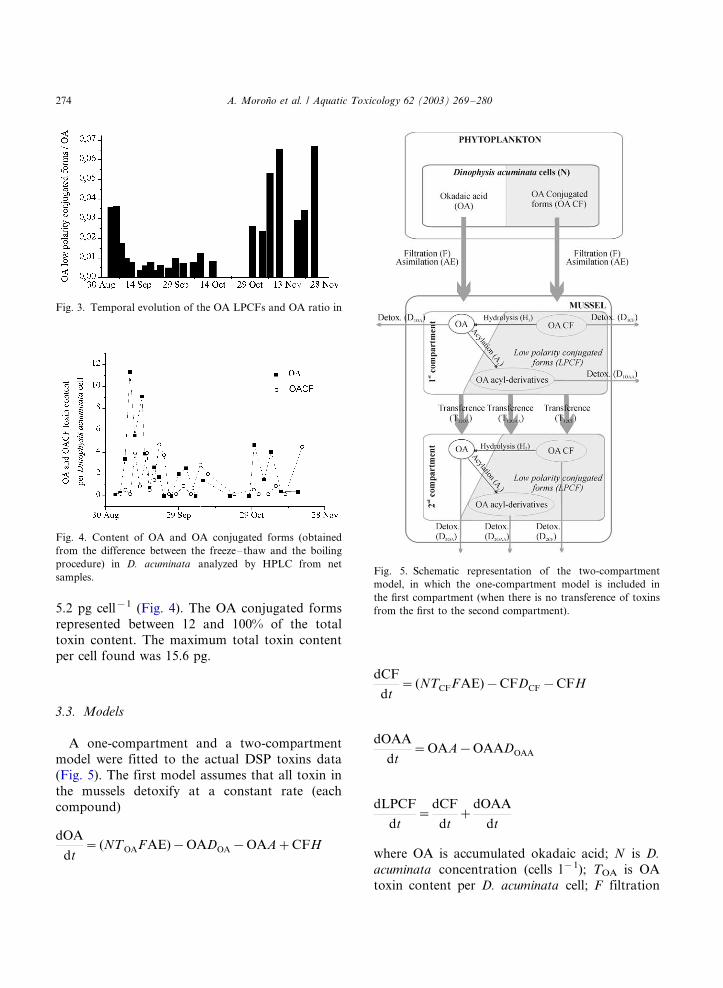
5.2 pg cell�1 (Fig. 4). The OA conjugated forms
represented between 12 and 100% of the total
toxin content. The maximum total toxin content
per cell found was 15.6 pg.
3.3. Models
A one-compartment and a two-compartment
model were fitted to the actual DSP toxins data
(Fig. 5). The first model assumes that all toxin in
the mussels detoxify at a constant rate (each
compound)
dOA
dt�(NTOAFAE)�OADOA�OAA�CFH
dCF
dt�(NTCFFAE)�CFDCF�CFH
dOAA
dt�OAA�OAADOAA
dLPCF
dt�
dCF
dt�
dOAA
dt
where OA is accumulated okadaic acid; N is D.
acuminata concentration (cells l�1); TOA is OA
toxin content per D. acuminata cell; F filtration
Fig. 3. Temporal evolution of the OA LPCFs and OA ratio in
Fig. 4. Content of OA and OA conjugated forms (obtained
from the difference between the freeze�/thaw and the boiling
procedure) in D. acuminata analyzed by HPLC from net
samples.Fig. 5. Schematic representation of the two-compartment
model, in which the one-compartment model is included in
the first compartment (when there is no transference of toxins
from the first to the second compartment).
A. Morono et al. / Aquatic Toxicology 62 (2003) 269�/280274

rate (l day�1); AE is assimilation efficiency; DOA isOA detoxification rate; A is OA acylation rate; CF
is accumulated conjugated forms; H is hydrolysis
rate; TCF is contents of conjugated forms per D.
acuminata cell; DCF is detoxification rate of
conjugated forms; OAA is OA acyl-derivatives;
DOAA is detoxification rate of OA acyl-derivatives;
and LPCF is accumulated low polarity conjugated
forms.In the two-compartment model it is assumed
that, for each toxin, there are two different pools
that detoxify at different rates, one fast (the first
compartment) and another slow (second compart-
ment). There is also a transfer of toxins from the
first to the second compartment (Fig. 5).
dOA1
dt�(NTOAFAE)�OA1D1OA�OA1A1
�OA1T12OA�CF1H
dCF1
dt� (NTCFFAE)�CF1DCF�CF1H
�CF1T12CF1
dOAA1
dt�OA1A�OAA1D1OAA�OAA1T12OAA1
dLPCF1
dt�
dCF1
dt�
dOAA1
dt
dOA2
dt�OA1T12OA�OA2A2�OA2D2OA�CF2H2
dCF2
dt�CF1T12CF1
�CF2D2CF�CF2H2
dOAA2
dt�OA2A2�OAA1T12OAA1
�OAA2DOAA2
dLPCF2
dt�
dCF2
dt�
dOAA2
dt
T12 is the transfer rate of OA or its derivativesfrom the first to the second compartment. The
remaining acronyms are the same as in the one-
compartment model with a number to indicate the
compartment.
The OA derivatives may be comprised of a great
variety of compounds with different solubility and
it is possible that not all of them were detected in
the mussels (especially DTX-4 type compoundsand some diol-esters). Taking this into account,
the two models were run again assuming that only
a variable percent of the OA derivatives had been
detected.
3.4. Model fitting
The one-compartment model provided a very
good fit for the OA toxin content in mussels (r2�/
0.92) as well as its LPCF (r2�/0.94) (Fig. 6).Notwithstanding, the model was not able to attain
the maximum concentration of the observed OA
data. The estimated rates (Table 1) suggest that
most of the LPCF detected in the mussels were OA
conjugated forms assimilated from the phyto-
plankton cells, with the acylation rate of OA being
very low. For the OA acyl-derivatives the esti-
mated detoxification rate is zero indicating thatthere is an almost permanent accumulation of
these compounds, even when this accumulation
must be slow because of the also slow acylation
rate. With the two-compartment model it was
Fig. 6. Fit of the one- and two-compartment models to the OA
(A) and LPCFs (B) in mussels of a raft located in the Rıa de
Vigo (NW, Spain).
A. Morono et al. / Aquatic Toxicology 62 (2003) 269�/280 275
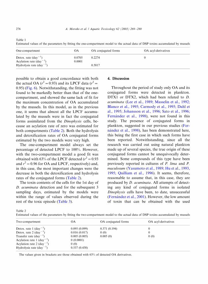
possible to obtain a good concordance with both
the actual OA (r2�/0.93) and its LPCF data (r2�/
0.95) (Fig. 6). Notwithstanding, the fitting was not
found to be markedly better than that of the one-
compartment, and showed the same lack of fit for
the maximum concentration of OA accumulated
by the mussels. In this model, as in the previous
one, it seems that almost all the LPCF accumu-
lated by the mussels were in fact the conjugated
forms assimilated from the Dinophysis cells, be-
cause an acylation rate of zero was estimated for
both compartments (Table 2). Both the hydrolysis
and detoxification rates of OA conjugated forms
estimated by the two models were very high.
The one-compartment model always set the
percentage of detected LPCF to 100%. However,
with the two-compartment model a good fit was
obtained with 63% of the LPCF detected (r2�/0.93
and r2�/0.96 for OA and LPCF, respectively) and,
in this case, the most important changes were the
decrease in both the detoxification and hydrolysis
rates of the conjugated forms (Table 2).The toxin contents of the cells for the 1st day of
D. acuminata detection and for the subsequent 3
sampling days, estimated by the models were
within the range of values observed during the
rest of the toxic episode (Table 3).
4. Discussion
Throughout the period of study only OA and its
conjugated forms were detected in plankton.
DTX1 or DTX2, which had been related to D.
acuminata (Lee et al., 1989; Masselin et al., 1992;
Blanco et al., 1995; Carmody et al., 1995; Dahl et
al., 1995; Johansson et al., 1996; Sato et al., 1996;
Fernandez et al., 1998), were not found in this
study. The presence of conjugated forms in
plankton, suggested in our previous studies (Fer-
nandez et al., 1998), has been demonstrated here,
this being the first case in which such forms have
been reported. Notwithstanding, since all the
research was carried out using natural plankton
made up of several species, the true origin of these
conjugated forms cannot be unequivocally deter-
mined. Some compounds of this type have been
previously reported in cultures of P. lima and P.
maculosum (Yasumoto et al., 1989; Hu et al., 1993,
1995; Quilliam et al., 1996). It seems, therefore,
reasonable to assume that, in this case, they are
produced by D. acuminata . All attempts of detect-
ing any kind of conjugated forms in isolated
Dinophysis cells have been, to date, unsuccessful
(Fernandez et al., 2001). However, the low amount
of toxin that can be obtained with the used
Table 1
Estimated values of the parameters by fitting the one-compartment model to the actual data of DSP toxins accumulated by mussels
One-compartment OA OA conjugated forms OA acyl-derivatives
Detox. rate (day�1) 0.0705 0.2274 0
Acylation rate (day�1) 0.0001
Hydrolysis rate (day�1) 0.5817
Table 2
Estimated values of the parameters by fitting the two-compartment model to the actual data of DSP toxins accumulated by mussels
Two-compartment OA OA conjugated forms OA acyl-derivatives
Detox. rate 1 (day�1) 0.095 (0.099) 0.371 (0.194) 0
Detox. rate 2 (day�1) 0.016 (0.017) 0 (0) 0
Transfer rate (day�1) 0.005 (0.005) 0.005 (0) 0 (0)
Acylation rate 1 (day�1) 0 (0.0001)
Acylation rate 2 (day�1) 0 (0)
Hydrolysis rate (day�1) 0.537 (0.438)
The values given in brackets are those obtained with 63% of detected OA derivatives.
A. Morono et al. / Aquatic Toxicology 62 (2003) 269�/280276
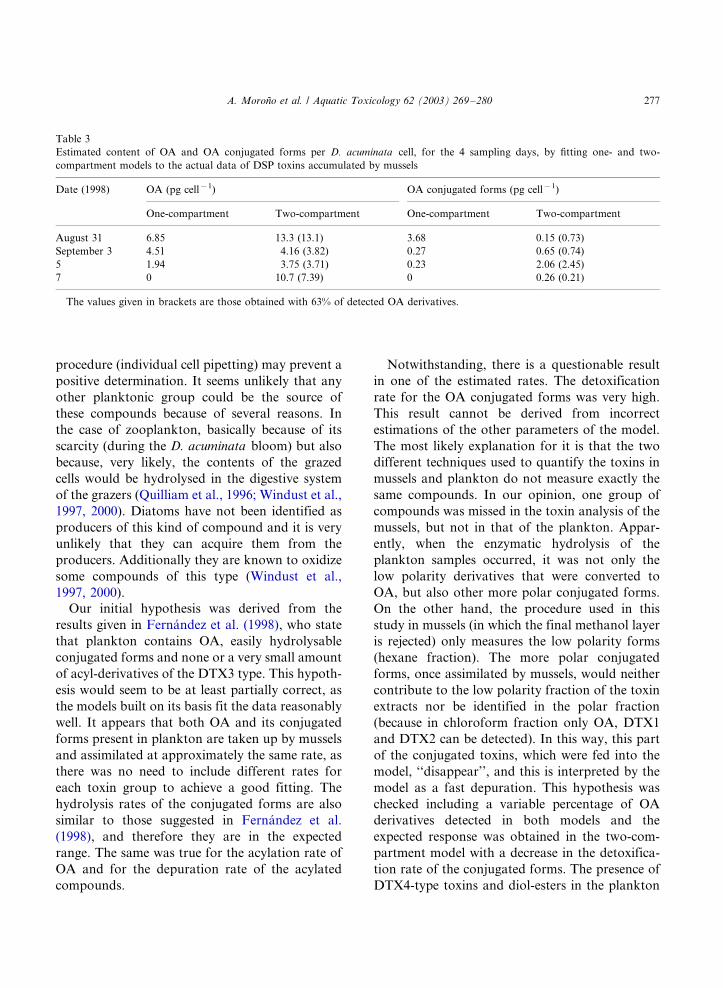
procedure (individual cell pipetting) may prevent a
positive determination. It seems unlikely that any
other planktonic group could be the source of
these compounds because of several reasons. In
the case of zooplankton, basically because of its
scarcity (during the D. acuminata bloom) but also
because, very likely, the contents of the grazed
cells would be hydrolysed in the digestive system
of the grazers (Quilliam et al., 1996; Windust et al.,
1997, 2000). Diatoms have not been identified as
producers of this kind of compound and it is very
unlikely that they can acquire them from the
producers. Additionally they are known to oxidize
some compounds of this type (Windust et al.,
1997, 2000).
Our initial hypothesis was derived from the
results given in Fernandez et al. (1998), who state
that plankton contains OA, easily hydrolysable
conjugated forms and none or a very small amount
of acyl-derivatives of the DTX3 type. This hypoth-
esis would seem to be at least partially correct, as
the models built on its basis fit the data reasonably
well. It appears that both OA and its conjugated
forms present in plankton are taken up by mussels
and assimilated at approximately the same rate, as
there was no need to include different rates for
each toxin group to achieve a good fitting. The
hydrolysis rates of the conjugated forms are also
similar to those suggested in Fernandez et al.
(1998), and therefore they are in the expected
range. The same was true for the acylation rate of
OA and for the depuration rate of the acylated
compounds.
Notwithstanding, there is a questionable result
in one of the estimated rates. The detoxification
rate for the OA conjugated forms was very high.
This result cannot be derived from incorrect
estimations of the other parameters of the model.
The most likely explanation for it is that the two
different techniques used to quantify the toxins in
mussels and plankton do not measure exactly the
same compounds. In our opinion, one group of
compounds was missed in the toxin analysis of the
mussels, but not in that of the plankton. Appar-
ently, when the enzymatic hydrolysis of the
plankton samples occurred, it was not only the
low polarity derivatives that were converted to
OA, but also other more polar conjugated forms.
On the other hand, the procedure used in this
study in mussels (in which the final methanol layer
is rejected) only measures the low polarity forms
(hexane fraction). The more polar conjugated
forms, once assimilated by mussels, would neither
contribute to the low polarity fraction of the toxin
extracts nor be identified in the polar fraction
(because in chloroform fraction only OA, DTX1
and DTX2 can be detected). In this way, this part
of the conjugated toxins, which were fed into the
model, ‘‘disappear’’, and this is interpreted by the
model as a fast depuration. This hypothesis was
checked including a variable percentage of OA
derivatives detected in both models and the
expected response was obtained in the two-com-
partment model with a decrease in the detoxifica-
tion rate of the conjugated forms. The presence of
DTX4-type toxins and diol-esters in the plankton
Table 3
Estimated content of OA and OA conjugated forms per D. acuminata cell, for the 4 sampling days, by fitting one- and two-
compartment models to the actual data of DSP toxins accumulated by mussels
Date (1998) OA (pg cell�1) OA conjugated forms (pg cell�1)
One-compartment Two-compartment One-compartment Two-compartment
August 31 6.85 13.3 (13.1) 3.68 0.15 (0.73)
September 3 4.51 4.16 (3.82) 0.27 0.65 (0.74)
5 1.94 3.75 (3.71) 0.23 2.06 (2.45)
7 0 10.7 (7.39) 0 0.26 (0.21)
The values given in brackets are those obtained with 63% of detected OA derivatives.
A. Morono et al. / Aquatic Toxicology 62 (2003) 269�/280 277
would produce the response found, because theyhave the two characteristics already mentioned: (a)
they (or at least some of them) are polar enough to
be excluded from the low polarity fraction of the
toxin extracts; and (b) they are easily hydrolysable,
thus contributing to the hydrolysable fraction of
the plankton samples. Quilliam et al. (1996)
observed that the enzymatic conversion of DTX-
4 type compounds to diol-ester, when the cells ofP. lima are disrupted, is very fast (minutes)
followed by a slower (hours) enzymatic conversion
of diol-esters to OA. Windust et al. (1997) also
observed that the increase in OA concentration
due to the hydrolysis of DTX-4 and OA diol-esters
may occur in a sterile medium, without the
presence of any organism. The hydrolysis rates
observed by these authors were very similar tothose estimated by the two models applied in this
study. However, the possibility that this relatively
polar and highly hydrolysable fraction is com-
prised of another substance cannot be overlooked.
OA is progressively acylated and therefore
transformed to low polarity derivatives, probably
of the DTX3 type, as demonstrated by Suzuki et
al. (1999), Suzuki and Mitsuya (2001). The rate atwhich these acylated compounds are formed is
low, but they are nearly not eliminated, supporting
the findings by Fernandez et al. (1998) on the same
bivalve species. The combination of these two
characteristics of their kinetics limits the toxic
relevance of the acylated forms to the final phase
of long lasting DSP episodes, in the case of M.
galloprovincialis . In other bivalve species this maynot be the case, as suggested by the data obtained
by Vale and Sampayo (1999), which probably need
much higher acylation rates to take place.
The toxin content per D. acuminata cell ob-
tained in this study, and the values for the first 4
days estimated from the fitting of the model, are
within the range of values observed in different
places for this species (Cembella, 1989; Lee et al.,1989; Andersen et al., 1996; Johansson et al.,
1996), and in the range of values previously
observed and, at the same time, in Galician Rıas
(Blanco et al., 1995; Fernandez et al., 2001;
Maneiro et al., 2000).
No clear compartmentalization of the toxins
was found given that a one-compartment model
was sufficient to explain the intoxication�/detox-ification processes. Bauder et al. (1996), Holmes et
al. (1999) observed that the OA and its derivatives
accumulated in the d.g. of Argopecten irradians
and Perna viridis show seemingly biphasic detox-
ification kinetics. All of this indicates that prob-
ably even when it is possible that a small second
compartment exist in the d.g., its importance
would be, in such a case, so small that it wouldnot justify the increment in the complexity of the
model.
5. Conclusion
The hypothesis suggested by Fernandez et al.
(1998) seems to be correct, this being the first
report of OA conjugated forms in natural plank-
ton. Moreover, OA is acylated at a very low rate
and the ingested LPCF are quickly hydrolysed. It
seems also that there are some hydrolysable toxinderivatives in the plankton that are not low
polarity compounds, but no production of sub-
stances of this kind has been described for
Dinophysis species. Further studies applying LC-
MS/MS techniques will be done to identify the
nature of the different conjugated forms and
accurately determine its contribution to the DSP
toxin content in both Dinophysis cells and mussels.
Acknowledgements
We wish to express our gratitude to J. Correa
and S. Muniz for their technical assistance in the
HPLC-FD analysis of the samples. The authors
would also like to thank to the staff of the
Department of Oceanographic Conditions and
Phytoplankton (F. Amoedo, C. Dıaz, P. Garcıa,
I. Lemos, A. Lopez, J. Mourino and S. Roura) and
to the Sampling Department (S. Bastos, M.J.Crespo, P. Ferreiro, M.T. Garcıa, M. Isorna, M.
Madrinan) for their technical assistance. This
experiment was partially supported by the projects
CITMAR 98-0978 and PGIDT-CIMA-99/03 (Plan
Galego de Investigacion e Desenvolvemento Tec-
noloxico).
A. Morono et al. / Aquatic Toxicology 62 (2003) 269�/280278
References
Andersen, P., Hald, B., Emsholm, H., 1996. Toxicity of
Dinophysis acuminata in Danish coastal waters. In: Yasu-
moto, T., Oshima, Y., Fukuyo, Y. (Eds.), Harmful and
Toxic Algal Blooms. Intergovernmental Oceanographic
Commission of UNESCO, New York, pp. 281�/284.
Bauder, A.G., Cembella, A.D., Quilliam, M.A., 1996. Dy-
namics of diarrhetic shellfish toxins from the dinoflagellate,
Prorocentrum lima , in the bay scallop, Argopecten irradians .
In: Yasumoto, T., Oshima, Y., Fukuyo, Y. (Eds.), Harmful
and Toxic Algal Blooms. Intergovernmental Oceanographic
Commission of UNESCO, New York, pp. 433�/436.
Blanco, J., Fernandez, M., Marino, J., Reguera, B., Mıguez, A.,
Maneiro, J., Cacho, E., Martınez, A., 1995. From Dino-
physis spp. toxicity to DSP outbreaks: a preliminary model
of toxin accumulation in mussels. In: Lassus, P., Arzul, G.,
Erard, E., Gentien, P., Marcaillou, C. (Eds.), Harmful
Marine Algal Blooms. Lavoisier, Paris, pp. 777�/782.
Blanco, J., Morono, A., Franco, J., Reyero, M.I., 1997. PSP
detoxification kinetics in the mussel Mytilus gallo-
provincialis . One- and two-compartment models and the
effect of some environmental variables. Mar. Ecol. Prog.
Ser. 158, 165�/175.
Blanco, J., Fernandez, M., Mıguez, A., Morono, A., 1999.
Okadaic acid depuration in the mussel Mytilus gallo-
provincialis : one- and two-compartment models and the
effect of environmental conditions. Mar. Ecol. Prog. Ser.
176, 153�/163.
Carmody, E.P., James, K.J., Kelly, S.S., Thomas, K., 1995.
Complex diarrhetic shellfish toxin profiles in Irish mussels.
In: Lassus, P., Arzul, G., Erard, E., Gentien, P., Marcaillou,
C. (Eds.), Harmful Marine Algal Blooms. Lavoisier, Paris,
pp. 273�/278.
Cembella, A.D., 1989. Occurrence of okadaic acid, a major
diarrhetic shellfish toxin, in natural populations of Dino-
physis spp. from the eastern coast of North America. J.
Appl. Phycol. 1, 307�/310.
Dahl, E., Rogstad, A., Aune, T., Hormazabal, V., Underdal,
B., 1995. Toxicity of mussels related to occurrence of
Dinophysis especies. In: Lassus, P., Arzul, G., Erard, E.,
Gentien, P., Marcaillou, C. (Eds.), Harmful Marine Algal
Blooms. Lavoisier, Paris, pp. 783�/788.
Fernandez, M.L., Mıguez, A., Cacho, E., Martınez, A., 1996.
Detection of okadaic acid esters in the hexane extracts of
Spanish mussels. Toxicon 34, 381�/387.
Fernandez, M.L., Mıguez, A., Morono, A., Cacho, E., Martı-
nez, A., Blanco, J., 1998. Detoxification of low polarity
toxins (DTX3) from mussels Mytilus galloprovincialis in
Spain. In: Reguera, B., Blanco, J., Fernandez, M.L., Wyatt,
T. (Eds.), Harmful Algae. Xunta de Galicia and Intergo-
vernmental Oceanographic Commission of UNESCO, pp.
449�/452.
Fernandez, M.L., Reguera, B., Ramilo, I., Martınez, A., 2001.
Toxin of Dinophysis acuminata , D. acuta and D. caudata
from the Galician Rıas Bajas. In: Hallegraeff, G.M.,
Blackburn, S.I., Bolch, C.J., Lewis, R.J. (Eds.) Harmful
Algal Blooms 2000. Intergovernmental Oceanographic
Commision of UNESCO, pp. 360�/363.
Holmes, M.J., Ming Teo, S.L., Woo Khoo, H., 1999. Persistent
low concentrations of diarrhetic shellfish toxins in green
mussels Perna viridis from the Johor Strait, Singapore: first
record of diarrhetic shellfish toxins from South-East Asia.
Mar. Ecol. Prog. Ser. 181, 257�/268.
Hu, T., Marr, J., de Freitas, A.S.W., Quilliam, M.A., Walter,
J.A., Wright, J.L.C., 1992. New diol esters isolated from
cultures of the dinoflagellates Prorocentrum lima and
Prorocentrum concavum . J. Nat. Prod. 55, 1631�/1637.
Hu, T., de Freitas, A.S.W., Doyle, J., Jackson, D., Marr, J.,
Nixon, E., Pleasance, S., Quilliam, M.A., Walter, J.A.,
Wright, J.L.C., 1993. New DSP toxin derivatives isolated
from toxic mussels and the dinoflagellates, Prorocentrum
lima and Prorocentrum concavum . In: Smayda, T.J., Shi-
mizu, Y. (Eds.), Toxic Phytoplankton Blooms in the Sea.
Elsevier Science Publishers BV, pp. 507�/512.
Hu, T., Curtis, J.M., Walter, J.A., McLachlan, J.L., Wright,
J.L.C., 1995. Identification of DTX4, a new water-soluble
phosphatase inhibitor from the toxic dinoflagellate Proro-
centrum lima . J. Chem. Soc. Chem. Commun., 597�/599.
James, K.J., Bishop, A.G., Gillman, M., Kelly, S.S., Roden, C.,
Draisci, R., Lucentini, L., Gianetti, L., 1998. The diarrhetic
shellfish poisoning toxins of Dinophysis acuta : identification
and isolation of dinophysistoxin-2 (DTX-2). In: Reguera,
B., Blanco, J., Fernandez, M.L., Wyatt, T. (Eds.), Harmful
Algae. Xunta de Galicia and Intergovernmental Oceano-
graphic Commission of UNESCO, pp. 489�/492.
Johansson, N., Graneli, E., Yasumoto, T., Carlsson, P.,
Legrand, C., 1996. Toxin production by Dinophysis acumi-
nata and D. acuta cells grown under nutrient sufficient and
deficient conditions. In: Yasumoto, T., Oshima, Y., Fu-
kuyo, Y. (Eds.), Harmful and Toxic Algal Blooms. Inter-
governmental Oceanographic Commission of UNESCO,
New York, pp. 277�/280.
Lee, J.S., Yanagi, T., Kenma, R., Yasumoto, T., 1987.
Fluorometric determination of diarrhetic shellfish toxins
by high performance liquid chromatography. Agric. Biol.
Chem. 51, 877�/881.
Lee, J.S., Igarashi, T., Fraga, S., Dahl, E., Hovgaard, P.,
Yasumoto, T., 1989. Determination of diarrhetic shellfish
toxins in various dinoflagellate species. J. Appl. Phycol. 1,
147�/152.
Lindhal, O., 1986. A dividable hose for phytoplankton sam-
pling. ICES, CM, L, 26, annex 3.
Marino, J., Maneiro, J., Blanco, J., 1998. The harmful algae
monitoring programme of Galicia: good value for money.
In: Reguera, B., Blanco, J., Fernandez, M.L., Wyatt, T.
(Eds.), Harmful Algae. Xunta de Galicia and Intergovern-
mental Oceanographic Commission of UNESCO, pp. 437�/
440.
McMahon, T., Raine, R., Silke, J., 1998. Oceanographic
control of harmful phytoplankton blooms around south-
western Ireland. In: Reguera, B., Blanco, J., Fernandez,
M.L., Wyatt, T. (Eds.), Harmful Algae. Xunta de Galicia
A. Morono et al. / Aquatic Toxicology 62 (2003) 269�/280 279

and Intergovernmental Oceanographic Commission of UN-
ESCO, pp. 128�/130.
Maneiro, I., Frangopoulos, M., Guisande, C., Fernandez, M.,
Reguera, B., Riveiro, I., 2000. Zooplancton as a potential
vector of diarrhetic shellfish poisoning toxins through the
food web. Mar. Ecol. Prog. Ser. 201, 155�/163.
Marr, J.C., Hu, T., Pleasance, S., Quilliam, M., Wright, J.,
1992. Detection of new 7-O-acyl derivatives of diarrhetic
shellfish poisoning toxins by liquid chromatography-mass
spectrometry. Toxicon 30, 1621�/1630.
Masselin, P., Lassus, P., Bardouil, M., 1992. High performance
liquid chromatography analysis of diarrhetic toxins in
Dinophysis spp. from the French coast. J. Appl. Phycol. 4,
385�/389.
Morono, A., Maneiro, J., Pazos, Y., Blanco, J., 1998. Model-
ling the accumulation of PSP toxins in Galician mussels:
results and perspectives. In: Reguera, B., Blanco, J.,
Fernandez, M.L., Wyatt, T. (Eds.), Harmful Algae. Xunta
de Galicia and Intergovernmental Oceanographic Commis-
sion of UNESCO, pp. 441�/444.
Navarro, E., Iglesias, J.I.P., Perez-Camacho, A., Labarta, U.,
Beiras, R., 1991. The physiological energetics of mussels
(Mytilus galloprovincialis Lmk) from different cultivation
rafts in the Rıa de Arosa (Galicia, NW Spain). Aquaculture
94, 197�/212.
Quilliam, M.A., Hardstaff, W.R., Ishida, N., McLachlan, J.L.,
Reeves, A.R., Ross, N.W., Windust, A.J., 1996. Production
of Diarrhetic Shellfish Poisoning (DSP) toxins by Prorocen-
trum lima in culture and development of analytical methods.
In: Yasumoto, T., Oshima, Y., Fukuyo, Y. (Eds.), Harmful
and Toxic Algal Blooms. Intergovernmental Oceanographic
Commission of UNESCO, New York, pp. 289�/292.
Reguera, B., Bravo, I., Marcaillou-Le Baut, C., Masselin, P.,
Fernandez, M.L., Mıguez, A., Martınez, A., 1993. Mon-
itoring of Dinophysis spp. and vertical distribution of
okadaic acid on mussel rafts in Rıa de Pontevedra (NW,
Spain). In: Smayda, T.J., Shimizu, Y. (Eds.), Toxic Phyto-
plankton Blooms in the Sea. Elsevier Science Publishers BV,
pp. 553�/558.
Sato, S., Koike, K., Kodama, M., 1996. Seasonal variation of
okadaic acid and dinophysistoxin-1 in Dinophysis spp. in
association with the toxicity of scallop. In: Yasumoto, T.,
Oshima, Y., Fukuyo, Y. (Eds.), Harmful and Toxic Algal
Blooms. Intergovernmental Oceanographic Commission of
UNESCO, New York, pp. 285�/288.
Silvert, W.L., Cembella, A.D., 1995. Dynamics modeling of
phycotoxin kinetics in the blue mussel, Mytilus edulis , with
implications for other marine invertebrates. Can. J. Fish.
Aquat. Sci. 52, 521�/531.
Silvert, W.L., Bricelj, M., Cembella, A.D., 1998. Dynamic
modelling of PSP toxicity in the surfclam (Spisula solidis-
sima ): multicompartmental kinetics and biotransformation.
In: Reguera, B., Blanco, J., Fernandez, M.L., Wyatt, T.
(Eds.), Harmful Algae. Xunta de Galicia and Intergovern-
mental Oceanographic Commission of UNESCO, pp. 437�/
440.
Subba Rao, D.V., Pan, Y., Zitko, V., Budgen, G., Mackeigan,
K., 1993. Diarrhetic shellfish poisoning (DSP) associated
with a subsurface bloom of Dinophysis norvegica in Bedford
Basin, eastern Canada. Mar. Ecol. Prog. Ser. 97, 117�/126.
Suzuki, T., Mitsuya, T., 2001. Comparison of dinophysistoxin-
1 and esterified dinophysistoxin-1 (dinophysistoxin-3) con-
tents in the scallop Patinopecten yessoensis and the mussel
Mytilus galloprovincialis . Toxicon 39, 905�/908.
Suzuki, T., Mitsuya, T., Imai, M., Yamasaki, M., 1997. DSP
toxin contents in Dinophysis fortii and scallops collected at
Mutsu Bay, Japan. J. Appl. Phycol. 8, 509�/515.
Suzuki, T., Ota, H., Yamasaki, M., 1999. Direct evidence of
transformation of dinophysistoxin-1 to 7-O -acyl-dinophy-
sistoxin-1 (dinophysistoxin-3) in the scallop Patinopecten
yessoensis . Toxicon 37, 187�/198.
Vale, P., 1999. Caracterizacao de toxinas DSP na costa
portuguesa. Doctoral thesis, University of Lisboa, Portugal.
Vale, P., Sampayo, M.A., 1999. Esters of okadaic acid and
dinophysistoxin-2 in Portuguese bivalves related to human
poisonings. Toxicon 37, 1109�/1121.
Widdows, J., Fieth, P., Worrall, C.M., 1979. Relationships
between seston, available food and feeding activity in the
common mussel Mytilus edulis . Mar. Biol. 50, 195�/207.
Windust, A.J., Quilliam, M.A., Wright, J.L.C., Mclachlan, J.L.,
1997. Comparative toxicity of the diarrhetic shellfish
poisons, okadaic acid, okadaic acid diol-ester and dinophy-
sistoxin-4, to the diatom Thalassiosira weissflogii . Toxicon
35, 1591�/1603.
Windust, A.J., Hu, T., Wright, J.L.C., Quilliam, M.A., Mcla-
chlan, J.L., 2000. Oxidative metabolism by Thalassiosira
weissflogii (Bacillariophyceae) of a diol-ester of okadaic
acid, the diarrhetic shellfish poisoning. J. Phycol. 36, 342�/
350.
Yasumoto, T., Murata, M., Oshima, Y., Sano, M., Matsumoto,
G., Clardy, J., 1985. Diarrhetic shellfish toxins. Tetrahedron
41, 1019�/1025.
Yasumoto, T., Murata, M., Lee, J., Korigoe, K., 1989.
Polyether toxins produced by dinoflagellates. In: Natori,
S., Hashimoto, K., Ueno, T. (Eds.), Mycotoxins and
Phycotoxins ‘88. Elsevier, Amsterdam, pp. 375�/382.
A. Morono et al. / Aquatic Toxicology 62 (2003) 269�/280280
Capítulo II – Resumen de resultados y discusión - 211 -
RESUMEN DE RESULTADOS Y DISCUSIÓN Artículo 6
“Okadaic acid depuration in the mussels Mytilus galloprovincialis: one and two
compartment models and the effect of environmental conditions”.
En los trabajos que constituyen el artículo 6 se estudió la cinética de detoxificación del
AO y el efecto sobre la misma de diferentes parámetros ambientales, mediante la
transferencia de mejillones contaminados a distintas zonas libres de toxinas y con distintas
condiciones ambientales. El efecto de las condiciones ambientales y del peso del cuerpo del
mejillón en las tasas de depuración fue estudiado mediante un análisis de componentes
principales, mediante un análisis de regresión múltiple y mediante el ajuste de cuatro modelos
de la cinética de depuración: de 1 y 2 compartimentos, cada uno de ellos en dos versiones,
esto es, incluyendo y sin incluir los efectos de las variables ambientales y el peso del cuerpo.
Resultados y discusión
El AO fue la única toxina detectada en concentraciones relevantes. Su tasa de
depuración varió entre 0.22 y 0.02 días -1, siendo estos resultados similares a los encontrados
en nuestros trabajos previos (Blanco et al., 1995).
Las variables ambientales estudiadas presentaron una covariación muy alta durante el
estudio, lo cual condujo a que en el análisis de componentes principal quedaran, en general,
asociadas en un solo componente. Esa misma covariación hizo muy difícil el establecimiento
de una relación entre la tasa de depuración y cada uno de los factores ambientales
determinados, ya que aunque podía detectarse un ligero efecto no pudo atribuirse de forma
inequívoca y concluyente a una variable determinada. De todas formas, y a la vista de los
datos obtenidos, no parece probable que la temperatura y la salinidad sean los factores
ambientales que afecten la tasa de depuración. En el caso de la temperatura, porque aparece
una relación negativa y por tanto opuesta a lo esperable tanto para procesos fisiológicos como
físico-químicos. En el segundo caso porque no es previsible un efecto positivo de la salinidad
sobre la tasa de depuración de compuestos que nos son hidrosolubles. Las variables que
pueden tener influencia quedan, por tanto, restringidas a la fluorescencia y la transmisión de
luz, las cuales son índices de la cantidad de alimento de que disponen los mejillones. La
transmisión está relacionada (inversamente) con la concentración de seston total y la
fluorescencia con la cantidad de fitoplancton. Por consiguiente, lo más probable es que pueda

Capítulo II – Resumen de resultados y discusión - 212 -
existir un pequeño efecto de la cantidad de alimento del que disponen los mejillones sobre la
velocidad de depuración, si bien los datos no son concluyentes.
El peso corporal no mostró un efecto constante sobre la tasa de depuración a lo largo
del período estudiado, pero la mayor parte de las observaciones son coherentes con los
procesos de dilución de la concentración de toxinas derivados del aumento de la biomasa.
Todos los modelos describieron bien la depuración observada, explicando el 97, 96 y
99 % de la varianza (1 compartimento, 1 compartimento con variables ambientales y 2
compartimentos respectivamente). Sin embargo, se observaron diferencias en los aspectos
cualitativos del ajuste, lo que se muestra en la manera en que se distribuyen los residuales. El
modelo de 1 compartimento sin condiciones ambientales no describió correctamente la parte
final de la depuración ni en el contenido de toxinas, que fue infraestimado (residuales
positivos), ni en lo que se refiere a las tendencias, mostrando los datos reales una tendencia a
decrecer que no fue observada en la respuesta esperada del modelo. Cuando los resultados de
la regresión global entre la tasa de depuración y las variables ambientales fueron incluidos, el
modelo se ajustó cualitativamente mejor a los datos observados. El modelo de 2
compartimentos (sin incluir las variables ambientales y el peso del cuerpo) se ajustó bien a los
datos reales. Los valores obtenidos para el primer periodo de muestreo fueron mucho más
próximos a los valores observados que en los dos modelos anteriores y los residuales se
agruparon hacia el centro. Los parámetros estimados para este modelo fueron una tasa de
depuración para el primer y segundo compartimento de 0.17 y 0.10 dia -1 respectivamente,
una tasa de transferencia entre el primero y el segundo compartimento de 0.31 dia -1 y una
contribución inicial de las toxinas en el primer compartimento de 99.9 %.
Al incluir las variables ambientales en el modelo de dos compartimentos la tasa de
transferencia entre compartimentos se estima que es 0 y el porcentaje inicial en el primer
compartimento es el 100%, lo cual quiere decir que se comporta como un solo
compartimento. Por tanto la variación no explicada por el modelo simple de un
compartimento, que es el que se usa habitualmente para calcular las tasas de depuración,
puede ser explicada de dos formas excluyentes: 1) por medio de la incorporación de las
variables ambientales (lo que supondría que realmente el ambiente afecta a la depuración), ó
2) por medio de la incorporación de un segundo compartimento. Si se incluye el efecto del
ambiente no hay lugar para el segundo compartimento y viceversa. Todo esto implica que, si
se pretende estimar adecuadamente el efecto de las variables externas del sistema, sea
Capítulo II – Resumen de resultados y discusión - 213 -
necesario previamente tener muy claro cuales son los modelos adecuados para describir la
cinética.
Las tasas de depuración encontradas fueron similares a las estimaciones previas en la
misma área, si bien las primeras fueron obtenidas optimizando el ajuste a un modelo de
acumulación-depuración (Blanco et al., 1995).
Como se muestra en varias contribuciones a esta tesis doctoral, parte de esta
incertidumbre en relación a los verdaderos efectos de factores ambientales en la tasa de
depuración radica en el hecho de que una correcta estimación requiere un sólido conocimiento
conceptual y matemático de la cinética de las toxinas implicadas (Moroño et al., 1998;
Fernández et al, 1998; Blanco et al., 1999) y este conocimiento ha empezado a emerger en
años recientes con el descubrimiento y la demostración de la contribución real de muy
diversos compuestos al contenido total de toxinas en los bivalvos (Vale and Sampayo, 2002 a;
MacKenzie et al., 2002; Moroño et al. 2003) y con el desarrollo de modelos matemáticos de
acumulación.
Artículo 7.
“Detection of okadaic acid esters in the hexane extracts of Spanish mussels”.
En los trabajos que dieron lugar al artículo 7 se estudió el contenido en toxinas del
grupo del AO en mejillones mediante HPLC-FD y se investigó la presencia de derivados
acilados de baja polaridad “DTX3” en la fracción de hexano proveniente del proceso de
purificación del extracto de mejillón y mediante la realización de una hidrólisis alcalina del
mismo.
Resultados y discusión
El AO fue la toxina mayoritaria en los mejillones. Todos los mejillones contaminados con
AO, contenían derivados del AO de menor polaridad, muy probablemente del tipo “DTX3”,
que se hicieron evidentes tras la hidrólisis alcalina de los extractos de hexano, que transforma
estos compuestos en AO. En todos los mejillones analizados, la cantidad de dichos derivados,
expresada en equivalentes de AO fue considerablemente menor que la de AO.
El presente trabajo demostró por primera vez la presencia de derivados del AO de baja
polaridad en mejillones españoles. Se han descrito dos tipos diferentes de este tipo de
derivados que tras ser sometidos a una hidrólisis alcalina dan lugar al AO: los diol ésteres, en
los que la esterificación tiene lugar en el grupo carboxilo (Yasumoto et al., 1987; Hu et al.,
Capítulo II – Resumen de resultados y discusión - 214 -
1993) y los derivados acilados (DTX3), en los que el grupo hidroxilo de la posición C7 está
esterificado con un ácido graso (Yasumoto et al., 1987; Marr et al., 1992). Si bien los
derivados del primer tipo habían sido descritos en los dinoflagelados bentónicos
Prorocentrum concavum y P. lima, y esta última especie está presente en las Rías Bajas, en
áreas próximas a las zonas de producción, su implicación en la intoxicación de mejillones no
ha sido demostrada hasta la fecha. Por esta razón, y teniendo en cuenta los hallazgos previos
de acil derivados en mejillones irlandeses (Marr et al., 1992), planteamos como hipótesis que
los derivados de baja polaridad solubilizados en la capa de hexano pertenecen a este grupo.
A la vista de los resultados obtenidos, parece razonable que cualquier procedimiento
de ensayo o análisis utilizado para determinar las toxinas DSP presentes en las muestras de
moluscos debe tener en cuenta que la fase de hexano, usualmente utilizada para la
purificación de las muestras y que es posteriormente descartada, puede ser muy rica en
derivados del AO de menor polaridad. Es importante mencionar que algunos protocolos de
bioensayo en ratón utilizados para la monitorización de toxinas en los moluscos incluyen
lavados con hexano para evitar los falsos positivos que pudieran derivarse de la presencia de
ácidos grasos en las muestras. Aunque en el caso de las muestras de mejillón analizadas en
este trabajo, estos compuestos sólo alcanzaron valores elevados en el caso de que ya
estuvieran contaminados con elevadas concentraciones de AO, las ventajas de utilizar los
lavados con hexano deben ser reconsideradas a la vista de la posibles pérdidas de toxinas que
se producen durante este proceso, que sin duda debe ser evitado en el análisis de muestras de
origen o perfil de toxinas desconocido. Es importante mencionar que los acil derivados de la
DTX1 han sido documentados como las toxinas mayoritarias en vieiras japonesas y que la
presencia de DTX3 en almejas (Scrobicularia plana) y navajas (Solen marginatus) de aguas
portuguesas fue relacionada con una reciente intoxicación diarreica en Portugal (Vale and
Sampayo, 2002c). Así, dependiendo de la especie de molusco, de su origen y de diferentes
factores que puedan afectar la actividad metabólica, la contribución de los acil derivados
puede ser más o menos importante, y debe ser tenida en cuenta en los programas de
monitorización de las toxinas. Se sugiere por tanto que el paso de hidrólisis alcalina de la fase
de hexano obtenida tras la partición líquido-líquido sea incluída en los procedimientos de
HPLC-FD.
Capítulo II – Resumen de resultados y discusión - 215 -
Artículo 8.
“Detoxification of low polarity toxins (DTX3) from mussels Mytilus galloprovincialis in
Spain”.
En los trabajos correspondientes al artículo 8 se estudió la cinética de detoxificación del AO,
DTX2 y DTX3 mediante la transferencia de mejillones contaminados a una zona libre de
toxinas y el análisis por HPLC-FD de los niveles de toxinas durante el periodo de depuración.
La variación temporal de DTX3 fue monitorizada mediante la hidrólisis alcalina de los
extractos de hexano de los mejillones y posterior determinación por HPLC-FD de las
correspondientes toxinas precursoras obtenidas tras la hidrólisis. Para la estimación de las
tasas de acilación y detoxificación se utilizaron dos modelos de dos compartimentos, el
primero incluyó la acilación de AO y DTX2 mientras que el segundo se estableció sin incluir
este proceso, y considerando que las dos toxinas principales y sus correspondientes derivados
tienen sus propias dinámicas y son independientes entre sí.
Resultados y discusión
El AO y la DTX2 fueron las toxinas mayoritarias en los mejillones, siendo la cantidad de
AO siempre superior a la de DTX2.
Se detectó la presencia de derivados de AO y DTX2 de baja polaridad en todos los
mejillones analizados, si bien siempre en menor proporción que las correspondientes toxinas
con el grupo carboxílico libre.
En lo que respecta al proceso de eliminación de las toxinas, la concentración de AO y
DTX2 disminuyó en un 85% y en un 70% respectivamente, en un periodo de 38 días.
El proceso de eliminación de las toxinas se puede describir como bifásico (rápido-lento),
con una primera fase en la que se elimina la mayor parte de las toxinas (85% del AO y 70%
de DTX2) y que tiene una duración aproximada de 38 días, y una segunda fase en que la
eliminación es mucho más lenta y los niveles remanentes de toxinas permanecen detectables
durante mucho tiempo.
La tasa de depuración depende de las toxinas. El AO se elimina más rápidamente que la
DTX2 y a su vez las toxinas carboxílicas se eliminan más rápidamente que los derivados
acilados correspondientes.
Capítulo II – Resumen de resultados y discusión - 216 -
La cinética de depuración del AO y la DTX2 puede describirse razonablemente bien
mediante modelos de uno o de dos compartimentos, si bien este último describe mejor la
rápida detoxificación del primer periodo y la progresiva ralentización observada en el
segundo periodo. Ambos modelos estiman una velocidad de depuración de la DTX2 más baja
que la correspondiente al AO. El mismo comportamiento se observa en el caso de los
correspondientes derivados acilados.
Los derivados acilados presentan una cinética de depuración muy diferente a la de las
correspondientes toxinas polares, sugiriendo importantes diferencias en el mecanismo
implicado. Ni siquiera el modelo de dos compartimentos describe razonablemente bien la
eliminación de los derivados de baja polaridad para los que se observa una rápida eliminación
al principio que decrece considerablemente con el tiempo, alcanzando un nivel que permanece
prácticamente constante e incluso aumenta ligeramente en el periodo central del experimento.
La inclusión de la tasa de acilación en el modelo mejora considerablemente el ajuste, del
modelo a los datos reales, pero produce estimaciones anormalmente altas de las velocidades
de depuración, que podrían ser producidas por una inexactitud de dichas estimaciones o bien
por una estructura inadecuada del modelo. Esto último parecería razonable en el caso de que
otros mecanismos intervinieran en la cinética de detoxificación de dichos derivados. Un
posible mecanismo para el que no es necesario asumir tasas iniciales de depuración muy altas
podría ser el siguiente: los mejillones asimilan del fitoplancton toxinas DSP tales como diol
ésteres o compuestos del tipo DTX4/DTX5. Estos compuestos son rápidamente hidrolizados a
AO y DTX2 por acción de las esterasas presentes en las glándulas digestivas o por aquellas
liberadas por las células planctónicas y simultáneamente se produce una lenta acilación del
AO y la DTX2. En este caso el modelo interpretaría como depuración lo que en realidad sería
una hidrólisis rápida de derivados de baja polaridad que estarían presentes en el plancton.
Teniendo en cuenta las cinéticas de depuración observadas, aquellos episodios tóxicos en
los que esté implicada la DTX2 serán de duración más prolongada que aquellos en los que
únicamente esté presente el AO. Asimismo, en el caso de que se produzcan derivados acilados
en gran cantidad, se prolongará notablemente el periodo necesario para la eliminación de las
toxinas.
La toxicidad remanente al final del experimento es considerable y puede permanecer por
largos periodos de tiempo, en muchos casos por debajo del límite de detección de los métodos
Capítulo II – Resumen de resultados y discusión - 217 -
oficiales de control. El riesgo de una exposición crónica a pequeñas cantidades de estas
toxinas debe ser tenido en cuenta a la hora de valorar la trascendencia para la salud humana de
la actividad de promoción de crecimiento tumoral exhibida por este grupo de toxinas.
Artículo 9
“Accumulation and transformation of DSP toxins in mussels Mytilus galloprovincialis
during a toxic episode caused by Dinophysis acuminata”.
En los trabajos correspondientes al artículo 9 se determinó la concentración de toxinas
DSP en plancton y en mejillones durante varios episodios de DSP causados por D. acuminata
y se implementaron una serie de modelos que fueron ensayados con los datos obtenidos, con
el propósito de estimar la importancia de los diferentes procesos implicados en la asimilación,
transformación y eliminación de toxinas.
Resultados
El AO fue la toxina principal detectada en los mejillones (entre 0.18 y 10.1 µg g-1).
Además del AO, de detectaron derivados conjugados de baja polaridad (muy probablemente
DTX3) que contribuyeron hasta un máximo de un 6.2 % del contenido total de toxinas
(expresado en equivalentes de AO). La razón entre los derivados del AO y el AO varió con el
tiempo, mostrando un descenso inicial, seguido de un incremento hasta niveles superiores a
los detectados al comienzo del episodio tóxico.
Parte del AO estaba presente en el plancton en forma de derivados conjugados (muy
probablemente diol ésteres ó compuestos del tipo DTX4, como se deduce del incremento en
las cantidades de AO observado tras la hidrólisis enzimática de las muestras de plancton.
El contenido de AO por célula de D. acuminata varió entre “no detectado” y 11.3 pg
cel-1 y el contenido en formas conjugadas entre “no detectado” y 5.2 pg cel -1. Las formas
conjugadas representaron entre un 12 y el 100 % del contenido total de toxinas. El contenido
total máximo encontrado fue de 15.6 pg cel -1.
Se verifica la hipótesis formulada en nuestros trabajos previos (Fernández et al.,
1998), en la que se proponía que existen en Dinophysis derivados de baja polaridad del ácido
okadaico y de la DTX2 que se hidrolizan rápidamente en el mejillón para dar las toxinas
libres, ya que los modelos desarrollados en base a esta hipótesis explicaron razonablemente
bien los datos observados.
Se implementaron diversos modelos, de complejidad creciente, en los que se incluyó
la hidrólisis de los derivados de baja polaridad existentes en el plancton, para tratar de
describir correctamente la cinética de acumulación de toxinas observada. El modelo de un
Capítulo II – Resumen de resultados y discusión - 218 -
compartimento explicó bien el contenido de AO en mejillones. También explicó bien el
contenido de los derivados conjugados de baja polaridad. Las tasas estimadas sugieren que la
mayor parte de los derivados del AO detectados en los mejillones fueron formas conjugadas
asimiladas de las células de fitoplancton, siendo la tasa de acilación muy baja.
En lo que respecta a los derivados acilados, la tasa de eliminación necesaria para
obtener el mejor ajuste es cero, indicando que existe una casi permanente acumulación de
estos compuestos, aunque esta acumulación sea lenta debido a la baja tasa de acilación.
Con el modelo de 2 compartimentos se obtuvo una buena concordancia con los datos
reales tanto para el AO como para sus derivados de baja polaridad. Sin embargo el ajuste no
fue considerablemente mejor que el del modelo de un compartimento. Al igual que en el
modelo de 1 compartimento, parece que casi todos los derivados de baja polaridad
acumulados por los mejillones fueron formas conjugadas asimiladas de las células de
Dinophysis, ya que se estimó una tasa de acilación cero en los dos compartimentos. Las tasas
de hidrólisis y de eliminación de las formas conjugadas del AO estimadas por ambos modelos
fueron muy elevadas.
Parecería por tanto que tanto el AO como sus formas conjugadas presentes en el
plancton son ingeridas y asimiladas por los mejillones a una velocidad similar, ya que no
hubo necesidad de introducir diferentes tasas de asimilación para cada grupo de toxinas. Las
tasas de hidrólisis de las formas conjugadas fueron similares a las encontradas en Fernández
et al., (1998), así como las tasas de acilación del AO y las tasas de depuración de los
compuestos acilados.
La tasa de detoxificación de las formas conjugadas del AO fue muy alta. Este
resultado no puede estar derivado de estimaciones incorrectas de los otros parámetros del
modelo. La explicación más probable es que las dos diferentes técnicas utilizadas para
cuantificar las toxinas en plancton y en los mejillones no miden exactamente el mismo rango
de compuestos. En nuestra opinión, existe un grupo de compuestos que no es detectado en el
análisis realizado sobre los mejillones (hidrólisis alcalina de los extractos de hexano) pero que
si es detectado en la hidrólisis enzimática que se desarrolla en los extractos de plancton.
Aparentemente, mediante la hidrólisis enzimática no sólo los derivados de baja
polaridad presentes en el plancton se convierten a AO sino también las formas conjugadas de
mayor polaridad. El procedimiento utilizado en el análisis de los mejillones solo mide las
formas conjugadas de baja polaridad. Las formas conjugadas de mayor polaridad que
pudieran ser asimiladas por los mejillones a partir del plancton no serían solubilizadas en la
fracción de hexano. De esta manera, una parte de las toxinas conjugadas que fueron incluidas

Capítulo II – Resumen de resultados y discusión - 219 -
dentro del modelo desaparecerían aparentemente, y esto es interpretado por el modelo como
una depuración rápida. Esta hipótesis se comprobó mediante la introducción de un porcentaje
variable de los derivados de AO en ambos modelos y la respuesta esperada apareció, en el
modelo de dos compartimentos, como un descenso en la tasa de detoxificación de las formas
conjugadas.
La presencia de toxinas del tipo DTX4 y diol ésteres en el plancton encajaría bien con
la respuesta encontrada, ya que este tipo de compuestos exhibe las dos características
previamente mencionadas: 1) son compuestos suficientemente polares como para ser
excluidos de la fracción de hexano de los extractos y 2) son fácilmente hidrolizables,
contribuyendo de esta manera a la fracción obtenida a partir de la hidrólisis enzimática de los
extractos de plancton.
El AO es progresivamente acilado y por tanto transformado en derivados de baja
polaridad, probablemente del tipo DTX3. La velocidad a la cual se producen estos
compuestos acilados es baja, pero su tasa de eliminación es practicamente nula,
confirmándose de esta manera los resultados que encontramos en los trabajos previos. La
combinación de estas dos características de su cinética limita la relevancia de las formas
aciladas desde el punto de vista de su toxicidad en Mytilus galloprovincialis a la fase final de
periodos de episodios de DSP que sean muy prolongados.
No se encuentra una clara compartimentalización de las toxinas, dado que el modelo
de un compartimento fue suficiente para explicar los procesos de intoxicación-detoxificación.
Es probable que en el caso de que exista un segundo compartimento en la glándula digestiva,
su importancia sea tan pequeña que no justifique el incremento de complejidad que supone el
uso del modelo de dos compartimentos.
Se confirman las hipótesis del trabajo realizado anteriormente (Fernández et al., 1998),
siendo estos trabajos la primera documentación de la existencia en plancton de formas
conjugadas derivadas del AO.

Capítulo II – Conclusiones - 221 -
CONCLUSIONES
Sobre los perfiles de toxinas en el fitoplancton y el mejillón y sobre el origen de las
intoxicaciones
La notable diferencia entre los perfiles de toxinas de P. lima y los perfiles
correspondientes a los moluscos contaminados durante episodios de DSP corroboran que la
inmensa mayoría de los episodios tóxicos de tipo DSP que se registran en el mejillón de
Galicia no son debidos a Prorocentrum lima y, por tanto, D. acuminata, D. acuta y D.
caudata deben ser los organismos principales que contribuyen a la toxicidad DSP de los
moluscos de las Rías Gallegas. La monitorización de las poblaciones de estas últimas especies
debe ser, por tanto, considerada en los programas de control de las aguas de producción. El perfil de toxinas de los mejillones depende de las especies fitoplanctónicas
presentes. Los episodios de D. acuminata dan lugar a perfiles tóxicos en mejillón en los que el
AO es la toxina predominante, mientras que la presencia de D. acuta da lugar a perfiles más
complejos, en los que además de AO y DTX2 se observan cantidades importantes de
PTX2SA. Es presumible que la presencia de D. caudata, organismo productor de PTX2, en el
plancton contribuya también a la aparición de PTX2-sa en mejillones.
Los derivados de baja polaridad forman parte del perfil de toxinas DSP de los
mejillones si bien siempre en menor proporción que las correspondientes toxinas principales
(AO y DTX2).
La fracción de hexano que se obtiene en la partición líquido-líquido realizada para
eliminar los lípidos no polares de los extractos y que es usualmente descartada, puede ser muy
rica en derivados de baja polaridad. Es recomendable la inclusión de una hidrólisis alcalina de
dichos extractos para la investigación de la presencia de estos compuestos.
Sobre la acumulación, transformación y eliminación de las toxinas en el mejillón
En lo que respecta a la ingestión, acumulación y transformación de las toxinas por
parte de los moluscos, los resultados obtenidos sugieren que el AO y la DTX2 se encuentran,
al menos en parte, en forma de derivados sulfatados o diol ésteres en las células de los
dinoflagelados. Los mejillones ingieren y asimilan estas toxinas del fitoplancton que son
rápidamente hidrolizadas a AO y DTX2 por acción de las esterasas presentes en las glándulas
digestivas o por aquéllas liberadas por las células planctónicas. Simultáneamente se produce
una lenta acilación del AO y la DTX2. En relación a las pectenotoxinas, los resultados
Capítulo II – Conclusiones - 222 -
sugieren que la PTX2, que es la pectenotoxina predominante en fitoplancton es convertida en
el correspondientes secoácido una vez que es ingerida y asimilada por el mejillón.
La tasa de depuración depende de las toxinas. El AO se elimina más rapidamente que
la DTX2 y a su vez las toxinas principales (AO y DTX2) se eliminan más rápidamente que los
derivados de baja polaridad correspondientes. Teniendo en cuenta las cinéticas de depuración
observadas, aquellos episodios tóxicos en los que esté implicada la DTX2 serán de duración
más prolongada que aquellos en los que únicamente esté presente el AO. Asimismo, en el
caso de que se produzcan derivados de baja polaridad en gran cantidad, se prolongará
notablemente el periodo necesario para la eliminación de las toxinas.
Los derivados de baja polaridad presentan una cinética de depuración muy diferente a
la de las correspondientes toxinas de origen, sugiriendo la implicación de varios tipos de
compuestos. Se observa una rápida eliminación al principio que decrece considerablemente
con el tiempo, alcanzando un nivel que permanece prácticamente constante e incluso aumenta
ligeramente en el periodo central del experimento. La explicación más plausible es que los
derivados poco polares procedentes de las celulas, probablemente diol ésteres, se hidrolizan
sufriendo una aparente eliminación rápida que en realidad es una transformación a las toxinas
correspondientes con el carboxilo libre, que a su vez se esterifican lentamente con ácidos
grasos para dar acil-derivados de tipo DTX3 que se eliminan muy lentamente.
La velocidad a la cual se producen los compuestos acilados es baja, pero su tasa de
eliminación es prácticamente nula. La combinación de estas dos características de su cinética
limita la relevancia de las formas aciladas, desde el punto de vista de su contribución a la
toxicidad en M. galloprovincialis, a la fase final de periodos de episodios de DSP que sean
muy prolongados.
No se encuentra una clara compartimentalización de las toxinas dentro de la glándula
digestiva, dado que el modelo de un compartimento fue suficiente para explicar los procesos
de intoxicación-detoxificación. En el caso de que exista un segundo compartimento en la
glándula digestiva, su importancia es pequeña, dando lugar a una ligera toxicidad remanante.
En algunos casos, la toxicidad remanente no es despreciable y puede permanecer por
largos periodos de tiempo, en muchos casos por debajo del límite de detección de los métodos
oficiales de control. El riesgo de una exposición crónica a pequeñas cantidades de estas
toxinas debe ser tenido en cuenta a la hora de valorar la trascendencia para la salud humana de
la actividad de promoción de crecimiento tumoral exhibida por este grupo de toxinas.

Capítulo II – Conclusiones - 223 -
Si bien el proceso de depuración de las toxinas está potencialmente afectado por
factores ambientales tales como la temperatura y disponibilidad de alimento, ninguno de estos
factores pudo ser inequívocamente vinculado a la velocidad de depuración. Los resultados no
dan lugar a conclusiones claras a este respecto por lo que podría concluirse que si la
temperatura incide en la tasa de depuración lo hace de forma poco relevante. La salinidad
tampoco parece afectar a la depuración. La tasa de depuración se incrementa con la
concentración de fitoplancton, si bien el efecto registrado fue pequeño.


Capítulo III - 225 -
CAPITULO III
BIOENSAYOS PARA LA DETECCION
DE TOXINAS MARINAS

Capítulo III – Introducción - 227 -
INTRODUCCIÓN
El objetivo del trabajo que constituye el Capítulo III fue mejorar la capacidad de
monitorización y control de las toxinas marinas en los moluscos mediante el análisis y
valoración de los diferentes bioensayos disponibles para su detección.
En el caso de las toxinas DSP, objeto de esta tesis, no ha existido un protocolo único
de bioensayo en ratón acordado y reconocido internacionalmente y que haya sido sometido a
un proceso formal de estandarización y validación. Si bien es cierto que la mayor parte de los
países que realizan el control de estas toxinas en los moluscos han venido utilizando ensayos
in vivo, existen diferencias en cuanto al animal usado (rata o ratón albino), en cuanto al modo
de administración de la toxinas (ingestión oral en el caso de la rata, o inyección
intraperitoneal en el caso del ratón) y en cuanto a la naturaleza y proporción de los solventes
utilizados para la extracción de las toxinas. Esto se ha traducido en una diferente
especificidad, selectividad y recuperabilidad de los bioensayos y por tanto en una falta de
equivalencia de los mismos, de tal manera que algunos protocolos pueden detectar todo el
rango de sustancias lipofílicas bioactivas potencialmente presentes en los mariscos
contaminados, mientras que la mayor especificidad de otros protocolos limita el espectro de
toxinas detectables por los mismos. Existen igualmente discrepancias en cuanto a los criterios
de positividad del ensayo.
En el presente trabajo que dio lugar al artículo 10 “In vivo assays for phycotoxins” se
muestran los diferentes ensayos in vivo para la detección de toxinas DSP, evaluándose para
cada uno de ellos propiedades tales como la especificidad, la selectividad y la recuperabilidad,
así como el rango de toxinas detectables. Se discuten y valoran los criterios de positividad. El
ámbito del estudio no se restringió a las toxinas del grupo DSP sino que se abarcaron también
los ensayos in vivo utilizados para la determinación de toxinas PSP, NSP, Azaspirácidos,
Espirólidos y Gymnodimina, valorándose su aplicabilidad, selectividad, sensibilidad,
debilidades y fortalezas.
Al final del capítulo se presenta un resumen del artículo así como las conclusiones más
relevantes del mismo.


Capítulo III – Artículo 10 - 229 -
ARTÍCULO 10
In vivo assays for phycotoxins.
Fernández, M.L., Richard, D.J.A. and Cembella, A. D. In: Hallegraeff, G.M.,
Cembella, A.D. and Anderson, D.M., (eds.). Manual on Harmful Marine
Microalgae. Paris. UNESCO Publishing. pp.347-380.


1
13
In vivo assays for phycotoxins M.L. Fernández, D.J.A. Richard and A.D. Cembella 13.1 INTRODUCTION The phycotoxins of principal concern to seafood safety involving human health and ecotoxicology in marine trophic webs include a wide diversity of compounds produced primarily by eukaryotic microalgae and cyanobacteria. These biologically active compounds range from low molecular weight components, such as the secondary amino acid, domoic acid (mol. wt. = 312), to complex polycyclic macromolecules, e.g., the cyclic polyether, maitotoxin (mol. wt. = 3422). Many phycotoxic syndromes result from simultaneous exposure to several closely-related but structurally distinct toxin derivatives. Toxin potencies are also highly variable, not only between different classes of phycotoxins, but also among congeners of the same toxin family. Differences in toxin structure and potency, and complications introduced by complex organic matrices, clearly pose a challenge to the development and application of robust bioassays. Whole animal bioassays provide a measure of total toxicity based upon the biological response of the animal to the toxins. Some oral feeding tests have been developed for phycotoxicity screening, and one such technique, the rat bioassay (Kat, 1983), is still used for DSP toxicity testing in a few European countries. Nevertheless, most mammalian bioassays for phycotoxins depend upon intraperitoneal (i.p.) injection of an extract of shellfish in an aqueous solution, or in a less polar solvent followed by evaporation and resuspension of the extract in a solvent compatible with injection. The choice of extraction solvent depends on the solubility properties of the toxins tested. Laboratory mice, which have been shown to react uniformly to toxins (e.g., Charles River CD-1 and CF-1, Swiss-Webster, etc.), are used routinely in phycotoxin bioassays. Following injection of the extract, subsequent observations are made to identify the time- and dose-dependent appearance of typical symptoms (morbidity and mortality) caused by the toxins. Toxicity values are generally interpolated from standard curves or tables, or from LD50 determinations. Survival time of mice is generally used for the measurement of overall toxicity, expressed in mouse units (MU) that can be converted into toxin-specific units (e.g., µg saxitoxin equivalents [STXeq]) based upon the toxicity response when calibrated with toxin reference standards. The principal advantage of a well-administered and properly calibrated mammalian bioassay, compared to physico-chemical analysis or many in vitro methods, is that the toxicity determination is directly proportional to human toxicity effects. Unfortunately, although this is true in theory, it is often difficult to accurately extrapolate results derived from an alternate route of toxin administration (often i.p. injection) to human oral potency. Yet such bioassays can often be rapidly and conveniently conducted without complex equipment or the extensive clean-up procedures often required for more sophisticated analytical methods. In addition, toxicity determination does not require availability of standards for all analogues of toxicological interest, as is the case with instrumental analytical methods. Mammalian bioassays also screen inadvertently for the presence of unknown or poorly defined toxic components in the extract matrix, which may ultimately be found to have human health significance. For example, the first indications of the toxicity associated with the ASP syndrome, later related to domoic acid (Wright et al., 1989), were revealed in the course of routine AOAC mouse bioassays for PSP toxicity using acidic aqueous mussel extracts. Skilled
2
bioassay analysts noted that the aberrant symptoms of ASP were distinguishable from the classic symptomology of PSP intoxication and consequent death.
Whole animal bioassays have numerous inherent and operational deficiencies when applied to the accurate quantitation of phycotoxins. High capital investment and maintenance costs are often associated with the installation and operation of bioassay facilities involving live animals, especially mammals. Whole animal bioassays are somewhat labour intensive to perform and cannot be readily automated. An additional drawback of mammalian bioassays is the high variability among laboratories, due mainly to a number of variables that can affect the results, such as specific animal characteristics (strain, sex, age, weight), general state of health, diet, stress conditions, pH of the injected extracts and environmental conditions, such as temperature, humidity, photoperiod. Most of these parameters acquire special relevance when sample toxicity levels are near the regulatory limit. For these reasons, careful monitoring and optimisation of the assay conditions is required to obtain reproducible and reliable assessment of toxicity and to reduce inconsistencies within and among laboratories. In the case of laboratories that are not equipped with the facilities needed to raise animals, they may be obtained from reliable and qualified suppliers. This requires careful planning so that animals of the proper size and condition are always available for assays. It should be noted that the use of laboratory animals is subject to regulations specifying the maintenance conditions (e.g., cage size, number of animals per cage, etc.), to assure quality of the results and welfare of the animals (EU Directive 86/609/CEE, Guide for the Care and Use of Laboratory Animals, Institute of Laboratory Animal Resources, National Research Council, NIH Pub. No. 85-24). Mammalian bioassays are susceptible to a host of artifacts and inaccuracies that can bias the validity of the results. False results (positive or negative) can occur due to interference by substances coextracted during the sample preparation or to an inappropriate choice of extraction solvents or clean-up method. Many mammalian bioassays for phycotoxins have a poor dynamic range. Furthermore, dilution of the toxic analyte in the sample to achieve death times in the working range of the assay will also dilute other components in the matrix and this can lead to non-linearity of the standard dose-response curve. The assays tend to be more reliable for phycotoxins with small LD50 values and short death times (i.e., high acute toxicity). Compared to instrumental analytical methods, whole animal bioassays are often much less sensitive (by up to five orders of magnitude) and precise (±20% is typical under optimal conditions). For example, for the AOAC mouse bioassay for PSP toxicity, the acceptable regulatory limit for human consumption of shellfish adopted in many countries (800 µg STXeq kg-1 soft tissue) is only twice the nominal toxicity detection limit (ca. 400 µg STXeq kg-1). This provides little security margin for technical errors. Moreover, no definitive qualitative information is provided on the nature of the toxin components. This is particularly a problem for samples that contain multiple toxin analogues that vary in specific toxicity, or where the co-occurrence of different phycotoxins can lead to synergistic or antagonistic biological responses, e.g. the simultaneous presence of a Na+ channel activator and a blocker. Nevertheless, despite the numerous technical and ethical problems inherent to mammalian bioassays and the poor information provided on specific toxin composition, such assays are widely employed for phycotoxin monitoring in seafood. For mammalian bioassays, only those that have been used in regulatory programs are presented in detail in this chapter. 13.2 PARALYTIC SHELLFISH POISONING (PSP) Paralytic shellfish poisoning (PSP) is a neurotoxic syndrome resulting primarily from the blockage of neuronal and muscular Na+ channels. Binding to the Na+ channel prevents propagation of the action potential that is essential to the conduction of nerve impulse and muscle contraction. In vertebrates, the peripheral nervous system is particularly affected;

3
typical symptoms of poisoning include tingling and numbness of the extremities (progressing to muscular incoordination), respiratory distress, and muscular paralysis leading to death by asphyxiation in extreme cases. The PSP toxins include saxitoxin (STX) and approximately two dozen naturally occurring tetrahydropurine derivatives that vary in toxicity, chemical stability, and relative abundance in shellfish and dinoflagellates. These toxins may be divided into three groups: a) highly potent carbamate toxins, including (STX), neosaxitoxin (NEO), gonyautoxins (GTX1-GTX4), b) weakly toxic N-sulfocarbamoyl toxins (B1, B2, C1-C4), and c) decarbamoyl (dc-) analogues of intermediate toxicity (see Chapter 7) (Fig. 13.1). Analysis of these compounds in natural matrices is complicated by the presence of multiple components which vary in specific toxicity (by up to two orders of magnitude), the susceptibility to chemical conversion during sample processing or storage, and the potential for substantial bioconversion which can occur in shellfish tissues. Nevertheless, these differences in specific toxicity among the various toxin derivatives do not necessarily have to be considered when determining integrated toxicity using assay methods that purport to yield an estimate of net toxicity. Fig.13.1 Specific toxicity of PSP toxin analogues (µg STXeq [saxitoxin equivalents] per
µmol. Values calculated from corrected data (Y. Oshima, pers. commun.) originally published in Oshima (1995), based upon mouse bioassays performed with mouse strain ddY (sensitivity: 1 MU [mouse unit] = 0.23 µgSTXeq umol-1). Abbreviations: STX = saxitoxin, NEO = neosaxitoxin, GTX = gonyautoxin, dc = decarbamoyl, 11-0H = 11-hydroxy.
13.2.1 AOAC mouse bioassay The mouse bioassay for the determination of PSP toxicity was first applied by Sommer and Meyer (1937) to the assay of acidic extracts of mussels from California. In subsequent years, the general procedure was further standardized and validated in a series of inter-collaborative studies (McFarren, 1959). This reference method (AOAC, 1990) is the only procedure recognized internationally for quantifying PSP toxicity and it is used worldwide in PSP monitoring programs, albeit with some variation in the acceptable regulatory limit for toxicity.
0
100
200
300
400
500
600
STX
GTX
1
NEO
GTX
4
GTX
3
dcS
TX
dcG
TX3
GTX
2
11-O
H-S
TX
dcG
TX2 C2 B1
B2
C4
C3
C1
µg S
TXeq
µm
ol-1

4
The PSP mouse bioassay for shellfish toxicity involves acidic aqueous extraction of the tissue (whole animal or selected organs) followed by i.p. injection of 1 mL of the extract supernatant into each of three mice. The mice are observed for classical PSP symptoms, such as jumping in the early stages, ataxia, ophthalmia, paralysis, gasping, and death (usually in <15 minutes) by respiratory arrest. The time from initial injection to mouse death is recorded and the toxicity is determined (in mouse units [MU]) from Sommer's table. One mouse unit is defined as the amount of PSP toxin required to kill a 20 g mouse within 15 minutes. The bioassay is only quantitative when the mouse death occurs after 5 minutes. Samples with a mean death time of less than 5 minutes will require dilution so that death times of 5-7 minutes are achieved. For the inexperienced, several dilutions may be needed to obtain a result within this range. The precision of this assay is often given as ±20% (C.V.) (Hurst et al., 1985), but precision of <10% is achievable with properly trained assayists. Upon dilution, accuracy will improve because of the reduction in Na+ levels naturally present in tissue. Following a standardization procedure, mouse units may be converted to toxicity units (µg STXeq kg-1) soft tissue. The tolerance level agreed upon by most countries is 800 µg STXeq kg-1 wet weight of edible tissue. Although the method has been extensively validated, there remain many caveats to be considered to minimize variability within and among laboratories. For this reason, bioassay standardization and periodic checking of the conversion factor (CF) is crucial. Interlaboratory comparisons are useful only if calibration with a known standard is carried out. The use of mouse units for comparison is ill advised because different strains of mice, environmental holding conditions, and assayist technique will affect the conversion values. Another important parameter influencing the PSP toxin bioassay results is the pH during extraction. In the AOAC (1990) method, extraction in 0.1 M hydrochloric acid, followed by heating at 100 oC, typically establishes a pH range between 2 and 4. Under the hot acidic conditions required in the AOAC protocol, a substantial proportion of the labile but low potency N-sulfocarbamoyl toxins (C1-C4, B1, B2) are converted to their respective high toxicity carbamate analogues (approximately 40-60% conversion, depending upon the tissue matrix buffering capacity, A. Cembella, unpub. data). Epimerisation also occurs with this hot acid treatment, resulting in the conversion of β- to α-epimers, but this usually has a minor effect on net toxicity. The PSP toxins are least stable at alkaline pH, yet heating under strongly acidic conditions (e.g., pH 2) can also lead to chemical transformation, with the degree of conversion depending upon the pH (Nagashima et al., 1991). Between pH 3 to 4, all PSP components are in a range of optimal stability. Low pH of the injected extract can also lead to mouse bioassay artifacts caused by acidosis. A decrease in the acceptable pH range established in the AOAC (1990) procedure is highly advisable in order to reduce variability and improve reproducibility of the assay. A crucial issue to resolve when applying the mouse bioassay or any other toxicity assay to the determination of PSP toxicity in various matrices is to establish a priori what is to be measured - maximum potential toxicity, actual in situ toxicity, or the toxicity of the prepared extract, such as that yielded by the AOAC protocol. In addition to the in vivo conversion of PSP toxins in shellfish to more potent analogues, particularly via transformation of the low potency N-sulfocarbamoyl derivatives to carbamate analogues, such conversions may also occur during cooking or other processing of shellfish for human consumption. The precautionary principle seems to defend the use of the maximum potential toxicity approach, despite the possible overestimation of human health risk. In fact, the standard AOAC procedure determines toxicity according to neither extremes, but instead yields an intermediate value. In view of the inconsistent and incomplete hydrolysis of N-sulfocarbamoyl toxin derivatives in PSP toxin-containing samples extracted according to the AOAC method, some authors have recommended a ten-fold increase (to 1.0 M) in the hydrochloric acid concentration (Hall et al., 1990). When followed by heating of the extract, this modification yields toxin values that closely approximate the maximum potential toxicity,
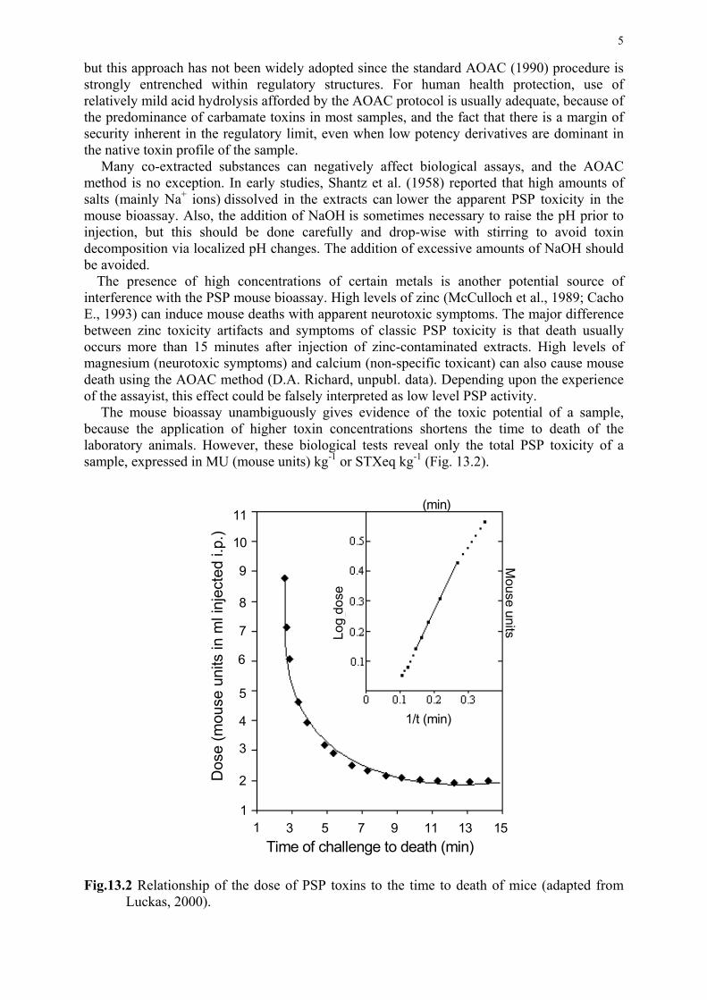
5
but this approach has not been widely adopted since the standard AOAC (1990) procedure is strongly entrenched within regulatory structures. For human health protection, use of relatively mild acid hydrolysis afforded by the AOAC protocol is usually adequate, because of the predominance of carbamate toxins in most samples, and the fact that there is a margin of security inherent in the regulatory limit, even when low potency derivatives are dominant in the native toxin profile of the sample. Many co-extracted substances can negatively affect biological assays, and the AOAC method is no exception. In early studies, Shantz et al. (1958) reported that high amounts of salts (mainly Na+ ions) dissolved in the extracts can lower the apparent PSP toxicity in the mouse bioassay. Also, the addition of NaOH is sometimes necessary to raise the pH prior to injection, but this should be done carefully and drop-wise with stirring to avoid toxin decomposition via localized pH changes. The addition of excessive amounts of NaOH should be avoided. The presence of high concentrations of certain metals is another potential source of interference with the PSP mouse bioassay. High levels of zinc (McCulloch et al., 1989; Cacho E., 1993) can induce mouse deaths with apparent neurotoxic symptoms. The major difference between zinc toxicity artifacts and symptoms of classic PSP toxicity is that death usually occurs more than 15 minutes after injection of zinc-contaminated extracts. High levels of magnesium (neurotoxic symptoms) and calcium (non-specific toxicant) can also cause mouse death using the AOAC method (D.A. Richard, unpubl. data). Depending upon the experience of the assayist, this effect could be falsely interpreted as low level PSP activity.
The mouse bioassay unambiguously gives evidence of the toxic potential of a sample, because the application of higher toxin concentrations shortens the time to death of the laboratory animals. However, these biological tests reveal only the total PSP toxicity of a sample, expressed in MU (mouse units) kg-1 or STXeq kg-1 (Fig. 13.2). Fig.13.2 Relationship of the dose of PSP toxins to the time to death of mice (adapted from
Luckas, 2000).
1/t (min)
6
3 7 9 135Time of challenge to death (min)
1511
2
5
7
8
1
9
10
11
3
4
1
1/t (min)
Log
dose
Dos
e (m
ouse
uni
ts in
ml i
njec
ted
i.p.)
(min)
Mouse units
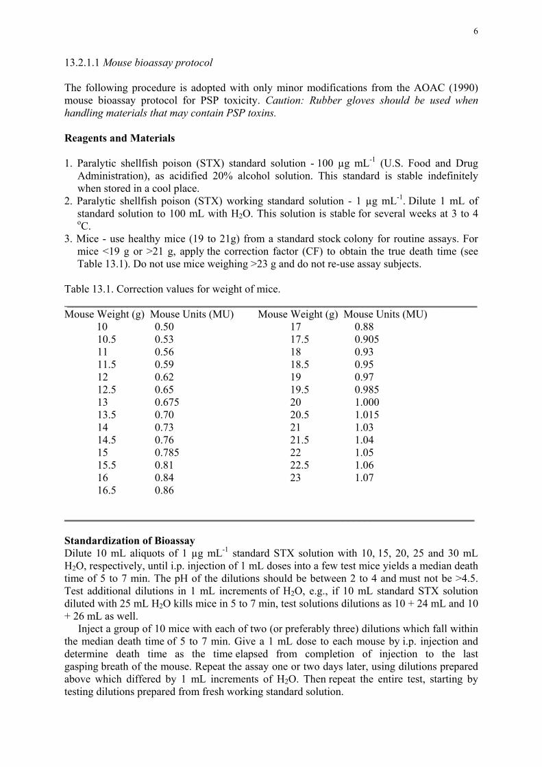
6
13.2.1.1 Mouse bioassay protocol The following procedure is adopted with only minor modifications from the AOAC (1990) mouse bioassay protocol for PSP toxicity. Caution: Rubber gloves should be used when handling materials that may contain PSP toxins. Reagents and Materials 1. Paralytic shellfish poison (STX) standard solution - 100 µg mL-1 (U.S. Food and Drug
Administration), as acidified 20% alcohol solution. This standard is stable indefinitely when stored in a cool place.
2. Paralytic shellfish poison (STX) working standard solution - 1 µg mL-1. Dilute 1 mL of standard solution to 100 mL with H2O. This solution is stable for several weeks at 3 to 4 oC.
3. Mice - use healthy mice (19 to 21g) from a standard stock colony for routine assays. For mice <19 g or >21 g, apply the correction factor (CF) to obtain the true death time (see Table 13.1). Do not use mice weighing >23 g and do not re-use assay subjects.
Table 13.1. Correction values for weight of mice. ___________________________________________________________________________ Mouse Weight (g) Mouse Units (MU) Mouse Weight (g) Mouse Units (MU)
10 0.50 17 0.88 10.5 0.53 17.5 0.905 11 0.56 18 0.93 11.5 0.59 18.5 0.95 12 0.62 19 0.97 12.5 0.65 19.5 0.985 13 0.675 20 1.000 13.5 0.70 20.5 1.015 14 0.73 21 1.03 14.5 0.76 21.5 1.04 15 0.785 22 1.05 15.5 0.81 22.5 1.06 16 0.84 23 1.07 16.5 0.86 ___________________________________________________________________________ Standardization of Bioassay Dilute 10 mL aliquots of 1 µg mL-1 standard STX solution with 10, 15, 20, 25 and 30 mL H2O, respectively, until i.p. injection of 1 mL doses into a few test mice yields a median death time of 5 to 7 min. The pH of the dilutions should be between 2 to 4 and must not be >4.5. Test additional dilutions in 1 mL increments of H2O, e.g., if 10 mL standard STX solution diluted with 25 mL H2O kills mice in 5 to 7 min, test solutions dilutions as 10 + 24 mL and 10 + 26 mL as well. Inject a group of 10 mice with each of two (or preferably three) dilutions which fall within the median death time of 5 to 7 min. Give a 1 mL dose to each mouse by i.p. injection and determine death time as the time elapsed from completion of injection to the last gasping breath of the mouse. Repeat the assay one or two days later, using dilutions prepared above which differed by 1 mL increments of H2O. Then repeat the entire test, starting by testing dilutions prepared from fresh working standard solution.
7
Calculate median death time for each group of 10 mice injected with each dilution. If all groups of 10 mice injected with any one dilution gave median death times <5 or >7 min, disregard results from this dilution in subsequent calculations. However, if any groups of 10 mice injected with a given dilution gave median death times between 5 and 7 min. include all groups of 10 mice assayed at this dilution, even though some of the median death times may be <5 or >7 min. From the median death time for each group of 10 mice at each selected dilution, determine the MU mL-1 from Sommer's Table (see Table 13.2). Divide calculated µg PSP toxin (STX) mL-1 by MU mL-1 to obtain the conversion factor (CF), expressed as µg PSP toxin (or µgSTXeq) per MU. Calculate the average CF value, and use this as a reference to verify routine assays. Individual CF values may vary significantly even within a given laboratory if the assay techniques and the condition of the mice are not rigidly controlled. Verification of PSP Toxin Conversion Factor (CF) for Routine Assays Check CF value periodically as follows: if shellfish tissues are assayed less than once a week, determine the CF value on each day assays are performed by injecting five mice with the appropriate dilution of working standard. If assays are carried out on several days during a given week, only one verification need be made per week using a dilution of standard such that the median death time falls within 5 to 7 min. The CF thus determined should agree with the average value within ±20%. If the CF value does not lie within this range, complete the group of 10 mice by adding five additional specimens to the five already injected, and inject a second group of 10 mice with same dilution of standard. Average the CF determined for the second group with that of the first group - this is the new CF value. Repeated checks of CF value normally produce results within ±20%. If wider variations are found frequently, the possibility of uncontrolled or unrecognised variables in the technique or in the response of mice to PSP toxin should be investigated before proceeding with routine assays. Sample Preparation and Extraction
a) Clams, oysters, and mussels, etc. (whole tissues) - Thoroughly clean the outside of shellfish with fresh water. Open by cutting adductor muscles. Rinse inside with fresh water to remove sand or other foreign material. Remove meat from the shell by separating adductor muscles and tissue connections at the hinge. Do not use heat or anaesthetics before opening the shell, and do not cut or damage the soft tissues at this stage. Collect ca. 100 to 150 g of shellfish tissue in a glazed dish. As soon as possible, transfer tissues to a No. 10 sieve without layering, and drain for 5 min. Pick out pieces of shell and discard drainings. Grind tissues in household-type grinder with 1/8 - 1/4'' (3-6 mm) holes or in a blender until homogeneous. b) Scallops, etc. (selected tissues) - Separate edible tissues (e.g., adductor muscle) and apply the extraction procedure to this portion alone. Drain and homogenize as in Step a).
c) Canned shellfish - Place entire contents of can (tissue and liquid) in a blender and blend
until homogenous or grind three times through a meat chopper. For large cans, drain tissues for 2 min on a No. 8-12 sieve and collect all liquid. Determine wet weight of tissue and volume of liquid. Recombine portions of each sample in proportionate amounts and blend (or grind) until homogeneous.
Weigh 100 g of well-mixed sample into a tared beaker. Add 100 mL 0.1N HCl, stir thoroughly, and check pH (should be <4.0, preferably ca. 3.0). If necessary, adjust pH as indicated in this section. Heat mixture and boil gently for 5 min, then let cool to room

8
temperature. Adjust cooled mixture to pH 2.0 to 4.0 (never >4.5), as determined by BHD Universal Indicator, phenol blue, Congo red paper, or by pH meter. To lower pH, add 5N HCl drop-wise with stirring; to raise pH, add 0.1N NaOH drop-wise with constant stirring to prevent local alkalinisation and consequent destruction or conversion of PSP toxins. Transfer mixture to a graduated cylinder and dilute to 200 mL. Return mixture to the beaker, stir to homogeneity, and let settle until a portion of the supernatant is translucent and can be decanted free of solid particles large enough to block a 26-gage hypodermic needle. If necessary, centrifuge the mixture or supernatant for 5 min at 3,000 x g or filter through paper to obtain enough liquid to perform the bioassay. Mouse Assay Intraperitoneally inject each test mouse with 1 mL of acidic extract. Note the time of inoculation and observe mice carefully for the time of death as indicated by the last gasping breath. Record death time by stopwatch or clock with sweep second hand. One mouse may be used for the initial determination, but two or three are preferred. If death time or median death time of several mice is <5 min, prepare dilutions to obtain death times of 5 to 7 min. If death time of one or two mice injected with the undiluted is >7 min, a total of three mice must be inoculated to establish sample toxicity. If large dilutions are necessary, adjust the pH of the dilution by drop-wise addition of dilute HCl (0.1 or 0.01N) to pH 2.0 to 4.0 (never >4.5). Inoculate three mice with a dilution that gives death times of 5 to 7 min. Calculation of Toxicity Determine median death times of mice, including survivors, and from Sommer's Table 13.2 determine the corresponding toxicity in mouse units (MU). If test animals weigh <19 g or >21 g, apply a correction for each mouse by multiplying MU corresponding to death time by the weight correction factor; then determine median number of MU for this group. [Consider death time of survivors as >60 min or equivalent to <0.875 MU in calculating the median.) Convert MU to µg PSP toxin (STXeq) by multiplying by the CF value: µg STXeq kg-1 tissue = (µg mL-1) X dilution factor X 2000 Consider any value >800 µg STXeq kg-1 as hazardous and unsafe for human consumption. 13.2.2. Fly bioassay The house fly (Musca domestica) bioassay (Ross et al., 1985) was developed as a viable alternative to the AOAC mouse assay for screening PSP toxins in shellfish. This method counters many of the disadvantages of the mouse bioassay, including those related to the high cost of mammalian bioassays, the difficulties in transporting and maintaining mice in acceptable facilities, animal rights concerns, and the interference due to salt effects. In brief, flies are temporarily immobilized on a cold plate and injected (in batches of 20) with standard AOAC extracts in a dilution series. After 10 minutes, flies are binary scored for motor ability – “0” = free-movement and/or ability to flip from dorsal position; “1” = positive, lack of motor ability. Interpretation of the results is based upon the fact that there is a linear relationship between the probit of the fraction of flies affected and the logarithm of the dose. The method proved to be reliable, with a mean ED50 of 0.25±0.02 µg mL-1 (381 pg per fly) when tested with USFDA certified STX standard. A collaborative study involving the assay of clam samples contaminated by PSP toxins and extracted according to the AOAC protocol was undertaken to compare the fly assay, the AOAC mouse assay, and a fluorescence-detection HPLC method.
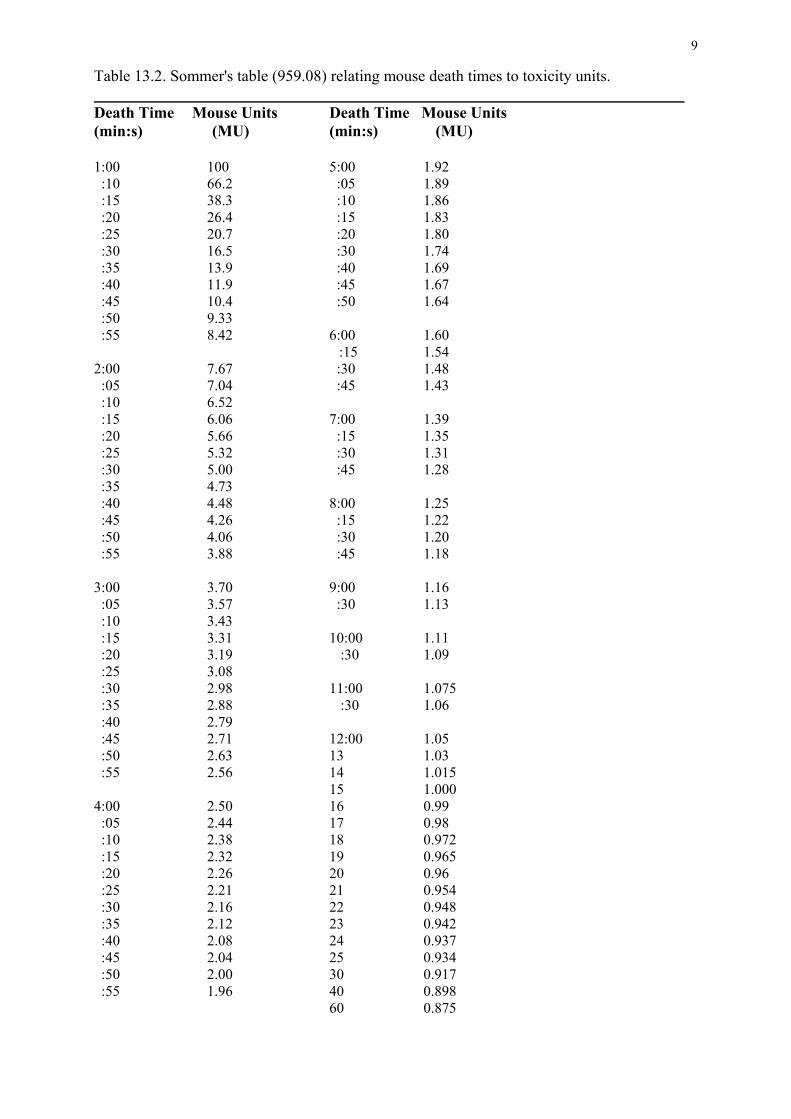
9
Table 13.2. Sommer's table (959.08) relating mouse death times to toxicity units. __________________________________________________________________________ Death Time Mouse Units Death Time Mouse Units (min:s) (MU) (min:s) (MU) 1:00 100 5:00 1.92 :10 66.2 :05 1.89 :15 38.3 :10 1.86 :20 26.4 :15 1.83 :25 20.7 :20 1.80 :30 16.5 :30 1.74 :35 13.9 :40 1.69 :40 11.9 :45 1.67 :45 10.4 :50 1.64 :50 9.33 :55 8.42 6:00 1.60 :15 1.54 2:00 7.67 :30 1.48 :05 7.04 :45 1.43 :10 6.52 :15 6.06 7:00 1.39 :20 5.66 :15 1.35 :25 5.32 :30 1.31 :30 5.00 :45 1.28 :35 4.73 :40 4.48 8:00 1.25 :45 4.26 :15 1.22 :50 4.06 :30 1.20 :55 3.88 :45 1.18 3:00 3.70 9:00 1.16 :05 3.57 :30 1.13 :10 3.43 :15 3.31 10:00 1.11 :20 3.19 :30 1.09 :25 3.08 :30 2.98 11:00 1.075 :35 2.88 :30 1.06 :40 2.79 :45 2.71 12:00 1.05 :50 2.63 13 1.03 :55 2.56 14 1.015 15 1.000 4:00 2.50 16 0.99 :05 2.44 17 0.98 :10 2.38 18 0.972 :15 2.32 19 0.965 :20 2.26 20 0.96 :25 2.21 21 0.954 :30 2.16 22 0.948 :35 2.12 23 0.942 :40 2.08 24 0.937 :45 2.04 25 0.934 :50 2.00 30 0.917 :55 1.96 40 0.898 60 0.875

10
The fly bioassay showed a high degree of congruence with the mouse bioassay results, often with slightly higher values, but with a lower detection limit (250 µg STXeq kg-1) for shellfish extracts than with the mouse bioassay (approx. 350-400 µg STXeq kg-1). Unfortunately, in spite of the fact that attempts were made to introduce the use of the fly bioassay, particularly in developing countries in Asia, this method never achieved common usage. Perhaps one of the difficulties is the high degree of manual dexterity required for the injection of minute quantities (1.5 µL) of sample into the abdomen-thorax junction without damage to the test subject. 13.3 AMNESIC SHELLFISH POISONING (ASP) Amnesic shellfish poisoning (ASP), caused by the neurotoxin domoic acid, was first identified in Canada in shellfish extracts from eastern Prince Edward Island (Wright et al., 1989). The memory loss associated with extreme cases of human intoxication from shellfish contaminated by domoic acid led to the naming of the phycotoxic syndrome known as amnesic shellfish poisoning. Domoic acid is an excitatory amino acid derivative acting as a glutamate agonist on the kainate receptors of the central nervous system. This secondary amino acid is considered to be a more potent neuroexcitor than kainic acid, which when systemically injected into specific parts of the brain is known to have degenerative effects. Domoic acid is considered to be the primary toxin involved in ASP, although isomeric forms (e.g., iso-domoic acid) of lesser potency also occur naturally. 13.3.1 Bioassay protocols The AOAC mouse bioassay for PSP toxicity can also detect domoic acid at concentrations ca. 40 µg mL-1 injected. This procedure was initially used when ASP toxicity was first detected in leftover mussels implicated in human poisonings, and to direct the purification scheme in identifying domoic acid as the cause of ASP in shellfish extracts from eastern Prince Edward Island, Canada (Wright et al., 1989). The AOAC extraction procedure can yield substantial recovery of domoic acid from shellfish tissue (ca. 80%), and subsequent modification of the extraction method using methanol:water improves extraction efficiency but the tolerance level established in Canada and subsequently adopted by certain other countries is 20 µg g-1. Therefore the AOAC bioassay procedure is too insensitive to be used with confidence for regulatory purposes to quantify this toxin and it has been abandoned in favour of chemical analytical methods (see Chapter 8) by most regulatory authorities.
Under certain circumstances, a rat or mouse bioassay could be used to detect the presence of low level domoic acid by careful monitoring of selected symptoms (Tasker et al., 1990, Doucette et al., 2000), but such a method would not meet established criteria for regulatory purposes. In the event all other means of analysis are unavailable, special extraction techniques designed to increase sensitivity (e.g., extraction of the digestive gland only) can be used in combination with the mouse bioassay and observing for symptoms as described in this chapter. This method can only be described as qualitative at best and should not be used instead of the universally accepted chemical analytical methods. The typical signs of the presence of domoic acid in the AOAC bioassay procedure are a unique scratching syndrome of the shoulders by the hind leg, followed by convulsions. Other clinical signs of domoic acid toxicity in mice, in the following general chronological order, may include: circling, head nodding, mastication, forelimb tremor, “wet dog” shakes, loss of postural control, profuse salivation, “praying” and rearing, scratching of the scapular region by the ipsilateral hind paw, myoclonic jerks, barrel rotation, generalized tremor, kyphosis, rigidity, general clonic-toxic convulsions, and death. The time of observation, as opposed to that of the AOAC procedure, must be extended to 4 hours. Mouse deaths associated with

11
mussels containing domoic acid were never observed after 135 minutes (Quilliam et al., 1989; Todd, 1990). 13.4 DIARRHETIC SHELLFISH POISONING (DSP) AND RELATED POLYETHER
TOXIN SYNDROMES Three groups of polyether compounds have been historically included in the “DSP toxin complex”: 1) okadaic acid (OA) and analogues such as the dinophysistoxins, DTX1, DTX2 and acyl-derivatives (DTX3), 2) pectenotoxins (PTXs) - separable into two groups, one having a cyclic ester structure (“macrolide”) and the other a “seco acid” group, in which the ester bond in the cyclic structure is hydrolysed, and 3) yessotoxins (YTXs), disulfated polyether compounds (see Chapter 8). These compound have a relatively high molecular weight and share lipophilicity and solubility properties, being extracted with the same generic extraction procedures. The traditional DSP mouse bioassay involving acetone extraction (Yasumoto et al., 1978) can detect not only OA and DTXs, but also PTXs and YTXs that may co-occur with OA in phytoplankton and shellfish. For this reason, PTX and YTX have been often associated with “DSP”. However, their biological activity is different from that of the OA group, and only toxins belonging to the latter group have been reported to produce diarrhoeic effects in humans (i.e., classic DSP symptoms). There is general agreement in the classification of OAs and DTXs, as DSP toxins, but the inclusion of PTXs and YTXs in the “DSP complex” has always been a subject of great controversy. The toxicity values for various lipophilic phycotoxins found in shellfish and toxigenic microalgae are presented in Table 13.3. In humans, typical symptoms following the consumption of DSP toxin-contaminated shellfish include acute diarrhea, nausea, vomiting and, in some cases, abdominal pain. Although no human mortalities from DSP have been reported, the affliction can be highly debilitating for several days. Okadaic acid and certain DTXs are potent inhibitors of at least two sub-classes of protein phosphatases (PP1 and PP2A) and this mode of action may be linked to the observed diarrhoea, degenerative changes in absorptive epithelium of the small intestine, and to tumor promotion (Fujiki et al., 1988). Toxicological and epidemiological data on PTX and YTX are scarce, and the action mechanism of these toxins has not yet been fully elucidated. By i.p. injection into mice, PTX1 has been shown to cause damage to the liver but no diarrhoea was observed (Terao et al., 1986). Pectenotoxin-2 is known to elicit extremely potent cytotoxic activities (at nM to pM concentrations) against numerous human cancer cell lines (Jung et al., 1995) and by oral administration into mice, can cause diarrhoea and severe injuries to the liver and intestine, with oral toxicity very similar to i.p. toxicity (230 µg kg-1) (Ishige et al., 1988). As members of both the OA and PTX groups are produced by Dinophysis species, it is very difficult to assess separately the contribution of PTX to human intoxication. Recently, the presence of PTX2 seco-acid in shellfish was correlated with a toxic outbreak in Australia (Quilliam et al., 2000), but the co-occurrence of DTX esters makes this relationship to illness difficult to confirm.
With respect to YTXs, the i.p lethal dose ranges from 100 to 500 µg kg-1, depending on the analogue, with the heart being the target organ (Terao et al., 1990). Oral toxicity is at least an order of magnitude order lower than i.p toxicity (Ojino et al., 1997, Aune et al., 2001). Diarrhoea has not been observed by oral administration, therefore the appropriateness of classifying these compounds as “DSP toxins” is questionable. Studies carried out with human lymphocytes suggest that YTX acts on cyclic adenosine monophosphate, increasing the cytosolic levels of calcium (De la Rosa et al., 2001).
Although the precautionary principle suggests that the presence of the three groups of toxins should be monitored in shellfish, there is no general agreement concerning which toxins should be included in the regulatory programmes (see Chapter 24). Furthermore, the definition of acceptable testing procedures, the standardization of methods and action limits to
12
be applied for regulatory purposes represents a difficult administrative and scientific challenge. The current situation is far from ideal, and leads to inconsistencies and a lack of equivalence between the toxin control approaches used by different countries. Table 13.3 Acute toxicity of various lipophilic phycotoxins after i.p. injection into mice. Only those lipophilic toxins found in shellfish and the corresponding toxigenic microalgae are included; toxicity values for ciguatoxins and other analogues found primarily in fish tissues are available from Lewis (1995). ___________________________________________________________________________ Toxin Group Analogue Toxicity (µg kg-1) Pathology Okadaic acid OA 200a diarrhoea;tumor promotionDinophysistoxin DTX1 160a diarrhoea DTX3 500a diarrhoea Pectenotoxin PTX1 250a hepatotoxic PTX2 230a hepatotoxic; diarrhoea PTX3 350a * PTX4 770a * PTX6 500i * PTX7 >5000j * PTX8 >5000j * PTX9 >5000j * Yessotoxin YTX 100a cardiotoxic HydroxyYTX 500b * TrinorYTX 220b * HomoYTX 100c * 45-hydroxyhomoYTX 500c * DesulfoYTX 500d * CarboxyYTX 500e * Azaspiracid AZA 200f diarrhoea AZA2 110g diarrhoea AZA3 140g diarrhoea AZA4 470h diarrhoea AZA5 1000h diarrhoea Gymnodimine 96k unconfirmed Brevetoxin BTX-B1 50i neurological BTX-B2 300i neurological BTX-B3 >300i neurological Spirolide B 200l unconfirmed des-methyl-C 40m unconfirmed
Data from: aYasumoto et al. (1989); bSatake et al., 1996; cSatake et al., 1997; dDaiguji et al. 1998; eCiminiello et al.,2000; fSatake et al, 1998; gOfuji et al., 1999a,b; hOfuji et al., 2001; icited in Yasumoto, 2000; j Sasaki et al., 1998; kMiles et al., 1999; lHu et al., 1995; mD.A. Richard, A.D. Cembella and M.A. Quilliam, unpubl. data. *In the case of PTX and YTX groups, oral toxicity and cytotoxicity studies have been carried out only for PTX1, PTX2 and YTX. For other analogues, the pathology is likely to be similar or identical to that of the parent compounds, but is currently undefined.

13
13.4.1 Bioassay protocols Despite the worldwide application of mammalian bioassays for DSP toxicity, there are large differences in the performance of the procedures and in the classification criteria employed among countries. Assay selectivity, specificity, and DSP toxin recovery depends greatly upon the selection, purity, and ratios of the organic solvents used for extraction and clean-up steps (see also Chapter 8). Bioassay procedures as diverse as the oral dosage rat bioassay and the i.p. injection mouse assay are not strictly homologous, because the former technique quantifies only the diarrhoeagenic effect of certain DSP toxin components (OA and analogues), whereas the latter assay yields an estimate of overall "DSP toxicity". Most extraction procedures applied to DSP toxins in shellfish samples can also coextract NSP toxins and certain other lipophilic ichthyotoxins that can yield non-specific artifacts in mammalian bioassays. There may also be interference by free fatty acids, which can vary with the particular shellfish species, as well as spatially and temporally. These components may be toxic to mice by i.p. injection, yielding false positive reactions for "DSP toxicity", but innocuous orally to humans and negative by the rat oral bioassay (Takagi et al.,1984; Lawrence et al., 1994).
The mouse bioassay for DSP toxicity is considered to be quantitative, although as the prolonged observation time would suggest, it is less reliable as a determinant of acute toxicity than is the corresponding AOAC mouse assay for PSP toxicity. Furthermore, the precise cause of death in rodents by i.p. administration of DSP toxins is not yet known. Phosphatase inhibition activity is strongly indicated as the toxicity mechanism leading to death, but the results of the i.p. bioassay cannot be simply extrapolated to quantify human oral toxicity or diarrhoeic symptoms. The DSP toxicity procedure as originally established (Yasumoto et al., 1978) involved the calculation of i.p. toxicity in mouse units (MU), with 50 MU kg-1 of whole soft tissue accepted as the regulatory limit in Japan. A mouse unit was defined as the minimum quantity of toxins required to kill two of three mice within 24 hours of i.p. injection; this corresponds to 3.2 µg DTX1 or 4 µg OA for the ddY mouse strain used in Japan, but may vary somewhat with the strain. As the calculation of toxicity using LD50 methods can be labourious and time- consuming, regulatory laboratories in most countries currently use mouse survival time for the evaluation of DSP toxicity. There is no international consensus on the appropriate observation period - the acceptable criteria can vary from "two of three mouse deaths in less than five hours" to "two of three mouse deaths in less than 24 hours". Such decisions are both operational and administrative, but they are also somewhat dependent upon the specific procedure for sample preparation prior to mouse bioassay, since various components of differing toxicity may be selectively extracted.
On the basis of an interlaboratory study of the mouse bioassay (adult mice) and the suckling mouse test (Marcaillou-Le Baut et al., 1985), the survival time considered as a positive result (adult mouse assay) was reduced from 24 to 5 hours. The study showed that the survival time of adult mice beyond 5 hours corresponded to a toxicity lower than that revealed by the suckling mouse test, and that the survival three of three mice for more than 5 hours was considered a negative response. In addition, the same investigators studied the relation between the survival time of three mice and OA concentration for observations of death time more than 5 hours, and found that <4 µg OA in an extract matrix was necessary to yield a death time greater than 5 hours. Consequently, a mouse survival time of more than 5 hours was considered lower than the regulatory action level, and therefore a negative result for DSP toxicity (Marcaillou-Le Baut and Masselin, 1990).
14
Further studies comparing mouse bioassay results and HPLC-FD determination of OA
showed that survival times of around 5 hours corresponded with an OA concentration of approximately (and in some cases above) 2 mg OA kg-1 of hepatopancreas. Vieytes et al. (1996) showed a relation between the death times in the mouse bioassay and values obtained in HPLC analysis. An analysis of 127 samples by both methods showed that reduction of the observation time from 24 to 5 hours could increase (by 35-76%) negative bioassays for toxin levels between 2 and 3 mg OA kg-1 of hepatopancreas. Vale and Sampayo (1996) found that although death times between 5 and 24 hours usually corresponded to OA + DTX2 concentrations of about 2 mg OAeq kg-1 of hepatopancreas, in some samples, appreciable amounts of DSP toxins (>2 mg OAeq kg-1 of hepatopancreas) resulted in survival times longer than 5 hours. When evaluating the level of consumer protection provided by the different toxicity criteria, the considerable degree of uncertainty associated to the mouse bioassay protocols should be taken into account. Although the purpose of the assay is to assess toxicity of the edible part of the molluscs (usually the whole body of soft tissues), the assay is normally carried out using the hepatopancreas as the analytical sample, assuming, with reference to abundant previous data on toxin distribution in tissues, that most DSP toxins are accumulated in the digestive tissues. Although the digestive tissues (primarily hepatopancreas) accumulate most of the DSP toxins, a certain amount can be retained in other soft tissues (gill, mantle, siphon, gonads, etc.). When considering the percentage of hepatopancreas to whole shellfish soft tissues, significant inter- and intra-specific differences are evident, and the percentage will also depend on the season and location for a given species (hepatopancreas percentage variations of between 7 and 25 % are routinely observed). Figure 13.3 shows the DSP toxicity results expressed in relation to whole shellfish body weight of soft tissues that corresponds to the different survival times used as the toxicity criterion.
These data are based on the assumption that when mice are injected with an extract equivalent to 5 g of hepatopancreas, a survival time of 24 hours corresponds to 0.8 mg OAeq kg-1 and a survival time of 5 hours corresponds to 2 mg OAeq kg-1. By introducing different possible percentages of hepatopancreas in relation to whole shellfish soft tissue weight, different results are obtained. If a range of between 10 and 25% is considered, a survival time of 24 hours would correspond to a toxicity between 80 and 200 µg OAeq kg-1 whole shellfish soft tissue, whilst a 5 hour survival time would correspond to a toxicity value of between 200 and 500 µg OAeq kg-1, concentrations very close to or even higher than those reported to produce diarrhoea (total dose of 40-48 µg OA).
Evidently, when DSP toxins other than OA, DTX1 and DTX2 are present in shellfish, the time/dose response may vary, and a delay in the appearance of the symptoms may occur. This is the case for DTX3, for which an observation time of 24 hours is required (Terao et al., 1993). Therefore, although the use of a 5-hour mouse test for DSP toxicity may improve the accuracy of the assay, it also increases the likelihood that shellfish with toxic levels of DSP analogues being accepted and placed on the market.
The assays described in the subsequent section are used for DSP toxicity determination in different countries, but the range of substances detected as well as the sensitivity may differ among protocols. These facts should be taken into account when making a decision on which method to use. Albeit in worldwide use for many years, none of these procedures has been evaluated in a formal collaborative study and although information on detection limit is in some cases available, performance characteristics, such as accuracy and precision are not fully known.
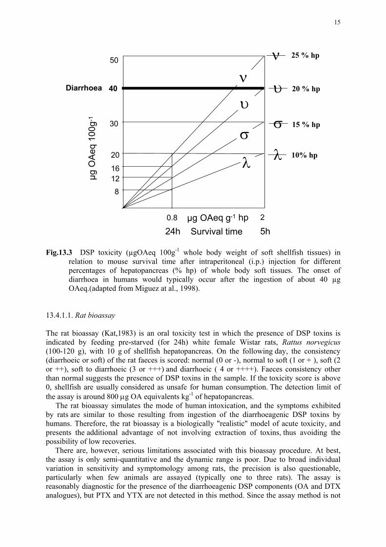
15
Fig.13.3 DSP toxicity (µgOAeq 100g-1 whole body weight of soft shellfish tissues) in
relation to mouse survival time after intraperitoneal (i.p.) injection for different percentages of hepatopancreas (% hp) of whole body soft tissues. The onset of diarrhoea in humans would typically occur after the ingestion of about 40 µg OAeq.(adapted from Miguez at al., 1998).
13.4.1.1. Rat bioassay The rat bioassay (Kat,1983) is an oral toxicity test in which the presence of DSP toxins is indicated by feeding pre-starved (for 24h) white female Wistar rats, Rattus norvegicus (100-120 g), with 10 g of shellfish hepatopancreas. On the following day, the consistency (diarrhoeic or soft) of the rat faeces is scored: normal (0 or -), normal to soft (1 or + ), soft (2 or ++), soft to diarrhoeic (3 or +++) and diarrhoeic ( 4 or ++++). Faeces consistency other than normal suggests the presence of DSP toxins in the sample. If the toxicity score is above 0, shellfish are usually considered as unsafe for human consumption. The detection limit of the assay is around 800 µg OA equivalents kg-1 of hepatopancreas. The rat bioassay simulates the mode of human intoxication, and the symptoms exhibited by rats are similar to those resulting from ingestion of the diarrhoeagenic DSP toxins by humans. Therefore, the rat bioassay is a biologically "realistic" model of acute toxicity, and presents the additional advantage of not involving extraction of toxins, thus avoiding the possibility of low recoveries. There are, however, serious limitations associated with this bioassay procedure. At best, the assay is only semi-quantitative and the dynamic range is poor. Due to broad individual variation in sensitivity and symptomology among rats, the precision is also questionable, particularly when few animals are assayed (typically one to three rats). The assay is reasonably diagnostic for the presence of the diarrhoeagenic DSP components (OA and DTX analogues), but PTX and YTX are not detected in this method. Since the assay method is not
ν
σσ
λ λ
υ υ
ν50
40
30
20
16128
µg O
Aeq
100
g-1
µg OAeq g-1 hp
Diarrhoea
0.8 2
24h 5hSurvival time
10% hp
15 % hp
20 % hp
25 % hp
16
very specific, other diarrhoeic agents including intestinal pathogens associated with poor animal health could also complicate the interpretation of faeces consistency. 13.4.1.2. Intraperitoneal mouse bioassays All of the mouse bioassay protocols detailed in this section require prior extraction and concentration of the lipophilic components by liquid/liquid partitioning. An advantage of the extraction protocols based upon liquid/liquid partitioning of toxin components is that by modifying the acetone volumes used for the initial extraction, the protocols can be applied to DSP toxin determination on whole shellfish soft-tissues. This can be very useful in the case of small shellfish species or when extracting eviscerated specimens. The acetone volumes required may vary depending upon the shellfish species, but 100 g of homogenized whole shellfish tissues are usually used as the minimum test portion. When using whole shellfish soft tissues, the accuracy of the assay improves. However, the high volumes of organic solvents required, the laboriousness of the procedure and the toxicity of the solvents used in the partitioning, have limited the wide application of this approach in monitoring programs that require the analysis of a high number of samples. Nevertheless, it is worth mentioning that a mouse bioassay procedure based upon extraction of whole soft tissues is used for NSP/DSP toxicity screening in New Zealand (described in Section 13.5.1.2). When inoculating mice with an extract corresponding to 25 g of whole soft tissues and using a 24 hour observation period, the detection limit of the assay is approximately 40 MU kg-1 or 160 µg OAeq kg-1. 13.4.1.2.1. Method 1 The original mouse bioassay procedure of Yasumoto et al. (1978) was developed for bioassay-guided fractionation of toxic extracts, but it is widely applied for routine monitoring of DSP toxicity in shellfish. According to this method, a 20 g sample of homogenized hepatopancreas is extracted with 50 mL of acetone. The slurry is filtered into an evaporation flask and the tissue residue re-extracted twice with volumes of 50 mL of acetone and the filtrates combined. The solvent is removed by rotary evaporation under reduced pressure and the residue is resuspended in 1% Tween 60 to a final concentration of 5 g equivalents mL-1. One mL of this solution is injected intraperitoneally into each of three mice weighing between 18 and 20 g, the mice observed for 24 to 48 hours, and toxicity determined on the basis of the survival time. The calculation of mouse units may require serial dilution of sample extracts until the minimum dose that produces death in 24 hours is reached. Under the conditions described here, the detection limit of the assay is 0.2 MU g-1 or 800 µg OA equivalents kg-1 hepatopancreas.
This procedure detects the presence of OA and DTXs, as well as the PTX and YTX groups. In addition, other lipophilic toxins, such as spirolides, gymnodimine, azaspiracids, brevetoxins and ciguatoxins are co-extracted, if present in shellfish. Mouse symptoms and death times may be similar depending on the bioactive compounds present in the samples, and in some circumstances it may be difficult to discriminate which substances are the cause of death of the mice. A potential pitfall of this bioassay procedure is associated with the aqueous-lipid residue obtained after solvent evaporation. Depending upon factors such as water content of the shellfish, resuspension of the final residue to achieve a specific concentration (5 g hepatopancreas mL-1 Tween 60) may be inconsistent, thereby negatively affecting the accuracy of the assay. The high temperatures sometimes required for the total removal of the water may also produce low toxin recoveries (Croci et al., 1994).
Interference by low levels of PSP and ASP toxins, even at concentrations undetectable by the AOAC (1990) mouse bioassay, can also present problems. Some of these polar
17
components can be coextracted along with DSP toxins by acetone extraction, and they are concentrated during the evaporation procedure, yielding a final extract that can be highly toxic to mice. Fast mouse deaths (minutes to a few hours) with neurotoxic symptomology do not permit the prolonged observations required for DSP toxin detection. The presence of certain free fatty acids can also interfere with this assay (Takagi et al.,1984).
Among all the bioassays for DSP detection, this method is the least selective, and when positive results are obtained, it is not always feasible to assign the response to the presence of known toxins. However, after many years of routine application throughout the world, it has been proven to be reasonably effective to screen for the presence of DSP and other polyether toxins, and for public health protection.
13.4.1.2.2. Method 2 In this modified method (Yasumoto et al., 1984), a 20g sample of homogenized hepatopancreas is extracted thrice with 50 mL of acetone. The extracts are filtered, then the filtrate is collected and the solvent removed by rotary evaporation. The residue is made up to 15 to 20 mL with water and the suspension is extracted thrice with 50 mL of diethyl-ether. The combined organic layers are backwashed twice with small quantities of water and evaporated to dryness. As in the original procedure described in Method 1, the residue is resuspended in 1% Tween 60 to a concentration of 5 g equivalents hepatopancreas mL-1 Tween 60 prior to i.p. injection. Toxicity is determined on the basis of the survival time or in mouse units. The assay is very suitable for use with a wide variety of shellfish tissues. With this procedure, possible interferences with PSP and ASP toxins are removed in the water layer. High amounts of salts that are concentrated during the evaporation step, and could cause artifactual mouse deaths, can also be removed during the water:ether partitioning step. A drawback of this extraction procedure is the poor solubility of YTX in diethyl-ether; depending upon the pH and lipid content of the sample, this toxic component may be lost in the water layer. A substantial improvement in YTX recovery is achieved if dichloromethane or ethyl acetate is used instead of diethyl-ether. 13.4.1.2.3 Method 3 The extraction procedure for this DSP mouse bioassay method was adapted from that applied for the validation of the HPLC-FD method (Lee et al., 1987)). In this protocol, homogenized hepatopancreas is extracted with 100% acetone. After evaporation of the solvent, the residue is redissolved in 80% methanol, which is extracted twice with petroleum ether to remove contaminant non-polar lipids and then with chloroform. The chloroform phase is evaporated to dryness and the residue is redissolved in 1% Tween 60 to be administered by i.p. injection into mice. One mL portions of this solution are administered into each of two mice at doses equivalent to 2.5 to 5 g of hepatopancreas. Mouse behaviour and survival time is recorded for a 24-hour period. Losses of low polarity DSP toxins (DTX3) can occur during the petroleum ether washing step. 13.4.1.2.4 Method 4 In this procedure (Le Baut and Masselin, 1990), hepatopancreas (20g) is extracted thrice with 100% acetone and the filtrates collected. After evaporation of the acetone, the residue is resuspended in 25 mL of methanol:water (80:20). The methanolic phase is washed twice with 25 mL of hexane, the hexane phases are discarded, and the methanolic layer is evaporated to dryness. The resuspension of the residue in 1% Tween 60 and i.p. injection protocols are identical to those described for the methods in Sections 13.4.1.2.1, 13.4.1.2.2 and 13.4.1.2.3.

18
One of the main advantages of this assay is that low polarity free fatty acids that could interfere with the assay are removed by the hexane wash. The final residue has a relatively low lipid content and it thus easier to accurately resuspend in Tween 60, resulting in fewer false positive responses. The main deficiency of the procedure is that the hexane washing step, besides removing free fatty acids, also preferentially solubilises DTX3, the acyl-derivatives of OA and DTX. If these components are present in shellfish samples, substantial loss of toxins can occur (Fernandez et al., 1996). PSP toxins and interfering salts are not removed in this procedure. 13.4.1.3. Suckling mouse bioassay The suckling mouse bioassay, a method usually applied for detection of bacterial enterotoxins, has been used to test the enteropathogenicity of OA, DTX1 and DTX3 (Hamano et al., 1986). The procedure involves the intragastric introduction via Teflon tubing of 0.1 mL of shellfish extracts suspended in 1% Tween 60, to four- to five-day old mice. After the treatment, the mice are kept at 25 ºC for 4 hours and then sacrificed with chloroform. The whole intestine is removed and the fluid accumulation ratio (FA) is determined as the ratio of the weight of intestine to that of the remaining body: FA = gut weight/(body weight-gut weight).
The detection limit of the test is lower than that of the i.p. mouse bioassay, and positive results can be observed at a level of 0.1 MU, corresponding to an FA value of 0.09. Pectenotoxins, YTXs and free fatty acids are not detected by this procedure. Although under optimal conditions results can be obtained within 4 hours, with a sensitivity higher that that of the mouse assay, accuracy and precision is a concern and the assay is difficult to be applied routinely. For example, wounding of the mice can take place during administration of the sample. 13.4.1.4. Daphnia magna bioassay Daphnia magna is a fresh water cladoceran invertebrate traditionally used as a standard ecotoxicological indicator organism for the testing of different toxicants. A D. magna bioassay was developed by Vernoux et al. (1993) for OA screening of mussel extracts, based on the measurement of the inhibition of Daphnia mobility due to the presence of OA. The test is carried out in glass tubes each containing 10 mL of growth medium and five Daphnia individuals. Standardized culture conditions are crucial to avoid variability of responses. A linear correlation (r = 0.74) has been established between results of the Daphnia bioassay and an HPLC-FD method involving pre-column derivatization. As a larger number of animals can be used, precision can potentially be higher than that of the mouse bioassay, and sensitivity is ten times higher. In addition to OA, other co-extracted lipophilic toxins may be detected. Although the method was proposed as an alternative to the mouse assay for OA screening, there have not been further reports on its application. It is therefore difficult to assess its value as a tool for routine DSP toxin monitoring. 13.5 NEUROTOXIC SHELLFISH POISONING (NSP) TOXINS The toxins implicated in neurotoxic shellfish poisoning (NSP), known collectively as "brevetoxins", are considered to be primarily ichthyotoxins. In humans, the symptoms of NSP intoxication include respiratory distress, as well as eye and nasal membrane irritation, caused principally by exposure to sea-spray aerosols and by direct contact with toxic blooms while swimming. The brevetoxins are also accumulated in shellfish, which when consumed by humans, cause a toxic syndrome somewhat similar to PSP intoxication. Many of these lipid-soluble cyclic polyether compounds have been characterized, including brevetoxins A, B
19
and C (BTX A, B and C) [synonyms: PbTx-1, PbTx-2 and PbTx-8, respectively], GB3, 5 and 6 [synonyms: PbTx-3, PbTx-4 and PbTx-5, respectively] and PbTx-6 and PbTx-7, but undoubtedly there exist other undescribed derivatives. Due to the chemical lability of the brevetoxins, analysis of these compounds continues to be problematic (see also Chapter 8). All of these derivatives exert their toxic effect by specific binding to site-5 of voltage-sensitive Na+ channels, leading to channel activation at normal resting potential. 13.5.1 Bioassay protocols 13.5.1.1 APHA protocol The official method for the determination of NSP toxins is the American Public Health Association (APHA, 1985) procedure (originally Irwin, 1970), based on a diethyl-ether extraction of shellfish tissue. The APHA protocol for NSP is used extensively in the United States, where the problem of NSP is most prevalent and acute. Apparatus Electric blender Analytical balance Hot plate Explosion-proof centrifuge with 250 mL centrifuge cups; covered with foil Explosion-proof chemical hood 1000 mL separatory funnel 400 mL beakers. Disposable syringes with 26-gauge needles (do not reuse) Stopwatch, mechanical or electronic, registering to at least 1 s Reagents Hydrochloric acid (HCl), concentrated Sodium chloride (NaCl) Cottonseed oil Diethyl ether, anhydrous, AR (ACS). Use ether only from a previously unopened container- peroxides reduce apparent toxicity. Test Animals Healthy albino male mice (Swiss-Webster strain) weighing 20±1 g are preferable, however, mice weighing between 15 and 25 g may be used. Do not reuse surviving mice. Since mice often eat excessively and may store 1 to 2 g of food in the digestive tract, feed them sufficiently but do not overfeed. Sample Preparation and Extraction Clean, shuck, and drain shellfish. The number of shellfish required for 100 g of homogenate varies from two large clams to eight to 10 small oysters. Homogenize shellfish tissues in an electric blender at high speed for 5 min. Weigh 100 g of homogenate into a pre-weighed 400 mL beaker and add 5 g NaCl and 1 mL conc. HCL. Stir well. Heat mixture to boiling and cook for 5 min; stir frequently. Let cool to room temperature and transfer to a 1000 mL separatory funnel. Rinse beaker with ether and add rinse to separatory funnel. Perform all subsequent steps under an explosion-proof hood. Add 100 mL ether to homogenate, stopper, and shake vigorously (venting frequently) for 5 min. Centrifuge at 2000 rpm for 15 min. Carefully decant upper clear yellow ether phase into a 1000 mL separatory funnel, keeping solids in the centrifuge bottle. Repeat extraction three more times until the total amount of ether used is 400 mL. Drain off and discard any bottom
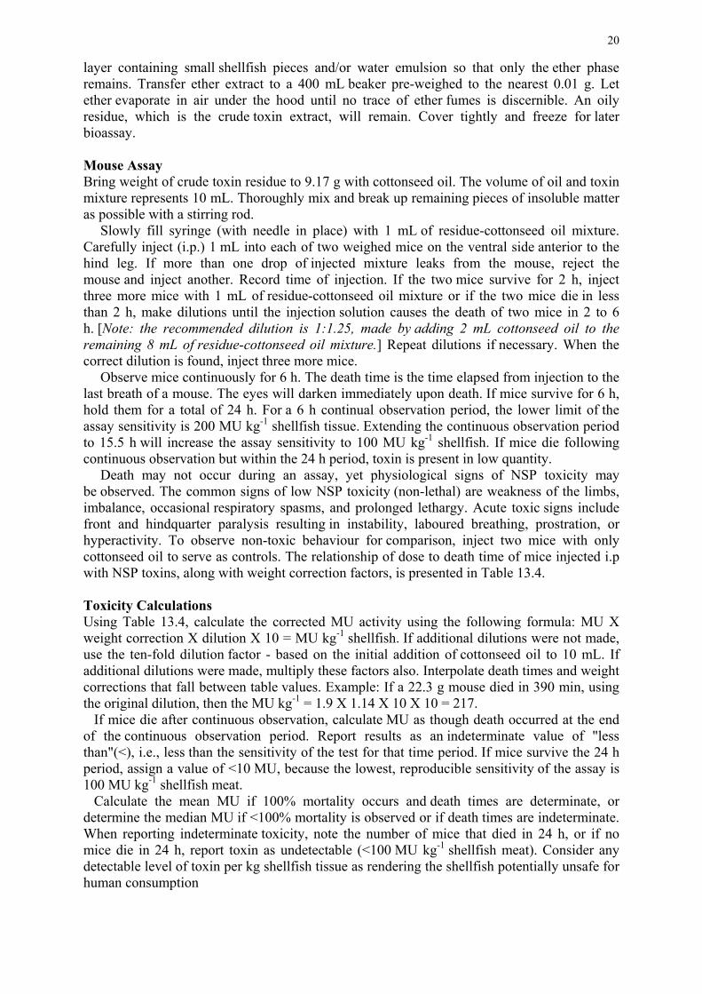
20
layer containing small shellfish pieces and/or water emulsion so that only the ether phase remains. Transfer ether extract to a 400 mL beaker pre-weighed to the nearest 0.01 g. Let ether evaporate in air under the hood until no trace of ether fumes is discernible. An oily residue, which is the crude toxin extract, will remain. Cover tightly and freeze for later bioassay. Mouse Assay Bring weight of crude toxin residue to 9.17 g with cottonseed oil. The volume of oil and toxin mixture represents 10 mL. Thoroughly mix and break up remaining pieces of insoluble matter as possible with a stirring rod. Slowly fill syringe (with needle in place) with 1 mL of residue-cottonseed oil mixture. Carefully inject (i.p.) 1 mL into each of two weighed mice on the ventral side anterior to the hind leg. If more than one drop of injected mixture leaks from the mouse, reject the mouse and inject another. Record time of injection. If the two mice survive for 2 h, inject three more mice with 1 mL of residue-cottonseed oil mixture or if the two mice die in less than 2 h, make dilutions until the injection solution causes the death of two mice in 2 to 6 h. [Note: the recommended dilution is 1:1.25, made by adding 2 mL cottonseed oil to the remaining 8 mL of residue-cottonseed oil mixture.] Repeat dilutions if necessary. When the correct dilution is found, inject three more mice. Observe mice continuously for 6 h. The death time is the time elapsed from injection to the last breath of a mouse. The eyes will darken immediately upon death. If mice survive for 6 h, hold them for a total of 24 h. For a 6 h continual observation period, the lower limit of the assay sensitivity is 200 MU kg-1 shellfish tissue. Extending the continuous observation period to 15.5 h will increase the assay sensitivity to 100 MU kg-1 shellfish. If mice die following continuous observation but within the 24 h period, toxin is present in low quantity. Death may not occur during an assay, yet physiological signs of NSP toxicity may be observed. The common signs of low NSP toxicity (non-lethal) are weakness of the limbs, imbalance, occasional respiratory spasms, and prolonged lethargy. Acute toxic signs include front and hindquarter paralysis resulting in instability, laboured breathing, prostration, or hyperactivity. To observe non-toxic behaviour for comparison, inject two mice with only cottonseed oil to serve as controls. The relationship of dose to death time of mice injected i.p with NSP toxins, along with weight correction factors, is presented in Table 13.4. Toxicity Calculations Using Table 13.4, calculate the corrected MU activity using the following formula: MU X weight correction X dilution X 10 = MU kg-1 shellfish. If additional dilutions were not made, use the ten-fold dilution factor - based on the initial addition of cottonseed oil to 10 mL. If additional dilutions were made, multiply these factors also. Interpolate death times and weight corrections that fall between table values. Example: If a 22.3 g mouse died in 390 min, using the original dilution, then the MU kg-1 = 1.9 X 1.14 X 10 X 10 = 217. If mice die after continuous observation, calculate MU as though death occurred at the end of the continuous observation period. Report results as an indeterminate value of "less than"(<), i.e., less than the sensitivity of the test for that time period. If mice survive the 24 h period, assign a value of <10 MU, because the lowest, reproducible sensitivity of the assay is 100 MU kg-1 shellfish meat. Calculate the mean MU if 100% mortality occurs and death times are determinate, or determine the median MU if <100% mortality is observed or if death times are indeterminate. When reporting indeterminate toxicity, note the number of mice that died in 24 h, or if no mice die in 24 h, report toxin as undetectable (<100 MU kg-1 shellfish meat). Consider any detectable level of toxin per kg shellfish tissue as rendering the shellfish potentially unsafe for human consumption
21
___________________________________________________________________________ Table 13.4. Relationship of dose to death time and weight of mice injected i.p. with NSP
toxins (brevetoxins) extracted from shellfish. ___________________________________________________________________________ Death time (min) Mouse Units Mouse Weight Weight Correction Factor (20 g mice) (MU mL-1) (g) ___________________________________________________________________________ 8 10.0 15 0.69 10 9.0 16 0.75 12 8.0 17 0.81 14 7.0 18 0.87 16 6.0 19 0.94 18 5.0 20 1.00 20 4.5 21 1.06 30 4.0 22 1.12 38 3.8 23 1.18 45 3.6 24 1.24 60 3.4 25 1.30 83 3.2 26 1.36 105 3.0 140 2.8 180 2.6 234 2.4 300 2.2 360 2.0 435 1.8 540 1.6 645 1.4 780 1.2 930 1.0 ___________________________________________________________________________ 13.5.1.2 New Zealand protocol After the emergence of NSP in New Zealand in 1993, a management strategy to monitor these toxins together with DSP toxins was developed by the MAF Regulatory Authority. The sample preparation method used for the screening of NSP and DSP toxins is based on acetone extraction of these lipophilic components, followed by partitioning into dichloromethane (Hannah et al., 1995). Death of two of three mice within 6 hours is indicative of the presence of NSP or DSP toxins. In this protocol, the presence of DSP toxin is confirmed using an ELISA test and NSP levels are confirmed by the official APHA method, to avoid interferences due to gymnodimine (see Section 13.6.3). Materials and Equipment
High speed flame proof blender Blender cups and lids Whatman 541 filter paper Cotton wool Round bottom flasks - 500 mL and 1000 mL
22
Separating funnels - 500 mL Rotary evaporator Buchner funnels and flasks Glass vials - 20 mL Balance Hot plate Solvents:
Acetone, analytical reagent (AR) grade Dichloromethane (AR)
Sodium sulphate (anhydrous AR), heated in a muffle furnace at 400 oC for 16 h Sterile Tween 60/Saline, 1%:0.85%. Sterilised by autoclaving. Healthy mice weighing 20 ± 2g Syringes with 26 to 23 gauge needles. Stopwatch Coloured paints/markers for coding mice.
Extraction Procedure Blend 100g of shellfish soft tissues at high speed for 60 to 120 s until homogeneous. Sample homogenates shall be extracted within 2 h of blending. Add 300 mL acetone and homogenise for 20 s. Filter the extract through filter paper (Whatman 541) using a Buchner funnel into a 1000 mL round bottom flask. Return the solid residue to the blender cup. Add 200 mL acetone and homogenise for 20 s. Filter as before into the same flask. Filter combined extracts through cotton wool to remove any solid material that has precipitated from the extract upon contact with the cold glassware.
Remove acetone by rotary evaporation at 35±2 ºC. Extracts may be covered and refrigerated at this point if overnight holding is necessary. Add 100 mL dichloromethane to the aqueous-lipid residue and transfer the mixture to a 500 mL separating funnel. Gently shake the funnel and allow layers to separate. Vigorous shaking may create toxin-containing emulsions at the interface. Draw lower layer through anhydrous Na2SO4 (pre-rinsed with dichloromethane) into a 500 mL round bottom flask. (This step is to remove any remaining water from the extract.) Add 80 mL dichloromethane to the funnel. Vigorously shake the funnel and allow layers to separate. Drain the lower layer through anhydrous Na2SO4 into the same flask. Wash through anhydrous Na2SO4 with 20 mL dichloromethane and collect in the same flask. Remove dichloromethane by rotary evaporation at 35 ± 2 ºC. Transfer the residue, with minimum volume dichloromethane, to a 20 mL glass vial. Remove dichloromethane by rotary evaporation or under a stream of nitrogen on a warm (40 ºC) hot block in a fume hood, and then by rotary evaporation to a constant weight (± 5 mg).
Accurately weigh the residue and resuspend in Tween 60/saline such that each millilitre of final volume contains the equivalent of 20g of shellfish tissue. For a 100g sample, the final volume required is 5 mL. The weight of Tween/saline required is determined as follows: 5.00 - Volume of lipid extract, where: Volume of lipid extract = Weight of lipid/0.917. If cottonseed oil is used, the final weight = 4.59g. [Note: The use of cotton seed oil is optional for samples that are difficult to suspend in Tween 60/saline.The density of extracted lipid is assumed to be 0.917 as per Recommended Procedures. However, changes in extraction solvent may result in different lipid extracts with different densities. This should be considered if anomalous results are encountered.] Mouse Bioassay for DSP/NSP toxicity Colour code each of three mice in order to accurately keep track of elapsed times. Inoculate 1 mL of 20g mL-1 intraperitoneally into each of three mice (wt: 18 to 22 g). Note and record the time of inoculation to the nearest 5 s. Observe continuously for 6 h. [In practice this means that the mice must be monitored every few minutes so that a reasonably accurate time of
23
death maybe noted.] The time of death is determined by the last gasping breath. Analysts should note the manner of mouse death. This could provide useful information if non-NSP toxins are encountered. Record the time of death and calculate death times for each mouse. Continue observation for 24 hours.
Interpretation Two or more deaths within 6 hours Suspect NSP (>200 MU kg-1). Confirm
NSP using ether extraction (APHA method). Check for DSP toxins using ELISA
One or no deaths in 6 hours NSP not detected (<200 MU kg-1)
Any deaths in 6 to 24 hours Check for DSP toxins using ELISA No deaths in further 18 hours DSP not detected 13.6 CYCLIC IMINE/AMINE SHELLFISH TOXINS 13.6.1 AZASPIRACIDS Azaspiracids (AZAs) are a recently identified group of shellfish toxins, presumably of dinoflagellate origin, that are responsible for an emerging type of shellfish intoxication in Europe, and named azaspiracid poisoning (AZP) (Ofuji et al., 1999a,b). Cases of AZP were first reported in the Netherlands in November 1995, caused by the ingestion of mussels from Killary Harbour, on the west coast of Ireland (McMahon and Silke, 1996; Satake et al., 1998). Acetone extracts of mussel samples from this harvest area gave strongly positive results following i.p. injection in mice and oral feeding of hepatopancreas to rats. Mice showed aberrant neurological symptoms, such as respiratory difficulties, spasms, slow progressive paralysis of the limbs, and at higher doses, death within 20 minutes, clearly differing from those of DSP toxicity. Human symptoms caused by AZA toxins are similar to those caused by OA and DTXs, and include nausea, vomiting, severe diarrhea and stomach cramps. Neurological disorders have not been reported. In vivo studies with mice have shown that the target organs of AZAs are the digestive tract, liver, pancreas, thymus and spleen (Ito et al. 1998, 2000). Azaspiracids are chemically characterized by a trispiro-assembly, a spiro-ring unique in nature fused with a 2,9 dioxabicyclo-nonane ring and a carboxylic acid (Satake et al., 1998) (see Chapter 8). In addition to the original azaspiracid structure (AZA-1), four analogues have been characterised: AZA2 (8-methylazaspiracid), AZA3 (22-demethylazaspiracid) (Ofuji et al.,1999b), AZA4 (3 hydroxy-22-demethyazaspiracid) and AZA5 (23–hydroxy-22-demethyl-azaspiracid) (Ofuji et al., 2001). Chemical structure of AZA4 and AZA5, suggest that they are oxidized metabolites of AZA3. The anatomical distribution of AZAs in shellfish differs from that of DSP toxins, and the toxins can be readily disseminated from the digestive tract into other tissues. Therefore, whole shellfish tissues should be considered as potentially toxic and included in the assay sample.
24
13.6.1.1 Bioassay Protocols
13.6.1.1.1 Mouse bioassay Recent findings on the tissue distribution of AZAs have led to the reconsideration of the applicability of the existing mouse bioassay procedures for polyether toxins. The mouse bioassay detailed in Method 1 (Section 13.4.2.2.1) (Yasumoto et al., 1978) may be used for the detection of AZA in shellfish, but because only the hepatopancreas is extracted in this procedure, and AZA is distributed throughout the entire shellfish soft tissues, a significant amount would not be taken into account. A bioassay using a whole shellfish tissue extract seems more appropriate. A specific mouse bioassay procedure has not yet been developed for AZA, but 100% acetone can be used for the initial extraction of fresh whole shellfish, followed by a further extraction of the tissues with methanol (twice) and liquid/liquid partitioning with diethyl-ether after evaporation of the extracting solvents (T. Yasumoto, pers. communication). The lower limits of detection, using a 24 hour observation period, is reported to be 2 to 4 µg AZA per 20g mouse (Ofuji et al., 2001). If mice are inoculated with an extract corresponding to 25 g, the detection limit would correspond to 80 to 160 µg AZA kg-1 shellfish tissue. 13.6.1.1.2 Rat bioassay Azaspiracids produce diarrheic effects, and therefore (in principle) the rat bioassay outlined in Section 13.4.1.1 can be used for their detection. Unfortunately, given the absence of quantitative standards for toxicity testing, the detection limit of this oral test has not yet been determined. 13.6.2 SPIROLIDES In 1991, routine biotoxin monitoring of bivalve molluscs for DSP toxins at aquaculture sites along the eastern shore of Nova Scotia, Canada, revealed a novel and highly potent toxic response in mice after i.p. injections of lipophilic extracts. The symptoms, which included rapid deaths and usually preceded by apparent neurological symptoms, were very different from those associated with known shellfish toxins, including those responsible for DSP or PSP intoxication. The symptoms of this novel “fast-acting toxin” in mice, after i.p. administration of contaminated shellfish extracts, vary depending on survival times and may include piloerection, splaying of the back legs, ataxia, ophthalmia, abdominal muscle spasms, hyperextensions of the back, tail whipping and arching towards the head, and immediately prior to death, front leg tremor, extension of the hind legs and loss of bladder control (D.A. Richard, unpubl. data). Extremely high doses tend to produce non-specific symptoms while survival times approaching the LD50 produce more specific symptomology. Elevated levels of spirolides in shellfish have not been categorically linked to any human intoxication.
Eventually, a group of cyclic imines named spirolides, consisting of a spiro-linked tricyclic ether ring system and an unusual seven-membered spiro-linked cyclic iminium moiety (see Chapter 8 for structures) were isolated from shellfish and later plankton samples (Hu et al.,1995, 1996, 2001), and the causative organism was identified as the dinoflagellate Alexandrium ostenfeldii (Cembella et al., 2000). Spirolides belonging to groups A, B, C, and D are biologically active, whereas spirolides E and F, isolated from shellfish (Hu et al., 1996), are not. Biological inactivity of the latter compounds, which may be degradation metabolites, is presumably due the opening of the cyclic imine ring.
In toxicological studies, the oral and intraperitoneal toxicities of spirolides in mice were determined to be approximately 1 mg kg-1 and 40 µg kg-1, respectively (Richard et al., 2001) (Fig. 13.4). The pharmacological effects of spirolides were also characterized by subjecting

25
mice to various drugs (e.g., atropine, physostigmine, propanolol and epinephrine), followed by a challenge of a spirolide-rich extract of A. ostenfeldii cultures. The results suggested that spirolides affect the muscarinic acetylcholine receptors in mammalian systems, however this needs to be confirmed with specific receptor assays. Recent neuropathological studies with rodents exposed to purified spirolides (Pulido et al., 2001) have shown evidence that the hippocampus and brain stem are possible target regions in mice. At the transcriptional level, the early injury markers (EIM)-Hsp72, c-fos, and c-jun, and the muscarinic acetylcholine receptors were altered between control and spirolide-challenged animals.
Spirolides are not yet considered to be of concern to shellfish consumers. Therefore, there is no existing regulatory protocol and no action levels for closure of shellfish harvesting have been established. Nevertheless, shellfish producers have voluntarily withheld shellfish from the market until the unusual mouse toxicities associated with spirolides have abated. 13.6.2.1 Mouse Bioassay Protocol As is the case for gymnodimine and certain other “fast acting toxins” of dinoflagellate origin, the recognition of characteristic symptoms of spirolide toxicity in mice first arose as a result of routine bioassays directed at other lipophilic toxins. Thus, the use of conventional extraction and preparation protocols for DSP toxins, such as the acetone extraction procedure (Yasumoto et al., 1978) described in Section 13.4.1.2.1 are adequate for extraction of spirolides, and no specific modifications are required in the bioassay. Due to the highly acute
Fig.13.4 Toxicity of spirolides in mice through intraperitoneal (i.p.) and oral (i.g.) dosing. The
curves are logistic dose-response curve fits (SigmaPlot). The i.p. data points are means of duplicate tests, whereas those for the i.g. tests are means of 6-8 replicate tests, with standard deviations shown as error bars. (adapted from Richard et al., 2001 and D.A. Richard, M.A. Quilliam and A.D. Cembella, unpublished data).
Dose (µg kg-1)
10 100 1000
Toxi
city
(1/(t
ime
of d
eath
), m
in-1
)
0.0
0.1
0.2
0.3
0.4 i.p.
i.g.
26
toxicity of spirolides administered intraperitoneally into mice and the specific symptomology, the mouse bioassay is an effective method of monitoring spirolide toxicity in shellfish. The survival time of i.p. injected mice is typically 3 to 20 minutes and is often, but not always, preceded by neurological symptoms, including convulsions. If a mouse survives past 20 minutes, even while demonstrating symptoms, it will normally recover fully and quickly. When extracts produce symptoms typical for spirolides, and other lipophilic toxins are not confirmed by analytical methods such as LC-MS, to be present, interpretation of the bioassay is rather straightforward. When using the mouse bioassay, the synergistic or antagonistic effects due to the presence of other co-extracted lipophilic toxins in shellfish matrices remains undetermined. At the very least, the presence of spirolides may mask the presence, and in turn the interpretation of mouse bioassay results for DSP toxicity, for which long observation periods are required and extended death times are common.
13.6.3 GYMNODIMINE In early 1994, intraperitoneal toxicity in mice was detected during the analysis of shellfish extracts prepared by the New Zealand Health Department using the acetone extraction method (Yasumoto et al., 1978) followed by dichloromethane partition (Hannah et al., 1995). Dredge oysters (Tiostrea chilensis), in particular, were found to be highly toxic, but further analysis did not demonstrate any detectable level of DSP or NSP toxins. Furthermore, the symptoms and dose response in mice did not match other known marine toxins. Undiluted shellfish extracts fed to rats and mice elicited no signs of oral toxicity (MacKenzie et al., 1996).
Isolation and purification of the novel toxin gymnodimine, a pentacyclic compound incorporating a C24 carboxylic acid and a fused azine (see Chapter 8), was accomplished with reference to a bioassay-guided fractionation of toxic shellfish (MacKenzie, 1994; Seki et al., 1995, 1996; MacKenzie et al., 1996). Gymnodimine is stable in acidic solutions but labile in basic solutions (Miles et al., 1999). The mode of action of the “fast acting toxicity” of gymnodimine has not been clearly elucidated, but symptoms are strongly characteristic of neurotoxicity. It should be noted that gymnodimine shares a high degree of structural homology with spirolides and pinnatoxin (see Chapter 8), thus cleavage of the nitrogenous ring will lead to deactivation (Stewart et al., 1997). In early studies, intraperitoneal mouse lethality (ddY strain) for gymnodimine was found to be 450 µg kg-1 (Seki et al., 1995). Administration of strongly toxic extracts yielded rapid and non-specific mortality in mice. Death times between 5 and 15 minutes produced neurological symptoms such as curled tail, jumping and paralysis (Seki et al., 1996). Yet more recent studies on gymnodimine indicate higher potency than originally believed, with i.p. mouse toxicity values of 96 µg kg-1 (Miles et al., 1999). Based on preliminary work indicating low oral potency in rodents (MacKenzie et al., 1996), and after a one-year closure of areas affected by gymnodimine, the New Zealand Marine Biotoxin Management Board made the decision that in cases where only gymnodimine was detected further shellfish area closures were not required. While there has been no indication that gymnodimine has any effects on humans, the New Zealand Marine Biotoxin Technical Committee, has decided that more formal Organisation for Economic Co-operation and Development (Paris) toxicity studies should be conducted, and these are presently underway (Fall 2001). 13.6.3.1 Mouse Bioassay Protocol Since gymnodimine is readily extracted by the Yasumoto protocol for DSP toxins (Section 13.4.2.2.1), it is presently considered to be an undesirable artifact of this DSP toxin extraction procedure. No specific mouse bioassay procedure has been developed for the monitoring of this compound in shellfish tissues. During the original investigative phase, it was determined
27
that the ether extraction protocol had the effect of reducing the toxicity of shellfish extracts due to gymnodimine by greater than 90% (MacKenzie et al., 1996). Confirmation of gymnodimine as the cause of a positive mouse bioassay is most conveniently done using LC-MS methods (Stirling et al., 2002; see Chapter 8). Partitioning an acidified acetone extract between water and ethyl acetate (in place of dichloromethane) can also reduce the amount of gymnodimine detected in the mouse bioassay (T. Yasumoto, pers. comm.). CONCLUSIONS There is still a paucity of simple, rapid, robust and sensitive procedures that can be used with reliance by regulatory laboratories for public health control of seafood toxicity caused by phycotoxins. This is particularly true for the toxins associated with PSP, DSP, NSP and for ciguatera poisoning. In the near future, certain in vitro assays (see Chapter 12) may be applied as routine screening tools for regulatory purposes, but thus far, in spite of increasing ethical considerations, most countries rely primarily or exclusively upon mammalian bioassays. In some countries, regulations against the use of experimental animals may complicate effective phycotoxin monitoring, since alternative toxicological procedures are not readily available. In any case, for the foreseeable future it is likely that both regulatory regimes and toxicological researchers will remain dependent upon effective whole animal bioassays for screening of acute toxicity effects and for studies on the effects of chronic toxin exposure. ACKNOWLEDGMENTS The authors thank Phil Busby, Takeshi Yasumoto, Ana Martinez, Aurea Míguez, Emiliano Cacho, Penny Truman, Nancy Lewis and Michael Quilliam for critical comments and other contributions to the methods and procedures outlined in this chapter. REFERENCES Association of Official Analytical Chemists (1990). Paralytic Shellfish Poison.
Biological method. Final action. In: Hellrich, K. (ed.), Official Methods of Analysis. 15th Edition, sec 959.08. Association of Official Analytical Chemists, Arlington, Virginia, pp. 881-882.
American Public Health Association (1985). Method for Ptychodiscus brevis toxins. Laboratory Procedures for the Examination of Seawater and Shellfish, 5th Ed. American Public Health Association, Inc., Washington, D.C., pp. 64-80.
Aune, T., Yndestad, M. (1993). Diarrhetic shellfish poisoning. In: Falconer, I. (ed), Algal Toxins In Seafood And Drinking Water. Academic Press, London, pp. 87-104.
Aune, T., Sorby, R., Yasumoto, T., Ramstad, H., Landsverk, T. (2001). Comparison of oral and intraperitoneal toxicity of yessotoxin towards mice. Toxicon 40: 77-82.
Cacho, E. (1993). Zinc, cadmio y cobre en ostras de distintas procedencias. Interferencias en la determinacíon de PSP por bioensayo. In: Mariño, J., Maneiro, J. (eds), Tercera Aula Ibirica sobre Fitoplancton Tóxico y Biotoxinas Marinas. Xunta de Galicia, Santiago de Compostela, Spain, pp. 79-82.
Cembella, A.D., N.I. Lewis and M.A. Quilliam. 2000. The marine dinoflagellate Alexandrium ostenfeldii (Dinophyceae) as the causative organism of spirolide shellfish toxins. Phycologia 39: 67-74.
Ciminiello, P., Fattorusso, E., Forino, M., Poletti, R., Viviani, R. (2000). A new analogue of yessotoxin, carboxyyessotoxin, isolated from Adriatic Sea mussels. Eur. J. Org. Chem. 2000: 291-295.

28
Croci, L., Toti, L., De Medici, D., Cozzi, L. (1994). Diarrhetic shellfish poison in mussels: Comparison of methods of detection and determination of the effectiveness of depuration. Int. J. Food Microbiol. 24: 337-342.
Daiguji, M., Satake, M., Ramstadt, H., Aune, T., Yasumoto, T. (1998). Structure and fluorometric HPLC determination of 1-desulfoyessotoxin, a new yessotoxin analog, isolated from mussels from Norway. Nat. Tox. 6: 235-239.
De la Rosa, L.A., Alfonso, A., Vilariño, N., Vieytes, M.R., Botana, L.M. (2001). Modulation of cytosolic calcium levels of human lymphocytes by yessotoxin, a novel marine phycotoxin. Biochem. Pharmacol. 61: 827-833.
Doucette, T.A., Strain, S.M., Allen, G.V., Ryan, C.L., Tasker, R.A.R. (2000). Comparative behavioural toxicity of domoic acid and kainic acid in neonatal rats. Neurotoxicol. Teratol. 22: 863-869.
Fernández, M. L., Míguez, A., Cacho, E., Martínez, A. (1996). Detection of okadaic acid esters in the hexane extracts of Spanish mussels. Toxicon 34: 381-387.
Fujiki, H., Suganuma, M., Suguri, H., Yoshizawa, S., Takagi, K., Uda, N., Wakamatsu ,K., Yamada, K., Murata, M., Yasumoto, T., Sugimura, T. (1988). Diarrhetic shellfish toxin, dinophysistoxin-1 is a potent tumor promoter on mouse skin. Jpn. J. Cancer Res. 79: 1089-1093.
Hall, S., Strichartz, G., Moczydlowski, E., Ravindran, A., Reichardt, P.B. (1990). The saxitoxins. Sources, chemistry, and pharmacology. In: Hall, S., Strichartz, G. (eds), Marine Toxins. Origin, Structure, and Molecular Pharmacology, ACS Symposium Series 418. American Chemical Society, Washington, D.C., pp. 29-65.
Hamano, Y., Kinoshita, Y., Yasumoto, T. (1986). Enteropathogenicity of diarrhetic shellfish toxins in intestinal models. J. Food Hyg. Soc. Japan. 27: 375-379.
Hannah, D.J., Till, D.G., Deverall, T., Jones, P.D., Frémy, J-M. (1995). Extraction of lipid-soluble marine biotoxins. J. AOAC Int. 78: 480-483.
Hu, T., Curtis, J. M., Oshima, Y., Quilliam, M.A., Walter, J.A., Watson-Wright, W.M., Wright, J.L.C. (1995). Spirolides B and D, two novel macrocycles isolated from the digestive glands of shellfish. J. Chem. Soc., Chem. Commun. 2159-2161.
Hu, T., Curtis, J. M., Walter, J. A., Wright, J. L. C. (1996). Characterization of biologically inactive spirolides E and F: identification of the spirolide pharmacophore. Tet. Lett. 37: 7671-74.
Hu, T., Burton, I.W., Cembella, A.D., Curtis, J.M., Quilliam, M.A., Walter, J.A., Wright, J.L.C. (2001). Characterization of spirolides A, C, and 13-desmethyl-C, new marine toxins isolated from toxic plankton and contaminated shellfish. J. Nat. Prod. 64: 308-312.
Hurst, J.W., Selvin, R., Sullivan, J.J., Yentsch, C.M., Guillard, R.R.L. (l985). Intercomparison of various assay methods for the detection of shellfish toxins. In: Anderson, D.M., White, A.W., Baden, D.G. (eds), Toxic Dinoflagellates. Elsevier-North Holland, New York, pp. 427-432.
Irwin, N. (1970) In: Recommended Procedures for the Examination of Seawater and Shellfish, 4th Ed. American Public Health Association, Washington, D.C., pp. 61-65.
Ishige, M., Satoh, N., Yasumoto, T. (1988). Pathological studies on the mice administered with the causative agent of diarrhetic shellfish poisoning (okadaic acid and pectenotoxin-2. Report from the Hokkaido Institute of Health 38, pp. 15-19. (in Japanese with an English abstract)
Ito E., Terao, K., McMahon, T., Silke, J., Yasumoto, T. (1998). Acute pathological changes in mice caused by crude extracts of novel toxins isolated from Irish mussels. In: Reguera, B., Blanco, J., Fernández, M.L., Wyatt, T. (eds), Harmful Algae. IOC (UNESCO) and Xunta de Galicia, Santiago de Compostela, Spain, pp. 588-589.

29
Ito E., Satake M., Ofuji K., Kurita N., McMahon T., James K.J., Yasumoto T. (2000). Multiple organ damage caused by new toxin azaspiracid, isolated from mussels produced in Ireland. Toxicon 38: 917-930.
Jung, J.H., Sim, C.J., Lee, C.O. (1995).Cytotoxic compounds from a two-sponge association. J. Nat. Prod. 58: 1722-1726.
Kat, M. (1983). Diarrhetic mussel poisoning in the Netherlands related to the dinoflagellate Dinophysis acuminata. Antonie V. Leeuwenhoek 49, p. 417.
Lawrence, J.F., Chadha, R.K., Ratnayake, W.M.N., Truelove, J.F. (1994) An incident of elevated levels of unsaturated free fatty acids in mussels from Nova Scotia and their toxic effects in mice after intraperitoneal injection. Nat. Tox. 2: 318-321.
Lee, J.S., Yanagi, T., Kenma, R., Yasumoto, T. (1987). Fluorometric determination of diarrhetic shellfish toxins by high-performance liquid chromatography. Agric. Biol. Chem. 51: 877-881.
Lewis, R.J. (1995). Detection of ciguatoxins and related benthic dinoflagellate toxins: in vivo and in vitro methods. In: Hallegraeff, G.M., Anderson, D.M, Cembella, A.D. (eds), Manual on Harmful Marine Microalgae. IOC Manuals and Guides No. 33, IOC-UNESCO, Paris, pp. 135-161.
Luckas, B. (2000). Phycotoxins in the marine environment: control of marine organisms for contamination with algal toxins. Int. J. Environ. Pollut. 13: 148-172.
Marcaillou-Le Baut, C., Lucas, D., Le Dean, L. (1985). Dinophysis acuminata toxins: status of toxicity bioassays in France. In: Anderson, D.M., White, A.W., Baden, D.G. (eds), Toxic Dinoflagellates. Elsevier-North Holland, New York, pp. 427-432.
Marcaillou-Le Baut, C., Masselin, P. (1990). Recent data on diarrhetic shellfish poisoning in France. In: Granéli, E., Sundström, B., Edler, L., Anderson, D.M. (eds), Toxic Marine Phytoplankton. Elsevier, New York, pp.485-488.
McCulloch, A., Boyd, R., De Freitas, A., Foxall, R., Jamieson, W., Laycock, M., Quilliam, M., Wright, J. (1989). Zinc from oyster tissue as causative factor in mouse deaths in official bioassay for paralytic shellfish poison. J. Assoc. Off. Anal. Chem.72: 384-386.
McFarren, E.F. (1959). Report on collaborative studies of the bioassay for paralytic shellfish poison. J. Assoc. Off. Anal. Chem. 42: 263-271.
McMahon, T., Silke, J. (1996). Winter toxicity of unknown aetiology in mussels. Harmful Algae News 14: p. 2.
MacKenzie, L. (1994) More blooming problems – toxic algae and shellfish biotoxins in the South Island (January-May 1994). Seafood New Zealand, June 1994, pp. 47-52.
MacKenzie, L., Haywood, A., Adamson, J., Truman, P., Till, D., Seki, T., Satake, M., Yasumoto, T. (1996). Gymnodimine contamination of shellfish in New Zealand. In: Yasumoto, T., Oshima,Y., Fukuyo, Y. (eds), Harmful And Toxic Algal Blooms. IOC (UNESCO), Paris, pp. 93-96.
Miguez, A., Fernández, M.L., Cacho, E., Martinez, A. (1998). Mouse survival time as a DSP toxicity criterion. In: Reguera, B., Blanco, J., Fernández, M.L., Wyatt, T. (eds), Harmful Microalgae. Xunta de Galicia and IOC (UNESCO), Santiago de Compostela, Spain, pp. 239-240.
Miles, C.O., Hawkes, A.D., MacKenzie, A.L., Munday, R., Towers, N.R., Prinsep, M.R. (1999). Chemistry and toxicity of gymnodimine. In: Marine Biotoxin Science Workshop No. 12, New Zealand Ministry of Agriculture and Forestry, pp. 94-98
Nagashima, Y., Noguchi, T., Tanaka, M., Hashimoto, K. (1991). Thermal degradation of paralytic shellfish poison. J. Food Sci. 56:1572-1575.
Ofuji, K., Satake, M., Oshima, Y., McMahon, T., James, K.J., Yasumoto, T. (1999a). A sensitive and specific determination method for azaspiracids, principal toxins of azaspiracid poisoning, by liquid chromatography mass spectrometry. Nat. Tox.7: 247-250.

30
Ofuji, K., Satake, M., McMahon, T., Silke, J., James, K.J., Naoki, H., Oshima, Y., Yasumoto, T. (1999b). Two analogs of azaspiracid isolated from mussels, Mytilus edulis, involved in human intoxication in Ireland. Nat Tox. 7: 99-102 .
Ofuji, K., Satake, M., McMahon, T., James, K.J., Naoki, H., Oshima, Y., Yasumoto, T. (2001). Structures of azaspiracid analogs, azaspiracid-4 and azaspiracid-5, causative toxins of azaspiracid poisoning in Europe. Biosci. Biotechnol. Biochem. 65: 740-742.
Ojino, H., Kumagai, M., Yasumoto, T. (1997). Toxicologic evaluation of yessotoxin. Nat. Tox. 5: 255-259.
Oshima, Y. (1995). Post-column derivatization HPLC methods for paralytic shellfish poisons. In: Hallegraeff, G.M., Anderson, D.M., Cembella, A.D. (eds), Manual on Harmful Marine Microalgae. IOC Manuals and guides No. 33. IOC-UNESCO, Paris, pp.81-94.
Pulido, O., Richard, D., Clausen, J., Murphy, M., Smyth, P., Mueller, R., MacKinnon, S., Quilliam, M., Gill, S. (2001). Toxicological neuropathy from domoic acid to spirolides: the Health Canada experience. In: Proc. of the Seventh Canadian Workshop on Harmful Marine Algae, May 23-25, Nanaimo, Canada, in press.
Quilliam, M., Sim, P.G., McCulloch, A.W., McInnes, A.G. (1989). High-performance liquid chromatography of domoic acid, a marine neurotoxin, with application to shellfish and plankton. Intern. J. Environ. Anal. Chem. 36: 139-154.
Quilliam, M., Eaglesham, G., Hallegraeff, G., Quaine, J., Richard, D., Nunez, P. (2000). Detection and identification of toxins associated with a shellfish poisoning incident in New South Wales, Australia. Ninth International Conference on Harmful Algal Blooms, 7-11 Feb. 2000, Hobart, Tasmania, Australia, (Abstract), p. 48.
Richard, D., Arsenault, E., Cembella, A., Quilliam, M. (2001). Investigations into the toxicology and pharmacology of spirolides, a novel group of shellfish toxins. In: Hallegraeff, G.M., Blackburn, S.I., Bolch, C.J, Lewis, R.J. (eds), Proceedings of the Ninth International Conference on Harmful Microalgae. IOC (UNESCO), Paris, pp. 383-386.
Ross, M.R, Siger, A., Abbott, B.C. (1985). The house fly: an acceptable subject for paralytic shellfish toxin bioassay. In: Anderson, D.M., White, A.W., Baden, D.G. (eds), Toxic Dinoflagellates. Elsevier-North Holland, New York, pp. 433-438.
Sasaki, K., Wright, J.L.C., Yasumoto, T. (1998). The identification and characterization of pectenotoxins (PTX)4 and PTX7 as spiroketal stereoisomers of two previously unreported pectenotoxins. J. Org. Chem. 63: 2475-2480.
Satake, M., Terasawa, K., Kadowaki, Y., Yasumoto, T. (1996). Relative configuration of yessotoxin and isolation of two new analogs from toxic scallop. Tet. Lett. 37: 5955-5958.
Satake, M., Tubaro, A., Lee, J.-S., Yasumoto, T. (1997). Two new analogs of yessotoxin, homoyessotoxin and 45-hydroxyhomoyessotoxin, isolated from mussels of the Adriatic Sea. Nat. Tox. 5: 107-110.
Satake, M., Ofuji, K., Naoki, H., James, K., Furey, A., McMahon, T., Silke, J., Yasumoto, T. (1998). Azaspiracid, a new marine toxin having unique spiro ring assemblies, isolated from Irish mussels, Mytilus edulis. J. Am. Chem. Soc. 120:38 9967-9968.
Seki, T., Satake, M., MacKenzie, L., Kaspar, H.F., Yasumoto, T. (1995). Gymnodimine, a new marine toxin of unprecedented structure isolated from New Zealand oysters and the dinoflagellate Gymnodinium sp. Tet. Lett. 36: 7093-7096.
Seki, T., Satake, M., MacKenzie, L., Kaspar, H.F., Yasumoto, T. (1996). Gymnodimine, a novel toxic imine isolated from the Foveaux Strait oysters and Gymnodinium sp. In: Yasumoto, T., Oshima, Y., Fukuyo, Y. (eds), Harmful And Toxic Algal Blooms. IOC (UNESCO), Paris, pp. 495-498.
Shantz, E.J., McFarren, E.F., Schaffer, M.L., Lewie, K.H. (1958). Purified poison for bioassay standardization. J. Assoc. Off. Anal. Chem. 41: 160-168
Sommer, H., Meyer, K.F. (1937). Paralytic shellfish poisoning. Arch. Pathol. 24: 560-598. Stewart, M., Blunt, J.W., Munro, M.H.G., Robinson, W.T., Hannah, D.J. (1997). The absolute
stereochemistry of the New Zealand shellfish toxin gymnodimine. Tet. Lett. 38: 4889-4890
31
Stirling, D.J., LeBlanc, Y., Quilliam, M.A. (2002). Analytical methods for yessotoxin and gymnodimine. J. AOAC Int., in press.
Takagi, T., Hayashi, K., Itabashi, Y. (1984). Toxic effect of free unsaturated fatty acids in the mouse assay of diarrhetic shellfish toxin by intraperitoneal injection. Nippon Suisan Gakkaishi 50: 1413-1418.
Tasker, R.A.R., Connell, B.J., Strain, S.M. (1990). Pharmacology of systemically administered domoic acid in mice. Can. J. Phys. Pharma. 69: 378-382.
Terao K., Ito E., Yanagi T., Yasumoto, T. (1986). Histopathological studies on experimental marine toxin poisoning. I. Ultrastructural changes in the small intestine and liver of suckling mice induced by dinophysistoxin-1 and pectenotoxin-1. Toxicon 24: 1141-1151.
Terao, K., Ito, E., Ohkusu, M., Yasumoto, T. (1993). A comparative study of the effects of DSP toxins on mice and rats. In: Smayda, T. J., Shimizu, Y. (eds), Toxic Phytoplankton Blooms in the Sea. Elsevier, Amsterdam, pp. 581-586.
Terao, K., Ito, E., Oarada, M., Murata, M., Yasumoto, T. (1990). Histopathological studies on experimental marine toxin poisoning. The effects in mice of yessotoxin isolated from Patinopecten yessoensis and of a desulfated derivative. Toxicon 28: 1095-1104.
Todd, E.C.D. (1990). Amnesic shellfish poisoning- a new seafood toxin syndrome. In: Granéli, E., Sundström, B., Edler, L., Anderson, D.M. (eds.), Toxic Marine Phytoplankton. Elsevier, Amsterdam, pp. 504-508.
Vale, P., Sampayo, A. (1996). DTX-2 in Portuguese bivalves. In: Yasumoto, T., Oshima, Y, Fukuyo, Y. (eds.), Harmful and Toxic Algal Blooms. IOC (UNESCO), Paris, pp. 539-542.
Vieytes, J.M., Leira, F., Botana, L.M., Vieytes, M.R. (1996). Determination of DSP toxins: comparative study of HPLC and bioassay to reduce the observation time of the mouse bioassay. Arch. Toxicol. 70: 440-443.
Vernoux, J.P., Le Baut, C., Masselin, P., Marais, C., Baron, B., Choumiloff, R., Proniewski, F., Nizard, G., Bohec, M. ( 1993 ). The use of Daphnia magna for the detection of okadaic acid in mussel extracts. Food Addit.Contam. 10: 603-608.
Wright, J.L.C., Boyd, R.K., De Freitas, A.S.W., Falk, M., Foxall, R.A., Jamieson, W.D., Laycock, M.V., McCulloch, A.W., McInnes, A.G., Odense, P., Pathak, V., Quilliam, M.A., Ragan, M., Sim, P.G., Thibault, P., Walter, J.A., Gilgan, M., Richard, D.J.A., Dewar, D. (1989). Identification of domoic acid, a neuroexcitatory amino acid, in toxic mussels from eastern P.E.I. Can. J. Chem. 67: 481-490.
Yasumoto, T. (2000). Historic considerations regarding seafood safety. In: Botana, L.M. (ed), Seafood and Freshwater Toxins. Marcel Dekker, New York, pp. 1-17.
Yasumoto, T., Oshima, Y., Yamaguchi, M. (1978). Occurrence of a new type of shellfish poisoning in the Tohoku district. Bull. Jap. Soc. Scient. Fish. 44: 1249-1255.
Yasumoto, T., Murata, M., Lee, J.S., Torigoe, K. (1989). Polyether toxins produced by dinoflagellates. In: Natori, S., Hashimoto, K., Ueno, Y. (eds), Mycotoxins and Phycotoxins. Elsevier, Amsterdam, pp. 375-382.
Yasumoto, T., Murata, M., Oshima, Y., Matsumoto, K., Clardy, J. (1984). Diarrhetic shellfish poisoning. In: Ragelis, E.P. (ed), Seafood Toxins, ACS Symposium Series 262. American Chemical Society, Washington, D.C., pp. 207-214.
Capítulo III – Artículo 10 Resumen - 263 -
RESUMEN
Hasta la fecha, la mayor parte de los programas de control de toxinas marinas en los
alimentos han estado basados en ensayos in vivo. El principio general de estos ensayos se
basa en la administración al animal de la muestra a analizar o de un extracto de la misma, ya
sea por vía oral o, lo que es más usual, mediante inyección intraperitoneal, seguida de la
observación de los síntomas y/o de la determinación del tiempo de supervivencia, parámetro
que se utiliza frecuentemente para la cuantificación a través de curvas dosis-respuesta
previamente establecidas.
Los bioensayos por administración oral simulan el modo de intoxicación en humanos
y en la mayoría de ellos no se realiza una extracción química de las toxinas, evitándose así
posibles pérdidas asociadas a este proceso. En la práctica, la aplicación de este tipo de
ensayos con fines de control sanitario conlleva un alto grado de inexactitud derivado de la
dificultad para establecer una relación cuantitativa entre la cantidad de toxina ingerida por el
animal y la respuesta observada.
En la administración de la muestra o toxina por vía intraperitoneal, funciones
asociadas con el tracto gastrointestinal como absorción, distribución y metabolización, que
podrían tener influencia en un aumento o disminución de la toxicidad, no son tenidas en
cuenta (Sullivan et al., 1993). Sin embargo, los bioensayos por inyección intraperitoneal,
como herramientas para cuantificar la toxicidad, pueden ser realizados en condiciones más
controladas y la relación dosis-respuesta se establece de forma más exacta y precisa. Los
extractos serán acuosos u orgánicos dependiendo de las propiedades de solubilidad de las
toxinas, y pueden ser purificado en mayor o menor grado a través de diferentes procesos de
partición líquido/líquido con distintos solventes, que permitan separar las diferentes
sustancias presentes en la muestra en función de su polaridad.
Tras la administración del extracto tóxico al animal, se procede a la observación y
evaluación de la sintomatología característica de los diferentes síndromes tóxicos y a la
determinación del tiempo de supervivencia, parámetro que en la mayoría de los bioensayos se
utiliza para la valoración de la toxicidad. Esta valoración ha de ir precedida de una calibración
previa de la respuesta de los animales con las toxinas a determinar. En la práctica se calibra
con una sola de las toxinas del grupo, obteniéndose una curva dosis respuesta, y se expresa
posteriormente la toxicidad en equivalentes de esta toxina.
Capítulo III – Artículo 10 Resumen - 264 -
Para la obtención de resultados de confianza y reproducibles, es necesario mantener
una colonia estandarizada de ratones en condiciones ambientales controladas y realizar una
calibración frecuente de la colonia con la/s toxina/s a medir. Los factores ambientales que
merecen especial atención son la temperatura, la humedad, los ciclos de luz/oscuridad y los
ciclos de renovación del aire. Existen otros parámetros que pueden afectar la respuesta o la
supervivencia de los animales, siendo muy importantes la cepa de ratones, la edad, el peso, el
sexo y el estado general. Estos parámetros adquieren especial importancia cuando la toxicidad
está próxima a los límites establecidos en las regulaciones sanitarias, de tal forma que a partir
de pequeñas variaciones en los resultados se podría derivar la aceptación o el rechazo de una
mercancía destinada al consumo humano. El grado de confianza y la precisión del ensayo
están directamente relacionados con la calidad y el estado de los animales utilizados. En el
caso de que los laboratorios no dispongan de animalarios, se pueden utilizar ratones
procedentes de proveedores o empresas cualificadas y certificadas para su cría y suministro.
Se deberá realizar una planificación adecuada de las peticiones de animales de tal forma que
se respete el periodo de acondicionamiento necesario, de manera que los animales se
encuentren en condiciones óptimas de estado general y de peso para la realización del ensayo.
Los bioensayos en ratón son poco selectivos, de baja especificidad y sensibilidad,
pueden producir falsos positivos debido a interferencias de las matrices biológicas y la
variabilidad entre laboratorios es alta (alrededor de un 20%). Sin embargo, la baja
especificidad es de gran utilidad desde el punto de vista de la estimación del riesgo para la
salud pública, puesto que permite la detección de nuevas toxinas o congéneres,
proporcionando de esta forma un alto grado de protección al consumidor.
Bioensayos para la detección de toxinas DSP
En el caso de las toxinas históricamente incluidas en el grupo DSP, no ha existido un
protocolo de bioensayo acordado y reconocido internacionalmente y que haya sido sometido a
un proceso formal de estandarización y validación. Si bien es cierto que la mayor parte de los
países que realizan el control de estas toxinas en los moluscos han venido utilizando ensayos
in vivo, existen diferencias en cuanto al animal usado (rata o ratón albino), en cuanto al modo
de administración de la toxinas (ingestión oral en el caso de la rata, o inyección
intraperitoneal en el caso del ratón) y en cuanto a la naturaleza y proporción de los solventes
utilizados para la extracción de las toxinas. Esto se ha traducido en una diferente
especificidad, selectividad y recuperabilidad de los bioensayos y por tanto en una falta de
equivalencia de los mismos, de tal manera que algunos protocolos pueden detectar todo el
Capítulo III – Artículo 10 Resumen - 265 -
rango de sustancias lipofílicas bioactivas potencialmente presentes en los mariscos
contaminados, mientras que la mayor especificidad de otros protocolos limita el espectro de
toxinas detectables por los mismos. Han existido igualmente discrepancias en cuanto a los
criterios de positividad del ensayo.
Los principales bioensayos utilizados para la detección de toxinas DSP son los siguientes:
El bioensayo de la rata
En el bioensayo de la rata (Kat et al., 1983), ratas Wistar (Rattus norvegicus 100-120g)
son alimentadas con el hepatopáncreas de los moluscos a analizar. Al día siguiente se valora
la consistencia de las heces: normal (0 ó -), normal-blanda (1 ó +), blanda (2 ó ++), blanda-
diarreica (3 ó +++) o diarreica (4 ó ++++). Una consistencia distinta a la normal (> 0) es
interpretada como presencia de toxinas DSP en la muestra y se traduce en la calificación de
dicho alimento como “no apto para el consumo”.
El bioensayo de la rata es un ensayo de administración oral, y por tanto simula el modo de
intoxicación en humanos. Los síntomas que muestra la rata son similares a los del síndrome
diarreico en humanos. Por otra parte no se realiza una extracción química de las toxinas de tal
forma que se evitan posibles pérdidas de toxinas asociadas a este proceso. Con este
procedimiento únicamente las toxinas DSP que producen síntomas genuinamente diarreicos,
fundamentalmente el AO y derivados y probablemente algunas pectenotoxinas son detectadas.
El ensayo es muy sensible para la detección del AO y análogos. Las yesotoxinas no son
detectadas por este ensayo.
El ensayo es cualitativo y poco específico, de tal forma que agentes diarreicos diferentes al
AO y derivados pueden producir resultados positivos e influir en el diagnóstico. La precisión
es baja debido a la amplia variación individual en la sensibilidad y sintomatología de las ratas.
En los últimos años, el bioensayo de la rata ha recobrado valor desde el punto de vista del
control sanitario, debido a la aparición de un nuevo grupo de ficotoxinas, el azaspirácido y
análogos, responsables del síndrome tóxico “AZP” (envenenamiento por azaspirácido). La
intoxicación cursa con sintomatología diarreica, de tal forma que la presencia de estas toxinas
en los moluscos es detectable mediante este bioensayo.
Capítulo III – Artículo 10 Resumen - 266 -
Bioensayo de Yasumoto (1978)
El protocolo del bioensayo en ratón para determinación de toxinas DSP, basado en el
ensayo propuesto por Yasumoto en 1978 (Yasumoto et al., 1978), ha sido utilizado en
numerosos países para el control sanitario de DSP. En el procedimiento original, las toxinas
son extraídas del hepatopáncreas de los moluscos con acetona. Tras evaporación del solvente
y resuspensión del residuo en solución salina estéril, el extracto final es inyectado
intraperitonealmente en cada uno de tres ratones albinos (20g), procediéndose a la
observación de los mismos durante un tiempo entre 24 y 48 horas, para realizar el cálculo de
la toxicidad en lo que se denomina “unidades ratón” (UR). Los síntomas mostrados por el
ratón son postración y debilidad general. En el caso de este grupo de toxinas, se define la
unidad ratón como la cantidad mínima de toxina administrada por inyección intraperitoneal
que se necesita para producir la muerte de un ratón de 20 g en 24 horas. Según los estudios de
Yasumoto, una unidad ratón corresponde a 4 microgramos de ácido okadaico y 3.2
microgramos de DTX1. En el caso de tiempos de supervivencia inferiores a 24 horas, la
determinación de la toxicidad en unidades ratón requiere sucesivas diluciones del extracto
hasta encontrar aquella que produce la muerte de los ratones en 24 horas. El límite que se
estableció originalmente en Japón fue de 5 UR/100g carne. Dado que este procedimiento es
largo y laborioso, en la práctica se suele utilizar el tiempo de supervivencia del ratón para la
evaluación de la toxicidad. El bioensayo en ratón de Yasumoto (1978) detecta AO y
derivados, Pectenotoxinas y Yesotoxinas, y además otras posibles toxinas liposolubles que
podrían estar presentes en los moluscos como son: espirólidos, gimnodimina, azaspirácido,
brevetoxinas y ciguatoxinas. La sintomatología del ratón es muy diferente dependiendo de las
sustancias presentes, pero no siempre es posible discernir qué tipo de toxinas o sustancias han
producido la muerte del ratón. Aunque por su carácter poco selectivo, el ensayo es susceptible
de interferencias (toxinas DSP, ASP, sales, ácidos grasos, etc.), este ensayo ha demostrado
gran eficacia en la protección de la salud pública a lo largo de muchos años de aplicación en
numerosos países.
Bioensayo en ratón de Yasumoto (1984)
Con el propósito de eliminar las interferencias producidas por las toxinas DSP en el
bioensayo propuesto en 1978, se desarrolló un protocolo alternativo (Yasumoto et al., 1984).
Tras la evaporación de la acetona del extracto bruto, se procede a una redisolución del
extracto lípido-acuoso en éter dietílico, posteriores lavados de la fase etérea con pequeños
Capítulo III – Artículo 10 Resumen - 267 -
volúmenes de agua, evaporación del éter y de nuevo resuspensión del extracto en solución
salina estéril para posterior inoculación en los ratones. Con este procedimiento no sólo las
toxinas PSP sino también otras interferencias de tipo polar debidas a toxinas ASP o sales, que
podrían estar presentes en las muestras, son eliminadas en la fase acuosa. Aunque los lavados
del extracto etéreo con agua representan una mejora en cuanto a la eliminación de
interferencias, toxinas lipídicas de polaridad superior al ácido okadaico, como es el caso de
las yesotoxinas son parcialmente eliminadas en la partición con agua, y por tanto, de estar
presentes en las muestras, podrían ser subestimadas. Este inconveniente puede ser obviado
sustituyendo el éter dietílico por diclorometano, solvente que solubiliza no solo al AO y
análogos y las PTXs sino también las YTXs.
Una ventaja adicional de los protocolos que incluyen particiones líquido/líquido con agua
es que variando adecuadamente las cantidades de los solventes permiten su aplicación a la
determinación de DSP sobre el cuerpo entero del molusco (muy útil en el caso de moluscos
de muy pequeño tamaño) o sobre productos eviscerados. El realizar la detección de toxinas
DSP a partir del cuerpo entero de los moluscos produce resultados de mayor exactitud y es
más susceptible de estandarización, pero la mayor laboriosidad del proceso así como la
elevada toxicidad del diclorometano ha limitado su aplicación en rutina en programas de
control que requieran el procesado y análisis de un número muy elevado de muestras. Sin
embargo, con el reciente descubrimiento de la ocurrencia en moluscos del azaspirácido y
análogos, responsables del AZP, y la constatación de que estas toxinas no se concentran en las
glándulas digestivas de los moluscos, sino que se distribuyen por todos los tejidos del
molusco, se está considerando la introducción en rutina del análisis del cuerpo entero para de
esta forma posibilitar el control sanitario de AZP, a la espera de que se desarrollen ensayos
más específicos.
Bioensayo en ratón para eliminación de ácido grasos
Existe otra variación del método original de Yasumoto que tras la evaporación de la
acetona, introduce una redisolución del extracto en metanol al 80% y lavados de la fase
metanólica con hexano (Le Baut et al., 1990).Tras descartar las fases de hexano, el extracto
metanólico es evaporado y resuspendido en solución salina estéril para su posterior
inoculación. Si bien con este procedimiento se eliminan las interferencias de ácidos grasos, las
toxinas menos polares del grupo del AO, como son los acil-derivados (DTX3), son eliminadas
en los lavados con hexano y por tanto no pueden ser detectadas.
Capítulo III – Artículo 10 Resumen - 268 -
Tiempo de supervivencia como criterio de positividad
El protocolo original del bioensayo en ratón para la detección de toxinas diarreicas se
diseñó para que la determinación y expresión de la toxicidad se efectuara en unidades ratón
(UR) estableciéndose un límite de 5 UR/ 100 g carne (Yasumoto et al. 1984). La unidad ratón
se definió como la cantidad mínima de toxina que administrada intraperitonealmente produce
la muerte de un ratón de 20 g en 24 horas, esto es, 4 microgramos en el caso del ácido
okadaico y 3.2 microgramos en el caso de la DTX1. En el caso de tiempos de supervivencia
inferiores a 24 horas, la determinación de la toxicidad en unidades ratón requiere sucesivas
diluciones del extracto hasta encontrar aquella que produce la muerte de los ratones en 24
horas. Dado que este procedimiento es largo y laborioso, en rutina se utiliza el tiempo de
supervivencia del ratón para la valoración de la toxicidad, de tal forma que en la práctica
habitual de los laboratorios de control la constatación de la muerte de “dos de tres ratones
inyectados antes de 24 horas” se considera como resultado positivo, y no se realizan
posteriores diluciones del extracto para el cálculo de las unidades ratón. Si bien durante
algunos años se produjeron discrepancias en el criterio de positividad derivadas del hecho de
que, ciertos países redujeron el tiempo de supervivencia considerado como resultado positivo
de 24 a 5 horas (la base para esta reducción, y sus repercusiones se discuten en profundidad a
lo largo del trabajo), en la actualidad, se ha uniformizado dicho criterio a través de la
Decisión 2002/225/CE que establece como criterio de positividad la muerte de dos de tres
ratones antes de 24 horas.
La revisión efectuada sobre los diferentes protocolos de bioensayo en ratón para la
determinación de DSP muestra que no son equivalentes. Entre todos ellos, el bioensayo
desarrollado inicialmente por Yasumoto et al. (1978) para la detección de toxinas DSP y
sujeto posteriormente a modificaciones para aumentar su selectividad (Yasumoto et al., 1984)
ha sido ampliamente utilizado y ha demostrado su valor en la protección de la salud pública.
En los últimos años, la presión para reducir o eliminar el uso de animales de
experimentación junto con el desarrollo de métodos alternativos tales como ensayos in vitro o
métodos químicos ha dado lugar a modificaciones en la legislación de manera que en la
actualidad, y como se discutirá en el capítulo siguiente, se ha abierto la posibilidad de utilizar
métodos de determinación de toxinas alternativos a los bioensayos con animales siempre que
sean sometidos a procesos previos de validación y que se demuestre que proporcionan un
nivel de protección equivalente al proporcionado por los bioensayos en ratón. Mientras tanto,

Capítulo III – Artículo 10 Resumen - 269 -
al igual que en el caso de las toxinas del grupo PSP y NSP, el bioensayo en ratón permanece
como un elemento fundamental en los programas de control sanitario de las toxinas DSP.


Capítulo III – Artículo 10 Conclusiones - 271 -
CONCLUSIONES
Hasta muy recientemente, los bioensayos en ratón han sido los únicos métodos
reconocidos para la determinación de toxinas PSP, DSP y NSP con fines de control sanitario y
han demostrado su eficacia en la protección de la salud pública.
La baja especificidad de los bioensayos ha sido de gran utilidad desde el punto de vista
de la estimación del riesgo, puesto que detectan un amplio rango de toxinas y permiten la
detección de nuevas toxinas o congéneres, proporcionando de esta forma un alto grado de
protección al consumidor.
Los diferentes protocolos de bioensayo en ratón para la determinación de DSP no son
equivalentes. Entre todos ellos, el bioensayo desarrollado inicialmente por Yasumoto et al.
(1978) para la detección de toxinas DSP y sujeto posteriormente a modificaciones para
aumentar su selectividad (Yasumoto et al., 1984) ha sido ampliamente utilizado y ha
demostrado su valor en la protección de la salud pública.
En los últimos años, la presión para reducir o eliminar el uso de animales de
experimentación junto con el desarrollo de métodos alternativos ha dado lugar a
modificaciones en la legislación de manera que se ha abierto la posibilidad de utilizar
métodos alternativos a los bioensayos con animales siempre que sean sometidos a procesos
previos de validación y que se demuestre que proporcionan un nivel de protección equivalente
al proporcionado por los bioensayos.
Es previsible que en el futuro próximo los bionsayos dejen de ser utilizados como
herramientas rutinarias de análisis con fines de control sanitario, si bien es importante que
permanezcan como herramientas de investigación para la detección de nuevos compuestos o
nuevos grupos de toxinas.
Mientras tanto, el bioensayo en ratón permanece como un elemento fundamental en
los programas de control sanitario por lo que es importante seguir realizando esfuerzos
orientados a la validación e intercalibración de los bioensayos utilizados en la actualidad para
el control de toxinas DSP.


Capítulo IV - 273 -
CAPITULO IV
GESTIÓN Y MITIGACIÓN DE LOS
EPISODIOS TÓXICOS


Capítulo IV – Introducción - 275 -
INTRODUCCIÓN
El objetivo del presente trabajo fue contribuir a una mejor gestión y mitigación del
impacto de las toxinas marinas en la salud pública y en la explotación y comercialización de
los moluscos mediante la revisión y valoración de diferentes estrategias, regulaciones e
iniciativas desarrolladas en el ámbito comunitario e internacional.
Ha habido numerosos intentos de reducir el impacto de las toxinas marinas en la salud
pública y en las actividades económicas. La adquisición de capacidad de predicción y el
conocimiento de los mecanismos de intoxicación y depuración son acciones importantes
puesto que permiten la planificación de las actividades acuícolas Son igualmente relevantes
las iniciativas orientadas a la reducción de la exposición de los moluscos a las microalgas
tóxicas y aquellas dirigidas a la eliminación de las toxinas una vez que ya han sido
incorporadas a los moluscos tanto las aplicables a los moluscos vivos como aquellas
orientadas a la obtención de productos marinos libres de toxinas.
Dado que una vez que los moluscos son contaminados, las estrategias de mitigación
son limitadas, la mayor parte de los países han desarrollado programas de monitorización y
control del fitoplancton tóxico en las aguas de producción y de las toxinas en los moluscos y
han establecido límites y regulaciones para el control de las toxinas en los moluscos. Los
aspectos cubiertos por los programas de monitorización y control dependen mucho del país y
área geográfica considerada. Algunos de estos programas han recibido el estatus de
regulaciones oficiales, estableciendo los requisitos para las áreas de producción y para la
comercialización, así como los métodos y los límites para el control sanitario.
En el artículo 11 “Management of shellfish resources” se muestra una perspectiva
global de la producción de moluscos y de los programas de monitorización de las toxinas
marinas, valorándose las iniciativas existentes para el control y mitigación de los episodios
tóxicos, las regulaciones relativas a PSP, DSP, ASP, NSP y azaspirácidos y las iniciativas de
armonización llevadas a cabo en diferentes ámbitos geográficos.
Al final del capítulo se presenta un resumen y las conclusiones más relevantes del
mismo.

Capítulo IV – Artículo 11 - 277 -
ARTÍCULO 11
Management of shellfish resources.
Fernández, M.L., Shumway, S. and Blanco, J. In “ Manual on Harmful Marine
Microalgae”. G.M.Hallegraeff, D.M. Anderson and A.D. Cembella (Eds).
Monograph on Oceanographic Methodology. Unesco Puiblishing


24 Management of shellfish resources
M. L. Fernández, S. Shumway, J.Blanco.
24.1 INTRODUCTION
Molluscan shellfish aquaculture has experienced a dramatic worldwide increase in the last decade. Although the species cultured, the farming and harvesting techniques, and the local consumption habits, vary depending on the geographical area, coastal production areas are regularly affected by a range of toxin-producing phytoplankton that even in low concentrations can contaminate shellfish, causing human illnesses and severe economic losses to industry.
There have been many attempts to reduce the impact of phycotoxins on shellfish aquaculture. Knowledge of the rates of intoxification and detoxification of filter-feeding shellfish is important because selection for shellfish species that accumulate low levels of toxins and show rapid depuration could minimize the effects of toxic outbreaks. Once shellfish product is contaminated, mitigation strategies are relatively limited, and most countries have implemented monitoring programmes to protect public health and reduce economic impacts. Topics covered by these programmes depend on the country but may include monitoring of oceanographic variables (weather, currents, nutrients), toxic microalgal species, and toxin content of seafood. Toxicity is determined by a range of testing procedures and the results are compared with regulatory limits to prevent unsafe products being placed on the market. Some of these programmes have been agreed as official regulations that lay down requirements for production areas and marketing, as well as methods and limits to be used for sanitary control. When reviewing existing regulations, discrepancies in methods and limits are evident. Disparities are especially significant where free trade and unrestricted movement of goods and services exist. The lack of uniformity and in some cases the paucity of regulations can lead to public health risks and unfair competition. Future elimination of trade barriers calls for harmonization of safety requirements. This chapter provides a summary of available information on rates of loss of toxins by bivalve molluscs, a global overview of shellfish production and monitoring programmes, and a summary of available information on regulations regarding paralytic (PSP), diarrhetic (DSP), amnesic (ASP) , neurotoxic (NSP) shellfish toxins and azaspiracids. Current activities on harmonization of phycotoxin control approaches carried out in the European Union (EU), in the Asia Pacific Economic Cooperation (APEC) area and in the USA are also described.
24.2 SHELLFISH PRODUCTION WORLDWIDE
Due to a considerable decrease in wild fisheries catches, shellfish production has grown worldwide in the last decade, but this expansion has been especially notable in the Asia-Pacific region. Countries of the Association of Southeast Asian Nations (ASEAN) are traditionally significant consumers of seafood products and rely heavily on them for subsistence and export income. Some of them have become world producers of several
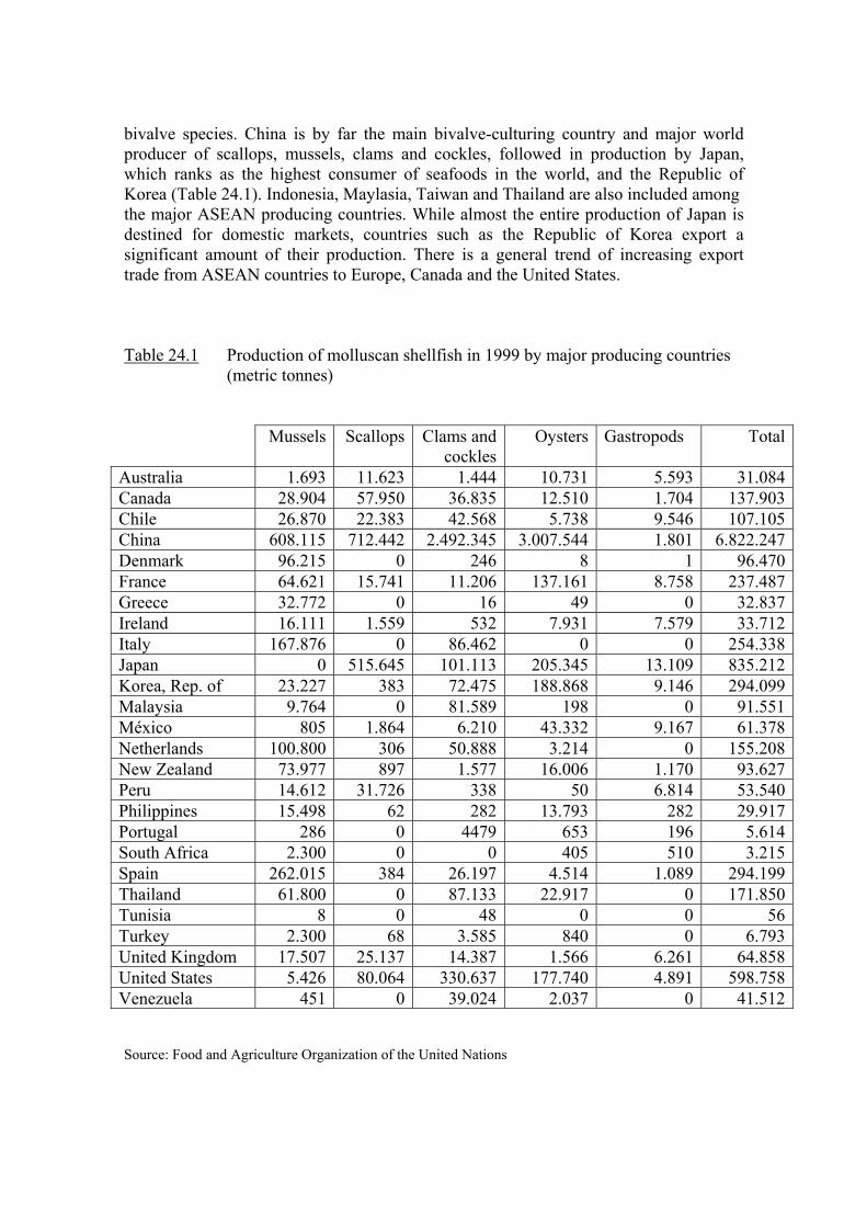
bivalve species. China is by far the main bivalve-culturing country and major world producer of scallops, mussels, clams and cockles, followed in production by Japan, which ranks as the highest consumer of seafoods in the world, and the Republic of Korea (Table 24.1). Indonesia, Maylasia, Taiwan and Thailand are also included among the major ASEAN producing countries. While almost the entire production of Japan is destined for domestic markets, countries such as the Republic of Korea export a significant amount of their production. There is a general trend of increasing export trade from ASEAN countries to Europe, Canada and the United States. Table 24.1 Production of molluscan shellfish in 1999 by major producing countries
(metric tonnes)
Mussels Scallops Clams and cockles
Oysters Gastropods Total
Australia 1.693 11.623 1.444 10.731 5.593 31.084Canada 28.904 57.950 36.835 12.510 1.704 137.903Chile 26.870 22.383 42.568 5.738 9.546 107.105China 608.115 712.442 2.492.345 3.007.544 1.801 6.822.247Denmark 96.215 0 246 8 1 96.470France 64.621 15.741 11.206 137.161 8.758 237.487Greece 32.772 0 16 49 0 32.837Ireland 16.111 1.559 532 7.931 7.579 33.712Italy 167.876 0 86.462 0 0 254.338Japan 0 515.645 101.113 205.345 13.109 835.212Korea, Rep. of 23.227 383 72.475 188.868 9.146 294.099Malaysia 9.764 0 81.589 198 0 91.551México 805 1.864 6.210 43.332 9.167 61.378Netherlands 100.800 306 50.888 3.214 0 155.208New Zealand 73.977 897 1.577 16.006 1.170 93.627Peru 14.612 31.726 338 50 6.814 53.540Philippines 15.498 62 282 13.793 282 29.917Portugal 286 0 4479 653 196 5.614South Africa 2.300 0 0 405 510 3.215Spain 262.015 384 26.197 4.514 1.089 294.199Thailand 61.800 0 87.133 22.917 0 171.850Tunisia 8 0 48 0 0 56Turkey 2.300 68 3.585 840 0 6.793United Kingdom 17.507 25.137 14.387 1.566 6.261 64.858United States 5.426 80.064 330.637 177.740 4.891 598.758Venezuela 451 0 39.024 2.037 0 41.512 Source: Food and Agriculture Organization of the United Nations
Among countries from the American continent, the USA leads in the production of bivalves, being ranked as the second world producer of clams and cockles and third producer of oysters and scallops, followed in importance by Canada which, together with the USA, is seeking new products with more added value (Vieytes and Leira, 2000). The increase in the last few years in Chile’s production of scallops and the rise in clam and cockle production of Mexico and Venezuela is noteworthy. Mexico is the second American producer of oysters, and Chile and Peru are the most important South American producers of mussels.
In Europe, the main molluscan shellfish producers are Spain, France, Italy, Denmark and the Netherlands. Total production in 1997 was about 1 million tonnes. Mussel production is of great important in this area, with Spain, Italy and the Netherlands leading the production of cultured mussels and Denmark the harvest of wild mussels. In recent years, Greece has strongly increased its mussel catch and Ireland has strengthened its mussel industry, mainly destined for export markets. The markets for fresh and frozen mussels are almost exclusively in the EU. Italy leads clam production and France is the major producer of oysters. The Netherlands is the biggest producer of cockles and the United Kingdom the top producer of scallops and gastropods. Export activities are very important in the EU, although exchanges are mainly between Member States. The Netherlands is by far the main mussel-exporting country, exporting mainly live mussels to neighbouring countries such as Belgium and Germany. Other exporting countries are Spain, Ireland, Germany and Denmark. Quantitatively, France, Belgium and Germany are the main importers, while the Netherlands tends to import in times of low production.
In the Pacific region, New Zealand and Australia are important producers of scallops and oysters. In recent years, the mussel production of New Zealand has experienced a sharp increase, with almost the total production destined to be exported as frozen mussels (half-shell product) (data on shellfish production from FAO and Vieytes and Leira, 2000)
The data on shellfish production in Table 24.1 illustrate the importance of seafoods in densely populated geographical areas that strongly depend on these resources for local consumption and where the exploitation of wild and cultured bivalves represents a significant part of their economic production. The impact of toxic phytoplankton on public health and economy can be enormous in developing countries but also in developed countries which, although less vulnerable to phycotoxins in shellfish, have also suffered toxic outbreaks and enormous economic losses in fisheries and tourism.
24.3 PROCESSES INVOLVED IN HARMFUL ALGAL EPISODES
24.3.1. Ingestion and incorporation of toxic or noxious algae and substances.
Some species of microalgae can produce harmful effects on shellfish populations either affecting them directly or by causing toxin accumulation that make them toxic or undesirable to man (reviewed in Blanco-Pérez, 2001). In some cases, large blooms of microalgae modify substantially characteristics of the water such as pH, oxygen content, viscosity, etc., producing delays of shellfish growth, alterations of the typical shape or even mortalities. This alteration of the medium may be, sometimes, also accompanied by the production of substances that can be toxic for some shellfish species, increasing

the probability of producing mortalities, and leading to large economical losses. In some other cases the microalgae produce substances that are accumulated in the shellfish, and, while not having a significant effect on them, turn the organisms undesirable –by giving them a strange color (e.g. the ciliate Mesodinium rubrum) or taste (e.g. diatoms of the genus Rhizosolenia )- or toxic to man. By far the later one is the most adverse effect.
Shellfish (bivalve molluscs, gastropods, crabs, lobsters and others) accumulate phycotoxins mostly by direct filtration of the algal cells or by feeding on contaminated organisms (e.g. carnivores and scavengers). The accumulation of a particular toxin is regulated by the balance of the toxin intake and loss from/to the environment and by the transformations to/from other toxins. The toxicity of the molluscs depends on the concentration of the different toxins in the soft tissues –that in turn depends on the toxin burden and the weight of the tissues- and also on the potency of each of toxin. Rates of toxin accumulation of filter-feeding shellfish from toxic algae are toxin- and species-specific because the food ingestion of some mollusc species can be affected by the presence of toxic algae, and also because mollusc species differ substantially in their retention capability for each group of toxins. Some species of bivalve are known to reduce or completely supress the toxic phytoplankton ingestion, in some cases to avoid toxic food but in some other because an adverse effect of the toxic species on the mechanism of the food capture (reviewed in Blanco-Pérez, 2001). Several PSP producing species of the genus Alexandrium have been shown to produce both kind of responses on several shellfish species (see Shumway and Cucci, 1987, Bricelj et al 1998, Lassus et al. 1999 for PSP). One species of particular interest is the northern quahog or hard clam, Mercenaria mercenaria which during an outbreak of a bloom of Alexandrium tamarense in Massachusetts, remained not toxic when most bivalve species attained large toxin concentrations. Studies in our laboratory (Shumway) have shown that in the presence of A. tamarense the quahog first retracts its siphons and then completely isolates itself from the external environment by means of shell valve closure. Efforts to induce toxicity by feeding A. tamarense were unsuccessful.
No effect on the feeding behavior by DSP producing species has been found up to date, neither in natural episodes –usually produced by Dinophysis species very low concentration in the water- nor in the experimental intoxification of Argopecten irradians with Prorocentrum lima (Bauder et al. 2001). Pseudo-nitzschia multiseries which produces ASP toxins, had no effect on Mytilus californianus (Whyte et al 1995) but induced the valve closure in Crassostrea gigas (Jones 1995)- The same effect is produced by Gymnodinium breve (NSP) on C. gigas and Braquiodontes recurvis (Sievers 1969).
The toxin contents and composition of the cells has been shown to have some effect, at least in PSP intoxification of M. edulis and M. mercenaria (Bricelj et al. 1990).
As with the organic matter, not all the toxin contained in the cells is absorbed by the mollusc. The main variables that seem to control the process are the gut passage time (the time that the food is maintained in the digestive system) and the digestibility and/or the postingestive selection of the toxic cells (Moroño et al. 2001; Bauder et al 2001). Toxic history and genetic differences have also an effect on the incorporation of toxins into molluscs.
Toxicity of individual shellfish in any given area is highly variable (see White et al., 1993; Chebib et al., 1993, for PSP; Gilgan et al. 1990; Douglas et al. 1997; Arévalo
et al. 1998; Blanco et al. 2002a,b, for ASP), probably mainly as a consequence of the also high inter-individual variability in the rates of food acquisition of most molluscs.
24.3.2. Detoxification
The rate of toxin loss is also toxin- and species-specific. The majority of available information concerns bivalve molluscs (see also Shumway, 1990 and Bricelj and Shumway, 1998), and, as these are the species most commonly reared in aquaculture, they are focused on here. Table 24.2 summarizes selected data on toxin retention for a number of bivalve species. Little is known about the factors that regulate the detoxification rate. Prakash et al. (1971) showed that PSP detoxification varied with season in the eastern coast of Canada and it is frequently assumed that low water temperatures retard toxin loss (see Shumway and Cembella 1993). Notwithstanding, temperature seems to have different effects for different toxins and species (no effect: PSP from Saxidomus giganteus (Madenwald 1985), Mytilus galloprovincialis (Blanco et al. 1997); slower loss: DSP from M. galloprovincialis (Blanco et al. 1999); faster loss: ASP from Mytilus edulis (Silvert and Subba Rao, 1993; Novaczek et al. 1992), and Pecten maximus (Blanco et al., in prep). Among the other factors studied to date, probably the food amount is the most important one for PSP and DSP, increasing slightly the detoxification rate (Sampayo et al. 1990; Marcaillou-Le Baut et al. 1993; Blanco et al. 1997, 1999). Metabolic processes in general seem to have little or no influence in regulating the loss of DSP and PSP toxins from mussels M. galloprovincialis, as no difference was found between groups with completely aerobic and partially anaerobic metabolisms (Moroño et al. 1998). However, it seems that metabolic processes may affect domoic acid detoxification in Pecten maximus (Blanco et al., unpublished data).
The rate of detoxification is highly dependent on the site of toxin storage within
the animal. Toxins in the digestive gland are usually eliminated much more readily than toxins bound in other organs, with the noticeable exceptions of DSP toxins in Argopecten irradians (Bauder et al. 2001) and domoic acid in P. maximus (Blanco et al. 2002).
24.3.3. Toxin transformations.
Most toxins undergo transformation in the organisms where they are accumulated, in some cases because the conditions (pH, redox potential, etc) are different from the producer, and in some other cases because of the enzimatic activityof the accumulator. As toxin analogues usually differ in their toxic potency, the transformations produce a change in the whole toxicity of the animal without any input or loss of toxin. Many PSP toxins interconvert easily (see Bricelj and Shumway 1998 for bivalves, Arakawa et al. 1998, for crabs), producing increases or decreases of the toxicity. A suite of derivatives of okadaic acid and dinophysistoxins 1 and 2 (e.g. diol esters, DTX4-like compounds) exhibiting different toxic potency ( can be present in the phytoplankton cells (eg. ) and, at least in some cases, they can be reverted to the parent toxins by the enzimes (during digestion of after it) of the accumulator inducing, very likely, significant changes in its toxicity. Simultaneously, OA, DTX1 and DTX2 can be converted to the corresponding acyl-derivatives (DTX3) (Fernández et al. 1998; Moroño et al. in press). ASP toxins have been less studied, with regard to this aspect.
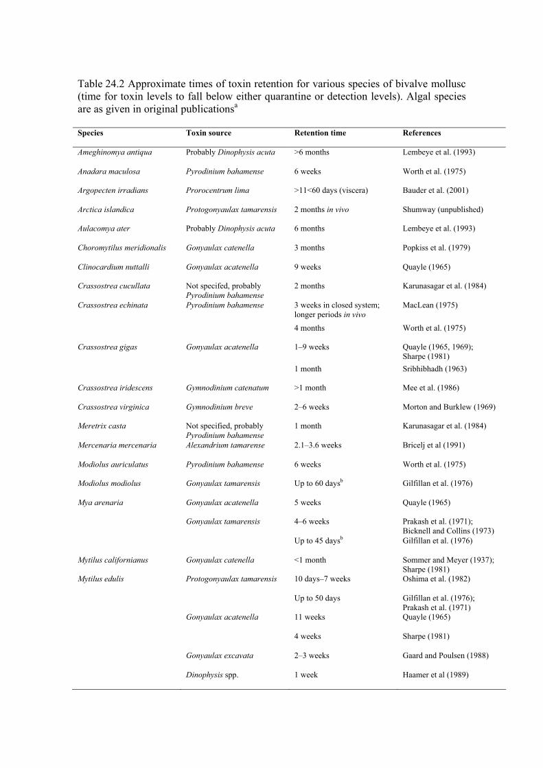
Table 24.2 Approximate times of toxin retention for various species of bivalve mollusc (time for toxin levels to fall below either quarantine or detection levels). Algal species are as given in original publicationsa
Species Toxin source Retention time References
Ameghinomya antiqua Probably Dinophysis acuta >6 months Lembeye et al. (1993)
Anadara maculosa Pyrodinium bahamense 6 weeks Worth et al. (1975)
Argopecten irradians Prorocentrum lima >11<60 days (viscera) Bauder et al. (2001)
Arctica islandica Protogonyaulax tamarensis 2 months in vivo Shumway (unpublished)
Aulacomya ater Probably Dinophysis acuta 6 months Lembeye et al. (1993)
Choromytilus meridionalis Gonyaulax catenella 3 months Popkiss et al. (1979)
Clinocardium nuttalli Gonyaulax acatenella 9 weeks Quayle (1965)
Crassostrea cucullata Not specifed, probably Pyrodinium bahamense
2 months Karunasagar et al. (1984)
Crassostrea echinata Pyrodinium bahamense 3 weeks in closed system; longer periods in vivo
MacLean (1975)
4 months Worth et al. (1975)
Crassostrea gigas Gonyaulax acatenella 1–9 weeks Quayle (1965, 1969); Sharpe (1981)
1 month Sribhibhadh (1963)
Crassostrea iridescens Gymnodinium catenatum >1 month Mee et al. (1986)
Crassostrea virginica Gymnodinium breve 2–6 weeks Morton and Burklew (1969)
Meretrix casta Not specified, probably Pyrodinium bahamense
1 month Karunasagar et al. (1984)
Mercenaria mercenaria Alexandrium tamarense 2.1–3.6 weeks Bricelj et al (1991)
Modiolus auriculatus Pyrodinium bahamense 6 weeks Worth et al. (1975)
Modiolus modiolus Gonyaulax tamarensis Up to 60 daysb Gilfillan et al. (1976)
Mya arenaria Gonyaulax acatenella 5 weeks Quayle (1965)
Gonyaulax tamarensis 4–6 weeks Prakash et al. (1971); Bicknell and Collins (1973)
Up to 45 daysb Gilfillan et al. (1976)
Mytilus californianus Gonyaulax catenella <1 month Sommer and Meyer (1937); Sharpe (1981)
Mytilus edulis Protogonyaulax tamarensis 10 days–7 weeks Oshima et al. (1982)
Up to 50 days Gilfillan et al. (1976); Prakash et al. (1971)
Gonyaulax acatenella 11 weeks Quayle (1965)
4 weeks Sharpe (1981)
Gonyaulax excavata 2–3 weeks Gaard and Poulsen (1988)
Dinophysis spp. 1 week Haamer et al (1989)
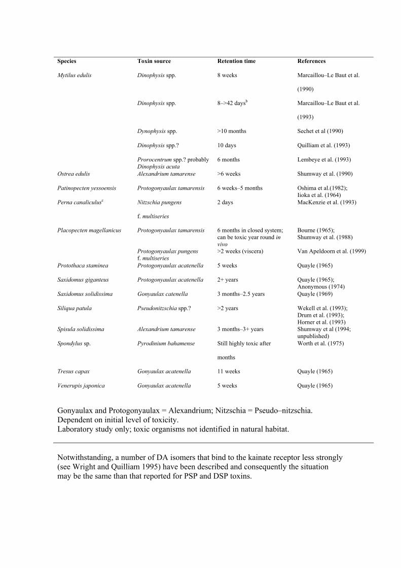
Species Toxin source Retention time References
Mytilus edulis Dinophysis spp. 8 weeks Marcaillou–Le Baut et al.
(1990)
Dinophysis spp. 8–>42 daysb Marcaillou–Le Baut et al.
(1993)
Dynophysis spp. >10 months Sechet et al (1990)
Dinophysis spp.? 10 days Quilliam et al. (1993)
Prorocentrum spp.? probably Dinophysis acuta
6 months Lembeye et al. (1993)
Ostrea edulis Alexandrium tamarense >6 weeks Shumway et al. (1990)
Patinopecten yessoensis Protogonyaulax tamarensis 6 weeks–5 months Oshima et al.(1982); Iioka et al. (1964)
Perna canaliculusc Nitzschia pungens
f. multiseries
2 days MacKenzie et al. (1993)
Placopecten magellanicus Protogonyaulax tamarensis 6 months in closed system; can be toxic year round in vivo
Bourne (1965); Shumway et al. (1988)
Protogonyaulax pungens f. multiseries
>2 weeks (viscera) Van Apeldoorn et al. (1999)
Protothaca staminea Protogonyaulax acatenella 5 weeks Quayle (1965)
Saxidomus giganteus Protogonyaulax acatenella 2+ years Quayle (1965); Anonymous (1974)
Saxidomus solidissima Gonyaulax catenella 3 months–2.5 years Quayle (1969)
Siliqua patula Pseudonitzschia spp.? >2 years Wekell et al. (1993); Drum et al. (1993); Horner et al. (1993)
Spisula solidissima Alexandrium tamarense 3 months–3+ years Shumway et al (1994; unpublished)
Spondylus sp. Pyrodinium bahamense Still highly toxic after
months
Worth et al. (1975)
Tresus capax Gonyaulax acatenella 11 weeks Quayle (1965)
Venerupis japonica Gonyaulax acatenella 5 weeks Quayle (1965)
Gonyaulax and Protogonyaulax = Alexandrium; Nitzschia = Pseudo–nitzschia. Dependent on initial level of toxicity. Laboratory study only; toxic organisms not identified in natural habitat. Notwithstanding, a number of DA isomers that bind to the kainate receptor less strongly (see Wright and Quilliam 1995) have been described and consequently the situation may be the same than that reported for PSP and DSP toxins.
24.3.4. Changes in shellfish biomass. The toxicity of an organism depends not only on the toxin burden and toxin
potency but also on the amount of biomass in which the toxins are distributed. Any alteration of the weight of soft tissues, not paralleled by an equivalent modification in the toxin burden, produces changes in the toxicity per unit of biomass. For PSP and DSP contaminated shellfish, weight is not lost at the same rate than the toxins). In such cases,. when the weight decreases, the same amount of toxin is concentrated in a smaller biomass, and the toxicity per unit of biomass of the organism increases. On the other hand, when the organism increases its weight the toxicity per unit of biomass decreases because the toxins are “diluted” in a larger biomass Those situations have been found in several studies both in natural and laboratory conditions (see Bricelj and Cembella 1995; Blanco et al., 1997, 1999). With some combinations of species and toxins, the situation may be different from that already described. Thus, the loss of domoic acid from the king scallop, seems to be related to loss of weight (Blanco et al. 2002) and consequently toxicity per unit of biomass is relatively independent from weight changes.
24.3.6. Toxin retention time.
Toxin retention time by shellfish depends on the mechanisms analized above and they are, therefore, species- and toxin- specific (Table 24.2). The differences of retention time are very large. The mussels Mytilus galloprovincialis and M. edulis retain domoic acid for hour or days (Novaczek et al, 1992; Blanco et al. 2002a) while the scallop Pecten maximus (Blanco et al. 2002b) retains this toxin for more than two years with a behaviour similar to that of Saxidomus giganteus and Spisula solidissima with PSP toxins (Quayle, 1965; Blogoslawski and Stewart, 1978; Chambers and Magnusson, 1950; Cembella and Todd, 1993; Shumway et al., 1994).
Few data are available on the retention times of toxins by crabs and carnivorous gastropods. The general trend for PSP toxins appears to be towards long-term retention (Desbiens and Cembella, 1993; Shumway et al., 1995; Shumway, unpublished data) but ASP can be eliminated from dungeness crab Cancer magister at a relatively fast rate (Lund et al. 1997).
Generalizations regarding the uptake and retention of phycotoxins by shellfish should be avoided and the species- and toxin- specific characteristics should be taken into account before choosing a species to be reared in areas prone to toxic algal blooms, and when designing monitoring and sampling regimes.
24.4 STRATEGIES TO AVOID INTOXICATION AND INTOXIFICATION OF SHELLFISH.
The exposure of shellfish to microalgal blooms can be reduced mainly by two procedures: reducing the algal biomass bloom or changing the location of the shellfish population. So far, the only strategy that has been proved to be efficient against the development of an algal bloom was the dispersion of clay over a proliferation of Cochlodinium polykrikoides in Korea (Kim et al. 1998), but it presents the drawback of an obvious environmental impact. Other strategies (reviewed in Blanco-Pérez 2001 and partially in Elbrachter and Schnepff, 1998) such as the use of ultrasounds or chlorine to kill the algal cells or the use of several viral, bacterial, fungus or protozoan parasites that lise cells, have been experimentally tried, but they have not been used in real
blooms. The second possible way of actuation consists in relocating the shellfish population (specially if they are cultured) in, or restrict the culture to, places or depths at which the concentration of the harmful population is minimal (Desbiens and Cembella, 1993; Lodeiros et al. 1998; Franzosini et al., 2000).
24.5 STRATEGIES TO ELIMINATE TOXINS FROM SHELLFISH.
Two groups of techniques are being used to eliminate toxins from the mollusc. The aim of the first group is to obtain live non-toxic mollusc, and includes techniques such as temperature or salinity stress, ozone or chlorine treatments, transplantation, etc that accelerate the loss of toxins from the organisms or, at least, prevent re-intoxication. The aim of the second group is to obtain products free of toxins after applying specific processing procedures ..
24.5.1. Strategies to obtain living shellfish free of toxins.
Various attempts have been made to detoxify live shellfish contaminated with paralytic shellfish poisons to reduce the duration of ‘off-market’ times. The most obvious method is to transfer shellfish to waters free of the toxic organisms and with environmental conditions that favor depuration. Unfortunately, the small effect of the environmental conditions on PSP and DSP detoxification rate of the mussel Mytilus galloprovincialis (Blanco et al 1997, 1999) makes transplantation more useful to prevent reintoxication than to increase the depuration rate. Notwithstanding, Blanco et al. (manuscript in prep) recently found that the king scallop Pecten maximus depurates domoic acid much faster when suspended in rafts than in their natural conditions. Transplantation is being used in several countries to take benefit in those two aspects. Detoxification of PSP toxins using temperature or salinity stress has also been tried with marginal success (Gilfillan et al., 1976; Blogoslawski and Neve, 1979). Instantaneous electrical shock treatments accelerated toxin excretion in scallops (Kodama et al., 1989). Reduced pH has been tried as a means of detoxifying butter clams, but with no success (Anonymous, 1966; Neal, 1967). Chlorination has been used in France; however, this process alters the flavour of the shellfish and thus decreases marketability.
Ozone has been touted as an effective means of reducing toxicity although its usefulness is questionable. Several early studies reported ozone to be effective in the inactivation of PSP toxins in shellfish exposed to Alexandrium tamarense, A. catenella and Karenia brevis blooms (Thurberg, 1975; Blogoslawski et al., 1975, 1979; Dawson et al., 1976; Blogoslawski and Stewart, 1978). Blogoslawski et al. (1973) also suggested that ozone could be used to inactivate Karenia (Gymnodinium) brevis toxins. More recently, preliminary studies by Gacutan et al. (1984, 1985) demonstrated that both ozone gas and PVP-iodide-iodine may effectively inactivate PSP toxins from Perna viridis contaminated by Pyrodinium bahamense. However, a subsequent study by White et al. (1985) produced results contradictory to previous studies in that no detoxification occurred in Mya arenaria exposed to ozone treatments. In a review (Blogoslawski, 1988), it was again suggested that ozonized sea water can be of value in detoxification of shellfish contaminated recently by the vegetative cells of toxic (PSP) dinoflagellates. In a study during a red-tide outbreak, it was shown that ozone treatment of the sea water does prevent shellfish (Mytilus edulis, Mya arenaria and Geukensia demissa) from accumulating paralytic shellfish poison. Blogoslawski concluded that inactivation could be achieved in bivalves exposed to and contaminated by motile PSP dinoflagellate cells without measurably altering the physical state of the treated bivalves and that this
inactivation could be achieved in a marketable species such as Mya within an economically feasible time-frame (Blogoslawski et al., 1979). Most recently, Fletcher et al. (1998) concluded that ozone was ineffective in reducing toxins from K. brevis in the oyster C. gigas. Ozone is useless in detoxifying cysts or in bivalves that have ingested cysts or have the toxins bound in their tissue over long periods. Furthermore, detoxification of algal toxins, especially paralytic shellfish poisons, over long periods is not economically feasible. We do not recommend ozone as a practical or safe means of eliminating algal toxins from shellfish. At present the economic feasibility of efficiently detoxifying shellfish on a large scale in artificial systems is not promising, perhaps with the exception of the oyster Crassostrea gigas contaminated with PSP toxins which responds to increased food supply with a substantial increment in its rate of toxin loss (Lassus pers., com.) and that has large commercial value. In areas prone to regular outbreaks of toxic algal species, aquaculturists and commercial fishermen alike must still depend on reliable monitoring systems to warn of toxic shellfish and plan their activities accordingly. Through the combined efforts of an intensive monitoring programme and culture of ‘rapid release’ species (e.g. Mytilus edulis), bivalves known to avoid toxic dinoflagellates (e.g. Mercenaria, most oysters) or scallops (adductor muscles rarely if ever toxic), economic losses can be kept to a minimum (see also Shumway et al., 1988).
24.5.2. Strategies to obtain shellfishery products free of toxins.
The second group of techniques range from the simple process of selective evisceration to cooking or more complex industrial processing techniques. Some species accumulate toxins preferentially in one or in a few organs. In these cases an easy way to eliminate most of the toxin is to remove such organ/s (evisceration). Obviously, this procedure can only be used when the size of the species is big enough to allow the dissection of the toxic anatomical part and the species has enough commercial value to compensate the usually large economical cost of dissection. This procedure is currently being applied in the EU for the king scallop Pecten maximus contaminated with ASP toxins. Recent EU legislation (Decision 2002/226/EC) has established an an exception to the general regulation for the exploitation of bivalve molluscs, in order to allow this procedure under specific conditions, that is being useful in mitigating the economic impact of the very long detoxification time of this species. The same procedure could be used with other bivalve species such as the razor clam Siliqua patula with ASP toxins(Horner et al 1993), or Argopecten irradians with DSP toxins (Bauder et al., 2001), but also with gasteropods as Haliotis tuberculata and Haliotis midae, that concentrate most of PSP in epipodial fringe (Bravo et al., 1999, 2001; Pitcher et al., 2001).
Very recently, the basis for a procedure to eliminate DSP toxins from molluscs by means of critical point extraction have been set up by González et al. (2002), pointing to an interesting approach to eliminate DSP toxins but also other lipophilic toxins. The technique requires dehydration of the product and therefore it will be only useful for products that require or, at least, tolerate this process.
Cooking has also been promoted as a possible means of detoxifying shellfish contaminated with paralytic shellfish poisons. Although cooking does not eliminate the danger of intoxication, it may effectively reduce toxicity to safe levels if initial levels of toxicity are low. Pan-frying seems to be more effective to reduce PSP levels than other
methods of cooking (Medcof et al., 1947; MacDonald, 1970). When clams or mussels are steamed or boiled, toxins lost from the tissues are contained in the cooking liquid rendering the fluids extremely toxic. Vieites et al. (1999) showed that more than 30% of the PSP toxins contained in mussels (Mytilus galloprovincialis) are transferred to the water after cooking them at 97ºC for two minutes, while real destruction of the toxin was about 20%.
Commercial canning has been shown to reduce toxicity (paralytic shellfish poisons) of soft-shell clams, Mya arenaria, by as much as 90%. A toxicity level of 160 µg STXeq 100 g–1 for soft-shell clams and mussels (Mytilus edulis) to be canned was established in the 1950s in Atlantic Canada and remains in effect today (Cembella and Todd, 1993),. Noguchi et al. (1980) (see also Nagashima et al., 1991) showed that toxicity levels of PSP-infested scallops could be reduced during canning processes. They demonstrated that during retorting (110 °C, 80 min, or 122 °C, 22 min) most of the PSP toxins could be eliminated (maximum initial level 102 MU g–1 digestive gland) whereas heating (70 °C, 20 min) followed by washing was less effective in reducing the toxicity below the quarantine limit of 4 MU g–1. Recent efforts in Spain (Berenguer et al., 1993) have demonstrated that toxicity levels of Mediterranean cockles (Acanthocardia tuberculatum) may be significantly reduced via canning. Total toxicity of cockles (initial levels of approximately 800 g STXeq 100 g–1) was reduced to < 35 g STXeq 100 g–1 after cooking. These authors demonstrated that the decreases in PSP toxicity obtained by commercial processing are sufficient to warrant canning as a practical means of obtaining a legal and acceptable product when the toxin levels are moderate. Similar attempts have been made to detoxify surfclams (Spisula solidissima) via canning, but results to date are inconclusive. During the canning process of PSP contaminated Mytilus galloprovincialis, about 30% of the toxin goes to the packing liquid and about 33% is destroyed (when packaged in brine) (Vieites et al., 1999). It has been also shown that the thermal treatments associated with canning induce transformations between the different PSP toxins, with the consequent changes in toxicity (Vieites et al., 1999; Indrasena and Gill, 1999). The effectiveness of canning as a means of reducing PSP-toxicity levels below quarantine levels seems to be dependent on the initial profile of toxins and , in some cases, on the initial level of toxicity and should be approached with great caution.
Domoic acid was shown to decrease from the scallop tissues using freezing followed by canning (Leira et al. 1998), but it seems that only the freezing step had an effect in reducing the overall toxin content of the product
With the exception of the study by Berenguer et al. (1993), no useful methods have been devised for effectively reducing phycotoxins in contaminated live shellfish. All methods tested to date have been unsafe, too slow, economically unfeasible or yielded products unacceptable in appearance and taste. Given the global increase in harmful algal blooms and the continually growing interest in culture of bivalve molluscs, further efforts are needed to develop effective means of detoxifying shellfish contaminated with phycotoxins. Failing the development of any such methods, increased efforts need to be expended in monitoring shellfish for the presence of phycotoxins.
24.6 STRATEGIES TO MINIMIZE THE RISK OF TOXIC SHELLFISH FOR HUMAN HEALTH
The strategies to minimize the risk for human health are based on the definition of allowable toxin levels in seafoods and in the determination of those levels in the exploited shellfish populations, which includes the use of suitable methodology and the establishment of adequate monitoring programmes.
24.6.1 Allowable toxin levels and methods: the basis of the regulation
Regulations on action levels should ideally be based on a risk assessment that comprises hazard identification (which involves identification of the toxins causing adverse health effects), hazard characterization (which implies evaluation of the adverse health effects associated with the presence of the toxins in seafoods), exposure assessment related to the evaluation of likely intake of phycotoxins, and risk characterization (which involves quantitative estimations of the probability of occurrence of adverse health effects in a given population) (Aune, 2001) . Epidemiological data and toxin kinetics studies, action mechanism, target organs and dose-response relationships are required for the risk assessment process. In addition, the capability of testing procedures to identify and quantify toxin levels in food should be taken into account when establishing regulations. In the case of some marine toxins, especially some polyether toxins, there is a lack of such studies, and the action levels have been set on the basis of sparse toxicity data mainly originating from human poisoning episodes, the detection limits of the available methods or following the specifications of other countries. Robust toxicological studies are lengthy and expensive to conduct and involve the use of large amounts of pure toxins, which are not available in most cases.
Accurate methods of quantification have to be available, because legislation calls for methods of control. However, the development and validation of suitable testing methods has also been hampered by the lack of pure toxins and certified reference materials (Van Egmond et al., 1992). Most countries still rely on animal bioassays to detect PSP and DSP but the ethical aspects of these tests have led to growing resistance from animal-welfare groups. It should also be borne in mind that it is not realistic to establish an action level lower than the actual limit of detection, although this might be desirable from the toxicity point of view. The elucidation of new toxins, as well as the wide range of seafood that can act as toxin vectors, make the establishment of rational guidelines and regulations more difficult. Despite the dilemmas, a number of countries have established limits and regulations for phycotoxins in shellfish.
Paralytic shellfish poisoning (PSP) Among all the seafood poisonings, PSP represents one of the most severe threats to public health, affecting most coastal areas in the world. Saxitoxin (STX) and analogues are mainly produced by a number of dinoflagellates belonging to the genera Alexandrium, Gymnodinium and Pyrodinium (Yasumoto and Murata, 1993). Although bivalve molluscs are the main PSP vectors, gastropods, crustaceans and pelagic fish have also been reported as vectors of these toxins (Shumway, 1995). Many affected countries have established regulations including methods and action limits. Table 24.3 shows countries known to have PSP regulations in force.
The main method used for regulatory purposes is the mouse bioassay (AOAC, 1990) that has been validated and standardized by the Association of Official
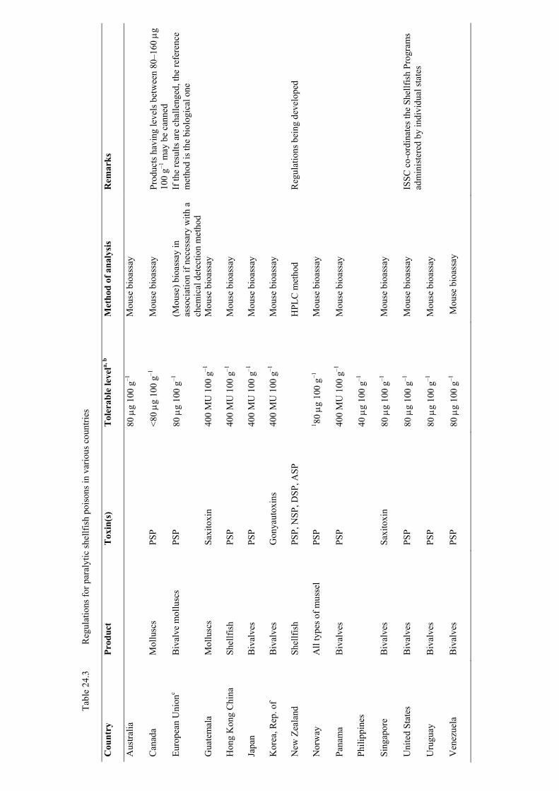
Tabl
e 24
.3
Reg
ulat
ions
for p
aral
ytic
shel
lfish
poi
sons
in v
ario
us c
ount
ries
Cou
ntry
Pr
oduc
t T
oxin
(s)
Tol
erab
le le
vela,
b
Met
hod
of a
naly
sis
Rem
arks
Aus
tralia
80
µg
100
g–1
Mou
se b
ioas
say
Can
ada
Mol
lusc
s PS
P <8
0 µg
100
g–1
M
ouse
bio
assa
y Pr
oduc
ts h
avin
g le
vels
bet
wee
n 80
–160
µg
100
g–1 m
ay b
e ca
nned
Eu
rope
an U
nion
c B
ival
ve m
ollu
scs
PSP
80 µ
g 10
0 g–1
(M
ouse
) bio
assa
y in
as
soci
atio
n if
nece
ssar
y w
ith a
ch
emic
al d
etec
tion
met
hod
If th
e re
sults
are
cha
lleng
ed, t
he re
fere
nce
met
hod
is th
e bi
olog
ical
one
Gua
tem
ala
Mol
lusc
s Sa
xito
xin
400
MU
100
g–1
M
ouse
bio
assa
y
Hon
g K
ong
Chi
na
Shel
lfish
PS
P 40
0 M
U 1
00 g
–1
Mou
se b
ioas
say
Japa
n B
ival
ves
PSP
400
MU
100
g–1
M
ouse
bio
assa
y
Kor
ea, R
ep. o
f B
ival
ves
Gon
yaut
oxin
s 40
0 M
U 1
00 g
–1
Mou
se b
ioas
say
New
Zea
land
Sh
ellfi
sh
PSP,
NSP
, DSP
, ASP
HPL
C m
etho
d R
egul
atio
ns b
eing
dev
elop
ed
Nor
way
A
ll ty
pes o
f mus
sel
PSP
1 80 µ
g 10
0 g–1
M
ouse
bio
assa
y
Pana
ma
Biv
alve
s PS
P 40
0 M
U 1
00 g
–1
Mou
se b
ioas
say
Phili
ppin
es
40 µ
g 10
0 g–1
Sing
apor
e B
ival
ves
Saxi
toxi
n 80
µg
100
g–1
Mou
se b
ioas
say
Uni
ted
Stat
es
Biv
alve
s PS
P 80
µg
100
g–1
Mou
se b
ioas
say
ISSC
co-
ordi
nate
s the
She
llfis
h Pr
ogra
ms
adm
inis
tere
d by
indi
vidu
al st
ates
U
rugu
ay
Biv
alve
s PS
P 80
µg
100
g–1
Mou
se b
ioas
say
Ven
ezue
la
Biv
alve
s PS
P 80
µg
100
g–1
Mou
se b
ioas
say
Analytical Chemists (AOAC). This reference method is the only procedure recognized internationally for quantifying PSP toxicity, although with some variations in the acceptable regulatory level and in the units used for expression of the toxicity: 40 µg PSP/100 g, 80 µg PSP/100 g or 200 MU/100 g, 400 MU/100 g (Van Egmond et al., 1992). Most countries have agreed on 80 µg saxitoxin (STX) equivalent/100 g shellfish meat.
The effectiveness of some procedures (evisceration, canning, etc.) in reducing PSP toxicity has led some countries to establish different limits depending on the form of consumption of the shellfish. Thus, in Canada and the United States, shellfish destined for canning or subjected to an evisceration step might be harvested with PSP toxin amounts higher than 80 µg/100 g (Cembella and Todd, 1993; NSSP, 1990). In the case of EU legislation, the application of detoxification procedures is limited by Council Directive 91/492/EEC that prohibits the harvesting of toxic shellfish. There is only one exception (in the case of PSP), and this applies to the species Acantochardia tuberculata destined to be canned, allowing Spain to authorize harvesting when PSP levels in the edible parts exceed 80 µg STXeq per 100 g tissue but are less than 300 µg STXeq per 100 g tissue (Decision 96/77/EC). The bivalves must undergo a heat treatment clearly defined in the Annex of the EU Decision, the final product must not
contain a PSP toxicity level detectable by the mouse bioassay, and each lot must be tested (Burdaspal et al., 1998).
Diarrhetic shellfish poisoning (DSP) Among all the groups of marine toxins, those included in the DSP group are the
subject of greatest controversy. Three groups of toxins have historically been included in the DSP group: okadaic acid (OA) and dinophysitoxins (DTXs) produced by Dinophysis and Prorocentrum species , pectenotoxins (PTXs) produced by some Dinophysis species and yessotoxins (YTXs), produced by Gonyaulax grindleyi and Lingulodinium polyedra (Yasumoto et al., 1989; Yasumoto and Murata, 1990; Satake et al., 1998; Tubaro et al., 1998). These bioactive compounds share polyether cyclic structure and solubility properties, and extraction procedures and conventional mouse bioassays do not discriminate among them. However, their biological activity and toxicological properties are significantly different. There is no general consensus as to which liposoluble toxins should be regarded as DSP toxins, which ones should be monitored and regulated and which are the most appropriate testing procedures and acceptable levels.
Concerning sanitary control of DSP toxins, there are disparities in methods and in the criteria for positive results. Mammalian bioassays are widely applied for DSP toxicity determination. However, there are great differences in the performance of the procedures. Assay selectivity, specificity and toxin recovery depend greatly on the selection and ratio of the organic solvents used for extraction. Bioassay procedures as diverse as the oral dosage rat bioassay (Kat, 1983) and the intra-peritoneal injection mouse assay are not equivalent, because the former technique quantifies only the diarrhetic effect of certain DSP toxins whereas the latter assay provides an estimate of total DSP toxicity (Cembella et al., 1995).
Regarding regulatory levels, most countries have set the limit at the detection limit of the analytical method used. The first country to establish a limit was Japan – 5 MU/100 g shellfish tissue, based on an epidemiological study (Yasumoto et al., 1978). This limit has also been established in the Republic of Korea and New Zealand. In
practice, most countries applying mouse bioassays use survival time for the determination of the toxicity, but without a consensus on the appropriate observation period, the acceptable criteria can vary between ‘two out of three mouse deaths in less than 5 h’ to ‘two of three mouse deaths in less than 24 h’.
Chile, Thailand, Turkey, Uruguay and Venezuela regulate DSP on the basis of the Yasumoto mouse bioassay (1978) with a survival time of 24 h as the criterion for a positive result. Canada and the USA have no official regulations on DSP, although Canada may issue informal advice on positive results by mouse assay and/or the fluorescence high-performance liquid chromatography (HPLC) method.
Regarding DSP control in the European Union, the EU Directive 91/492 established that the the customary biological method must not give a positive result for the presence of DSP in the edible part of the molluscs, but it did not clarify the interpretation of a positive results and which biological methods should be used. An important progress has been recently made, with the approval of the Decision 2002/225/EC that lays down detailed rules for the implementation of the Directive 91/492 as regards the maximum levels and the methods of quantification of OA and Dinophysistoxins, Pectenotoxins, Yessotoxins and Azaspiracids in bivalve molluscs, echinoderms, tunicates, and marine gastropods. Regarding maximum levels, the following has been stated: A) The maximum level of okadaic acid, dinophysistoxins, and pectenotoxins, together shall be 160 µg of okadaic acid equivalents/ kg edible part (the whole body or any part edible separately). B) The maximum level of yessotoxins shall be 1 mg of yessotoxin equivalents/ kg edible part (the whole body or any part edible separately). C) The maximum level of azaspiracids shall be 160 µg of azaspiracid equivalents/ kg edible part (the whole body or any part edible separately). Concerning detection methods, the new Decision states that in addition to biological testing methods, alternative detection methods such as chemical methods and in vitro assays should be accepted if it is demonstrated that the performance of the chosen method is not less effective than the performance of the biological method and that their implementation provides and equivalent level of public health protection. The compounds to be detected are: OA and Dinophysistoxins, PTX1 and PTX2, YTX, 45 OHYTX, homo YTX and 45 OH homo YTX AZA1, AZA2 and AZA 3. However, if new analogues of public health significance are discovered they should be included in the determinations. Standards have to be available before chemical analysis will be possible and total toxicity will be calculated using conversion factors based on the toxicity data available for each toxin. The performance of these methods should be defined after validation following and internationally agreed protocol. Concerning biological methods, it is stated that a suite of mouse bioassay procedures differing in the test portion and in the solvents used for the extraction and purification steps can be used, but that sensitivity and selectivity depend on the choice of the solvents and that this should be taken into account when making a decision on the method to be used in order to cover the full range of toxins. With regard to the mouse bioassay, it has been established that the death of two out of three mice within 24 hours after inoculation into each of them of an extract, equivalent to 5 g of hepatopancreas or 25 g of whole body, should be considered as a positive result for the presence of one or more of the toxins of the groups regulated in the Decision at levels above those allowed.
Although the full implementation of this new Decision on the basis of chemical methods and/or in vitro assays still requires big efforts on the development of toxin
standards, toxicological studies and validation of alternative methods, it represents an important step towards harmonization of phycotoxin control approaches.
Amnesic shellfish poisoning (ASP)
ASP is produced by the ingestion of seafood contaminated with domoic acid (DA), an excitatory amino-acid produced by some species of the pennate diatom genus Pseudo-nitzschia. The first toxic outbreak took place in Canada in 1987 and toxic episodes, although not human poisonings, have been reported in other regions. Countries that have established regulations for ASP are Canada, the United States, New Zealand (Trusewich et al., 1995; Wright et al., 1989; NSSP, 1990), Chile, Peru, and European Union Member States (EU Directive 91/492/EEC amended by Directive 97/61/EC). The limit is 20 mg/kg edible meat. The US Federal Food and Drug Administration (FDA) has set a quarantine level of 30 ppm of DA in cooked viscera of dungeness crabs (Shumway, 1995). HPLC methods have been established for regulatory purposes. In order to minimize the economic impact produced by the slow ASP detoxification process of certain pectinidae, the EU has recently approved the Decision 2002/226/EC, that allows the harvesting of Pecten maximus and Pecten jacobeous with DA levels higher than 20 mg / kg and lower than 250 mg / kg if they are destined to be placed on the market after total removal of hepatoancreas, soft tissues or any other toxic contaminated part. On the basis of this Decision, a restricted harvesting regime of scallops with DA concentration (whole body) > 20 µg /g and intended to be processed, can be initiated if DA concentration in whole scallop (whole body) is < 250 µg /g and if the DA concentration of the anatomical parts intended to be placed on the market and analyzed separately, (edible parts), is < 4.6 µg /g DA. The latter condition was introduced to take into account the large inter-animal variability which exists in the concentration of DA in scallop organs. It is not a lowering of the regulatory limit. It assists in ensuring that the chance of the DA concentration in the scallop edible part exceeding 20µg/g is less than 1 in 1000 upon harvesting ( probability “p” of 0.1%). It is based on the analysis of 10 pooled individual edible parts and a 50 % coefficient of variation (as a measure of inter-animal variability in DA concentrations).
In addition to the conditions above for allowing harvesting, additional requirements concerning sampling, number of individuals to make up the analytical sample, transport conditions, authorized establishments, testing of the end product and traceability measures have been also established in the EU Decision.
Neurotoxic shellfish poisoning (NSP)
NSP is a seafood intoxication produced after the ingestion of shellfish contaminated with brevetoxins produced by the dinoflagellate Karenia (Gymnodinium) brevis and related species. The United States and New Zealand are currently the main areas affected directly by NSP (MacFarren et al., 1965; Ishida et al., 1995). The testing procedure in use in the USA is the mouse bioassay (APHA, 1985) and the acceptable level is 20 MU/100 g. In New Zealand, a mouse bioassay equivalent to the APHA method (Hannah et al., 1995) is in use and 20 MU/100 g is also employed as acceptable level (Trusewich et al., 1995). The EU Directive makes no mention of NSP toxins as they have not been reported to occur at significant levels in European coastal waters.
Azaspiracid shellfish poisoning (AZP)
Azaspiracids Poisoning (Ofuji et al., 1999, añadir) is caused by the ingestion of shellfish contaminated with Azaspiracids, a novel group of polyether toxins, recently identified in shellfish from Ireland, Norway and United Kingdom (James et al., 2000 añadir) and reponsible of several outbreaks. AZP symptoms are similar to those of DSP and include nausea, vomiting, severe diarrhoea and stomach cramps. Protoperidinium crassipes has been identified as an algal source of AZAs (Yasumoto, unpublished data). Recently, the control of AZAs in shellfish has been regulated in the EU Decision 2002/225/EC that establishes a maximum level of AZAs of 160 µg / kg edible part. In contrast with other lipophilic toxins, such as okadaic acid and analogues that mainly remain within the hepatopancreas, azaspiracids can be accumulated in the whole shellfish tissues. In addition to LC-MS methods, a modification of the Yasumoto mouse bioassay (Yasumoto et al., 1984) that involves the analysis of whole shellfish tissues is suitable for the detection of AZAs in shellfish at the levels established in the Decision (Yasumoto, manuscript in prep.).
24.6.2 Monitoring programmes: putting regulation into practice.
Once established the basis of their regulation, a large number of countries worldwide have established phycotoxin monitoring programmes to prevent risks for public health and minimize economic losses for aquaculture and fisheries. The basic components of a programme -the authorities responsible for its application, the involvement of the industry and the organization of the inspection services- very much depend on the country or geographical area. In many cases the programme includes surveillance for potential toxic algal species (identification and quantification) and monitoring of toxin content in shellfish by means of a range of testing procedures. Results are compared with allowed levels established in the regulations to prevent unsafe shellfish being placed on the market. In addition, some countries complement phytoplankton and toxicity data with the monitoring of biological, chemical and physical parameters such as water temperature, salinity, nutrients, chlorophyll, water stratification, current circulation, etc. This information, together with the weather and oceanographic conditions, constitute valuable prediction tools. Information on water-mass distribution and movements obtained from satellite imagery is also used to predict the appearance of phytoplankton populations. The application of airborne spectrographic sensing technologies and information provided by moored installations of ocean optical equipment have been proposed as effective monitoring tools.
Whatever the features of the programme, an algal and shellfish sampling protocol over time and space is designed, including definition of adequate location and number of sampling sites, and sufficient sampling frequency to document spatio-temporal changes of phytoplankton and toxins in shellfish and to cover the risks of rapid rises in shellfish toxicity. Taking into account the changing nature of phytoplankton populations, sampling should be carried out at least weekly (Andersen, 1996), intensifying during the development of a toxic episode.
The design of the programme is usually done on the basis of oceanographic information, historical data on phytoplankton occurrence in the area, local needs, harvesting periods, the knowledge of the detoxification kinetics of the species under control, etc. Algal and shellfish sampling is usually performed by official authorities,
although in some countries local producers, industry and even volunteers may carry out sampling under the supervision of the authorities.
The economic importance of the resource and therefore the available funding, and also its spatial concentration, usually determines the features and extent of the programme, which may be mostly funded by government, e.g. Galician Rias (north-western Spain); by industry, e.g. Denmark; or by both, e.g. New Zealand. The more information is available on the spatio-temporal evolution of the phytoplankton populations in a given production area, the better the assessment of closure or opening of small zones in a short period. On the contrary, if the costs of regular monitoring are not affordable, production areas may be kept closed for prolonged periods when a risk of toxic episodes exists. In extreme cases of chronic bloom situations and no available funds, production areas may be permanently closed to avert loss of life, as in the case of some shellfish farms in ASEAN regions (Corrales and Maclean, 1995).
In addition to a suitable sampling programme, an adequate transport system for the samples should be developed, including suitable conditions to preserve them until they reach the laboratory. After the identification and quantification of potentially toxic algal species, and after shellfish testing by biological and chemical methods, results are evaluated and delivered to decision-makers. An extremely important issue that determines the efficacy and reliability of the control system is the time between sampling and issuing of results. This should be as short as possible (preferably not longer than one to two days) in order to make decisions on prohibition of harvesting.
Information obtained from monitoring of toxic phytoplankton is used for different goals: as an early warning tool, to set threshold levels for increasing sampling frequency and toxin testing in shellfish, and even as a criterion to close production areas when the number of potentially toxic algal cells exceeds certain critical concentrations. There is no general rule to define harmful concentration of cells and the criteria depend very much on the specific toxic species, ranging from hundreds (Dinophysis spp.) to millions (Pseudo-nitzschia spp.) of cells. However, correlation between cell counts and toxicity in shellfish is not always good. Significant inter- and intra-species variability in toxin profile and toxin content has been reported for many phytoplankton species even from the same area and over a short period. In addition, some shellfish species can store toxicity for long periods, so that toxicity remains many months after the toxic cells bloomed. ASP toxicity in scallops can remain even years after Pseudo-nitzschia blooms. The contribution of cysts to shellfish toxicity also cannot be underestimated.
Some countries rely on both cell counts and shellfish toxin content to make decisions on closure of production areas, while others only take decisions based on toxicity in shellfish. Although information on both phytoplankton and toxin content is highly desirable, shellfish testing is more reliable from the point of view of protecting consumers.
The strategy used to monitor the Galician Rías, an area of intense aquaculture and consequently with most of the resources concentrated in a relatively small area, illustrates how intensive a monitoring programme can be in time and space if the value of the resources is significant. The design of the programme was based on the fact that toxic events are possible at any time of year and that toxicity levels affect only small areas, whereas harvesting closures previously affected wider areas. To better predict the appearance, evolution and disappearance of toxic species, the production areas have been divided into small zones and subzones and the exploited species have been differentiated in terms of intoxication-detoxification dynamics and harvesting

schedules. Depending on factors such as oceanographic and weather conditions and the levels of toxic species found, various action plans including different sampling frequencies (from weekly to daily) have been established (Fig. 24.1). All data generated from the programme – oceanographic variables, phytoplankton composition, weather forecast, surface temperatures, toxin analysis – are processed using the same computer software, which updates the action plan twice a day. Any change in the plan is notified by fax and e-mail to all the organizations of the administration, industry and research centres. The number of sampling sites and frequency of sampling have increased, but the duration of quarantine periods and the extent of affected areas have been reduced considerably, allowing better management of the resources and minimizing economic impacts (Mariño et al., 1998).
A monitoring programme like the Galician one, may be unfeasible, specially from the economical point of view in other areas in which the resources are more dispersed or/and have less economical and social importance. Within the United States, the State of Maine has one of the most comprehensive monitoring programmes for paralytic shellfish poisons (see NSSP, 1990, Part 1, Section C, Appendix A). This programme was necessitated by yearly occurrences of toxic shellfish and has been used as a template for establishing monitoring programmes in many other regions worldwide. Its purpose is to ensure that only safe shellfish are harvested. Years of practical experience have afforded the opportunity to continually modify the sampling programme to better reflect increasing knowledge of potential toxic areas as well as the changing utilization of shellfish. Instead of ‘primary-key stations’, areas of similar toxic patterns are used. At the beginning of the PSP testing year, shellfish samples are collected from each of these areas to determine the background level of toxicity. Sampling stations from these areas are sampled each week from April to October, regardless of toxin patterns. When shellfish show any toxicity, sampling is expanded until stations of no toxicity are found. This sampling programme allows for closures to be made in a safe manner. Maine’s law and regulations require the immediate closure of toxic shellfish harvest areas, and embargo or confiscation of all suspect shellfish. When necessary, licences and certificates may be suspended. Administrative actions are accomplished on a same-day basis. Maine’s PSP Monitoring Program is continually modified to reflect increasing knowledge as to where toxic shellfish are likely to occur, as well as changing utilization of shellfish. The program is carried out on a yearly basis from April until October. Based on areas of similar toxin patterns, the coast of Maine has been divided into 18 areas from south-west to north-eastAt the beginning of the PSP testing year, shellfish samples (mussels, Mytilus edulis; clams, Mya arenaria) are collected weekly from ‘primary’ sampling stations from these areas. Early-season shellfish samples determine the background level of toxicity in an area (hopefully below the limit of detection sensitivity of the test). Whenever there is any rise of toxin in an area the sampling of shellfish is increased to adjacent sampling stations so that when a closure is necessary adequate data are available to make a proper public-health-oriented decision. Closures are made with safety zones in place and frequently on the basis of previous PSP closures for that area. Shellfish are returned to the laboratory under refrigeration and tested as soon as possible under the standard mouse bioassay. Whenever toxin levels are found approaching the quarantine level of 80 µg STXeq per 100 g of shellfish, the area is closed to shellfish harvesting. Closures are made on a species basis if adequate information is available to demonstrate that not all species are toxic. Species closures require increased sampling of the ‘toxin-free’ shellfish. In areas where it can be
justified, increased sampling will permit a partial opening of an area where non-toxicity is proven. Shellfish such as the ocean quahog, Arctica islandica, are sampled from contract fishing boats by department personnel. Areas where quahogs and similar species cannot be sampled are closed because they cannot be regarded as safe without sampling. Reopening of closed areas is dependent on continued toxin levels less than 80 µg. Openings are made after evaluations of current and historical records. Areas in which there is a high-value shellfish resource and where there is reason to believe that there will not be another rise of toxin may be reopened after at least two weekly samplings below 80 µg.
24.6.3 The need for harmonization.
24.6.3.1 General Agreement on Tariffs and Trade The World Trade Organization (WTO) emerged as a consequence of historical efforts and negotiations intended to achieve the liberalization of international trade. The WTO includes the General Agreement on Tariffs and Trade (GATT) and comprises about 125 countries that represent more than 90% of international trade. The basic policy of the organization is the progressive elimination of the barriers that protected domestic markets in the past. Within this framework, sanitary standards or regulations may be used as non-tariff barriers. The GATT agreements on sanitary and phytosanitary measures acknowledge the right of WTO members to establish regulations for health protection, if they are based on scientific evidence and risk assessment and do not discriminate among members where identical or similar conditions prevail. Sanitary regulations shall not be applied in a manner that constitutes a disguised restriction to international trade. Thus, when establishing a suitable level of sanitary protection, each country will consider the aim of reducing as much as possible the negative effects on trade so that regulations do not involve a restriction higher than that required (Council Decision 94/800/EC). Based on GATT agreements, trade relationships shall be carried out based on the principles of harmonization, equivalency and transparency. This means the establishment of common sanitary measures following international standards, the acceptance of different sanitary measures whenever the achievement of the appropriate level of protection is demonstrated, and the elaboration of regulations with absolute transparency. WTO members should provide information on their sanitary regulations and notify any change. GATT agreements encourage the uniformity of sanitary regulations in order to eliminate risks to consumers and reduce obstacles to trade. However, when considering existing regulations for marine toxins, discrepancies between methods and limits are evident, even between countries for which a free trade and unrestricted circulation of seafood are already in place. Current attempts at harmonization in the EU, the APEC area and the USA are described below.
24.6.3.2 European approaches to marine toxin control and harmonization
A large number of European coastal countries are affected by toxic events related to harmful algal blooms. Toxic episodes due to PSP have been registered since the 1950s and DSP and ASP toxins have been the cause of prolonged closures of shellfish production areas. On the other hand, a new group of toxins, azaspiracids (AZAs) responsible of the toxic syndrome, Azaspiracid Poisoning (AZP) might affect a number of EU countries. So far, there are no records of shellfish contamination due to the
presence of brevetoxins, but occasional reports have been made of microcystins and nodularin in molluscs that might represent an additional risk for shellfish consumers. Within Europe, the European Union, currently made up of 15 countries (Austria, Belgium, Denmark, Finland, France, Germany, Greece, Ireland, Italy, Luxembourg, the Netherlands, Portugal, Spain, Sweden and the United Kingdom) is an area of free trade, with unrestricted circulation of merchandises, and with border control carried out only for imports from countries outside the EU. Along the last decade, a number of Directives dealing with food safety legislation have been elaborated and implemented to ensure consumer protection, high quality of products and fair competition. .
Regarding molluscan shellfish, the process of harmonization is at an advanced stage. EU Directives 91/492/EEC and 91/493/EEC lay down the health conditions for the production and placing on the market of live bivalves and fishery products respectively. Directive 91/492 establishes the control of the shellfish production areas and the different activities of harvesting, transport, depuration, storage, processing and marketing, as well as microbiological and chemical parameters and marine toxin content. Directive 97/61/EC modifies and complements Directive 91/492/EEC concerning traceability of mollusc batches from harvesting to marketing and includes domoic acid control. According to Directive 91/492/EEC, a public health control system must be established by the responsible authority in order to verify whether the requirements laid down in the Directive are complied with. In relation to marine toxins, this control system must include:
1. Periodic monitoring of live bivalve molluscs and production areas in order to check
the possible presence of toxin-producing phytoplankton in the waters and biotoxins in shellfish.
2. Sampling plans that will take into account possible variations in production and relaying areas in the presence of toxic phytoplankton. The sampling must be carried out periodically to detect changes in the composition of the toxic phytoplankton populations and their geographical distribution. Information leading to suspicion of accumulation of toxins in mollusc flesh must be followed by an intensive sampling, increasing the number of sampling points and the number of samples. Toxicity tests shall be carried out using the molluscs of the affected area which are most susceptible to contamination. If the results of a sampling plan show that marketing live bivalve molluscs may constitute a hazard to human health, the responsible authority must close the production area as regards molluscs until the situation has been restored. Marketing molluscs from that area may not be re-authorized until new sampling has provided satisfactory test results.
3. Laboratory tests of the end product to verify that the level of marine biotoxins does not exceed safety limits.
4. Inspection of establishments at regular intervals. 5. Check on storage and transport conditions for consignments of live bivalve molluscs.
Traceability is another important aspect regulated in Directive 97/61/EC. This implies the implementation of a registration and labelling procedure, so that if a health problem occurs after placing shellfish on the market, the batch can be traced back to the dispatching establishment and the harvesting area of origin.
Regarding methods and limits for the control of PSP, DSP and ASP, Directive 91/492/EEC states:
The total PSP content in the edible part of the shellfish must be lower than 80 µg g/100 g of tissue, measured by a biological testing method that can be associated with a chemical acknowledged method. In the case of discrepancies, the reference method shall be the biological method.
The normal biological testing method must not give positive results to the presence of DSP in the edible part of molluscs.
The total ASP content in the edible part of the shellfish must be lower than 20 µg domoic acid/g by an HPLC method.
The Directive is vague, and neither specific biological methods nor criteria of aceptability have been established for PSP and DSP control. In the case of DSP, the action limit is dependent on the detection limit of the biological method used. Concerning monitoring programmes, the criteria for the design of the sampling plans in relation to the ideal number and location of sampling stations, sampling points, sampling frequency and criteria to close and re-open the areas have not been established. The need to eliminate inconsistencies among EU Member States and harmonize European trade led the European Commission to nominate a National Reference Laboratory on Marine Toxins (EU-NRL) in each Member State and a Community Reference Laboratory (EU-CRL, Vigo, Spain), to co-ordinate the activities of the network of EU-NRLs and create a forum for discussion and agreements on analytical methods, performance criteria, and action levels for toxin control. The tasks of the EU-CRL are 1) to inform NRLs on methods and intercomparative testing, 2) to coordinate the application of methods by organizing intercomparative testing, 3) to coordinate the investigation of methods and inform on advances in the area,4) to organize training courses, 5) to collaborate with laboratories from third countries 6) to assist the EU Commission in case of discrepancies, and 7) to assist the NRLs on accreditation activities. The tasks of the NRL are 1) to coordinate the activities of the national laboratories, 2) to assist the competent authority to organize the system for monitoring marine biotoxins, 3) to organize comparative tests between the various national laboratories and disseminate the information supplied by the EU-CRL to the competent authority and national laboratories.
The interaction between the EU-NRLs network takes place through annual meetings, and by means of Working Groups (WGs) hosted by the EU Commission that deals with different marine toxin issues and that are integrated by experts from, the network of EU-NRL s and also by experts outside the network that may join the WGs to improve the capabilities. The WGs might deal with items related to the activities of the EU-NRLs, or with specific issues under the request of the EU Commission or the Standing Veterinary Committee. The outcomes of some of the WGs in the way of conclusions and/or recommendations have been used as a pre-normative study to elaborate new regulations. In the last few years, big efforts have been done by the EU-NRL network and the EU Commission to achieve equivalent approaches for the control

of marine toxins in shellfish. For PSP sanitary control, experts of the EU National Reference Laboratories on Marine Toxins agreed that the biological method mentioned in Directive 91/492/EEC should be that described in the AOAC Official Methods of Analysis (1990) and the tolerated level as specified in the Directive. As some recently developed technologies such as the receptor binding assays, some immunoassays (MIST Alert) have shown that they may be suitable alternatives to the mouse assay, it is the intention of the EU-NRLs group to evaluate and assess these methodologies with the goal of open a way to apply them if it is demonstrated that thay provide a level of protection equivalent level to that of the mouse bioassay. Regarding DSP control, Working Group performed a risk assessment on the basis of the available data on OAs PTXs,YTXs and AZP with the goal of recommending allowance levels and suitable detection methods. The recommendations and conclusions of the WG were the scientific basis of the Decision 2002/225/EC establishing limits and methods for the control of OA, DTXs, PTXs YTXs and AZAs that have been already shown in section 24.6.1. Similarly, an intense work was carried out by another WG on ASP in scallops that was the basis of the Decision 2002/226/EC, also discussed in section 24.6.1 .The issue of sampling plans has also been adressed by the EU-NRL network, and the conclusions of a WG may be included in the near future in the EU regulations. Other current activities of the EU-NRL network include the preparation and distribution of toxins standards and reference materials, proficiency tests, inter-laboratory studies and validation of analytical methods. In addition to the EU-NRLs activity, it is worth mentioning the work of the European Standardization Committee (CEN), a committee integrated by members of national standardization bodies from 19 european countries and that produces standard methods to enable food manufacturers to determine with reasonable certainty whether a production batch may be put on the market and to enable regulatory authorities to determine whether foodstuffs on the market comply with legal limits. Within CEN, the Working Group 5- covered within Technical Committee 275 ( Food analysis-Horizontal Methods)- works on the selection and elaboration of methods that are to become EU standards. CEN standards are not necessarily official EU methods but the EU Commission may make use of them for substances subject to legal limits. The CEN WG 5 works closely with the EU-NRLs network and both are currently elaborating a report on performance criteria for analytical methods for phycotoxins. The criteria will be based on the results of validation studies and published data and will include scope and definitions, tables with precision (RSDr and RSDR), recovery experiments for various phycotoxins, information about extraction solvents, applicability and food types.
EU procedure for international seafood trade In relation to countries outside the EU, Directive 91/492/EEC states that provisions applied to imports of live bivalve molluscs from third countries shall be at least equivalent to those governing the production and placing on the market of Community products. Inspections shall be carried out on the spot by experts from the Commision and the Member States to verify whether the conditions of production and marketing can be considered as being equivalent to those applied in the Community. In deciding on equivalency, particular account shall be taken of legislation of the country, organization of the responsible authority and inspection services, facilities for monitoring the implementation of their legislation, health conditions of production and marketing, monitoring of production areas in relation to microbiological and
environmental contamination and the presence of marine biotoxins, information on toxic phytoplankton, in particular species not occurring in Community waters, and assurances on the compliance with the standards in Directive 91/492/EEC.
EU Decision 97/20/EC of 17 December 1996 establishes a list of countries outside the EU that fulfil the requirements of equivalence concerning production of bivalve molluscs, echinoderms, tunicates and marine gastropods. This decision has undergone modifications in the last few years (Decisions 97/565/EC, 98/571/EC, 2000/332/EC, 2001/38/EC, 2001/255/EC). Imports from the countries listed in the Decision are allowed and, after veterinary control at the border inspection posts, products can enter the Community.
The list of authorized countries could be modified if new agreements of equivalence are signed or if the current agreements are modified. Information about EU legislation currently in force or in preparation is available at the web site:
http://europa.eu.int 24.6.3.3 APEC approaches to toxin control and harmonization
A similar programme leading towards harmonization of sanitary controls is being carried out among the countries belonging to the Asia Pacific Economic Cooperation. APEC comprises 18 coastal and archipelago economies with highly diversified marine resources that constitute a significant portion of the regional economic production. Economies belonging to the network are Australia, Brunei, Canada, China, Chinese Taipei, Hong Kong China, Indonesia, Japan, Malaysia, Mexico, New Zealand, Papua New Guinea, the Philippines, the Republic of Korea, Singapore, Thailand and the USA. One of the goals of APEC is to eliminate trade barriers and establish an area of free trade by the year 2020, with unrestricted movement of merchandise, including fish and shellfish products. The occurrence of toxic phytoplankton is a common phenomenon in all the APEC economies, which has affected shellfish resources through impacts on public health and ecosystems. Economic losses have been severe in some countries of this region. Currently, there are great differences in the capabilities of the different APEC economies to cope with toxic algal events, and monitoring programmes differ in geographical coverage, resources monitored, procedures used, toxins analysed and regulatory levels. Some APEC countries have already developed extensive expertise, and are extremely well equipped with analytical instrumentation to cope with phycotoxins, as in the case of Canada, Japan, New Zealand or the USA, while others are still beginning to implement monitoring programmes and have not established regulations to restrict the harvesting or marketing of shellfish contaminated with phycotoxins. As these differences would constitute a serious obstacle for the establisment of a free trade area, a Red Tide/Toxic Algae Project has been set up by the Working Group on Marine Resource Conservation (MRC) in order to achieve common seafood safety standards and legislation (APEC, 1997). The project includes a large-scale regional programme whose main goal is to provide the training, infrastructure and supervision necessary to establish uniformity in policies, procedures and capabilities so that each economy can certify that its seafood is free from phycotoxins, using procedures and standards that are acceptable to all economies within the region. Hence fishery products can be exported and imported freely among the economies. The human resource development and technical exchange is met through focused working groups and task teams, technical workshops, symposia, exchange visits and consultant reports. Within the framework of the project, those countries with greater expertise and

regulatory infrastructure will provide training and technical assistance to other APEC countries lacking the capabilities needed to face toxic events.
The fundamental principle for APEC economies in seafood certification and trade with respect to marine algal toxins is the application of performance-based criteria wherever possible. The current prescriptive analytical methods are considered barriers to trade. This is a fundamental concern of APEC members. There is an urgent need to encourage member economies to develop and accept cost-effective, validated methods for analysis of seafood toxins that can be used for regulation in monitoring programmes without jeopardizing the trade of safe seafood. The equivalency mechanism can be effectively used to measure the performance of different member economy programmes in meeting agreed levels for seafood safety. This approach implies that technical regulations should be designed in such a way that they are performance-based rather than prescriptive in nature. Currently, there is a process for designating APEC Reference Laboratories for Algal Toxin Analysis and Standards to provide advice on analytical methods and standards, promote the development of standard reference materials and assist in the development of a distribution network for standards and reference materials.
A task team on Algal Biotoxin Regulation has proposed that all APEC economies adopt the following action limits: For PSP toxins: the internationally accepted limit of not more than 80 µg STX
equivalent/100 g consumable product For ASP toxins: the internationally accepted limit of 20 ppm shellfish meat For NSP toxins: the action limits currently used in the USA, which includes detectable
NSP toxin levels in shellfish tissues, as measured by the APHA (1985) method, and Karenia brevis concentrations exceeding 5000 cells/L (ISSC, 1997). This is also the cell density that triggers public health warnings in New Zealand, which uses an action limit in shellfish tissues of 20 MU/100 g. In the USA, harvesting areas are reopened when mouse bioassay results indicate that shellfish from closed areas contain <20 MU/100 g.
For DSP toxins: for the present, accept the absence of toxins using bioassays (either rat or mouse) as the limit, subject to change as improvements occur in understanding of the toxins or in the availability of detection methods. Furthermore, the use of alternative detection methods is welcomed, in so far as the economy demonstrates that the performance of the chosen alternative is not less than the performance of the bioassay.
Toxic plankton: it is recommended that economies consider plankton monitoring as a tool that can help make toxicity monitoring more cost effective.
Interactions between APEC and the EU are currently in progress in order to establish equivalent aproaches for phycotoxin control and regulations that eliminate obstacles to trade (information on APEC activities from: report of the Program Steering Committee, report of the Task Team on Analytical Methods and Standards and report of the Task Team on Algal Biotoxin Regulations, APEC Marine Resource Conservation Working Group. Project on Management of Red Tides and Harmful Algal Blooms (MRT/HAB), People´s Republic of China, April 2000 )
24.6.3.4 USA approach to harmonization
Interstate Shellfish Sanitation Conference and National Shellfish Sanitation Program (ISSC/NSSP, USA)
In the United States, the FDA is responsible for assuring that all food items shipped in interstate commerce are safely prepared, packed, and always held under sanitary conditions. Items must be correctly labelled and the conditions in which the food is prepared and held must also be safe and sanitary. In 1925, the FDA was authorized to receive aid from state and local authorities in order to enforce laws to prevent and suppress the transmission of communicable disease. This led to the creation of the National Shellfish Sanitation Program (NSSP). The NSSP includes members of the FDA, state control agencies and the shellfish industry, which set standards to ensure that sanitary conditions exist in the production and interstate shipment of shellfish, on a voluntary basis. The Interstate Shellfish Sanitation Conference (ISSC), formed in 1982, consists of members of state and federal control agencies, the shellfish industry and the academic community. A voluntary organization, it is open to all persons interested in ensuring that shellfish reach the consumer under safe, sanitary conditions. The ISSC provides up-to-date sanitation guidelines for the regulation of harvesting, processing and shipping of shellfish. It also provides a forum for all interested persons to air their concerns regarding shellfish sanitation, and disseminates information on recent developments via publications, meetings and work with academic institutions as well as trade associations. Any country shipping shellfish to the United States must comply with these regulations (Hurst, pers. comm.). Closure of contaminated areas
An area shall be closed to the harvest of shellfish immediately if the meats of shellfish harvested from that area contain 80 micrograms of Paralytic Shellfish Poison toxins per 100 grams of shellfish meats or contain concentrations of other toxins or contaminates known to be harmful to consumer health. The Commissioner may also close surrounding areas of lower toxicity levels to provide a margin of safety in the event of rapidly changing toxicity levels.
The Commissioner may close areas or fisheries if sufficient current information is not available to assure above conditions do not exist or current information does not permit prediction that the above conditions are unlikely to occur (NSSP, 1990, Chapter 23.30).
Repeal of polluted or contaminated area closures
The Commissioner shall repeal polluted or contaminated area closure regulations when sanitary surveys reveal that pollution or contamination conditions no longer exist and that shellfish may be harvested from the area without threat to the public health (NSSP, 1990, Chapter 23.40).
Toxins other than PSP covered under NSSP Manual requirements, January 1990 The US Department of Marine Resources (DMR) acknowledges that there may be toxins other than PSP in shellfish. The DMR conducts a limited sampling programme
for amnesic shellfish poison (ASP), domoic acid, in conjunction with its PSP sampling programme. Information from Canada concerning domoic acid is available on an up-to-date basis. Closures will be made whenever domoic acid levels reach 20 ppm. Diarrhetic shellfish poisoning (DSP) has not been reported from North America. Due to the presence of Dinophysis spp., the DMR recognizes that cases are likely to occur, but it cannot assay for DSP toxins at this time. Any area suspected of containing ASP, DSP or any other toxin defined or undefined will be closed under Chapter 23.30 until such time that area is deemed toxin free.
In order to sell shellfish to EU countries, they must be accompanied by a Health Certificate. The FDA has indicated that these certificates will be issued if the dealer involved is in compliance with applicable laws and regulations. This will involve giving consent for the FDA to access quality control, production and other relevant records. If the dealer is in compliance with NSSP regulations, the EU requirements other than for DSP will be met. DSP may or may not be a problem if the dealer has any records concerning the presence of toxic algae in the harvest waters.
There is at present no test for DSP in the USA. It is recommended that anyone contemplating shipping shellfish to EU countries collected data in their harvest areas on the presence or absence of potentially toxic plankton. It is reasonable to expect the FDA to make the states assume the responsibility of determining any safety standards that is realistically theirs. If and when a dealer asks for certification to the EU market it must be pointed out that there is no way of determining a negative DSP assay. USA procedure for international seafood trade Those interested in selling shellfish in the international market should contact their respective National Health agency and their National Natural Resource agency for their current regulations concerning shellfish. In order to market safe shellfish, regulations must be in place and enforced concerning the sanitary suitability of the harvest area(s) and the safe processing of the shellfish. The exporting nation is responsible for determining the safety of shellfish being exported. If the potential shellfish market is the United States, there must be in place a Memorandum of Understanding (MOU) between the national agency responsible for shellfish safety and the FDA. Nations that have current MOUs with the FDA are Australia, Canada, Chile, Iceland, Japan, Mexico, New Zealand, the Republic of Korea and the United Kingdom. The MOUs may restrict harvest areas and selected species of shellfish. Shipments of shellfish to EU Member States must meet the EU sanitation requirements under Council Directive 91/492/EEC ), which requires a Certificate of Health by a competent authority from the nation of origin. This certificate must address measures ensuring that the shellfish meet EU standards for shellfish sanitation. Other countries may also require a MOU from the nation of origin concerning safety of shellfish being imported.
REFERENCES
ANDERSEN, P. 1996. Design and Implementation of Some Harmful Algal Monitoring Systems. Paris, UNESCO Publishing. (IOC Technical Series, 44.)
ANONYMOUS. 1966. Acid Pre-treatment to Remove Paralytic Poison from Butter Clams (Saxidomus giganteus). US Department of the Interior, Fish and Wildlife Service, Bureau of Commercial Fisheries. (Tech. Report, 72.)
AOAC. 1990. Paralytic shellfish poison. Biological method. Final action. In: K. Herlich (ed.), Official Methods of Analysis, 15th ed., sec. 959.08, pp. 881–2. Arlington, Va., Association of Official Analytical Chemists.
ARAKAWA, O.; NOGUCHI, T.; ONOUE, Y. 1998. Transformation of gonyautoxins in the xanthid crab Atergatis floridus. Fisheries Science 64, pp. 334-7.
ARÉVALO, F.F.; BERMÚDEZ, M.; SALGADO, C. 1998. ASP toxicity in scallops: individual variability and tissue distribution. In: B. Reguera, J. Blanco, M.L. Fernández, T. Wyatt (eds) Harmful microalgae. Xunta de Galicia and the IOC of UNESCO, Santiago de Compostela, pp. 499-502.
APHA. 1985. Method for Ptychodiscus brevis toxins. Laboratory Procedures for the Examination of Seawater and Shellfish. 5th ed., pp. 64–80. Washington, D.C., American Public Health Association Inc.
AUNE , T. Risk assessment of toxins associated with DSP, PSP and ASP in seaffod. 2001. In “Mycotoxins and Phycotoxins in perspective at the turn of the millenium” De Koe, W., Samsom, R.A.,van Egmond H., Gilbert, J. Sabino, M. (eds). ISBN, Wageningen, the Netherlands. pp 515-526.
BAUDER, A. G.; CEMBELLA, A. D.; BRICELJ, V. M.; QUILLIAM, M. A. 2001. Uptake and
fate of diarrhetic shellfish poisoning toxins from the dinoflagellate Prorocentrum lima in the bay scallop Argopecten irradians. Mar. Ecol. Prog. Ser., No. 213, pp. 39–52.
BERENGUER, J. A.; GONZALEZ, L.; JIMENEZ, T. M.; LEGARDA, J. B.; OLMEDO, J. B.; BURDASPAL, P. A. 1993. The effect of commercial processing on the paralytic shellfish poison (PSP) content of naturally-contaminated Acanthocardia tuberculatum L. Food Additives and Contaminants, No. 10, pp. 217–30.
BICKNEL,W.J.; COLLINS, J.C. 1973. The paralytic shellfish poisoning incident in Massachusetts. Atlantic City: American Public Health Association centennial meeting, 13 Nov. 1972. Commonwealth. Atlantic City, New Jersey, USA.
BLOGOSLAWSKI, W. J. 1988. Ozone depuration of bivalves containing PSP: Pitfalls and possibilities. J. Shellfish Res., No. 7, pp. 702–5.
BLANCO, J.; BERMÚDEZ DE LA PUENTE, M.; ARÉVALO, F.; SALGADO, C.; MOROÑO, A. 2002. Depuration of mussels (Mytilus galloprovincialis) contaminated with domoic acid. Aquat. Living Resour. 15, pp. 53-60.
BLANCO, J.; FERNÁNDEZ, M.; MARIÑO, J.; REGUERA, B.; MÍGUEZ, A.; MANEIRO, J.; CACHO, E.; MARTÍNEZ, A. 1995. From Dinophysis spp. toxicity to DSP outbreaks: A preliminary model of toxin accumulation in mussels. In: P. Lassus, G. Arzul, E. Erard, P. Gentien, C. Marcaillou (eds) Harmful marine algal blooms. Lavoisier, Intercept Ltd, Paris, pp. 777-82.
BLANCO, J.; FERNÁNDEZ, M.L.; MÍGUEZ, A.; MOROÑO, A. 1999. Okadaic acid depuration in the mussel Mytilus galloprovincialis: one- and two-compartment models and the effect of environmental condition. Mar. Ecol. Prog. Ser. 176, pp. 153-63.
BLANCO, J.; MOROÑO, A.; FRANCO, J.; REYERO, M.I. 1997. PSP detoxification kinetics in the mussel Mytilus galloprovincialis. One- and two-compartment models and the effect of some environmental variables. Mar. Ecol. Prog. Ser. 1158, 165-75.
BLANCO-PÉREZ, J. 2001. Episodios nocivos por fitoplancton. In: A.N. Maeda-Martínez (ed) Los Moluscos Pectínidos de Iberoamérica: Ciencia y Acuicultura. Editorial Limusa, México, pp. 285-324

BLOGOSLAWSKI, W. J.; STEWART, M. E. 1978. Paralytic shellfish poison in Spisula solidissima: Anatomical location and ozone detoxification. Mar. Biol., No. 45, pp. 261–4.
BLOGOSLAWSKI, W.; BROWN, C.; RHODES, E.; BROADHURST, M. 1975. Ozone disinfection of a sea water supply system. In: R. G. Rice and M. E. Browning (eds), Proc. First Int. Symp. Ozone for Water and Wastewater Treatment, pp. 674–87. Syracuse, N.Y., International Ozone Institute.
BLOGOSLAWSKI, W.; NEVE, R. 1979. Detoxification of shellfish. In: D. L. Taylor and H. H. Seliger (eds), Toxic Dinoflagellate Blooms, p. 473. New York, Elsevier/North-Holland.
BLOGOSLAWSKI, W.; STEWART, M. E.; HURST, J. W. JR.; KERN, F. G. III. 1979. Ozone detoxification of paralytic shellfish poison in the softshell clam (Mya arenaria). Toxicon, No. 17, pp. 650–4.
BLOGOSLAWSKI, W.; THURBERG, F. P.; DAWSON, M. A. 1973. Ozone inactivation of a Gymnodinium breve toxin. Water Research, No. 7, pp. 1701–3.
BOND, R. M.; LACHANCE, A. 1959. Toxicity Records 1959. St Andrews, N.B., Canada, Dept. Fish. Fish Insp. Lab.
BOURNE, N. 1965. Paralytic shellfish poison in sea scallops (placopecten magellanicus, Gmelin). J. Fish. Res. Bd. Canada, 22, pp. 1137-49.
BRAVO, I.; FRANCO, J.M.; ALONSO, A.; DIETRICH, R.; MOLIST, P. 2001. Cytological study and immunohistochemical location of PSP toxins in foot skin of the ormer, Haliotis tuberculata, from the Galician coast (NW Spain). Mar. Biol. 138(4), pp. 709-15.
BRAVO, I.; REYERO, M.I.; CACHO, E.; FRANCO, J.M. 1999. Paralytic shellfish poisoning in Haliotis tuberculata from the Galician coast: geographical distribution, toxicity by lengths and parts of the mollusc. Aquat. Toxicol. 46, pp. 79-85.
BRICELJ, V. M.; LEE, J. H.; CEMBELLA, A. D. 1991. Influence of dinoflagellate cell toxicity on uptake and loss of paralytic shellfish toxins in the northern quahog Mercenaria mercenaria. Mar. Ecol. Prog. Ser., No. 74, pp. 33–46.
BRICELJ, V.M.and SHUMWAY, S. 1998.Paralytic shellfish toxins in bivalve molluscs: occurrence, transfer kinetics and biotransformation. Reviews in Fisheries Science 6: 315-383.
BRICELJ, V.M.; WARD, J.E.; CEMBELLA, A.D.; MACDONALD, B.A. 1998. Application of video-endoscopy to the study of bivalve feeding on toxic dinoflagellates. In: B. Reguera, J. Blanco, M.L. Fernández, T. Wyatt (eds) Harmful Microalgae. Xunta de Galicia and IOC of UNESCO, Santiago de Compostela, pp. 453-6.
BURDASPAL, P.; BUSTOS, J.; LEGARDA, T.; OLMEDO, J.; VIGO, M.; GONZALEZ, L.; BERENGUER, T. 1998. Commercial processing of Acanthocardia Tuberculatum naturally contaminated with PSP. Evaluation after one year industrial experience. In: B. Reguera, J. Blanco, M. L. Fernández and T. Wyatt (eds), Harmful Algae, pp. 241–4. Proc. 8th Int. Conf. Harmful Algae, Paris, IOC of UNESCO.
CEMBELLA, A. D.; MILENKOVIC, L.; DOUCETTE, G.; FERNANDEZ, M. L. 1995. In vitro biochemical methods and mammalian bioassays for phycotoxins. In: G. M. Hallegraeff, D. M. Anderson, A. D. Cembella and H. O. Enevoldsen (eds), Manual on Harmful Marine Algae, pp. 177–228. Paris, UNESCO Publishing. (IOC Manuals and Guides, 33.)
CEMBELLA, A. D.; TODD, E. 1993. Seafood toxins of algal origin and their control in Canada. In: I. R. Falconer (ed.), Algal Toxins in Seafood and Drinking Water, pp. 129–44. New York, Academic Press.
CHAMBERS, J. S.; MAGNUSSON, H. W. 1950. Seasonal variations in toxicity of butter clams from selected Alaska beaches. U.S. Fish. Wild. Spec. Sci. Rep. Fish., No. 53, 19 pp.
CHEBIB, H. A.; CEMBELLA, A. D.; ANDERSON, P. 1993. Differential paralytic shellfish toxin accumulation and detoxification kinetics in transplanted populations of Mytilus edulis exposed to natural blooms of Alexandrium excavatum. In: T. J. Smayda and Y. Shimizu (eds), Toxic Phytoplankton Blooms in the Sea, pp. 383–6. New York, Elsevier Science Inc.
CORRALES, R.; MACLEAN, J. L. 1995. Impacts of harmful algae on seafarming in the Asia-Pacific areas. J. Appl. Phycol., No. 7, pp. 151–62.
DAWSON, M. A.; THURBERG, F. P.; BLOGOSLAWSKI, W. J.; SASNER, J. J.; IKAWA, M. 1976. Inactivation of paralytic shellfish poison by ozone treatment. In: G. Ruggieri and H. Webber (eds), Proc. Fourth Food – Drugs from the Sea Conference, p. 152. Washington, D.C., Marine Technology Society.
DESBIENS, M.; CEMBELLA, A. D. 1993. Minimization of PSP toxin accumulation in cultured blue mussels (Mytilus edulis) by vertical displacement in the water column. In: T. J. Smayda and Y. Shimizu (eds), Toxic Phytoplankton Blooms in the Sea, pp. 395–9. New York, Elsevier Science Inc.
DOUGLAS, D. J.; KENCHINGTON, E.R.; BIRD, C.J.; POCKLINGTON, R.; BRADFORD, B.; SILVERT, W. 1997. Accumulation of domoic acid by the sea scallop (Placopecten magellanicus) fed cultured cells of toxic Pseudo-nitzschia multiseries. Can. J. Fish. Aquat. Sci. 54, pp. 907-913.
DRAISCI, R.; LUCENTINI, L.; GIANNETTI, L.; BORIA, P.; POLETTI, R. 1997. First report of Pectenotoxin-2 (PTX-2) in algae (Dinophysis fortii) related to seafood poisoning in Europe. Toxicon, Vol. 34, No. 8, pp. 923–5.
DRUM, A.S.; SIEBENS, T.L.; CRECELIUS, E.A.; ELSTON, R.A. 1993. Domoic acid in the Pacific razor clam Siliqua patula (Dixon, 1789). J. Shellfis Res. 12; pp. 443-50.
ELBRÄCHTER, M.; SCHNEPF, E. 1998. Parasites of harmful algae. In: D.M. Anderson, A.D. Cembella, & G.M. Hallegraeff (eds.), Physiological ecology of harmful algal blooms. Springer-Verlag, Berlin. pp. 351-369.
EUROPEAN COMMISSION. 1992. FAO. Rome, Food and Agriculture Organization of the United Nations, www.fao.org. FERNÁNDEZ, M.L.; MÍGUEZ, A.; MOROÑO, A.; CACHO, E.; MARTÍNEZ, A.; BLANCO, J.
1998. Detoxification of low polarity toxins (DTX3) from mussels Mytilus galloprovincialis in Spain. In: B. Reguera, J. Blanco, M.L. Fernández, T. Wyatt (eds) Harmful Algae. Xunta de Galicia and IOC of UNESCO, Santiago de Compostela. Pp. 449-52..
FLETCHER, G. C.; HAY, B. E.; SCOTT, M. G. 1998. Detoxifying Pacific oysters (Crassostrea gigas) from the neurotoxic shellfish poison (NSP) produced by Gymnodinium breve. J. Shellfish Res., No. 17, p. 1637.
GAARD, E.; POULSON, M. 1988. Blooms of the toxic dinoflagellate Gonyaulax excavata in a Faroese fjord. ICES.C.m. 1988/L:6:1-11.
GACUTAN, R. Q.; TABBU, M. Y.; AUJERO, E.; ICATLO, F. JR. 1985. Paralytic shellfish poisoning due to Pyrodinium bahamense var. compressa in Mati, Davao Oriental, Philippines. Mar. Biol., No. 87, pp. 223–7.
GACUTAN, R. Q.; TABBU, M. Y.; DE CASTRO, T.; GALLEGO, A. B.; BULALACAO, M. 1984. Detoxification of Pyrodinium-generated shellfish poisoning toxin in Perna viridis from Western Samar, Philippines. In: A. W. White, M. Anraku and K.-K. Hooi (eds), Proceedings of a Consultative Meeting Held in Singapore 11–14 September 1984, pp. 80–5. Southeast Asian Fisheries Development Center and International Development Research Centre.
GILFILLAN, E. S.; HURST, J. W. JR.; HANSEN, S. A.; LEROYER, C. P. III. 1976. Final Report to the New England Regional Commission. USA, New England Regional Commission, 51 pp.
GILGAN, M.W.; BURNS, B.G.; LANDRY, G.J. 1990. Distribution and magnitude of domoic acid contamination of shellfish in Atlantic Canada during 1988. Elsevier Science Publishing Co., pp. 469-74.
GONZÁLEZ, J.C.; FONTAL, O.I.; VIEYTES, M.R.; VIEYTES, J.M.; BOTANA, L.M. 2002. Basis for a new procedure to eliminate diarrheic shellfish toxins from a contaminated matrix. J. Agric. Food Chem. 50(2), pp. 400-5.
HAAMER, J.; ANDERSSON, P.O.; LANGE, S.; LL, X.P.; EDEBO, L. 1989. Effects of transplantation and sumping of mussels on their contents of okadaic acid. Abstracts, Fourth International Conference on Toxic Marine Phytoplankton. Lund, Sweden.
HANNAH, D. J.; TILL, D. G.; DEVERALL, T.; JONES, P. D.; FRY, J. M. 1995. Extraction of lipid-soluble marine biotoxins. J. AOAC Int., No. 78, pp. 480–3.
HORNER, R. A.; KUSSKE, M.B.; MOYNIHAN, B.P.; SKINNER, R.N.; WEKELL, J.C. 1993. Retention of domoic acid by Pacific razor clams, Siliqua patula (Dixon, 1789): Preliminary study. J. Shellfish Res. 12, pp. 451-6.
IIOKA, K.; NAKANO, W.; ISHIMODA, S.; NAGAYAMA, K.; SATO, T. 1964. Studies on toxicity of shellfish (Chlamys nipponensis Akazara Kuroda) (II) Change of its toxicity during 1963. J. Food Hyg. Soc. Japan, 5, 391-94.
INDRASENA, W. M.; GILL, T.A. 1999. Thermal degradation of paralytic shellfish poisoning toxins in scallop digestive glands. Food Res. Internat. 32, pp. 49-57.
ISHIDA, H.; NOZAWA, A.; TOTORIBE, K.; MURAMATSU, N.; NUKAYA, H.; TSUJI, K.; KASPAR, H.; YAMAGUCHI, K.; YASUMOTO, T.; BERKETT, N.; KOSUGE, T. 1995. Brevetoxin B1, a new polyether marine toxin from the New Zealand shellfish, Austrovenus stutchburyi. Tetrahedron Lett., No. 36, pp. 725–8.
ISSC. 1997. JAMES, K.J., FUREY, A., SATAKE, M., YASUMOTO, T. 2000. Azaspiracids
poisoning (AZP) – a new shellfish toxic syndrome in Europe. IXth International Conference on Harmful Algal Blooms. Tasmania.Australia. IN PRESS.
JONES, T. O.; WHYTE, J.N.C.; TOWNSEND, L.D.; GINTER, N.G.; IWAMA, G.K. 1995. Effects of domoic acid on haemolimph pH, pCO2, and pO2 in the Pacific oyster, Crassostrea gigas and the California mussel, Mytilus californianus. Aquat. Toxicol. 31, pp. 43-55.
KARUNASAGAR, I,; GOWDA, H.S.V.; SUBBURAJ, M.; VENUGOPAL, M.N.; KARUNSAGAR, I. 1984. Outbreak of paralytic shellfish poisoning in Mangalore, West Coast of India. Curr. Sci.; 53, 247-49.
KAT, M. 1983. Diarrhetic mussel poisoning in The Netherlands related to the dinoflagellate Dinophysis acuminata. Antonie van Leeuwenhoek, No. 49, pp. 417–27.
KIM, H.G. 1998. Cochlodinium polykrikoides blooms in Korean coastal waters and their mitigation. In: B. Reguera, J. Blanco, M.L. Fernández, & T. Wyatt (eds.),
Harmful algae. Xunta de Galicia and IOC of UNESCO, Santiago de Compostela. pp. 227-228.
KODAMA, M.; OGATA, T.; SATO, S.; INOGUCHI, N.; SHIMIZU, M.; DAIDO, H. 1989. Secretion of paralytic shellfish toxins from scallop. Toxicon, No. 27, p. 55.
LASSUS, P.; BARDOUIL, M.; BELIAEFF, B.; MASSELIN, P.; NAVINER, M.; TRUQUET, P. 1999. Effect of a continuous supply of the toxic dinoflagellate Alexandrium minutum Halim on the feeding behavior of the Pacific oyster (Crassostrea gigas Thunberg). J. Shellfish Res. 18, pp. 211-6.
LEE, J. S.; TANGEN, K.; DAHL, E.; HOVGAARD, P; YASUMOTO, T. 1988. Diarrhetic shellfish toxins in Norwegian mussels. Bull. Jpn. Soc. Sci. Fish., No. 54, pp. 1953–7.LEIRA, F. J.; VIEITES, J.M.; BOTANA, L.M.; VIEYTES, M.R. 1998. Domoic acid levels of naturally contaminated scallops as affected by canning. J. Food Sci. 63, pp. 1081-3.
LEMBEYE, G.; YASUMOTO, T.: ZHAO, J.: FERNANDEZ, R. 1993. DSP outbreak in Chilean fiords. In: D.M. Anderson, A.W. White and D.G. Baden (eds.) Toxic dinoflagellates, pp. 525-29. Elsevier Science Publishers, New York.
LODEIROS, C.J.; RENGEL, J.J.; FREITES, L.; MORALES, F.; HIMMELMAN, J.H. 1998. Growth and survival of the tropical scallop Lyropecten (Nodipecten) nodosus maintained in suspended culture at three depths. Aquaculture 165, pp. 41-50.
LUND, J. A. K.; BARNETT, H.J.; HATFIELD, C.L.; GAUGLITZ, J.E.J.; WEKELL, J.C.; RASCO, B. 1997. Domoic acid uptake and depuration in dungeness crab (Cancer magister Dana 1852). J. Shellfish Res. 16, pp. 225-31.
MACDONALD, E. M. 1970. The occurrence of paralytic shellfish poison in various species of shore animals along the Strait of Juan de Fuca in the state of Washington. M.S. thesis, Seattle, University of Washington, 65 pp.
MACFARREN, E. F.; TANABE, H.; SILVA, F. J.; WILSON, W. B.; CAMPBELL, J. E.; LEWIS, K. L. 1965. Toxicon, No. 3, pp. 111–23.
MACKENZIE, A.L.; WHITE, D.A.; SIM, P.G.; HOLLAND, A.J. 1993. Domoic acid and the New Zealand greenshell mussel (Perna canaliculus). In: T.J. Smayda, and Y. Shimizu (eds.), Toxic Phytoplankton Blooms in the Sea, pp. 395-99. Elsevier Science Publishers, New York.
MACLEAN, J.L. 1993. Developing-country aquaculture and harmful algal blooms, pp. 252-84. In: R.S.V. Pullin, H. Rosenthal and J.L. Maclean (eds.) Environment and aquaculture in developing countries. ICLARM Conf. Proc. 31, 359 p.
MADENWALD, N. D. 1985. Effect of water temperature on the loss of paralytic shellfish poison from the butter clam, Saxidomus giganteus. In: D. M. Anderson, A. W. White and D. G. Baden (eds), Toxic Dinoflagellates, pp. 479–84. New York, Elsevier Science Inc.
MARCAILLOU-LE BAUT, C.; BARDIN, B.; BARDOUIL, M.; BOHEC, M.; LE DEAN, L.; MASSELIN, P.; TRUQUET, P. 1990. Etude de la décontamination de moules toxiques (Toxines diarrehéiques) en laboratoire et en milieu naturel. Rapport IFREMER/DERO-90-02-MR : 21 pp. (France).
MARCAILLOU-LE BAUT, C.; BARDIN, B.; BARDOUIL, M.; BOHEC, M.; LE DEAN, L.; MASSELIN, P.; TRUQUET, P. 1993. DSP depuration rates of mussels reared in a laboratory and an aquaculture pond. In: T. J. Smayda and Y. Shimizu (eds), Toxic Phytoplankton Blooms in the Sea, pp. 531–5. New York, Elsevier Science Inc.
MARIÑO, J.; MANEIRO, J.; BLANCO, J. 1998. The harmful algae monitoring programme of Galicia: good value for money. In: B. Reguera, J. Blanco, M. L. Fernández and

T. Wyatt (eds), Harmful Algae, pp. 229–32. Proc. 8th Int. Conf. Harmful Algae, Paris, Intergovernmental Oceanographic Commission of UNESCO.
MEDCOF, J. C.; LEIM, A. H.; NEEDLER, A. B.; NEEDLER, A. W. H.; GIBBARD, J.; NAUBERT, J. 1947. Paralytic shellfish poisoning on the Canadian Atlantic coast. Bull. Fish. Res. Bd. Can., No. 75, pp. 1–32.
MEE, L.D.; ESPINOSA, M.; DIAZ, G. 1996. Paralytic shellfish poisoning with a Gymnodinium catenatum red tide on the Pacific coast of Mexico. Mar. Envirm. Res., 19, 77-92.
MOROÑO, A.; ARÉVALO, F.; FERNÁNDEZ, M.L.; MANEIRO, J.; PAZOS, Y.; SALGADO, C.; BLANCO, J. 2002 (In Press). Accumulation and transformation of DSP toxins in mussels Mytilus galloprovincialis LMK during a toxic episode caused by Dinophysis acuminata. Aquat. Toxicol.
MOROÑO, A.; FERNÁNDEZ, M.L.; FRANCO, J.M.; MARTÍNEZ, A.; REYERO, I.; MÍGUEZ, A.; CACHO, E.; BLANCO, J. 1998. PSP and DSP detoxification kinetics in mussel, Mytilus galloprovincialis: Effect of environmental parameters and body weight. In: B. Reguera, J. Blanco, M.L. Fernández, T. Wyatt (eds) Harmful Algae. Xunta de Galicia and IOC of UNESCO , Santiago de Compostela. pp. 445-8.
MOROÑO, A.; FRANCO, J.; MIRANDA, M.; REYERO, M.I.; BLANCO, J. 2001. The effect of mussel size, temperature, seston volume, food quality and volume-specific toxin concentration on the uptake rate of PSP toxins by mussels (Mytilus galloprovincialis Lmk). J. Exp. Mar. Biol. Ecol. 257, pp. 117-32.
MOROÑO, A.; MANEIRO, J.; PAZOS, Y.; BLANCO, J. 1998. Modelling the accumulation of PSP toxins in galician mussels: results and perspectives. In: B. Reguera, J. Blanco, M.L. Fernández, T. Wyatt (eds) Harmful Algae. Xunta de Galicia and IOC of UNESCO, Santiago de Compostela. Pp. 441-4.
MORTON, R.A.; BURKLEW, M.A. 1969. Florida shellfish toxicity following blooms of the dinoflagellate Gymnodinium breve. Florida Dept. Nat. Resour. Mar. Res. Lav. Tech. Ser., 60, 1-26.
NAGASHIMA, Y.; NOGUCHI, T.; TANAKA, M.; HASHIMOTO, K. 1991. Thermal degradation of paralytic shellfish poison. J. Food Science, No. 56, pp. 1572–5.
NEAL, R. A. 1967. Fluctuation in level of paralytic shellfish toxin in four species of lamellibranch molluscs near Ketchikan, Alaska, 1963–65. Ph.D. thesis, Seattle, University of Washington, 164 pp.
NOGUCHI, T.; UEDA, Y.; ONOUE, Y.; KONO, M.; KOYAMA, K.; HASHIMOTO, K.; TAKEUCHI, T.; SENO, Y.; MISHIMA, S. 1980. Reduction in toxicity of highly PSP infested scallops during canning process and storage. Bull. Jpn. Soc. Sc. Fish., No. 46, pp. 1339–44.
NOVACZEK, I.; MADHYASTHA, M.S.; ABLETT, R.F.; DONALD, A.; JOHNSON, G.; NIJJAR, M.S.; SIMS, D.E. 1992. Depuration of domoic acid from live blue mussels (Mytilus edulis). Can. J. Fish. Aquat. Sci. 49, pp. 312-8.
NSSP. 1990. National Shellfish Sanitation Program. Manual of Operations. Part I: Sanitation of Shellfish Growing Areas. 1990 revision. Washington D.C., US Department of Health and Human Services, Public Health Service, Food and Drug Administration, C22–C24.
OFUJI, K., SATAKE, M., MCMAHON, T., SILKE, J., JAMES, K.J., NAOKI, H., OSHIMA, Y., YASUMOTO, T. 1999. Two analogues of Azaspiracids isolated from mussels Mytilus edulis, involved in human intoxication in Ireland. Natural toxins 7: 99-102.

OSHIMA, Y.; YASUMOTO, T.: KODAMA, M.: OGATA, T. FUKUYO, Y.: MATSUURA, F. 1982. Features of shellfish poisoning in Tohoku district. Bull. Jap. Soc. Scient. Fish. 48, pp. 525-30.
PITCHER, G.C.; FRANCO, J.M.; DOUCETTE, G.J.; POWELL, C.L.; MOUTON, A. 2001.Paralytic shellfish poisoning in the abalone Haliotis midae on the west coast of South Africa. J. Shellfish Res. 20(2), pp. 895-904.
POPKISS, M.E.; HORSTMAN, D.A.; HARPUR, D. 1979. Paralytic shellfish poisoning: A report of 17 cases in Cape Town. S. Afr. Med. J. 55. pp. 107-1023.
PRAKASH, A.; MEDCOF, J. C.; TENNANT, A. D. 1971. Paralytic shellfish poisoning in eastern Canada. Bull. Fish. Res. Bd. Can., No. 177, 87 pp.
QUAYLE, D. B. 1965. Animal Detoxification. Proc. Joint Sanitation Seminar on North Pacific Clams, September 24–25, 1965. 11 pp.
QUAYLE, D. B. 1969. Paralytic shellfish poisoning in British Columbia, Bull. Fish. Res. Bd. Can. 168:68p.
QUILLIAM, M.A.; GILGAN, M.W.; PLEASANCE, S.; DEFREITAS, A.W.; DOUBLAS, D.; FRITZ, L.; HU, T.; MARR, J.C.; SMYTH, C.; WRIGHT, J.L.C. 1993. Confirmation of an incident of diarrhetic shellfish poisoning in eastern Canada. In: T.J. Smayda and Y. Shimizu (eds.) Toxic Phytoplankton Blooms in the Sea, pp. 547-52. Elsevier Science Publishers, New York.
SAMPAYO, M.A.; ALVITO, P.; FRANCA, S.; SOUSA, I. 1990. Dinophysis spp. toxicity and relation to accompanying species. In: E. Granéli, B. Sundström, L. Edler, D.M.M. Anderson (eds) Toxic Marine Phytoplankton. Elsevier, New York, pp.215-20.
SATAKE, M.; OFUJI, K.; JAMES, K.; FUREY, A.; YASUMOTO, T. 1998. New toxic event caused by Irish mussels. In: B. Reguera, J. Blanco, M. L. Fernández and T. Wyatt (eds), Harmful Algae, pp. 468–9. Proc. 8th Int. Conf. Harmful Algae, Paris, Intergovernmental Oceanographic Commission of UNESCO.
SAUNDERS, S.; SAMPLE, T.; MATSUDA, R. 1982. Paralytic Shellfish Poisoning: Its History, Processes and Impacts as Applicable to Puget Sound. Report of Task Group, Seattle, Water Pollution Control Department, 47 p.
SHARPE, C.A. 1981. Paralytic shellfish poison, California – Summer 1980. In: State of California Dept. Health Services – Sanitary Engineering Section. 75 p. (USA)
SHUMWAY, S. E. 1990. A review of the effects of algal blooms on shellfish and aquaculture. J. World Aq. Soc., No. 21, pp. 65–104.
SHUMWAY, S. E. 1995. Phycotoxin-related shellfish poisoning: bivalve molluscs are not the only vectors. Rev. Fish. Sc., No. 3, pp. 1–1.
SHUMWAY, S. E.; BARTER, J. L.; SHERMAN-CASWELL, S. 1990. Auditing the impact of toxic algal blooms on oysters. Environ. Auditor, No. 2, pp. 41–56.
SHUMWAY, S. E.; CEMBELLA, A. D. 1993. The impact of toxic algae on scallop culture and fisheries. Rev. Fish. Sci., No. 1, pp. 121–50.
SHUMWAY, S. E.; CUCCI, T. L. 1987. The effects of the toxic dinoflagellate Protogonyaulax tamarensis on the feeding and behavior of bivalve molluscs. Aq. Toxicol., No. 10, pp. 9–27.
SHUMWAY, S. E.; SHERMAN, S. A.; CEMBELLA, A. D.; SELVIN, R. 1994. Accumulation of paralytic shellfish toxins by surfclams, Spisula solidissima (Dillwyn, 1897) in the Gulf of Maine: Seasonal changes, distribution between tissues and notes on feeding habits. Nat. Tox., No. 2, pp. 236–51.
SHUMWAY, S. E.; SHERMAN-CASWELL, S.; HURST, J. W. 1988. Paralytic shellfish poisoning in Maine: monitoring a monster. J. Shellfish Res., No. 7, pp. 643–52.
SHUMWAY, S. E.; VAN EGMOND, H. P.; HURST, J. W.; BEAN, L. L. 1995. Management of shellfish resources. In: G. M. Hallegraeff, D. M. Anderson and A. D. Cembella (eds), Manual on Harmful Marine Microalgae, pp. 433–48. Paris, UNESCO Publishing. (IOC Manuals and Guides, 33.)
SIEVERS, A. M. 1969. Comparative toxicity of Gonyaulax monilata and Gymnodinium breve to annelids, crustaceans, mollusks and fish. J. Protozool. 16, pp. 401-4.
SILVERT, W.; SUBBA RAO, D.V. 1991. Dynamic model of the flux of domoic acid, a neurotoxin, through a Mytilus edulis population. Can. J. Fish. Aquat. Sci. 48, pp. 400-5.
SILVERT, W. L.; CEMBELLA, A.D. 1995. Dynamic modelling of phycotoxin kinetics in the blue mussel, Mytilus edulis, with implications for other marine invertebrates. Can. J. Fish. Aquat. Sci. 52, pp. 521-31.
SOMMER, H.; MEYER, K.F. 1937. Paralytic shellfish poisoning. Arch. Path., 24, pp.560-98.
SRIBHIBHADH, A. 1963. Seasonal variations of shellfish toxicity in the California mussel, Mytilus californianus Conrad, and the Pacific oyster, Crassostrea gigas (Thunberg), along the Strait of Juan de Fuca and in Willapa Bay. M.S. thesis, Seattle, University of Washington, 171 pp.
TERAO, K.; ITO, E.; OARADA, M.; MURATA, M.; YASUMOTO, T. 1990. Histopathological studies on experimental marine toxin poisoning. The effects in mice of yessotoxin isolated from Pactinopecten yessoensis and of a desulfated derivative. Toxicon, No. 28, pp. 1095–104.
THURBERG, F. P. 1975. Inactivation of red-tide toxins by ozone treatment. In: W. J. Blogoslawski and R. G. Rice (eds), Aquatic Application of Ozone, p. 50. International Ozone Institute.
TRUSEWICH, B.; SIM, J.; BUSBY, P.; HUGHES, C. 1995. Management of marine toxins in New Zealand. In: T. Yasumoto, Y. Oshima and Y. Fukuyo (eds), Harmful and Toxic Algal Blooms, pp. 27–30. Paris, UNESCO Publishing.
TUBARO, A.; SIDARI, L.; DELLA LOGGIA, R.; YASUMOTO, T. 1998. Occurrence of yessotoxin-like toxins in phytoplankton and mussels from Northern Adriatic Sea. In: B. Reguera, J. Blanco, M. L. Fernández and T. Wyatt (eds), Harmful Algae, pp. 470–2. Proc. 8th Int. Conf. Harmful Algae, Paris, Intergovernmental Oceanographic Commission of UNESCO.
VAN APELDOORN, M. E.; VAN EGMOND, H. P.; SPEIJERS, G. J. A. 1999. Amnesic Shellfish Poisoning: A Review. National Institute of Public Health and the Environment, the Netherlands, 53 pp. (RIVM Report 388802 019.)
VAN EGMOND, H. P.; SPEIJERS, G. J. A.; VAN DE TOP, H. J. 1992. Current situation on worldwide regulations for marine phycotoxins. J. Nat. Tox., No. 1, pp. 67–85.
VIEITES, J. M.; BOTANA, L.M.; VIEYTES, M.R.; LEIRA, F.J. 1999. Canning process that diminishes paralytic shellfish poison in naturally contaminated mussels (Mytilus galloprovincialis). J. Food Protection 62, pp. 515-9.
VIEITES, J. M.; LEIRA, F. 2000. Incidence of marine toxins on industrial activity. In: L. Botana (ed.), Seafood and Freshwater Toxins,. pp. 741–60. Marcel Dekker, Inc. New York.
WEKELL, J.C.; GAUGLITZ, E.J.; BARNETT, H.J.; HATFIELD, C.L. 1994. The occurrence of domoic acid in west coast molluscan and crustacean shellfish and finfish during 1991-1993. In: Proceedings of the 6th International Conference on Toxic Marine Phytoplankton. Nantes, France. P. Lassus (ed.), in press.
WHITE, A. W.; MARTIN, J. L.; LEGRESLEY, M.; BLOGOSLAWSKI, W. J. 1985. Inability of ozonation to detoxify shellfish toxins in the soft-shell clams. In: D. M. Anderson, A. W. White and D. G. Baden (eds), Toxic Dinoflagellates, pp. 473–8. New York, Elsevier Science Inc.
WHITE, A. W.; SHUMWAY, S. E.; NASSIF, J.; WHITTAKER, D. K. 1993. Variation in levels of paralytic shellfish toxins among individual shellfish. In: T. J. Smayda and Y. Shimizu (eds), Toxic Phytoplankton Blooms in the Sea, pp. 441–6. New York, Elsevier Science Inc.
WHYTE, J.N.C.; GINTHER, N.G.; TOWNSEND, T.D. 1995. Accumulation and depuration of domoic acid by the mussel, Mytilus californianus. In: P. Lassus, G. Arzul, E. Erard, P. Gentien, C. Marcaillou (eds) Harmful marine algal blooms. Technique et documentation-Lavoisier, Intercept Ltd, París, pp. 531-7.
WORTH, G.K. MACLEAN, J.L.; PRICE, M.J. 1975. Paralytic shellfish poisoning in Papua New Ginea, 1972. Pac Sci., 29, pp. 1-5.
WRIGHT, J. L. C.; BOYD, R. K.; DE FREITAS, A. S. W.; FALK, M.; FOXALL, R. A.; JAMIESON, W. D.; LAYCOCK, M. V.; MCCULLOCH, A. W.; MCINNES, A. G.; ODENSE, P.; PATHAK, V. P.; QUILLIAM, M. A.; RAGON, M. A.; SIM, P. G.; THIBAULT, P.; WALTER, J. A. 1989. Identification of domoic acid, a neuroexcitatory amino acid, in toxic mussels from eastern Prince Edward Island. Can. J. Chem., No. 67, pp. 481–90.
WRIGHT, J.L.C. ;QUILLIAM, M.A. 1995. Methods for domoic acid, the amnesic shellfish poisons. In: Hallegraeff, G.M., Anderson, D.M., Cembella, A.D. IOC manual on harmful marine microalgae. Paris., IOC manuals and guides 33. UNESCO. pp. 97-113.
YASUMOTO, T.; OSHIMA, Y.; YAMAGUCHI, M. (1978). Occurrence of a new type of shellfish poisoning in the Tohoku district. Bull. Jap. Soc. Scient. Fish., 44, 1249-1255.
YASUMOTO, T.; MURATA, M. 1990. Polyether toxins involved in seafood poisoning. In: S. Hall and G. Strichartz (eds), Marine Toxins, Origin, Structure, and Molecular Pharmacology, pp. 120–32. Washington, D.C., American Chemical Society. (ACS Symposium Series, 418.)
YASUMOTO, T.; MURATA, M. 1993. Marine toxins. Chem. Rev., No. 93, pp. 1897–909. YASUMOTO, T.; MURATA, M.; LEE, J. S.; TORIGOE, K. 1989. Polyether toxins produced
by dinoflagellates. In: S. Natori, K. Hashimoto and Y. Ueno (eds), Mycotoxins and Phycotoxins, pp. 375–82. New York, Elsevier Science Inc.
YASUMOTO, T.; MURATA, M.; OSHIMA, Y.; MATSUMOTO, K.; CLARDY, J. 1984. Diarrhetic shellfish poisoning. In: E. P. Ragelis (ed.), Seafood Toxins, pp. 207–14. Washington, D.C., American Chemical Society. (ACS Symposium Series, 262.)
Capítulo IV – Artículo 11 Resumen - 315 -
RESUMEN
El objetivo del presente trabajo fue contribuir a una mejor gestión y mitigación del
impacto de las toxinas marinas en la salud pública y en la explotación y comercialización de
los moluscos mediante la revisión y valoración de diferentes estrategias, regulaciones e
iniciativas desarrolladas en el ámbito comunitario e internacional.
Ha habido numerosos intentos de reducir el impacto de las toxinas marinas en la
explotación de los moluscos a partir de diferentes aproximaciones. La adquisición de la
capacidad de predicción mediante el control intensivo de las poblaciones fitoplanctónicas, y
de las condiciones fisico-químicas que condicionan su desarrollo es un paso importante
puesto que permite conocer con cierta anticipación el inicio de los episodios tóxicos y
predecir su severidad y duración, para de esta manera establecer las estrategias más adecuadas
de cara a la protección de los consumidores y del recurso.
La exposición de los moluscos a las microalgas tóxicas puede ser en principio
reducida mediante dos tipos de procedimientos: 1) actuando sobre la proliferación algal para
conseguir su eliminación (dispersión de arcilla, uso de ultrasonidos, cloración, virus,
bacterias, parásitos, etc) ó 2) ubicando los moluscos en zonas de baja concentración de
fitoplancton tóxico. La revisión de las iniciativas llevadas a cabo para la eliminación de las
poblaciones de fitoplancton muestra que la mayor parte de ellas han sido únicamente
ensayadas a nivel experimental, con la excepción de la utilización de arcilla, que si bien
resultó eficaz para alcanzar el objetivo perseguido, conlleva un considerable impacto
ambiental
El conocimiento de los mecanismos de intoxicación y depuración es valioso a efectos
de la planificación de las actividades acuícolas. Tanto la capacidad de acumulación de toxinas
como la tasa de depuración dependen del grupo de toxinas pero también de la especie de
molusco, de manera que es posible seleccionar aquellas especies que acumulen niveles bajos
de toxinas y que presenten unas tasas de depuración más elevada. En lo que respecta a los
factores ambientales que regulan la depuración, parece que la cantidad de alimento disponible
es el más relevante, aumentado ligeramente la tasa de depuración. El sitio de almacenamiento
de las toxinas en los moluscos influye también en dicha tasa, de manera que las toxinas
ligadas a las glándulas digestivas se eliminan en general más rápidamente que las toxinas
ligadas a otros tejidos.
En lo que respecta a la eliminación de las toxinas una vez que ya han sido
incorporadas a los moluscos existen dos estrategias principales. La primera está dirigida a los
Capítulo IV – Artículo 11 Resumen - 316 -
moluscos vivos e incluye técnicas tales como la inducción de estrés con variaciones de
temperatura ó salinidad, el tratamiento con cloro ó ozono y la transferencia de los moluscos a
zonas libres de toxinas y con condiciones ambientales que favorezcan la depuración. La
eliminación de toxinas a gran escala mediante el uso de ozono o en sistemas artificiales no
parece prometedora desde el punto de vista de su viabilidad económica. La transferencia de
los moluscos a áreas libres de fitoplancton tóxico es el método más obvio, si bien el pequeño
efecto de las condiciones ambientales en la depuración de las toxinas DSP hace que este
sistema sea más efectivo a efectos de prevenir una mayor contaminación de los moluscos que
en lo que se refiere a aumentar su tasa de depuración.
La segunda estrategia está dirigida a la obtención de productos marinos libres de
toxinas y comprende sistemas tales como la eliminación de las partes tóxicas de los moluscos
mediante la evisceración selectiva o la aplicación de técnicas más complejas de procesado
industrial. La evisceración resulta más viable en el caso de aquellas especies con un tamaño
suficientemente grande que permita la disección de las partes tóxicas y cuyo valor comercial
compense o justifique los costes de dicha disección. Este procedimiento se aplica en la UE
para la detoxificación de vieiras (Pecten maximus) contaminadas con ácido domoico (ASP)
bajo condiciones estrictamente reguladas en la legislación. En lo que respecta al procesado
industrial, se han ensayado con diferente éxito diversas técnicas de congelación y preparación,
incluyendo la elaboración de conservas bajo condiciones específicas dirigidas a la eliminación
de las toxinas. Aunque existen algunas excepciones, como es el caso del procesado para la
elaboración de conserva de Acanthocardia tuberculata que elimina de manera considerable la
toxicidad PSP, la mayor parte de las técnicas de procesado han resultado inviables desde el
punto de vista económico ó han dado lugar a productos no aceptables por su textura o sabor.
Dado que una vez que los moluscos son contaminados, las estrategias de mitigación
son limitadas, la mayor parte de los países han desarrollado programas de monitorización y
control del fitoplancton tóxico en las aguas de producción y de las toxinas en los moluscos.
En lo que respecta al control de las toxinas, las estrategias encaminadas a minimizar el riesgo
para la salud pública se basan en el establecimiento de los niveles máximos permitidos y en la
determinación de los niveles de toxinas en los moluscos mediante la aplicación de métodos de
ensayo ó análisis.
En relación a la definición de los niveles máximos, el análisis de riesgo es la
metodología fundamental para ello e incluye las siguientes etapas: 1) identificación del tóxico
2) caracterización y evaluación de los efectos adversos para la salud asociados a las toxinas 3)
valoración de la exposición al mismo a través de la evaluación de la ingesta más probable y 4)

Capítulo IV – Artículo 11 Resumen - 317 -
caracterización del riesgo mediante la estimación cuantitativa de la probabilidad de ocurrencia
de efectos adversos en una población dada. Este procedimiento requiere la disponibilidad de
datos epidemiológicos, la realización de estudios sobre la cinética y los mecanismos de acción
de las toxinas, la identificación de los órganos diana y la realización de estudios de la relación
dosis- respuesta. La capacidad de los procedimientos de identificación y cuantificación de
toxinas debe también ser tenida en cuenta a efectos de su regulación.
En el caso de algunas familias de toxinas marinas, y especialmente en el caso de las
toxinas DSP, los estudios mencionados anteriormente son muy limitados, de manera que los
límites han sido establecidos en base a los escasos datos epidemiológicos derivados de las
intoxicaciones registradas en humanos, sobre la base .de los límites de detección de los
métodos disponibles o bien siguiendo las regulaciones de otros países. Los estudios
toxicológicos han estado limitados por la escasez de toxinas puras en suficiente cantidad lo
que también ha dificultado el desarrollo y validación de métodos adecuados de determinación.
A pesar de estas limitaciones, un gran número de países han establecido límites y
regulaciones para el control de las toxinas en los moluscos, y específicamente para las toxinas
PSP, DSP, ASP, NSP y Azaspirácidos. Los aspectos cubiertos por los programas de
monitorización y control dependen mucho del país y área geográfica considerada. En la
mayor parte de los casos los programas incluyen la vigilancia de la presencia de especies
fitoplanctónicas potencialmente tóxicas (identificación y cuantificación) y la determinación
del contenido en toxinas de los moluscos. Adicionalmente algunos países o regiones
complementan estos datos con la monitorización de parámetros físicos, químicos y biológicos
(temperatura del agua, salinidad, nutrientes, clorofila, corrientes, estratificación de la columna
de agua, etc) y con el conocimiento de las condiciones oceanográficas y meteorológicas.
Algunos de estos programas han recibido el status de regulaciones oficiales, estableciendo los
requisitos para las áreas de producción y para la comercialización, así como los métodos y los
límites para el control sanitario.
La revisión de las regulaciones pone de manifiesto la existencia de discrepancias en
métodos y límites. Las disparidades son especialmente relevantes en las áreas geográficas en
las que existe libre movimiento de mercancías y servicios. La falta de uniformidad, y en
algunos casos, la carencia de regulaciones puede dar lugar a riesgos para la salud pública y a
competencia desleal.
En lo que respecta a las regulaciones relativas al control de las toxinas históricamente
incluidas en el grupo DSP, han sido objeto de gran controversia, existiendo discrepancias en
lo que se refiere a qué toxinas deben ser incluidas dentro de esta clasificación y cuáles deben

Capítulo IV – Artículo 11 Resumen - 318 -
ser objeto de monitorización y control, así como en lo que respecta a los niveles máximos
admisibles y a los métodos más adecuados para su detección y cuantificación. Todo esto ha
ocasionado discrepancias en los métodos de detección y cuantificación utilizados y en los
criterios de positividad. Ha existido un consenso general en la pertinencia de clasificar el
ácido okadaico y derivados dentro del grupo de toxinas DSP, por sus documentados efectos
diarreicos, pero ha existido un debate en la comunidad científica en lo que se refiere a la
idoneidad de la inclusión de las pectenotoxinas y yesotoxinas en este grupo y en lo que
respecta al riesgo que representan para la salud humana y por tanto a la necesidad o no de
regulación de las mismas.
A pesar de las dificultades y de las carencias y limitaciones de los datos disponibles,
en los últimos años se han realizado esfuerzos considerables para racionalizar y mejorar la
legislación sanitaria relativa a estas familias de toxinas, llevándose a cabo iniciativas de
armonización en diferentes marcos y ámbitos (Red Europea de Laboratorios Nacionales de
Referencia de Toxinas Marinas, Red de paises pertenecientes a la Cooperación económica
Asia Pacífico, APEC, etc) que dieron lugar primeramente a cambios significativos en la
legislación europea y posteriormente a una nueva propuesta de niveles y criterios de
aceptabilidad de métodos que muy posiblemente dará lugar a nuevas modificaciones a nivel
comunitario e internacional.
Capítulo IV – Artículo 11 Conclusiones - 319 -
CONCLUSIONES Sobre estrategias de gestión y mitigación
La adquisición de la capacidad de predicción mediante el control intensivo de las
poblaciones fitoplanctónicas, y de las condiciones fisico-químicas que condicionan su
desarrollo es un paso importante para establecer las estrategias más adecuadas de cara a la
protección de los consumidores y del recurso.
El conocimiento de los mecanismos de intoxicación y depuración es valioso a efectos
de la planificación de las actividades acuícolas pues permite predecir la duración de los
episodios tóxicos a la vez que seleccionar aquellas especies que acumulen niveles bajos de
toxinas y que presenten unas tasas de depuración más elevada.
Una vez que los moluscos están contaminados, su transferencia a zonas libres de
fitoplancton tóxico y con condiciones ambientales que aumenten la tasa de depuración parece
hoy por hoy la estrategia de mitigación más efectiva para favorecer la eliminación de las
toxinas.
En el caso de moluscos destinados a la elaboración, la evisceración y ciertos métodos
específicos de procesado se han perfilado como las estrategias más viables para la eliminación
de ciertas toxinas (ASP y PSP) en algunas especies de moluscos, si bien no han sido
desarrolladas o implantadas hasta la fecha para la eliminación de las toxinas DSP.
Es importante avanzar en la optimización de los sistemas de control para reducir sus
costes y para reducir los tiempos de cierre de las áreas que sean ocasionados por limitaciones
de los mismos.
Sobre el análisis de las regulaciones y de los sistemas de control de las toxinas
La mayor parte de los países han implantado la legislación que regula los programas
de control de toxinas y fitoplancton tóxico. Dependiendo del pais y área geográfica dichos
programas pueden incluir la monitorización de variables oceanográficas (viento, corrientes,
nutrientes), las especies de microalgas tóxicas y el contenido en toxinas de los moluscos.
La revisión de las regulaciones pone de manifiesto la existencia de discrepancias
especialmente relevantes en las áreas geográficas en las que existe libre movimiento de
mercancías y servicios. En lo que respecta a las regulaciones relativas al control de las toxinas
DSP, han existido discrepancias en lo que se refiere a qué toxinas deben ser incluidas dentro
de esta clasificación y cuáles deben ser objeto de monitorización y control, así como en lo que
Capítulo IV – Artículo 11 Conclusiones - 320 -
respecta a los niveles máximos admisibles y a los métodos más adecuados para su detección y
cuantificación
Los estudios toxicológicos son escasos y limitados por la escasez de toxinas puras, los
datos sobre exposición e ingesta son también escasos o imprecisos. La falta de toxinas puras y
material certificado de referencia ha dificultado igualmente el desarrollo y validación de
métodos adecuados de determinación.
Es necesario avanzar en la obtención de datos epidemiológicos, toxicológicos y de
ingesta que permitan un establecimiento de niveles máximos sobre una base científica sólida.
Es necesario avanzar en la armonización de las regulaciones para evitar que distintos
niveles de restricción puedan repercutir en la salud pública y en el mercado.
En los últimos años se han realizado esfuerzos considerables para racionalizar y
mejorar la legislación sanitaria relativa a estas familias de toxinas, llevándose a cabo
iniciativas de armonización en diferentes marcos y ámbitos que dieron lugar primeramente a
cambios significativos en la legislación europea y posteriormente a una nueva propuesta de
niveles y criterios de aceptabilidad de métodos que muy posiblemente dará lugar a nuevas
modificaciones a nivel comunitario e internacional.

Conclusiones Generales - 321 -
CONCLUSIONES GENERALES

Conclusiones Generales - 323 -
CONCLUSIONES GENERALES Sobre las especies productoras, los perfiles y el contenido en toxinas de las mismas
(CAPÍTULOS I y III).
El perfil de toxinas de Dinophysis acuminata está compuesto mayoritariamente por AO. Es
posible que una parte importante del AO se encuentre en las células en forma de diol ésteres o
de compuestos tipo DTX4 / DTX5.
El perfil de toxinas de Dinophysis acuta está compuesto por PTX2, AO y DTX2, esta última
toxina siempre presente en menor cantidad que el AO. Al igual que en el caso de D.
acuminata, existen indicios sólidos de que una parte importante del AO y de la DTX2 se
encuentren en las células en forma de diol ésteres o de compuestos tipo DTX4 /DTX5.
El perfil de toxinas de D. caudata está dominado por PTX2, observándose concentraciones de
toxina por célula muy elevadas (del orden de 100 pg cel-1).
Se observa una gran variabilidad temporal y espacial inter e intraespecífica en el contenido de
toxina por célula.
Las toxinas principales que contienen las diferentes cepas de Prorocentrum lima son AO,
DTX1 y DTX2. AO y DTX2 aparecen en forma libre y esterificada mientras que DTX1 lo
hace en forma libre. Existe una notable heterogeneidad en el contenido y perfil de toxinas de
las diferentes cepas.
Sobre los perfiles de toxinas en el fitoplancton y el mejillón y sobre el origen de las
intoxicaciones (CAPÍTULOS I y II).
D. acuminata, D. acuta y D. caudata son los principales agentes que contribuyen a la
toxicidad DSP de los moluscos de las Rías Gallegas.
No hay indicios de que P.lima contribuya a los episodios de toxicidad DSP en los moluscos
de las Rías Gallegas. Los episodios de D. acuminata dan lugar a perfiles tóxicos en mejillón en los que el AO es la
toxina predominante, mientras que la presencia de D. acuta da lugar a perfiles más complejos,
en los que además de AO y DTX2 se observan cantidades importantes de PTX2SA.
Conclusiones Generales - 324 -
Es presumible que la presencia de D. caudata, organismo productor de PTX2, en el plancton
contribuya también a la aparición de PTX2SA en mejillones.
Los derivados de baja polaridad forman parte del perfil de toxinas DSP de los mejillones si
bien siempre en menor proporción que las correspondientes toxinas principales (AO y DTX2).
Sobre la acumulación, transformación y eliminación de las toxinas en el mejillón
(CAPÍTULOS I y II).
El AO y la DTX2 se encuentran, al menos en parte, en forma de derivados sulfatados o diol
ésteres en las células de los dinoflagelados. Los mejillones ingieren y asimilan estas toxinas
del fitoplancton, siendo las formas conjugadas rápidamente hidrolizadas a AO y DTX2 por
acción de las esterasas presentes en las glándulas digestivas o por aquéllas liberadas por las
células planctónicas. Simultáneamente se produce una lenta acilación del AO y la DTX2.
La PTX2 es convertida mayoritariamente en el correspondientes secoácido una vez que es
ingerida y asimilada por el mejillón.
La tasa de depuración depende de las toxinas. El AO se elimina más rápidamente que la
DTX2 y a su vez las toxinas principales (AO y DTX2) se eliminan más rápidamente que los
derivados de baja polaridad correspondientes.
La tasa de depuración se incrementa con la concentración de fitoplancton, si bien el efecto
registrado fue pequeño. La temperatura y la salinidad no inciden de manera relevante en la
depuración.
No se encuentra una clara compartimentalización de las toxinas dentro de la glándula
digestiva.
Sobre los bioensayos (CAPÍTULO III).
Hasta muy recientemente, los bioensayos en ratón han sido los únicos métodos
reconocidos para la determinación de toxinas PSP, DSP y NSP con fines de control sanitario y
han demostrado su eficacia en la protección de la salud pública.
Conclusiones Generales - 325 -
La baja especificidad de los bioensayos ha sido de gran utilidad desde el punto de vista
de la estimación del riesgo, puesto que detectan un amplio rango de toxinas y permiten la
detección de nuevas toxinas o congéneres, proporcionando de esta forma un alto grado de
protección al consumidor.
En los últimos años, la presión para reducir o eliminar el uso de animales de
experimentación junto con el desarrollo de métodos alternativos ha dado lugar a
modificaciones en la legislación de manera que se ha abierto la posibilidad de utilizar
métodos alternativos a los bioensayos con animales siempre que sean sometidos a procesos
previos de validación y que se demuestre que proporcionan un nivel de protección equivalente
al proporcionado por los bioensayos.
Es previsible que en un futuro próximo los bioensayos dejen de ser utilizados como
herramientas rutinarias de análisis con fines de control sanitario, si bien es importante que
permanezcan como herramientas de investigación para la detección de nuevos compuestos o
nuevos grupos de toxinas.
Mientras tanto, es importante seguir realizando esfuerzos orientados a la validación e
intercalibración de los bioensayos utilizados en la actualidad para el control de toxinas DSP.
Sobre estrategias de gestión y mitigación (CAPÍTULO IV).
La adquisición de la capacidad de predicción mediante el control intensivo de las
poblaciones fitoplanctónicas, y de las condiciones fisico-químicas que condicionan su
desarrollo es un paso importante para establecer las estrategias más adecuadas de cara a la
protección de los consumidores y del recurso.
El conocimiento de los mecanismos de intoxicación y depuración es valioso a efectos
de la planificación de las actividades acuícolas pues permite predecir la duración de los
episodios tóxicos a la vez que seleccionar aquellas especies que acumulen niveles bajos de
toxinas y que presenten unas tasas de depuración más elevada.
Una vez que los moluscos están contaminados, su transferencia a zonas libres de
fitoplancton tóxico y con condiciones ambientales que aumenten la tasa de depuración parece
hoy por hoy la estrategia de mitigación más efectiva para favorecer la eliminación de las
toxinas.
En el caso de moluscos destinados a la elaboración, la evisceración y ciertos métodos
específicos de procesado se han perfilado como las estrategias más viables para la eliminación

Conclusiones Generales - 326 -
de ciertas toxinas (ASP y PSP) en algunas especies de moluscos, si bien no han sido
desarrolladas o implantadas hasta la fecha para la eliminación de las toxinas DSP.
Es importante avanzar en la optimización de los sistemas de control para reducir sus
costes y para reducir los tiempos de cierre de las áreas que sean ocasionados por limitaciones
de los mismos.
Sobre el análisis de las regulaciones y de los sistemas de control de las toxinas
(CAPÍTULO IV).
La mayor parte de los países han implantado la legislación que regula los programas
de control de toxinas y fitoplancton tóxico. Dependiendo del pais y área geográfica dichos
programas pueden incluir la monitorización de variables oceanográficas (viento, corrientes,
nutrientes), las especies de microalgas tóxicas y el contenido en toxinas de los moluscos.
La revisión de las regulaciones pone de manifiesto la existencia de discrepancias
especialmente relevantes en las áreas geográficas en las que existe libre movimiento de
mercancías y servicios. En lo que respecta a las regulaciones relativas al control de las toxinas
DSP, han existido discrepancias en lo que se refiere a qué toxinas deben ser incluidas dentro
de esta clasificación y cuáles deben ser objeto de monitorización y control, así como en lo que
respecta a los niveles máximos admisibles y a los métodos más adecuados para su detección y
cuantificación.
Los estudios toxicológicos son escasos y limitados por la escasez de toxinas puras, los
datos sobre exposición e ingesta son también escasos o imprecisos. La falta de toxinas puras y
material certificado de referencia ha dificultado igualmente el desarrollo y validación de
métodos adecuados de determinación.
Es necesario avanzar en la obtención de datos epidemiológicos, toxicológicos y de
ingesta que permitan un establecimiento de niveles máximos sobre una base científica sólida.
Es necesario avanzar en la armonización de las regulaciones para evitar que distintos
niveles de restricción puedan repercutir en la salud pública y en el mercado.
En los últimos años se han realizado esfuerzos considerables para racionalizar y
mejorar la legislación sanitaria relativa a estas familias de toxinas, llevándose a cabo
iniciativas de armonización en diferentes marcos y ámbitos que dieron lugar primeramente a
cambios significativos en la legislación europea y posteriormente a una nueva propuesta de
niveles y criterios de aceptabilidad de métodos que muy posiblemente dará lugar a nuevas
modificaciones a nivel comunitario e internacional.
Perspectivas - 327 -
PERSPECTIVAS
Los trabajos que constituyen esta tesis doctoral han contribuido a avanzar en el
conocimiento sobre las especies de dinoflagelados y sobre las toxinas causantes de los
episodios de toxicidad DSP en los moluscos de las Rías Gallegas y a mejorar el conocimiento
sobre los mecanismos de acumulación, transformación y eliminación de las mismas por parte
de los moluscos.
Se ha desvelado una notable complejidad en los perfiles de toxinas, tanto en lo que se
refiere a los dinoflagelados, que presentan notables variaciones intraespecíficas, como en lo
que respecta a los moluscos, que contribuyen a esta complejidad a través de una complicada
cinética de transformación y eliminación de las toxinas. Esto da lugar a importantes desafíos
tanto en el terreno analítico, de cara a a conseguir determinar con exactitud los perfiles
originales de las células y de los moluscos, como en el terreno de las regulaciones sanitarias
que deben ser capaces de abordar esta complejidad de manera segura y eficaz desde el punto
de vista de la protección de la salud pública pero sin producir trabas innecesarias a la
explotación y comercialización de los moluscos. Para ello es importante seguir trabajando
hacia una correcta y armonizada regulación de los niveles tolerables, a partir de la obtención
de datos epidemiológicos, toxicológicos y de ingesta y hacia el desarrollo de sistemas de
control efectivos y optimizados en términos de coste económico.
Desde el punto de vista de la predicción, a la vista de los conocimientos actuales,
puede considerarse realista la implementación de modelos predictivos de la acumulación de
toxinas en bivalvos que permitan estimar la duración de los episodios tóxicos y por tanto
optimizar las frecuencias de muestreo del sistema de control.
En lo que se refiere a la mitigación, es importante incidir en el desarrollo de métodos y
estrategias de reducción de la acumulación de toxinas en los bivalvos, bien actuando sobre los
procesos que regulan la acumulación y depuración (aproximación fisiológica) como sobre los
propios organismos (aproximación genética).Por ello, se hace necesario encontrar las bases
fisiológicas de las que depende la depuración de las toxinas y diseñar tratamientos específicos
enfocados a modificar los procesos más relevantes para la eliminación.


Referencias - 329 -
REFERENCIAS

Referencias - 331 -
REFERENCIAS
Alfonso, A., de la Rosa, L., Vieytes, M. R., Yasumoto, T. and Botana, L.M. (2003). Yessotoxin, a novel phycotoxin, activates phosphodiesterase activity. Effect of yessotoxin on cAMP levels in human lymphocytes. Biochemical Pharmacology, 65: 193-208. Amzil, Z., Pouchus, Y. F., Le Boterf, J., Roussakis, C., Verbist, J. F., Marcaillou-LeBaut, C., and Masselin, P. (1992). Short-time cytotoxicity of mussel extracts: a new bioassay for okadaic acid detection. Toxicon, 30:1419-1425.
Amzil, Z., Quilliam, M.A., Hu, T. and Wright, J.L.C. (1999). Winter accumulation of Paralytic Shellfish Toxins in digestive glands of mussels from Arcachon and Toulon (France) without detectable toxic plankton species revealed by interference in the mouse bioassay for lipophilic toxins. Natural Toxins, 7: 271-277. Andersen, P., Hald, B. and Emsholm, H. (1996). Toxicity of Dinophysis acuminata in Danish coastal waters. In: T. Yasumoto, Y. Oshima, and Y. Fukuyo (eds). Harmful and Toxic Algal Blooms, IOC of UNESCO, Paris, pp. 281-4. Anonymous (2002). Decisión de la Comisión 2002/225/CE de15 de Marzo de 2002, por la que se establecen normas detalladas para la aplicación de la Directiva 91/492/CEE del Consejo. Off. J. Eur. Communities L75: 62-64. Anonymous (2004). Reglamento CE No 852/2004 del Parlamento Europero y del Consejo de 29 de Abril de 2004, por el que se establecen normas específicas para la organización de controles oficiales de los productos de origen animal destinados al consumo humano. Off. J. Eur. Communities. L226: 3-21. Anonymous (2004). Reglamento CE No 853/2004 del Parlamento Europero y del Consejo de 29 de Abril de 2004, por el que se establecen normas específicas de higiene de los alimentos de origen animal. Off. J. Eur. Communities. L226: 22-82. Anonymous (2004). Reglamento CE No 854/2004 del Parlamento Europero y del Consejo de 29 de Abril de 2004 por el que se establecen las normas específicas para la organización de los controles oficiales para los productos de origen animal destinados al consumo humano. Off. J. Eur. Communities. L226: 83-127. Arévalo, F., Pazos, Y., Correa, J., Salgado, C., Moroño, A., Paz, B. and Franco, J. (2006). First reported case of yessotoxins in mussels in the Galician Rías during a bloom of Lingulodinium polyedrum STEIN (Dodge). In: Henshilwood, K., Deegan, B., McMahon, T., Cusack, C., Keavenery, S., Silke, J., O´Cinneide, M., Lyons, D. and Hess, P. (eds). Molluscan Shellfish Safety. The Marine Institute, Rinville, Oranmore, Galway, Ireland Aune, T., Yasumoto, T. and Engeland, E. (1991). Light and electron microscopic studies on effects of marine algal toxins freshly prepared hepatocytes. J. Toxicol. Environ. Health, 34: 1-9. Aune, T. (2001). Risk assessment of toxins associated with DSP, PSP and ASP in seafood. In: “Mycotoxins and Phycotoxins in perspective at the turn of the millenium”. Proceedings of the Xth International IUPAC Symposium on Mycotoxins and Phycotoxins. W.K. de Joe, R.A.
Referencias - 332 -
Samson, H.P. van Egmond, J.Gilbert and M. Sabino (eds.). Wageningen, the Netherlands, pp. 515-526. Aune, T., Torgersen, T., Arff, J. and Tangen, K. (2002). Detection of pectenotoxin in Norwegian blue mussels. Abstracts of the Xth International Conference on Harmful Algae, Florida, USA, p. 17. Aune, T., Sorby, R., Yasumoto, T., Ramstad, H. and Landsverk, T. (2002). Comparison of oral and intraperitoneal toxicity of yessotoxin toward mice. Toxicon, 40: 77-82. Bauder, A.G., Cembella, A.D., Bricelj, V.M. and Quilliam, M.A. (2001). Uptake and fate of diarrhetic shellfish poisoning toxins from the dinoflagellate Prorocentrum lima in the bay scallop Argopecten irradians. Mar. Ecol. Prog. Ser., 213: 39-52. Bauder, A., Cembella, A.D., Quilliam, M.A. and Ross, N. (1996). Kinetics of diarrhetic shellfish toxins in the bay scallop, Argopecten irradians. Can. Tech. Rep. Fish. Aquat. Sci., pp. 104-107. Berven, G., Sætre, F., Halvorsen, K. and Seglen, P.O. (2001). Effects of the diarrhetic shellfish toxin, okadaic acid, on cytoskeletal elements, viability and functionality of rat liver and intestinal cells. Toxicon, 39: 349-62. Bialojan, C. and Takai, A. (1988). Inhibitory effect of a marine-sponge toxin, okadaic acid, on protein phosphatases. Specificity and kinetics. Biochem. J., 256: 283-90. Blanco, J., Fernández, M., Mariño, J., Reguera, B., Míguez, A., Maneiro, J., Cacho, E. and Martínez, A. (1995). From Dinophysis spp. toxicity to DSP outbreaks: A preliminary model of toxin accumulation in mussels. In: P. Lassus, G. Arzul, E. Erard, P. Gentien and C. Marcaillou (eds.). Harmful Marine Algal Blooms, Lavoisier, Intercept Ltd, Paris, pp. 777-82. Blanco, J., Moroño, A., Franco, J. and Reyero, M.I. (1997). PSP detoxification kinetics in the mussel Mytilus galloprovincialis. One- and two-compartment models and the effect of some environmental variables. Mar. Ecol. Prog. Ser., 158: 165-75. Blanco, J., Fernández, M.L., Míguez, A. and Moroño, A. (1999). Okadaic acid depuration in the mussel Mytilus galloprovincialis: one- and two-compartment models and the effect of environmental conditions. Mar. Ecol. Prog. Ser., 176: 153-63. Blanco, J., Acosta, C.P., Bermúdez de la Puente, M. and Salgado, C. (2002a). Depuration and anatomical distribution of the amnesic shellfish poisoning (ASP) toxin domoic acid in the king scallop Pecten maximus. Aquatic Toxicology, 60: 111-21. Blanco, J., Bermúdez de la Puente, M., Arévalo, F., Salgado, C. and Moroño, A. (2002b). Depuration of mussels (Mytilus galloprovincialis) contaminated with domoic acid. Aquat. Living Resour., 15: 53-60. Bravo, I., (1991). Results of preliminary studies in the field and in culture of the toxic dinoflagellate Prorocentrum lima (Ehrenberg) Dodge. Revue Internationale d´Oceanographie Medicale, 189-196.
Referencias - 333 -
Bravo, I., Ramilo, I., Franco, J.M., Maroño, E., Fraga, S., (1997). Colección de cultivos de microalgas nocivas del Centro Oceanografico de Vigo. Resultados experimentales de algunas especies. Inf. Tec. Inst. Esp. Oceanogr., 168, 32 pp. Bravo, I., Fernández, M.L., Ramilo, I. and Marínez, A. (2001). Toxin composition of the toxic dinoflagellate Prorocentrum lima isolated from different locations along the Galician coast (NW Spain). Toxicon, 39: 1537-45. Burgess, V. and Shaw, G. (2001). Pectenotoxins - an issue for public health - A review of their comparative toxicology and metabolism. Environment International, 27: 275-83. Campos, M.J., Fraga, S., Mariño, J. and Sanchez, F.J. (1982). Red tide monitoring program on NW Spain. Report of 1977-1981. International Council for the Exploration of the Sea. C.M. 1982/L:27, 8 pp. Carmody, E.P., James, K.J., Kelly, S.S. and Thomas, K. (1995a). Complex diarrhetic shellfish toxin profiles in Irish mussels. In: P., Lassus, G., Arzul, E., Erard, P., Gentien and C. Marcaillou (eds.). Harmful Marine Algal Blooms, Lavoisier, Intercept Ltd, Paris, pp. 273-8. Carmody, E.P., James, K.J. and Kelly, S.S. (1995b).Diarrhetic shellfish poisoning: evaluation of enzyme-linked inmunosorbent assays methods for dinophysistoxin-2 determination. J. AOAC Int., 78: 1403-8. Chen, D.Z.X., Boland, M.P., Smillie, M.A., Klix, H., Ptak, K., Anderson, R.J. and Holmes, C.F.B. (1993). Identification of protein-phosphatase inhibitors of the microcystin class in the marine environment. Toxicon, 31: 1407-1414. Chin, J.D., Quilliam, M.A., Fremy, J.M., Mohatrapa, S.K. and Sikorska, H.M. (1995). Screening for okadaic acid by immunoassay. J. AOAC, Int., 78: 508-13. Ciminiello, P., Fattorusso, E., Forino, M., Magno, S., Poletti, R., Satake, M., Viviani, R. and Yasumoto, T. (1997). Yessotoxin in mussels of the Northern Adriatic Sea. Toxicon, 35: 177-83. Ciminiello, P., Fattorusso, E., Forino, M., Magno, S., Poletti, R. and Viviani, R. (1998). Isolation of adriatoxin, a new analogue of yessotoxin, from mussels of the Adriatic Sea. Tetrahedron Lett., 39: 8897-900. Ciminiello, P., Fattorusso, E., Forino, M., Magno, S., Poletti, R. and Viviani, R. (1999). Isolation of 45-hydroxyyessotoxin from mussels ofthe Adriatic Sea. Toxicon, 37: 689-93. Ciminiello, P., Fattorusso, E., Forino, M., Poletti, R., Viviani, R. (2000) A new analogue of yessotoxin, carboxyyessotoxin, isolated from Adriatic Sea mussels.Eur. J. Org. Chem. 2000: 291-295.
Cohen, P. (1989). The structure and regulation of protein-phosphatases. Annual Review in Biochemistry, 58: 453-508. Cohen, P., Holmes, C.F. and Tsukitani, Y. (1990). Okadaic acid: a new probe for the study of cellular regulation. Trends Biochem. Sci., 15: 98-102.

Referencias - 334 -
Cordier, S., Monfort, C., Miossec, L., Richardson, S. and Belin, C. (2000). Ecological analysis of digestive cancer mortality related to contamination by diarrhetic shellfish poisoning toxins along the coasts of France. Environmental Research, 84: 145-50. Croci, L., Toti, L., De Medici, D. and Cozzi, L. (1994). Diarrhetic shellfish poison in mussels: comparison of methods of detection and determination of the efectiveness of depuration. Int. J. Food Microbiol., 24: 337-42. Dahl, E. and Yndestad, M. (1985). Diarrhetic shellfish poisoning (DSP) in Norway in the autumn 1984 related to the occurrence of Dinophysis spp.In: D.M. Anderson, A.D. Cembella and G.M. Hallegraeff (eds). Physiological ecology of harmful algal blooms, Springer-Verlag, Berlin, pp. 495-500. Dahl, E., Rogstad, A., Aune, T., Hormazabal, V. and Underdal, B. (1995). Toxicity of mussels related to occurrence of Dinophysis species. In: P. Lassus, G. Arzul, E. Erard, P. Gentien and C. Marcaillou (eds.). Harmful Marine Algal Blooms, Lavoisier, Intercept Ltd, Paris, pp. 783-8. Daiguji, M., Satake, M., James, K.J., Bishops, A., MacKenzie, L., Naoki, H. and Yasumoto, T. (1998). Structures of new pectenotoxin analogs, pectenotoxin-2 seco acid and 7-epi-pectenotoxin-2 seco acid, isolated from a dinoflagellate and greenshell mussels. Chem Lett. Chem Soc Jpn., pp. 653-4. Desbiens, M. and Cembella, A. (1993). Growth of the blue mussel Mytilus edulis on toxic Alexandrium fundyense and effects of gut passage on dinoflagellate cells. In: T.J. Smayda and Y. Shimizu (eds). Toxic phytoplankton blooms in the sea, Elsevier Science, New York, USA, pp. 395-9. Dickey, R.W., Granade, H.R. and Bencsath, F.A. (1993). Improved analytical methodology for the derivatization and HPLC -fluorometric determination of okadaic acid in phytoplankton and shellfish. In: T.J. Smayda and Y. Shimizu (eds). Toxic phytoplankton blooms in the sea, Elsevier Science, New York, USA ,pp. 495-9. Draisci, R., Lucentini, L., Giannetti, L., Boria, P. and Poletti, R. (1996). First report of pectenotoxin-2 (PTX2) in algae (Dinophysis fortii) related to seafood poisoning in Europe.Toxicon, 34: 923-935. Draisci, R., Giannetti, L., Lucentini, L., Marchiafava, C., James, K.J., Bishop, A.G., Healy, B.M. and Kelly, S.S. (1998a). Isolation of a new okadaic acid analogue from phytoplankton implicated in diarrhetic shellfish poisoning. Journal of Chromatography A, 798: 137-45. Draisci, R., Lucentini, L., Giannetti, L., Boria, P., James, K.J., Furey, A., Gillman, M., Kelly, S. (1998b). Determination of diarrheic shellfish toxins in mussels by microliquid chromatography-tandem mass spectrometry. J. AOAC Int., 81: 441-447. Draisci, R., Palleschi, L., Giannetti, L., Lucentini, L., James, K.J., Bishop, A.G., Satake, M. and Yasumoto, T. (1999). New approach to the direct detection of known and new diarrhoeic shellfish toxins in mussels and phytoplankton by liquid chromatography-mass spectrometry. Journal of Chromatography A, 847: 213-21.
Referencias - 335 -
Draisci, R., Palleschi, L., Ferretti, E., Furey, A., James, K.J., Satake, M. and Yasumoto, T. (2000). Development of a method for the identification of azaspiracid in shellfish by liquid chromatography-tandem mass spectrometry. Journal of Chromatography A, 871: 13-21. Espenes, A., Aasen, J., Satake, M., Smith, A., Eraker, N., Aune, T. (2004). Toxicity of yessotoxin towards mice upon repeated oral exposure. 5th International Conference on Molluscan Shellfish Safety. Galway. Ireland 2004. Book of Abstracts, p. 31. Fernández, M.L., Miguez, A., Cacho, E. and Martinez, A. (1996). Detection of okadaic acid esters in the hexane extracts of Spanish mussels. Toxicon, 34: 381-7. Fernández, M.L., Míguez, A., Moroño, A., Cacho, E., Martínez, A. and Blanco, J. (1998). Detoxification of low polarity toxins (DTX3) from mussels Mytilus galloprovincialis in Spain. In: B., Reguera, J., Blanco, M.L., Fernández and T. Wyatt (eds). Harmful Algae, Xunta de Galicia and IOC of UNESCO, Santiago de Compostela, pp. 449-52. Fernández, M.L., Reguera, B., Ramilo, I. and Martinez, A. (2001). Toxin content of Dinophysis acuminata, D. acuta and D. caudata from the Galician Rías Baixas. In: G.M. Hallegraeff, S.I. Blackburn, C.J. Bolch and R.J. Lewis (eds). Harmful Algal Blooms 2000, IOC of UNESCO, pp. 360-3. Fernández, M.L., Míguez, A., Martínez, A., Moroño, A., Arévalo, F., Pazos, Y., Salgado, C., Correa, J., Blanco, J., González-Gil, S. and Reguera, B. (2003a). First report of pectenotoxin-2 in phytoplankton net-hauls and mussels from the Galician Rías Baixas (NW Spain) during proliferations of Dinophysis acuta and Dinophysis caudata. In A. Villalba, B. Reguera, J.L. Romalde and R. Beiras (eds). Molluscan Shellfish Safety, Xunta de Galicia and IOC of UNESCO, pp. 75-83. Fernández, M.L., Richard, D.J.A. and Cembella, A. D. (2003b). In vivo assays for phycotoxins. In: Hallegraeff, G.M., Cembella, A.D. and Anderson, D.M., (eds.). Manual on Harmful Marine Microalgae. Paris. UNESCO Publishing. pp. 347-380. Fernández, M.L., Shumway, S. and Blanco, J. (2003c). Management of shellfish resources. In: Hallegraeff, G.M., Cembella, A.D. and Anderson, D.M., (eds.). Manual on Harmful Marine Microalgae. Paris. UNESCO Publishing. pp. 657-692. Fernández, M.L., Reguera, B., Gonzalez-Gil, S. and Míguez, A. (2006). Pectenotoxin 2 in single cells isolates of Dinophysis caudata and Dinophysis acuta from the Galician Rias (NW Spain). Toxicon , 48: 477-490. Fessard, V., J. Diogene, A. Dubreuil, J.P: Quod, M. Durand Clement, C. Legay and S. Puiseaux-Dao. (1994). Selection of cytotoxic responses to maitotoxin and okadaic acid and evaluation of toxicity of dinoflagellate extracts. Natural Toxins, 2: 322-328. Fladmark, K.E., Serres, M.H., Larsen, N.L., Yasumoto, T., Aune, T. and Døskeland, S.O. (1998). Sensitive detection of apotogenic toxins in suspension cultures of rat and salmon hepatocytes. Toxicon, 36: 1101-14.
Referencias - 336 -
Fujiki, H., Suganuma, M., Suguri, H., Yoshizawa, S., Takagi, K., Uda, N., Wakamatsu, K., Yamanda, K., Murata, M., Yasumoto, T. and Sugimura, T. (1988). Diarrhetic shellfish toxins, dinophysistoxin-1 is a potent tumor promoter on mouse skin. Jpn. J. Cancer Res., 79: 1089-93. Gago, A., de la Fuente, E., Rodriguez, J.A., Alvito, P. and Sousa, I. (1993). Okadaic acid as the main component of diarrhetic shellfish toxins in molluscs from the west coast of Spain and Portugal. In: T.J. Smayda and Y. Shimizu (eds). Toxic phytoplankton blooms in the sea, Elsevier Science, New York, USA, pp. 537-40. Gago-Martínez, A., Rodríguez-Vázquez, J.A., Thibault, P., and Quilliam, M.A. (1996). Simultaneous occurrence of diarrhetic and paralytic shellfish poisoning toxins in Spanish mussels in 1993. Nat. Toxins, 4 (2): 72-79. González, J.C., Fontal, O.I., Vieytes, M.R., Vieytes, J.M. and Botana, L.M. (2002). Basis for a new procedure to eliminate diarrheic shellfish toxins from a contaminated matrix. Journal of Agricultural and Food Chemistry, 50: 400-5. Haamer, J., Anderson, P.-O., Lindahl, O., Lange, S., Li X. P. and Edebo, L. (1990). Effects of transplantation and reinmersion of mussels Mytilus edulis Linnaeus 1728, on their contents in okadaic acid. J. Shellfish Res., 9: 109-12. Hess, P., McMahon, T, Slattery, D., Swords, D., Dowling, G., McCarron, M., Clarke, D., Gibbons, W., Silke, W. and O’Cinneide, M. (2003). Use of LC-MS testing to identify lipophilic toxins, to establish local trends and interspecies differences and to test the comparability of LC-MS testing with mouse bioassay an example from the Irish Biotoxin Monitoring Programme. In: A. Villalba, B. Reguera, J.L. Romalde and R. Beiras (eds). Molluscan Shellfish Safety, Xunta de Galicia and IOC of UNESCO, pp. 57-66. Holmes, C.F.B. (1991). Liquid chromatography-linked protein phosphatase bioassay; a highly sensitive marine bioscreen for okadaic acid and related diarrhetic shellfish toxins. Toxicon, 29: 469-77. Honkanen, R.E., Mowdy, D.E., Dickey, R.W. (1996). Detection of DSP toxins, okadaic acid, and dinophysistoxin-1 in shellfish by serine / threonine protein phosphatase assay. J. AOAC Int., 79(6): 1336–1343. Horner, R.A., Kusske, M.B., Moynihan, B.P., Skinner, R.N. and Wekell, J.C. (1993). Retention of domoic acid by Pacific razor clams, Siliqua patula (Dixon, 1789): preliminary study. J. Shellfish Res., 12: 451-6. Hu, T, Marr, J. (1992 a). New diol esters isolated from cultures of the dinoflagellates Prorocentrum lima and Prorocentrum concavum. Journal of Natural Products, 55(11): 1631-1637. Hu, T., Doyle, J., Jackson, D., Marr, J., Nixon, E., Pleasance, S., Quilliam, M.A., Walter, J.A., Wright, J.L.C. (1992 b). Isolation of a new Diarrhetic Shellfish Poison from Irish mussels. J. Chem. Soc. Chem. Commun., 54: 39-41.
Referencias - 337 -
Hu, T., de Freitas, A.S.W., Doyle, J., Jackson, D., Marr, J., Nixon, E., Pleasance, S., Quilliam, M.A., Walter, J.A. and Wright, J.L.C. (1993). New DSP toxin derivatives isolated from toxic mussels and the dinoflagellates, Prorocentrum lima and Prorocentrum concavum. In: T.J. Smayda and Y. Shimizu (eds). Toxic phytoplankton blooms in the sea, Elsevier Science, New York, USA, pp. 507-12. Hu, T., Curtis, J.M, Walter, J.A., MacLachlan, J.L., Wright, J.L.C. (1995 a). Two new water-soluble DSP toxin derivatives from the dinoflagellate Prorocentrum maculosum: Possible storage and excretion products. Tetrahedron letters, 36 (51): 9273-9276. Hu, T., Curtis, J.M., Walter J.A., Wright, L.C. (1995 b). Identification of DTX-4, a New Water-soluble Phosphatase Inhibitor from the Toxic Dinoflagelate Prorocentrum lima. J. Chem. Soc. Chem. Commun., 1995: 597-599. Hu, T., Burton, I., Curtis, J.M., Quilliam, M.A., Walter, J.A., Windust, A.J. and Wright, J.L.C. (1999). Oxidative transformation of a naturally occurring okadaic acid diol ester by the diatom Thalassiosira weissflogi. Tetrahedron Letters, 40: 3981-4. Ito, S. and Tsukada, K. (2001). Matrix effect and correction by standard addition in quantitative liquid chromatographic-mass spectrometric analysis of diarrhetic shellfish poisoning toxins. J. Chromatogr. A, 943: 30-46. Ito, E. (2006). Verification of diarrhetic activities of PTX-2 and okadaic acid in vivo. Programme and abstracts of 12th International Conference on Harmful Algae, Copenhagen, Denmark, 4-8 September 2006 James, K.J., Bishop, A.G., Gillman, M., Kelly, S.S., Roden, C., Draisci, R., Lucentini, L., Giannetti, L. and Boria, P. (1997). Liquid chromatography with fluorometric mass spectrometric and tandem mass spectrometric detection for the investigation of the seafood-toxin-producing phytoplankton, Dinophysis acuta. J. Chromatography A, 777: 213-21. James, K.J., Bishop, A.G., Gillman, M., Kelly, S.S., Roden, C., Draisci, R., Lucentini, L. and Giannetti, L. (1998 a). The diarrhoeic shellfish poisoning toxins of Dinophysis acuta: identification and isolation of dinophysistoxin-2 (DTX-2). In: B. Reguera, J. Blanco, M.L. Fernández and T. Wyatt (eds). Harmful Algae, Xunta de Galicia and IOC of UNESCO, Santiago de Compostela, pp. 489-92. James, K.J., Furey, A., Healy, B., Kelly, S. S., Twohig, M. (1998 b). Luminarin-3, a new fluorimetric reagent for the HPLC determination of diarrhoeic shellfish poisoning toxins. In: Reguera, B., Blanco, J., Fernandez, M.L., Wyatt, T. (eds). Harmful Algae. Xunta de Galicia and IOC of UNESCO. Santiago de Compostela, pp. 519-520. James, K.J., Bishop A.G., Healy, B.M., Roden, C., Sherlock, I.R., Twohig, M., Draisci, R., Giannetti, L., Lucentini. L. (1999a). Efficient isolation of the rare diarrhoeic shellfish toxin, dinophysistoxin-2, from marine phytoplankton.Toxicon, 37(2): 343-357.
James, K.J., Bishop, A.G., Draisci, R., Palleschi, L., Marchiafava, C., Ferretti, E., Satake, M. and Yasumoto, T. (1999b). Liquid chromatographic methods for the isolation and identification of new pectenotoxin-2 analogues from marine phytoplankton and shellfish. Journal of Chromatography A, 844: 53-65.
Referencias - 338 -
Johansen, M. and Rundberget, T. (2006). 12th
Internacional Conference on Harmful Algae, Copenhague, 4-8 September 2006, Programme and Abstracts, p.41. Johansson, N., Graneli, E., Yasumoto, T., Carlsson, P. and Legrand, C. (1996). Toxin production by Dinophysis acuminata and D. acuta cells grown under nutrient sufficient and deficient conditions. In: T. Yasumoto, Y. Oshima, and Y. Fukuyo (eds). Harmful and Toxic Algal Blooms, IOC of UNESCO, Paris, pp. 277-80. Kat, M. (1979). The occurrence of Prorocentrum species and coincidental gastrointestinal illness of mussel consumers. In: D.L. Taylor and H.W. Seliger (eds). Elsevier, New York, pp. 215-20. Kat, M. (1983). Dinophysis acuminata blooms in the Dutch coastal area related to diarrhetic mussel poisoning in the Dutch Waddensea. Sarsia, 6: 81-4. Kim, H.G. (1998). Cochlodinium polykrikoides blooms in Korean coastal waters and their mitigation. In: B., Reguera, J., Blanco, M.L., Fernández and T. Wyatt (eds). Harmful Algae, Xunta de Galicia and IOC of UNESCO, Santiago de Compostela, pp. 227-8. Kitts, D.D., Smith, D.S., Beitler, M.K. and Liston, J. (1992). Presence of Paralytic shellfish poisoning toxins and soluble proteins in toxic butter clams (Saxidomus giganteus). Biochem. Biophys. Res. Comm., 184: 511-7. Krogh, P., L. Edler, E. Granéli and U. Nyman (1985). Outbreak of Diarrhetic Shellfish Poisoning on the west coast of Sweden. In: Anderson, D.M., White, A.W. & Baden, D.G. (eds.). Toxic Dinoflagellates. Elsevier, New York, pp. 501-504. Kumagai, M., Yanagi, T., Murata, M., Yasumoto, T., Kat, M., Lassus, P., Rodriguez-Vazquez, J.A., (1986). Okadaic acid as the Causative Toxin of Diarrhetic Shellfish Poisoning in Europe. Agric. Biol. Chem., 50: 2853-7. Landsberg, J.H., Balazs, G.H., Steidinger, K.A., Baden, D.G., Work, T.M. and Russell, D.J. (1999). The potential role of natural tumor promoters in marine turtle fibropapillomatosis. Journal of Aquatic Animal Health, 11: 199-210. Lawrence, J.E., Bauder, A.G., Quilliam, M.A. and Cembella, A.D. (1998). Prorocentrum lima: a putative link to diarrhetic shellfish poisoning in Nova Scotia, Canada. Harmful Algae, Xunta de Galicia and IOC of UNESCO, Santiago de Compostela, pp.78-9. Lawrence, J.E., Grant, J., Quilliam, M.A., Bauder, A.G., Cembella, A.D. (2000). Colonization and growth of the toxic dinoflagellate Prorocentrum lima and associated fouling macroalgae on mussels in suspended culture. Mar. Ecol. Progr. Ser., 201: 147-154. Laycock, M.V., Jellett, J.F., Easy, E.J., Donovan, M.A. (2006). First report of a new rapid assay for diarrhetic shellfish poisoning toxins. Harmful Algae, 5: 74-78. Lee, J-S., Yanagi, T., Kenna, R., Yasumoto, T., (1987). Fluorometric determination of diarrhetic shellfish toxins by high-performance liquid chromatography. Agric. Biol. Chem., 51: 877-81.
Referencias - 339 -
Lee, J.-S., Igarashi, T., Fraga, S., Dahl, E., Hovgaard, P. and Yasumoto, T. (1989 a). Determination of diarrhetic shellfish toxins in various dinoflagellate species. Jour. Appl. Phycol., 1:147-52. Lee, J.S., Murata, M.and Yasumoto, T. (1989 b). Analytical methods for the determination of diarrhetic shellfish toxins. In: Natori, S., Hashimoto, K., Ueno, Y. (eds). Mycotoxins and Phycotoxins 88. Tokyo. Elsevier Science, pp 327-334. Leira, F., Cabado, A.G., Vieytes, M.R., Román, Y., Alfonso, A., Botana, L.M., Yasumoto, T., Malaguti, C. and Rossini, G.P. (2002). Characterization of f-actin depolymerization as a major toxic event induced by pectenotoxin-6 in neuroblastoma cells. Biochemical Pharmacology, 63: 1979-88. Levine, L., Fujiki H., Yamada K., Ojika M., Gjika H.B. and Van Vanakis H. (1988). Production of antibodies and development of a radioinmunoassay for okadaic acid. Toxicon, 26: 1123-8. Lodeiros, C.J., Rengel, J.J. and Freites, L. (1998). Growth and survival of the tropical scallop Lyropecten (Nodipecten) nodosus maintained in suspended culture at three depths. Aquaculture, 165 (1-2): 41-50. Luckas, B. (1992). Phycotoxins in seafood - toxicological and chromatographic aspects. J. Chromatogr., 624: 439-56. Masselin, P., Lassus, P., and Bardouil, M. (1992). High performance liquid chromatography analysis of diarrhetic toxins in Dinophysis spp. from the French coast. J. Appl. Phycol., 4:385-389. MacKenzie, A., White, D.A., Sim, P.G. and Holland, A.J. (1993). Domoic acid and the New Zealand Greenshell mussel (Perna canaliculus). In: T.J. Smayda and Y. Shimizu (eds). Toxic phytoplankton blooms in the sea, Elsevier Science, New York, USA, pp. 607-12. MacKenzie, L., Truman, P., Satake, M., Yasumoto, T., Adamson, J., Mountfort, D. and White, D. (1998). Dinoflagellate blooms and associated DSP toxicity in shellfish in New Zealand. In: B. Reguera, J. Blanco, M.L. Fernández and T. Wyatt (eds). Harmful Algae, Xunta de Galicia and IOC of UNESCO, Santiago de Compostela, pp. 74-7. Mackenzie, L., Suzuki, T. and Adamson, J. (2001). Elimination and differential transformation of Yessotoxin by the greenshell mussel Perna canaliculus and the blue mussel Mytilus galloprovincialis. In: Hallegraeff, G.M., Blackburn, S.I., Bolch, C.J. and Lewis, R.J., (eds.) Harmful Algal Blooms 2000, Paris: IOC of UNESCO, pp. 371-374. MacKenzie, L., Holland, P., McNabb, P., Beuzenberg, V., Selwood, A. and Suzuki, T. (2002). Complex toxin profiles in phytoplankton and Greenshell mussels (Perna canaliculus), revealed by LC–MS/MS analysis. Toxicon, 40: 1321-30. Marcaillou-Le-Baut, C., Bardin, B., Bardouil, M., Bohec, M., Le Dean, L., Masselin, P. and Truquet, P. (1993). DSP depuration rates of mussels reared in a laboratory and an aquaculture
Referencias - 340 -
pond. In: Smayda, T.J., Shimizu, Y. (eds). Toxic Phytoplankton Blooms in the Sea. Elsevier, New York, pp. 531-535. Marr, J.C., Jackson, A.E., McLachlan J.L. (1992). Ocurrence of Prorocentrum lima, a DSP toxin-producing species from the Atlantic coast of Canada. J. Appl. Phycol., 4: 17-24. Marr, J.C., Mc Dowell, L.M., Quilliam, M.A. (1994). Investigation of derivatization reagents for the analysis of DSP toxins by liquid chromatography with fluorescence detection. Nat. Toxins, 2: 302-311. Morton, S.L., Bomber, J.W. and Tindall, P.M. (1994). Environmental effects on the production of okadaic acid from Prorocentrum hoffmannianum Faust I. temperature, light, and salinity. J. Exp. Mar. Biol. Ecol., 178: 67-77. McNabb, P. and Holland, P. (2003). Using liquid chromatography mass spectrometry to manage shellfish harvesting and protect public health. In: Proceedings of the 4th Internacional Conference on Molluscan Shellfish Safety. Villalba, A., Reguera, B., Romalde, J., Beiras, R. (eds). Consellería de Pesca e Asuntos Marítimos da Xunta de Galicia and Intergovernmental Oceanographic Commission of Unesco, pp. 179-186. Miles, C.O., Wilkins, A.L., Munday, R., Dines, M.H., Hawkes, A.D., Briggs, L.R., Sandvik, M., Jensen, D.J., Cooney, J.M., Holland, P.T., Quilliam, M.A., MacKenzie, A.L., Beuzenberg, V. and Towers, N.R. (2004 a). Isolation of pectenotoxin-2 from Dinophysis acuta and its conversion to pectenotoxin-2 seco acid, and preliminary assesment of their acute toxicities. Toxicon, 43: 1-9. Miles, C.O., Sandvik, M., Petersen, D., Quilliam, M.A., Naustvoll, L.J., Rundberget, T., Torgersen, T., Hovgaard, P., Jensen, D.J., Samdal, I.A., Cooney, J.M. (2004 b). A novel pectenotoxin, PTX-12, in Dinophysis spp. and shellfish from Norway. Toxicon, 17: 1423-1433. Miles, C.O., Wilkins, A.L., Hawkes, A.D., Jensen, D.J., Selwood, A.I., Beuzenberg, V., MacKenzie, A.L., Cooney, J.M. and Holland, P.T. (2006). Isolation and identification of pectenotoxins-13 and -14 from Dinophysis acuta in New Zealand. Toxicon, 48: 152-159. Moroño, A., Fernández, M.L., Franco, J.M., Martínez, A., Reyero, I., Míguez, A., Cacho, E. and Blanco, J. (1998). PSP and DSP detoxification kinetics in mussel, Mytilus galloprovincialis: Effect of environmental parameters and body weight. In: B. Reguera, J. Blanco, M.L.,Fernández and T. Wyatt (eds). Harmful Algae, Xunta de Galicia and IOC of UNESCO, Santiago de Compostela, pp. 445-8. Moroño, A., Arévalo, F., Fernández, M.L., Maneiro, J., Pazos, Y., Salgado, C. and Blanco, J. (2003). Accumulation and transformation of DSP toxins in mussels Mytilus galloprovincialis during a toxic episode caused by Dinophysis acuminata. Aquatic Toxicology, 62: 269-80. Mounfort, D. O., Kennedy, G., Garthwaite, I., Quilliam, M., Truman, P. and Hannah, D. (1999). Evaluation of the phosphatase inhibition assay in the determination of okadaic acid in mussels. Toxicon, 37: 909-22.

Referencias - 341 -
Mounfort, D.O., Suzuki, T. and Truman, P. (2001). Protein phosphatase inhibition assay adapted for determination of total DSPcontaminated mussels. Toxicon, 39: 383-90. Murata, M., Shimitami, M., Sugitani, H., Oshima, Y. and Yasumoto, T. (1982). Isolation and structural elucidation of the causative toxin of the diarrhetic shellfish poisoning. Nippon Suisan Gakkaishi, 48: 549-52. Murata, M., Kumagai, M., Lee, J.S. and Yasumoto, T. (1987). Isolation and structure of Yessotoxin, a novel polyether compound implicated in diarrhetic shellfish poisoning. Tetrahedron Letters, 28: 5869-72. Murata, M. and Yasumoto, T. (1990). Polyether toxins implicated in ciguatera and seafood poisoning. Microb. Toxins Foods Feeds: Cell. Mol. Modes Action, Meeting 1988, Plenum, New York, pp 589-96. Norte, M., Padilla, A., Fernandez, J.J. and Souto, M.L. (1994). Structural Determination and Biosynthetic Origin of Two Ester Derivatives of Okadaic Acid Isolated From Prorocentrum lima. Tetrahedron 50(30): 9175-9180. Novaczek, I., Madhyastha, M.S., Ablett, R.F., Johnson, G., Nijjar, M.S. and Sims, D.E. (1991). Uptake, disposition and depuration of domoic acid by blue mussels (Mytilus edulis). Aquat. Toxicol., 21: 103-18. Novaczek, I., Madhyastha, M.S., Ablett, R.F., Donald, A., Johnson, G., Nijjar, M.S. and Sims, D.E. (1992). Depuration of domoic acid from live blue mussels (Mytilus edulis). Can. J. Fish. Aquat. Sci., 49: 312-8. Ofuji, M., Satake, M., Oshima, Y., McMahon, T., James, K.J. and Yasumoto, T. (1999b). A sensitive and specific determination method for azaspiracids by liquid chromatography mass spectrometry. Nat. Tox., 7: 247-50. Ogino, H., Kumagai M. and Yasumoto, T. (1997). Toxicologic evaluation of yessotoxin. Natural Toxins, 5: 255-259. Oshima, Y., Blackburn, S. and Hallegraeff, G. (1993). Comparative study on paralytic shellfish toxins profiles of the dinoflagellate Gymnodinium catenatum from three different countries. Mar. Biol., 116: 471-476. Park, M.G., Kim, S., Kim, H.S., Myung, G., Kang, Y.G. and Yih, W. (2006). First successful culture of the marine dinoflagellate Dinophysis acuminata. Aquatic Microbial Ecology, 45: 101-106. Pillet, S., Pereira, A., Braekman, J. C. and Houvenaghel, G. (1995). Patterns in long term accumulation of okadaic acid and DTX-1 in blue mussels, Mytilus edulis, experimentally fed with the DSP-containing alga Prorocentrum lima. In: P.,Lassus, G.,Arzul, E.,Erard, P. Gentien and C. Marcaillou (eds.). Harmful Marine Algal Blooms, Lavoisier, Intercept Ltd, Paris, pp. 487-92. Pleasance, S., Quilliam, M.A., de Freitas, A.S.W., Marr, J.C. and Cembella, A.D. (1990). Ion-spray mass spectrometry of marine toxins. II. Analysis of diarrhetic shellfish toxins in
Referencias - 342 -
plankton by liquid chromatrografy/mass spectrometry. Rapid Commun. Mass Spectrom., 4: 206-213. Pleasance, S., Quilliam, M.A., Marr, J.C. (1992). Ion-spray mass spectrometry of marine toxins. IV. Determination of diarrhetic shellfish poisoning toxins in mussel tissue by liquid chromatrography/mass spectrometry. Rapid Commun. Mass Spectrom., 6: 121-127. Quilliam, M.A., Gilgan, M.W., Pleasance, S., Defreitas, A.S.W., Douglas, D., Fritz, L., Hu, T., Marr, J.C., Smyth, C. and Wright, J.L.C., (1993). Confirmation of an incident of Diarrhetic Shellfish Poisoning in eastern Canada. In: Smayda, T.J., Shimizu, Y. (eds). Toxic Phytoplankton Blooms in the Sea, Elsevier, New York, pp. 547-552. Quilliam, M. A. (1995). Analysis of diarrhetic shellfish poisoning toxins in shellfish tissue by liquid chromatography with fluorometric and mass spectrometric detection. J. AOAC. International, 78: 555-70. Quilliam, M.A., Hardstaff, W.R., Ishida, N., McLachlan, J.L., Reeves, A.R., Rose, N.W. and Windust, A.J. (1996). Production of Diarrhetic Shellfish Poisoning (DSP) toxins by Prorocentrum lima in culture and development of analytical methods. In: Yasumoto, T., Oshima, Y., Fukuyo, Y. (eds). Harmful and Toxic Algal Blooms, IOC of UNESCO, Sendai, pp. 289-292. Quilliam, M.A., Gago-Martínez, A. and Rodríguez-Vázquez, J.A. (1998). Improved method for preparation and use of 9-anthryldiazomethane for derivatization of hydroxycarboxylic acids. Application to diarrhetic shellfish poisoning toxins. Journal of Chromatography, A 807 (2): 229-239. Quilliam, M., Eaglesham, G., Hallegraeff, G., Quaine, J., Richard, D. and Nunez, P. (2000) Detection and identification of toxins associated with a shellfish poisoning incident in New South Wales, Australia. In: Proc. 9th Int. Conf. Harmful Algal Blooms, (abstract), p.48. Ramstad, H., Larsen, S. and Aune, T. (2001a). The repeatibility of two HPLC methods and the PP2A assay in the quantification of diarrhetic toxins in blue mussels (Mytilus edulis). Toxicon, 39: 515-522. Ramstad, H., Shen, J.L., Larsen, S. and Aune, T. (2001b). The validity of two HPLC methods and a colorimetric PP2A assay related to the mouse bioassay in quantification of diarrhetic toxins in blue mussels (Mytilus edulis). Toxicon, 39: 1387-1391. Reguera, B., Bravo, I., Mariño, J., Campos, M.J., Fraga, S., and Carbonell, A. (1993). Trends in the occurrence of Dinophysis spp in Galician coastal waters. In: Smayda T.J., and Shimizu Y. (eds.). Toxic Phytoplankton Blooms in the Sea. Elsevier, Amsterdam, pp. 559-564. Rodríguez Vázquez, J.A., Gago, A., Oshima, Y., Sugino, K., Lee, J.S. and Yasumoto, T. (1989). Analyses of toxins in mussels from the Atlantic coast of Spain. In: Natori, S., Hashimoto, K., Ueno, Y. (eds). Mycotoxins and Phycotoxins 88. Elsevier, Amsterdam, pp. 367-375.
Referencias - 343 -
Rosa de la, L., Alfonso, A., Vilariño, N., Vieytes, M. and Botana, L.M. (2001). Modulation of cytosolic calcium levels of human lymphocytes by yessotoxin, a novel marine phycotoxin. Biochem. Pharmacol., 61: 827-833. Sampayo, M.A., Alvito, P., Franca, S. and Sousa, I. (1990). Dinophysis spp. toxicity and relation to accompanying species. In: E. Granéli, B. Sundström, L. Edler, and D.M. Anderson (eds.). Toxic Marine Phytoplankton, Elsevier, New York, pp. 215-20. Sasaki, K., Wright, J.L.C. and Yasumoto, T. (1998). The identification and characterization of pectenotoxins PTX4 and PTX7 as spiroketal stereoisomers of two previously reported pectenotoxins. J. Org. Chem., 63: 2475-80. Sasaki, K., Takizawa, A., Tubaro, A., Sidari, L., Della Loggia, R. and Yasumoto, T. (1999). Fluorometric analysis of pectenotoxin-2 in microalgal samples by high performance liquid chromatography. Nat. Toxins, 7: 241-6. Satake, M., Terasawa, K., Kadowaki, Y and Yasumoto, Y. (1996). Relative configuration of yessotoxin and isolation of two new analogues from toxic scallops. Tet. Lett., 37, pp. 5955-8. Satake, M., Tubaro, A., Lee, J.-S. and Yasumoto, Y. (1997). Two new analogues of yessotoxin, homoyessotoxin and 45-hydroxyhomoyessotoxin, isolated from mussels of the Adriatic Sea. Nat. Tox., 5: 107-10. Satake, M., Ofuji, K., Naoki, H., James, K.J., Furey, A., McMahon, T., Silke, J. and Yasumoto, T. (1998). Azaspiracid, a new marine toxin having unique spiro ring assemblies, isolated from Irish mussels, Mytilus edulis, J. Am. Chem. Soc., 120: 9967-8. Satake, M., Ichimura, T., Sekiguchi, K., Yoshimatsu S. and Oshima, Y. (1999). Confirmation of Yessotoxin and 45, 46, 47-Trinoryessotoxin Production by Protoceratium reticulatum collected in Japan. Nat. Toxins, 7: 147-50. Sato, S., Koike, K. and Kodama, M. (1996). Seasonal variation of okadaic acid and dinophysistoxin-1 in Dinophysis spp. in association with the toxicity of scallops. In: T. Yasumoto, Y. Oshima and Y. Fukuyo (eds). Harmful and Toxic Algal Blooms, IOC of UNESCO, Paris, pp. 285-8. Sekiguchi, R., Suzuki, M., Takahashi, N., Yamamoto, M., Watai, M., Suzuki, T. and Yasumoto, T. (2006). Development of simple and rapid assays for diarrhetic shellfish toxins and yessotoxins based on enzyme inhibition and ELISA. 12th
Internacional Conference on Harmful Algae, Copenhague, 4-8 September (2006), Programme and Abstracts, p.41. Shen, J.L., Ganzlin, G., Luckas, B. (1991). HPLC determination of DSP toxins. In: Fremy, J.M. (ed.).Proceedings of Symposium on Marine Biotoxins, Paris, France, Edition CNEVA, Maisons Alforts, ISBN 2-908473-02-X. pp 101-106. Shewstowsky, W. S Holmes C. F. B., Hu T., Marr J., Wright J.L.C., Chin J. and Sikorsku H. (1993). An anti-okadaic acid -anti -idyotipic antibody bearing an internal image of okadaic acid inhibits protein phophatase PP1 and PP2A catalytic activity. Biochem. Biophys. Res. Commun., 193: 302-10.
Referencias - 344 -
Shumway, S. E. and Cembella, A.D. (1993). The impact of toxic algae on scallop culture and fisheries. Rev. Fish. Sci., 1: 121-50. Silvert, W.L. and Cembella, A.D. (1995). Dynamic modelling of phycotoxin kinetics in the blue mussel, Mytilus edulis, with implications for other marine invertebrates. Can. J. Fish. Aquat. Sci., 52: 521-31. Simon, J. F. and Vernoux J.P. (1994). Highly sensitive assay of okadaic acid using protein phophatase and paranitrophenyl phosphate. Nat. Tox., 2: 293-301. Stabell, O. B., Steffenak, I. and Aune, T. (1992). An evaluation of the mouse bioassay applied to extracts of diarrhetic shellfish toxins. Food and Chemical Toxicology, 30: 139-44. Stobo, L. A., Lacaze Jpcl; Scott, A. C.; Gallacher, S.; Smith, E. A., and Quilliam, M. A. (2005). Liquid Chromatography With Mass Spectrometry - Detection of Lipophilic Shellfish Toxins. Journal of AOAC International, 31; 88(5):1371-1382. Subba Rao, D.V., Pan, Y., Zitko, V. and Mackeigan, K. (1993). Diarrhetic shellfish poisoning (DSP) associated with a subsurface bloom of Dinophysis norvegica in Bedford Basin, Eastern Canada. Mar. Ecol. Prog. Ser., 97: 117-26. Suganuma, M., Fujiki, H., Suguri, H., Yoshizawa, S., Hirota, M., Nakayasu, M., Ojiva, M., Wakamatsu, K., Yamada, K. and Sugimura, T. (1988). Okadaic acid: an additional non-phorbol-12-tetradecanoate 13-acetate-type tumor promotor. Proc. Natl. Acad. Aci. USA, 85: 1768-1771. Sullivan, J.J. (1993). Methods of Analysis for Algal Toxins: Dinoflagellate and Diatom Toxins. In: Algal Toxins in Seafood and Drinking Water. Ian R.Falconer (ed.). Academic Press London: 29-48. Suzuki, T., Yoshizawa, R. and Yamasaki, M. (1995). Diarrhetic Shellfish Toxins Determined by High-Performance Liquid Chromatography-Fluorometry in Mussels, Mytilus corsicus, from the Niigata Coast of Japan. UJNR Technical Report, 24: 97-100. Suzuki, T., Mitsuya, T., Imai, M., Yamasaki, M., (1997). DSP toxin contents in Dinophysis fortii and scallops collected at Mutsu Bay, Japan. J. Appl. Phycol., 8 (6): 509-515. Suzuki, T., Mitsuya, T., Matsubara, H. and Yamasaki, M. (1998). Determination of pectenotoxin-2 after solid-phase extraction from seawater and from the dinoflagellate Dinophysis fortii by liquid chromatography with electrospray mass spectrometry and ultraviolet detection. Evidence of oxidation of pectenotoxin-2 to pectenotoxin-6 in scallops. Journal of Chromatography A, 815: 155-60. Suzuki, T., Ota, H., and Yamasaki, M. (1999). Direct evidence of transformation of dinophysistoxin-1 to 7-O-acyl-dinophysistoxin-1 (dinophysistoxin-3) in the scallop Patinopecten yessoensis. Toxicon, 37 (1): 187-198. Suzuki, T. and Yasumoto, T. (2000). Liquid chromatography-electrospray ionization mass spectrometry of the diarrhetic shellfish-poisonig toxins okadaic acid, dinophysistoxin-1 and pectenotoxin-6 in bivalves. Journal of Chromatography A, 874: 199-206.
Referencias - 345 -
Suzuki T. and Mitsuya T. (2001a). Comparison of dinophysistoxin-1 and esterified dinophysistoxin-1 (dinophysistoxin-3) contents in the scallop Patinopecten yessoensis and the mussel Mytilus galloprovincialis. Toxicon, 39: 905-8. Suzuki, T., MacKenzie, L., Stirling, D. and Adamson, J. (2001b). Pectenotoxin-2 seco acid: a toxin converted from pectenotoxin-2 by the New Zealand greenshell mussel, Perna canaliculus. Toxicon, 39: 507-14. Suzuki, T., Mackenzie, L., Stirling, D. and Adamson, J. (2001c). Conversion of pectenotoxin-2 to pectenotoxin-2 seco acid in the New Zealand scallop, Pecten novaezelandiae. Fisheries Science, 67: 506-10. Suzuki, T., Beuzenberg, V., MacKenzie, L. and Quilliam, M.A. (2003). Liquid chromatography-mass spectrometry of spiroketal stereoisomers of pectenotoxins and the analysis of novel pectenotoxin isomers in the toxic dinoflagellate Dinophysis acuta from New Zealand. Journal of Chromatography, A, 992: 141-50. Suzuki, T., Beuzenberg, V., MacKenzie, L. and Quilliam, M.A. (2004).Discovery of okadaic acid esters in the toxic dinoflagellate Dinophysis acuta from New Zealand using liquid chromatography-tandem/mass spectrometry. Rapid Communications in Mass Spectrometry, 18: 1131-1138. Svensson, S. (2003). Depuration of okadaic acid (diarrhetic shellfish toxin) in mussels, Mytilus edulis (Linnaeus), feeding on different quantities of nontoxic algae. Aquaculture, 218: 277-91. Takai, A. and Mieskes G. (1991). Inhibitory effect of okadaic acid on the p-nitrophenyl phosphate phosphatase activity on protein phophatases. Biochem. J., 275: 233-9. Tangen, K. (1983). Shellfish poisoning and the occurrence of potentially toxic dinoflagellates in Norwegian waters. Sarsia, 68: 1-7. Ten-Hage L., Delaunay N., Pichon V., Coute A., Puiseux-Dao S. and Turquet J. (2000). Okadaic acid production from the marine benthic dinoflagellate Prorocentrum arenarium Faust (dinophyceae) isolated from Europa island coral reef ecosystem (SW Indian Ocean)’, Toxicon, 38: 1043-54. Terao, K., Ito, E., Yanagi, T. and Yasumoto, T. (1986). Histopathological studies on experimental marine toxin poisoning. I. Ultrastructural changes in the small intestine and liver of suckling mice induced by dinophysistoxin 1 and pectenotoxin 1. Toxicon, 24: 1141-1151. Terao, K., Ito, E., Oarada, M., Murata, M. and Yasumoto, T. (1990). Histopathological studies on experimental marine toxin poisoning. The effects on mice of yessotoxin isolated from Patinopecten yessoensis and of a desulfated derivative. Toxicon, 28: 1095-104. Terao, K., Ito, E., Ohkusu, M. and Yasumoto, T. (1993). A comparative study of the effects of DSP-toxins on mice and rats. In: Smayda, T.J., Shimizu, Y. (eds.). Toxic Phytoplankton Blooms in the Sea, Elsevier, New York, pp. 581-6.
Referencias - 346 -
Tubaro, A., Florio, C., Luxich, E., Sosa, S., Della Loggia, R. and Yasumoto, T. (1996). A protein phosphatase 2a inhibition assay for a fast and sensitive assessment of okadaic acid contamination in mussels. Toxicon, 34: 743-52. Tubaro, A., Sidari, L., Della Loggia, R. and Yasumoto, T. (1998). Occurrence of yessotoxin-like toxins in phytoplankton and mussels from the northern Adriatic Sea. In: B. Reguera, J. Blanco, M.L. Fernández and T. Wyatt (eds). Harmful Algae, Xunta de Galicia and IOC of UNESCO, Santiago de Compostela, pp. 470-2. Tubaro, A., Sosa, S., Altinier, G., Soranzo, M.R., Satake, M., Della Loggia, R. and Yasumoto, T. (2004 a). Short-term oral toxicity of homoyessotoxins, yessotoxin and okadaic acid in mice. Toxicon, 43: 439-445. Tubaro, A., Sosa, S., Carbonato, M., Altinier, G., Vita, F., Melato, M., Satake, M. and Yasumoto, T. (2004 b). Oral and intraperitoneal acute toxicity studies of yessotoxin and homoyessotoxins in mice. Toxicon, 41: 783-792. Usagawa, T., Nishimura M., Itoh Y., Uda T. and Yasumoto T. (1989). Preparation of monoclonal antibodies against okadaic acid prepared from the sponge Halicondria okadai. Toxicon, 27: 1323-30. Vale, P. and Sampayo, M.A. (1996). DTX-2 in Portuguese bivalves. In: Yasumoto, T., Oshima, Y., and Fukuyo, Y. (eds.). Harmful and Toxic Algal Blooms. IOC of UNESCO Publishers, pp. 539-542. Vale, P., Sampayo, M.A.de M. and Quilliam, M. (1998). DSP complex toxin profiles in relation with Dinophysis spp occurrence and domoic acid confirmation by LC-MS in Portuguese bivalves. In: Reguera, B., Blanco, J., Fernández, M.L., and Wyatt, T. (eds.). Harmful Algae. Xunta de Galicia and IOC of UNESCO Publishers, pp. 503-506. Vale, P. and Sampayo, M.A. de M. (1999). Esters of okadaic acid and dinophysistoxin-2 in Portuguese bivalves related to human poisonings. Toxicon, 37: 1109-21. Vale P. and Sampayo M.A. de M. (2000). Dinophysistoxin-2: a rare diarrhoeic toxin associated with Dinophysis acuta.Toxicon, 38: 1599-606. Vale P. and Sampayo M.A.de M. (2002a). Pectenotoxin-2 seco acid, 7-epi-pectenotoxin-2 seco acid and pectenotoxin-2 in shellfish and plankton from Portugal. Toxicon, 40: 989-96. Vale, P. and Sampayo, M.A. de M. (2002b). Esterification of DSP toxins by Portuguese bivalves from the northwest coast determined by LC-MS- a widespread phenomenon. Toxicon, 40: 33-42. Vale, P. and Sampayo, M.A. de M. (2002c). First confirmation of human diarrhoeic poisonings by okadaic acid esters after ingestion of razor clams (Solen marginatus) and green crabs (Carcinus maenas) in Aveiro Lagoon, Portugal and detection of okadaic acid esters in phytoplankton. Toxicon, 40: 989-96.
Referencias - 347 -
Vale, P. and Sampayo, M.A. de M. (2002d). Dinophysistoxin-2: a rare diarrhoeic toxin associated with Dinophysis acuta. Toxicon, 38: 1599-1606. Vieytes, M. R., Fontal, O., Leira, F., Vieites Baptista de Sousa, J. M. and Botana, L. (1997). A fluorescent microplate assay for diarrheic shellfish toxins. Analytical Biochemistry, 248: 258-64. Whyte, J.N.C., Ginther, N.G. and Townsend, T.D. (1995). Accumulation and depuration of domoic acid by the mussel, Mytilus californianus. In: P. Lassus, G. Arzul, E. Erard, P. Gentien and C. Marcaillou (eds.). Harmful Marine Algal Blooms, Lavoisier, Intercept Ltd, Paris, pp. 531-7. Wilkins, A.L., Rehmann, N., Torgersen, T., Rundberget, T., Keogh, M., Petersen, D., Hess, P., Rise, F. and Miles, C.O. (2006). Identification of fatty acid esters of pectenotoxin-2 seco acid in blue mussels (Mytilus edulis) from Ireland. Journal of Agricultural and Food Chemistry, 54: 5672-5678. Windust, A., J., Quilliam, M.A., Wright, J.L.C. and McLachlan, J.L. (1997). Comparative toxicity of the diarrhetic shellfish poisons, okadaic acid, okadaic acid diol-ester and dinophysistoxin-4, to the diatom Thalassiosira weissflogii. Toxicon, 35: 1591-603. Windust, A.J., Hu, T.M., Wright, J.L.C., Quilliam, M.A. and McLachlan, J.L. (2000). Oxidative metabolism by Thalassiosira weissflogii (Bacillariophyceae) of a diol-ester of okadaic acid, the diarrhetic shellfish poisoning. Journal of Phycology, 36: 342-50. Wohlgeschaffen, G.D., Mann, K.H., Subba Rao, D.V. and Pocklington, R. (1992). Dynamics of the phycotoxin domoic acid: accumulation and excretion in two commercially important bivalves. J. Appl. Phycol., 4: 297-310. Yasumoto, T., Oshima, Y. and Yamaguchi, M. (1978). Occurrence of a new type of shellfish poisoning in the Tohoku district. Nippon Suisan Gakkaishi, 44: 1249-55. Yasumoto, T., Oshima, Y. and Yamaguchi, M. (1979). Occurrence of a new type of toxic shellfish in Japan and chemical properties of the toxin. In: D.L. Taylor and H.W. Seliger (eds.). Toxic dinoflagellate blooms, Elsevier, New York, pp. 395-8. Yasumoto, T., Oshima, Y., Sugawara, W., Fukuyo, Y., Oguri, H., Igarashi, T. and Fujita, N. (1980 a). Identification of Dinophysis fortii as the causative organism of diarrhetic shellfish poisoning. Bulletin of the Japanese Society of Scientific Fisheries, 46: 1405-11. Yasumoto, T., Oshima, Y., Murakami, Y., Nakajima, I., Bagnis, R., Fukuyo, Y. (1980 b). Toxicity of benthic dinoflagellates found in coral reef. Bull. Japan Soc. Sci. Fish., 46(3): 327-331. Yasumoto, T., Murata, M., Oshima, Y., Matsumoto, G.K. and Clardy, J. (1984). Diarrhetic Shellfish Poisoning. In: E.P. Ragelis (ed.). Seafood toxins, ACS Symposium Series 269, Washington D.C., pp. 207-14. Yasumoto, T., Murata, M., Oshima, Y., Sano, M., Matsumoto, G. K. and Clardy, J. (1985 a). Diarrhetic shellfish toxins. Tetrahedron, 41: 1019-25.
Referencias - 348 -
Yasumoto, T. (1985 b). Recent progress in the chemistry of dinoflagellate toxins. In: D.M. Anderson, A.D. Cembella and G.M. Hallegraeff (eds). Physiological ecology of harmful algal blooms, Springer-Verlag, Berlin, pp. 259-70. Yasumoto, Y., Seino, N., Murakami, Y. and Murata, M. (1987). Toxins produced by benthic dinoflagellates. Biological Bulletin, 172: 128-31 Yasumoto, T., Murata, T., Lee, J.S., Torigoe, K. (1989). Polyeter toxins produced by dinoflagellates. In: Natori, S., Hashimoto, K., Ueno, Y. (eds). Mycotoxins and Phycotoxins, Elsevier, Amsterdam, pp. 375-382. Yasumoto, T. (1990). Recent progress in the chemistry of dinoflagellate toxins. In: E. Granéli, E.B. Sündstrom, L. Edler and D.M. Anderson (eds). Toxic Marine Phytoplankton, Elsevier, New York, pp. 259-70. Yasumoto, T., Fukui, M., Sasaki, K. and Sugiyama, K. (1995). Determinations of marine toxins in foods. J. AOAC International, 78: 574-82. Yasumoto, T. and Takizawa, A. (1997). Fluorometric measurement of yessotoxins in shellfish by high-pressure liquid chromatography. Bioscience Biotechnology and Biochemistry, 61: 1775-7. Yuzao, Q., Ziaping, Z., Ying, H., Songhui, L., Congju, Z. and Yaquing, L. (1993). Occurrence of red tides on the coast of China. In: T.J. Smayda and Y. Shimizu (eds). Toxic phytoplankton blooms in the sea, Elsevier Science, New York, USA, pp. 43-6. Zhao, J., Lembeye, G., Cenci, G., Wall, B. and Yasumoto, T. (1993). Determination of okadaic acid and dinophysistoxin-1 in mussels form Chile, Italy and Ireland. In: T.J. Smayda and Y. Shimizu (eds). Toxic phytoplankton blooms in the sea, Elsevier Science, New York, USA., pp. 587-92. Zhou, J. and Fritz, L. (1994). Okadaic acid antibody localizes to chloroplasts in the DSP-toxin-producing dinoflagellates Prorocentrum lima and Prorocentrum maculosum. Phycologia, 33: 455-61. Zhou, M., Li, J., Luckas, B., Yu, R., Yan, T., Hummert, C. and Kastrup, S. (1999). A recent shellfish toxin investigation in China. Marine Pollution Bulletin, 39: 331-4.

