enbrio
TRANSCRIPT
7/30/2019 enbrio
http://slidepdf.com/reader/full/enbrio 1/10
Error-prone mammalian female meiosis from silencingthe spindle assembly checkpoint without normalinterkinetochore tensionAgnieszka Kolanoa,b, Stéphane Bruneta,b, Alain D. Silkc,d, Don W. Clevelandd,1, and Marie-Hélène Verlhaca,b,1
aCentre Interdisciplinaire de Recherche en Biologie, Unité Mixte de Recherche-Centre National de la Recherche Scientifique 7241/Institut National de la Santéet de la Recherche Médicale U1050, Collège de France, 75005 Paris, France; bMemolife Laboratory of Excellence and Paris Science Lettre, 75005 Paris, France;cKnight Cancer Institute and Department of Dermatology, Oregon Health and Science University, Portland, OR 97239; and dLudwig Institute for CancerResearch and Department of Cellular and Molecular Medicine, University of California at San Diego, La Jolla, CA 92093
Contributed by Don W. Cleveland, March 20, 2012 (sent for review January 7, 2012)
It is well established that chromosome segregation in female
meiosis I (MI) is error-prone. The acentrosomal meiotic spindlepoles do not have centrioles and are not anchored to the cortex via
astral microtubules. By Cre recombinase-mediated removal in
oocytes of the microtubule binding site of nuclear mitotic appa-
ratus protein (NuMA), which is implicated in anchoring micro-
tubules at poles, we determine that without functional NuMA,microtubules lose connection to MI spindle poles, resulting in
highly disorganized early spindle assembly. Subsequently, very
long spindles form with hyperfocused poles. The kinetochores of
homologs make attachments to microtubules in these spindles butwith reduced tension between them and accompanied by align-
ment defects. Despite this, the spindle assembly checkpoint is
normally silenced and the advance to anaphase I and first polar
body extrusion takes place without delay. Females without
functional NuMA in oocytes are sterile, producing aneuploid eggs
with altered chromosome number. These findings establish that in
mammalian MI, the spindle assembly checkpoint is unable tosustain meiotic arrest in the presence of one or few misaligned
and/or misattached kinetochores with reduced interkinetochore
tension, thereby offering an explanation for why MI in mammals
is so error-prone.
kinetochore tension | mouse
Accurate segregation of chromosomes during cell divisionrequires the assembly of a bipolar microtubule spindle. In
most animal cells, bipolar mitotic spindle formation relies oncentrosomes acting as major microtubule organizing centers(MTOCs). At mitosis onset, duplicated centrosomes rapidly promote spindle bipolarization (1), defining spindle poles as wellas the spindle axis along which chromosome attachment andsegregation will take place (reviewed in 2). Most oocytes, by contrast, lose their centrioles during oogenesis, and thereforelack canonical centrosomes (reviewed in 3). Mammalian meioticspindle poles are not anchored to the cell cortex via astralmicrotubules. However, robust kinetochore fibers and spindle
poles are established late in meiosis I (MI) (4), raising thequestion of how acentrosomal spindle poles, microtubules, andkinetochores cooperate to enable proper chromosome attach-ment and segregation.
Errors in meiotic chromosome segregation lead to the for-mation of aneuploid eggs, greatly compromising further embryodevelopment. MI in human females is error-prone and is a lead-ing cause of spontaneous abortion and congenital defects (5). It isnow generally accepted that the spindle assembly checkpoint(SAC), also known as the mitotic/meiotic checkpoint, acts duringMI in mouse oocytes to ensure successful attachment of thekinetochores of homologous chromosomes (whereas the sisterkinetochores on each duplicated chromosome attach to micro-tubules from the same spindle pole) (6–9).
The molecular switch that silences the female meiotic check-point, as well as its robustness to prevent premature anaphase I,is unresolved. Studies using oocytes from females lacking a key synaptonemal complex gene (Scp3) have shown that passagethrough MI can occur in the presence of a few univalent chro-mosomes (10). Suppressing meiotic recombination in mouseoocytes lacking Mhl1, a protein essential for meiotic recom-bination, leads to premature separation of most homologs and
major abnormalities in meiotic spindle assembly accompanied by chronic SAC-dependent meiotic arrest (11) or delay (12), de-pending on the genetic background. Efforts in male meiosis ininsects have established that despite attachment to spindlemicrotubules, a single unpaired homolog provokes chronic SAC-dependent meiotic arrest (13) that is overcome by application of mechanical tension to the kinetochore of the unpaired homolog.No study to date has directly addressed the consequences of perturbing spindle tension on MI progression and SAC silencingin oocytes.
Work performed in Xenopus egg extracts has shown that thenuclear mitotic apparatus protein (NuMA) and dynein partici-pate in focusing microtubules at spindle poles and allow thetethering of centrosomes to spindle microtubules (14, 15). To
perturb meiotic spindle structure in oocytes and development of tension between kinetochores of homologs after bioriented at-tachment, we have now exploited an allele of NuMA in which theexon encoding the microtubule binding domain is selectively deletable by the Cre recombinase (16). In somatic cells, whenmitosis takes place in the presence of this mutated form of NuMA, centrosomes establish initially focused spindle poles;however, as mitosis progresses, centrosomes are ejected frompoles and focusing is lost (16).
We now use selective inactivation of NuMA to demonstratethat oocytes mutant for NuMA uniformly become aneuploid;thus, females are sterile, establishing an essential role for NuMA in female meiosis. Moreover, without functional NuMA, kinet-ochores of homologous chromosomes in mammalian female MI
make bioriented attachments to spindles without development of normal tension between kinetochores. We exploit this discovery to test whether SAC silencing at the kinetochore requires simple
Author contributions: A.K. and M.-H.V. designed research; A.K. and S.B. performed re-
search; A.D.S. and D.W.C. contributed new reagents/analytic tools; A.K., S.B., D.W.C., and
M.-H.V. analyzed data; and A.K., S.B., A.D.S., D.W.C., and M.-H.V. wrote the paper.
The authors declare no conflict of interest.
Freely available online through the PNAS open access option.
1To whom correspondence may be addressed. E-mail: [email protected] or marie-helene.
See Author Summary on page 10761 (volume 109, number 27).
This article contains supporting information online at www.pnas.org/lookup/suppl/doi:10.
1073/pnas.1204686109/-/DCSupplemental.
E1858–E1867 | PNAS | Published online May 2, 2012 www.pnas.org/cgi/doi/10.1073/pnas.1204686109

7/30/2019 enbrio
http://slidepdf.com/reader/full/enbrio 2/10

7/30/2019 enbrio
http://slidepdf.com/reader/full/enbrio 3/10
NuMA fl ox/ Δ 22 females, which produced oocytes homozygous forΔ 22, were sterile; that is, they never gave rise to live pups (Fig.1 F ), thereby demonstrating that NuMA and its microtubulebinding are essential for female meiosis.
NuMA Is Required for Early Steps of Meiotic Spindle Assembly. Toidentify why loss of meiotic spindle-bound NuMA produced fe-male sterility, the events of meiotic maturation were examined.The size and competence (oocytes need to reach a certain size
during follicular growth to become competent to resume meio-sis) of NuMA Δ 22 oocytes present in ovaries of sexually maturefemales were comparable to controls, demonstrating that follic-ular growth was not affected by ZP3-Cre–dependent homozy-gosity for NuMA Δ 22. Indeed, when grown in culture, NuMA mutant oocytes initiated meiosis as ef ficiently as wt ones: 96% of Δ 22 oocytes initiated nuclear envelope breakdown (NEBD) vs.93% of controls, and with normal kinetics (50% underwentNEBD within 30–45 min of transfer to culture medium). In ad-dition, most NuMA-deficient oocytes extruded a first polar body (PB1) with normal kinetics (see Fig. 5 A and B).
Meioticspindle assemblyin the absence of functional NuMA wasanalyzed by time-lapse spinning disk microscopy using EB3-GFP,a (+)-end microtubule tracker, previously shown to track micro-tubule ends in mouse oocytes (4, 19). Following NEBD in wt
oocytes, microtubules assembled between chromosomes andMTOCs formed the poles of bipolar spindles within∼3h(Fig.2 A–C and Movie S1). In contrast,Δ 22 oocytes exhibited impaired early steps of spindle assembly (Fig. 2 B and Movie S2). After NEBD,chromosomes and microtubules were distributed over an areatwiceas large as in wt oocytes (Fig. 2 B and D and Movie S2). Theseenlarged spindles eventually bipolarized after a 1-h delay compared
with control spindles (bipolarization at 4 ± 1 h in Δ 22 compared with 2 h 50 min ± 40 min in wt; P = 0.003; Fig. 2 B and C).
Four hours following NEBD, the resulting bipolar spindles were elongated (seen in live and fi xed samples), with a twistedlong axis and scattered chromosomes (Fig. 2 B and E and MovieS2). In addition, rather than the classic barrel shape character-istic of controls, the spindle poles were splayed (Fig. 2 B and E).
Localization of hepatoma up-regulated protein (HURP), amarker of the central spindle domain (4, 20), was normal in theabsence of microtubule-binding NuMA (Fig. S1 A), whereastargeting protein for Xklp2 (TPX2), a marker of the peripolardomain, was abnormal, with its positioning spreading towardspindle extremities in the presence of NuMA Δ 22 (Fig. S1 B).
In the absence of microtubule-bound NuMA, spindle polesunderwent a surprising reorganization during MI. Whereasspindles maintained a stable barrel shape throughout the 8 h of MI in controls (Fig. 2G and Movie S3), they progressively closedto yield more focused poles before anaphase in NuMA Δ 22oocytes (Fig. 2G and Movie S4). Astral-like structures were oftenobserved at the extremities (Fig. 2 F , arrowhead). Spindles of NuMA Δ 22 oocytes remained 40% longer than controlsthroughout all the latter stages of MI (Fig. 2 H ). This phenotype
was maintained following PB1 extrusion for MII spindles, whichelongated with hyperfocused poles (Fig. 2 G and H ).
Meiotic Spindle Pole Shaping by NuMA Is a Two-Step Process. Theformation of functional meiotic spindle poles in oocytes is di-
vided into two phases. In the first, microtubules self-organizeinto a bipolar structure within the first hours of MI. Sub-sequently, components of the final MTOCs, originally scatteredalong the spindle axis, accumulate at spindle ends, where they organize and generate robust spindle poles (4). Our observationsof aberrant spindle poles in the Δ 22 NuMA mutant oocytes,together with the known function of NuMA at spindle poles inmitotic cells (reviewed in 21), prompted us to analyze the natureof these meiotic defects more closely. To do this, we followedbehavior of MTOCs by immunofluorescence analysis of peri-
centrin, a major MTOC component, and time-lapse microscopicanalysis of Venus-tagged Polo kinase 1, which has previously been shown to associate with MTOCs in meiosis (22). Analysis of both time-lapse microscopy and fi xed samples clearly showedthat the kinetics of MTOCs sorting to the spindle poles weresimilar in control and mutant oocytes (Fig. 3 A and C). Thisresult is consistent with previous findings that MTOC sortingdepends on the presence of a central array of microtubules as-sembled by HURP and that HURP domain formation is not
impaired in the absence of NuMA (Fig. S1 A). Further, theamount of these MTOC components accumulated at the poles
was comparable between controls and in the absence of micro-tubule-bound NuMA (Fig. 3 D).
In contrast, the organization and final shape of MTOCs werestrikingly different in the absence of NuMA binding to microtubules(Fig. 3 A). Analysis of the circularity of individual MTOCs indicatedthat in controls, MTOCs formed large elongated structures (witha low index of circularity) circumscribing the poles. NuMA Δ 22mutant oocytes exhibited mostly spherical MTOCs characterized by a circularity index close to 1 (Fig. 3 A and E), mimicking the shapeof mitotic ones. Interestingly, these round MTOCs were poorly attached to spindle extremities and had a strong tendency to detachfrom spindle poles at the end of MI (Figs. 2 F and 3 B, arrowheads).NuMA is therefore required for proper organization of the polardomain of female mouse meiotic spindles.
NuMA-Dependent Meiotic Spindle Pole Organization Is Required for
Efficient Chromosome Congression. To examine the role of NuMA-dependent organization of spindle poles in meiotic chromosomemovement, chromosome congression toward the MI spindle center
was assessed. In controls, chromosomes progressively aligned atmetaphase, with anaphase I ensuing immediately following spindlemigration to the cell cortex and with concomitant polar body ex-trusion (Fig. 2G and Movie S3). Analysis of live imaging of NuMA Δ 22 oocytes revealed that chromosomes were dispersed duringearly steps of MI, normal congression was impaired, and a tightmetaphase alignment was never achieved (Fig. 2G and Movie S4).Quantitative analysis of chromosome congression using fi xed sam-
ples confirmed these observations: although many chromosomesdid congress towards the spindle center in NuMA Δ 22 mutants,close alignment of chromosomes was never achieved (Fig. 3C). Inaddition, at late MI, without microtubule-bound NuMA, one ormore pairs of homologs frequently persisted at poles (Fig. 3C).
NuMA Is Required for Accurate Chromosome Segregation and Oocyte
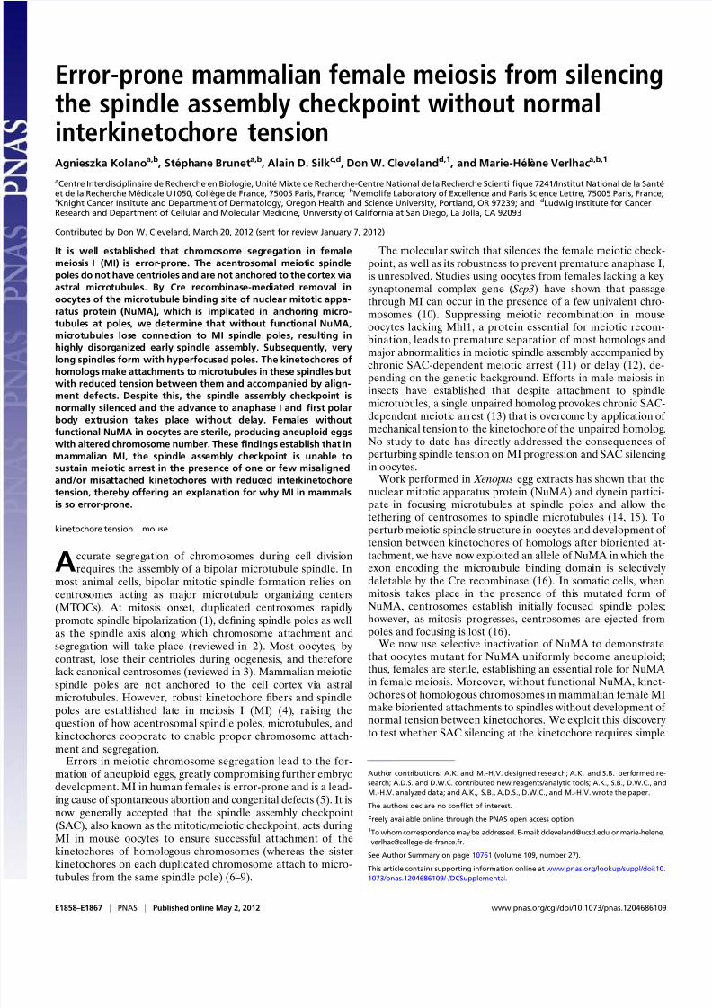
Euploidy. In wt oocytes, anaphase I takes place about 8–8.5 h afterNEBD and after the last chromosome has aligned (Fig. 4 A andMovie S5). In mutant oocytes, anaphase I also typically took placearound 8–8.5 h after NEBD despite the presence of misalignedchromosomes (Fig. 4 A and Movie S6). In addition, lagging chro-mosomes were often observed during anaphase (Fig. 4 A, whitearrows). Within the same NuMA Δ 22 spindle, chromosomes ini-tially at or near spindle poles either became bioriented and con-
gressed to metaphase (Fig. 4 B, white arrow) or remainedmotionless at the poles for long periods (Fig. 4 B, white asterisk).
Quantification of the chromosome number of MII oocytesshowed that, as expected, most Δ 22 oocytes were aneuploid incontrast to control oocytes (80% compared with less than 20% incontrols; Fig. 4 C and D). Chromosome abnormalities involvedmostly the loss or gain of one or two chromosomes (Fig. 4 D).These data show that proper assembly of spindle poles by NuMA is essential to support accurate chromosome congression andsegregation in female meiosis, providing an explanation for fe-male sterility in the absence of functional NuMA.
Anaphase Is Not Delayed Despite Chromosome Congression Defects
in the Absence of Spindle-Bound NuMA. The previous observationsunambiguously establish that without spindle-bound NuMA,
E1860 | www.pnas.org/cgi/doi/10.1073/pnas.1204686109 Kolano et al.

7/30/2019 enbrio
http://slidepdf.com/reader/full/enbrio 4/10
D
0
200
400
600
800
a r e a o c c u p
i e d b y
M T ( µ m 2 )
0 1 2 3 4 5
time after NEBD (h)
wt (n=10)
(n=10)E NEBD+4h
w t
MTs DNA
H
C
wt (n=17) (n=17)
0
60
120
180
240
300
360
***
b i p o
l a r i t y s e
t u p
( m i n a
f t e r
N E B D )
B
w t
00:00 01:00 02:00 03:00 04:00
00:00 01:00 02:00 03:00 04:00
MTs DNA
G
w t
04:40 06:00 07:20 08:40 10:00 11:20
04:20 05:40 07:00 08:20 09:40 11:00
MTs DNA
*PB1
*PB1
*PB1
*MII
*MII
s p i n d l e l e n g t h ( µ m )
wt (MI n=6; MII n=22)
(MI n=20; MII n=33)
***
NEBD +7h
60
50
40
30
20
10
0
***
MII
F
w t
NEBD+7h
NEBD NEBD+3h NEBD+8h
A bipolarization metaphase
Fig. 2. NuMA is required for early MI spindle assembly. ( A) Scheme represents the process of MI spindle assembly in control oocytes. After NEBD, bi-
polarization takes ∼3 h. During the remaining 5 h before metaphase I, MTOCs are sorted and clustered to the poles, and bivalents biorient and congress to
the equator. Bivalents are red with yellow kinetochores, microtubules are gray, and MTOCs are dark gray. ( B) Mutant oocytes have defective early meiotic
spindle assembly. NuMA is required to restrict the area of microtubule (MT) assembly at early steps: In the absence of functional NuMA, long MTs form,
which organize slowly into roughly bipolar structures with unfocused poles. wt oocytes (n = 20; Upper ); Δ 22 oocytes (n = 19; Lower ). Oocytes express EB3-
GFP (gray) and H2B-RFP (red). All times on all figures are hours and minutes after NEBD. (Scale bars: 10 μm unless otherwise specified.) (C ) Histogram shows
that the spindle bipolarization setup is delayed in Δ 22 oocytes. Bipolarity was scored when two poles were distinguishable. The mean time for Δ 22 oocytes
is 288 ± 60 min (green) compared with 172 ± 40 min (gray) for wt oocytes (***P = 0.003). (D) Graph shows the surface occupied by MTs at early stages of MI
spindle formation (wt , gray curve; Δ 22, green curve). (E ) Early defects result in long twisted spindles with broad poles and dispersed chromosomes, as
observed on fixed oocytes at NEBD + 4 h. wt (n = 6; Upper ); Δ 22 mutant (n = 6; Lower ). (F ) Metaphase I spindles in Δ 22 oocytes eventually resemble mitotic
ones. They are elongated with hyperfocused poles presenting astral-like MTs (white arrowhead) not observed in controls, which are barrel-shaped. wt (n =
6; Upper ); Δ 22 mutant (n = 20; Lower ). ( G) Spindle pole focusing during late stages of MI in mutant oocytes. Chromosome congress on a loose metaphase
plate in Δ 22 (n = 30; Lower ) compared with wt (n = 14; Upper ) oocytes. Oocytes express EB3-GFP (gray) and H2B-RFP (red). The white star indicates the PB1.
(H ) Histogram of mean spindle length of MI and MII spindles from wt and Δ 22 oocytes. Mutant spindles (green) are 40% longer than controls (gray). The
mean length of MI spindles in controls is 30.0 ± 4.5 μm and 43.6 ± 6.7 μm in Δ 22 (***P = 0.0001), and the mean length of MII spindles in wt is 25.1 ± 3.3 μm
and 41.7 ± 7.6 μm in Δ 22 (***P < 0.001).
Kolano et al. PNAS | Published online May 2, 2012 | E1861
C E L L B I O L O G Y
P N A S
P L U S

7/30/2019 enbrio
http://slidepdf.com/reader/full/enbrio 5/10
anaphase onset is triggered despite the continued presence of misaligned chromosomes resulting from defective spindle pole
assembly. Indeed, counting of normal and NuMA Δ 22 oocytesrevealed that 60% of mutant oocytes completed MI, extruding
N E B D + 8 h
N E B D + 6 h
N E B D + 4 h
N E B D + 2 h
N E B D + 8 h
N E B D + 6 h
N E B D + 4 h
N E B D + 2 h
MTOCs DNA
wt
0 5 10 15 2 00 5 10 15 20 25 25
0
0.25
0.5
0.75
1
50
100
150
200
250
wt
01:20 03:20 05:20 07:20
C wt
0 1 1.1
spindle axis (a.u)
MTOCschromosomes
50
0
50
0
50
0
50
0
50
0
50
0
50
0
50
0
0 1 1.1
spindle axis (a.u)
% o
f t o t a l a r e a
D E
individual MTOC size (a.u)
M T O C c i r c u l a r i t y
M T O C a r e a a t o n e p o l e ( a . u )
A B
w t
MTs DNA
07:30
09:30
09:00
08:30
08:00
01:20 03:20 05:20 07:20
Fig. 3. NuMA is essential for the formation of meiotic barrel-shaped spindle poles. ( A) MTOCs from mutant oocytes coalesce into a unique MTOC in MI. Atthe end of MI, MTOCs form structures made of discrete entities in wt oocytes (n = 10; Upper ), whereas they appear compact in Δ 22 oocytes (n = 18; Lower ),
reflecting the morphology of poles. (Scale bars: 10 μm unless otherwise specified.) Oocytes express Venus-Plk1 (green) and H2B-RFP (red). (B) Large MTOC
detaches from spindle microtubules (MTs) at the end of MI as observed in live mutant oocytes. Oocytes express EB3-GFP (green) and H2B-RFP (red). Smaller
images (Right ) correspond to a zoomed-in version of larger images (Left , Inset ). The white arrowheads show an MTOC detaching from the spindle pole (n = 20
oocytes). (C ) Kinetics of MTOCs sorting to the poles during MI are comparable in wt (Left ) and Δ 22 (Right ) oocytes, but chromosome congression is inefficient
in mutants. Graphs show the quantitative analysis of MTOC distribution and chromosome congression along the spindle axis at 2 h ( wt , n = 8; Δ 22, n = 7), 4 h
(wt , n = 8; Δ 22, n = 10), 6 h (wt , n = 9; Δ 22, n = 14), and 8 h (wt , n = 10; Δ 22, n = 12) after NEBD. The graphs clearly show that chromosome congression is less
efficient in mutants with few chromosomes stuck at spindle poles. (D) Mean area occupied by MTOCs at NEBD + 8 h in Δ 22 oocytes (green circles) is com-
parable to the area for wt oocytes (gray circles). Mutant oocytes do not lose MTOC material during their sorting to spindle poles. (E ) Shape of MTOCs at NEBD
+ 8 h at the poles differs between wt and Δ 22 oocytes. The mean circularity shape factor is closer to 1 (with 1 corresponding to a perfect circle) in mutants
(greencircles) comparedwith controls (gray circles), showing that most MTOCs are circular in MI spindles, whereas controls have various shapes. a.u., arbitrary unit.
E1862 | www.pnas.org/cgi/doi/10.1073/pnas.1204686109 Kolano et al.
7/30/2019 enbrio
http://slidepdf.com/reader/full/enbrio 6/10
a PB1, compared with 79% of control oocytes. Very surprisingly,despite the presence of misaligned chromosomes whose kinet-ochores would have been predicted to continue to generate the“ wait anaphase” SAC signal, polar body extrusion occurred
without significant delay in oocytes without spindle-boundNuMA, yielding overall kinetics of polar body extrusion essen-tially indistinguishable from normal oocytes (Fig. 5 B). Further-more, kinetics of cyclin B1-GFP degradation were identical innormal and mutant oocytes (Fig. S2 A), reinforcing the notion
that progression to anaphase I was comparable with or withoutspindle-bound NuMA.
Robust SAC Signaling in Oocytes Depleted of Spindle-Bound NuMA.
Prior work with preying mantid (13) and grasshopper (23, 24)spermatocytes had implicated the SAC in delaying meioticanaphase until all chromosomes had stably attached to spindlemicrotubules and tension had developed between kinetochoresof paired homologous chromosomes. Although many proteinsinvolved in generation of the SAC signal are known (reviewed in25) and no work in any system has implicated NuMA or a NuMA homolog as a component of the signaling pathway, the absenceof MI delay in the NuMA Δ 22 oocytes called into question therobustness of SAC signaling in these oocytes. To examine SACsignaling more directly, low doses of nocodazole (100 nM), which
have been shown to maintain an activated SAC and delay ana-phase I onset (26), were added to normal and Δ 22 oocytesstarting 6 h after NEBD. The ensuing disruption of spindle mi-crotubule assembly continued activation of the SAC, whichdelayed polar body extrusion by 6 h in control oocytes (Fig. 5C).Similar treatment produced an even longer delay in polar body extrusion in oocytes without spindle-bound NuMA (Fig. 5C),consistent with nocodazole amplifying the spindle defects in-herent to the Δ 22 mutant and demonstrating that the loss of
spindle-bound NuMA (and presence of theΔ 22 protein) doesnot diminish SAC signaling or its sustained activation.
Further, we determined SAC signaling in Δ 22 oocytes fol-lowing inhibition of Aurora B, a mitotic kinase whose activity isdispensable for nocodazole-mediated mitotic arrest by the SACbut necessary for sustaining taxol-dependent SAC activation (27,28). Addition (to 100 nM) of the Aurora B inhibitor hesperadinto Δ 22 or normal oocytes at NEBD + 4 h (a time when Δ 22oocytes are not yet bipolar) inhibited SAC signaling, advancingpolar body extrusion in both oocyte genotypes (Fig. S2 B).However, similar to what was seen in the case of nocodazoleaddition, SAC signaling was, if anything, enhanced in Δ 22oocytes (i.e., it was sustained an additional hour compared withcontrol oocytes). Lastly, we analyzed the effect of reversine, aninhibitor of the Mps1 kinase (29), whose activity is essential for
08:00 08:20 08:40 09:00
07:00 07:20 07:40 08:00
w t
PB1
*
PB1
*
PB1
*
% o
f o o c y t e s
0
20
40
60
80
100
1 7 1 8 1 9 2 0 2 1 2 2 1 7 1 8 1 9 2 0 2 1 2 2
wt (n=22)
(n=19)
number of chromosomes/ oocyte
**
*
**
*
MTs DNA
06:30 07:00 07:30
08:00 08:30 09:00
A
D
B
C
w
t
20 chromosomes
21 chromosomes
chromosome spreads in MII
Δ 2 2
Δ 2 2
Δ 2 2
Δ22
Fig. 4. NuMA is required for proper chromosome segregation. ( A) Anaphase I in wt (n = 14; Upper ) and Δ 22 (n = 30; Lower ) oocytes expressing H2B-RFP.
Lagging chromosomes are observed in mutants (white arrows). (Scale bars: 10 μm unless otherwise specified.) (B) Two different behaviors are shown on the
same spindle for chromosomes lost at poles. One chromosome (Left ; white arrow) is under tension, demonstrates bipolar attachment, and moves to the
metaphase plate. The other chromosome (Right ; white asterisk) stays at the pole. NuMA Δ 22 oocytes express EB3-GFP (green) and H2B-RFP (red). (C ) Examples
of chromosome spreads from MII wt (Upper ) and Δ 22 (Lower ) oocytes used to determine oocyte ploidy after thefi
rst meiotic division. Chromosomes are gray.Deconvolution was applied to images of chromosome spreads using Huygens Professional deconvolution software (SVI) to improve counting. (Scale bar:
5 μm). (D) Histograms represent the percentage of wt (gray) and Δ 22 (green) oocytes for each category of ploidy (17–22 chromosomes) determined by chro-
mosome spreading as in C .
Kolano et al. PNAS | Published online May 2, 2012 | E1863
C E L L B I O L O G Y
P N A S
P L U S

7/30/2019 enbrio
http://slidepdf.com/reader/full/enbrio 7/10
the SAC (29, 30). Treatment with reversine accelerated themetaphase I-to-anaphase I transition by 2–3 h similarly in bothcontrol oocytes, as previously reported (26), and Δ 22 oocytes(Fig. 5C), consistent with comparable SAC signaling in the twooocyte genotypes.
Taken together, these observations indicate that NuMA is nota component of SAC activity and that the SAC is fully active inNuMA Δ 22 oocytes. Consequently, the PB1 extrusion in the pres-ence of misaligned chromosomes in Δ 22 oocytes is not attributableto an inherent inability to initiate or sustain SAC activation.
SAC Cannot Delay Anaphase I Despite Reduced Tension at
Kinetochores. A widely accepted model is that in MI, the SAC wait anaphase signal is silenced by tension applied between thekinetochores of paired homologous chromosomes after attach-ment (13, 24; reviewed in 25, 31). Moreover, the lack of micro-tubule anchoring to the abnormal spindle poles in the absence of bound NuMA has been shown to reduce the tension applied tokinetochores in mammalian mitosis (16). We therefore de-termined interkinetochore tension in MI by measuring the dis-tance between kinetochores in each bivalent after attachment
NEBD+3h
NEBD+7h
NEBD+7h + NZ
Mad2 DNA
NEBD+7h
w t
CREST DNA*
*
P B 1 e x t r u s i o n ( % )
0
20
40
60
80
100
( n = 2 0 2 )
( n = 2 6 7 )
wt
***
100
75
50
25
0
wt (n=59)
(n=68)n=68)
wt (n=59)
(n=68)
6 7 8 9 10 11 12
time after NEBD (h)
P B 1 e x t r u s i o n ( % )
d
time after NEBD (h)
0
25
50
75
100
4 6 8 10 12 14 16 18 20 22
P B 1 e x t r u s i o n ( % )
m e a n i n t e r k i n e t o c h o r e d i s t a n c e ( µ m )
2
3
4
5
6
( n = 1 3 )
( n = 1 1 )
wt
***
A B
FE
C D
G 9
M a d 2 s t a i n i n g i n t e n s i t y ( a . u )
7
6
5
4
3
2
1
0
8
( n = 1 6 8 )
( n = 1 0 2 )
( n = 8 6 )
( n = 2 0 6 )
( n = 1 4 2 )
( n = 2 1 4 )
***
***
**
***
wt
Fig. 5. SAC does not detect reduced tension on kinetochores in late MI. ( A) Histogram represents the rate of PB1 extrusion in wt (gray) vs.Δ 22 (green) oocytes.
Sixtypercentof NuMAΔ 22 oocytes extrude PB1 comparedwith 79% in controls(***P < 0.0001).(B) Graphrepresents the kineticsof PB1 extrusionin wt (gray) vs.Δ 22 (green) oocytes.Mutant oocytes extrude PB1withkineticsidenticalto controls:the mean time is 8 h and30± 50minfor wt oocytes and 8 h and 40min± 1 h
for Δ 22 oocytes (P = 0.3, not statistically significant). For all kinetics, the percentage of PB1 is calculated only from the population of oocytes extruding a polar
body. (C ) Graph represents the kinetics of PB1 extrusion in wt (gray) vs. Δ 22 (green) oocytes after treatment with reversine or with low doses of nocodazole.
Reversine (100 nM) was added at NEBD, nocodazole (100 nM) was added at NEBD + 6 h, and oocytes were scored for PB1. Mutant oocytes respond to reversine
like controls do: The mean time of PB1 is 6 h and 20 min ± 1 h and 10 min for wt oocytes (gray dotted line) and 6 h and 20 min ± 40 min for Δ 22 oocytes (green
dotted line) treated with reversine and 8 h and 50 min ± 40 min for wt oocytes (gray solid line) and 8 h and 20 min ± 50 min for Δ 22 oocytes (green solid line)
treated with DMSO. The difference in mean PB1 timing was extremely significant (P < 0.0001) between controls and reversine-treated oocytes. Mutant oocytes
arealsodelayedafter nocodazoletreatment:The mean time ofPB1 is15 h and20 min±2hand20minforwt oocytes (graybroken line)and 16h and50 min±2 h
for Δ 22 oocytes (green broken line) treated with nocodazole. The difference in mean PB1 timing was extremely significant (P < 0.0001) between controls and
nocodazole-treated oocytes,and the difference between wt andΔ 22 oocytes treated withnocodazole was significant (P = 0.0253).( D) Representative images of
metaphase I (NEBD + 7 h) plate from wt (Upper ) vs. Δ22 (Lower ) oocytes. Kinetochores are labeled with CREST (green). The white stars indicate unaligned
chromosomes. (Scale bars: 10 μm unless otherwise specified.) (E ) Quantification of interkinetochore distance from bivalents in oocytes as labeled in D. Tension
exerted on kinetochoresis reducedin Δ 22 oocytes:The meanvalue of the interkinetochoredistance(d in thecartoon)forΔ 22 oocytes (green)is 4.4±0.4 μm,and
it is 5.0 ± 0.4 μm for wt oocytes (gray; ***P = 0.001). n, number of oocytes analyzed. For each oocyte, 20 interkinetochore distances were measured. (F ) Mad2 is
lost from all kinetochores, even unaligned ones, in Δ 22 oocytes (Middle) at NEBD + 7 h. Kinetochores in mutants are positive for Mad2 staining at NEBD + 3 h
(Top) or at NEBD + 7 h after 30 min of treatment with 10 μΜ nocodazole (Bottom). (Left ) Z-projections of confocal stacks. (Right ) Magnifications of selected
chromosomes. [Scale bar: 5 μm (Right ).] (G) Quantification of Mad2 kinetochore labeling in wt (gray) vs.Δ 22 (green) oocytes at 3 h, 7h post-NEDB, and 7 h post-
NEBD after nocodazole treatment as in F . n, number of kinetochores observed. ** = 0.01; *** < 0.0001.
E1864 | www.pnas.org/cgi/doi/10.1073/pnas.1204686109 Kolano et al.

7/30/2019 enbrio
http://slidepdf.com/reader/full/enbrio 8/10
(Fig. 5 D). Average interkinetochore distance was significantly reduced (from 5.0 ± 0.4 μm to 4.4 ± 0.4 μm) in NuMA Δ 22 vs.control oocytes (Fig. 5 E). In the NuMA Δ 22 oocytes, the inter-kinetochore spacing on unaligned chromosomes near the poles
was very significantly reduced (only 2.2 ± 0.9 μm), with spacingon some of these between 1 and 1.4 μm.
We could not accurately determine the distribution of inter-kinetochore distances for homologs under no tension, becausemicrotubule assembly disruptors like nocodazole induce chro-
mosome clumping that precludes a complete assessment. How-ever, in the subset of chromosomes that were suf ficiently separated from the others, interkinetochore distances of thesepresumptively unattached kinetochores were 1.3 ± 0.2 μm. Thus,not only were aligned chromosomes with bipolar attachments inthe NuMA Δ 22 oocytes under reduced interkinetochore tension,but those at poles were under little, if any, tension.
HURP association onto kinetochores and microtubules at theend of MI enables visualization of K-fibers present in the oocyte(4). As expected, in control oocytes, chromosomes that hadcongressed to the equator of the spindle had HURP-decoratedmicrotubules emanating from their kinetochores (Fig. S3).Similar figures were observed in NuMA Δ 22 oocytes, even forunaligned chromosomes located in the vicinity of the spindlepoles (Fig. S3). Thus, the misorganization of spindle poles inmutant oocytes does not interfere with proper establishmentof K-fibers.
Mad2 Disassociates from Kinetochores with Normal Kinetics Even
with Reduced Tension. The spindle checkpoint component Mad2localizes to unattached kinetochores (6) but is lost as stable ki-netochore-microtubule interactions are established (6, 32) or inmale meiosis after tension is applied (13, 23, 24). In both normaland NuMA Δ 22 oocytes, Mad2 was localized at kinetochores atNEBD + 3 h (Fig. 5 F and G). Subsequently, Mad2 stainingdisappeared, even from chromosomes that failed to achievemetaphase alignment in the NuMA Δ 22 oocytes (Fig. 5 F and G;NEBD + 7 h). Short treatment (30 min) of wt or NuMA mutantoocytes with 10 μM nocodazole induced the reappearance of
Mad2 staining on kinetochores, demonstrating competency forcheckpoint reactivation at unattached kinetochores (Fig. 5 F and G).
Thus, the SAC is normally activated and then satisfied at in-dividual kinetochores in NuMA mutant oocytes, even in thepresence of imperfectly aligned chromosomes near the spindlecenter and unaligned chromosomes whose kinetochores areunder sharply reduced tension at poles. Some of these mis-aligned polar chromosomes are likely bioriented with reducedtension but with silenced SAC signaling. For others, with inter-kinetochore spacing similar to that in the absence of micro-tubules and more random orientations relative to the spindle axisthan would be expected for correctly attached bivalents, thesebivalents are unlikely to be attached in a bipolar manner. Rather,at least some of these are likely to be attached unstably or at-
tached in an aberrant manner (e.g., with both kinetochores of a bivalent attached to microtubules from the most proximal poleso as to produce syntelic attachment). Nevertheless, SAC sig-naling is silenced, demonstrating that SAC satisfaction in thisexample of female meiosis in mammals is not mediated by de-
velopment of normal interkinetochore tension.
NuMA-Dependent Spindle Pole Assembly in Meiosis. NuMA, to-gether with dynein, anchors minus-ends of microtubules to thecentrosomes of mitotic spindles. This function is not essential forearly stages of mitotic spindle bipolarization, which is ensured by the two centrosomes, but becomes essential when robust bundlesof kinetochore fibers form and transmit strong pulling forcesbetween spindle poles and kinetochores. In the absence of functional NuMA, centrosomes are ejected from poles, poles
splay open, and the equilibrium of forces essential for properchromosome segregation is compromised. We show here that
without centrosomes, NuMA is essential in the early stages of spindle assembly in mammalian meiosis. Our data establish thatNuMA is required for shaping meiotic spindle poles, throughactivities that can be divided into two temporal phases. Duringthe course of initial microtubule sorting into a bipolar array, atthe time when MTOCs are dispersed along the spindle axis,NuMA ensures the cohesion of microtubule minus-ends and
each of the two spindle poles. In the second phase of meioticspindle formation, MTOCs are sorted toward these poles. Dur-ing this phase, NuMA, probably with its partner dynein, acts asa microtubule spacer to maintain appropriate pole shape andallow deposition and further reorganization of MTOCs into thecharacteristic barrel shape of the meiotic spindle.
Strikingly, we have established that MTOC sorting to the polestakes place normally in the absence of functional NuMA. Thiscan be explained by the fact that HURP, which is required to sortMTOCs to the poles, is appropriately localized to the centralspindle region in NuMA-depleted oocyte spindles. Also note-
worthy is that overexpression of lateral geniculate nucleus de- velopment (LGN), known to inhibit NuMA in mitosis (33, 34),induces spindle elongation and mitotic-like pole phenotypes thatare strikingly similar to the ones we have observed using geneticNuMA disruption (35). This suggests that a correct balance of LGN and NuMA is required for proper organization of meioticspindle poles.
Understanding the High Error Rate of Female Mammalian Meiosis.
Evidence of missegregation in mouse MI of univalent, achias-matic chromosomes after blocking almost all homologous re-combination (by deletion of Mlh1) led Hunt and colleagues (12)to postulate that the oocyte SAC was unable to ensure stablebipolar attachment of bivalents before anaphase onset. Our ev-idence now demonstrates that the presence in mouse oocytes of poorly aligned chromosomes with sharply reduced interkineto-chore tension does not delay, much less prevent, onset of mei-otic anaphase I.
This outcome is strikingly different from the chronic delay seen in mouse oocytes with defective spindle architecture (4, 6,12) and in insect spermatocytes (13, 23, 24). Indeed, efforts usingmicromanipulation of chromosomes in centriolar male meiosisin insects had initially concluded that tension generated by spindle forces between kinetochores of bivalents was required tosilence SAC signaling by those kinetochores, thereby permittingadvance to anaphase (12, 23). Additional efforts identified aninterplay between microtubule attachment and tension, withincreased tension stabilizing microtubule attachment (24, 36),and tension was again concluded to be essential for checkpointsilencing. Similarly, in maize meiosis, release of kinetochore-associated MAD2 is correlated with increased interkinetochoredistance (i.e., tension) rather than initial attachment (37). Incontrast to these examples, our evidence demonstrates that in
mice, female MI is not delayed in response to reduced inter-kinetochore tension.
Most recently, it was proposed that the well-known high errorrate of mammalian female meiosis is attributable to a high fre-quency of initial misattachment of kinetochores followed by in-complete error correction (38). This study neither followedchromosome movement into anaphase nor assessed SAC sig-naling. Although initial errors in attachment will produce anincreased error frequency if uncorrected, our evidence demon-strates that a central determinant of a high error rate is that SACsignaling in mouse oocytes is silenced prematurely, even in theabsence of spindle-induced tension and in the presence of mul-tiple misaligned/misattached chromosomes. Moreover, our evi-dence establishes that even global reduction of tension onbivalent kinetochores (e.g., in NuMA-depleted spindles) is not
Kolano et al. PNAS | Published online May 2, 2012 | E1865
C E L L B I O L O G Y
P N A S
P L U S

7/30/2019 enbrio
http://slidepdf.com/reader/full/enbrio 9/10
suf ficient to delay anaphase, much less support sustained SACsignaling, producing anaphase initiation with normal kinetics andaneuploid oocytes instead. Thus, the SAC in mouse oocytes hasa surprisingly low threshold for satisfaction and/or a poor sig-naling capacity. Recognizing this, our identification of an in-herently weakened SAC that is silenced without normal tension,coupled with the initial errors of attachment (including merotelicand syntelic attachments) seen in an otherwise unperturbedmeiosis (38), provides an explanation for why female MI in
mammals is so error-prone.
Experimental ProceduresMouse Strains and Genotyping. Zp3-Cre [C57BL/6-Tg(Zp3-cre)93Knw/J]
breeding pairs were obtained from Jackson Laboratories. NuMA fl ox/wt
mice were produced on a C57BL/6 background (16). For measurements of
Cre-mediated NuMA exon 22 excision, genotyping was performed using the
following primers: Cre 5′-GCG GTC TGG CAG TAA AAA CTA TC-3′ and 5′-GTG
AAA CAG CAT TGC TGT CAC TT-3′, and NuMA 5′-AAC CGC ATC GCA GAG
TTG CAG -3′ and 5′-GAG GAG TGG TGG CAA CAG TAG-3′.
Mouse Oocyte Collection and Culture. Oocytes were collected from 8- to 12-
wk-old female mice into M2 + BSA medium supplemented with 150 μg/mL
dibutyryl cAMP (dbcAMP; Sigma) to ensure a block in prophase I, as pre-
viously described (39). Resumption of meiosis was triggered by culturing
oocytes in dbcAMP-free medium. All drugs were stored in DMSO at −20 °C
and diluted in M2 + BSA. Nocodazole (Sigma) was used at 100 nM or 10 μM,reversine (Cayman Chemical Company) was used at 100 nM, and hesperadin
(Calbiochem) was used at 100 nM as described by Kitajima et al. (38).
Plasmid Construction and in Vitro Transcription of cRNA.We usedthe following
constructs: pRN3-histone-RFP (40), pRN3-EB3-GFP (19), and pRN3-cyclin
B1-GFP (41). Plk1 (a gift from Erich Nigg, Biozentrum, Basel, Switzlerland)
was subcloned into pSpe3-Venus (a gift from Alex McDougall, UMR 7009,
Villefranche-sur-Mer, France). All cRNAs were synthesized using the T3
mMessage mMachine Kit (Ambion) and resuspended in RNase-free water as
previously described (42).
Microinjection. Injection of in vitro-transcribed cRNAs into the cytoplasm of
prophase I-arrested oocytes was performed using an Eppendorf Femtojet
microinjector as described (43), and the oocytes were further kept for 2 h in
dbcAMP arrest to allow expression of fusion proteins. Oocytes were then
released from the prophase I stage by transferring and washing intodbcAMP-free M2 medium.
LiveImaging of Oocytes. Spinning disk movieswere acquired using a Plan APO
40× /1.25 N.A. objective on a Leica DMI6000B microscope, enclosed in
a thermostatic chamber set at 37 °C (Life Imaging Services), and equipped
with a CoolSnap HQ2/CCD-camera (Princeton Instruments) coupled to
a Sutter filter wheel (Roper Scientific) and a Yokogawa CSU-X1-M1 confocal
scanner. MetaMorph software (Universal Imaging) was used to collect
the data.
Chromosome Spreads. Chromosome spreads of metaphase II-arrested oocytes
were prepared according to the method of Tarkowski (44) and stained with
propidium iodide (5 mg/mL in PBS; Molecular Probes) for 20 min. Image
acquisition was carried out on a Leica SP5/AOBS confocal microscope
equipped with a Plan APO 100× /1.4 N.A. objective.
Immunofluorescence. Oocytes were prepared for fixation as described by
Kubiak et al. (45). Microtubules were fixed with 0.1% glutaraldehyde as
described by de Pennart et al. (46). For NuMA labeling, oocytes were fixed in
ice-cold 100% methanol. For MTOC, TPX2, HURP, and CREST staining, 4%
paraformaldehyde was used, and for Mad2 staining, 4% (vol/vol) formal-
dehyde (FA) with 0.15% Triton X-100 in Pipes Hepes EDTA Mops (PHEM)
buffer was used as described by Wassmann et al. (6).
Rabbit polyclonal antibody against human NuMA (ab36999; Abcam) was
used at a ratio of 1:100. Rat monoclonal antibody against tyrosinated
α-tubulin (YL 1/2; Serotec) was used at a ratio of 1:200. Human α-CREST (HCT-
100; Immunovision) was used at a ratio of 1:60. Mouse antipericentrin (BDTransduction Laboratories) was used at a ratio of 1:500. Rabbit–α-mHURP
(sc-98809; Santa Cruz) was used at a ratio of 1:50. Rabbit α-human TPX2 (a
gift from Oliver Grüss, ZMBH, Heidelberg, Germany) was used at a ratio of
1:500. Rabbit α-Mad2 (a gift from Katja Wassmann, UMR 7622, Paris, France)
was used at a ratio of 1:200. As secondary antibodies, anti-rabbit Cy2 or Cy3,
anti-mouse dye-light 488, anti-mouse Cy3, anti-rat Cy2 (all from Jackson
Laboratories), and anti-human Alexa 488 (Molecular Probes) were used at
a ratio of 1:200. Chromatin was stained for 20 min with Hoechst (5 μg/mL;
Invitrogen) or propidium iodide (5 μg/mL; Molecular Probes). Citifluor AF-1
was used as a mounting medium.
Image acquisition of fixed oocytes was carried out on the SP5/AOBS
confocal microscope equipped with a Plan APO 63× /1.4 N.A. objective.
Quantification Analysis. The measurement of NuMA fluorescence intensity at
the poles in control and Δ 22 oocytes was performed on maximum projec-
tions of oocytes stained with NuMA using MetaMorph software. Afterbackground subtraction, two identical squares allowed us to measure the
mean integrated intensity at poles.
The measurement of spindle length was performed using Volocity 4.1
software (Improvision)to obtain a 3D reconstructionof spindlesafter fixation
(for this, we used the Z-stack acquired on the SP5/AOBS confocal microscope).
The measurement of interkinetochore distance was performed using
Imaris software (Bitplans Scientific Software) on Z-stack images of oocytes
stained with CREST.
The measurement of MTOC and chromosome distribution was performed
using Z-projection of oocytes stained with pericentrin. For this purpose, we
used ImageJ software (MacBiophotonics). The signals of chromosomes and
pericentrin were binarized to assess the area of individual chromosomes or
MTOCs (arbitrary units). The coordinates were then plotted along the spindle
axis (0–1.1).
The measurement of cyclin B1-GFP fluorescence intensity was performed
on maximum projections. Afterbackground subtraction, a circular framewitha diameter slightly smaller than the oocyte diameter allowed us to measure
the maximum integrated fluorescence intensity within each cell. The cyclin
B1-GFP intensity was measured inside this circular frame for all oocytes at
recorded time points.
Normalization and correction were performed using Microsoft Excel
software. Statistical analysis was performed using online GraphPad software.
ACKNOWLEDGMENTS. We thank Jérémie Teillon (Plate-Forme de Microsco-pie, Centre Interdisciplinaire de Recherche en Biologie) for his help withdeconvolution of chromosome spreads. A.K. is a recipient of an AgenceNationale pour la Recherche postdoctoral fellowship (Grant ANR08-BLAN-0136-01 to M.-H.V.). S.B. is an Institut National de la Santé et de la RechercheMédicale fellow. This work was supported by Ligue Nationale Contre leCancer Grant EL/2009/LNCC/MHV, Agence Nationale pour la Recherche GrantANR08-BLAN-0136-01 (to M.-H.V.), and National Institutes of Health GrantGM 29513 (to D.W.C.).
1. Toso A, et al. (2009) Kinetochore-generated pushing forces separate centrosomes
during bipolar spindle assembly. J Cell Biol 184:365–372.
2. Tanenbaum ME, Medema RH (2011) Mechanisms of centrosome separation and
bipolar spindle assembly. Dev Cell 19:797–806.
3. Manandhar G, Schatten H, Sutovsky P (2005) Centrosome reduction during
gametogenesis and its significance. Biol Reprod 72:2–13.
4. Breuer M, et al. (2010) HURP permits MTOC sorting for robust meiotic spindle
bipolarity, similar to extra centrosome clustering in cancer cells. J Cell Biol 191:
1251–1260.
5. Hassold T, Hunt P (2001) To err (meiotically) is human: The genesis of human
aneuploidy. Nat Rev Genet 2:280–291.
6. Wassmann K, Niault T, Maro B (2003) Metaphase I arrest upon activation of the Mad2-
dependent spindle checkpoint in mouse oocytes. Curr Biol 13:1596–1608.
7. Homer HA, et al. (2005) Mad2 prevents aneuploidy and premature proteolysis of
cyclin B and securin during meiosis I in mouse oocytes. Genes Dev 19:202–207.
8. Niault T, et al. (2007) Changing Mad2 levels affects chromosome segregation and
spindle assembly checkpoint control in female mouse meiosis I. PLoS ONE 2:e1165.
9. McGuinness BE, et al. (2009) Regulation of APC/C activity in oocytes by a Bub1-
dependent spindle assembly checkpoint. Curr Biol 19:369–380.
10. Kouznetsova A, Lister L, Nordenskjold M, Herbert M, Hoog C (2007) Bi-orientation of
achiasmatic chromosomes in meiosis I oocytes contributes to aneuploidy in mice. Nat
Genet 39:966–968.
11. Woods LM, et al. (1999) Chromosomal influence on meiotic spindle assembly:
Abnormal meiosis I in female Mlh1 mutant mice. J Cell Biol 145:1395–1406.
12. Nagaoka SI, Hodges CA, Albertini DF, Hunt PA (2011) Oocyte-specific differences in
cell-cycle control create an innate susceptibility to meiotic errors. Curr Biol 21:
651–657.
13. Li X, Nicklas RB (1997) Tension-sensitive kinetochore phosphorylation and the
chromosome distribution checkpoint in praying mantid spermatocytes. J Cell Sci 110:
537–545.
E1866 | www.pnas.org/cgi/doi/10.1073/pnas.1204686109 Kolano et al.

7/30/2019 enbrio
http://slidepdf.com/reader/full/enbrio 10/10
14. Merdes A, Ramyar K, Vechio JD, Cleveland DW (1996) A complex of NuMA and
cytoplasmic dynein is essential for mitotic spindle assembly. Cell 87:447–458.
15. Khodjakov A, Copenagle L, Gordon MB, Compton DA, Kapoor TM (2003) Minus-end
capture of preformed kinetochore fibers contributes to spindle morphogenesis. J Cell
Biol 160:671–683.
16. Silk AD, Holland AJ, Cleveland DW (2009) Requirements for NuMA in maintenance
and establishment of mammalian spindle poles. J Cell Biol 184:677–690.
17. Tang CJ, Hu HM, Tang TK (2004) NuMA expression and function in mouse oocytes and
early embryos. J Biomed Sci 11:370–376.
18. Lewandoski M, Wassarman KM, Martin GR (1997) Zp3-cre, a transgenic mouse line for
the activation or inactivation of loxP-flanked target genes specifically in the female
germ line. Curr Biol 7:148–151.
19. Schuh M, Ellenberg J (2007) Self-organization of MTOCs replaces centrosome
function during acentrosomal spindle assembly in live mouse oocytes. Cell 130:484–498.
20. Yang G, Cameron LA, Maddox PS, Salmon ED, Danuser G (2008) Regional variation of
microtubule flux reveals microtubule organization in the metaphase meiotic spindle.
J Cell Biol 182:631–639.
21. Fant X, Merdes A, Haren L (2004) Cell and molecular biology of spindle poles and
NuMA. Int Rev Cytol 238:1–57.
22. Pahlavan G, et al. (2000) Characterization of polo-like kinase 1 during meiotic
maturation of the mouse oocyte. Dev Biol 220:392–400.
23. Li XT, Nicklas RB (1995) Mitotic forces control a cell-cycle checkpoint. Nature 373:
630–632.
24. Nicklas RB, Waters JC, Salmon ED, Ward SC (2001) Checkpoint signals in grasshopper
meiosis are sensitive to microtubule attachment, but tension is still essential. J Cell Sci
114:4173–4183.
25. Musacchio A, Salmon ED (2007) The spindle-assembly checkpoint in space and time.
Nat Rev Mol Cell Biol 8:379–393.
26. Hached K, et al. (2011) Mps1 at kinetochores is essential for female mouse meiosis I.
Development 138:2261–2271.
27. Carvalho A, Carmena M, Sambade C, Earnshaw WC, Wheatley SP (2003) Survivin isrequired for stable checkpoint activation in taxol-treated HeLa cells. J Cell Sci 116:
2987–2998.
28. Hauf S, et al. (2003) The small molecule Hesperadin reveals a role for Aurora B in
correcting kinetochore-microtubule attachment and in maintaining the spindle
assembly checkpoint. J Cell Biol 161:281–294.
29. Santaguida S, Tighe A, D’Alise AM, Taylor SS, Musacchio A (2011) Dissecting the role
of MPS1 in chromosome biorientation and the spindle checkpoint through the small
molecule inhibitor reversine. J Cell Biol 190:73–87.
30. Perreira M, et al. (2005) “Reversine” and its 2-substituted adenine derivatives as
potent and selective A3 adenosine receptor antagonists. J Med Chem 48:4910–4918.
31. Pinsky BA, Biggins S (2005) The spindle checkpoint: Tension versus attachment. Trends
Cell Biol 15:486–493.
32. Brunet S, et al. (1999) Kinetochorefibers are not involved in the formation of the first
meiotic spindle in mouse oocytes, but control the exit from the first meiotic M phase.
J Cell Biol 146:1–11.
33. Du Q, Stukenberg PT, Macara IG (2001) A mammalian Partner of inscuteable binds
NuMA and regulates mitotic spindle organization. Nat Cell Biol 3:1069–1075.
34. Du Q, Taylor L, Compton DA, Macara IG (2002) LGN blocks the ability of NuMA to bind
and stabilize microtubules. A mechanism for mitotic spindle assembly regulation. Curr
Biol 12:1928–1933.
35. Guo X, Gao S (2009) Pins homolog LGN regulates meiotic spindle organization in
mouse oocytes. Cell Res 19:838–848.
36. King JM, Nicklas RB (2000) Tension on chromosomes increases the number of
kinetochore microtubules but only within limits. J Cell Sci 113:3815–3823.
37. Yu HG, Muszynski MG, Kelly Dawe R (1999) The maize homologue of the cell cycle
checkpoint protein MAD2 reveals kinetochore substructure and contrasting mitotic
and meiotic localization patterns. J Cell Biol 145:425–435.
38. Kitajima TS, Ohsugi M, Ellenberg J (2011) Complete kinetochore tracking reveals
error-prone homologous chromosome biorientation in mammalian oocytes. Cell 146:
1–14.
39. Terret M-E, et al. (2003) DOC1R: A MAP kinase substrate that control microtubule
organization of metaphase II mouse oocytes. Development 130:5169–5177.
40. Tsurumi C, Hoffmann S, Geley S, Graeser R, Polanski Z (2004) The spindle assembly
checkpoint is not essential for CSF arrest of mouse oocytes. J Cell Biol 167:1037–1050.
41. Ledan E, Polanski Z, Terret M-E, Maro B (2001) Meiotic maturation of the mouse
oocyte requires an equilibrium between cyclin B synthesis and degradation. Dev Biol
232:400–413.
42. Verlhac MH, et al. (2000) Mos activates MAP kinase in mouse oocytes through two
opposite pathways. EMBO J 19:6065–6074.43. Verlhac M-H, Lefebvre C, Guillaud P, Rassinier P, Maro B (2000) Asymmetric division in
mouse oocytes: With or without Mos. Curr Biol 10:1303–1306.
44. Tarkowski AK (1966) An air-drying method for chromosome preparation from mouse
eggs. Cytogenetics 5:394–400.
45. Kubiak JZ, Weber M, Géraud G, Maro B (1992) Cell cycle modification during the
transition between meiotic M-phases in mouse oocytes. J Cell Sci 102:457–467.
46. de Pennart H, Houliston E, Maro B (1988) Post-translational modifications of tubulin
and the dynamics of microtubules in mouse oocytes and zygotes. Biol Cell 64:375–378.
Kolano et al. PNAS | Published online May 2, 2012 | E1867
C E L L B I O L O G Y
P N A S
P L U S