ecv hemorragico tx agudo
TRANSCRIPT
8/7/2019 ECV Hemorragico TX Agudo
http://slidepdf.com/reader/full/ecv-hemorragico-tx-agudo 1/9
REVIEW ARTICLE
CME
The Acute Management of Intracerebral Hemorrhage:A Clinical Review
Justine Elliott, FRCA, and Martin Smith, FRCA
Intracerebral hemorrhage (ICH) is a devastating disease with high rates of mortality andmorbidity. The major risk factors for ICH include chronic arterial hypertension and oralanticoagulation. After the initial hemorrhage, hematoma expansion and perihematoma edemaresult in secondary brain damage and worsened outcome. A rapid onset of focal neurologicaldeficit with clinical signs of increased intracranial pressure is strongly suggestive of adiagnosis of ICH, although cranial imaging is required to differentiate it from ischemic stroke.ICH is a medical emergency and initial management should focus on urgent stabilization ofcardiorespiratory variables and treatment of intracranial complications. More than 90% ofpatients present with acute hypertension, and there is some evidence that acute arterial bloodpressure reduction is safe and associated with slowed hematoma growth and reduced risk ofearly neurological deterioration. However, early optimism that outcome might be improvedby the early administration of recombinant factor VIIa (rFVIIa) has not been substantiated bya large phase III study. ICH is the most feared complication of warfarin anticoagulation, andthe need to arrest intracranial bleeding outweighs all other considerations. Treatment optionsfor warfarin reversal include vitamin K, fresh frozen plasma, prothrombin complex concen-trates, and rFVIIa. There is no evidence to guide the specific management of antiplatelettherapy–related ICH. With the exceptions of placement of a ventricular drain in patients withhydrocephalus and evacuation of a large posterior fossa hematoma, the timing and nature ofother neurosurgical interventions is also controversial. There is substantial evidence thatmanagement of patients with ICH in a specialist neurointensive care unit, where treatment isdirected toward monitoring and managing cardiorespiratory variables and intracranialpressure, is associated with improved outcomes. Attention must be given to fluid andglycemic management, minimizing the risk of ventilator-acquired pneumonia, fever control,provision of enteral nutrition, and thromboembolic prophylaxis. There is an increasingawareness that aggressive management in the acute phase can translate into improvedoutcomes after ICH. (Anesth Analg 2010;110:1419 –27)
Intracerebral hemorrhage (ICH) is a spontaneous ex-travasation of blood into brain parenchyma. The overallincidence is 12 to 15 cases per 100,000 population per
year,1 and it is the cause of 10% to 15% of first-ever strokes.2
It is more common in the elderly3 and in those of African4
or Asian ethnicity,5 and the incidence is substantially
increased in those receiving anticoagulant therapy.6 Al-
though ICH accounts for only 10% to 30% of all stroke-related admissions to hospital, it is one of the major causes
of stroke-related death and disability. Overall mortality
approaches 50% at 30 days,7,8 and approximately half of allICH-related mortality occurs within the first 24 hours after
the initial hemorrhage.9 Functional outcome in survivors is
also poor with fewer than 20% being independent at 6months.2 In up to 40% of cases, the hemorrhage extends
into the ventricles (intraventricular hemorrhage [IVH]) andthis is associated with obstructive hydrocephalus andworsened prognosis.5 Other factors associated with pooroutcome include large hematoma volume (30 mL), pos-terior fossa location, older age, and admission mean arterialblood pressure (MAP) 130 mm Hg.8,9
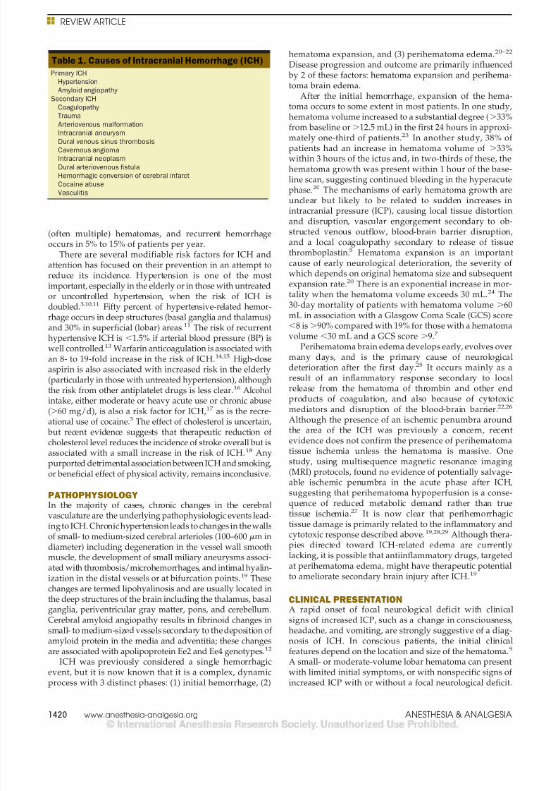
More than 85% of ICH occurs as a primary (spontane-ous) event related to rupture of small penetrating arteriesand arterioles that have been damaged by chronic arterialhypertension or amyloid angiopathy. Sixty percent to 70%of primary ICH is hypertension related10 and, in theelderly, amyloid angiopathy accounts for up to one-third ofthe cases.11 Secondary ICH can be related to multiple
causes (Table 1).11
This review discusses the current understanding of thepathophysiology of spontaneous and anticoagulation-related ICH and presents consensus evidence for its acutemanagement.
RISK FACTORSNonmodifiable risk factors for ICH include male gender,older age, and African or Asian ethnicity.3–5 Cerebralamyloid angiopathy is an important risk factor in theelderly and can occur in isolation or in association withAlzheimer disease or familial apolipoprotein syn-
dromes.11,12 Amyloid angiopathy usually results in lobar
From the Department of Neuroanaesthesia and Neurocritical Care, TheNational Hospital for Neurology and Neurosurgery, University CollegeLondon Hospitals NHS Foundation Trust, Queen Square, London, UK.
Accepted for publication January 11, 2010.
Supported by the Department of Health’s Institute for Health ResearchCentres funding scheme (to MS).
Address correspondence and reprint requests to Dr. Martin Smith, Depart-ment of Neuroanaesthesia and Neurocritical Care, Box 30, The NationalHospital for Neurology and Neurosurgery, University College LondonHospitals, Queen Square, London WC1N 3BG, UK. Address e-mail [email protected].
Copyright © 2010 International Anesthesia Research SocietyDOI: 10.1213/ANE.0b013e3181d568c8
May 2010 • Volume 110 • Number 5 www.anesthesia-analgesia.org 1419
8/7/2019 ECV Hemorragico TX Agudo
http://slidepdf.com/reader/full/ecv-hemorragico-tx-agudo 2/9
(often multiple) hematomas, and recurrent hemorrhageoccurs in 5% to 15% of patients per year.
There are several modifiable risk factors for ICH andattention has focused on their prevention in an attempt to
reduce its incidence. Hypertension is one of the mostimportant, especially in the elderly or in those with untreatedor uncontrolled hypertension, when the risk of ICH isdoubled.3,10,11 Fifty percent of hypertensive-related hemor-rhage occurs in deep structures (basal ganglia and thalamus)and 30% in superficial (lobar) areas.11 The risk of recurrenthypertensive ICH is 1.5% if arterial blood pressure (BP) iswell controlled.13 Warfarin anticoagulation is associated withan 8- to 19-fold increase in the risk of ICH.14,15 High-doseaspirin is also associated with increased risk in the elderly(particularly in those with untreated hypertension), althoughthe risk from other antiplatelet drugs is less clear.16 Alcoholintake, either moderate or heavy acute use or chronic abuse
(
60 mg/d), is also a risk factor for ICH,
17
as is the recre-ational use of cocaine.3 The effect of cholesterol is uncertain,but recent evidence suggests that therapeutic reduction ofcholesterol level reduces the incidence of stroke overall but isassociated with a small increase in the risk of ICH.18 Anypurported detrimental association between ICH and smoking,or beneficial effect of physical activity, remains inconclusive.
PATHOPHYSIOLOGYIn the majority of cases, chronic changes in the cerebralvasculature are the underlying pathophysiologic events lead-ingto ICH. Chronic hypertension leads to changes in the wallsof small- to medium-sized cerebral arterioles (100–600 m indiameter) including degeneration in the vessel wall smooth
muscle, the development of small miliary aneurysms associ-ated with thrombosis/microhemorrhages, and intimal hyalin-ization in the distal vessels or at bifurcation points.19 Thesechanges are termed lipohyalinosis and are usually located inthe deep structures of the brain including the thalamus, basalganglia, periventricular gray matter, pons, and cerebellum.Cerebral amyloid angiopathy results in fibrinoid changes insmall- to medium-sized vessels secondary to the deposition ofamyloid protein in the media and adventitia; these changesare associated with apolipoprotein Ee2 and Ee4 genotypes.12
ICH was previously considered a single hemorrhagicevent, but it is now known that it is a complex, dynamicprocess with 3 distinct phases: (1) initial hemorrhage, (2)
hematoma expansion, and (3) perihematoma edema.20–22
Disease progression and outcome are primarily influencedby 2 of these factors: hematoma expansion and perihema-toma brain edema.
After the initial hemorrhage, expansion of the hema-toma occurs to some extent in most patients. In one study,hematoma volume increased to a substantial degree (33%from baseline or12.5 mL) in the first 24 hours in approxi-
mately one-third of patients.23
In another study, 38% ofpatients had an increase in hematoma volume of 33%within 3 hours of the ictus and, in two-thirds of these, thehematoma growth was present within 1 hour of the base-line scan, suggesting continued bleeding in the hyperacutephase.20 The mechanisms of early hematoma growth areunclear but likely to be related to sudden increases inintracranial pressure (ICP), causing local tissue distortionand disruption, vascular engorgement secondary to ob-structed venous outflow, blood-brain barrier disruption,and a local coagulopathy secondary to release of tissuethromboplastin.5 Hematoma expansion is an importantcause of early neurological deterioration, the severity of
which depends on original hematoma size and subsequentexpansion rate.20 There is an exponential increase in mor-tality when the hematoma volume exceeds 30 mL.24 The30-day mortality of patients with hematoma volume 60mL in association with a Glasgow Coma Scale (GCS) score8 is90% compared with 19% for those with a hematomavolume 30 mL and a GCS score 9.7
Perihematoma brain edema develops early, evolves overmany days, and is the primary cause of neurologicaldeterioration after the first day.25 It occurs mainly as aresult of an inflammatory response secondary to localrelease from the hematoma of thrombin and other endproducts of coagulation, and also because of cytotoxicmediators and disruption of the blood-brain barrier.22,26
Although the presence of an ischemic penumbra aroundthe area of the ICH was previously a concern, recentevidence does not confirm the presence of perihematomatissue ischemia unless the hematoma is massive. Onestudy, using multisequence magnetic resonance imaging(MRI) protocols, found no evidence of potentially salvage-able ischemic penumbra in the acute phase after ICH,suggesting that perihematoma hypoperfusion is a conse-quence of reduced metabolic demand rather than truetissue ischemia.27 It is now clear that perihemorrhagictissue damage is primarily related to the inflammatory andcytotoxic response described above.19,28,29 Although thera-pies directed toward ICH-related edema are currently
lacking, it is possible that antiinflammatory drugs, targetedat perihematoma edema, might have therapeutic potentialto ameliorate secondary brain injury after ICH.19
CLINICAL PRESENTATIONA rapid onset of focal neurological deficit with clinicalsigns of increased ICP, such as a change in consciousness,headache, and vomiting, are strongly suggestive of a diag-nosis of ICH. In conscious patients, the initial clinicalfeatures depend on the location and size of the hematoma.9
A small- or moderate-volume lobar hematoma can presentwith limited initial symptoms, or with nonspecific signs ofincreased ICP with or without a focal neurological deficit.
Table 1. Causes of Intracranial Hemorrhage (ICH)
Primary ICH
Hypertension
Amyloid angiopathy
Secondary ICH
Coagulopathy
Trauma
Arteriovenous malformation
Intracranial aneurysm
Dural venous sinus thrombosis
Cavernous angioma
Intracranial neoplasm
Dural arteriovenous fistula
Hemorrhagic conversion of cerebral infarct
Cocaine abuse
Vasculitis
REVIEW ARTICLE
1420 www.anesthesia-analgesia.org ANESTHESIA & ANALGESIA
8/7/2019 ECV Hemorragico TX Agudo
http://slidepdf.com/reader/full/ecv-hemorragico-tx-agudo 3/9
Even a small-sized lesion in the posterior fossa can, how-
ever, be fatal. Larger hematomas, whatever their location,can result in immediate loss of consciousness and rapidprogression to death because of critically increased ICP ordirect brainstem compression.30 More than 90% of patientspresent with acute hypertension (160/100 mm Hg),31 anddysautonomia, causing hyperventilation, tachycardia,bradycardia, central fever, and hyperglycemia, is also com-mon. Clinical deterioration occurs in 30% to 50% of pa-tients, usually within the first 24 hours, and may beattributable to any combination of hematoma expansion,perihematoma edema, hydrocephalus, and seizures.
DIAGNOSIS AND INTERVENTIONICH cannot be differentiated from other causes of stroke by
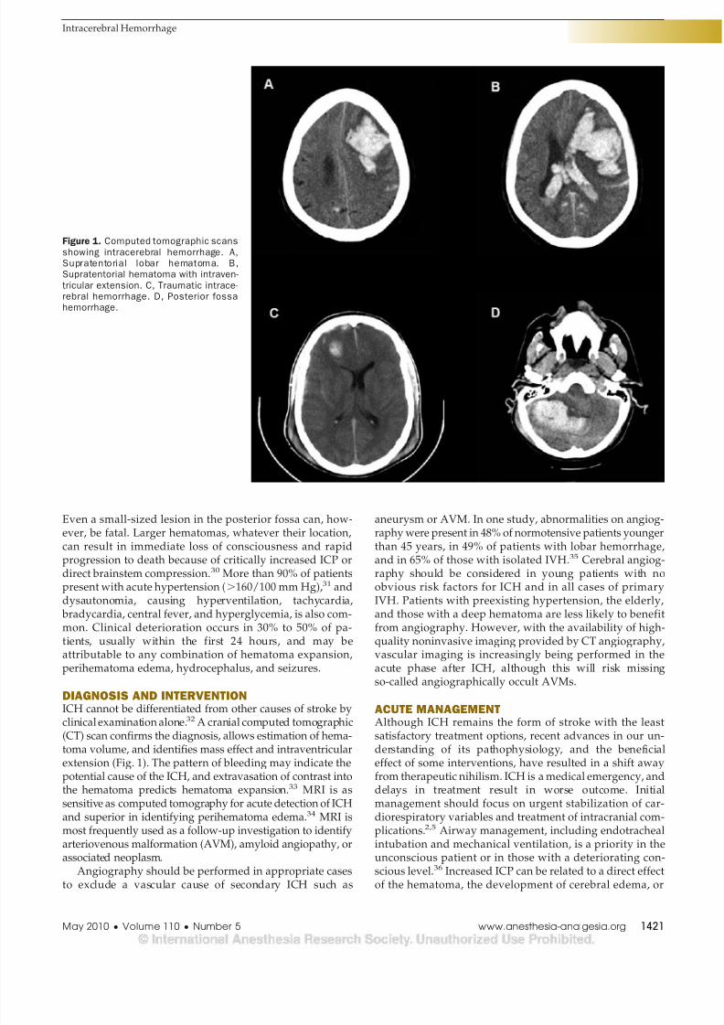
clinical examination alone.32 A cranial computed tomographic(CT) scan confirms the diagnosis, allows estimation of hema-toma volume, and identifies mass effect and intraventricularextension (Fig. 1). The pattern of bleeding may indicate thepotential cause of the ICH, and extravasation of contrast intothe hematoma predicts hematoma expansion.33 MRI is assensitive as computed tomography for acute detection of ICHand superior in identifying perihematoma edema.34 MRI ismost frequently used as a follow-up investigation to identifyarteriovenous malformation (AVM), amyloid angiopathy, orassociated neoplasm.
Angiography should be performed in appropriate casesto exclude a vascular cause of secondary ICH such as
aneurysm or AVM. In one study, abnormalities on angiog-
raphy were present in 48% of normotensive patients youngerthan 45 years, in 49% of patients with lobar hemorrhage,and in 65% of those with isolated IVH.35 Cerebral angiog-raphy should be considered in young patients with noobvious risk factors for ICH and in all cases of primaryIVH. Patients with preexisting hypertension, the elderly,and those with a deep hematoma are less likely to benefitfrom angiography. However, with the availability of high-quality noninvasive imaging provided by CT angiography,vascular imaging is increasingly being performed in theacute phase after ICH, although this will risk missingso-called angiographically occult AVMs.
ACUTE MANAGEMENTAlthough ICH remains the form of stroke with the leastsatisfactory treatment options, recent advances in our un-derstanding of its pathophysiology, and the beneficialeffect of some interventions, have resulted in a shift awayfrom therapeutic nihilism. ICH is a medical emergency, anddelays in treatment result in worse outcome. Initialmanagement should focus on urgent stabilization of car-diorespiratory variables and treatment of intracranial com-plications.2,5 Airway management, including endotrachealintubation and mechanical ventilation, is a priority in theunconscious patient or in those with a deteriorating con-scious level.36 Increased ICP can be related to a direct effectof the hematoma, the development of cerebral edema, or
Figure 1. Computed tomographic scans
showing intracerebral hemorrhage. A,Supratentorial lobar hematoma. B,Supratentorial hematoma with intraven-
tricular extension. C, Traumatic intrace-rebral hemorrhage. D, Posterior fossahemorrhage.
Intracerebral Hemorrhage
May 2010 • Volume 110 • Number 5 www.anesthesia-analgesia.org 1421
8/7/2019 ECV Hemorragico TX Agudo
http://slidepdf.com/reader/full/ecv-hemorragico-tx-agudo 4/9
hydrocephalus. The usual emergency measures to controlICP should be considered in unconscious patients or inthose who present with clinical signs of brainstem hernia-tion. Early placement of a ventricular drain in patients withhydrocephalus can be life saving.
BP CONTROLBP monitoring and management is critical after ICH, butthe targets for treatment remain controversial. Even inpreviously normotensive patients, hypertension is a verycommon finding31 and associated with worse outcome,probably because excessive hypertension is a cause ofhematoma expansion.37,38 In a recent multicenter study,systolic BP (SBP)140 to 150 mm Hg after ICH doubled therisk of subsequent death or dependency.39
The risks of a sudden therapeutic reduction in BP afterischemic stroke are well known,40 but it is possible thatthese same concepts may not apply after ICH because of theabsence of an ischemic penumbra around small-volumeICHs.27 A small, single-center study suggested that BPreduction in patients with acute ICH is safe and that
aggressive reduction might reduce the risk of neurologicaldeterioration in the first 24 hours after admission.41 Tworecently completed multicenter studies have providedmore robust preliminary data on BP control after ICH. Inthe INTEnsive blood pressure Reduction in Acute Cerebralhemorrhage Trial (INTERACT), 203 patients were random-ized to a low-target SBP of 140 mm Hg to be achievedwithin 1 hour and maintained for at least 24 hours afterICH, and 201 were randomized to a more conservative SBPtarget of 180 mm Hg.42 This pilot study established thesafety of decreasing BP early after ICH, determined by theabsence of a significant excess risk of death, dependency, orcardiovascular morbidity, and demonstrated a tendency
toward reduction of hematoma expansion within the first 6hours. However, this study excluded patients with theseverest injury (admission GCS score 3–5) and thereforethose who would presumably be most at risk from acutetherapeutic reductions in BP. The study was also notpowered to detect clinical outcomes, so INTERACT2 hasbeen designed to assess in 2800 patients whether earlyintensive BP-decreasing therapy can reduce death anddisability after ICH. The Antihypertensive Treatment inAcute Cerebral Hemorrhage (ATACH) study evaluated thefeasibility and safety of 3 escalating levels of antihyperten-sive treatment with IV nicardipine in patients with ICH-related acute hypertension.43 Preliminary data from this
study suggest that reduction of SBP to 110 to 140 mm Hg inthe first 24 hours after ICH is well tolerated and associatedwith a reduced risk of hematoma expansion, neurologicaldeterioration, and in-hospital mortality.* Only patients with apresentation GCS score 8 and hematoma volume 60 mLare being recruited into the ATACH study, so its results willbe relevant only to the less severe end of the ICH spectrum.
The continued controversy over the targets for BPcontrol after ICH is reflected in current management guid-ance. The American Heart Association/American StrokeAssociation recommends cautious management of severe
hypertension with continuous infusion of antihypertensivedrugs such as labetalol, esmolol, or nicardipine accordingto the following guidelines.2 If SBP is200 mm Hg or meanarterial blood pressure (MAP) 150 mm Hg, aggressive BPreduction, guided by frequent BP monitoring (at least every5 minutes), should be considered. If SBP is 180 mm Hg (orMAP 130 mm Hg) and there is no evidence or suspicionof increased ICP, a modest reduction in BP to 160/90 mm
Hg (MAP 110 mm Hg) is recommended. The EuropeanUnion Stroke Initiative (EUSI), however, recommends BPtargets determined by the patient’s premorbid state.44 Anupper limit of SBP of 180 mm Hg and a diastolic BP of 105mm Hg is recommended for patients with known hyper-tension or signs of chronic hypertension (e.g., electrocar-diogram or retinal changes) and, if treatment is necessary,the recommended target BP is 160/100 mm Hg (or MAP120 mm Hg). In patients without known hypertension, theupper recommended limits are 160/95 mm Hg, and thetarget BP is 150/90 mm Hg (or MAP 110 mm Hg).However, the EUSI also recommends that mean BP reduc-tion should always be limited to 20% of baseline. In
patients with an ICP monitor in place, both sets of guidancerecommend that BP management should be targeted to main-tain cerebral perfusion pressure between 60 and 70 mmHg.2,44 The optimal timing of conversion from IV to oralantihypertensive therapy is unknown but, in stable patients,sometime between 24 to 72 hours is usually recommended.11
Data to guide management of the lower limits of BPafter ICH are virtually nonexistent. Individualized manage-ment based on premorbid BP, age, cause of ICH, andpresence of increased ICP is recommended, but SBP shouldbe maintained 90 mm Hg in all cases.5
HEMOSTATIC THERAPY
There has been interest in the application of hemostatictherapy to minimize hematoma expansion and improveoutcome after ICH. In 2005, a phase IIb placebo-controlledstudy showed that treatment with recombinant factor VII(rFVIIa), a potent initiator of hemostasis, within 4 hours ofICH significantly reduced hematoma growth in associationwith reduced mortality and improved functional outcomein survivors at 3 months.45 This improvement was seendespite a small increase in thromboembolic complicationsin the rFVIIa-treated patients (7% vs 2% for rFVIIa andplacebo, respectively, P 0.12). However, a subsequentphase III trial in 841 patients, the Factor VII for AcuteHemorrhagic Stroke (FAST) study, failed to replicate theseclinical outcomes.46 In this 2-dose study (rFVIIa 20 and 80g/kg), the dose-related reduction in hematoma expansiondid not translate into a beneficial effect on the risk of deathor severe disability. Post hoc analysis of the FAST datasuggests that rFVIIa might be effective in a subgroup ofyounger patients (70 years) with baseline ICH volume60 mL if administered within 2.5 hours of the onset ofsymptoms.47 On balance, current evidence suggests thatany potential benefit of rFVIIa is offset by a modest increasein the risk of thromboembolic complications.48 Early inves-tigation with CT angiography might identify patients mostat risk of hematoma expansion and who therefore mighthave the most to gain from treatment with rFVIIa.49 Furtherstudies are therefore urgently required to define more
*Available at: http://www.strokecenter.org/trials/TrialDetail.aspx?tid602.Accessed December 14, 2009.
REVIEW ARTICLE
1422 www.anesthesia-analgesia.org ANESTHESIA & ANALGESIA
8/7/2019 ECV Hemorragico TX Agudo
http://slidepdf.com/reader/full/ecv-hemorragico-tx-agudo 5/9
accurately the potential target population that might ben-efit from rFVIIa.
ANTICOAGULATION AND ICH
Oral AnticoagulationICH is the most serious complication of warfarin antico-
agulation. The risk of ICH approximately doubles for eachincrease of one in the international normalized ratio(INR),50 and an INR 3 is associated not only with largerinitial hematoma volume51 but also with an increasedfrequency of hematoma expansion and higher incidence ofneurological deterioration in the first 24 to 48 hours.52
Warfarin-related ICH has a very high mortality, withreported rates up to 67%.14,15 The need to arrest intracranialbleeding outweighs all other considerations and, althoughthere is often a reluctance to reverse anticoagulation inpatients considered to be at high risk of thrombotic com-plications (e.g., those with mechanical heart valves),53 theevidence overwhelmingly supports the correction of coagu-lopathy in all patients.54,55 There is a relatively short time
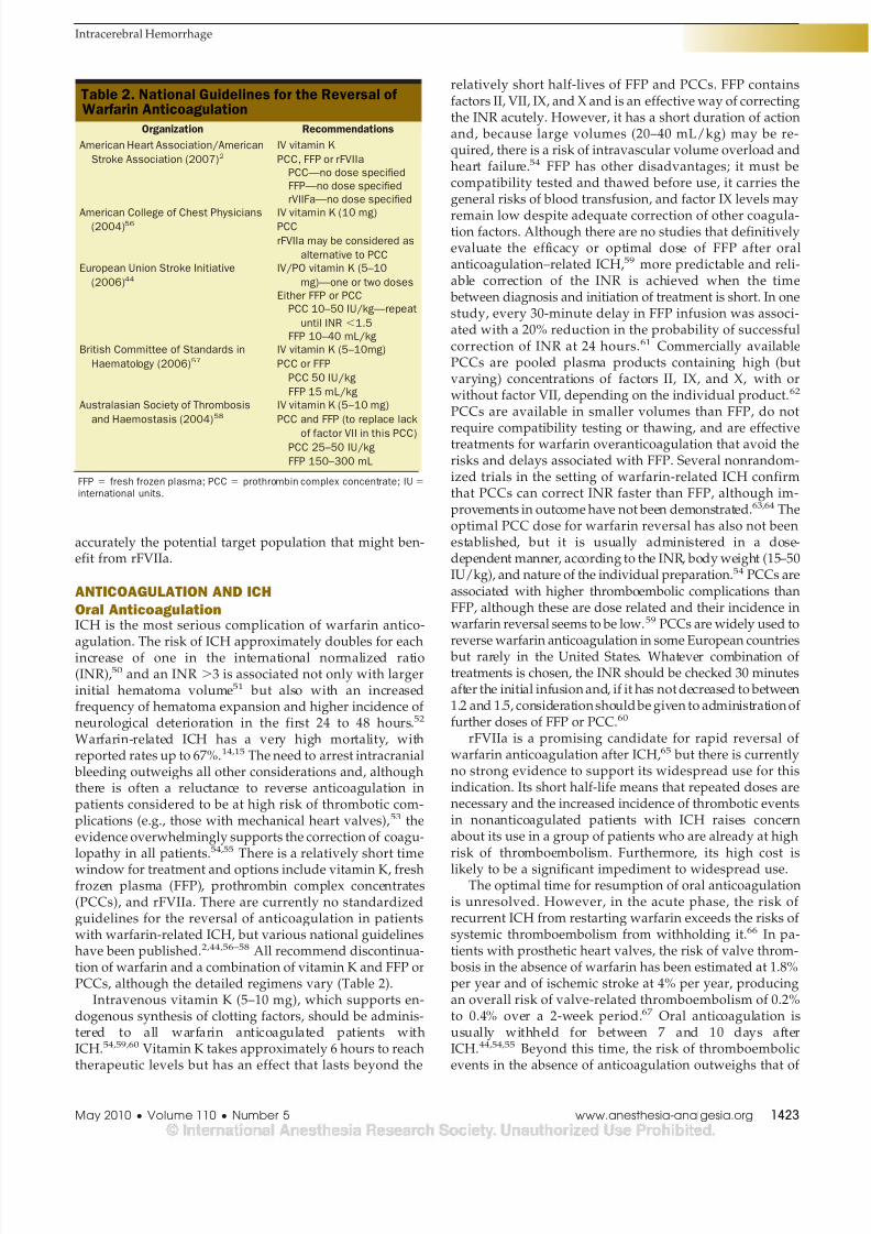
window for treatment and options include vitamin K, freshfrozen plasma (FFP), prothrombin complex concentrates(PCCs), and rFVIIa. There are currently no standardizedguidelines for the reversal of anticoagulation in patientswith warfarin-related ICH, but various national guidelineshave been published.2,44,56–58 All recommend discontinua-tion of warfarin and a combination of vitamin K and FFP orPCCs, although the detailed regimens vary (Table 2).
Intravenous vitamin K (5–10 mg), which supports en-dogenous synthesis of clotting factors, should be adminis-tered to all warfarin anticoagulated patients withICH.54,59,60 Vitamin K takes approximately 6 hours to reachtherapeutic levels but has an effect that lasts beyond the
relatively short half-lives of FFP and PCCs. FFP containsfactors II, VII, IX, and X and is an effective way of correctingthe INR acutely. However, it has a short duration of actionand, because large volumes (20–40 mL/kg) may be re-quired, there is a risk of intravascular volume overload andheart failure.54 FFP has other disadvantages; it must becompatibility tested and thawed before use, it carries thegeneral risks of blood transfusion, and factor IX levels may
remain low despite adequate correction of other coagula-tion factors. Although there are no studies that definitivelyevaluate the efficacy or optimal dose of FFP after oralanticoagulation–related ICH,59 more predictable and reli-able correction of the INR is achieved when the timebetween diagnosis and initiation of treatment is short. In onestudy, every 30-minute delay in FFP infusion was associ-ated with a 20% reduction in the probability of successfulcorrection of INR at 24 hours.61 Commercially availablePCCs are pooled plasma products containing high (butvarying) concentrations of factors II, IX, and X, with orwithout factor VII, depending on the individual product.62
PCCs are available in smaller volumes than FFP, do not
require compatibility testing or thawing, and are effectivetreatments for warfarin overanticoagulation that avoid therisks and delays associated with FFP. Several nonrandom-ized trials in the setting of warfarin-related ICH confirmthat PCCs can correct INR faster than FFP, although im-provements in outcome have not been demonstrated.63,64 Theoptimal PCC dose for warfarin reversal has also not beenestablished, but it is usually administered in a dose-dependent manner, according to the INR, body weight (15–50IU/kg), and nature of the individual preparation.54 PCCs areassociated with higher thromboembolic complications thanFFP, although these are dose related and their incidence inwarfarin reversal seems to be low.59 PCCs are widely used toreverse warfarin anticoagulation in some European countriesbut rarely in the United States. Whatever combination oftreatments is chosen, the INR should be checked 30 minutesafter the initial infusion and, if it has not decreased to between1.2 and 1.5, consideration should be given to administration offurther doses of FFP or PCC.60
rFVIIa is a promising candidate for rapid reversal ofwarfarin anticoagulation after ICH,65 but there is currentlyno strong evidence to support its widespread use for thisindication. Its short half-life means that repeated doses arenecessary and the increased incidence of thrombotic eventsin nonanticoagulated patients with ICH raises concernabout its use in a group of patients who are already at highrisk of thromboembolism. Furthermore, its high cost is
likely to be a significant impediment to widespread use.The optimal time for resumption of oral anticoagulation
is unresolved. However, in the acute phase, the risk ofrecurrent ICH from restarting warfarin exceeds the risks ofsystemic thromboembolism from withholding it.66 In pa-tients with prosthetic heart valves, the risk of valve throm-bosis in the absence of warfarin has been estimated at 1.8%per year and of ischemic stroke at 4% per year, producingan overall risk of valve-related thromboembolism of 0.2%to 0.4% over a 2-week period.67 Oral anticoagulation isusually withheld for between 7 and 10 days afterICH.44,54,55 Beyond this time, the risk of thromboembolicevents in the absence of anticoagulation outweighs that of
Table 2. National Guidelines for the Reversal of Warfarin Anticoagulation
Organization Recommendations
American Heart Association/American
Stroke Association (2007)2IV vitamin K
PCC, FFP or rFVIIa
PCCno dose specified
FFPno dose specified
rVIIFano dose specified
American College of Chest Physicians(2004)56
IV vitamin K (10 mg)PCC
rFVIIa may be considered as
alternative to PCC
European Union Stroke Initiative
(2006)44IV/PO vitamin K (5–10
mg)one or two doses
Either FFP or PCC
PCC 10–50 IU/kgrepeat
until INR 1.5
FFP 10–40 mL/kg
British Committee of Standards in
Haematology (2006)57IV vitamin K (5–10mg)
PCC or FFP
PCC 50 IU/kg
FFP 15 mL/kg
Australasian Society of Thrombosis
and Haemostasis (2004)58IV vitamin K (5–10 mg)
PCC and FFP (to replace lack
of factor VII in this PCC)PCC 25–50 IU/kg
FFP 150–300 mL
FFP fresh frozen plasma; PCC prothrombin complex concentrate; IU
international units.
Intracerebral Hemorrhage
May 2010 • Volume 110 • Number 5 www.anesthesia-analgesia.org 1423
8/7/2019 ECV Hemorragico TX Agudo
http://slidepdf.com/reader/full/ecv-hemorragico-tx-agudo 6/9
recurrent ICH after its reintroduction.68,69 However, survi-vors of lobar ICH with atrial fibrillation should not beoffered long-term anticoagulation because the risks ofrecurrent hemorrhage outweigh the potential benefits.69
The role of IV heparin, or subcutaneous low-molecular-weight heparin (LMWH), as temporary therapy prior toreinstitution of warfarin is unclear.54
Antiplatelet DrugsWith an increasingly elderly population, there has been adramatic increase in the number of patients receivinglong-term antiplatelet medication. Aspirin is associatedwith an absolute risk increase in ICH of 12 events per 10,000persons, although this must be put into the context of anoverall benefit of aspirin in terms of reduced risk ofmyocardial infarction and ischemic stroke.70 High-doseaspirin increases the risk of ICH further in the elderly,particularly in association with untreated hypertension.16
The risk of ICH is increased even more by the combinationof aspirin and clopidogrel.71 Antiplatelet therapy is also anindependent predictor of hematoma expansion.23 There are
no data confirming the efficacy of platelet replacement orother specific interventions after antiplatelet therapy–related ICH, and further studies on this issue are required.
NEUROSURGERYThe value of placement of a ventricular drain in patientswith hydrocephalus is undisputed, but the timing andnature of other neurosurgical interventions are morecontroversial.72,73 One meta-analysis failed to show a sta-tistically significant reduction in the odds of death withsurgical intervention (odds ratio, 0.84; 95% confidenceinterval, 0.67–1.07) compared with standard medical
therapy.
74
The Surgical Trial in Intracerebral Hemorrhage(STICH) randomized 1033 patients with supratentorial ICHto surgery within 72 hours or conservative management; nooutcome benefit of hematoma evacuation compared withstandard medical therapy was demonstrated.75 AlthoughSTICH suggests that early surgery is ineffective, it does notconfirm that it is useless in all cases because the study wasbased on clinical equipoise; patients who the local investi-gator thought might benefit from hematoma evacuationwere not recruited into the study. The STICH trial also didnot set out to differentiate deep-seated ICH with IVH andhydrocephalus from more superficial lobar ICH for whichthe prognosis is much better. Only 222 patients with lobarICH were randomized, possibly because so many neuro-
surgeons believed that such patients should undergo sur-gery. There is some evidence to support this view; a posthoc analysis of the STICH data showed that a subgroup ofpatients with superficial hematomas and no IVH gainedbenefit from surgery.76 Because the mean time to surgerywas24 hours, STICH also does not exclude the possibilitythat earlier surgery might have been beneficial in somepatients. However, there is evidence from other sourcesthat ultraearly surgery (within 4 hours of ictus) is associ-ated with an increased risk of rebleeding and highermortality (75%).77 These controversies provided impetusfor the continuing STICH-II study that will evaluate therole of early surgery in superficial supratentorial lobar
hematomas in patients without IVH.† In contrast to supra-tentorial lesions, there is better evidence that patients witha posterior fossa hematoma benefit from early surgicalevacuation because of the high risk of deterioration.78 Theplace of decompressive craniectomy after ICH is not estab-lished, although in a small series, 6 of 11 patients (54.5%)treated with hemicraniectomy had a good functional out-come.79 These findings suggest that a randomized con-
trolled trial of decompressive craniectomy after ICH iswarranted.
Hematoma aspiration via minimally invasive surgery(MIS) offers some advantages over conventional surgery,including the possibility for local anesthesia, reduced op-erating time, and reduced tissue trauma.80 Thrombolysis,with or without clot aspiration, can also be performedusing MIS, but one meta-analysis concluded that, althoughintraventricular thrombolysis is safe, there is no definiteevidence of efficacy.81 However, preliminary data from theMinimally Invasive Surgery plus rtPA for Intracerebralhemorrhage Evacuation (MISTIE) trial suggest that MISplus recombinant tissue plasminogen activator (rtPA) of-
fers greater clot resolution than conventional medicaltherapy.82 A recent preliminary report of the Clot LysisEvaluating Accelerated Resolution on IntraventricularHemorrhage (CLEAR-IVH) trial also confirms that low-dose rtPA can be safely administered to stable IVH clotsand may increase lysis rates.83
OTHER INTENSIVE CARE MANAGEMENTPatients with depressed conscious level require ventilatorysupport as well as cardiovascular and ICP monitoring andmanagement in an intensive care unit. However, close obser-vation in an intensive care environment is recommended formany nonventilated patients for at least the first 24 hours
because the risk of neurological deterioration is greatestduring this period.25 Systemic medical complications, includ-ing pneumonia, neurogenic lung injury, hyperglycemia, andfever, are common after ICH and associated with increasedintensive care unit and hospital length of stay and worsenedoutcome.84 There is substantial evidence that management ina specialist neurointensive care unit results in improvedoutcomes after ICH.85 The exact reasons for this remainunclear, although the delivery of consensus-based, protocol-ized treatment strategies by a dedicated multiprofessionalteam familiar with the interactions between the injured brainand nonneurological organ systems, as well as early transferto a multidisciplinary stroke rehabilitation unit, are likely toplay a role. Attention has also focused on the major role thattherapeutic nihilism and self-fulfilling prophesies of doomcan have on determining outcome when patients with ICHare cared for by nonspecialist teams.86
Control of ICPThere is a high risk of increased ICP after large-volumeICH, particularly in the presence of IVH.87 Although thereis limited evidence for the monitoring and management ofICP after ICH, many neurocritical care units continuouslymonitor ICP in all sedated ICH patients requiring mechani-cal ventilation. Although the majority of the evidence base
†Available at: http://www.ncl.ac.uk/stich/. Accessed December 14, 2009.
REVIEW ARTICLE
1424 www.anesthesia-analgesia.org ANESTHESIA & ANALGESIA

8/7/2019 ECV Hemorragico TX Agudo
http://slidepdf.com/reader/full/ecv-hemorragico-tx-agudo 7/9
and consensus guidance for the treatment of intracranialhypertension relates to traumatic brain injury, similar prin-ciples are applied empirically to the ICH patient popula-tion. Standard medical treatment of increased ICP shouldtherefore be initiated as appropriate, a detailed discussionof which is beyond the scope of this review.
Anticonvulsant Therapy
Approximately 8% of patients with ICH develop clinicalseizures within 30 days of the ictus, and continuous elec-troencephalographic monitoring demonstrates subclinicalseizure activity in up to 25%.88 Seizures are more likely tooccur in the presence of a lobar hematoma.89 The use ofprophylactic anticonvulsant medication after ICH is con-troversial, although one small study showed that it doesreduce the risk of early seizures.89 Current guidance doesnot recommend universal prophylaxis, but that therapyshould be considered in selected patients with lobarICH.2,44 If seizures do occur, they should be treated aggres-sively in the usual manner.
Glycemic ControlHyperglycemia worsens cerebral ischemic injury, and admis-sion hyperglycemia is associated with increased 30-day mor-tality after ICH.90 However, the targets for glycemic controlare unclear, and there is increasing evidence that “tight”glycemic control with insulin infusion can be associated witha critically low cerebral extracellular glucose concentrationafter brain injury.91 Until further data become available,systemic glucose levels should not be treated in the acutephase after ICH unless 10.0 mmol/L (180 mg/dL).92
General TherapyGeneral measures, including fluid management, fever control,
provision of enteral nutrition, and prevention of aspirationpneumonia and bedsores, are the same as for patients withischemic stroke.2,44,93 Thromboembolic prophylaxis withcompression stockings and intermittent pneumatic compres-sion is recommended in all patients from admission. Subcu-taneous low-molecular-weight heparin should be consideredafter 24 to 48 hours, when it does not seem to result in anincreased risk of recurrent hemorrhage.94
SUMMARYICH is a devastating disease, and the long-standing contro-versy over its optimal management remains largely unre-solved. However, there is optimism that new insights into
its pathophysiology will lead to the introduction of tar-geted management strategies. A greater understanding ofthe dynamic processes that occur after ICH is likely toresult in the development of therapies aimed at the preven-tion of neurological deterioration and improve outcome byminimizing hematoma expansion, perihematoma edema,and secondary neuronal damage. An awareness of theadverse effects of systemic physiological disturbances isalso likely to lead to the introduction of evidence-basedtreatments that were previously delivered on an empiricalbasis. Continuing randomized, controlled trials will clarifythe correct approach to early BP management and theindications for surgical interventions after ICH. Promising
future treatments include the development of antiinflam-matory drugs that inhibit or reduce perihematoma edema,surgical techniques that maximize hematoma removalwhile minimizing damage to normal tissue, and thrombo-lytic therapy for IVH. There is a short time window for thestabilization and acute management of patients with ICH,and an increasing recognition that focused management ina specialist neurocritical care unit is associated with im-
proved outcome. The days of treatment nihilism are beingreplaced by an appreciation that aggressive management inthe acute phase can translate into improved outcomes.
REFERENCES
1. Gebel JM, Broderick JP. Intracerebral hemorrhage. Neurol Clin2000;18:419–38
2. Broderick J, Connolly S, Feldmann E, Hanley D, Kase C,Krieger D, Mayberg M, Morgenstern L, Ogilvy CS, Vespa P,Zuccarello M. Guidelines for the management of spontaneousintracerebral hemorrhage in adults: 2007 update: a guidelinefrom the American Heart Association/American Stroke Asso-ciation Stroke Council, High Blood Pressure Research Council,and the Quality of Care and Outcomes in Research Interdisci-plinary Working group. Stroke 2007;38:2001–23
3. Ariesen MJ, Claus SP, Rinkel GJ, Algra A. Risk factors forintracerebral hemorrhage in the general population: a system-atic review. Stroke 2003;34:2060–5
4. Flaherty ML, Woo D, Haverbusch M, Sekar P, Khoury J,Sauerbeck L, Moomaw CJ, Schneider A, Kissela B, KleindorferD, Broderick JP. Racial variations in location and risk ofintracerebral hemorrhage. Stroke 2005;36:934–7
5. Mayer SA, Rincon F. Treatment of intracerebral haemorrhage.Lancet Neurol 2005;4:662–72
6. Hart RG, Tonarelli SB, Pearce LA. Avoiding central nervoussystem bleeding during antithrombotic therapy: recent dataand ideas. Stroke 2005;36:1588–93
7. Broderick JP, Brott TG, Duldner JE, Tomsick T, Huster G.Volume of intracerebral hemorrhage. A powerful and easy-to-use predictor of 30-day mortality. Stroke 1993;24:987–93
8. Fogelholm R, Murros K, Rissanen A, Avikainen S. Long termsurvival after primary intracerebral haemorrhage: a retrospec-tive population based study. J Neurol Neurosurg Psychiatry2005;76:1534–8
9. Hemphill JC III, Bonovich DC, Besmertis L, Manley GT,Johnston SC. The ICH score: a simple, reliable grading scale forintracerebral hemorrhage. Stroke 2001;32:891–7
10. Thrift AG, McNeil JJ, Forbes A, Donnan GA. Three importantsubgroups of hypertensive persons at greater risk of intrace-rebral hemorrhage. Melbourne Risk factor Study group. Hy-pertension 1998;31:1223–9
11. Qureshi AI, Tuhrim S, Broderick JP, Batjer HH, Hondo H,Hanley DF. Spontaneous intracerebral hemorrhage. N EnglJ Med 2001;344:1450–60
12. Skidmore CT, Andrefsky J. Spontaneous intracerebral hemor-rhage: epidemiology, pathophysiology, and medical manage-
ment. Neurosurg Clin N Am 2002;13:281–813. Arakawa S, Saku Y, Ibayashi S, Nagao T, Fujishima M. Bloodpressure control and recurrence of hypertensive brain hemor-rhage. Stroke 1998;29:1806–9
14. Fang MC, Go AS, Chang Y, Hylek EM, Henault LE, JensvoldNG, Singer DE. Death and disability from warfarin-associatedintracranial and extracranial hemorrhages. Am J Med2007;120:700–5
15. Rosand J, Eckman MH, Knudsen KA, Singer DE, GreenbergSM. The effect of warfarin and intensity of anticoagulation onoutcome of intracerebral hemorrhage. Arch Intern Med2004;164:880–4
16. Saloheimo P, Juvela S, Hillbom M. Use of aspirin, epistaxis,and untreated hypertension as risk factors for primary intra-cerebral hemorrhage in middle-aged and elderly people.Stroke 2001;32:399 – 404
Intracerebral Hemorrhage
May 2010 • Volume 110 • Number 5 www.anesthesia-analgesia.org 1425
8/7/2019 ECV Hemorragico TX Agudo
http://slidepdf.com/reader/full/ecv-hemorragico-tx-agudo 8/9
17. Thrift AG, Donnan GA, McNeil JJ. Heavy drinking, but notmoderate or intermediate drinking, increases the risk of intra-cerebral hemorrhage. Epidemiology 1999;10:307–12
18. Amarenco P, Bogousslavsky J, Callahan A III, Goldstein LB,Hennerici M, Rudolph AE, Sillesen H, Simunovic L, Szarek M,Welch KM, Zivin JA. High-dose atorvastatin after stroke ortransient ischemic attack. N Engl J Med 2006;355:549–59
19. Rincon F, Mayer SA. Novel therapies for intracerebral hemor-rhage. Curr Opin Crit Care 2004;10:94–100
20. Brott T, Broderick J, Kothari R, Barsan W, Tomsick T, Sauer-
beck L, Spilker J, Duldner J, Khoury J. Early hemorrhagegrowth in patients with intracerebral hemorrhage. Stroke1997;28:1–5
21. Mayer SA, Lignelli A, Fink ME, Kessler DB, Thomas CE,Swarup R, Van Heertum RL. Perilesional blood flow andedema formation in acute intracerebral hemorrhage: a SPECTstudy. Stroke 1998;29:1791–8
22. Xi G, Fewel ME, Hua Y, Thompson BG Jr, Hoff JT, Keep RF.Intracerebral hemorrhage: pathophysiology and therapy. Neu-rocrit Care 2004;1:5–18
23. Broderick JP, Diringer MN, Hill MD, Brun NC, Mayer SA,Steiner T, Skolnick BE, Davis SM. Determinants of intracere-bral hemorrhage growth: an exploratory analysis. Stroke2007;38:1072–5
24. Tuhrim S, Horowitz DR, Sacher M, Godbold JH. Validationand comparison of models predicting survival following intra-
cerebral hemorrhage. Crit Care Med 1995;23:950–425. Mayer SA, Sacco RL, Shi T, Mohr JP. Neurologic deterioration
in noncomatose patients with supratentorial intracerebralhemorrhage. Neurology 1994;44:1379–84
26. Hua Y, Keep RF, Hoff JT, Xi G. Brain injury after intracerebralhemorrhage: the role of thrombin and iron. Stroke2007;38:759–62
27. Schellinger PD, Fiebach JB, Hoffmann K, Becker K, OrakciogluB, Kollmar R, Juttler E, Schramm P, Schwab S, Sartor K, HackeW. Stroke MRI in intracerebral hemorrhage: is there a perihem-orrhagic penumbra? Stroke 2003;34:1674–9
28. Huang FP, Xi G, Keep RF, Hua Y, Nemoianu A, Hoff JT. Brainedema after experimental intracerebral hemorrhage: role ofhemoglobin degradation products. J Neurosurg 2002;96:287–93
29. Sansing LH, Kaznatcheeva EA, Perkins CJ, Komaroff E, Gut-man FB, Newman GC. Edema after intracerebral hemorrhage:
correlations with coagulation parameters and treatment.J Neurosurg 2003;98:985–92
30. Andrews BT, Chiles BW III, Olsen WL, Pitts LH. The effectof intracerebral hematoma location on the risk of brain-stemcompression and on clinical outcome. J Neurosurg1988;69:518–22
31. Qureshi AI, Ezzeddine MA, Nasar A, Suri MF, Kirmani JF,Hussein HM, Divani AA, Reddi AS. Prevalence of elevatedblood pressure in 563,704 adult patients with stroke present-ing to the ED in the United States. Am J Emerg Med2007;25:32–8
32. Goldstein LB, Simel DL. Is this patient having a stroke? JAMA2005;293:2391–402
33. Murai Y, Ikeda Y, Teramoto A, Goldstein JN, Greenberg SM,Smith EE, Lev MH, Rosand J. Contrast extravasation on CTangiography predicts hematoma expansion in intracerebral
hemorrhage. Neurology 2007;69:61734. Kidwell CS, Chalela JA, Saver JL, Starkman S, Hill MD,
Demchuk AM, Butman JA, Patronas N, Alger JR, Latour LL,Luby ML, Baird AE, Leary MC, Tremwel M, Ovbiagele B,Fredieu A, Suzuki S, Villablanca JP, Davis S, Dunn B, Todd JW,Ezzeddine MA, Haymore J, Lynch JK, Davis L, Warach S.Comparison of MRI and CT for detection of acute intracerebralhemorrhage. JAMA 2004;292:1823–30
35. Zhu XL, Chan MS, Poon WS. Spontaneous intracranial hem-orrhage: which patients need diagnostic cerebral angiography?A prospective study of 206 cases and review of the literature.Stroke 1997;28:1406–9
36. Gujjar AR, Deibert E, Manno EM, Duff S, Diringer MN.Mechanical ventilation for ischemic stroke and intracerebralhemorrhage: indications, timing, and outcome. Neurology1998;51:447–51
37. Fogelholm R, Avikainen S, Murros K. Prognostic value anddeterminants of first-day mean arterial pressure in spontaneoussupratentorial intracerebral hemorrhage. Stroke 1997;28:1396–400
38. Willmot M, Leonardi-Bee J, Bath PM. High blood pressure inacute stroke and subsequent outcome: a systematic review.Hypertension 2004;43:18–24
39. Zhang Y, Reilly KH, Tong W, Xu T, Chen J, Bazzano LA, QiaoD, Ju Z, Chen CS, He J. Blood pressure and clinical outcomeamong patients with acute stroke in Inner Mongolia, China.
J Hypertens 2008;26:1446–5240. Grossman E, Messerli FH, Grodzicki T, Kowey P. Should a
moratorium be placed on sublingual nifedipine capsules givenfor hypertensive emergencies and pseudoemergencies? JAMA1996;276:1328–31
41. Suri MF, Suarez JI, Rodrigue TC, Zaidat OO, Vazquez G,Wensel A, Selman WR. Effect of treatment of elevated bloodpressure on neurological deterioration in patients with acuteintracerebral hemorrhage. Neurocrit Care 2008;9:177–82
42. Anderson CS, Huang Y, Wang JG, Arima H, Neal B, Peng B,Heeley E, Skulina C, Parsons MW, Kim JS, Tao QL, Li YC, JiangJD, Tai LW, Zhang JL, Xu E, Cheng Y, Heritier S, MorgensternLB, Chalmers J. Intensive blood pressure reduction in acutecerebral haemorrhage trial (INTERACT): a randomised pilottrial. Lancet Neurol 2008;7:391–9
43. Qureshi AI. Antihypertensive Treatment of Acute Cerebral
Hemorrhage (ATACH): rationale and design. Neurocrit Care2007;6:56–66
44. Steiner T, Kaste M, Forsting M, Mendelow D, Kwiecinski H,Szikora I, Juvela S, Marchel A, Chapot R, Cognard C, Unter-berg A, Hacke W. Recommendations for the management ofintracranial haemorrhagepart I: spontaneous intracerebralhaemorrhage. The European Stroke Initiative Writing Commit-tee and the Writing Committee for the EUSI Executive Com-mittee. Cerebrovasc Dis 2006;22:294–316
45. Mayer SA, Brun NC, Begtrup K, Broderick J, Davis S, DiringerMN, Skolnick BE, Steiner T. Recombinant activated factor VIIfor acute intracerebral hemorrhage. N Engl J Med 2005;352:777–85
46. Mayer SA, Brun NC, Begtrup K, Broderick J, Davis S, DiringerMN, Skolnick BE, Steiner T. Efficacy and safety of recombinantactivated factor VII for acute intracerebral hemorrhage. N Engl
J Med 2008;358:2127–3747. Mayer SA, Davis SM, Skolnick BE, Brun NC, Begtrup K,
Broderick JP, Diringer MN, Steiner T. Can a subset of intrace-rebral hemorrhage patients benefit from hemostatic therapywith recombinant activated factor VII? Stroke 2009;40:833–40
48. Diringer MN, Skolnick BE, Mayer SA, Steiner T, Davis SM,Brun NC, Broderick JP. Risk of thromboembolic events incontrolled trials of rFVIIa in spontaneous intracerebral hemor-rhage. Stroke 2008;39:850 – 6
49. Wada R, Aviv RI, Fox AJ, Sahlas DJ, Gladstone DJ, Tomlinson G,Symons SP. CT angiography “spot sign” predicts hematomaexpansion in acute intracerebral hemorrhage. Stroke 2007;38:1257–62
50. Schulman S, Beyth RJ, Kearon C, Levine MN. Hemorrhagiccomplications of anticoagulant and thrombolytic treatment:American College of Chest Physicians Evidence-Based Clinical
Practice Guidelines (8th edition). Chest 2008;133:257S–98S51. Flaherty ML, Tao H, Haverbusch M, Sekar P, Kleindorfer D,
Kissela B, Khatri P, Stettler B, Adeoye O, Moomaw CJ, Brod-erick JP, Woo D. Warfarin use leads to larger intracerebralhematomas. Neurology 2008;71:1084–9
52. Cucchiara B, Messe S, Sansing L, Kasner S, Lyden P. Hema-toma growth in oral anticoagulant related intracerebral hem-orrhage. Stroke 2008;39:2993–6
53. Appelboam R, Thomas EO. The headache over warfarin inBritish neurosurgical intensive care units: a national survey ofcurrent practice. Intensive Care Med 2007;33:1946–53
54. Appelboam R, Thomas EO. Warfarin and intracranial haemor-rhage. Blood Rev 2009;23:1–9
55. Phan TG, Koh M, Wijdicks EF. Safety of discontinuation ofanticoagulation in patients with intracranial hemorrhage athigh thromboembolic risk. Arch Neurol 2000;57:1710–3
REVIEW ARTICLE
1426 www.anesthesia-analgesia.org ANESTHESIA & ANALGESIA
8/7/2019 ECV Hemorragico TX Agudo
http://slidepdf.com/reader/full/ecv-hemorragico-tx-agudo 9/9
56. Ansell J, Hirsh J, Poller L, Bussey H, Jacobson A, Hylek E. Thepharmacology and management of the vitamin K antagonists:the Seventh ACCP Conference on Antithrombotic and Throm-bolytic Therapy. Chest 2004;126:204S–33S
57. Baglin TP, Keeling DM, Watson HG. Guidelines on oralanticoagulation (warfarin): third edition2005 update. Br JHaematol 2006;132:277–85
58. Baker RI, Coughlin PB, Gallus AS, Harper PL, Salem HH,Wood EM. Warfarin reversal: consensus guidelines, on behalfof the Australasian Society of Thrombosis and Haemostasis.
Med J Aust 2004;181:492–759. Aiyagari V, Testai FD. Correction of coagulopathy in warfarin
associated cerebral hemorrhage. Curr Opin Crit Care2009;15:87–92
60. Goldstein JN, Rosand J, Schwamm LH. Warfarin reversal inanticoagulant-associated intracerebral hemorrhage. NeurocritCare 2008;9:277–83
61. Goldstein JN, Thomas SH, Frontiero V, Joseph A, Englel C,Snider R, Smith EE, Greenberg SM, Rosand J. Timing of freshfrozen plasma administration and rapid correction of coagu-lopathy in warfarin-related intracerebral hemorrhage. Stroke2006;37:151–5
62. Leissinger CA, Blatt PM, Hoots WK, Ewenstein B. Role ofprothrombin complex concentrates in reversing warfarin antico-agulation: a review of the literature. Am J Hematol 2008;83:137–43
63. Sjoblom L, Hardemark HG, Lindgren A, Norrving B, Fahlen M,Samuelsson M, Stigendal L, Stockelberg D, Taghavi A, WallrupL, Wallvik J. Management and prognostic features of intrace-rebral hemorrhage during anticoagulant therapy: a Swedishmulticenter study. Stroke 2001;32:2567–74
64. Yasaka M, Sakata T, Naritomi H, Minematsu K. Optimal doseof prothrombin complex concentrate for acute reversal of oralanticoagulation. Thromb Res 2005;115:455–9
65. Brody DL, Aiyagari V, Shackleford AM, Diringer MN. Use ofrecombinant factor VIIa in patients with warfarin-associatedintracranial hemorrhage. Neurocrit Care 2005;2:263–7
66. Hacke W. The dilemma of reinstituting anticoagulation forpatients with cardioembolic sources and intracranial hemor-rhage: how wide is the strait between Skylla and Karybdis?Arch Neurol 2000;57:1682–4
67. Crawley F, Bevan D, Wren D. Management of intracranial bleed-
ing associated with anticoagulation: balancing the risk of furtherbleeding against thromboembolism from prosthetic heart valves.J Neurol Neurosurg Psychiatry 2000;69:396–8
68. Claassen DO, Kazemi N, Zubkov AY, Wijdicks EF, RabinsteinAA. Restarting anticoagulation therapy after warfarin-associatedintracerebral hemorrhage. Arch Neurol 2008;65:1313–8
69. Eckman MH, Rosand J, Knudsen KA, Singer DE, GreenbergSM. Can patients be anticoagulated after intracerebral hemor-rhage? A decision analysis. Stroke 2003;34:1710– 6
70. He J, Whelton PK, Vu B, Klag MJ. Aspirin and risk ofhemorrhagic stroke: a meta-analysis of randomized controlledtrials. JAMA 1998;280:1930–5
71. Diener HC, Bogousslavsky J, Brass LM, Cimminiello C, CsibaL, Kaste M, Leys D, Matias-Guiu J, Rupprecht HJ. Aspirin andclopidogrel compared with clopidogrel alone after recent isch-aemic stroke or transient ischaemic attack in high-risk patients
(MATCH): randomised, double-blind, placebo-controlled trial.Lancet 2004;364:331–7
72. Fernandes HM, Gregson B, Siddique S, Mendelow AD. Sur-gery in intracerebral hemorrhage. The uncertainty continues.Stroke 2000;31:2511–6
73. Mendelow AD, Unterberg A. Surgical treatment of intracere-bral haemorrhage. Curr Opin Crit Care 2007;13:169–74
74. Teernstra OP, Evers SM, Kessels AH. Meta analyses in treat-ment of spontaneous supratentorial intracerebral haematoma.Acta Neurochir (Wien) 2006;148:521–8
75. Mendelow AD, Gregson BA, Fernandes HM, Murray GD,Teasdale GM, Hope DT, Karimi A, Shaw MD, Barer DH. Earlysurgery versus initial conservative treatment in patients withspontaneous supratentorial intracerebral haematomas in theInternational Surgical Trial in Intracerebral Haemorrhage(STICH): a randomised trial. Lancet 2005;365:387–97
76. Bhattathiri PS, Gregson B, Prasad KS, Mendelow AD. Intra-ventricular hemorrhage and hydrocephalus after spontaneousintracerebral hemorrhage: results from the STICH trial. ActaNeurochir Suppl 2006;96:65–8
77. Morgenstern LB, Demchuk AM, Kim DH, Frankowski RF,Grotta JC. Rebleeding leads to poor outcome in ultra-earlycraniotomy for intracerebral hemorrhage. Neurology2001;56:1294–9
78. Ott KH, Kase CS, Ojemann RG, Mohr JP. Cerebellar hemor-rhage: diagnosis and treatment. A review of 56 cases. Arch
Neurol 1974;31:160–779. Murthy JM, Chowdary GV, Murthy TV, Bhasha PS, Naryanan
TJ. Decompressive craniectomy with clot evacuation in largehemispheric hypertensive intracerebral hemorrhage. NeurocritCare 2005;2:258–62
80. Auer LM, Deinsberger W, Niederkorn K, Gell G, Kleinert R,Schneider G, Holzer P, Bone G, Mokry M, Korner E. Endo-scopic surgery versus medical treatment for spontaneous in-tracerebral hematoma: a randomized study. J Neurosurg1989;70:530–5
81. Lapointe M, Haines S. Fibrinolytic therapy for intraventricularhemorrhage in adults. Cochrane Database Syst Rev2002;CD003692
82. Morgan T, Zuccarello M, Narayan R, Keyl P, Lane K, HanleyD. Preliminary findings of the minimally-invasive surgery plusrtPA for intracerebral hemorrhage evacuation (MISTIE) clinical
trial. Acta Neurochir Suppl 2008;105:147–5183. Morgan T, Awad I, Keyl P, Lane K, Hanley D. Preliminary
report of the clot lysis evaluating accelerated resolution ofintraventricular hemorrhage (CLEAR-IVH) clinical trial. ActaNeurochir Suppl 2008;105:217–20
84. Naidech AM, Bendok BR, Tamul P, Bassin SL, Watts CM,Batjer HH, Bleck TP. Medical complications drive length ofstay after brain hemorrhage: a cohort study. Neurocrit Care2009;10:11–9
85. Diringer MN, Edwards DF. Admission to a neurologic/neurosurgical intensive care unit is associated with reducedmortality rate after intracerebral hemorrhage. Crit Care Med2001;29:635–40
86. Hemphill JC III, Newman J, Zhao S, Johnston SC. Hospitalusage of early do-not-resuscitate orders and outcome afterintracerebral hemorrhage. Stroke 2004;35:1130–4
87. Nilsson OG, Lindgren A, Brandt L, Saveland H. Prediction ofdeath in patients with primary intracerebral hemorrhage: aprospective study of a defined population. J Neurosurg2002;97:531–6
88. Claassen J, Jette N, Chum F, Green R, Schmidt M, Choi H,Jirsch J, Frontera JA, Connolly ES, Emerson RG, Mayer SA,Hirsch LJ. Electrographic seizures and periodic dischargesafter intracerebral hemorrhage. Neurology 2007;69:1356–65
89. Passero S, Rocchi R, Rossi S, Ulivelli M, Vatti G. Seizures afterspontaneous supratentorial intracerebral hemorrhage. Epilep-sia 2002;43:1175–80
90. Fogelholm R, Murros K, Rissanen A, Avikainen S. Admissionblood glucose and short term survival in primary intracerebralhaemorrhage: a population based study. J Neurol NeurosurgPsychiatry 2005;76:349–53
91. Oddo M, Schmidt JM, Carrera E, Badjatia N, Connolly ES,Presciutti M, Ostapkovich ND, Levine JM, Le RP, Mayer SA.Impact of tight glycemic control on cerebral glucose metabo-lism after severe brain injury: a microdialysis study. Crit CareMed 2008;36:3233–8
92. Prakash A, Matta BF. Hyperglycaemia and neurological injury.Curr Opin Anaesthesiol 2008;21:565–9
93. Olsen TS, Langhorne P, Diener HC, Hennerici M, Ferro J,Sivenius J, Wahlgren NG, Bath P. European Stroke InitiativeRecommendations for Stroke Managementupdate 2003. Ce-rebrovasc Dis 2003;16:311–37
94. Boeer A, Voth E, Henze T, Prange HW. Early heparin therapyin patients with spontaneous intracerebral haemorrhage.J Neurol Neurosurg Psychiatry 1991;54:466–7
Intracerebral Hemorrhage
May 2010 • Volume 110 • Number 5 www.anesthesia-analgesia.org 1427