bitis gabonica (gaboon viper) snake venom gland: …...bitis gabonica (gaboon viper) snake venom...
TRANSCRIPT
www.elsevier.com/locate/gene
Gene 337 (2004) 55–69
Bitis gabonica (Gaboon viper) snake venom gland: toward a catalog for
the full-length transcripts (cDNA) and proteins$
Ivo M.B. Francischettia,*, Van My-Phama, Jim Harrisonb, Mark K. Garfieldc, Jose M.C. Ribeiroa
aMedical Entomology Section, Laboratory of Malaria and Vector Research, National Institutes of Allergy and Infectious Diseases,
National Institutes of Health, 12735 Twinbrook Parkway, Building Twinbrook III, Room 2E-28, Rockville, MD 20852, USAbKentucky Reptile Zoo, 200 L and E Railroad, Slade, KY 40376, USA
cResearch Technologies Branch, National Institutes of Allergy and Infectious Diseases, National Institutes of Health, 12441 Parklawn Dr.,
Building Twinbrook II, Room 225, Rockville, MD 20852, USA
Received 17 November 2003; received in revised form 31 January 2004; accepted 18 March 2004
Received by J.A. Engler
Abstract
The venom gland of the snake Bitis gabonica (Gaboon viper) was used for the first time to construct a unidirectional cDNA phage
library followed by high-throughput sequencing and bioinformatic analysis. Hundreds of cDNAs were obtained and clustered into contigs.
We found mostly novel full-length cDNA coding for metalloproteases (P-II and P-III classes), Lys49-phospholipase A2, serine proteases
with essential mutations in the active site, Kunitz protease inhibitors, several C-type lectins, bradykinin-potentiating peptide, vascular
endothelial growth factor, nucleotidases and nucleases, nerve growth factor, and L-amino acid oxidases. Two new members of the recently
described short coding region family of disintegrin, displaying RGD and MLD motifs are reported. In addition, we have identified for the
first time a cytokine-like molecule and a multi-Kunitz protease inhibitor in snake venoms. The CLUSTAL alignment and the unrooted
cladograms for selected families of B. gabonica venom proteins are also presented. A significant number of sequences were devoid of
database matches, suggesting that their biologic function remains to be identified. This paper also reports the N-terminus of the 15 most
abundant venom proteins and the sequences matching their corresponding transcripts. The electronic version of this manuscript, available
on request, contains spreadsheets with hyperlinks to FASTA-formatted files for each contig and the best match to the GenBank and
Conserved Domain Databases, in addition to CLUSTAL alignments of each contig. We have thus generated a comprehensive catalog of
the B. gabonica venom gland, containing for each secreted protein: (i) the predicted molecular weight, (ii) the predicted isoelectric point,
(iii) the accession number, and (iv) the putative function. The role of these molecules is discussed in the context of the envenomation
caused by the Gaboon viper.
D 2004 Elsevier B.V. All rights reserved.
Keywords: Snake venom; Bitis gabonica; Metalloprotease; cDNA library; Viperidae; Toxins
0378-1119/$ - see front matter D 2004 Elsevier B.V. All rights reserved.
doi:10.1016/j.gene.2004.03.024
Abbreviations: BG-HP, B. gabonica hypothetical protein; bp, base pair;
BPP, bradykinin-potentiating peptide; ds, double stranded; EtdBr, ethidium
bromide; LAO, L-amino acid oxidases; NGF, nerve growth factor; NR,
nonredundant; ORF, open reading frame; pI, isoelectric point; PLA2,
phospholipase A2; PVDF, polyvinylidene difluoride; SDS/PAGE, sodium
dodecyl sulfate/polyacrylamide gel electrophoresis; VEGF, vascular
endothelial growth factor.$ Supplementary data associated with this article can be found, in the
online version, at doi: 10.1016/j.gene.2004.03.024
* Corresponding author. Tel.: +1-301-402-62-00; fax: +1-301-402-49-
41.
E-mail address: [email protected] (I.M.B. Francischetti).
1. Introduction
Snake venoms are complexmixtures of proteins, including
enzymes and other biologically active components (Aird,
2002). These components are responsible for the envenoma-
tion caused by snake bites and display mostly neurotoxic
(Harvey, 2001) or proteolytic (Bjarnason and Fox, 1995)
activities. The Gaboon viper, Bitis gabonica, is a large viper
widely distributed over West, Central, and East Africa. It
produces the largest amounts of venom of all poisonous
snakes, yielding in excess of 2 g of dried venom per milking.
Bites from Gaboon vipers appear to be rare, however, due at
least in part to the animal’s extremely placid nature. Actually,
I.M.B. Francischetti et al. / Gene 337 (2004) 55–6956
the majority of reported bites have occurred from handling
specimens in captivity (Marsh et al., 1997). In these cases,
unstable circulation, a severe coagulation disorder, and tissue
damage followed by necrosis are the most life-threatening
conditions associated with the envenomation (Marsh et al.,
1997).
As far as the biochemical composition of B. gabonica
venom is concerned, several activities have been reported
including arginine esterases (Viljoen et al., 1979), phospholi-
pase A2 (PLA2) (Botes and Viljoen, 1974), thrombin-like
enzyme(gabonase) (Pirkleet al., 1986), anti-platelet (gabonin)
(Huang et al., 1992), and metalloprotease (Marsh et al., 1997)
activities. Remarkably, information about the B. gabonica
snake venom gland at the molecular level is almost nonexis-
tent. In fact, only the N-terminus of gabonase (Pirkle et al.,
1986) in addition to the amino acid sequence of a B. gabonica
PLA2 has been reported (Botes and Viljoen, 1974). Further-
more, aGenBank searchwith the term ‘‘B. gabonica’’ displays
in September 2003only aPLA2and cytochromeb sequence at
the protein level and two housekeeping sequences at the
nucleotide level. The striking lack of information on the
molecular constituents ofB. gabonica venom led us to choose
this snake toperformavenomglandcDNAlibrary followedby
sequencing of the clones. Edman degradation of the most
abundant protein was performed in parallel, allowing us to
generate for the first time a comprehensive catalog containing
B. gabonica transcripts (cDNA) and proteins. Roles of the
components of Gaboon viper venom are discussed in the
contextofbothenvenomationand invitro activities previously
described for this venom.
2. Materials and methods
2.1. Reagents
All water used was of 18 MV quality and was produced
using a MilliQ apparatus (Millipore, Bedford, MA, USA).
Organic compounds were obtained from Sigma (St. Louis,
MO, USA) or as stated otherwise.
2.2. Snake venom gland and snake venom
B. gabonica venom and venom gland were obtained from
the same snake held in captivity at the Kentucky Reptile
Zoo (Slade, KY). Three days after milking the head was cut
and the gland immediately dissected and frozen in dry ice
under the supervision of Jim Harrison and Kristen L.
Wiley of Kentucky Reptile Zoo (http://www.geocities.com/
Kentuckyreptilezoo).
2.3. Sodium dodecyl sulfate/polyacrylamide gel electropho-
resis (SDS/PAGE)
Approximately 30 Ag of venom was treated with LDS
sample buffer (Invitrogen, San Diego, CA, USA) containing
SDS without reducing conditions and applied to a NU-PAGE
4–12% Bis-Tris gel (MES buffer) (Invitrogen) 1 mm thick.
The supplemental data of this paper contains detailed infor-
mation on B. gabonica SDS/PAGE and Edman degradation.
2.4. Snake venom gland cDNA library construction and
sequencing
A fragment was rapidly obtained from the center part of
the gland. Fragments were transferred to a sterile plastic
Petri dish located on the top of dry ice to avoid melting. B.
gabonica salivary gland mRNA was obtained using Micro-
Fast Track mRNA isolation kit (Invitrogen) according to the
manufacturer’s instructions. The PCR-based cDNA library
was made following the instructions for the SMART cDNA
library construction kit (Clontech, Palo Alto, CA, USA) as
described (Francischetti et al., 2002). Cycle sequencing
reactions using DTCS labeling kit from Beckman Coulter.
(Fullerton, CA, USA) was performed as reported (Franci-
schetti et al., 2002). The supplemental data of this paper
contains detailed information on B. gabonica cDNA library
construction and sequencing of B. gabonica cDNA library.
2.5. cDNA sequence clustering and bioinformatics
Other procedures were as in Francischetti et al. (2002)
except that clustering of the cDNA sequences was accom-
plished using the CAP program (see Supplemental data). The
electronic version of the complete tables (Microsoft Excel
format), with hyperlinks to web-based databases and to
BLAST results is available on request (ifrancischetti@niaid.
nih.gov). The supplemental data of this paper contains
detailed information on cDNA sequence clustering, sequenc-
ing information cleaning, blast search and other bioinfor-
matic analysis.
3. Results and discussion
In an attempt to improve our understanding of the
complexity of the proteins and transcripts expressed in B.
gabonica venom glands, we have performed SDS/PAGE
and a cDNA library using, respectively, the secreted proteins
and mRNA from this same tissue.
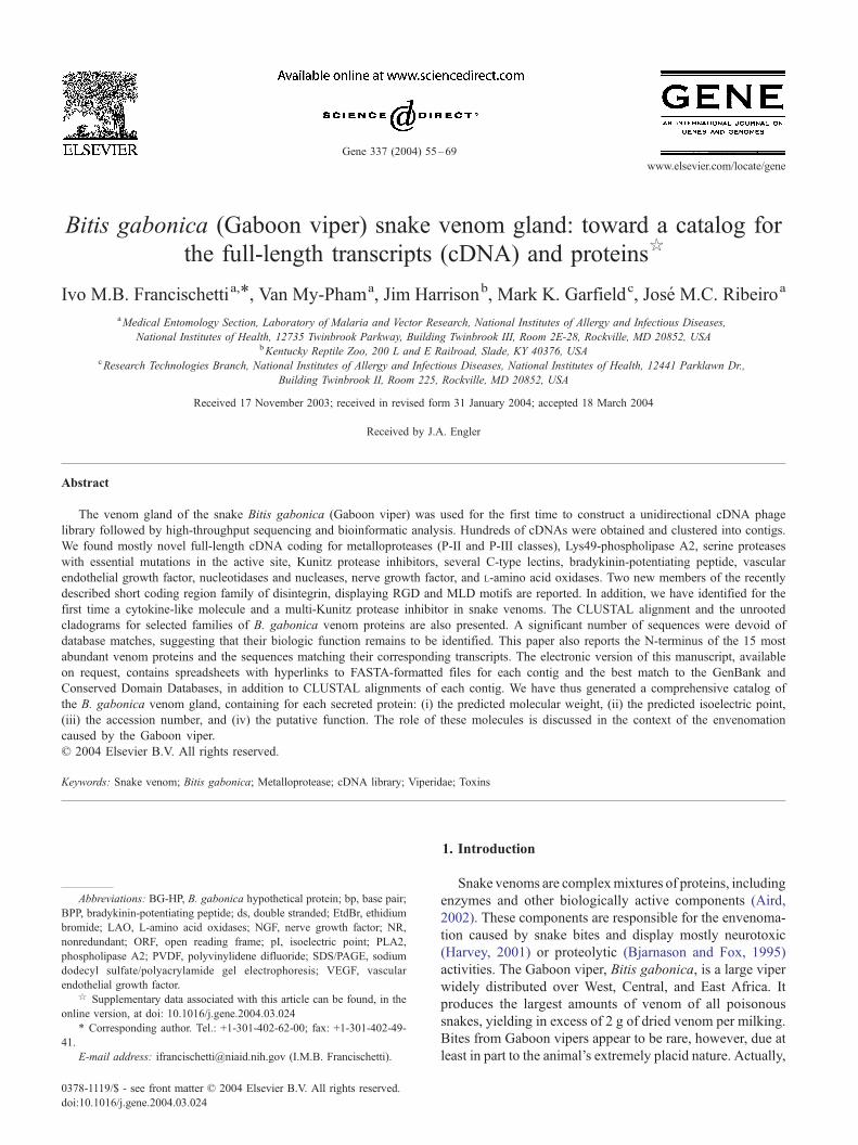
3.1. SDS/PAGE of B. gabonica snake venom gland
Fig. 1 shows the pattern of separation of B. gabonica
venom proteins by SDS-PAGE that have been stained by
Coomassie Blue. The gel shows 15 clearly visible stained
bands and many other slightly stained. The protein bands
were numbered from 1 to 15 according to their decreasing
apparent molecular weight, starting with the letter BG that
stands for B. gabonica. To identify these proteins, they were
transferred to PVDF membranes and the bands cut from the
membrane and submitted to Edman degradation. Amino-
Fig. 1. SDS-PAGE of B. gabonica venom gland proteins under denaturing non-reducing conditions. Thirty Ag of venom was applied to a 4–12% NU-PAGE
pre-cast gel, MES buffer. Standard molecular mass is shown on the left. The match found is shown on the right.
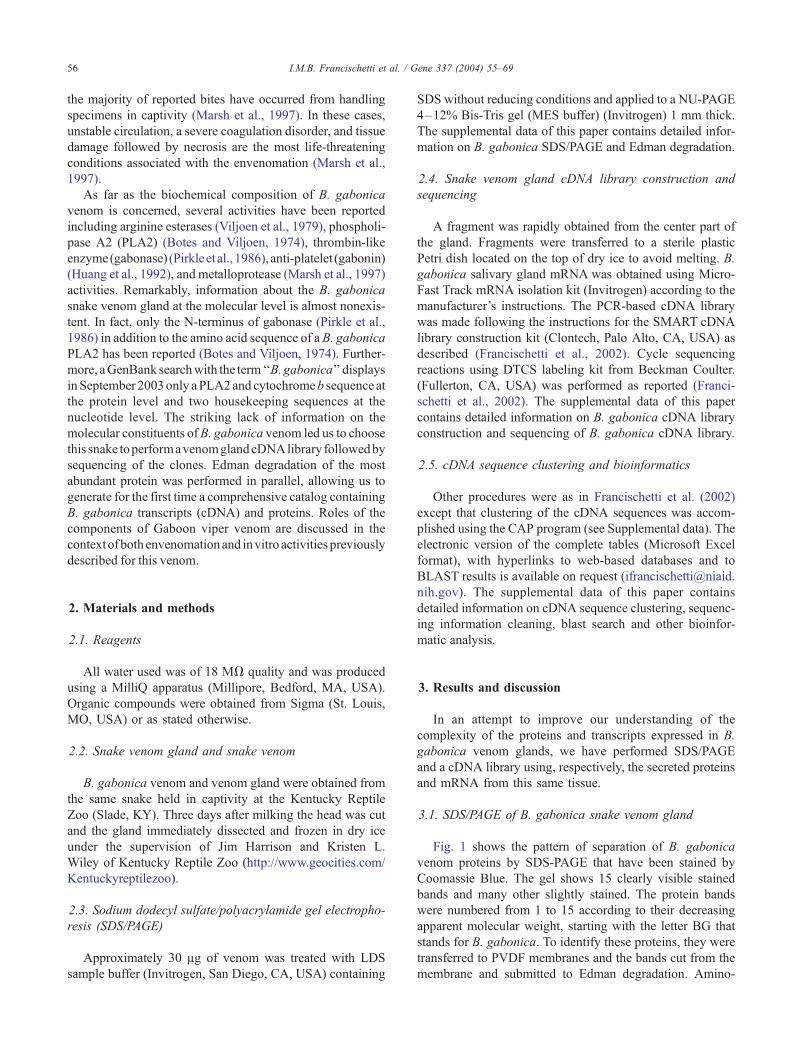
Fig. 2. The composition of B. gabonica cDNA coding for putative secreted
proteins. (A) Number of sequences (%) for a given contig. (B) Number of
contigs (%) for a given venom biologic function (e.g. phospholipase A2).
The total number of sequences in (A) or contigs in (B) includes only the
ones coding for putative secreted proteins. PLA2, phospholipase A2; BPP,
bradykinin-potentiating peptide; CRD lectin, carbohydrate-recognition
domain lectin, carbohydrate recognition domain containing lectin.
I.M.B. Francischetti et al. / Gene 337 (2004) 55–69 57
terminal information was successfully obtained for all bands
BG-2 to BG-15. To find matches to known proteins, the
sequences were blasted against the NR GenBank database
and to each cDNA sequence obtained in the mass-sequenc-
ing project of the B. gabonica venom gland described in this
paper (see Materials and methods).
3.2. cDNA library of the venom gland of B. gabonica
A cDNA library was constructed using the venom
gland of B. gabonica and about 600 of independent clones
randomly 5V sequenced. When a cluster analysis of all
sequences from this library was performed, 300 indepen-
dent contigs were organized. Subsequently, contigs were
blasted against the NR nucleotide database, and the
presence of signal peptides was predicted by submission
of the sequences to the SignalP server. Our analysis shows
that f 75% of all sequences have database hits; f 46%
of all sequences code for protein with a putative signal
peptide, 38% code for proteins with housekeeping func-
tion, and the remaining sequences could not be assigned
as housekeeping or secretory (unknown). It is thus clear
that cDNA for secretory proteins are highly represented in
our library, suggesting that in vivo these molecules are
preferentially expressed over housekeeping and unknown-
function proteins. Also, because the cDNA have been
obtained from a single animal, these variations do not
represent populational diversity, as the maximal number of
alleles would be 2.
Among the individual cDNA sequences containing pu-
tative signal peptides, 90% have hits in the GenBank
database. Fig. 2A shows the relative proportion of the
number of individual cDNA over the total number. These
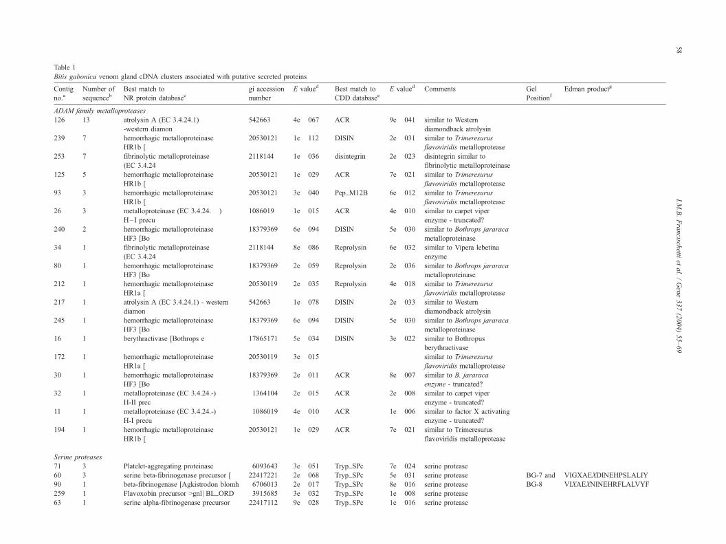
Table 1
Bitis gabonica venom gland cDNA clusters associated with putative secreted proteins
Contig
no.aNumber of
sequencebBest match to
NR protein databasecgi accession
number
E valued Best match to
CDD databaseeE valued Comments Gel
PositionfEdman productg
ADAM family metalloproteases
126 13 atrolysin A (EC 3.4.24.1)
-western diamon
542663 4e� 067 ACR 9e� 041 similar to Western
diamondback atrolysin
239 7 hemorrhagic metalloproteinase
HR1b [
20530121 1e� 112 DISIN 2e� 031 similar to Trimeresurus
flavoviridis metalloprotease
253 7 fibrinolytic metalloproteinase
(EC 3.4.24
2118144 1e� 036 disintegrin 2e� 023 disintegrin similar to
fibrinolytic metalloproteinase
125 5 hemorrhagic metalloproteinase
HR1b [
20530121 1e� 029 ACR 7e� 021 similar to Trimeresurus
flavoviridis metalloprotease
93 3 hemorrhagic metalloproteinase
HR1b [
20530121 3e� 040 Pep_M12B 6e� 012 similar to Trimeresurus
flavoviridis metalloprotease
26 3 metalloproteinase (EC 3.4.24.� )
H–I precu
1086019 1e� 015 ACR 4e� 010 similar to carpet viper
enzyme - truncated?
240 2 hemorrhagic metalloproteinase
HF3 [Bo
18379369 6e� 094 DISIN 5e� 030 similar to Bothrops jararaca
metalloproteinase
34 1 fibrinolytic metalloproteinase
(EC 3.4.24
2118144 8e� 086 Reprolysin 6e� 032 similar to Vipera lebetina
enzyme
80 1 hemorrhagic metalloproteinase
HF3 [Bo
18379369 2e� 059 Reprolysin 2e� 036 similar to Bothrops jararaca
metalloproteinase
212 1 hemorrhagic metalloproteinase
HR1a [
20530119 2e� 035 Reprolysin 4e� 018 similar to Trimeresurus
flavoviridis metalloprotease
217 1 atrolysin A (EC 3.4.24.1) - western
diamon
542663 1e� 078 DISIN 2e� 033 similar to Western
diamondback atrolysin
245 1 hemorrhagic metalloproteinase
HF3 [Bo
18379369 6e� 094 DISIN 5e� 030 similar to Bothrops jararaca
metalloproteinase
16 1 berythractivase [Bothrops e 17865171 5e� 034 DISIN 3e� 022 similar to Bothropus
berythractivase
172 1 hemorrhagic metalloproteinase
HR1a [
20530119 3e� 015 similar to Trimeresurus
flavoviridis metalloprotease
30 1 hemorrhagic metalloproteinase
HF3 [Bo
18379369 2e� 011 ACR 8e� 007 similar to B. jararaca
enzyme - truncated?
32 1 metalloproteinase (EC 3.4.24.-)
H-II prec
1364104 2e� 015 ACR 2e� 008 similar to carpet viper
enzyme - truncated?
11 1 metalloproteinase (EC 3.4.24.-)
H-I precu
1086019 4e� 010 ACR 1e� 006 similar to factor X activating
enzyme - truncated?
194 1 hemorrhagic metalloproteinase
HR1b [
20530121 1e� 029 ACR 7e� 021 similar to Trimeresurus
flavoviridis metalloprotease
Serine proteases
71 3 Platelet-aggregating proteinase 6093643 3e� 051 Tryp_SPc 7e� 024 serine protease
60 3 serine beta-fibrinogenase precursor [ 22417221 2e� 068 Tryp_SPc 5e� 031 serine protease BG-7 and VIGXAEXDINEHPSLALIY
90 1 beta-fibrinogenase [Agkistrodon blomh 6706013 2e� 017 Tryp_SPc 8e� 016 serine protease BG-8 VIXAEXNINEHRFLALVYF
259 1 Flavoxobin precursor >gnl | BL_ORD 3915685 3e� 032 Tryp_SPc 1e� 008 serine protease
63 1 serine alpha-fibrinogenase precursor 22417112 9e� 028 Tryp_SPc 1e� 016 serine protease
I.M.B.Francisch
ettiet
al./Gene337(2004)55–69
58
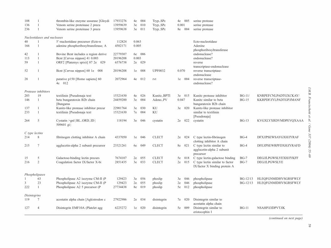
108 1 thrombin-like enzyme ussurase [Gloydi 17933276 4e� 004 Tryp_SPc 4e� 005 serine protease
136 1 Venom serine proteinase 2 precu 13959639 3e� 010 Tryp_SPc 0.001 serine protease
236 1 Venom serine proteinase 3 precu 13959638 3e� 011 Tryp_SPc 8e� 004 serine protease
Nucleotidases and nucleases
40 1 5’-nucleotidase precursor (Ecto-n 112824 0.063 Ecto-nucleotidase
166 1 adenine phosphoribosyltransferase; A 4502171 0.005 Adenine
phosphoribosyltransferase
42 1 Bovine Bcnt includes a region derive 22779307 6e� 006 endonuclease?
113 1 Bcnt [Cervus nippon] 41 0.003 20196208 0.003 endonuclease?
59 1 ORF2 [Platemys spixii] 87 2e� 029 6576738 2e� 029 reverse
transcriptase-endonuclease
52 1 Bcnt [Cervus nippon] 60 1e� 008 20196208 1e� 008 UPF0032 0.070 reverse transcriptase-
endonuclease
28 1 putative p150 [Homo sapiens] 60
4e� 012
2072964 4e� 012 rvt 1e� 004 reverse transcriptase-
endonuclease?
Protease inhibitors
203 19 textilinin [Pseudonaja text 15321630 4e� 026 Kunitz_BPTI 3e� 015 Kunitz protease inhibitor BG-11/ KNRPEFCNLPADTGXCKAY/
146 1 beta bungaratoxin B2b chain
[Bungarus
24459200 3e� 004 Adeno_PV 0.047 Kunitz similar to beta
bungaratoxin B2b chain
BG-15 KKRPDFXYLPADTGPXMANF
137 1 Kunitz-like protease inhibitor precur 22901764 3e� 030 KU 3e� 020 Kunitz-like protease inhibitor
233 1 textilinin [Pseudonaja text 15321630 7e� 004 KU similar to textilinin
[Pseudonaja]
264 5 Cystatin >gnl | BL_ORD_ID |
509601 gi |
118194 3e� 046 cystatin 2e� 022 cystatin BG-13 KVGXLYXRDVMDPEVQXAAA
C type lectins
214 8 fibrinogen clotting inhibitor A chain 4337050 1e� 046 CLECT 2e� 024 C type lectin-fibrinogen
clotting inhibitor A chain
BG-4 DFXXPSEWSAYGXHXYRAF
215 7 agglucetin-alpha 2 subunit precursor 23321261 6e� 049 CLECT 8e� 021 C type lectin similar to
agglucetin-alpha 2 subunit
precursor
BG-4 DFEXPSEWRPFDXHXYRAFD
15 5 Galactose-binding lectin precurs 7674107 2e� 055 CLECT 5e� 018 C type lectin-galactose binding BG-7 DEGXLPGWSLYEXHXYKFF
216 2 Coagulation factor IX/factor X-bi 2851435 3e� 033 CLECT 2e� 015 C type lectin similar to factor
IX/factor X binding protein A
BG-7 DEGXLPGWSLYE
Phospholipases
1 63 Phospholipase A2 isozyme CM-II (P 129423 3e� 056 phoslip 3e� 046 phospholipase BG-12/13 HLEQFGNMIDHVSGRSFWLY
3 23 Phospholipase A2 isozyme CM-II (P 129423 2e� 055 phoslip 2e� 046 phospholipase BG-12/13 HLEQFGNMIDHVSGRSFWLY
222 1 Phospholipase A2 5 precursor (P 27734438 8e� 019 phoslip 5e� 012 phospholipase
Disintegrins
119 7 acostatin alpha chain [Agkistrodon c 27922986 2e� 034 disintegrin 7e� 020 Disintegrin similar to
acostatin alpha chain
127 4 Disintegrin EMF10A (Platelet agg 6225272 1e� 020 disintegrin 5e� 009 Disintegrin similar to
eristocophin I
BG-11 NSAHPXXDPVTXK
(continued on next page)
I.M.B.Francisch
ettiet
al./Gene337(2004)55–69
59
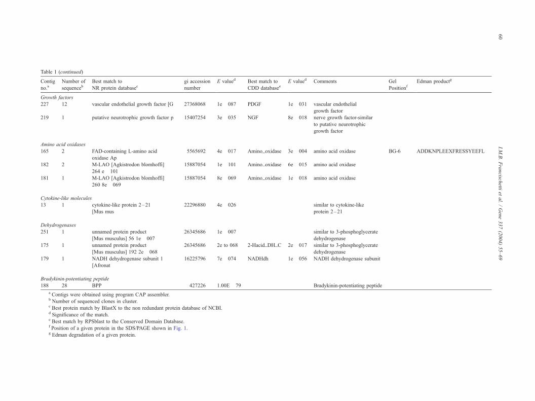
Table 1 (continued)
Contig
no.aNumber of
sequencebBest match to
NR protein databasecgi accession
number
E valued Best match to
CDD databaseeE valued Comments Gel
PositionfEdman productg
Growth factors
227 12 vascular endothelial growth factor [G 27368068 1e� 087 PDGF 1e� 031 vascular endothelial
growth factor
219 1 putative neurotrophic growth factor p 15407254 3e� 035 NGF 8e� 018 nerve growth factor-similar
to putative neurotrophic
growth factor
Amino acid oxidases
165 2 FAD-containing L-amino acid
oxidase Ap
5565692 4e� 017 Amino_oxidase 3e� 004 amino acid oxidase BG-6 ADDKNPLEEXFRESSYEEFL
182 2 M-LAO [Agkistrodon blomhoffi]
264 e� 101
15887054 1e� 101 Amino_oxidase 6e� 015 amino acid oxidase
181 1 M-LAO [Agkistrodon blomhoffi]
260 8e� 069
15887054 8e� 069 Amino_oxidase 1e� 018 amino acid oxidase
Cytokine-like molecules
13 1 cytokine-like protein 2–21
[Mus mus
22296880 4e� 026 similar to cytokine-like
protein 2–21
Dehydrogenases
251 1 unnamed protein product
[Mus musculus] 56 1e� 007
26345686 1e� 007 similar to 3-phosphoglycerate
dehydrogenase
175 1 unnamed protein product
[Mus musculus] 192 2e� 068
26345686 2e to 068 2-Hacid_DH_C 2e� 017 similar to 3-phosphoglycerate
dehydrogenase
179 1 NADH dehydrogenase subunit 1
[Afronat
16225796 7e� 074 NADHdh 1e� 056 NADH dehydrogenase subunit
Bradykinin-potentiating peptide
188 28 BPP 427226 1.00E� 79 Bradykinin-potentiating peptide
a Contigs were obtained using program CAP assembler.b Number of sequenced clones in cluster.c Best protein match by BlastX to the non redundant protein database of NCBI.d Significance of the match.e Best match by RPSblast to the Conserved Domain Database.f Position of a given protein in the SDS/PAGE shown in Fig. 1.g Edman degradation of a given protein.
I.M.B.Francisch
ettiet
al./Gene337(2004)55–69
60
I.M.B. Francischetti et al. / Gene 337 (2004) 55–69 61
sequences were organized into 60 contigs (70%). Fig. 2B
shows the relative proportion of the number of contigs for
each venom toxin family. Based on the distribution de-
scribed above, cDNAs coding for PLA2 are the most
abundant but organized in only three contigs. This indicates
that PLA2 in this venom are rather similar and highly
expressed. On the other hand, the metalloproteases are
organized in 18 different contigs, suggesting that these
enzymes may have evolved to perform other functions.
Finally, among the 30 sequences (10% of all sequences)
that do not have matches to any database, 25 contigs (30%)
were organized.
Among the housekeeping cDNAs, we have found
sequences involved in transcription and translation (ribo-
somal proteins, cAMP-dependent transcription factors, elon-
gation factors), metabolism (ATP synthase, amine oxidase,
glutathione S-transferase, guanine nucleotide-binding pro-
tein, cytochrome c oxidase, NADH-ubiquinone oxidoreduc-
tase chain I), processing (versican core protein precursor),
cell regulation (lithostatine), structural functions (microtu-
bule binding protein), storage (ferritin heavy chain), and
retrotransposable (L-1) elements. The complete list of the
sequences coding for proteins with secretory, housekeeping,
or undetermined function, with or without database hits, can
be obtained on request ([email protected]).
3.3. cDNA coding for putative secretory proteins
Table 1 describes the contigs we have found coding for
putative secreted proteins and, when available, their
corresponding N-terminus obtained by Edman degradation.
Matches to the NR, snake DNA, and Conserved Domains
Database in addition to accession numbers are also reported.
A detailed discussion on the sequences assigned by each
cluster and its participation in envenomation by B. gabonica
is presented below.
3.3.1. Metalloproteases
The metalloproteases make up the most complex group
of proteins, being composed of 18 contigs or 30% of all
assembled cDNAs. These findings are consistent with the
functional characterization of two hemorrhagic proteins
(HTa and HTb) in B. gabonica venom that were previously
shown to degrade collagen and affect endothelial cell
morphology (Marsh et al., 1997). Metalloproteases, the
primary proteins responsible for snake venom-induced
hemorrhage, belong to the reprolysin family of venom
metalloproteases (Bjarnason and Fox, 1995). These
enzymes are capable of hydrolyzing various components
of the extracellular matrix and have also been reported to
affect endothelial cells leading to apoptosis. These enzymes
are organized into four classes PI-PIV, according to size and
domain composition (Bjarnason and Fox, 1995; Jia et al.,
1997).
According to our cDNA library, we have found a number
of contigs containing partial sequences with homology to
the reprolysin, disintegrin or cysteine-rich domains of difer-
ent venom metalloprotease. However, no matches to the C-
type lectin domains of metalloproteases were found (Class
P-IV). It appears that B. gabonica venom contains the P-II
and P-III classes of metalloprotease although the presence of
the P-I and P-IV classes cannot be excluded. Contig 34 has
the longest cDNAwe have found in our library coding for a
metalloprotease and accordingly, it was extended with
appropriated primers in an attempt to identify its functional
domains. Although the pre- and pro-domains are not avail-
able, the regions coding for the metalloprotease and dis-
integrin domains were found, indicating that this enzyme
belongs to the P-II class and named herein B. gabonica
metalloprotease-4 (AY44228). The metalloprotease domain
is typical and contains the zinc-binding motif, but is unusual
in the sense that 5 instead of 6 cysteines are present. Since
the nucleotide sequence coding for this regions was repro-
ducible and unambiguous, and found in a reliable region of
the chromatogram, it is concluded that this is a true
substitution. Actually, it has been reported that the number
of cysteine in the metalloprotease domain of these enzymes
may differ (Kini et al., 2002). Of interest, atrolysin A, a
class III enzyme from Crotalus atrox also has an additional
cysteine residue in the proteinase domain. The oxidation
state or potential disulfide bond partner of this residue in
atrolysin is unknown (Bjarnason and Fox, 1995). At present,
however, it cannot be completely excluded that a mutation
may have occurred in this cDNA during the first or second
strand synthesis; or unlikely, the mRNA used to generate the
cDNA presents a mutation. As far as the disintegrin domain
is concerned a typical RGD sequence was found, and the
cysteine pattern was similar but again, not identical to the
disintegrin domain of most metalloproteases. In fact, the B.
gabonica metalloprotease-4 (AY442287) does not contain
8 aminoacids in the N-terminus including a cysteine that is
conserved in most P-II enzymes. Likewise, these amino
acids are missing in a metalloprotease from Macrovipera
lebetina (gi 2118144) (Fig. 3) suggesting that the amino acid
changes we have observed are consistent. Of note, the N-
terminus NSAHPCCDPVTXK (BG-12) obtained by Edman
degradation of B. gabonica venom proteins (Fig. 1) is
identical to the putative N-terminus coded by the
corresponding cDNA in contig 34. This finding strongly
suggests that this P-II metalloprotease when processed may
generate disintegrin peptides. We could not identify the N-
terminus of the metalloprotease domain in our proteome
study (Fig. 1), suggesting that these proteins may have their
N-terminus blocked as previously reported (Fox et al.,
2002). The CLUSTAL alignment of B. gabonica P-II metal-
loprotease (B. gabonica metalloprotease-4; AY442287) with
other P-II class enzymes is shown in Fig. 3A. The unrooted
cladogram is presented in Fig. 3B and shows that metal-
loproteases from B. gabonica and M. lebetina venoms are
the most closely related enzymes.
Table 1 shows that a number of other contigs (e.g. contig
16, AY430411 and 30, AY430412) contains the partial-length
Fig. 3. (A) Alignment of B. gabonica P-II metalloprotease. Proteins were deduced from a B. gabonica venom gland cDNA library. Asterisks, colons, and stops
below the sequences indicate identity, high conservation, and conservation of the amino acids, respectively. (B) Unrooted cladogram indicating the families of
P-II snake venom metalloprotease. The bar represents the degree of divergence among sequences. BG_metallo (B. gabonica metalloprotease-4; AY442287);
ML-metallo (M. lebetina metalloprotease; gi 2118144); AC_metallo (A. c. contortix metalloprotease; gi 7630286); GH_metallo (G. halys metalloprotease;
4106005); TF_metallo (T. flavoviridis metalloprotease; gi 14595995); PM_metallo (P. mucrosquamatus metalloprotease; gi 995748); CA_metallo (C. atrox
metalloprotease; gi 462320); and BJ_metallo (B. jararaca metalloprotease; gi 13194760). The conserved zinc-binding domain is boxed.
I.M.B. Francischetti et al. / Gene 337 (2004) 55–6962
I.M.B. Francischetti et al. / Gene 337 (2004) 55–69 63
sequences homologous to P-III metalloproteases from other
venoms (Jia et al., 1997). In addition, contig 172 (AY430412)
contains the full-coding region for the disintegrin-like and
cysteine- rich domains of a typical P-III venom metallopro-
teases. In fact, the protein coded by this cDNA has a SECD
motif in the disintegrin-like domain in addition to a conserved
pattern of cysteines commonly found in the cysteine-rich
region. This cDNA codes for a protein that can be aligned (not
shown) with the disintegrin/cysteine-rich domains of other P-
III metalloproteases including berythractivase, a prothrombin
activator from Bothrops erythromelas and other related
molecules (Silva et al., 2003). These enzymesmay participate
together with other venom components in the pathogenesis of
B. gabonica envenomation (Marsh et al., 1997).
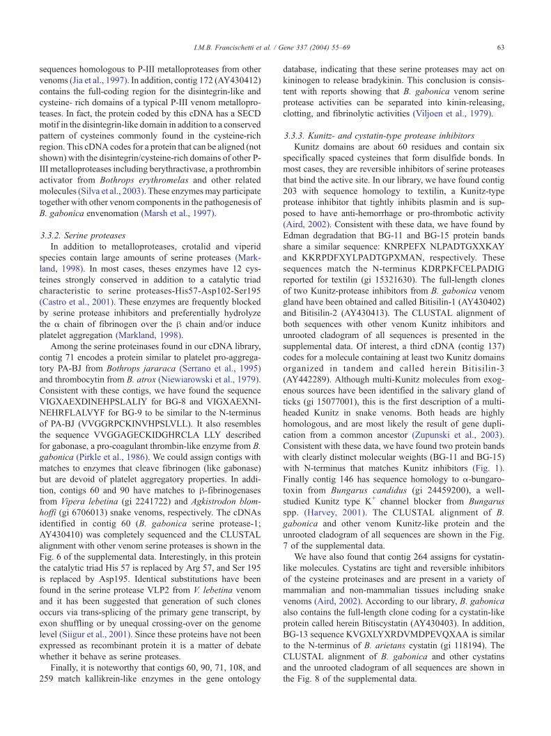
3.3.2. Serine proteases
In addition to metalloproteases, crotalid and viperid
species contain large amounts of serine proteases (Mark-
land, 1998). In most cases, theses enzymes have 12 cys-
teines strongly conserved in addition to a catalytic triad
characteristic to serine proteases-His57-Asp102-Ser195
(Castro et al., 2001). These enzymes are frequently blocked
by serine protease inhibitors and preferentially hydrolyze
the a chain of fibrinogen over the h chain and/or induce
platelet aggregation (Markland, 1998).
Among the serine proteinases found in our cDNA library,
contig 71 encodes a protein similar to platelet pro-aggrega-
tory PA-BJ from Bothrops jararaca (Serrano et al., 1995)
and thrombocytin from B. atrox (Niewiarowski et al., 1979).
Consistent with these contigs, we have found the sequence
VIGXAEXDINEHPSLALIY for BG-8 and VIGXAEXNI-
NEHRFLALVYF for BG-9 to be similar to the N-terminus
of PA-BJ (VVGGRPCKINVHPSLVLL). It also resembles
the sequence VVGGAGECKIDGHRCLA LLY described
for gabonase, a pro-coagulant thrombin-like enzyme from B.
gabonica (Pirkle et al., 1986). We could assign contigs with
matches to enzymes that cleave fibrinogen (like gabonase)
but are devoid of platelet aggregatory properties. In addi-
tion, contigs 60 and 90 have matches to h-fibrinogenasesfrom Vipera lebetina (gi 2241722) and Agkistrodon blom-
hoffi (gi 6706013) snake venoms, respectively. The cDNAs
identified in contig 60 (B. gabonica serine protease-1;
AY430410) was completely sequenced and the CLUSTAL
alignment with other venom serine proteases is shown in the
Fig. 6 of the supplemental data. Interestingly, in this protein
the catalytic triad His 57 is replaced by Arg 57, and Ser 195
is replaced by Asp195. Identical substitutions have been
found in the serine protease VLP2 from V. lebetina venom
and it has been suggested that generation of such clones
occurs via trans-splicing of the primary gene transcript, by
exon shuffling or by unequal crossing-over on the genome
level (Siigur et al., 2001). Since these proteins have not been
expressed as recombinant protein it is a matter of debate
whether it behave as serine proteases.
Finally, it is noteworthy that contigs 60, 90, 71, 108, and
259 match kallikrein-like enzymes in the gene ontology
database, indicating that these serine proteases may act on
kininogen to release bradykinin. This conclusion is consis-
tent with reports showing that B. gabonica venom serine
protease activities can be separated into kinin-releasing,
clotting, and fibrinolytic activities (Viljoen et al., 1979).
3.3.3. Kunitz- and cystatin-type protease inhibitors
Kunitz domains are about 60 residues and contain six
specifically spaced cysteines that form disulfide bonds. In
most cases, they are reversible inhibitors of serine proteases
that bind the active site. In our library, we have found contig
203 with sequence homology to textilin, a Kunitz-type
protease inhibitor that tightly inhibits plasmin and is sup-
posed to have anti-hemorrhage or pro-thrombotic activity
(Aird, 2002). Consistent with these data, we have found by
Edman degradation that BG-11 and BG-15 protein bands
share a similar sequence: KNRPEFX NLPADTGXXKAY
and KKRPDFXYLPADTGPXMAN, respectively. These
sequences match the N-terminus KDRPKFCELPADIG
reported for textilin (gi 15321630). The full-length clones
of two Kunitz-protease inhibitors from B. gabonica venom
gland have been obtained and called Bitisilin-1 (AY430402)
and Bitisilin-2 (AY430413). The CLUSTAL alignment of
both sequences with other venom Kunitz inhibitors and
unrooted cladogram of all sequences is presented in the
supplemental data. Of interest, a third cDNA (contig 137)
codes for a molecule containing at least two Kunitz domains
organized in tandem and called herein Bitisilin-3
(AY442289). Although multi-Kunitz molecules from exog-
enous sources have been identified in the salivary gland of
ticks (gi 15077001), this is the first description of a multi-
headed Kunitz in snake venoms. Both heads are highly
homologous, and are most likely the result of gene dupli-
cation from a common ancestor (Zupunski et al., 2003).
Consistent with these data, we have found two protein bands
with clearly distinct molecular weights (BG-11 and BG-15)
with N-terminus that matches Kunitz inhibitors (Fig. 1).
Finally contig 146 has sequence homology to a-bungaro-
toxin from Bungarus candidus (gi 24459200), a well-
studied Kunitz type K+ channel blocker from Bungarus
spp. (Harvey, 2001). The CLUSTAL alignment of B.
gabonica and other venom Kunitz-like protein and the
unrooted cladogram of all sequences are shown in the Fig.
7 of the supplemental data.
We have also found that contig 264 assigns for cystatin-
like molecules. Cystatins are tight and reversible inhibitors
of the cysteine proteinases and are present in a variety of
mammalian and non-mammalian tissues including snake
venoms (Aird, 2002). According to our library, B. gabonica
also contains the full-length clone coding for a cystatin-like
protein called herein Bitiscystatin (AY430403). In addition,
BG-13 sequence KVGXLYXRDVMDPEVQXAA is similar
to the N-terminus of B. arietans cystatin (gi 118194). The
CLUSTAL alignment of B. gabonica and other cystatins
and the unrooted cladogram of all sequences are shown in
the Fig. 8 of the supplemental data.
l. / Gene 337 (2004) 55–69
3.3.4. C-type lectins
C-type lectins are molecules containing a carbohydrate-
recognition domain (CRD). Most C-type lectins are Ca2 +
dependent; however, many of them have lost their sugar-
binding properties and have evolved to interact with platelet
receptors and/or blood coagulation factors (Markland,
1998). Notably, snake venoms are a rich source of C-type
lectins, and not surprisingly our library also contains a large
amount of cDNA coding for this family of proteins. Among
the cDNA we have sequenced, contig 218 assigns for a
fibrinogen-clotting inhibitor from Gloydius halys brevicau-
dus (gi 4337050) and contig 216 assigns for Factor IX/X-
Binding protein from Trimeresurus flavoviridis. Also, contig
215 assigns for a GPIb agonist from Agkistrodon acutus and
may affect platelet function either by direct agglutination of
platelets, through binding to von Willebrandt factor (Matsui
et al., 2002). Finally, contig 15 assigns for a galactose-
binding lectin from the venom of Trimeresurus stejnegeri
(gi 7674107). Consistent with these results described above,
we have found for BG-4 the sequence DFEXPSEW-
SAYGXHXYRAF in addition to BG-5 (DQGXLP-
DWSAYE QHXY), BG-7 (DEGXLPGWSLYE), and BG-
2 (DFGXLSDWSXYEQH) that resembles the N-terminus
of a C-type lectin from B. arietans DFQCPSEWSAYGQH-
CYR (Harrison et al., 2003). BG-3 (DFGA) and BG-10
(DQGALPDTSYHQHHYYP) are also similar to B. arie-
tans C-type lectin in addition to the DQDCLPDWSS-
HERHCY N-terminus of Echis pyramidum leakeyi C-
type lectin (gi 33243102). The full-length clones of three
B. gabonica C-type lectin have been obtained and named
B. gabonica C-type lectin-1 (AY439477), B. gabonica C-
type lectin-2 (AY429478), and B. gabonica C-type lectin-3
(AY429479). The CLUSTAL alignment of B. gabonica
and other venom C-type lectins and the unrooted clado-
gram of all sequences are shown in the Fig. 9 of the
supplemental data.
3.3.5. PLA2
Snake and other venoms are rich sources of PLA2
(E.C.3.1.1.4), a family of enzymes known to have edemato-
genic, antiplatelet, anticoagulant, mast cell degranulating, or
neurotoxic properties (Bon et al., 1994). On the basis of
primary structure and disulfide bond pairings, snake venom
PLA2s were classified as type I (Elapidae) or class II PLA2s
(Viperidae/Crotalidae). The catalytic site of class II PLAs2contains a highly conserved aspartic acid or lysine at
position 49 (Ownby et al., 1999).
In our library, contigs 1 and 3 assign to PLA2, similar to
one described in B. nasicornius venom (gi 67204), whereas
contig 222 matches a PLA2 described in E. pyramidum
leakeyi (gi 27734438). The cDNA sequenced in this library
code for Lys49-PLA2; no Asp49-PLA2 has been sequenced.
The presence of a PLA2 protein in this venom was confirmed
by the BG-14 sequence HLEQFGNMIDHVSGRSFWLY
that is similar to the N-terminus DLTQFGNMIN previously
reported for B. gabonica PLA2 (Botes and Viljoen, 1974).
I.M.B. Francischetti et a64
The full-length clone of B. gabonica PLA2 has been
obtained and named B. gabonica PLA2-1 (AY430410).
The CLUSTAL alignment of B. gabonica and Lys49-PLA2
and the unrooted cladogram of all sequences are shown in
the Fig. 10 of the supplemental data.
3.3.6. Disintegrins
Disintegrins are cysteine-rich, low-molecular-weight
platelet aggregation inhibitor polypeptides that usually
contain an RGD sequences or other motifs that are
recognized by integrins in different cell types (McLane
et al., 1998). In most cases, venom disintegrin are encoded
with a signal peptide, pre-peptide (pro-domain), metal-
loprotease, and disintegrin region on their common pre-
cursors (P-II class metalloproteases). It is suggested that
the metalloprotease/disintegrin precursor is cleaved by
protease(s), resulting in production of metalloprotease
and disintegrin (Bjarnason and Fox, 1995; McLane et
al., 1998). More recently, a new gene structure of the
disintegrin family was identified in Agkistrodon contortrix
contortix and A. p. piscivourus venoms and it consists of
signal peptide, pre-peptide (pro-domain), a disintegrin
domain and lacking the protease domain (Okuda et al.,
2002).
In our library, contigs 119 and 127 assign, respectively,
for disintegrin similar to acostatin from the venom of A.
contortrix contortrix (Okuda et al., 2002), and eristochophin
I from Eriscocophis macmahonii (gi 6225272). Interesting-
ly, cluster 119 contains sequences that code for two proteins
respectively called herein B. gabonica disintegrin-1 (gabo-
nin-1, AY430904) and B. gabonica disintegrin-2 (gabonin-
2, AY430505). Remarkably, these two protein sequences
were identical except for nine amino acids that occurs
between the cysteine residues that form the putative acidic
hairpin loop where the disintegrin domain is found. One of
these sequences contains a typical RGD sequence known to
bind to h3 integrins (McLane et al., 1998), whereas the
second sequence contains the motif MLDG, known to
interact with integrin a9h1 and to affect neutrophil function
(McLane et al., 1998). Since a typical signal peptide and a
pre-peptide region were found for both gabonin-1 and -2, it
is clear that these proteins together with acustatin and
piscivostatin a chains are new members of the short coding
region family of disintegrins (Okuda et al., 2002). The
CLUSTAL alignment of gabonin-1 and -2 with acostatin
and piscivostatin is shown in Fig. 4A. The schematic
domain structure of this family of protein is shown in Fig.
4B (Okuda et al., 2002).
Consistent with these contigs, Edman degradation of the
protein band BG-12 yields the sequence NSAHPXXDPV
TXK that matches the N-terminus NSANPCCDPITCK of
eristocophin (gi 265034). Since BG-12 N-terminus also
matches the N-terminus found for the disintegrin domain
of B. gabonica metalloprotease-4 (Fig. 3A), it is unclear
whether the protein we have identified as a disintegrin is a
processed form of a B. gabonica venom P-II metallopro-
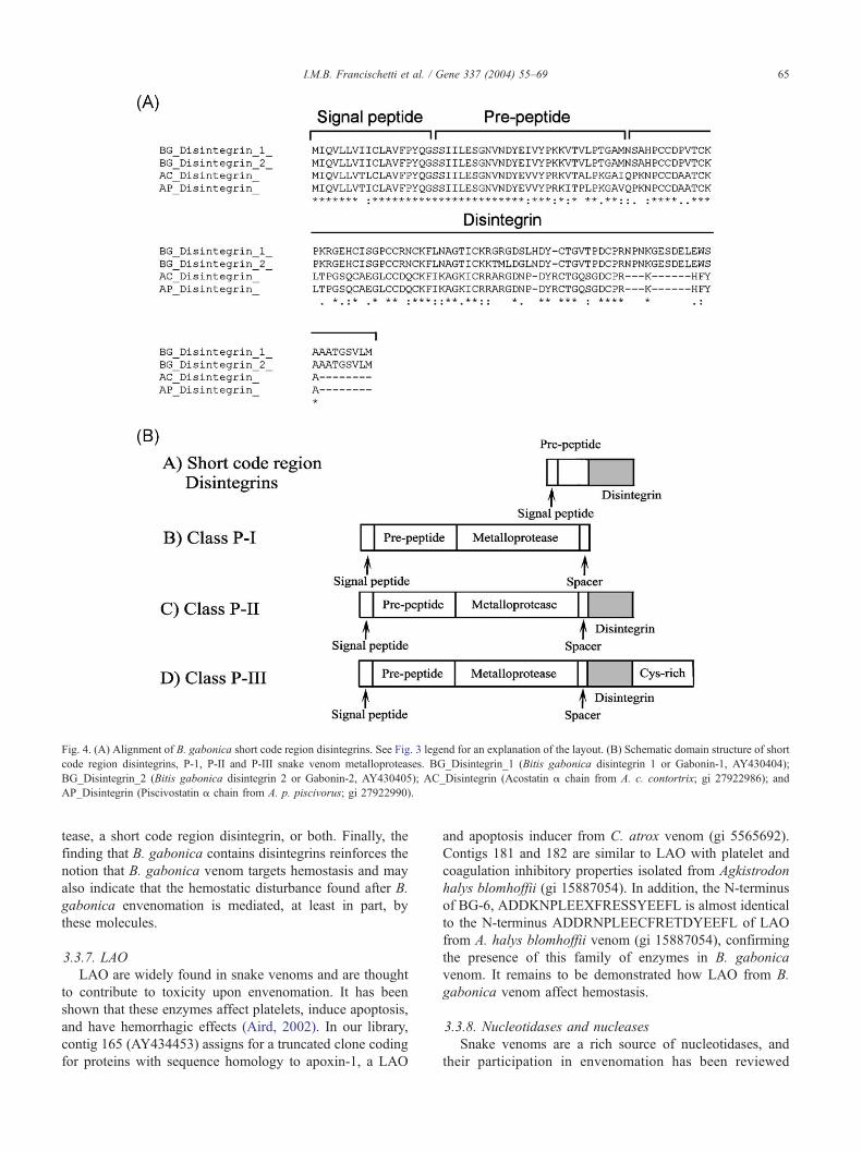
Fig. 4. (A) Alignment of B. gabonica short code region disintegrins. See Fig. 3 legend for an explanation of the layout. (B) Schematic domain structure of short
code region disintegrins, P-1, P-II and P-III snake venom metalloproteases. BG_Disintegrin_1 (Bitis gabonica disintegrin 1 or Gabonin-1, AY430404);
BG_Disintegrin_2 (Bitis gabonica disintegrin 2 or Gabonin-2, AY430405); AC_Disintegrin (Acostatin a chain from A. c. contortrix; gi 27922986); and
AP_Disintegrin (Piscivostatin a chain from A. p. piscivorus; gi 27922990).
I.M.B. Francischetti et al. / Gene 337 (2004) 55–69 65
tease, a short code region disintegrin, or both. Finally, the
finding that B. gabonica contains disintegrins reinforces the
notion that B. gabonica venom targets hemostasis and may
also indicate that the hemostatic disturbance found after B.
gabonica envenomation is mediated, at least in part, by
these molecules.
3.3.7. LAO
LAO are widely found in snake venoms and are thought
to contribute to toxicity upon envenomation. It has been
shown that these enzymes affect platelets, induce apoptosis,
and have hemorrhagic effects (Aird, 2002). In our library,
contig 165 (AY434453) assigns for a truncated clone coding
for proteins with sequence homology to apoxin-1, a LAO
and apoptosis inducer from C. atrox venom (gi 5565692).
Contigs 181 and 182 are similar to LAO with platelet and
coagulation inhibitory properties isolated from Agkistrodon
halys blomhoffii (gi 15887054). In addition, the N-terminus
of BG-6, ADDKNPLEEXFRESSYEEFL is almost identical
to the N-terminus ADDRNPLEECFRETDYEEFL of LAO
from A. halys blomhoffii venom (gi 15887054), confirming
the presence of this family of enzymes in B. gabonica
venom. It remains to be demonstrated how LAO from B.
gabonica venom affect hemostasis.
3.3.8. Nucleotidases and nucleases
Snake venoms are a rich source of nucleotidases, and
their participation in envenomation has been reviewed
I.M.B. Francischetti et al. / Gene 337 (2004) 55–6966
recently (Aird, 2002). In our library, contig 40 has a
truncated clone whose sequence is similar to the sequence
coding for an ectonucleotidase from the electric ray electric
lobe (gi 112824). Nucleotidases inhibit platelet aggregation,
and it appears that B. gabonica may affect platelet function
by removal of ADP. We have also identified cDNA coding
for endonucleases, a family of enzymes ubiquitously found
in snake venoms. Venom endonucleases work together with
venom and endogenous phosphodiesterase degrading
nucleic acids to free nucleotides, which serve as substrate
for 5V nucleotidases, which, in turn, liberate free nucleo-
sides. Adenosine, in particular, is a potent vasodilator and
inhibitor of platelet aggregation (Aird, 2002).
3.3.9. Growth factors
In our library, we have found cluster 227 that match
VEGF from Gallus gallus (gi 27368068). The VEGF are the
most potent vascular permeability factors known and char-
acteristically cause reversible increase in permeability and
have been described in venoms (Aird, 2002). The full-length
clone of B. gabonica VEGF has been obtained and named
B. gabonica VEGF (AY429481). This protein may be
involved in edema induced by B. gabonica bite. The
CLUSTAL alignment of B. gabonica and other venom
VEGF and the unrooted cladogram of all sequences are
shown in the Fig. 11 of the supplemental data. In addition,
cluster 219 (AY430406) in our library is similar to NGF
from B. jararacussu venom (gi 15407254). NGF is ubiqui-
tous in snake venoms and exhibit non-neuronal effects such
as the induction of plasma extravasation and histamine
release from whole blood cells. Although we could not find
the N-terminus of growth factors in any of the bands shown
in Fig. 1, this paper describes for the first time transcripts for
this family of proteins in B. gabonica venom gland.
3.3.10. BPP
We have found an abundant contig 188 containing 28
truncated cDNA (AY434452) whose sequence has matches
to the 3V untranslated region of BPP from A. halys blomhoffi
(gi 427226). BPP were first isolated in the venom of B.
jararaca snake and shown to display intense hypotensive
properties (Aird, 2002). We suggest that this family of
peptides is involved in hypotension associated with B.
gabonica envenomation.
3.3.11. Cytokine-like and unknown proteins
We have sequenced other cDNA (contig 28) whose
sequences are similar to a cytokine-like protein that inhibits
insulin secretion (Zhu et al., 2002). This is the first descrip-
tion of this family of proteins in snake venoms. By immu-
nohistochemistry it was shown that a member of this
cytokine family was expressed prominently in the vascular
endothelium, particularly in capillaries (Zhu et al., 2002).
The function of this cytokine-like protein in snake venom
remains to be determined, but it may be that it somehow
affects vascular biology. The full-length clone of B. gabon-
ica cytokine-like protein has been obtained and named B.
gabonica cytokine-like protein-1 (AY429480). The CLUS-
TAL alignment of B. gabonica and other cytokine-like
proteins and the unrooted cladogram of all sequences are
shown in Fig. 5.
Finally, no matches have been found for some clusters,
and these were assigned as unknowns (not shown, available
on request). In some selected cases, we have named hypo-
thetical proteins (HP) when a sequence without database
hits has an open-reading frame (ORF) containing metionine,
a stop codon and a putative signal peptide (Table 2).
3.4. A catalog toward the full-length cDNA and proteins
from the snake venom gland of B. gabonica
To gather the maximum amount of information about the
putative secreted proteins from the B. gabonica venom
gland, selected sequences presented in Table 1 were re-
sequenced and extended to obtain, when applicable and
possible, their full-length cDNA. The full coding sequences
with database hits were then blasted again to the NR protein
database and SignalP server to confirm, respectively, se-
quence similarity and the presence of a signal peptide. In the
event a signal peptide was predicted to exist, the molecular
weight and the pI of the mature protein were also calculated
and the putative function annotated. Most of the sequences
displayed in Table 2 are full-length clones, with the excep-
tion of the metalloproteases, LAO, and BPP (see below). It
may be that the cDNA coding for these proteins have an SfiI
site that is purposely cleaved during the cDNA library
construction (see Materials and methods). Although our
library is PCR-amplified, it is clear that the base changes
observed in different contigs, including contig 34 or 60 and
others are not artefactual. In fact, similar base changes have
been found for all individual sequences of a given contig;
actually, this diversity can be explained by accelerated
evolution that has been well-documented in snake venom
glands (Deshimaru et al., 1996). It is also known that PCR
based libraries which cDNAs have not been size-fractioned
may be enriched with small cDNAs. However, our libraries
have been constructed using low, medium and high molec-
ular weight cDNAs that have been separated by gel-filtra-
tion (see Material and methods in the Supplemental data).
This separation minimizes the preferential amplification of
small transcripts over larger ones, and the preferential
ligation of small-sized cDNAs over larger ones, in the
TripleX2 vector. Of note, the putative proteins coded by
the most abundant clusters have been identified in the SDS/
PAGE (e.g. PLA2, protease inhibitors, C-type lectins, serine
protease, and disintegrins) with the exception of the metal-
loproteases, which N-terminus are found to be frequently
blocked (Fox et al., 2002), and the BPP that are nor
appropriately separated by 4–12% PAGE due to its low
molecular weight. Accordingly, PCR-based libraries appear
to provide a reasonable qualitative estimate of the transcripts
expressed in a given tissue. Alternatively, construction of a
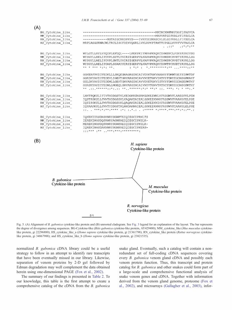
Fig. 5. (A) Alignment of B. gabonica cytokine-like protein and (B) unrooted cladogram. See Fig. 3 legend for an explanation of the layout. The bar represents
the degree of divergence among sequences. BG-Cytokine-like (Bitis gabonica cytokine-like protein, AY429480); MM_cytokine_like (Mus musculus cytokine-
like protein, gi 22296880); HS_cytokine_like_a (Homo sapiens cytokine-like protein, gi 21361798); RN_cytokine_like protein (Rattus norvegicus cytokine-
like protein, gi 34867988); and HS_cytokine_like_b (Homo sapiens cytokine-like protein, gi 23821535).
I.M.B. Francischetti et al. / Gene 337 (2004) 55–69 67
normalized B. gabonica cDNA library could be a useful
strategy to follow in an attempt to identify rare transcripts
that have been eventually missed in our library. Likewise,
separation of venom proteins by 2-D gel followed by
Edman degradation may well complement the data obtained
herein using one-dimensional PAGE (Fox et al., 2002).
The summary of our findings is presented in Table 2. To
our knowledge, this table is the first attempt to create a
comprehensive catalog of the cDNA from the B. gabonica
snake gland. Eventually, such a catalog will contain a non-
redundant set of full-coding cDNA sequences covering
every B. gabonica venom gland cDNA and possibly each
venom protein function. Thus, this transcript and protein
catalog for B. gabonica and other snakes could form part of
a large-scale and comprehensive functional analysis of
snake venom genes and cDNA. Together with information
derived from the venom gland genome, proteome (Fox et
al., 2002), and microarrays (Gallagher et al., 2003), infor-
Table 2
A catalog of Bitis gabonica venom gland cDNAs for secretory proteins
Sequence namea Contigb Clonec GenBankd Ne MWf SPg MWh pli Functionj
Bradykinin potent.peptide 188 partial AY43452 Y NA NA NA NA Hypotension
C-type lectin-1 15 full-length AY439477 Y 18624 23 16088 8.22 Anti-hemostatic
C-type lectin-2 215 full-length AY429478 Y 18156 21 15959 5.17 Anti-hemostatic
C-type lectin-3 214 full-length AY429479 Y 18094 23 15640 5.98 Anti-hemostatic
Cystatin (Bitiscystatin) 264 full-length AY430403 Y 15899 24 13325 7.01 Protease inhibitor
Ctokine-like protein 28 full-length AY429480 Y 26411 29 23138 9.07 Unknown
Disintegrin-1 (Gabonin-1) 119 full-length AY430404 Y 13791 20 11614 7.06 Plate inhibitor
Disintegrin-2 (Gabonin-2) 119 full-length AY430405 Y 13785 20 11608 6.31 Plate inhibitor
VEGFl 222 full-length AY429481 Y 22357 26 19303 7.8 Edema inducer
Hypothetical protein-1 99 full-length AY430407 Y 9740 13 8312 9.55 Unknown
Hypothetical protein-2 103 full-length AY430408 Y 7602 15 5719 7.24 Unknown
Kunitz inhibitor-1 (Bitisilin-1) 203 full-length AY430402 Y 9922 24 7498 8.26 Protease inhibitor
Kunitz inhibitor-2 (Bitisilin-2) 146 full-length AY43013 Y 10005 24 7581 9.3 Protease inhibitor
Kunitz inhibitor-3 (Bitisilin-3) 137 partial AY442289 Y NAk NA NA NA Protease inhibitor
L-amino acid oxidase 147 partial AY434453 Y NA NA NA NA Anti-hemostatic
Metalloprotease-1 16 partial AY430411 Y NA NA NA NA Hemmorhagic
Metalloprotease-2 30 partial AY430412 Y NA NA NA NA Hemmorhagic
Metalloprotease-3 172 partial AY442288 Y NA NA NA NA Hemmorhagic
Metalloprotease-4 34 partial AY442287 Y NA NA 32506 5.76 Hemmorhagic
Nerve Growth Factor 219 partial AY430406 Y NA NA NA NA Edema inducer
Phospholipase A2 1 full-length AY429476 Y 15745 16 13932 4.99 Edema inducer
Serine-protease-1 60 full-length AY430410 Y 16803 24 14104 9.07 Pro-hemostatic?
a Including putative secretory proteins only.b Contig, contig number.c Clone (partial of full-length).d Genbank, NR database accession number.e N, novel cDNA (Y= yes).f MW.; molecular mass before signal peptide removed.g SP-signal peptide.h MW.; molecular mass of the mature protein.i pl, Isoelectric point.j Function, putative function or possible biological activity.k NA, Not available.l VEGF, Vascular Endothelial Growth Factor.
I.M.B. Francischetti et al. / Gene 337 (2004) 55–6968
mation provided by this catalog could be an essential tool to
understand snakes physiology (Perales and Domont, 2002),
the molecular basis of envenomation, as well as to find
potential candidates for serum production (Theakston et al.,
2003) and/or tools to study cell biology and biochemistry
(Menez, 1998).
3.5. B. gabonica venom components and envenomation
Snake venom envenomation employs three well-integrat-
ed strategies including prey immobilization via hypotension,
prey immobilization via paralysis, and prey digestion (Aird,
2002). Although the identification of the toxin clusters does
not allow us to determine quantitatively the contribution of
each protein cluster in the envenomation, it allow us to
speculate about the mechanisms of envenomation by B.
gabonica venom. It is remarkable that proteins such as
metalloproteases, serine proteases, C-type lectins, PLA2,
Kunitz inhibitors, growth factors, and LAO account for
most of our sequences. As described above and reviewed
elsewhere (Aird, 2002), these proteins act on the hemostatic
system and/or affect vascular biology. In this respect, B.
gabonica venom resembles an expressed sequence tag
(EST) approach reported for Bothrops insularis, where a
large number of cDNA code for metalloproteases, BPP, C-
type lectins, serine protease, PLA2, and growth factors
(Junqueira-de-Azevedo and Ho, 2002). We have also found
an abundant cluster whose sequences match the 3/ untrans-
lated region cDNA of A. halys blomhoffi BPP, a family of
peptides also abundant in the B. insularis cDNA library
(Junqueira-de-Azevedo and Ho, 2002) (see Table 2). Be-
cause we also found sequences coding for kallikrein-like
enzymes, it is plausible that these enzymes and BPP are
primarily responsible for the hypotension associated with B.
gabonica and possibly B. insularis envenomation. The
similarity in the cDNA composition between B. gabonica
and B. insularis libraries is also consistent with the symp-
toms resulting from envenomation by Bitis and Bothrops
spp. that is characterized by consumption coagulopathy,
hypotension, and local damage (Aird, 2002).
4. Conclusion
It is worth noting that the description of the B. gabonica
venom gland cDNA database match biologic activities
I.M.B. Francischetti et al. / Gene 337 (2004) 55–69 69
described before for this venom, including the molecules
involved with hypotension, bleeding, digestion, and tissue
damage (Marsh et al., 1997). This indicates that an approach
combining cDNA library construction, massive sequencing,
and bioinformatic analysis, in addition to Edman degrada-
tion of the main proteins, may be useful to study exogenous
secretion from different venom glands, and to the develop-
ment of recombinant antigens for antibody production.
Acknowledgements
We thank Drs. Thomas E. Wellems, Robert W. Gwadz
and Thomas J. Kindt for encoragement and support, and
Brenda Rae Marshall for editorial assistance.
References
Aird, S.D., 2002. Ophidian envenomation and the role of the purines.
Toxicon 40, 335–393.
Bjarnason, J.B., Fox, J.W., 1995. Snake venom metalloendopeptidases:
reprolysins. Methods Enzymol. 248, 345–368.
Bon, C., Choumet, V., Delot, E., Faure, G., Robbe-Vincent, A., Saliou, B.,
1994. Different evolution of phospholipase A2 neurotoxins (beta-neuro-
toxins) from Elapidae and Viperidae snakes. Ann. N.Y. Acad. Sci. 710,
142–148.
Botes, D.P., Viljoen, C.C., 1974. Bitis gabonica venom. The amino acid
sequence of phospholipase A. J. Biol. Chem. 249, 3827–3835.
Castro, H.C., Silva, D.M., Craik, C., Zingali, R.B., 2001. Structural fea-
tures of a snake venom thrombin-like enzyme: thrombin and trypsin on
a single catalytic platform? Biochim. Biophys. Acta 1547, 183–195.
Deshimaru, M., Ogawa, T., Nakashima, K., Nobuhisa, I., Chijiwa, T.,
Shimohigashi, Y., Fukumaki, Y., Niwa, M., Yamashina, I., Hattori, S.,
Ohno, M., 1996. Accelerated evolution of crotalinae snake venom gland
serine proteases. FEBS Lett. 397, 83–88.
Fox, J.W., Shannon, J.D., Stefansson, B., Kamiguti, A.S., Theakston,
R.D.G., Serrano, S.M.T., Camargo, A.C.M., Sherman, N., 2002. Role
of discovery sciences in toxinology: examples in venom proteomics. In:
Menez, A. (Ed.), Perspectives in molecular toxinology. Wiley, West
Sussex, UK, pp. 97–106.
Francischetti, I.M., Valenzuela, J.G., Pham, V.M., Garfield, M.K., Ribeiro,
J.M.C., 2002. Toward a catalog for the transcripts and proteins (sia-
lome) from the salivary gland of the malaria vector Anopheles gambiae.
J. Exp. Biol. 205, 2429–2451.
Gallagher, P.G., Bao, Y., Serrano, M.T., Kamiguti, A.S., Theakston,
R.D.G., Fox, J.W., 2003. Use of microarrays for investigating the sub-
toxic effects of snake venoms: insights into venom-induced apoptosis in
human umbilical vein endothelial cells. Toxicon 41, 429–440.
Harrison, R.A., Oliver, J., Hasson, S.S., Bharati, K., Theakston, R.D.,
2003. Novel sequences encoding venom C-type lectins are conserved
in phylogenetically and geographically distinct Echis and Bitis viper
species. Gene 315, 95–102.
Harvey, A.H., 2001. Twenty years of dendrotoxins. Toxicon 39, 15–26.
Huang, T.F., Peng, H.C., Peng, I.S., Teng, C.M., Ouyang, C., 1992. An
antiplatelet peptide, gabonin, from Bitis gabonica snake venom. Arch.
Biochem. Biophys. 298, 13–20.
Jia, L.G., Wang, X.M., Shannon, J.D., Bjarnason, J.B., Fox, J.W., 1997.
Function of disintegrin-like/cysteine-rich domains of atrolysin A. Inhi-
bition of platelet aggregation by recombinant protein and peptide antag-
onists. J. Biol. Chem. 272, 13094–13102.
Junqueira-de-Azevedo, I.L., Ho, P.L., 2002. A survey of gene expression
and diversity in the venom glands of the pitviper snake Bothrops insu-
laris through the generation of expressed sequence tags (ESTs). Gene
299, 279–291.
Kini, R.M., Joseph, J.S., Rao, V.S., 2002. Prothrombin activator from snake
venoms. In: Menez, A. (Ed.), Perspectives in Molecular Toxinology.
Wiley, West Sussex, UK, pp. 341–355.
Markland, F.S., 1998. Snake venoms and the hemostatic system. Toxicon
36, 1749–1800.
Marsh, N., Gattullo, D., Pagliaro, P., Losano, G., 1997. The Gaboon viper,
Bitis gabonica: hemorrhagic, metabolic, cardiovascular and clinical
effects of the venom. Life Sci. 61, 763–769.
Matsui, T., Hamako, J., Matsushita, T., Nakayama, T., Fujimura, Y., Titani,
K., 2002. Binding site on human von Willebrand factor of bitiscetin, a
snake venom-derived platelet aggregation inducer. Biochemistry 41,
7939–7946.
McLane, M.A., Marcinkiewicz, C., Vijay-Kumar, S., Wierzbicka-Patynow-
ski, I., Niewiarowski, S., 1998. Viper venom disintegrins and related
molecules. Proc. Soc. Exp. Biol. Med. 219, 109–119.
Menez, A., 1998. Functional architectures of animal toxins: a clue to drug
design? Toxicon 36, 1557–1572.
Niewiarowski, S., Kirby, E.P., Brudzynsky, T.M., Stocker, K., 1979.
Thrombocytin, a serine protease from Bothrops atrox venom. 2. Inter-
cation with platelets and plasma clotting factors. Biochemistry 18,
3570–3577.
Okuda, D., Koike, H., Morita, T., 2002. A new gene structure of the
disintegrin family: a subunit of dimeric disintegrin has a short coding
region. Biochemistry 41, 14248–14254.
Ownby, C.L., Selistre de Araujo, H.S., White, S.P., Fletcher, J.E., 1999.
Lysine 49 phospholipase A2 proteins. Toxicon 37, 411–445.
Perales, J., Domont, G.B., 2002. Are inhibitors of metallopoteases, phos-
pholipases A2, and miotoxins members of the innate immune system?
In: Menez, A. (Ed.), Perspectives in Molecular Toxinology. Wiley, West
Sussex, UK, pp. 435–456.
Pirkle, H., Theodor, I., Miyada, D., Simmons, G., 1986. Thrombin-like
enzyme from the venom of Bitis gabonica. Purification, properties,
and coagulant actions. J. Biol. Chem. 261, 8830–8835.
Serrano, S.M., Mentele, R., Sampaio, C.A., Fink, E., 1995. Purification,
characterization, and amino acid sequence of a serine proteinase, PA-
BJ, with platelet-aggregating activity from the venom of Bothrops jar-
araca. Biochemistry 34, 7186–7193.
Siigur, E., Aaspollu, A., Siigur, J., 2001. Sequence diversity of Vipera
lebetina snake venom gland serine protease homologs—results of alter-
native-splicing or genome alteration. Gene 263, 199–203.
Silva, M.B., Schattner, M., Ramos, C.R., Junqueira-de-Azevedo, I.L.,
Guarnieri, M.C., Lazzari, M.A., Sampaio, C.A., Pozner, R.G., Ventura,
J.S., Ho, P.L., Chudzinski-Tavassi, A.M., 2003. A prothrombin activa-
tor from Bothrops erythromelas (jararaca-da-seca) snake venom: char-
acterization and molecular cloning. Biochem. J. 369, 129–139.
Theakston, R.D., Warrell, D.A., Griffiths, E., 2003. Report of a WHO
workshop on the standardization and control of antivenoms. Toxicon
4, 541–557.
Viljoen, C.C., Meehan, C.M., Botes, D.P., 1979. Separation of Bitis gabon-
ica (Gaboon adder) venom arginine esterases into kinin-releasing, clot-
ting and fibrinolytic factors. Toxicon 17, 145–154.
Zupunski, V., Kordis, D., Gubensek, F., 2003. Adaptative evolution in the
snake venom Kunitz/BPTI protein family. FEBS Lett. 547, 131–136.
Zhu, Y., Xu, G., Patel, A., McLaughlin, M.M., Silverman, C., Knecht, K.,
Sweitzer, S., Li, X., McDonnell, P., Mirabile, R., Zimmerman, D.,
Boyce, R., Tierney, L.A., Hu, E., Livi, G.P., Wolf, B., Abdel-Meguid,
S.S., Rose, G.D., Aurora, R., Hensley, P., Briggs, M., Young, P.R.,
2002. Cloning, expression, and initial characterization of a novel cy-
tokine-like gene family. Genomics 80, 144–150.