papel de la medicina nuclear en la mama diseminada mama y med... · papel de la medicina nuclear en...
TRANSCRIPT
Papel de la Medicina Nuclear en la mama diseminada

MU
GA
Densitometría ósea DENSITOMETRIAS
GAMMA OSEA

Cardiotoxicity of nonanthracycline cancer chemotherapy agents:
•Arrhythmias (eg, histone deacetylase inhibitors, nilotinib)•Myocardial necrosis causing a dilated cardiomyopathy and clinical heart failure (eg, sunitinib, alemtuzumab, imatinib)•Vasospasm or vasoocclusion resulting in angina or myocardial infarction (eg, 5-fluorouracil, particularly infusional administration, etoposide)•Pericarditis (eg, cytarabine, bleomycin)
Advantages — The equilibrium RVG has several advantages for LVEF measurement:
•High accuracy and reproducibility•Measurements do not rely on geometric assumptions regarding the shape of the LV•Global and regional LV systolic function can be assessed•Functional information for all cardiac chambers (both atria and ventricles) is readily available•Phase analysis of segmental ventricular contraction conveys information for regional dyssynergy•The technique can be used for accurate volumetric measurements with the use of an appropriate phantom and corrections for soft tissue attenuation •The patient's body habitus does not limit the technique and virtually all patients can be imaged•The noninvasive nature of the test minimizes associated risks•RVG is easy to perform and not time consuming (less than 30 minutes)
Ref. UpToDate, last updated: noviembre 24, 2009


I. PETI. PET--CT CT
II. Terapia metabII. Terapia metabóólicalica

PETPET--CT CT nono FDGFDG
DiagnDiagnóóstico Mx: stico Mx: PET FRENTE ESTUDIOS DE IMAGEN PET FRENTE ESTUDIOS DE IMAGEN
CONVENCIONALCONVENCIONAL
RespuestaRespuesta al tratamiento:al tratamiento:CUAL ES LA LIMITACICUAL ES LA LIMITACIÓÓN DEL PET FDG?N DEL PET FDG?
I. PETI. PET--CT CT

Tx Nx Mx
Informe AETS Noviembre 2005 la PET es esencial en el manejo clínico del paciente oncológico 92% de los pacientes, detectando nuevas lesiones en el 39%, modificando diagnóstico o estadio en un 57%, cambiando el tratamiento propuesto en un 79% (53% con cambio de modalidad), evitando pruebas invasivas o de riesgo en un 76%, terapias innecesarias en un 76% y de utilidad a juicio del clínico solicitante en el 88%.

• Mtx al diagnóstico 1-5% ptes• Riesgo de recurrencia del cáncer de mama
– 7-30% y Mtx 45-90% (Bongers et al, 2004)
• Latencia– 814 ptes N+ – 18% recurrencias en los 10 primeros años– Recurrencias hasta 23.5 años
DiagnDiagnóóstico Mx: stico Mx: PET FRENTE ESTUDIOS DE IMAGEN CONVENCIONALPET FRENTE ESTUDIOS DE IMAGEN CONVENCIONAL


Role of positron-emission tomography scan in the diagnosis and management of breast cancer. Almubarak M y cols. Oncology (Williston Park). 2009 Mar;23(3):255-61.
FDG-PET is a useful tool in staging advancedbreast cancer and assessing the extent of disease involvement when metastasis is
suspected. It might also aid in assessing early response to
therapy.
DiagnDiagnóóstico Mx: stico Mx: PET FRENTE ESTUDIOS DE IMAGEN CONVENCIONALPET FRENTE ESTUDIOS DE IMAGEN CONVENCIONAL
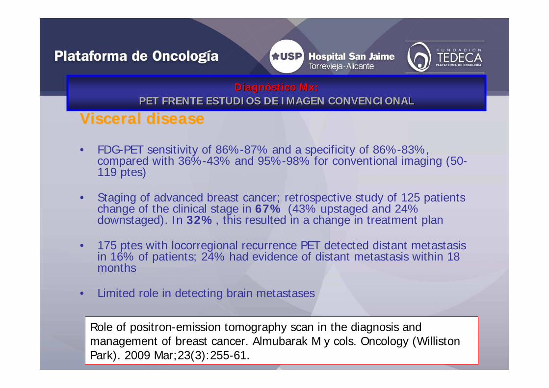
Visceral diseaseVisceral disease
• FDG-PET sensitivity of 86%-87% and a specificity of 86%-83%, compared with 36%-43% and 95%-98% for conventional imaging (50-119 ptes)
• Staging of advanced breast cancer; retrospective study of 125 patients change of the clinical stage in 67% (43% upstaged and 24% downstaged). In 32%, this resulted in a change in treatment plan
• 175 ptes with locorregional recurrence PET detected distant metastasis in 16% of patients; 24% had evidence of distant metastasis within 18 months
• Limited role in detecting brain metastases
Role of positron-emission tomography scan in the diagnosis and management of breast cancer. Almubarak M y cols. Oncology (Williston Park). 2009 Mar;23(3):255-61.
DiagnDiagnóóstico Mx: stico Mx: PET FRENTE ESTUDIOS DE IMAGEN CONVENCIONALPET FRENTE ESTUDIOS DE IMAGEN CONVENCIONAL

Role of positron-emission tomography scan in the diagnosis and management of breast cancer. Almubarak M y cols. Oncology (Williston Park). 2009 Mar;23(3):255-61.
High therapeutic impact

Role of positron-emission tomography scan in the diagnosis and management of breast cancer. Almubarak M y cols. Oncology (Williston Park). 2009 Mar;23(3):255-61.
High therapeutic impact
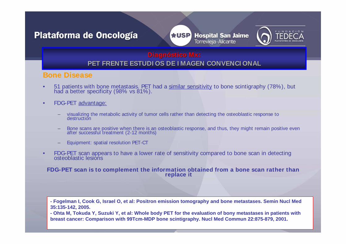
Bone DiseaseBone Disease• 51 patients with bone metastasis. PET had a similar sensitivity to bone scintigraphy (78%), but
had a better specificity (98% vs 81%).
• FDG-PET advantage:
– visualizing the metabolic activity of tumor cells rather than detecting the osteoblastic response to destruction
– Bone scans are positive when there is an osteoblastic response, and thus, they might remain positive even after successful treatment (2-12 months)
– Equipment: spatial resolution PET-CT
• FDG-PET scan appears to have a lower rate of sensitivity compared to bone scan in detecting osteoblastic lesions
FDG-PET scan is to complement the information obtained from a bone scan rather than replace it
- Fogelman I, Cook G, Israel O, et al: Positron emission tomography and bone metastases. Semin Nucl Med 35:135-142, 2005.- Ohta M, Tokuda Y, Suzuki Y, et al: Whole body PET for the evaluation of bony metastases in patients with breast cancer: Comparison with 99Tcm-MDP bone scintigraphy. Nucl Med Commun 22:875-879, 2001.
DiagnDiagnóóstico Mx: stico Mx: PET FRENTE ESTUDIOS DE IMAGEN CONVENCIONALPET FRENTE ESTUDIOS DE IMAGEN CONVENCIONAL
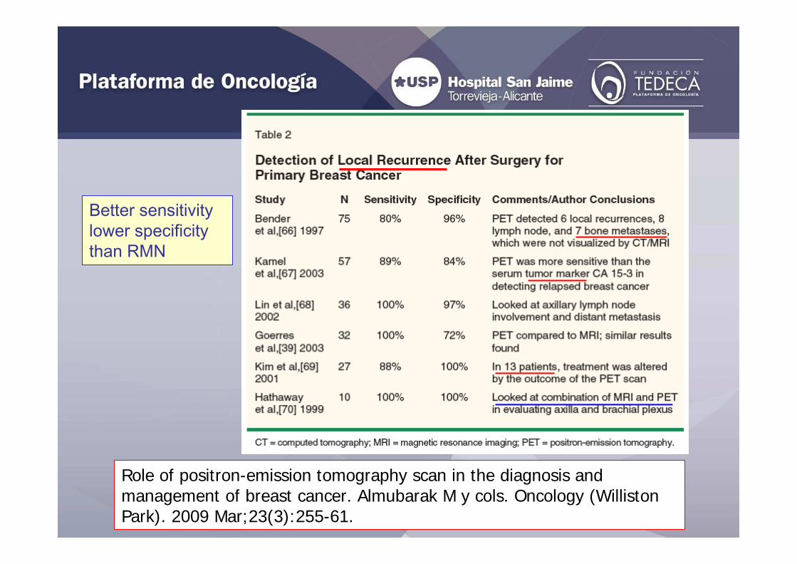
Role of positron-emission tomography scan in the diagnosis and management of breast cancer. Almubarak M y cols. Oncology (Williston Park). 2009 Mar;23(3):255-61.
Better sensitivity lower specificity than RMN

42 studies FDG-PET vs CI (US, CT, MRI, SMM) Pathology +/- 6 months follow upUS and MRI highest pooled specificity (96-93%)MRI and PET highest pooled sensitivity (95%)
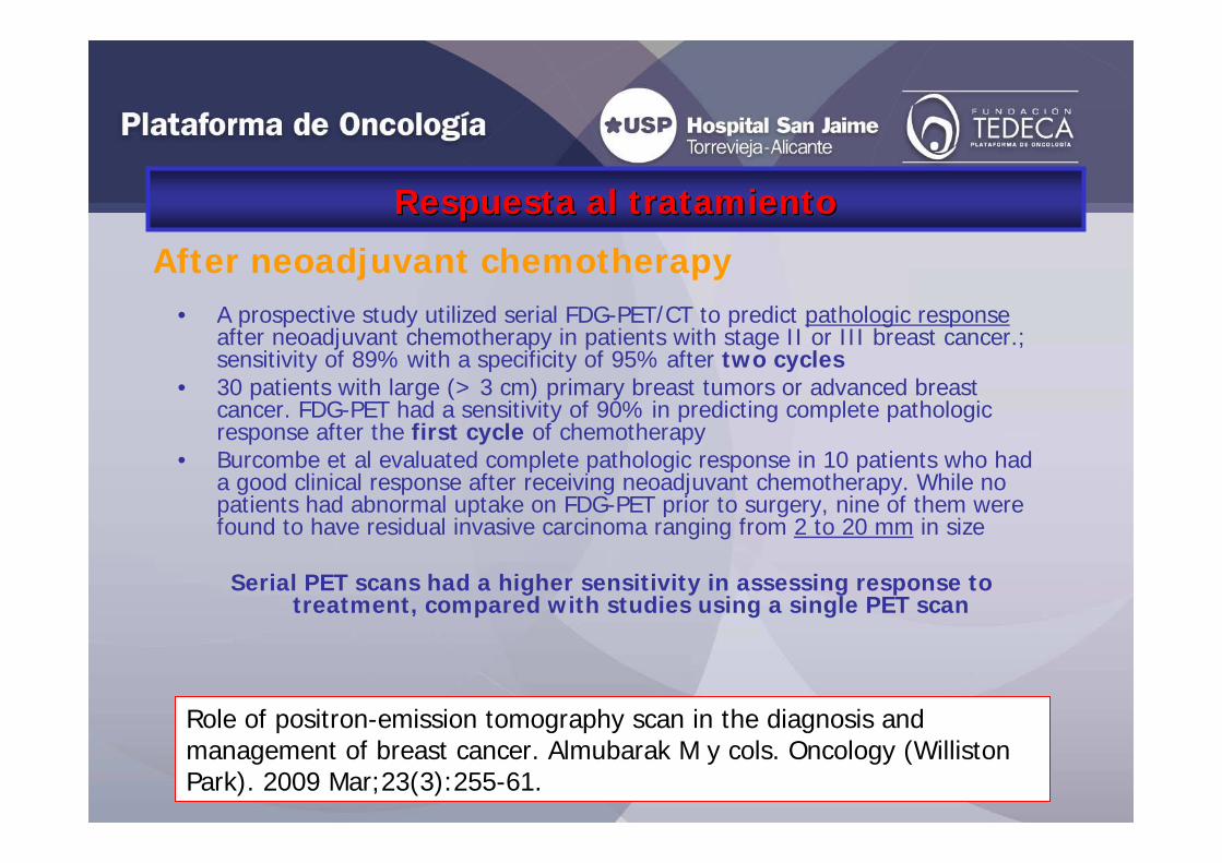
• A prospective study utilized serial FDG-PET/CT to predict pathologic responseafter neoadjuvant chemotherapy in patients with stage II or III breast cancer.; sensitivity of 89% with a specificity of 95% after two cycles
• 30 patients with large (> 3 cm) primary breast tumors or advanced breast cancer. FDG-PET had a sensitivity of 90% in predicting complete pathologic response after the first cycle of chemotherapy
• Burcombe et al evaluated complete pathologic response in 10 patients who had a good clinical response after receiving neoadjuvant chemotherapy. While no patients had abnormal uptake on FDG-PET prior to surgery, nine of them were found to have residual invasive carcinoma ranging from 2 to 20 mm in size
Serial PET scans had a higher sensitivity in assessing response to treatment, compared with studies using a single PET scan
After neoadjuvant chemotherapy
Role of positron-emission tomography scan in the diagnosis and management of breast cancer. Almubarak M y cols. Oncology (Williston Park). 2009 Mar;23(3):255-61.
RespuestaRespuesta al tratamientoal tratamiento

• Mortimer et al noted a greater degree of ER blockade in patients who had a decrease in SUV value on their FDG-PET scan.
• 20 patients with hormone-refractory or hormone receptor–negative metastatic breast cancer. Semiquantitative analysis of FDG-PET metabolic response predicated short-term and overall survival when assessed after three cycles of chemotherapy
Systemic and Hormonal TherapySystemic and Hormonal Therapy
Role of positron-emission tomography scan in the diagnosis and management of breast cancer. Almubarak M y cols. Oncology (Williston Park). 2009 Mar;23(3):255-61.
RespuestaRespuesta al tratamientoal tratamiento

18F-FDG PET correctly predicted the responses after thefirst cycle of chemotherapy and was more accurate than CI after the third cycle ofchemotherapy

•SUV decreased to 72% ±21% after the first cycle and 54% ±16% after the second
•Not responding to 94% ± 19% after the first cycle and 79% ±9% after the second cycle (differences statistically significant)
•Visual analysis predicted the response in all patients as early as after the first cycle of chemotherapy

•Statistically significant correlation between PET scan, circulating tumor cells, and CA 27.29
•CA 27.29 had a high PPV of 90%, low sensitivity of 59% in detecting metastatic disease shown on PET scan.
•Detection of more than 5 cells per 7.5 mL of blood had a positive predictive value of 100% with 100% specificity of having an abnormal PET scan
RespuestaRespuesta al tratamientoal tratamiento
Saad A, Abraham J: Role of tumor markers and circulating tumors cells in the management of breastcancer. Oncology (Williston Park) 22:726-744 (incl discussion), 2008

•A retrospective analyses of 115 MBC patients who started a new line of therapy and who had CTC counts and FDG-PET/CT scans performed at baseline and at 9 to 12 weeks during therapy (midtherapy)
•Detection of five or more CTCs during therapeutic monitoring can accurately predict prognosis in MBC beyond metabolic response
(Midtherapy CTC levels correlated with FDG-PET/CT response in 67%; Midtherapy CTC counts and FDG-PET/CT response predicted overall survival (P < .001 and P <.001, respectively). FDG-PET/CT predicted overall survival (P < .0086) in 31 (91%) of 34 discordant patients who had fewer than five CTCs at midtherap)
•FDG-PET/CT deserves a role in patients who have fewer than five CTCs at midtherapy
RespuestaRespuesta al tratamientoal tratamiento


• 18-F-FES• 18F-FLT• 15O-water• 18F-Fluoride
PETPET--CT CT nono FDGFDG

• Dehdashti F, Mortimer JE, Trinkaus K, et al. PET-based estradiol challenge as a predictive biomarker of response to endocrine therapy in women with estrogen receptor-positive breast cancer. Breast Cancer Res Treat. 2009;113:509–517
18F18F--FESFES
- 18F-FES uptake in breast cancer reflects estrogen receptor status- SUV of ≥2.0 is considered positive for estrogen receptor expression

Synthesis of new carbon-11 labeled cyclofenil derivatives for PET imaging of breast cancer estrogen receptors. Gao M, y colsAppl Radiat Isot. 2008 Apr;66(4):523-9. Epub 2007. Department of Radiology, Indiana University School of Medicine, USA.
•(11)C]methyl-2-{4-[bis(4-hydroxyphenyl)methylene]cyclohexyl}acetate([(11)C]16a)
•[(11)C]methyl-4- [bis(4hydroxyphenyl)methylene]cyclohexanecarboxylate([(11)C]16b)
•[(11)C]methyl-2-{3-[bis(4-hydroxyphenyl)methylene]cyclohexyl}acetate([(11)C]18a)
•[(11)C]methyl-3-[bis(4-hydroxyphenyl)methylene]cyclohexanecarboxylate([(11)C]18b)
OtherOther--FESFES

18F-FLT PET for treatment monitoring in metastatic breast cancer detected changes in breast cancer proliferation 1 wk-2 wks after the initiation of combination chemotherapy
- Kenny L, Coombes RC, Vigushin DM, Al-Nahhas A, Shousha S, Aboagye EO. Imaging early changes in proliferation at 1 week post chemotherapy: a pilot study in breast cancer patients with 39-deoxy-39-[18F]fluorothymidine positron emission tomography. Eur J Nucl Med Mol Imaging. 2007;34:1339–1347
- Pio BS, Park CK, Pietras R, et al. Usefulness of 39-[F-18]fluoro-39-deoxythymidine with positron emission tomography in predicting breast cancer responseto therapy. Mol Imaging Biol. 2006;8:36–42.

18F18F--FluorideFluoride

•El manejo de estos pacientes debe ser multidisciplinar e incluye:
Analgesia, Bifosfonatos
Radioterapia, Cirugía
Quimioterapia, Hormonoterapia
Radioisótopos
•El cáncer óseo metastásico es una complicación severa y común en la
enfermedad avanzada:
Hasta un 70% de los pacientes con c. próstata y mama
Hasta 30% en c. pulmón, vejiga y tiroides
II. Terapia metabII. Terapia metabóólicalica
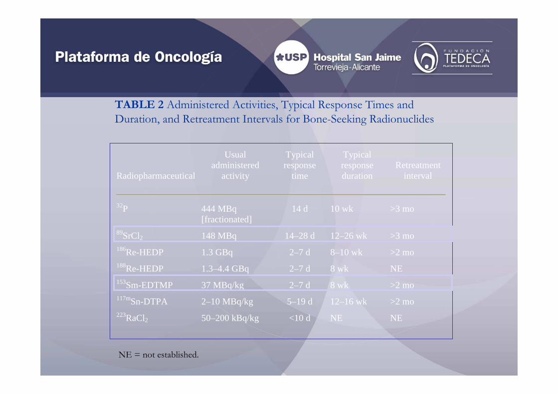
TABLE 2 Administered Activities, Typical Response Times and Duration, and Retreatment Intervals for Bone-Seeking Radionuclides
Radiopharmaceutical
Usual administered
activity
Typical response
time
Typical response duration
Retreatment interval
32P 444 MBq
[fractionated] 14 d 10 wk >3 mo
89SrCl2 148 MBq 14–28 d 12–26 wk >3 mo 186Re-HEDP 1.3 GBq 2–7 d 8–10 wk >2 mo 188Re-HEDP 1.3–4.4 GBq 2–7 d 8 wk NE 153Sm-EDTMP 37 MBq/kg 2–7 d 8 wk >2 mo 117mSn-DTPA 2–10 MBq/kg 5–19 d 12–16 wk >2 mo 223RaCl2 50–200 kBq/kg <10 d NE NE
NE = not established.

Anterior
99mTc-MDP 153Sm-EDTMP
Posterior
99mTc-MDP 153Sm-EDTMP
Criterios de selección de pacientes
•Gammagrafía ósea poco antes del tratamiento (FUNDAMENTAL)
•Hemograma / reserva hematopoyética
La mielotoxicidad reversible es el efecto adverso más frecuente en esta
terapia
Una infiltración difusa de M.O. (patrón “superscan” en gammagrafía ósea)
Es importante el timing entre este y otros tratamientos mielosupresores
•Bioquímica (función renal y hepática adecuadas):Una pobre función renal retrasará el aclaramiento del radiofármaco,
aumentando la dosis corporal total y potencialmente su toxicidad

TABLE 3 Criteria for Patient Selection for Bone-Seeking Radionuclide Therapy
Treatment indications
Treatment refractory to bone pain despite analgesics
Positive bone scan; abnormal uptake corresponding to pain sites
Hematology: Hb > 90 g/L; white cell count > 4 x 10⁹/L; platelets > 100 x 109/L
Renal function: urea < 12 mmol/L; creatinine < 200 mmol/L
Absolute contraindications
Pregnancy
Acute spinal cord compression
Acute or chronic renal failure; glomerular filtration rate < 30 mL/min
Precautions
Urinary incontinence; catheterize before treatment
Vesicoureteric or bladder outflow obstruction; consider ureteric stent or catheterize before treatment

Eficacia:
•Existen más estudios que incluyeron otros tumores (mama, pulmón,…)
•Alivio del dolor: 75% (30-85)
•Disminución en el uso de analgésicos: en 3-4 semanas
•Inicio de respuesta: 5-10 días / duración: hasta 4 meses
•Efectivo en dosis repetidas (se ha publicado que pueden mejorar la duración de la respuesta al dolor y la supervivencia)
(Turner JH et al. Eur J Cancer 1991; 27: 1084-1086)
•Se han publicado descensos en marcadores tumorales (PSA y FAlc) e intensidad en la gammagrafía ósea
(Collins C et al. J Nucl Med 1993; 34: 1839-1844)(Sartor O et al. Urology 2004; 63: 940-945)
153Sm-EDTMP (QUADRAMET®)

Eficacia (II):
•Con dosis única datos de prolongación de supervivencia contradictorios:
Resche I et al. Eur J Cancer 1997; 33: 1583-1591
- 76 próstata, 36 mama, 2 pulmones y 9 “grupo mixto”
- 18.5 MBq / kg vs. 37 MBq / kg
- aumento de supervivencia para la dosis > en mama (no en próstata)
Sartor O et al. Urology 2004; 63: 940-945
- 152 próstata hormonorrefractario
-37 MBq / kg 153Sm-EDTMP vs. placebo
- no diferencias en supervivencia global
153Sm-EDTMP (QUADRAMET®)

Experiencia USP San Jaime
• 28 tratamientos 153Sm-EDTMP (3 mama, 1 neuroendocrino, 1 esófago)
1- Mtx múltiples esqueleto axial y periféricas n=7 (Qt+zometa; RP) Tiempo a progresión 9 meses (bq y ósea). (OS=3.5 a.)
2- Mtx múltiples esqueleto axial y periféricas n=6 (Qt+H. RP)Tiempo a progresión >6meses (lost follow-up). (OS=5 a.)
3- Mtx esqueleto axial n=4, 5 meses (SD lost follow-up).

•RIT vs RT externa
II. Terapia metabII. Terapia metabóólica. Aspectos por resolverlica. Aspectos por resolver
•RIT adyuvante a RT externa
•RIT en combinación con QT
•RIT en consolidación
•Nuevos fármacos: EMISORES DE “CORTO ALCANCE”
117mSn-DTPA
223Ra (Alpharadin)

www.plataformadeoncologia.comwww.fundaciontedeca.org