odontologÍa en imÁgenes / dentistry in images cuerpo extraŒo Šrbito-maxilar · odontologÍa en...
TRANSCRIPT

MEDICINA ORALVOL. 7 / N.o 1
ENE.-FEB. 2002
7373
ODONTOLOGÍA EN IMÁGENES / DENTISTRY IN IMAGES
Cuerpo extra�o �rbito-maxilarOrbito-maxilar foreign body
AUTORES/AUTHORSMar�a Fe Garc�a Reija, Angel Espeso Ferrero, Mar�a Galdeano Arenas,Alberto Verrier Hern�ndez.
Servicio Regional de Cirug�a Maxilofacial. Hospital Universitario del R�o Hortega.Valladolid. Espa�a.Universidad de Santiago de Compostela. Espa�a.
Paciente de 18 a�os que ingresa en el Servicio de Cirug�aOral y Maxilofacial del Hospital Universitario P�o del R�oHortega (Valladolid) remitido por el Servicio de Urgenciaspor presentar un traumatismo orbitario a consecuencia de unaccidente casual.
En la exploraci�n f�sica se observ� un cuerpo extra�o (pa-lo de madera) penetrante a nivel de p�rpado inferior con tra-yecto hacia suelo de �rbita.
Examen oftalmol�gico: Midriasis arreactiva. Edema peri-f�rico de retina (porci�n superior e inferior). Limitaci�n en lamirada superior, por atrapamiento del m�sculo recto inferior.
Examen radiogr�fico: Cuerpo extra�o penetrante a trav�sdel suelo de �rbita izquierda, pared posterior del seno maxilarizquierdo hasta fosa pterigomaxilar.
Es intervenido bajo anestesia general realiz�ndose extrac-ci�n del cuerpo extra�o y reconstrucci�n del suelo orbitariomediante pr�tesis de polietileno de alta densidad. Reconstruc-ci�n y sutura del p�rpado inferior. Cierre por planos.
Patient of 18 years who enters in the Service of oral andmaxilofacial surgery of the University Hospital P�o del R�oHortega (Valladolid) sent by the Service of Urgencies, dis-playing an orbitarial traumatism as a result of an accident.
During the physical exploration a foreign body was obser-ved (a splinter of wood) penetrating at the low level of theeyelid with passage towards orbital floor.
Ophtalmological examination: arreactive midriasis. Pe-ripheral edema of the retina (superior and low portion). Li-mitation in the superior glance, by atrapment of the inferiorstraight muscle.
Radiological examination: penetrating foreign body th-rough the floor of the left orbit, latter wall of the left maxilarsinus until to the pterigomaxilar grave.
Foreign body extracted under general anesthesia, and re-construction of the orbitarial floor by means of a prothesis ofHD polyethylene. Reconstruction and suture of the inferioreyelid. Closure by planes.
Fig. 1. Vista de perfil. Cuerpo extra�o (palo de madera) con orificio deentrada a nivel del p�rpado inferior.
Profile view. Foreign body (splinter of wood) with orifice of entrance at thelevel of the inferior eyelid.
GARCêA A, y cols.
MEDICINA ORALVOL. 7 / N.o 1
ENE.-FEB. 2002
7474
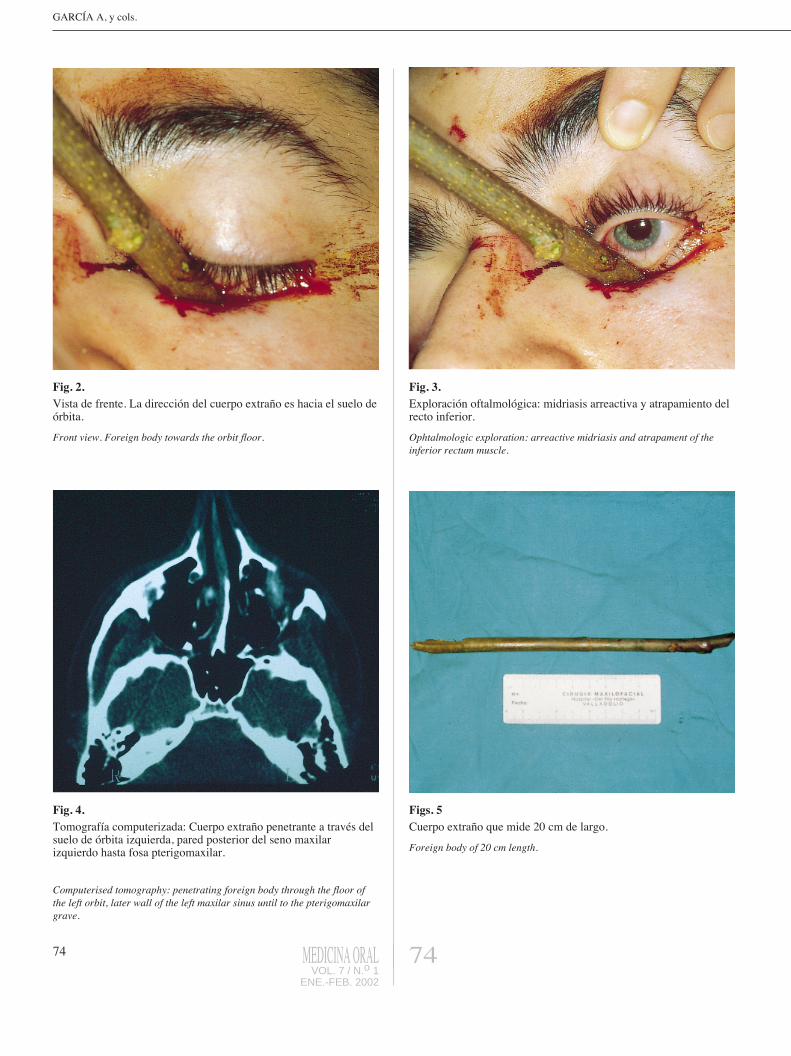
Fig. 2. Vista de frente. La direcci�n del cuerpo extra�o es hacia el suelo de�rbita.
Front view. Foreign body towards the orbit floor.
Fig. 3. Exploraci�n oftalmol�gica: midriasis arreactiva y atrapamiento delrecto inferior.
Ophtalmologic exploration: arreactive midriasis and atrapament of theinferior rectum muscle.
Fig. 4. Tomograf�a computerizada: Cuerpo extra�o penetrante a trav�s delsuelo de �rbita izquierda, pared posterior del seno maxilarizquierdo hasta fosa pterigomaxilar.
Computerised tomography: penetrating foreign body through the floor ofthe left orbit, later wall of the left maxilar sinus until to the pterigomaxilargrave.
Figs. 5Cuerpo extra�o que mide 20 cm de largo.
Foreign body of 20 cm length.