natural products in therapeutic management of
TRANSCRIPT

Review ArticleNatural Products in Therapeutic Management ofMultineurodegenerative Disorders by Targeting Autophagy
Sibhghatulla Shaikh,1,2 Khurshid Ahmad ,1,2 Syed Sayeed Ahmad,1,2 Eun Ju Lee,1,2
Jeong Ho Lim,1 Mirza Masroor Ali Beg ,3 Amit K. Verma,4 and Inho Choi 1,2
1Department of Medical Biotechnology, Yeungnam University, Gyeongsan 38541, Republic of Korea2Research Institute of Cell Culture, Yeungnam University, Gyeongsan 38541, Republic of Korea3Faculty of Medicine, Ala-Too International University, Bishkek, Kyrgyzstan4Department of Biotechnology, Jamia Millia Islamia, New Delhi, India
Correspondence should be addressed to Inho Choi; [email protected]
Sibhghatulla Shaikh and Khurshid Ahmad contributed equally to this work.
Received 7 May 2021; Revised 9 August 2021; Accepted 18 August 2021; Published 14 September 2021
Academic Editor: Xinfeng Li
Copyright © 2021 Sibhghatulla Shaikh et al. This is an open access article distributed under the Creative Commons AttributionLicense, which permits unrestricted use, distribution, and reproduction in any medium, provided the original work isproperly cited.
Autophagy is an essential cellular process that involves the transport of cytoplasmic content in double-membraned vesicles tolysosomes for degradation. Neurons do not undergo cytokinesis, and thus, the cell division process cannot reduce levels ofunnecessary proteins. The primary cause of neurodegenerative disorders (NDs) is the abnormal deposition of proteins insideneuronal cells, and this could be averted by autophagic degradation. Thus, autophagy is an important consideration whenconsidering means of developing treatments for NDs. Various pharmacological studies have reported that the activecomponents in herbal medicines exhibit therapeutic benefits in NDs, for example, by inhibiting cholinesterase activity andmodulating amyloid beta levels, and α-synuclein metabolism. A variety of bioactive constituents from medicinal plants areviewed as promising autophagy controllers and are revealed to recover the NDs by targeting the autophagic pathway. In thepresent review, we discuss the role of autophagy in the therapeutic management of several NDs. The molecular processresponsible for autophagy and its importance in various NDs and the beneficial effects of medicinal plants in NDs by targetingautophagy are also discussed.
1. Introduction
Autophagy is a fundamental, exceptionally well-controlledprocess in the eukaryotic cell recycling system under differentstates of cellular stress. Autophagy plays an essential role incell survival and maintenance by degrading cytoplasmicorganelles, macromolecules, and misfolded proteins and, thus,facilitates the recycling of cellular content as breakdown prod-ucts [1, 2]. The literal meaning of autophagy is “self-eating,”and this process involves the intracellular capture of unnec-essary proteins, lipids, and organelles and directing themfor their further degradation in the lysosomal compartment[3]. Thus, autophagy performs a vital role in the overallhomeostasis of proteins and, eventually, cells and retains
metabolic balance between the synthesis and degradation ofcytoplasmic materials and their subsequent reprocessing.Thus, autophagy is a natural cellular mechanism that sus-tains cellular homeostasis under various conditions [4].Many other functions of autophagy have been identified inthe presence of pathological processes such as infectious dis-eases, myocardial diseases, diabetes, neurodegenerative dis-eases (NDs), and various cancers [5, 6].
There are typically three types of autophagy: macroauto-phagy, microautophagy, and chaperone-mediated autoph-agy (CMA), all of which have specific roles that depend onthe cellular microenvironment, signals, and organs, but allusually involve proteolytic degradation of cytosolic compo-nents inside lysosomes [7]. Macroautophagy is the most
HindawiOxidative Medicine and Cellular LongevityVolume 2021, Article ID 6347792, 12 pageshttps://doi.org/10.1155/2021/6347792
important degradation pathway and involves the formationof double-membrane vesicles called autophagosomes incytoplasm. In fact, the term “autophagy” typically refers tothis process, unless otherwise specified. Macroautophagymay further be classified as mitophagy, nucleophagy, pexo-phagy, aggrephagy, and xenophagy [8]. Pathological studieshave shown that macroautophagy deficiencies may beinvolved in the development of NDs [9]. In microautophagy,cellular constituents are taken up by lysosomes by directengulfment, projection, or septation of lysosome membranes[10]. Macro- and microautophagy are both capable ofengulfing large cellular components through selective andnonselective mechanisms. CMA is an extensive form ofautophagy found in almost all cells and tissue types in highereukaryotes and involves the direct recognition of targetedproteins containing the KFERQ motif, which is recognizedby chaperone proteins, such as the heat shock protein(Hsc-70). These proteins are then delivered directly to lyso-somes via the lysosomal-associated membrane protein 2A(LAMP-2A) [11, 12]. Studies increasingly support the notionthat dysregulation of the CMA pathway plays a role in mul-tiple NDs including Alzheimer’s Disease (AD), Parkinson’sDisease (PD), Amyotrophic Lateral Sclerosis (ALS), andFrontotemporal Lobar Degeneration [12].
Neurons are of the postmitotic cell type and do not gothrough cytokinesis, and thus, cell division cannot reduceunnecessary deposited proteins. Therefore, the primarycause of NDs is pathological protein accumulation withinneuronal cells, and this could be prevented by autophagicdegradation. Hence, autophagy activation plays an impor-tant role in the treatment of NDs and provides a strategicplatform for disease management [13, 14]. In this review,we discussed the beneficial effects of medicinal plants onNDs by targeting autophagy. Before describing the roles ofherbal products in NDs, we provide a brief overview ofautophagic processes at a molecular level and their roles invarious NDs.
2. Molecular Process of Autophagy
Autophagy is highly inducible by starvation and other stress-related responses, and its activation causes a rapid increasein numbers of autophagosomes, which are generated on ornear the endoplasmic reticulum [15]. In addition, mem-branes acquired from the Golgi complex, mitochondria,and plasma membrane also contribute to autophagosomeformation [15]. Autophagosome establishment involves amulticomponent complex process, which is governed bymultiple Atg proteins and related proteins. Of the 35 Atgproteins known, Atg1–10, 12–14, 16, and 18 are the “coreAtg proteins” in yeast [16], and these proteins with Atg17,29, and 31 participate in autophagosome formation. Otherautophagic pathways such as pexophagy (autophagic perox-isome degradation) and cytoplasm-to-vacuole targetingshare core Atg proteins [17, 18]. Furthermore, core Atg pro-teins are highly conserved in eukaryotes, including mam-mals, and function in a similar hierarchical manner inyeast [19].
Studies have shown that mTOR is essential for promot-ing adult stem cell differentiation, progenitor cell growth,and proliferation and has a great impact on the multipotentstem cell population [20, 21]. Interruption or deregulation ofthe autophagy system has been implicated in neurodegener-ative issues such as AD, and Aβ assumes a significant role inthis autophagy framework. Autophagy plays a substantialrole in the generation and digestion of Aβ and the accumu-lation of tau. Much of the time, autophagy is directed by thephosphatidylinositol 3-phosphate kinase/AKT/mTOR/p70ribosomal protein S6 kinase signaling pathway [22].
Various mTOR-dependent and mTOR-independentautophagy modulators have been found to have beneficialeffects in the management of AD [23]. The mTOR pathwayappears to be an adaptable player, and its modulation canaffect both neuroprotective and cognitive processes. Althoughdirect targeting of mTOR does not appear to have therapeuticpotential, its indirect modulation by other signaling pathwaysis encouraging in this respect [24]. Targeting of rapamycincomplex 1 (mTORC1) promotes cell growth in nutrient-richenvironments by inducing the biosynthesis of proteins, lipid,and nucleotides and inhibiting cellular catabolism by sup-pressing the autophagic pathway [25, 26]. Aberrant mTORsignaling is associated with brain abnormalities and NDs.Even subtle defects in the mTOR pathway may have severeeffects, including neurological and psychiatric disorders.Conversely, mTOR inhibitors can be useful in several neuro-psychiatric alterations such as in brain cancer, brain ische-mia, schizophrenia, autism, and NDs. mTOR has beenlinked to synaptic plasticity and the activation of autophagy[27]. Under nutrient-rich conditions, by direct associationwith the ULK1-Atg13-FIP200 complex, mTOR suppressesautophagy and mediates the phosphorylation-dependentinhibitions of the autophagy-related kinase activities of -13(Atg13) and Unc-51-like kinase 1 (ULK1). Furthermore,the mTOR-mediated phosphorylations of Atg13 and ULK1are inhibited under starvation conditions or after treatmentwith rapamycin. Rapamycin inhibits mTOR, and this triggersautophagy by dephosphorylating ULK1, ULK2, and Atg13and activating ULK to phosphorylate FIP200 (FAK family-interacting protein of 200 kDa) [28, 29]. The existence ofmTOR-independent regulation of mammalian autophagyresulted from the observation that intracellular inositol andinositol 1,4,5-trisphosphate (IP3) levels negatively regulateautophagy [30]. Furthermore, inhibition of inositol mono-phosphatase decreased levels of free inositol and IP3, whichled to autophagy upregulation [31].
In postmitotic neuronal cells, basal autophagy movementis important, possibly because of their inability to dilute nox-ious components via cell division [32]. Autophagic activity isenhanced by various burdens, for example, by supplementstarvation, hypoxia, or inflammation [33]. During certainphysiological processes and pathological conditions, increasedautophagy leads to cell death, the elimination of microorgan-isms entering cells, and cancer suppression [34]. Then again,the reduced autophagic potential is related to aging [35]. Dur-ing autophagy, proteins are degraded into amino acids and,thus, provide a source of energy and raw materials for proteinsynthesis [36]. Hence, the dysregulation of autophagy may
2 Oxidative Medicine and Cellular Longevity

result in the aggregation of intracellular proteins. Further-more, different types of autophagy dysfunctions can lead toND or ND-like symptoms, such as the inhibition ofautophagosome-lysosome fusion [37], reduced lysosomalacidification [38], or intracellular protein deposition [39].
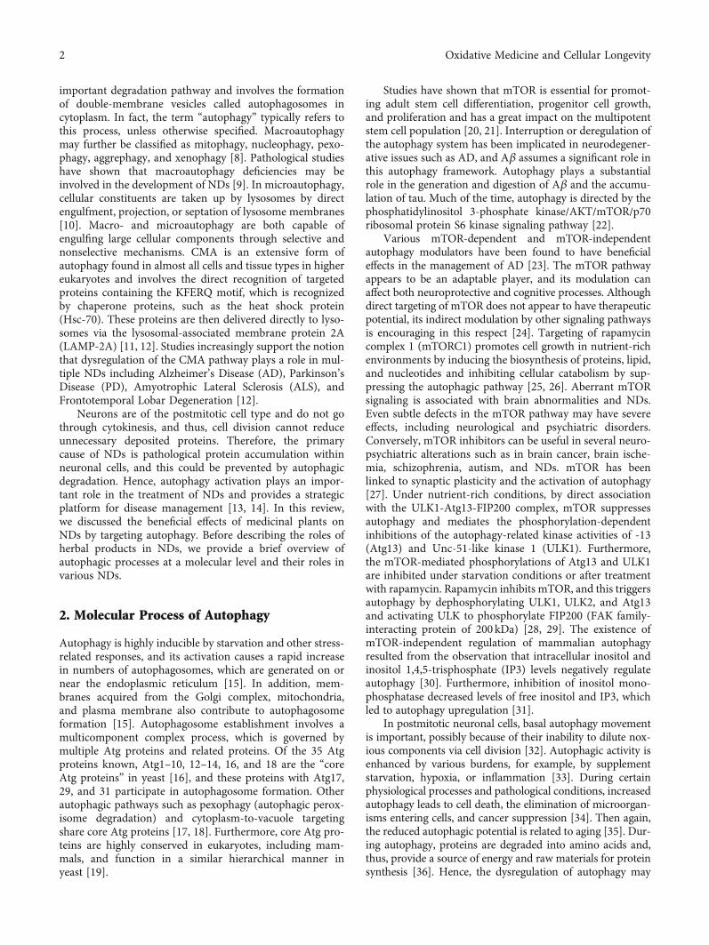
Two complexes, that is, mTORC1 and mTORC2, areresponsible for the regulation of autophagy. mTORC1 consistsof four different protein factors, viz., raptor (regulatory-associ-ated protein of mTOR), deptor (DEP-domain containingmTOR-interacting protein), PRAS40 (proline-rich Aktsubstrate of 40kDa), and mLST8 (mammalian lethal withSEC13 protein 8), whereas mTORC2 consists of rictor(rapamycin insensitive companion of mTOR), protor (proteinobserved with rictor), and mSIN1 (mammalian stress-activated mitogen-activated protein kinase-interacting pro-tein) along with mLST8 and deptor [40, 41]. Starvation resultsin the activation of the mTORC1 complex, which stimulatesautophagy resulting in the recycling of intracellular compo-nents as a source of energy [42]. In addition, the phosphoryla-tion of Akt by the mTORC2 complex results in the activationof the mTORC1 complex (Figure 1).
3. Autophagy, Immune Response,and Neurodegeneration
Most cellular stress-response pathways, including those thatregulate immunological responses and inflammation, inter-act with the autophagy machinery [43–45]. The autophagypathway/proteins have a complex reciprocal relationshipwith immunity and inflammation; autophagy proteins areinvolved in both the stimulation and suppression of immuneand inflammatory responses, and immune and inflamma-tory signals are involved in both the stimulation and sup-pression of autophagy [44].
Autophagic interference with type I interferon responsesoccurs either directly by targeting signaling molecules withinthe pathway, beginning with RIG-I-like receptors or cGAMPsynthase (a cytosolic DNA sensor) and progressing to thestimulator of the interferon gene (STING) and interferonregulatory factors, or indirectly by removing agonist sourcesthat activate these pathways [46–49]. The p62 receptorappears to have a function in preventing T-cell receptor-(TCR-) mediated NF-κB signaling via Bcl10. Although p62initiates signaling, it also functions as a receptor to degradeBcl10, which becomes ubiquitinated in response to TCRactivation. Therefore, this strategy may protect cells againstNF-κB hyperactivation as a result of TCR signaling [50].
Using fly genetics, researchers show that deregulation ofcyclin-dependent kinase 5 (Cdk5) activity disrupts autoph-agy, leads to an overactive innate immune response, andresults in dopamine neurodegeneration in Drosophila[51]. It was demonstrated that an overactive innate immuneresponse was sufficient to trigger neuronal cell death.Intriguingly, inhibiting the NF-κB transcription factor inneurons lowers neuronal loss and downregulates the innateimmune response genes in the Cdk5-deficient back-ground [52].
Many NDs are linked with inflammatory responses inglia, which may contribute to pathology, and autophagy in
glial cells may play a role in regulating these processes[53]. Microglia, as a key immune cell in the brain, influencesphagocytosis and inflammation in age-related NDs [54].When the LC3B and Atg7 genes were inhibited, microgliafailed to degrade extracellular Aβ, indicating that autophagyfunction impairment in microglia may contribute to CNS-degenerative neurological disease [55]. Astrocytes are spe-cialized glial cells in the brain and spinal cord and have beenlinked to the development of various NDs such as AD, PD,and ALS [56, 57]. Trifluoperazine-induced autophagy wasimplicated in astrocyte protection against bilirubin-inducedcytotoxicity [58]. Recently, it has been demonstrated thatAtg5 knockdown reduced astrocyte development in vivo,but Atg5 overexpression resulted in excessive astrocyte dif-ferentiation in vivo [59].
4. Autophagy and Neurodegenerative Disorders
4.1. Autophagy in Alzheimer’s Disease (AD). AD is character-ized by the depositions of Aβ and tau in the brain. Undernormal conditions, the production and clearance rates ofAβ are balanced, and Aβ is not deposited inside neuronalcells. Enhanced aggregation of Aβ peptides has been foundin AD patients, and it is well recognized that failure of theautophagic system is a characteristic of AD. Recently, itwas shown that autophagy enhanced the protein degrada-tions of Aβ and tau [60]. During autophagy, autophago-somes enclosing Aβ facilitate its degradation by fusingwith lysosomes. In addition, the microglial inflammatoryresponse is regulated by autophagy, and dysregulation ofautophagy damages neurons by exacerbating NLRP3 inflam-masome signaling [55].
The C-terminal fragments of the amyloid precursor pro-tein (APP) might be an etiological trigger for AD [61]. Thecleavage of APP by BACE-1 produces C99 fragments.Reductions in autophagy (inhibition of autophagosome pro-duction or prevention of autophagosome fusion with lyso-somes) result in increased C99 levels [62, 63]. Conversely,enhanced autophagy, either by mTOR suppression or bystarvation, enhances C99 clearance in degenerative lyso-somes [62, 64]. Also, lysosomes are disrupted by phagocyto-sis of the Aβ peptide, which results in the release of alysosomal proteolytic enzyme (cathepsin B), which, in turn,stimulates pyrin domain containing 3 inflammasomes andleads to the production of proinflammatory and neurotoxicfactors via the interleukin 1 beta pathway [65]. Stimulationof the autophagic system via cystatin B deletion decreasesAβ aggregation and has protective effects in mouse modelsof AD [66]. Tau protein stabilizes the microtubule, but itshyperphosphorylation reduces its affinity for microtubulesand results in microtubule entanglement. Thus, the elimina-tion of the tau protein by the autophagic system is requiredto address NDs [67, 68]. In addition, enhanced accumula-tion of the tau protein in the presence of the autophagicinhibitor (3-methyladenine) suggests that autophagy isrequired to prevent tau aggregation [69].
Various approaches used to upregulate the autophagicsystem have potential use for the management of AD [70].Rapamycin, an inhibitor of the mTOR pathway, decreased
3Oxidative Medicine and Cellular Longevity

Aβ deposition and prevented AD development by enhanc-ing autophagy in an animal model of AD [71, 72], and themultifunctional protein p62 has been linked to neuropatho-logical inclusions in various NDs and with the degradationsof Aβ and tau. The ubiquitin-binding domain and the LC3-(microtubule-associated protein 1 light chain 3-) interactingregions of p62 are two functional domains. Enhancing brainp62 expression promoted autophagy and led to cognitiveimprovement in an animal model of AD, whereas removingthe LC3-interacting region domain disrupted Aβ clearanceby preventing autophagy [73]. In another study, latrepirdinewas found to decrease Aβ aggregation by stimulating theAtg5-dependent autophagy in an animal model [70]. Thesereports indicate that autophagy is disrupted in AD and thatregulating the autophagic system offers a reasonable thera-peutic approach.
4.2. Autophagy in Parkinson’s Disease (PD). Autophagy andthe ubiquitin-mediated pathway eliminate misfolded pro-teins in healthy cells, but both of these pathways are dis-rupted in PD, which results in the aggregation of misfoldedproteins [74]. One of the important hallmarks of PD is thedeposition of misfolded α-synuclein into intraneuronalinclusions known as Lewy bodies (LBs). α-Synuclein is sus-ceptible to degradation by chaperone-mediated autophagy[75]. In familial PD, lysosomes are unable to engulf themutant α-synuclein because of its high affinity for the lyso-somal receptor (LAMP-2A), which in turn prevents α-synu-clein degradation by shielding the substrate from CMA [76].Furthermore, autophagosome and dysfunctional lysosomeaccumulations were found in postmortem PD brain samples[77], which highlighted the pathogenic role of autophagy inPD. Enhanced α-synuclein levels have been found in the
lysosomal dysfunction, indicating a close link betweenautophagy and α-synuclein degradation. Various studieshave reported that autophagy can degrade all forms of α-synuclein [77, 78] and that proteasomes also degradesmonomeric α-synuclein [79].
Overexpression of α-synuclein is caused by mutationsin SNCA, which encodes for α-synuclein, and these aresufficient to cause the progression of PD. Excess α-synu-clein levels damage the autophagy system by hinderingsmall GTPase Rab-1A [80]. Autophagy contributes in var-ious ways to the protection of neural cells, but α-synucleinaccumulation enhances protein aggregation levels, countersthe effective clearance of misfolded protein, and inducesneuronal cell apoptosis [80]. Moreover, α-synuclein muta-tion has been suggested to impair CMA [76, 81]. Theseresults indicate that the regulations of more than one typeof autophagy by α-synuclein mutations have toxic effectson neuronal cells.
4.3. Autophagy in Amyotrophic Lateral Sclerosis (ALS). ALSis a paralytic condition defined as motor neuronal dys-functions in the brain and spinal cord resulting in muscleatrophy. Mutations in TARDNA-binding protein and super-oxide dismutase 1 (SOD1) are common causes of familialALS [82], and it has been established that autophagy is linkedwith ALS. Rat LC3 is vital for autophagy, and the formationof LC3-II from LC3-I has been suggested to provide a simplemeans of controlling autophagy. LC3-II overexpression hasbeen reported in mutant SOD1G93A transgenic mice. Inaddition, enhanced autophagosomes were strongly associ-ated with reduced mTOR phosphorylation in various geneticALS models [83]. A growing number of studies have estab-lished that mutations in autophagy-associated proteins are
AD brain
Proteinaccumulation
Autophagy
DEPTOR
RAPTOR
mLST8
PRAS40
mTORC1
Induce autophagy
Inhibition
PROTOR
RICTOR mSIN1
mLST8
DEPTORmTORC2
Indirect autophagyregulator
Resistance torapamycin
Aktphosphorylate
mTORPathway
Role in organizationof actin-cytoskeleton
Intracellularconstituents
recycleProvide source
of energy
Starvation
Hyperactivity ofPI3-K / Akt / mTOR
Figure 1: Molecular pathways in autophagy.
4 Oxidative Medicine and Cellular Longevity

well correlated with the pathogenesis of ALS. The endosomalsorting complexes required for transport (ESCRTs) areresponsible for sorting transmembrane proteins into the innervesicles of the multivesicular body (MVB) during endocytosis.Reductions in ESCRT subunits inhibit either autophagosome-MVB fusion or amphisome-lysosome fusion and are consid-ered to be linked with ALS [84]. Ubiquilin-2 (a proteasomeshuttle factor) has an important role in the generation ofautophagosomes. Mice with mutated UBQLN2 exhibit neu-ronal loss, cognitive deficits, and short lifespans [85].
5. Beneficial Effects of Medicinal Plants onNDs by Targeting Autophagy
Medicinal herbs have become increasingly important in thequest for more effective and adjunctive treatments [86–89].Various pharmacological studies have reported that theactive components of herbal medicines show therapeuticbenefits in NDs via different mechanisms such as by increas-ing neurogenic activity, inhibiting cholinesterase activity,and controlling Aβ, tau, and α-synuclein metabolism by tar-geting autophagy [90–92]. Table 1 provides a summary ofclasses of natural compounds that reduce neurodegenerationby regulating autophagy (Figure 2).
5.1. Crude Extracts. Radix Polygalae extract was reported todecrease Aβ andmutant A53T α-synuclein levels by activatingAMPK/mTOR signaling to stimulate autophagy in Chinesehamster ovary cells (transfected with APP and BACE1) andPC-12 cells, respectively [93, 94]. Withania somnifera extracthad a protective effect in ALS by downregulating p62 (a clas-sical autophagy receptor), thereby promoting autophagy inthe motor neurons of SOD1G93A mice [95]. In another study,Ginkgo biloba extract repressed microglial inflammation andenhanced cognitive functions by regulating the mechanismmoderately involved in the activation of autophagy [96].
5.2. Saponins. Ginsenoside-Rg2, a bioactive compoundobtained from Panax ginseng induces autophagy in anAMPK/ULK1-dependent manner. Rg2 increased the clear-ance of aggregated proteins and enhanced cognitive func-tion by inducing autophagy in an AD mouse model [97].The protopanaxadiol derivative DDPU (1-(3,4-dimethoxy-phenethyl)-3-(3-dehydroxyl-20(s)-protopanaxadiol-3β-yl)-urea) increased Aβ clearance by inducing autophagy via thePI3K/AKT/mTOR signaling pathway by inhibiting PI3Kand decreased Aβ generation by restraining PERK/eIF2αsignaling-mediated BACE1 translation [98]. In addition,Radix Polygalae derived onjisaponin B enhanced mutantα-synuclein degradation by autophagy induction by activat-ing the AMPK/mTOR signaling pathway [93].
5.3. Alkaloids. Alkaloids are the important active compo-nents in herbal medicines and exert beneficial effects onNDs by inducing autophagy and inhibiting cholinesteraseactivity [99, 100]. Alkaloids isolated from Dendrobiumnobile enhanced autophagic flux via autophagosome genera-tion and stimulated Beclin-1 expression [101], and berberinehas been reported to stimulate autophagy by activating
Bcl2/Beclin-1 signaling, thus increasing Aβ clearance, and toimprove cognitive functions in a mouse model of AD [102].Furthermore, it has been reported that berberine can bypassthe blood-brain barrier [103]. TDP-43 (43 kDa nuclear proteinTARDNA-binding protein) is the main component of ubiqui-tinated inclusions in aggregated proteins in ALS [104, 105].Berberine has therapeutic potential in ALS as it reversesTDP-43 proteinopathy by disrupting mTOR/p70S6K signal-ing and stimulating the autophagic degradation pathway[106]. Corynoxine isolated from Uncaria rhynchophylla isan established inducer of autophagy and enhances autopha-gosome generation and the elimination of α-synuclein inPC12 cells [107]. Isorhynchophylline, a main tetracyclic oxi-ndole alkaloid obtained from U. rhynchophylla, has beenused to manage NDs in East Asia for centuries. This alkaloidinduces the Beclin-1-dependent autophagy-lysosome path-way and enhances the clearance of α-synuclein monomersand α-synuclein/synphilin-1 aggresomes from neuronalcells [108]. Angelica sinensis-derived n-butylidenephthalideenhanced motor functions in SOD1-ALS mice. The autoph-agy pathway was involved in the therapeutic mechanism, asn-butylidenephthalide treatment reduced LC3-II expressionand increased mTOR levels [109]. In addition, conophyllinefrom Ervatamia microphylla induced autophagy in Hunting-ton disease and PD models [100, 110].
5.4. Flavonoids. Studies have established that flavonoidsinfluence the autophagy system in some disorders [111, 112].Silibinin isolated from Silybum marianum reduced neuronaldamage via the BDNF/TrkB pathway by decreasing autophagyin the hippocampus [113]. In another study, wogoninenhanced autophagy by inhibiting the Akt/mTOR pathwayand increasing Aβ clearance [114]. Hesperetin recovered Aβdamage-induced glucose utilization by downregulating Aβ-stimulated autophagy [115], and kaempferol has beenreported to enhance autophagy and decrease ROS, apoptosis,and mitochondrial dysfunction in rotenone-exposed SH-SY5Y cells [116].
5.5. Polyphenols. Curcumin inhibits Aβ aggregation andameliorates cognitive functions. The mechanisms responsi-ble involve the stimulation of autophagy by downregulatingthe PI3K/Akt/mTOR pathway [117]. In amyloid-treated HT-22 cells, curcumin protected hippocampal neurons by inhi-biting the abnormal formation of Beclin-1 and autophago-somes [118]. In an in vitro dopaminergic neuron model ofPD, curcumin was involved in the modulation of autophagyand the clearing of α-synuclein aggregates [119]. Resveratrolis attracting attention because of its curative potential in ADand has been reported to reduce Aβ generation and restrainthe development of AD by inhibiting apoptosis and regulat-ing mitophagy [120]. Curcumin decreased the accumulationof A53T α-synuclein protein (related to early-onset PD) bydownregulating mTOR/p70 ribosomal protein S6 kinasesignaling and induced macroautophagy in SH-SY5Y cells[121]. In addition, resveratrol has been reported to protectagainst neural damage by activating mitophagy [122] andto stimulate autophagy and lysosomal degradation by regu-lating the AMPK/mTOR signaling pathway and reducing
5Oxidative Medicine and Cellular Longevity

Aβ synthesis in HEK293 and N2a cells [123]. In mice, orallyadministered resveratrol crossed the blood-brain barrier,stimulated brain AMPK, and decreased Aβ deposition inthe cerebral cortex [123]. The active component (2,3,5,4′-tetrahydroxystilbene-2-O-glycoside) in Radix PolygoniMultiflori was reported to hinder autophagy by decreasingBeclin-1 levels, thus enhancing cognitive function [124],and carnosic acid stimulated autophagy by activating theAMPK/mTOR pathway and inhibited Aβ deposition [125].
Resveratrol was observed to protect SH-SY5Y cells fromrotenone-stimulated apoptosis and to increase α-synucleindegradation in α-synuclein-expressing PC12 cell lines byinducing autophagy. The mechanism of α-synuclein degra-dation in a cellular model of PD involved the regulation ofmammalian SIRT1 (silent information regulator 2)/AMPK(AMP-activated protein kinase), which diminished LC3-IIprotein levels and increased α-synuclein clearance [126].Resveratrol improved mitochondrial oxidative function byregulating the AMPK and SIRT1 pathways and increased
macroautophagic flux by activating an LC3-independentpathway in early-onset PD fibroblasts [127]. In another study,resveratrol stimulated heme oxygenase-1 expression andinhibited dopaminergic cell death by controlling autophagicflux and, as a result, protected against rotenone-induced neu-ronal apoptosis in a PD model [128]. Corema album polyphe-nol fractions promoted nontoxic α-synuclein formation and,thus, reduced its toxicity and aggregation in cells by enhancingautophagic flux and reducing oxidative stress [129]. Inaddition, Arctium lappa-derived arctigenin inhibited the gen-eration and enhanced the clearance of Aβ by inducing autoph-agy by inhibiting AKT/mTOR signaling and AMPK/Raptorpathway activation in an animal model of AD [130].
5.6. Terpenes and Terpenoids. Recently, monoterpenes havebeen identified to be autophagy modulators [131]. Cubeben,a Piper cubeba sesquiterpene, decreased Aβ toxicity in pri-mary neuronal cells, recovered autophagy via PI3K/AMPKsignaling, and suppressed the inhibition of mTOR [132]. In
Table 1: Natural compounds that inhibit neurodegeneration via autophagy.
Natural sources Signaling Effects References
Crude extracts
Radix Polygalae AMPK/mTORDecrease Aβ and mutant A53T
α-synuclein levels[93, 94]
Withania somniferaDownregulate the p62
(a classical autophagy receptor)Promote autophagy in motor neuron [95]
Saponins
Ginsenoside-Rg2AMPK-ULK1-dependentand MTOR-independent
Aggregated protein clearance andenhanced cognitive function
[97]
DDPU PI3K/AKT/mTOR and PERK/eIF2α Clearance of Aβ and decreased Aβ generation [98]
Onjisaponin B AMPK/mTOR Enhances mutant α-synuclein degradation [93]
Alkaloids
Berberine Bcl2/Beclin-1 Clearance of Aβ and improves cognitive function [102]
Isorhynchophylline Beclin-1Clearance of α-synuclein monomers andα-synuclein/synphilin-1 aggresomes
[108]
n-Butylidenephthalide mTOR Enhanced motor functions [109]
Flavonoids
Silibinin BDNF/TrkB Reduces neuronal damage [113]
Wogonin Akt/mTOR Clearance of Aβ [114]
Hesperetin IRS-PI3K-Akt Recovers Aβ-damage glucose utilization [115]
Polyphenols
CurcuminPI3K/Akt/mTOR and mTOR/p70
ribosomal protein S6 kinase
Inhibits Aβ aggregation, improves cognitivefunction and decreased A53T α-synuclein
accumulation[117, 118]
Resveratrol AMPK/mTOR Decreased Aβ synthesis [123]
2,3,5,4′-tetrahydroxystilbene-2-O-glycoside
Beclin-1 Cognitive function [124]
Carnosic acid AMPK/mTOR Inhibits Aβ deposition [125]
Arctigenin AKT/mTOR Enhanced Aβ clearance [130]
Terpenes
Cubeben PI3K/AMPK/mTOR Decreased Aβ toxicity [132]
Geraniol Increased Atg5-7-12 Reduce α-synuclein [133]
Cucurbitacin E Regulate autophagy lysosomal pathway Eliminate toxic deposits [134]
6 Oxidative Medicine and Cellular Longevity

a PD model, geraniol (an acyclic monoterpene) protectedneurons against rotenone stress by restoring mitochondria,reducing α-synuclein levels, and increasing autophagic flux[133]. Cucurbitacin E (a terpenoid phytosterol) partiallyprotected PC12 neurons from PD simulating toxins, signifi-cantly decreased Beclin-1 autophagy, increased autophago-some activities, and eliminated toxic deposits [134].
6. Nanomaterials, Autophagy, and NDs
The need for innovative therapeutic approaches for NDs, aswell as the limits imposed by the BBB, is driving the use ofnanotechnology in the delivery of targeted drugs to theCNS. Because of their physical and chemical properties,nanomaterials can be excellent drug carriers to the brain[135, 136]. Nanoparticles (NPs) stimulated intracellularautophagy, enhanced autophagosome breakdown, increasedAβ clearance in brain cell cultures, and decreased Aβ-stim-ulated cytotoxicity [137]. The use of nanocarriers thatencapsulate molecules may improve drug transport throughthe BBB in NDs and target key brain areas for regenerativeprocesses [135]. Quercetin is a natural antioxidant that hasa low capacity to cross the BBB and is easily eliminated.Recently, it has been demonstrated that quercetin-modifiedgold-palladium NPs increase the clearance of intracellularAβ via autophagy activation and, thereby, decrease Aβ-induced neurotoxicity [138]. This study paves the way forNPs to encapsulate natural products capable of modulatingautophagy in the management of various NDs.
7. Conclusion and Future Prospects
Autophagy is an important process under normal and path-ologic conditions. Studies have shown that the dysregulationof autophagy is involved in the pathogeneses of neurological
disorders and suggested possible neuroprotective strategiesto mitigate neurological disorders by managing the autoph-agy system. Several bioactive compounds derived frommedicinal plants are believed to have the capabilities to con-trol autophagy and treat NDs by targeting autophagic path-ways. Regulation of the autophagic pathway is now viewedas an exciting drug developmental strategy because it isbelieved that the targeted control of autophagy offers ameans of managing NDs.
Conflicts of Interest
The authors declare that they have no conflicts of interest.
Authors’ Contributions
Inho Choi, Sibhghatulla Shaikh, and Khurshid Ahmaddesigned the study, and drafted the manuscript. Syed SayeedAhmad, Eun Ju Lee, Jeong Ho Lim, Mirza Masroor Ali Beg,and Amit K. Verma critically revised the manuscript. Sibh-ghatulla Shaikh and Khurshid Ahmad contributed equallyto this work.
Acknowledgments
This research was supported by the Basic Science ResearchProgram through the National Research Foundation of Korea(NRF) funded by the Ministry of Education (2020R1A6A1A03044512) and by the NRF funded by the Korean govern-ment (MSIP: Grant No. NRF-2021R1A2C2004177).
References
[1] L. Yu, Y. Chen, and S. A. Tooze, “Autophagy pathway: cellu-lar and molecular mechanisms,” Autophagy, vol. 14, no. 2,pp. 207–215, 2018.
Flavonoids
Crude extracts
Modulateautophagy
Decrease neurodegenerative disease
• AMPK/mTOR signaling activation• Inhibit Akt/mTOR signaling pathway• Inhibit mutant A53T 𝛼-synuclein levels• Downregulate the p62
Inhibit PI3K/AKT/mTOR signaling pathway
Polyphenols Saponins AlkaloidsTerpenes &terpenoids
Medicinalplants
Class of naturalcompounds
Activation &inhibition of
pathway
ActivateBcl2/Beclin1
signaling
Figure 2: Different classes of natural compounds that modulate autophagy and suppress neurodegeneration by activating or inhibitingdifferent molecular pathways.
7Oxidative Medicine and Cellular Longevity

[2] I. Dikic and Z. Elazar, “Mechanism and medical implicationsof mammalian autophagy,” Nature Reviews. Molecular CellBiology, vol. 19, no. 6, pp. 349–364, 2018.
[3] C. C. Tan, J. T. Yu, M. S. Tan, T. Jiang, X. C. Zhu, and L. Tan,“Autophagy in aging and neurodegenerative diseases: impli-cations for pathogenesis and therapy,” Neurobiology of Aging,vol. 35, no. 5, pp. 941–957, 2014.
[4] Z. Yin, C. Pascual, and D. J. Klionsky, “Autophagy: machin-ery and regulation,” Microbial Cell, vol. 3, no. 12, pp. 588–596, 2016.
[5] B. Levine and G. Kroemer, “Autophagy in the pathogenesis ofdisease,” Cell, vol. 132, no. 1, pp. 27–42, 2008.
[6] E.-L. Eskelinen and P. Saftig, “Autophagy: a lysosomal degra-dation pathway with a central role in health and disease,” Bio-chimica et Biophysica Acta (BBA)-Molecular Cell Research,vol. 1793, no. 4, pp. 664–673, 2009.
[7] Y. Wang, R. Singh, Y. Xiang, and M. J. Czaja, “Macroauto-phagy and chaperone-mediated autophagy are required forhepatocyte resistance to oxidant stress,” Hepatology, vol. 52,no. 1, pp. 266–277, 2010.
[8] J. D. Mancias and A. C. Kimmelman, “Mechanisms of selec-tive autophagy in normal physiology and cancer,” Journal ofMolecular Biology, vol. 428, no. 9, pp. 1659–1680, 2016.
[9] K. Inoue, J. Rispoli, H. Kaphzan et al., “Macroautophagy defi-ciency mediates age-dependent neurodegeneration through aphospho-tau pathway,” Molecular Neurodegeneration, vol. 7,no. 1, pp. 1–13, 2012.
[10] S. Schuck, “Microautophagy – distinct molecular mecha-nisms handle cargoes of many sizes,” Journal of Cell Science,vol. 133, no. 17, 2020.
[11] S. Kaushik and A. M. Cuervo, “The coming of age ofchaperone-mediated autophagy,” Nature Reviews MolecularCell Biology, vol. 19, no. 6, pp. 365–381, 2018.
[12] I. E. Alfaro, A. Albornoz, A. Molina et al., “Chaperone medi-ated autophagy in the crosstalk of neurodegenerative diseasesand metabolic disorders,” Frontiers in Endocrinology, vol. 9,p. 778, 2019.
[13] R. A. Nixon, “The role of autophagy in neurodegenerativedisease,” Nature Medicine, vol. 19, no. 8, pp. 983–997, 2013.
[14] B. Boland, W. H. Yu, O. Corti et al., “Promoting the clearanceof neurotoxic proteins in neurodegenerative disorders of age-ing,” Nature Reviews Drug Discovery, vol. 17, no. 9, pp. 660–688, 2018.
[15] N. Mizushima, T. Yoshimori, and Y. Ohsumi, “The role ofAtg proteins in autophagosome formation,” Annual Reviewof Cell and Developmental Biology, vol. 27, no. 1, pp. 107–132, 2011.
[16] H. Nakatogawa, K. Suzuki, Y. Kamada, and Y. Ohsumi,“Dynamics and diversity in autophagy mechanisms: lessonsfrom yeast,” Nature Reviews. Molecular Cell Biology, vol. 10,no. 7, pp. 458–467, 2009.
[17] Y. Chen and D. J. Klionsky, “The regulation of autophagy -unanswered questions,” Journal of Cell Science, vol. 124,no. 2, pp. 161–170, 2011.
[18] R. J. Youle and D. P. Narendra, “Mechanisms of mitophagy,”Nature Reviews. Molecular Cell Biology, vol. 12, no. 1, pp. 9–14, 2011.
[19] E. Itakura and N. Mizushima, “Characterization of autopha-gosome formation site by a hierarchical analysis of mamma-lian Atg proteins,” Autophagy, vol. 6, no. 6, pp. 764–776,2010.
[20] D. Meng, A. R. Frank, and J. L. Jewell, “mTOR signaling instem and progenitor cells,”Development, vol. 145, no. 1, 2018.
[21] Y. Mugume, Z. Kazibwe, and D. C. Bassham, “Target of rapa-mycin in control of autophagy: puppet master and signalintegrator,” International Journal of Molecular Sciences,vol. 21, no. 21, p. 8259, 2020.
[22] S. Fan, B. Zhang, P. Luan et al., “PI3K/AKT/mTOR/p70S6KPathway Is Involved in Aβ25-35-Induced Autophagy,”BioMed Research International, vol. 2015, Article ID161020, 9 pages, 2015.
[23] Q. Li, Y. Liu, and M. Sun, “Autophagy and Alzheimer's dis-ease,” Cellular and Molecular Neurobiology, vol. 37, no. 3,pp. 377–388, 2017.
[24] R. Franco, E. Martinez-Pinilla, G. Navarro, andM. Zamarbide, “Potential of GPCRs to modulate MAPKand mTOR pathways in Alzheimer's disease,” Progress inNeurobiology, vol. 149-150, pp. 21–38, 2017.
[25] F. Nazio and F. Cecconi, “mTOR, AMBRA1, and autophagy:an intricate relationship,” Cell Cycle, vol. 12, no. 16, pp. 2524-2525, 2013.
[26] Z. Yang and D. J. Klionsky, “Mammalian autophagy: coremolecular machinery and signaling regulation,” CurrentOpinion in Cell Biology, vol. 22, no. 2, pp. 124–131, 2010.
[27] L. Ryskalin, F. Limanaqi, A. Frati, C. L. Busceti, and F. Fornai,“mTOR-related brain dysfunctions in neuropsychiatric dis-orders,” International Journal of Molecular Sciences, vol. 19,no. 8, p. 2226, 2018.
[28] N. Mizushima, “The role of the Atg1/ULK1 complex inautophagy regulation,” Current Opinion in Cell Biology,vol. 22, no. 2, pp. 132–139, 2010.
[29] C. H. Jung, C. B. Jun, S. H. Ro et al., “ULK-Atg13-FIP200complexes mediate mTOR signaling to the autophagymachinery,” Molecular Biology of the Cell, vol. 20, no. 7,pp. 1992–2003, 2009.
[30] S. Sarkar, R. A. Floto, Z. Berger et al., “Lithium inducesautophagy by inhibiting inositol monophosphatase,” TheJournal of Cell Biology, vol. 170, no. 7, pp. 1101–1111,2005.
[31] A. Williams, S. Sarkar, P. Cuddon et al., “Novel targets forHuntington's disease in an mTOR-independent autophagypathway,” Nature Chemical Biology, vol. 4, no. 5, pp. 295–305, 2008.
[32] S. F. Funderburk, B. K. Marcellino, and Z. Yue, “Cell “self-eating” (autophagy) mechanism in Alzheimer's disease,”Mount Sinai Journal of Medicine: A Journal of Transla-tional and Personalized Medicine, vol. 77, no. 1, pp. 59–68, 2010.
[33] A. François, F. Terro, T. Janet, A. R. Bilan, M. Paccalin,and G. Page, “Involvement of interleukin-1β in theautophagic process of microglia: relevance to Alzheimer'sdisease,” Journal of Neuroinflammation, vol. 10, no. 1,pp. 1–22, 2013.
[34] D. Glick, S. Barth, and K. F. Macleod, “Autophagy: cellularand molecular mechanisms,” The Journal of Pathology,vol. 221, no. 1, pp. 3–12, 2010.
[35] D. C. Rubinsztein, G. Marino, and G. Kroemer, “Autophagyand aging,” Cell, vol. 146, no. 5, pp. 682–695, 2011.
[36] A. J. Meijer, S. Lorin, E. F. Blommaart, and P. Codogno, “Reg-ulation of autophagy by amino acids and MTOR-dependentsignal transduction,” Amino Acids, vol. 47, no. 10,pp. 2037–2063, 2015.
8 Oxidative Medicine and Cellular Longevity

[37] B. Boland, A. Kumar, S. Lee et al., “Autophagy induction andautophagosome clearance in neurons: relationship to autoph-agic pathology in Alzheimer's disease,” The Journal of Neuro-science, vol. 28, no. 27, pp. 6926–6937, 2008.
[38] H. M. Shen and N. Mizushima, “At the end of the autophagicroad: an emerging understanding of lysosomal functions inautophagy,” Trends in Biochemical Sciences, vol. 39, no. 2,pp. 61–71, 2014.
[39] M. García-Arencibia, W. E. Hochfeld, P. P. Toh, and D. C.Rubinsztein, “Autophagy, a guardian against neurodegenera-tion,” Seminars in Cell & Developmental Biology, vol. 21,no. 7, pp. 691–698, 2010.
[40] M. Laplante and D. M. Sabatini, “mTOR signaling in growthcontrol and disease,” Cell, vol. 149, no. 2, pp. 274–293, 2012.
[41] R. Zoncu, A. Efeyan, and D. M. Sabatini, “mTOR: fromgrowth signal integration to cancer, diabetes and ageing,”Nature Reviews. Molecular Cell Biology, vol. 12, no. 1,pp. 21–35, 2011.
[42] D. J. Klionsky and S. D. Emr, “Autophagy as a regulated path-way of cellular degradation,” Science, vol. 290, no. 5497,pp. 1717–1721, 2000.
[43] G. Kroemer, G. Marino, and B. Levine, “Autophagy and theintegrated stress response,” Molecular Cell, vol. 40, no. 2,pp. 280–293, 2010.
[44] B. Levine, N. Mizushima, and H. W. Virgin, “Autophagy inimmunity and inflammation,” Nature, vol. 469, no. 7330,pp. 323–335, 2011.
[45] T. Saitoh and S. Akira, “Regulation of innate immuneresponses by autophagy-related proteins,” The Journal of CellBiology, vol. 189, no. 6, pp. 925–935, 2010.
[46] T. Saitoh, N. Fujita, T. Hayashi et al., “Atg9a controlsdsDNA-driven dynamic translocation of STING and theinnate immune response,” Proceedings of the National Acad-emy of Sciences of the United States of America, vol. 106,no. 49, pp. 20842–20846, 2009.
[47] H. Konno, K. Konno, and G. N. Barber, “Cyclic dinucleotidestrigger ULK1 (ATG1) phosphorylation of STING to preventsustained innate immune signaling,” Cell, vol. 155, no. 3,pp. 688–698, 2013.
[48] Q. Liang, G. J. Seo, Y. J. Choi et al., “Crosstalk between thecGAS DNA sensor and beclin-1 autophagy protein shapesinnate antimicrobial immune responses,” Cell Host &Microbe, vol. 15, no. 2, pp. 228–238, 2014.
[49] M. C. Tal, M. Sasai, H. K. Lee, B. Yordy, G. S. Shadel, andA. Iwasaki, “Absence of autophagy results in reactive oxygenspecies-dependent amplification of RLR signaling,” Proceed-ings of the National Academy of Sciences of the United Statesof America, vol. 106, no. 8, pp. 2770–2775, 2009.
[50] S. Paul, A. K. Kashyap, W. Jia, Y. W. He, and B. C. Schaefer,“Selective autophagy of the adaptor protein Bcl10 modulatesT cell receptor activation of NF-κB,” Immunity, vol. 36, no. 6,pp. 947–958, 2012.
[51] S. Trunova and E. Giniger, “Absence of the Cdk5 activatorp35 causes adult-onset neurodegeneration in the centralbrain of Drosophila,” Disease Models & Mechanisms, vol. 5,no. 2, pp. 210–219, 2012.
[52] J. Spurrier, A. K. Shukla, K. McLinden, K. Johnson, andE. Giniger, “Altered expression of the Cdk5 activator-likeprotein, Cdk5alpha, causes neurodegeneration, in part byaccelerating the rate of aging,” Disease Models & Mecha-nisms, vol. 11, no. 3, 2018.
[53] E. Czirr and T. Wyss-Coray, “The immunology of neurode-generation,” The Journal of Clinical Investigation, vol. 122,no. 4, pp. 1156–1163, 2012.
[54] A. Plaza-Zabala, V. Sierra-Torre, and A. Sierra, “Autophagyand microglia: novel partners in neurodegeneration andaging,” International Journal of Molecular Sciences, vol. 18,no. 3, p. 598, 2017.
[55] M. H. Cho, K. Cho, H. J. Kang et al., “Autophagy in microgliadegrades extracellular β-amyloid fibrils and regulates theNLRP3 inflammasome,” Autophagy, vol. 10, no. 10,pp. 1761–1775, 2014.
[56] M. Madill, K. McDonagh, J. Ma et al., “Amyotrophic lateralsclerosis patient iPSC-derived astrocytes impair autophagyvia non-cell autonomous mechanisms,” Molecular Brain,vol. 10, no. 1, p. 22, 2017.
[57] H. Phatnani and T. Maniatis, “Astrocytes in neurodegenera-tive disease,” Cold Spring Harbor Perspectives in Biology,vol. 7, no. 6, 2015.
[58] M. Qaisiya, P. Mardesic, B. Pastore, C. Tiribelli, andC. Bellarosa, “The activation of autophagy protects neuronsand astrocytes against bilirubin-induced cytotoxicity,” Neu-roscience Letters, vol. 661, pp. 96–103, 2017.
[59] S. Wang, B. Li, H. Qiao et al., “Autophagy-related gene Atg5is essential for astrocyte differentiation in the developingmouse cortex,” EMBO Reports, vol. 15, no. 10, pp. 1053–1061, 2014.
[60] B. Caballero, Y. Wang, A. Diaz et al., “Interplay of pathogenicforms of human tau with different autophagic pathways,”Aging Cell, vol. 17, no. 1, p. e12692, 2018.
[61] L. Vaillant-Beuchot, A. Mary, R. Pardossi-Piquard et al.,“Accumulation of amyloid precursor protein C-terminalfragments triggers mitochondrial structure, function, andmitophagy defects in Alzheimer's disease models and humanbrains,” Acta Neuropathologica, vol. 141, no. 1, pp. 39–65,2021.
[62] A. E. González, V. C. Muñoz, V. A. Cavieres et al., “Autopha-gosomes cooperate in the degradation of intracellular C-terminal fragments of the amyloid precursor proteinviatheMVB/lysosomal pathway,” The FASEB Journal, vol. 31,no. 6, pp. 2446–2459, 2017.
[63] I. Lauritzen, R. Pardossi-Piquard, A. Bourgeois et al., “Intra-neuronal aggregation of the β-CTF fragment of APP (C99)induces Aβ-independent lysosomal-autophagic pathology,”Acta Neuropathologica, vol. 132, no. 2, pp. 257–276, 2016.
[64] L. K. Hein, P. M. Apaja, K. Hattersley et al., “A novel fluores-cent probe reveals starvation controls the commitment ofamyloid precursor protein to the lysosome,” Biochimica etBiophysica Acta (BBA) - Molecular Cell Research, vol. 1864,no. 10, pp. 1554–1565, 2017.
[65] A. Halle, V. Hornung, G. C. Petzold et al., “The NALP3inflammasome is involved in the innate immune responseto amyloid-β,” Nature Immunology, vol. 9, no. 8, pp. 857–865, 2008.
[66] D. S. Yang, P. Stavrides, M. Saito et al., “Defective macroauto-phagic turnover of brain lipids in the TgCRND8 Alzheimermouse model: prevention by correcting lysosomal proteolyticdeficits,” Brain, vol. 137, no. 12, pp. 3300–3318, 2014.
[67] E. Kesidou, R. Lagoudaki, O. Touloumi, K. N. Poulatsidou,and C. Simeonidou, “Autophagy and neurodegenerative dis-orders,” Neural Regeneration Research, vol. 8, no. 24,pp. 2275–2283, 2013.
9Oxidative Medicine and Cellular Longevity

[68] Y. S. Rajawat and I. Bossis, “Autophagy in aging and in neu-rodegenerative disorders,” Hormones (Athens, Greece), vol. 7,no. 1, pp. 46–61, 2008.
[69] T. Hamano, T. F. Gendron, E. Causevic et al., “Autophagic-lysosomal perturbation enhances tau aggregation in transfec-tants with induced wild-type tau expression,” The EuropeanJournal of Neuroscience, vol. 27, no. 5, pp. 1119–1130, 2008.
[70] J. W. Steele and S. Gandy, “Latrepirdine (Dimebon®), apotential Alzheimer therapeutic, regulates autophagy andneuropathology in an Alzheimer mouse model,” Autophagy,vol. 9, no. 4, pp. 617-618, 2013.
[71] A. Richardson, V. Galvan, A. L. Lin, and S. Oddo, “How lon-gevity research can lead to therapies for Alzheimer's disease:the rapamycin story,” Experimental Gerontology, vol. 68,pp. 51–58, 2015.
[72] L. Zhang, L. Wang, R. Wang et al., “Evaluating the effective-ness of GTM-1, rapamycin, and carbamazepine on autoph-agy and Alzheimer disease,” Medical Science Monitor,vol. 23, pp. 801–808, 2017.
[73] A. Caccamo, E. Ferreira, C. Branca, and S. Oddo, “Retractedarticle: p62 improves AD-like pathology by increasingautophagy,” Molecular Psychiatry, vol. 22, no. 6, pp. 865–873, 2017.
[74] R. Abdullah, I. Basak, K. S. Patil, G. Alves, J. P. Larsen, andS. G. Moller, “Parkinson's disease and age: the obvious butlargely unexplored link,” Experimental Gerontology, vol. 68,pp. 33–38, 2015.
[75] Y. Cai, J. Arikkath, L. Yang, M. L. Guo, P. Periyasamy, andS. Buch, “Interplay of endoplasmic reticulum stress andautophagy in neurodegenerative disorders,” Autophagy,vol. 12, no. 2, pp. 225–244, 2016.
[76] A. M. Cuervo, L. Stefanis, R. Fredenburg, P. T. Lansbury, andD. Sulzer, “Impaired degradation of mutant alpha-synucleinby chaperone-mediated autophagy,” Science, vol. 305,no. 5688, pp. 1292–1295, 2004.
[77] B. Dehay, J. Bove, N. Rodriguez-Muela et al., “Pathogeniclysosomal depletion in Parkinson's disease,” The Journal ofNeuroscience, vol. 30, no. 37, pp. 12535–12544, 2010.
[78] M. L. Hebron, I. Lonskaya, and C. E. Moussa, “Nilotinibreverses loss of dopamine neurons and improves motorbehavior via autophagic degradation of α-synuclein in Par-kinson's disease models,” Human Molecular Genetics,vol. 22, no. 16, pp. 3315–3328, 2013.
[79] J. L. Webb, B. Ravikumar, J. Atkins, J. N. Skepper, and D. C.Rubinsztein, “α-Synuclein is degraded by both autophagyand the proteasome∗,” The Journal of Biological Chemistry,vol. 278, no. 27, pp. 25009–25013, 2003.
[80] A. R. Winslow, C. W. Chen, S. Corrochano et al., “α-Synu-clein impairs macroautophagy: implications for Parkinson'sdisease,” The Journal of Cell Biology, vol. 190, no. 6,pp. 1023–1037, 2010.
[81] M. Martinez-Vicente, Z. Talloczy, S. Kaushik et al., “Dopa-mine-modified alpha-synuclein blocks chaperone-mediatedautophagy,” The Journal of Clinical Investigation, vol. 118,no. 2, pp. 777–788, 2008.
[82] P. Pasinelli and R. H. Brown, “Molecular biology of amyotro-phic lateral sclerosis: insights from genetics,” Nature Reviews.Neuroscience, vol. 7, no. 9, pp. 710–723, 2006.
[83] N. Morimoto, M. Nagai, Y. Ohta et al., “Increased autophagyin transgenic mice with a G93A mutant SOD1 gene,” BrainResearch, vol. 1167, pp. 112–117, 2007.
[84] M. Filimonenko, S. Stuffers, C. Raiborg et al., “Functionalmultivesicular bodies are required for autophagic clearanceof protein aggregates associated with neurodegenerative dis-ease,” The Journal of Cell Biology, vol. 179, no. 3, pp. 485–500, 2007.
[85] H. X. Deng, W. Chen, S. T. Hong et al., “Mutations inUBQLN2 cause dominant X-linked juvenile and adult-onsetALS and ALS/dementia,” Nature, vol. 477, no. 7363,pp. 211–215, 2011.
[86] E. J. Lee, S. Shaikh, K. Ahmad et al., “Isolation and character-ization of compounds from Glycyrrhiza uralensis as thera-peutic agents for the muscle disorders,” InternationalJournal of Molecular Sciences, vol. 22, no. 2, 2021.
[87] M. H. Baig, A. T. Jan, G. Rabbani et al., “Methylglyoxal andadvanced glycation end products: insight of the regulatorymachinery affecting the myogenic program and of its modu-lation by natural compounds,” Scientific Reports, vol. 7, no. 1,p. 5916, 2017.
[88] M. J. R. Howes, C. L. Quave, J. Collemare et al., “Moleculesfrom nature: reconciling biodiversity conservation and globalhealthcare imperatives for sustainable use of medicinal plantsand fungi,” Plants, People, Planet, vol. 2, no. 5, pp. 463–481,2020.
[89] S. Shaikh, E. J. Lee, K. Ahmad, S. S. Ahmad, J. H. Lim, andI. Choi, “A comprehensive review and perspective on naturalsources as dipeptidyl peptidase-4 inhibitors for managementof diabetes,” Pharmaceuticals (Basel), vol. 14, no. 6, p. 591,2021.
[90] S. G. Sreenivasmurthy, J. Y. Liu, J. X. Song et al., “Neurogenictraditional Chinese medicine as a promising strategy for thetreatment of Alzheimer's disease,” International Journal ofMolecular Sciences, vol. 18, no. 2, p. 272, 2017.
[91] S. F. Wang, M. Y.Wu, C. Z. Cai, M. Li, and J. H. Lu, “Autoph-agy modulators from traditional Chinese medicine: mecha-nisms and therapeutic potentials for cancer andneurodegenerative diseases,” Journal of Ethnopharmacology,vol. 194, pp. 861–876, 2016.
[92] S. S. Ahmad, M. B. Khan, K. Ahmad et al., “Biocomputationalscreening of natural compounds against acetylcholinester-ase,” Molecules, vol. 26, no. 9, p. 2641, 2021.
[93] A. G. Wu, V. K. Wong, S. W. Xu et al., “Onjisaponin Bderived from Radix Polygalae enhances autophagy and accel-erates the degradation of mutant α-synuclein and Huntingtinin PC-12 cells,” International Journal of Molecular Sciences,vol. 14, no. 11, pp. 22618–22641, 2013.
[94] H. Zhao, Z. C. Wang, K. F. Wang, and X. Y. Chen, “Aβ pep-tide secretion is reduced by Radix Polygalae-induced autoph-agy via activation of the AMPK/mTOR pathway,” MolecularMedicine Reports, vol. 12, no. 2, pp. 2771–2776, 2015.
[95] K. Dutta, P. Patel, and J. P. Julien, “Protective effects of With-ania somnifera extract in SOD1G93A mouse model of amyo-trophic lateral sclerosis,” Experimental Neurology, vol. 309,pp. 193–204, 2018.
[96] X. Liu, W. Hao, Y. Qin et al., “Long-term treatment withGinkgo biloba extract EGb 761 improves symptoms andpathology in a transgenic mouse model of Alzheimer 's dis-ease,” Brain, Behavior, and Immunity, vol. 46, pp. 121–131,2015.
[97] Y. Fan, N. Wang, A. Rocchi et al., “Identification of naturalproducts with neuronal and metabolic benefits throughautophagy induction,” Autophagy, vol. 13, no. 1, pp. 41–56,2017.
10 Oxidative Medicine and Cellular Longevity

[98] X. Guo, J. Lv, J. Lu et al., “Protopanaxadiol derivative DDPUimproves behavior and cognitive deficit in ADmice involvingregulation of both ER stress and autophagy,” Neuropharma-cology, vol. 130, pp. 77–91, 2018.
[99] D. Kaufmann, A. Kaur Dogra, A. Tahrani, F. Herrmann, andM. Wink, “Extracts from traditional Chinese medicinalplants inhibit acetylcholinesterase, a known Alzheimer's dis-ease target,” Molecules, vol. 21, no. 9, p. 1161, 2016.
[100] K. Umezawa, I. Kojima, S. Simizu et al., “Therapeutic activityof plant-derived alkaloid conophylline on metabolic syn-drome and neurodegenerative disease models,” Human Cell,vol. 31, no. 2, pp. 95–101, 2018.
[101] L. S. Li, Y. L. Lu, J. Nie et al., “Dendrobium nobile Lindl alka-loid, a novel autophagy inducer, protects against axonaldegeneration induced by Aβ25-35 in hippocampus neuronsin vitro,” CNS Neuroscience & Therapeutics, vol. 23, no. 4,pp. 329–340, 2017.
[102] M. Huang, X. Jiang, Y. Liang, Q. Liu, S. Chen, and Y. Guo,“Berberine improves cognitive impairment by promotingautophagic clearance and inhibiting production of β-amyloidin APP/tau/PS1 mouse model of Alzheimer's disease,” Exper-imental Gerontology, vol. 91, pp. 25–33, 2017.
[103] X. Wang, R. Wang, D. Xing et al., “Kinetic difference of ber-berine between hippocampus and plasma in rat after intrave-nous administration of Coptidis rhizoma extract,” LifeSciences, vol. 77, no. 24, pp. 3058–3067, 2005.
[104] M. Neumann, D. M. Sampathu, L. K. Kwong et al., “Ubiqui-tinated TDP-43 in frontotemporal lobar degeneration andamyotrophic lateral sclerosis,” Science, vol. 314, no. 5796,pp. 130–133, 2006.
[105] T. Arai, M. Hasegawa, H. Akiyama et al., “TDP-43 is a com-ponent of ubiquitin-positive tau-negative inclusions in fron-totemporal lobar degeneration and amyotrophic lateralsclerosis,” Biochemical and Biophysical Research Communi-cations, vol. 351, no. 3, pp. 602–611, 2006.
[106] C. F. Chang, Y. C. Lee, K. H. Lee et al., “Therapeutic effect ofberberine on TDP-43-related pathogenesis in FTLD andALS,” Journal of Biomedical Science, vol. 23, no. 1, p. 72, 2016.
[107] L. L. Chen, J. X. Song, J. H. Lu et al., “Corynoxine, a naturalautophagy enhancer, promotes the clearance of alpha-synuclein via Akt/mTOR pathway,” Journal of NeuroimmunePharmacology, vol. 9, no. 3, pp. 380–387, 2014.
[108] J. H. Lu, J. Q. Tan, S. S. Durairajan et al., “Isorhynchophylline,a natural alkaloid, promotes the degradation of alpha-synuclein in neuronal cells via inducing autophagy,” Autoph-agy, vol. 8, no. 1, pp. 98–108, 2012.
[109] K. W. Hsueh, T. W. Chiou, S. F. Chiang et al., “Autophagicdown-regulation in motor neurons remarkably prolongs thesurvival of ALS mice,” Neuropharmacology, vol. 108,pp. 152–160, 2016.
[110] Y. Sasazawa, N. Sato, K. Umezawa, and S. Simizu, “Cono-phylline protects cells in cellular models of neurodegenerativediseases by inducing mammalian target of rapamycin(mTOR)-independent autophagy∗,” The Journal of BiologicalChemistry, vol. 290, no. 10, pp. 6168–6178, 2015.
[111] H. S. Song, S. Jang, and S. C. Kang, “Bavachalcone from Cul-len corylifolium induces apoptosis and autophagy in HepG2cells,” Phytomedicine, vol. 40, pp. 37–47, 2018.
[112] X. M. Zhang, D. Q. An, L. L. Guo, N. H. Yang, and H. Zhang,“Identification and screening of active components fromZiziphora clinopodioides Lam. in regulating autophagy,”
Natural Product Research, vol. 33, no. 17, pp. 2549–2553,2019.
[113] X. Song, B. Liu, L. Cui et al., “Silibinin ameliorates anxiety/de-pression-like behaviors in amyloid β-treated rats by upregu-lating BDNF/TrkB pathway and attenuating autophagy inhippocampus,” Physiology & Behavior, vol. 179, pp. 487–493, 2017.
[114] Y. Zhu and J. Wang, “Wogonin increases β-amyloid clear-ance and inhibits tau phosphorylation via inhibition of mam-malian target of rapamycin: potential drug to treatAlzheimer's disease,” Neurological Sciences, vol. 36, no. 7,pp. 1181–1188, 2015.
[115] S. M. Huang, S. Y. Tsai, J. A. Lin, C. H. Wu, and G. C. Yen,“Cytoprotective effects of hesperetin and hesperidin againstamyloid β-induced impairment of glucose transport throughdownregulation of neuronal autophagy,”Molecular Nutrition& Food Research, vol. 56, no. 4, pp. 601–609, 2012.
[116] G. Filomeni, I. Graziani, D. De Zio et al., “Neuroprotection ofkaempferol by autophagy in models of rotenone-mediatedacute toxicity: possible implications for Parkinson's disease,”Neurobiology of Aging, vol. 33, no. 4, pp. 767–785, 2012.
[117] C. Wang, X. Zhang, Z. Teng, T. Zhang, and Y. Li, “Downreg-ulation of PI3K/Akt/mTOR signaling pathway in curcumin-induced autophagy in APP/PS1 double transgenic mice,”European Journal of Pharmacology, vol. 740, pp. 312–320,2014.
[118] L. Zhang, Y. Fang, X. Cheng et al., “The Potential ProtectiveEffect of Curcumin on Amyloid-β-42 Induced Cytotoxicityin HT-22 Cells,” BioMed Research International, vol. 2018,Article ID 8134902, 8 pages, 2018.
[119] T. Jaroonwitchawan, N. Chaicharoenaudomrung,J. Namkaew, and P. Noisa, “Curcumin attenuates paraquat-induced cell death in human neuroblastoma cells throughmodulating oxidative stress and autophagy,” NeuroscienceLetters, vol. 636, pp. 40–47, 2017.
[120] K. Drygalski, E. Fereniec, K. Korycinski et al., “Resveratroland Alzheimer's disease. From molecular pathophysiologyto clinical trials,” Experimental Gerontology, vol. 113,pp. 36–47, 2018.
[121] T. F. Jiang, Y. J. Zhang, H. Y. Zhou et al., “Curcumin amelio-rates the neurodegenerative pathology in A53T α-synucleincell model of Parkinson's disease through the downregulationof mTOR/p70S6K signaling and the recovery of macroauto-phagy,” Journal of Neuroimmune Pharmacology, vol. 8,no. 1, pp. 356–369, 2013.
[122] H. Wang, T. Jiang, W. Li, N. Gao, and T. Zhang, “Resveratrolattenuates oxidative damage through activating mitophagy inan in vitro model of Alzheimer 's disease,” Toxicology Letters,vol. 282, pp. 100–108, 2018.
[123] V. Vingtdeux, L. Giliberto, H. Zhao et al., “AMP-activatedprotein kinase signaling activation by resveratrol modulatesamyloid-β peptide metabolism,” The Journal of BiologicalChemistry, vol. 285, no. 12, pp. 9100–9113, 2010.
[124] H. Luo, Y. Li, J. Guo et al., “Tetrahydroxy stilbene glucosideimproved the behavioral disorders of APP695V717I trans-genic mice by inhibiting the expression of Beclin-1 andLC3-II,” Journal of Traditional Chinese Medicine, vol. 35,no. 3, pp. 295–300, 2015.
[125] J. Liu, H. Su, and Q. M. Qu, “Carnosic acid prevents beta-amyloid-induced injury in human neuroblastoma SH-SY5Ycells via the induction of autophagy,” NeurochemicalResearch, vol. 41, no. 9, pp. 2311–2323, 2016.
11Oxidative Medicine and Cellular Longevity

[126] Y. Wu, X. Li, J. X. Zhu et al., “Resveratrol-activated AMPK/-SIRT1/autophagy in cellular models of Parkinson's disease,”Neurosignals, vol. 19, no. 3, pp. 163–174, 2011.
[127] A. Ferretta, A. Gaballo, P. Tanzarella et al., “Effect of resver-atrol on mitochondrial function: implications in parkin-associated familiar Parkinson's disease,” Biochimica et Bio-physica Acta, vol. 1842, no. 7, pp. 902–915, 2014.
[128] T. K. Lin, S. D. Chen, Y. C. Chuang et al., “Resveratrol par-tially prevents rotenone-induced neurotoxicity in dopami-nergic SH-SY5Y cells through induction of hemeoxygenase-1 dependent autophagy,” International Journal ofMolecular Sciences, vol. 15, no. 1, pp. 1625–1646, 2014.
[129] D. Macedo, L. Tavares, G. J. McDougall et al., “(Poly)phenolsprotect from α-synuclein toxicity by reducing oxidative stressand promoting autophagy,” Human Molecular Genetics,vol. 24, no. 6, pp. 1717–1732, 2015.
[130] Z. Zhu, J. Yan, W. Jiang et al., “Arctigenin effectively amelio-rates memory impairment in Alzheimer's disease model micetargeting both β-amyloid production and clearance,” TheJournal of Neuroscience, vol. 33, no. 32, pp. 13138–13149,2013.
[131] M. Ashrafizadeh, Z. Ahmadi, T. Farkhondeh, andS. Samarghandian, “Autophagy as a molecular target of quer-cetin underlying its protective effects in human diseases,”Archives of Physiology and Biochemistry, pp. 1–9, 2019.
[132] X. Li, J. Song, and R. Dong, “Cubeben induces autophagy viaPI3K-AKT-mTOR pathway to protect primary neuronsagainst amyloid beta in Alzheimer's disease,” Cytotechnology,vol. 71, no. 3, pp. 679–686, 2019.
[133] K. R. Rekha and R. Inmozhi Sivakamasundari, “Geraniol pro-tects against the protein and oxidative stress induced by rote-none in an in vitro model of Parkinson's disease,”Neurochemical Research, vol. 43, no. 10, pp. 1947–1962, 2018.
[134] A. M. Arel-Dubeau, F. Longpre, J. Bournival et al., “Cucurbi-tacin E has neuroprotective properties and autophagic mod-ulating activities on dopaminergic neurons,” OxidativeMedicine and Cellular Longevity, vol. 2014, Article ID425496, 15 pages, 2014.
[135] X. Niu, J. Chen, and J. Gao, “Nanocarriers as a powerful vehi-cle to overcome blood-brain barrier in treating neurodegen-erative diseases: focus on recent advances,” Asian Journal ofPharmaceutical Sciences, vol. 14, no. 5, pp. 480–496, 2019.
[136] G. Modi, V. Pillay, and Y. E. Choonara, “Advances in thetreatment of neurodegenerative disorders employing nano-technology,” Annals of the New York Academy of Sciences,vol. 1184, no. 1, pp. 154–172, 2010.
[137] F. Gao, J. Zhao, P. Liu et al., “Preparation and in vitro evalu-ation of multi-target-directed selenium-chondroitin sulfatenanoparticles in protecting against the Alzheimer's disease,”International Journal of Biological Macromolecules, vol. 142,pp. 265–276, 2020.
[138] Y. Liu, H. Zhou, T. Yin et al., “Quercetin-modified gold-palladium nanoparticles as a potential autophagy inducerfor the treatment of Alzheimer's disease,” Journal of Colloidand Interface Science, vol. 552, pp. 388–400, 2019.
12 Oxidative Medicine and Cellular Longevity