isolation and characterization of a proteinase k-sensitive prp sc ...
TRANSCRIPT
Isolation and Characterization of a Proteinase K-Sensitive PrPSc Fraction†
Miguel A. Pastrana,‡ Gustavo Sajnani,‡ Bruce Onisko,§ Joaquı´n Castilla,| Rodrigo Morales,| Claudio Soto,| andJesu´s R. Requena*,‡
Prion Research Unit, Department of Medicine, School of Medicine, UniVersity of Santiago de Compostela, Rue de S. Franciscos/n, Santiago de Compostela, Galiza, Spain 15782, Western Regional Research Center, United States Department of Agriculture,
Room 3124, 800 Buchanan Street, Albany, California 94710, and Department of Neurology, UniVersity of Texas MedicalBranch, 301 UniVersity BouleVard, GalVeston, Texas 77555
ReceiVed July 31, 2006; ReVised Manuscript ReceiVed October 13, 2006
ABSTRACT: Recent studies have shown that a sizable fraction of PrPSc present in prion-infected tissues is,contrary to previous conceptions, sensitive to digestion by proteinase K (PK). This finding has importantimplications in the context of diagnosis of prion disease, as PK has been extensively used in attempts todistinguish between PrPSc and PrPC. Even more importantly, PK-sensitive PrPSc (sPrPSc) might be essentialto understand the process of conversion and aggregation of PrPC leading to infectivity. We have isolateda fraction of sPrPSc. This material was obtained by differential centrifugation at an intermediate speed ofSyrian hamster PrPScobtained through a conventional procedure based on ultracentrifugation in the presenceof detergents. PK-sensitive PrPSc is completely degraded under standard conditions (50µg/mL of proteinaseK at 37 °C for 1 h) and can also be digested with trypsin. Centrifugation in a sucrose gradient showedsPrPSc to correspond to the lower molecular weight fractions of the continuous range of oligomers thatconstitute PrPSc. PK-sensitive PrPSc has the ability to convert PrPC into protease-resistant PrPSc, as assessedby the protein misfolding cyclic amplification assay (PMCA). Limited proteolysis of sPrPSc using trypsinallows for identification of regions that are particularly susceptible to digestion, i.e., are partially exposedand flexible; we have identified as such the regions around residues K110, R136, R151, K220, and R229.PK-sensitive PrPSc isolates should prove useful for structural studies to help understand fundamental issuesof the molecular biology of PrPSc and in the quest to design tests to detect preclinical prion disease.
Since prions were defined as “proteinaceous infectiousparticles” and characterized as infectious agents composedalmost exclusively of protein (1), substantial and diverseevidence has accumulated lending support to a strongerdefinition, according to which prions would be, in fact,infectious proteins (2-5). The prion protein, PrP,1 is capableof adopting two basic conformations. Under the PrPC form,it is a monomeric, glycosylated,R-helix-rich protein attachedto cell membranes through a GPI anchor (2); in contrast,PrPSc appears as aâ-sheet-rich, polymeric, insoluble mol-ecule. PrPSc catalyzes the transformation of PrPC into morePrPSc through a poorly characterized molecular mechanism(2, 3, 6). The structure of PrPSc is yet unknown, althoughseveral models have been proposed (7-9). These and futuremodels need to conform to experimental constraints, suchas those derived from electron micrographs of two-dimensional crystals of PrPSc (7, 10) or chemical cross-linking studies (11).
Traditionally, in the absence of definitive conformation-specific antibodies, and without structural data, PrPSc hasbeen defined operationally by means of two physicochemical
characteristics that distinguish it from PrPC: its resistanceto proteinase K and its insolubility in detergents (2, 12). Thus,treatment of a sample with 50µg/mL PK for 1 h at 37°Ccompletely destroys PrPC; in contrast, PrPSc is partiallyresistant to this treatment, resulting in cleavage of the aminoterminal portion of this molecule, leaving a PK-resistant coretermed PrP 27-30, 60-70 residues shorter than PrPSc. Inthe presence of detergent, PrP 27-30 forms characteristicrod-shaped structures readily visible by transmission electronmicroscopy (13). The amino terminal stretch of PrPC
spanning from the amino terminus to about position 121 ishighly disordered, as surmised from NMR studies ofrecombinant PrP in solution, and it has been proposed thatpart of that stretch, up to the PK-cut position, is alsodisordered in PrPSc.
With regard to the second operational method to definePrPSc, insolubility in detergent, high-speed centrifugationunder “standard conditions” (100000g for 1 h at 4°C) allowspelleting PrPSc, leaving PrPC in the supernatant (12). Resis-tance to PK and insolubility under “standard conditions”,the two operational characteristics that define “bona fide”PrPSc (12), have been traditionally assumed to go hand inhand. Unexpectedly, more recent studies have revealed amore complex picture. Safar et al. first described theexistence of a PK-sensitive fraction of PrPSc (14). Theseauthors showed that denaturation with chaotropes enhancesthe immunoreactivity of PrPSc toward certain antibodies, aresult of the uncovering of partially buried epitopes, whichare equally available in native vs denatured PrPC (14).
† This work was supported by research grants from the SpanishMinistry of Science and Technology (EET 2001-4861 to JRR) and theNational Institutes of Health (NS049173 to CS).
* Corresponding author. Phone:+34-981-559904. Fax:+34-981-559904. E-mail: [email protected].
‡ University of Santiago de Compostela.§ United States Department of Agriculture.| University of Texas Medical Branch.
15710 Biochemistry2006,45, 15710-15717
10.1021/bi0615442 CCC: $33.50 © 2006 American Chemical SocietyPublished on Web 12/06/2006
Intriguingly, pretreatment of samples with PK considerablyweakens the denaturation-dependent immunoreactivity en-hancement effect, indicating the existence of a subset of PrPSc
molecules that are completely degraded by PK and that weretermed, in accordance, PK-sensitive PrPSc (sPrPSc). Subse-quent studies by Tzaban et al. showed that, when brain andcell homogenates from scrapie-infected animals and cultureswere subjected to sucrose gradients, PrP distributed in acontinuum of aggregation sizes. Although PrPSc is presentin the more dense fractions, corresponding to larger polymers,and was PK-resistant, PrPSc recovered from intermediatefractions, corresponding to smaller oligomers, was not (12).In contrast, homogenates from control brains or cell culturesonly contained PrP in the very light fractions, correspondingto monomeric or at the most, dimeric PrP (12). These resultssuggest that PrPSc is a heterogeneous collection of oligomersof different sizes and that resistance to PK is dependent, atleast in part, on its quaternary structure.
More recent studies highlight the importance of sPrPSc;in a recent study, as much as 80% of PrPSc in the brains ofindividuals who had died as a consequence of Creutzfeldt-Jakob disease (CJD) was estimated to be sPrPSc (15). Becausemany methods rely on the use of PK to distinguish betweenPrPSc and PrPC (2), a considerable underestimation of theamount of PrPSc present in samples might be expected. Thiswould be of particular importance in the quest to developanalytical methods to detect minute amounts of PrPSc inbiological fluids such as blood, which could constitute thebasis of a preclinical prion test. Beyond this practicalconsideration, sPrPSc might hold important clues on thestructure of PrPSc and the process that leads to its generationand propagation.
We reasoned that, starting from a sample of PrPSc isolatedthrough standard procedures that rely on ultracentrifugationin the presence of detergent, it would be possible to isolatea lighter fraction of smaller PrPSc oligomers that wouldremain in the supernatant at lower centrifugal forces. On thebasis of Tzaban’s results, we also reasoned that such smalleroligomers of PrPSc would be more sensitive to the effect ofPK. Thus, by operationally adjusting our experimentalconditions, one would possibly be able to obtain a fractionof sPrPSc. We describe here the isolation of such a fractionand its further characterization and introduce its possible usefor structural studies.
EXPERIMENTAL PROCEDURES
Isolation of PrPSc. PrPSc was isolated from brains ofterminally ill Syrian hamsters infected with the 263K strainof scrapie, using a slightly modified version of the procedureof Diringer et al. (16). This procedure involves low-speedcentrifugation of a 10% brain homogenate in 10% sarkosylto remove cell debris and centrifugation of the supernatant
at high speed (gav 149008g) through a sucrose cushion. Thepellet is resuspended in 0.1% of Z-3,14 detergent (N-tetradecyl-N,N-dimethyl-3-ammonio-1-propanesulphonate, Boe-hringer Mannheim, Germany) and subjected to three addi-tional cycles of pelleting at 149008g through sucrosecushions, followed by resuspension, first in this detergent,and later in deionized water at pH 8.5. Our modifications ofthe method consisted of inclusion of a cocktail of proteaseinhibitors (Complete, Roche Diagnostics, Penzberg, Ger-many) at a final concentration of 1X in all buffers usedthroughout the procedure up to the penultimate pellet, P145c,as defined in the mentioned study (16), which was resus-pended, as described, in 20 mM Tris/HCl, pH 8.5 buffer(T8.5) not supplemented with the protease inhibitor cocktail.Also, treatment with PK of this fraction was omitted. Thefinal pellet was resuspended in T8.5 containing 1% sarkosyland no protease inhibitors at concentrations between 1 and2 µg/µL by application of three to four 1 s pulses at anamplitude of 50% with a 4710 series probe ultrasonicshomogenizer (Cole Parmer, Chicago, IL). The stock suspen-sion thus prepared was frozen until further use; its puritywas assessed by SDS-PAGE with Coomassie blue stainingand MALDI and estimated to be approximately 80-85%.Several methods, all based on the same principle (pelletingin the presence of detergents) with minor variations in thenumber of centrifugation steps, centrifugation times, com-position of buffers, etc., have been described to isolate PrPSc
(17,18). Of note is that, when a control brain homogenate issubjected to the same procedure, no PrP is found in thepellets (Figure S1 in the Supporting Information and ref 17).Concentration of PrPSc was estimated by comparison of serialdilutions of this material with serial dilutions of a recom-binant Syrian hamster, SHaPrP (90-231), standard, a gener-ous gift of Giuseppe Legname, UCSF, on dot blots stainedwith Amido Black (19).
Isolation of sPrPSc. sPrPSc was isolated from total PrPSc
by ultracentrifugation at an intermediate speed. A 50-150µL portion of the PrPSc stock suspension (see above) washomogenized by application of three to four sonication pulsesof 1 s each, as described above, and spun in a TLXultracentrifuge (Beckman, Fullerton, CA) using a TLA-120-1rotor at 40 000 rpm (gav 56806g) for 2 h at 20°C. Thesupernatant, corresponding to sPrPSc (see below), wascollected, and the pellet was resuspended by brief sonicationin a volume of T8.5 containing 1% sarkosyl equivalent tothat of the supernatant. Fractions of supernatant and pelletwere treated with 50µg/mL PK at 37°C for 1 h; the reactionwas terminated with 2 M Pefabloc (Fluka, St. Louis, MO)and a fraction subjected to SDS-PAGE (20) and either stainedwith Coomassie blue or transferred to a PVDF membrane(Immobilon-P, Millipore, Billerica, MA) and analyzed byWestern blotting using mAbs 3F4 (Dako, Glostrup, Den-mark) or 6H4 (Prionics, Zurich, Switzerland) at 1:5000 and1:2000 dilutions, respectively.
Conformation-Dependent ImmunoreactiVity. To measurethe dependence of the immunoreactivity of sPrPSc and rPrPSc
on protein denaturation, we used the dot blot proceduredescribed by Serban et al. (21). Samples were diluted withdeionized water to an approximate concentration of 12.5 ng/µL, and 2 µL of each sample was spotted on a drynitrocellulose membrane. The dots were thoroughly air-dried.The membranes were then washed extensively with PBS
1 Abbreviations: CB, conversion buffer (150 mM NaCl, 1% TritonX-100, 4 mM EDTA, complete 1X in PBS); CJD, Creutzfeldt-Jakobdisease; GPI, glycosylinositol phospholipid; PBS, phosphate bufferedsaline; NBH, normal brain homogenate; PMCA, protein misfoldingcyclic amplification; PK, proteinase K; PrP, prion protein; PrPC, cellularprion protein isoform; PrPSc, scrapie prion protein isoform; sPrPSc, PK-sensitive PrPSc; rPrPSc, PK-resistant PrPSc; TIC, total ion current; TNS,10 mM Tris, pH 7.5, 150 mM NaCl, 1% sarkosyl buffer; TSEs,transmisible spongiform encephalopathies; T8.5, 20mM Tris/HCl, pH8.5 buffer; XIC, extracted ion chromatogram.
Isolation of a PK-Sensitive PrP Biochemistry, Vol. 45, No. 51, 200615711
containing 0.3% Tween-20 and incubated for 10 min at roomtemperature in PBS with or without 4 M guanidiniumhydrochloride; after thorough washing in PBS containing0.3% Tween-20, the membrane was probed with mAb 3F4.Signal intensities were quantitated using the LabWorks 3.0image analysis software (UVP, Cambridge, U.K.). Confor-mation dependence was calculated for each sample (N) as
whereI(N,0M)/I(N, 4M) are, respectively, blot signal intensitiesof a given sample with and without 4 M guanidinehydrochloride treatment.
Velocity Sedimentation in Sucrose Gradients.Samples ofsPrPSc or rPrPSc, ∼2.5µg, were diluted in 1.2 mL of 10 mMTris, pH 7.5, 150 mM NaCl, and 1% sarkosyl (TNS) andloaded on top of 10-60% sucrose step gradients (12).Gradients were formed in polyallomer (11× 34 mm) tubesfrom 600 µL of each of the following sucrose concentra-tions: 10, 15, 20, 25, 30, and 60% in water. The gradientswere spun for 1 h at 4°C at 50 000 rpm (gav 200620g) in aMLS-50 rotor in a Biosafe Optima MAX ultracentrifuge(Beckman Coulter, Fullerton, CA). Twelve 300µL fractions,following the load volume, were collected starting from thetop of the tube; fraction 12 included the pellet.
PMCA. Different fractions obtained from the sucrosegradient sedimentation experiment were diluted with aminimal amount of conversion buffer (CB), consisting ofPBS containing 150 mM NaCl, 1% Triton X-100, 4 mMEDTA, and Complete 1X to obtain samples of a similar PrPconcentration, as determined by Western blot. These sampleswere serially diluted in NBH (10% normal brain homogenate)prepared in CB from 1/20 to 1/320 (final volume 100µL).From each final dilution, 18µL aliquots were immediatelywithdrawn and frozen at-80 °C (control samples); theremaining 82µL portions were subjected to PMCA ampli-fication (22-24) in 0.2 mL PCR polypropylene tubes (FisherScientific, Hampton, NH). Tubes were inserted in an adaptorand placed on the plate holder of a microsonicator (MisonixModel 3000, Farmingdale, NY); the plate was then placedin the water bath of the sonicator and incubated withoutshaking. The sonicator was programmed to perform incuba-tion cycles of 30 min at 37°C followed by a 20 s pulse ofsonication at 60-80% potency for 48 h. Amplified andcontrol samples were incubated with 50µg/mL of PK for60 min at 37°C; the digestion reaction was stopped byaddition of Laemmli electrophoresis buffer and boiling.Samples were subjected to SDS-PAGE and Western blotting,as described above. In parallel experiments, sPrPSc wastreated with PK or buffer for 60 min at 37°C and then dilutedin CB as appropriate and subjected to PMCA.
Limited Proteolysis.sPrPSc (∼3.5 µg) was treated withtrypsin (Promega, Madison, WI) in T8.5 (final volume 10µL) at 37°C for 1 h at theindicated enzyme/substrate ratios(figure legends). SHaPrP (90-231), kept at-70 in 6Mguanidine hydrochloride, was freshly refolded in 50 mMphosphate buffer, pH 7.4, extensively dialyzed against thesame buffer, and treated with trypsin in a similar way.Reactions were stopped with Complete (Roche) and reactionmixtures analyzed by Western blot. Alternatively, sampleswere denatured by addition of solid guanidine hydrochlorideto a final concentration of 6 M, acidified with 10%
trifluoroacetic anhydride, and adsorbed to C-18 ZipTips(Millipore). Peptides were eluted according to the manufac-turer’s instructions and dried by centrifugal evaporation(SpeedVac, Savant, Farmingdale, NY). For analysis bynanospray LC/MS/MS, samples were redissolved in 40µLof 6 M guanidine hydrochloride.
Nanospray LC/MS/MS.NanoLC-ESI-MS-MS was donewith an Applied Biosystems (ABI/MDS Sciex, Toronto,Canada) model QStar Pulsar equipped with a ProxeonBiosystems (Odense, Denmark) nanoelectrospray source.Redissolved trypsin digests (20µL) were loaded automati-cally onto a C-18 trap cartridge, and after washing, thetrapped peptides were chromatographed on a reversed-phasecolumn (Vydac 238EV5.07515, 75µm × 150 mm; Hesperia,CA) fitted at the effluent end with a coated spray tip (FS360-50-5-CE, New Objective Inc., Woburn, MA). An LCPackings nanoflow LC system (Dionex, Sunnyvale, CA) withan autosampler, column switching device, loading pump, andnanoflow solvent delivery system was used to elute thecolumn. Elution solvents were A (0.5% acetic acid in water)and B (80% acetonitrile, 0.5% acetic acid). Samples wereeluted at 250 nL/min with the following gradient profile:8% B at 0 min to 80% B in a 15 min linear gradient (heldat 80% B for 5 min then back to 8% B for 10 min). TheQStar Pulsar was externally calibrated daily and operatedabove a resolution of 7000. The acquisition cycle time of 6s consisted of a single 1 s MS“survey” scan followed by a5 s MS/MS scan. Ions betweenm/z 400 and 1000 of chargestates between+2 and+5 having intensities greater than40 counts in the survey scan were selected for fragmentation.The dynamic exclusion window was set to always excludepreviously fragmented masses. A collision energy optimizedfor charge state andm/z was automatically selected by theAnalyst QS 1.1 software after adjusting parameters to obtainsatisfactory fragmentation of the Glu fibrinogen peptide (+2)and ACTH (+3 and+4). Nitrogen was used for the collisiongas, and the pressure in the collision cell ranged from 3×10⁻⁶ to 6 × 10⁻⁶ Torr. The externally calibrated TOF-MSsurvey scans were processed with the “LCMS Reconstruct”tool in the Analyst software. The output is a list of peptidemolecular weights calculated by deconvolution of multiplecharge states and then identification of the monoisotopic12Cspecies.
RESULTS
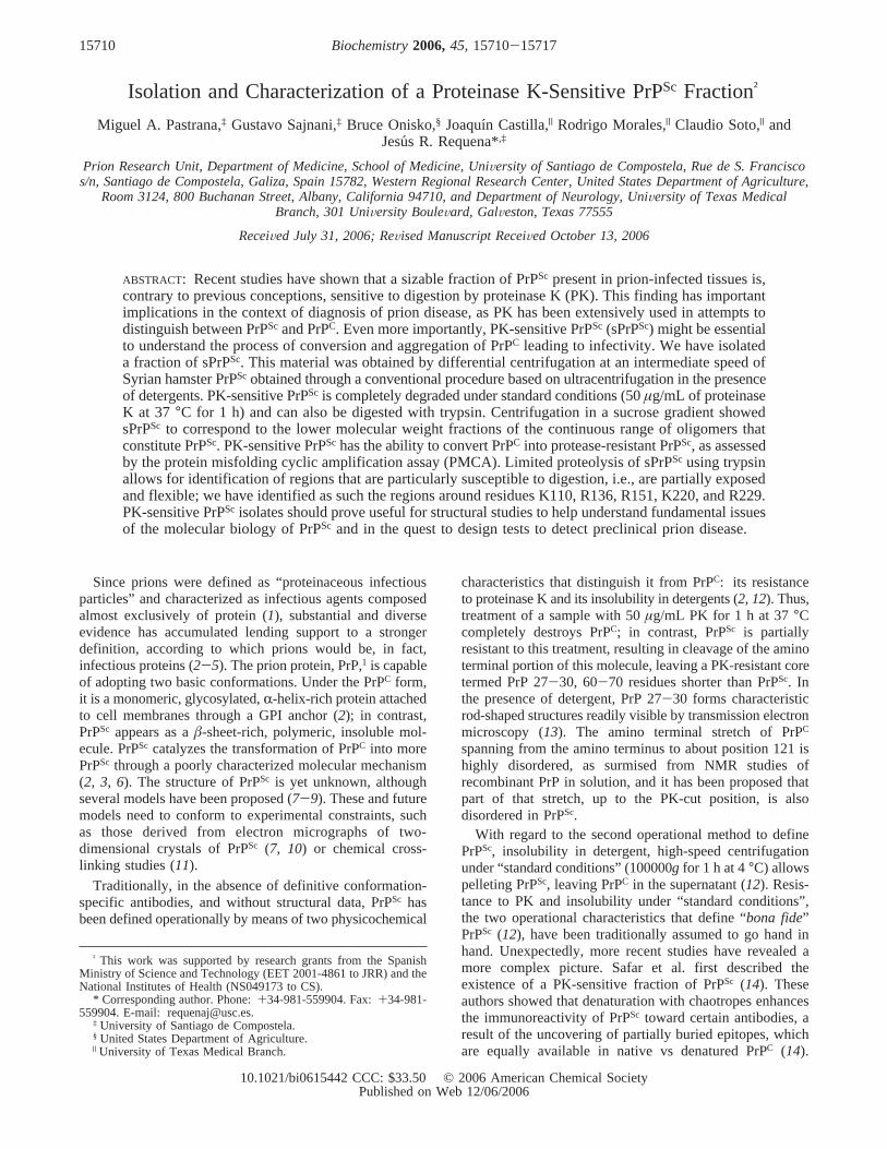
Isolation of an sPrPSc Fraction.Centrifugation of a PrPSc
suspension at an intermediate centrifugal force, as describedin the Experimental Procedures, generated pellets andsupernatants with divergent resistance to PK. Supernatants,typically making up 35-55% of the total starting PrPSc
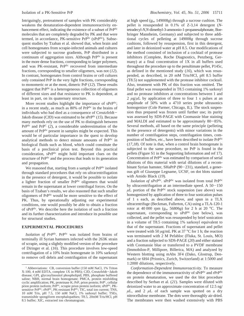
material, were much less resistant than pellets. By fine-tuningthe centrifugal force and length of centrifugation, we wereable to obtain a supernatant PrPSc fraction that was com-pletely hydrolyzed by the standard treatment with PK at 50µg/mL for 1 h at 37°C (sPrPSc), as judged by Western blotusing antibody 3F4 and Coomassie staining (Figure 1). Underthe same conditions, an equivalent amount of the pelletproduced the characteristic PK-resistant core (PrP 27-30)with an increased electrophoretic mobility, a consequenceof the trimming of 65-70 amino terminal residues. Wethereafter term this pellet fraction “rPrPSc”. Treatment ofsPrPSc with lower concentrations of PK showed that this
Y(N) ) I(N,0M)/I(N,4M)
15712 Biochemistry, Vol. 45, No. 51, 2006 Pastrana et al.
material exhibits, as expected, some resistance to degradationby this enzyme (Figure 2).
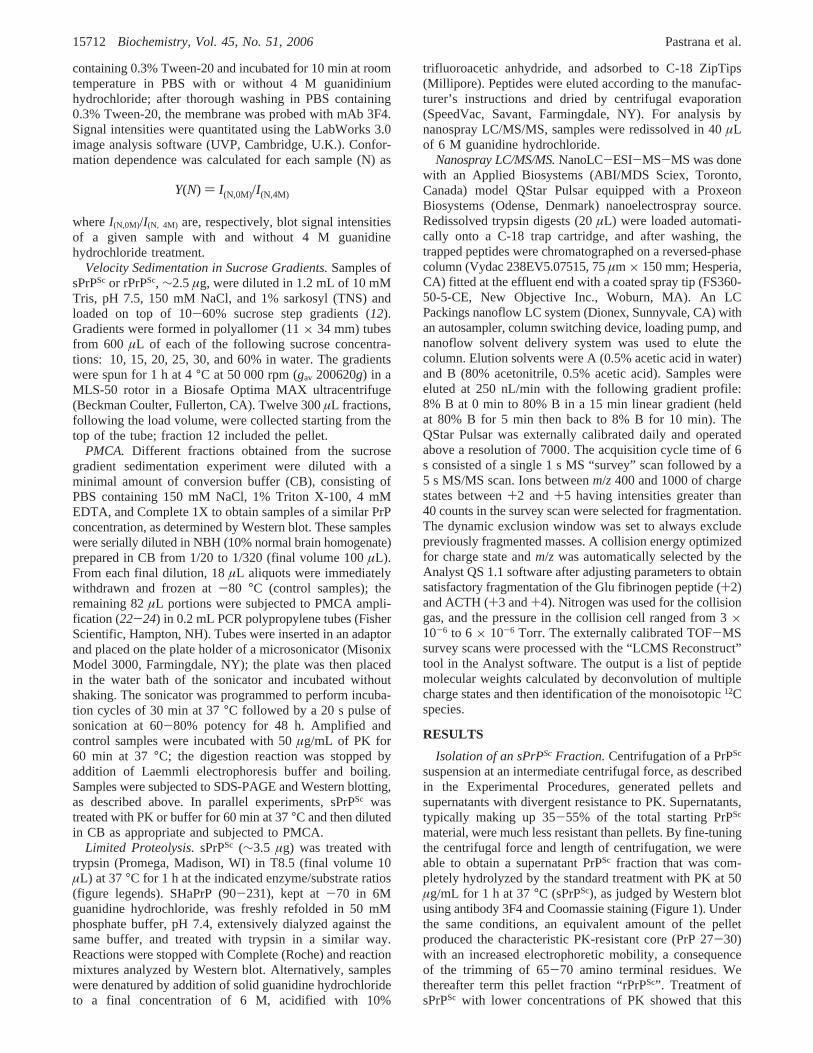
We next studied susceptibility of sPrPSc to trypsin.Treatment of sPrPSc with trypsin at 37°C for 1 h, at enzyme/substrate ratios in the range 1:20-1:60 (there was somevariability with different substrate and trypsin batches)resulted in a virtually complete disappearance of the protein,as assessed by Western blot using antibody 3F4 (Figure 3A);under similar conditions, an equivalent amount of rPrPSc,treated with the same ratio of trypsin, yielded a band ofapproximately the same intensity as that of the untreatedsample, albeit migrating slightly faster (Figure 3A). Thisapparent MW difference of 2000-3000 suggests clippingof rPrPSc at arginines 37 and 48 in the putatively unstructuredtail that, in contrast, is cleaved up to Gly90 by PK and agreeswith previously published results relative to total PrPSc (13).Further experiments showed that decreasing trypsin/substrateratios can be used to achieve limited proteolysis of sPrPSc
(Figure 3B). Western blots using mAb 6H4 (epitope 144-152) also showed a similar trypsin concentration-dependentsignal disappearance (Figure 3C), suggestive of a generalizedtryptic cleavage. When freshly refolded, recombinant SHaPrP-(90-231) was subjected to trypsin proteolysis under the sameconditions, complete disappearance of the 3F4 signal was
seen at an approximate enzyme/substrate ratio of 1:400,indicative of a much lower resistance to trypsin degradation.
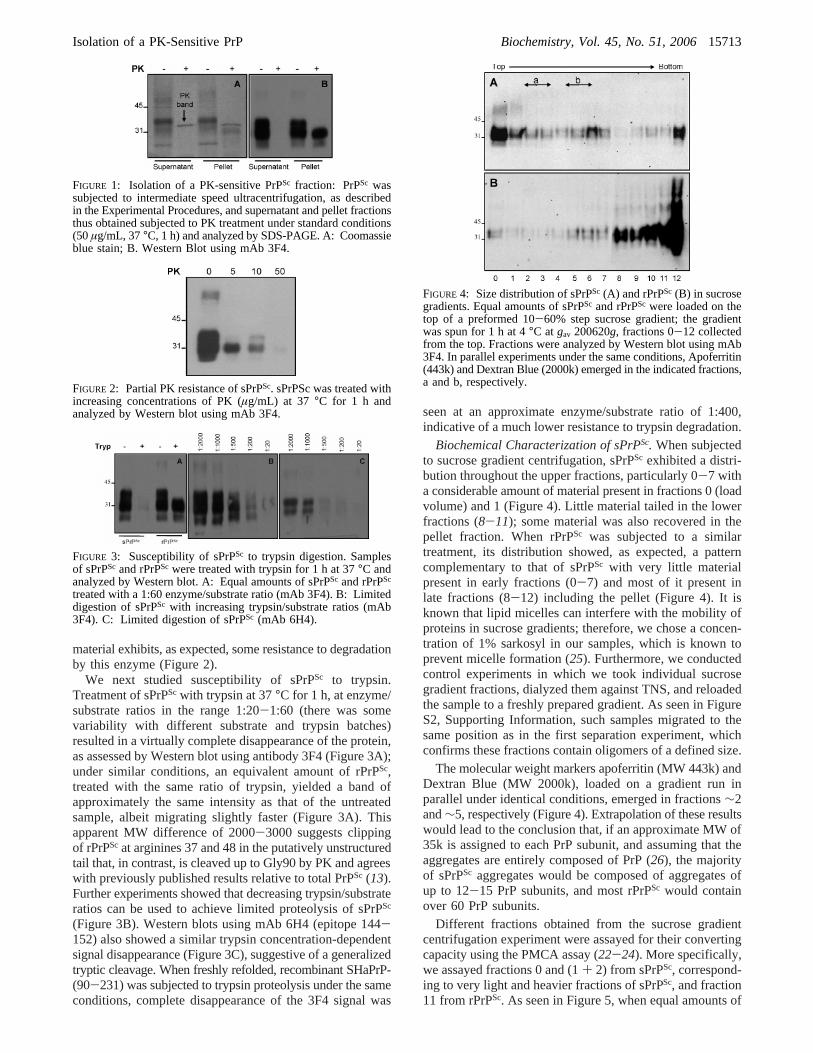
Biochemical Characterization of sPrPSc. When subjectedto sucrose gradient centrifugation, sPrPSc exhibited a distri-bution throughout the upper fractions, particularly 0-7 witha considerable amount of material present in fractions 0 (loadvolume) and 1 (Figure 4). Little material tailed in the lowerfractions (8-11); some material was also recovered in thepellet fraction. When rPrPSc was subjected to a similartreatment, its distribution showed, as expected, a patterncomplementary to that of sPrPSc with very little materialpresent in early fractions (0-7) and most of it present inlate fractions (8-12) including the pellet (Figure 4). It isknown that lipid micelles can interfere with the mobility ofproteins in sucrose gradients; therefore, we chose a concen-tration of 1% sarkosyl in our samples, which is known toprevent micelle formation (25). Furthermore, we conductedcontrol experiments in which we took individual sucrosegradient fractions, dialyzed them against TNS, and reloadedthe sample to a freshly prepared gradient. As seen in FigureS2, Supporting Information, such samples migrated to thesame position as in the first separation experiment, whichconfirms these fractions contain oligomers of a defined size.
The molecular weight markers apoferritin (MW 443k) andDextran Blue (MW 2000k), loaded on a gradient run inparallel under identical conditions, emerged in fractions∼2and∼5, respectively (Figure 4). Extrapolation of these resultswould lead to the conclusion that, if an approximate MW of35k is assigned to each PrP subunit, and assuming that theaggregates are entirely composed of PrP (26), the majorityof sPrPSc aggregates would be composed of aggregates ofup to 12-15 PrP subunits, and most rPrPSc would containover 60 PrP subunits.
Different fractions obtained from the sucrose gradientcentrifugation experiment were assayed for their convertingcapacity using the PMCA assay (22-24). More specifically,we assayed fractions 0 and (1+ 2) from sPrPSc, correspond-ing to very light and heavier fractions of sPrPSc, and fraction11 from rPrPSc. As seen in Figure 5, when equal amounts of
FIGURE 1: Isolation of a PK-sensitive PrPSc fraction: PrPSc wassubjected to intermediate speed ultracentrifugation, as describedin the Experimental Procedures, and supernatant and pellet fractionsthus obtained subjected to PK treatment under standard conditions(50µg/mL, 37°C, 1 h) and analyzed by SDS-PAGE. A: Coomassieblue stain; B. Western Blot using mAb 3F4.
FIGURE 2: Partial PK resistance of sPrPSc. sPrPSc was treated withincreasing concentrations of PK (µg/mL) at 37 °C for 1 h andanalyzed by Western blot using mAb 3F4.
FIGURE 3: Susceptibility of sPrPSc to trypsin digestion. Samplesof sPrPSc and rPrPSc were treated with trypsin for 1 h at 37°C andanalyzed by Western blot. A: Equal amounts of sPrPSc and rPrPSc
treated with a 1:60 enzyme/substrate ratio (mAb 3F4). B: Limiteddigestion of sPrPSc with increasing trypsin/substrate ratios (mAb3F4). C: Limited digestion of sPrPSc (mAb 6H4).
FIGURE 4: Size distribution of sPrPSc (A) and rPrPSc (B) in sucrosegradients. Equal amounts of sPrPSc and rPrPSc were loaded on thetop of a preformed 10-60% step sucrose gradient; the gradientwas spun for 1 h at 4°C atgav 200620g, fractions 0-12 collectedfrom the top. Fractions were analyzed by Western blot using mAb3F4. In parallel experiments under the same conditions, Apoferritin(443k) and Dextran Blue (2000k) emerged in the indicated fractions,a and b, respectively.
Isolation of a PK-Sensitive PrP Biochemistry, Vol. 45, No. 51, 200615713
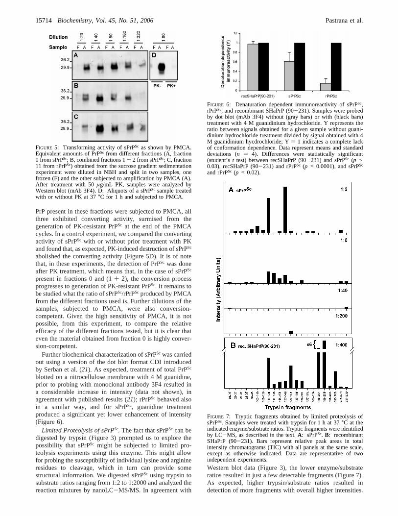
PrP present in these fractions were subjected to PMCA, allthree exhibited converting activity, surmised from thegeneration of PK-resistant PrPSc at the end of the PMCAcycles. In a control experiment, we compared the convertingactivity of sPrPSc with or without prior treatment with PKand found that, as expected, PK-induced destruction of sPrPSc
abolished the converting activity (Figure 5D). It is of notethat, in these experiments, the detection of PrPSc was doneafter PK treatment, which means that, in the case of sPrPSc
present in fractions 0 and (1+ 2), the conversion processprogresses to generation of PK-resistant PrPSc. It remains tobe studied what the ratio of sPrPSc/rPrPSc produced by PMCAfrom the different fractions used is. Further dilutions of thesamples, subjected to PMCA, were also conversion-competent. Given the high sensitivity of PMCA, it is notpossible, from this experiment, to compare the relativeefficacy of the different fractions tested, but it is clear thateven the material obtained from fraction 0 is highly conver-sion-competent.
Further biochemical characterization of sPrPSc was carriedout using a version of the dot blot format CDI introducedby Serban et al. (21). As expected, treatment of total PrPSc
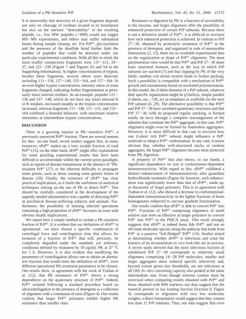
blotted on a nitrocellulose membrane with 4 M guanidine,prior to probing with monoclonal antibody 3F4 resulted ina considerable increase in intensity (data not shown), inagreement with published results (21); rPrPSc behaved alsoin a similar way, and for sPrPSc, guanidine treatmentproduced a significant yet lower enhancement of intensity(Figure 6).
Limited Proteolysis of sPrPSc. The fact that sPrPSc can bedigested by trypsin (Figure 3) prompted us to explore thepossibility that sPrPSc might be subjected to limited pro-teolysis experiments using this enzyme. This might allowfor probing the susceptibility of individual lysine and arginineresidues to cleavage, which in turn can provide somestructural information. We digested sPrPSc using trypsin tosubstrate ratios ranging from 1:2 to 1:2000 and analyzed thereaction mixtures by nanoLC-MS/MS. In agreement with
Western blot data (Figure 3), the lower enzyme/substrateratios resulted in just a few detectable fragments (Figure 7).As expected, higher trypsin/substrate ratios resulted indetection of more fragments with overall higher intensities.
FIGURE 5: Transforming activity of sPrPSc as shown by PMCA.Equivalent amounts of PrPSc from different fractions (A, fraction0 from sPrPSc; B, combined fractions 1+ 2 from sPrPSc; C, fraction11 from rPrPSc) obtained from the sucrose gradient sedimentationexperiment were diluted in NBH and split in two samples, onefrozen (F) and the other subjected to amplification by PMCA (A).After treatment with 50µg/mL PK, samples were analyzed byWestern blot (mAb 3F4). D: Aliquots of a sPrPSc sample treatedwith or without PK at 37°C for 1 h and subjected to PMCA.
FIGURE 6: Denaturation dependent immunoreactivity of sPrPSc,rPrPSc, and recombinant SHaPrP (90-231). Samples were probedby dot blot (mAb 3F4) without (gray bars) or with (black bars)treatment with 4 M guanidinium hydrochloride. Y represents theratio between signals obtained for a given sample without guani-dinium hydrochloride treatment divided by signal obtained with 4M guanidinium hycdrochloride; Y) 1 indicates a complete lackof conformation dependence. Data represent means and standarddeviations (n ) 4). Differences were statistically significant(student’st test) between recSHaPrP (90-231) and sPrPSc (p <0.03), recSHaPrP (90-231) and rPrPSc (p < 0.0001), and sPrPSc
and rPrPSc (p < 0.02).
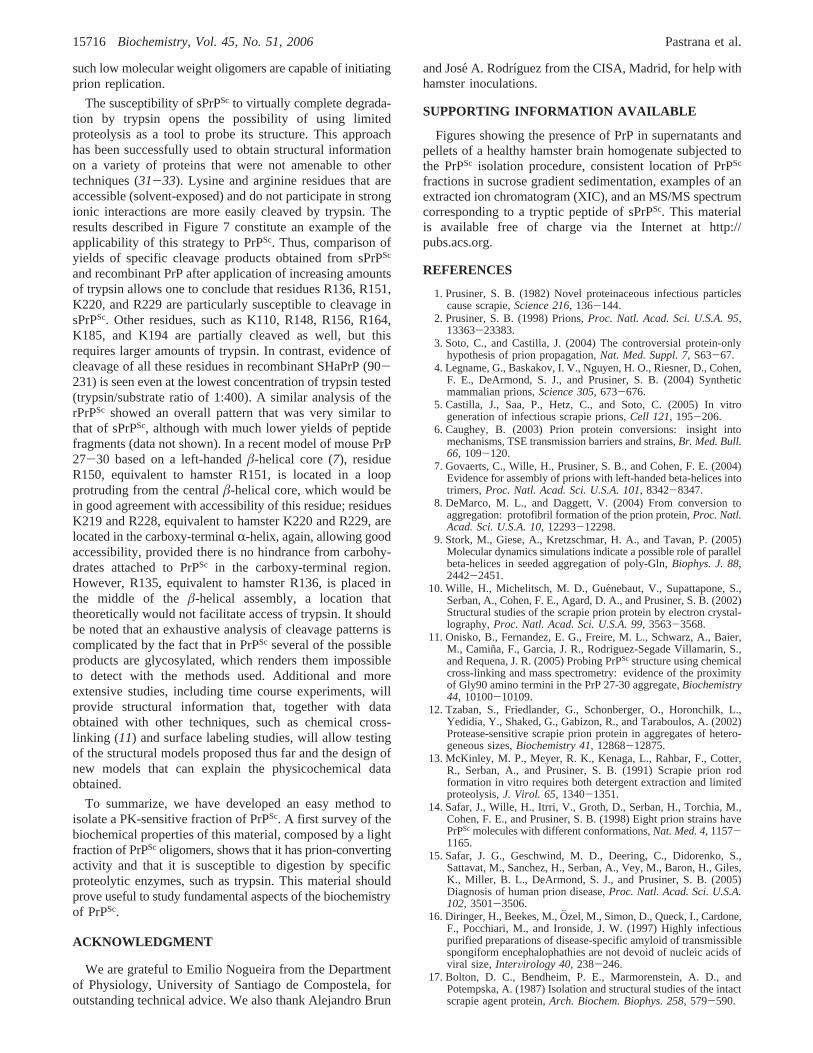
FIGURE 7: Tryptic fragments obtained by limited proteolysis ofsPrPSc. Samples were treated with trypsin for 1 h at 37°C at theindicated enzyme/substrate ratios. Tryptic fragments were identifiedby LC-MS, as described in the text.A: sPrPSc. B: recombinantSHaPrP (90-231). Bars represent relative peak areas in totalintensity chromatograms (TIC) with all panels at the same scale,except as otherwise indicated. Data are representative of twoindependent experiments.
15714 Biochemistry, Vol. 45, No. 51, 2006 Pastrana et al.
It is noteworthy that detection of a given fragment dependsnot only on cleavage of residues located at its boundariesbut also on the intrinsic “detectability” of the resultingpeptide; i.e., low MW peptides (<800) would not triggerMS-MS experiments, and others may suffer substantiallosses during sample cleanup, etc. For PrPSc, glycosylationand the presence of the disulfide bond further limit thenumber of peptides that could be detected under ourparticular experimental conditions. With all this in mind, themost readily conspicuous fragments were 137-151, 24-37, and 221-230 (Figure 7 and Figures S3 and S4 in theSupporting Information). At higher concentrations of trypsin,besides these fragments, several others were detected,including 111-136, 137-148, 152-164, and 157-164. Atan even higher trypsin concentration, intensity ratios of somefragments changed, indicating further fragmentation at previ-ously intact internal residues. As an example, peptides 111-136 and 137-148, which do not have any intact internal Kor R residues, increased steadily as the trypsin concentrationincreased, whereas fragments 111-148, 111-151, and 152-164 exhibited a bimodal behavior, with maximum relativeintensities at intermediate trypsin concentrations.
DISCUSSION
There is a growing interest in PK-sensitive PrPSc, apreviously unnoticed PrPSc fraction. There are several reasonsfor this: on one hand, recent studies suggest that, in manyinstances, sPrPSc makes up a very sizable fraction of totalPrPSc (15); on the other hand, sPrPSc might offer explanationsfor some puzzling experimental results that have beendifficult to accommodate within the current prion paradigm,such as reports of disease transmission in the absence of “PK-resistant PrP” (27), or the inherent difficulty to propagatesome prions, such as those causing some genetic forms ofdisease (28). Finally, the existence of sPrPSc has clearpractical implications, as it limits the usefulness of analyticaltechniques relying on the use of PK to detect PrPSc. Thisshould be carefully considered in the development of theurgently needed ultrasensitive tests capable of detecting PrPSc
in preclinical disease-suffering subjects and animals. Fur-thermore, the possibility of missing infected specimenscontaining a high proportion of sPrPSc becomes an issue withobvious health implications.
We report here a simple method to isolate a PK-sensitivefraction of PrPSc. It is evident that our definition of sPrPSc isoperational: we have chosen a specific combination ofcentrifugal force and centrifugation time that allows forisolation of a fraction of PrPSc that will, precisely, becompletely degraded under the standard, yet arbitrary,conditions defined by treatment by 50µg/mL PK at 37°Cfor 1 h. However, it is also evident that modifying theparameters of centrifugation allows one to obtain an alterna-tive fraction that would meet the definition of sPrPSc, weredifferent operational PK-treatment parameters to be chosen.Our results show, in agreement with the work of Tzaban etal. (12), that PK resistance of PrPSc shows a strongdependence on the quaternary structure of PrPSc. Indeed,PrPSc isolated following a standard procedure based onultracentrifugation in the presence of detergents is a collectionof oligomers with a continuum of sizes (Figure 4). Our resultsconfirm that larger PrPSc polymers exhibit higher PKresistance than smaller ones.
Resistance to digestion by PK is a function of accessibilityto the enzyme, and larger oligomers offer the possibility ofenhanced protection of certain PrP subunits. Because thereis not a definitive model of PrPSc, it is difficult to envisionhow such enhanced protection is achieved. In contrast to PrP27-30, obtained by proteolytic treatment of PrPSc in thepresence of detergent, and organized in rods of measurabledimensions (2, 13), there are no available experimental dataon the organization or shape of PrPSc oligomers. The mostparsimonious view would be that PrPSc and PrP 27-30 sharebasic structural features, such as the way in which PrPsubunits are stacked (7) and that clipping by PK of the verylabile, random coil amino termini leads to further packing.Such a possibility is compatible with a mechanism of PrPSc
growth and transmission based on nucleated polymerization.In this model, theâ-sheet domains of a PrP subunit, whatevertheir specific organization might be (â-helices, simple parallelâ-sheets, etc.;7,8), act as templates or scaffolds for the nextPrP subunit (6, 29). The alternative possibility is that PrPSc
and PrP 27-30 have unrelated quaternary structures and thatPrP 27-30, with its proposed regular stacking, is formedtotally de noVo through a complete rearrangement of thesubunits that constitute the PrPSc aggregate; in that case, PrPSc
oligomers might even be formed by featureless aggregates.However, it is more difficult in that case to envision howone â-sheet rich PrPSc subunit might influence a PrPC
molecule to adopt a PrPSc conformation. In any case, it seemsobvious that, whether well-structured stacks or randomaggregates, the larger PrPScoligomers become more protectedfrom PK digestion.
A property of PrPSc that also shows, in our hands, asignificant dependence on size is conformation-dependentimmunoreactivity. With regard to this, sPrPSc exhibited adistinct enhancement of immunoreactivity after guanidinehydrochloride treatment (Figure 6); however, such enhance-ment was significantly lower than that of rPrPSc, made up,as discussed, of larger polymers. This is in agreement withTzaban et al. (12), who showed a decrease in conformational-dependent immunoreactivity in the lighter fractions of brainhomogenates subjected to sucrose gradient fractionation.
Our results confirm that sPrPSc is able to convert PrPC intoPrPSc. Fractions of PrPSc comprising oligomers of smallrelative size were as effective as larger polymers to convertPrPC into PrPSc in the PMCA assay. This result stronglysuggests that sPrPSc is indeedbona fidePrPSc and not anoff-route molecular species along the pathway that leads fromPrPC to a putative “full-fledged” PrPSc (14). Studies aimedat determining whether sPrPSc is infectious and what thekinetics of its accumulationin ViVo look like are in process.A recent study showed that the most infectious fraction ofsolubilized PrP 27-30 corresponds to relatively smalloligomers comprising 14-28 PrP molecules; smaller andlarger aggregates show reduced specific infectivity and,beyond certain given size thresholds, are not infectious atall (30). In Vitro converting capacity also peaked at the sameintermediate size. Even though extreme caution must beexercised when comparing results obtained with PrPSc andthose obtained with MW markers, our data suggest that thematerial present in our loading fraction (fraction 0, Figure4) corresponds to oligomers with very low molecularweights; a direct interpolation would suggest that they containless than 12 PrP subunits. Thus, our data suggest that even
Isolation of a PK-Sensitive PrP Biochemistry, Vol. 45, No. 51, 200615715
such low molecular weight oligomers are capable of initiatingprion replication.
The susceptibility of sPrPSc to virtually complete degrada-tion by trypsin opens the possibility of using limitedproteolysis as a tool to probe its structure. This approachhas been successfully used to obtain structural informationon a variety of proteins that were not amenable to othertechniques (31-33). Lysine and arginine residues that areaccessible (solvent-exposed) and do not participate in strongionic interactions are more easily cleaved by trypsin. Theresults described in Figure 7 constitute an example of theapplicability of this strategy to PrPSc. Thus, comparison ofyields of specific cleavage products obtained from sPrPSc
and recombinant PrP after application of increasing amountsof trypsin allows one to conclude that residues R136, R151,K220, and R229 are particularly susceptible to cleavage insPrPSc. Other residues, such as K110, R148, R156, R164,K185, and K194 are partially cleaved as well, but thisrequires larger amounts of trypsin. In contrast, evidence ofcleavage of all these residues in recombinant SHaPrP (90-231) is seen even at the lowest concentration of trypsin tested(trypsin/substrate ratio of 1:400). A similar analysis of therPrPSc showed an overall pattern that was very similar tothat of sPrPSc, although with much lower yields of peptidefragments (data not shown). In a recent model of mouse PrP27-30 based on a left-handedâ-helical core (7), residueR150, equivalent to hamster R151, is located in a loopprotruding from the centralâ-helical core, which would bein good agreement with accessibility of this residue; residuesK219 and R228, equivalent to hamster K220 and R229, arelocated in the carboxy-terminalR-helix, again, allowing goodaccessibility, provided there is no hindrance from carbohy-drates attached to PrPSc in the carboxy-terminal region.However, R135, equivalent to hamster R136, is placed inthe middle of the â-helical assembly, a location thattheoretically would not facilitate access of trypsin. It shouldbe noted that an exhaustive analysis of cleavage patterns iscomplicated by the fact that in PrPSc several of the possibleproducts are glycosylated, which renders them impossibleto detect with the methods used. Additional and moreextensive studies, including time course experiments, willprovide structural information that, together with dataobtained with other techniques, such as chemical cross-linking (11) and surface labeling studies, will allow testingof the structural models proposed thus far and the design ofnew models that can explain the physicochemical dataobtained.
To summarize, we have developed an easy method toisolate a PK-sensitive fraction of PrPSc. A first survey of thebiochemical properties of this material, composed by a lightfraction of PrPScoligomers, shows that it has prion-convertingactivity and that it is susceptible to digestion by specificproteolytic enzymes, such as trypsin. This material shouldprove useful to study fundamental aspects of the biochemistryof PrPSc.
ACKNOWLEDGMENT
We are grateful to Emilio Nogueira from the Departmentof Physiology, University of Santiago de Compostela, foroutstanding technical advice. We also thank Alejandro Brun
and Jose´ A. Rodrıguez from the CISA, Madrid, for help withhamster inoculations.
SUPPORTING INFORMATION AVAILABLE
Figures showing the presence of PrP in supernatants andpellets of a healthy hamster brain homogenate subjected tothe PrPSc isolation procedure, consistent location of PrPSc
fractions in sucrose gradient sedimentation, examples of anextracted ion chromatogram (XIC), and an MS/MS spectrumcorresponding to a tryptic peptide of sPrPSc. This materialis available free of charge via the Internet at http://pubs.acs.org.
REFERENCES
1. Prusiner, S. B. (1982) Novel proteinaceous infectious particlescause scrapie,Science 216, 136-144.
2. Prusiner, S. B. (1998) Prions,Proc. Natl. Acad. Sci. U.S.A. 95,13363-23383.
3. Soto, C., and Castilla, J. (2004) The controversial protein-onlyhypothesis of prion propagation,Nat. Med. Suppl. 7, S63-67.
4. Legname, G., Baskakov, I. V., Nguyen, H. O., Riesner, D., Cohen,F. E., DeArmond, S. J., and Prusiner, S. B. (2004) Syntheticmammalian prions,Science 305, 673-676.
5. Castilla, J., Saa, P., Hetz, C., and Soto, C. (2005) In vitrogeneration of infectious scrapie prions,Cell 121, 195-206.
6. Caughey, B. (2003) Prion protein conversions: insight intomechanisms, TSE transmission barriers and strains,Br. Med. Bull.66, 109-120.
7. Govaerts, C., Wille, H., Prusiner, S. B., and Cohen, F. E. (2004)Evidence for assembly of prions with left-handed beta-helices intotrimers,Proc. Natl. Acad. Sci. U.S.A. 101, 8342-8347.
8. DeMarco, M. L., and Daggett, V. (2004) From conversion toaggregation: protofibril formation of the prion protein,Proc. Natl.Acad. Sci. U.S.A. 10, 12293-12298.
9. Stork, M., Giese, A., Kretzschmar, H. A., and Tavan, P. (2005)Molecular dynamics simulations indicate a possible role of parallelbeta-helices in seeded aggregation of poly-Gln,Biophys. J. 88,2442-2451.
10. Wille, H., Michelitsch, M. D., Gue´nebaut, V., Supattapone, S.,Serban, A., Cohen, F. E., Agard, D. A., and Prusiner, S. B. (2002)Structural studies of the scrapie prion protein by electron crystal-lography,Proc. Natl. Acad. Sci. U.S.A. 99, 3563-3568.
11. Onisko, B., Fernandez, E. G., Freire, M. L., Schwarz, A., Baier,M., Camina, F., Garcia, J. R., Rodriguez-Segade Villamarin, S.,and Requena, J. R. (2005) Probing PrPSc structure using chemicalcross-linking and mass spectrometry: evidence of the proximityof Gly90 amino termini in the PrP 27-30 aggregate,Biochemistry44, 10100-10109.
12. Tzaban, S., Friedlander, G., Schonberger, O., Horonchilk, L.,Yedidia, Y., Shaked, G., Gabizon, R., and Taraboulos, A. (2002)Protease-sensitive scrapie prion protein in aggregates of hetero-geneous sizes,Biochemistry 41, 12868-12875.
13. McKinley, M. P., Meyer, R. K., Kenaga, L., Rahbar, F., Cotter,R., Serban, A., and Prusiner, S. B. (1991) Scrapie prion rodformation in vitro requires both detergent extraction and limitedproteolysis,J. Virol. 65, 1340-1351.
14. Safar, J., Wille, H., Itrri, V., Groth, D., Serban, H., Torchia, M.,Cohen, F. E., and Prusiner, S. B. (1998) Eight prion strains havePrPSc molecules with different conformations,Nat. Med. 4, 1157-1165.
15. Safar, J. G., Geschwind, M. D., Deering, C., Didorenko, S.,Sattavat, M., Sanchez, H., Serban, A., Vey, M., Baron, H., Giles,K., Miller, B. L., DeArmond, S. J., and Prusiner, S. B. (2005)Diagnosis of human prion disease,Proc. Natl. Acad. Sci. U.S.A.102, 3501-3506.
16. Diringer, H., Beekes, M., O¨ zel, M., Simon, D., Queck, I., Cardone,F., Pocchiari, M., and Ironside, J. W. (1997) Highly infectiouspurified preparations of disease-specific amyloid of transmissiblespongiform encephalophathies are not devoid of nucleic acids ofviral size,InterVirology 40, 238-246.
17. Bolton, D. C., Bendheim, P. E., Marmorenstein, A. D., andPotempska, A. (1987) Isolation and structural studies of the intactscrapie agent protein,Arch. Biochem. Biophys. 258, 579-590.
15716 Biochemistry, Vol. 45, No. 51, 2006 Pastrana et al.
18. Caughey, B., Raymond, G. J., Priola, S. A., Kocisko, D. A., Race,R. E., Bessen, R. A., Lansbury, P. T., Jr., and Chesebro, B. (1999)Methods for studying prion protein (PrP) metabolism and theformation of protease-resistant PrP in cell culture and cell-freesystems,Mol. Biotechnol. 13, 45-55.
19. Beekes, M., Baldauf, E., Cassens, S., Diringer, H., Keyes, P., Scott,A. C., Wells, G. A. H., Brown, P., Gibbs, C. J., Jr., and Gajdusek,D. C. (1995) Western blot mapping of disease-specific amyloidin various animal species and humans with transmissible spongi-form encephalopathies using a high-yield purification method,J.Gen. Virol.76, 2567-2576.
20. Laemmli, U. K. (1970) Cleavage of structural proteins during theassembly of the head of bacteriophage T4,Nature 227, 680-685.
21. Serban, D., Taraboulos, A., DeArmond, S. J., and Prusiner, S. B.(1990) Rapid detection of Creutzfeldt-Jakob disease and scrapieprion proteins,Neurology 40, 110-117.
22. Saborio, G. P., Permanne, B., and Soto, C. (2001) Sensitivedetection of pathological prion protein by cyclic amplification ofprotein misfolding,Nature 411, 810-813.
23. Castilla, J., Saa´, P., and Soto, C. (2004) Cyclic Amplification ofPrion Protein Misfolding, in: Techniques in Prion Research(Lehman, S., and Grassi, J., Eds.), pp 198-213, Birkhouser, Basel.
24. Saa´, P., Castilla, J., and Soto, C. (2004). Cyclic Amplification ofPrion Protein Misfolding, inAmyloid Proteins: Methods andProtocol. (Sigurdsson, E. M., Ed.) pp 53-65, Humana Press,Totowa, NJ.
25. Riesner, D., Kellings, K., Post, K., Wille, H., Serban, A., Groth,D., Baldwin, M. A., and Prusine, r S. B. (1996). Disruption ofprion rods generates 10-nm spherical particles having highR-helical content and lacking scrapie infectivity,J. Virol. 70,1714-1722.
26. Chiesa, R., Piccardo, P., Quaglio, E., Drisaldi, B., Si-Hoe, S. L.,Takao, M., Ghetti, B., and Harris, D. A. (2003) Moleculardistinction between pathogenic and infectious properties of theprion protein,J. Virol. 77, 7611-7622.
27. Lasmezas, C. I., Deslys, J. P., Robain, O., Jaegly, A., Beringue,V., Peyrin, J. M., Fournier, J. G., Hauw, J. J., Rossier, J., andDormont, D. (1997) Transmission of the BSE agent to mice inthe absence of detectable abnormal prion protein,Science 275,402-405.
28. Brown, P., Gibbs, C. J., Rodgers-Johnson, P., Asher, D. M.,Sulima, M. P., Bacote, A., Goldfarb, L. G., and Gajdusek, D. C.(1994) Human spongiform encephalopathy: the National Institutesof Health series of 300 cases of experimentally transmitted disease,Ann. Neurol. 35, 513-529.
29. Come, J. H., Fraser, P. E., and Lansbury, P. T., Jr. (1993) A kineticmodel for amyloid formation in the prion diseases: Importanceof seeding,Proc. Natl. Acad. Sci. U.S.A. 90, 5959-5963.
30. Silveira, J. R., Raymond, G. J., Hughson, A. G., Race, R. E., Sim,V. L., Hayes, S. F., and Caughey, B. (2005) The most infectiousprion protein particles,Nature 437, 257-261.
31. Hubbard, S. J. (1998) The structural aspects of limited proteolysisof native proteins,Biochim. Biophys. Acta 1382, 191-206.
32. Kheterpal, I., Williams, A., Murphy, C., Bledsoe, B., and Wetzel,R. (2001) Structural features of the Aâ amyloid fibril elucidatedby limited proteolysis,Biochemistry 40, 11757-11767.
33. Neurath, H. (1980) Limited Proteolysis, Protein Folding andPhysiological Regulation, inProtein Folding(Jaenicke, R., Ed.)pp 501-523, Elsevier, Amsterdam, The Netherlands.
BI0615442
Isolation of a PK-Sensitive PrP Biochemistry, Vol. 45, No. 51, 200615717