insuficiencia renal aguda
TRANSCRIPT
INSUFICIENCIA RENAL AGUDA
DEFINICIÓN AKI
AKI como cualquiera de los siguientes:
Incremento de la creatinina sérica por 0.3 m/dl ( x 26.5 lmol/l) dentro de
las primeras 48 horas. o
Incremento de la creatinina sérica por 1.5 el valor basal, que sea
conocido o presumido que haya ocurrido dentro de los primeros 7 días o
Volumen urinario de 0.05 ml/kg/h en 6 horas.
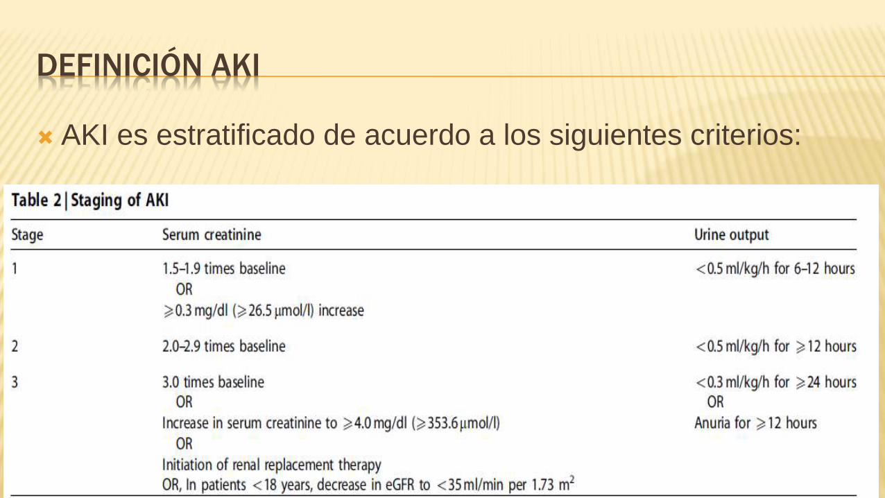
DEFINICIÓN AKI
AKI es estratificado de acuerdo a los siguientes criterios:
DEFINICIÓN AKI
2.1.3: The cause of AKI should be determined whenever possible. (Not Graded)
2.2.1: We recommend that patients be stratified for risk of AKI according to their susceptibilities and exposures. (1B)
2.2.2: Manage patients according to their susceptibilities and exposures to reduce the risk of AKI (see relevant guideline
sections). (Not Graded)
2.2.3: Test patients at increased risk for AKI with measurements of SCr and urine output to detect AKI. (Not Graded)
Individualize frequency and duration of monitoring based on patient risk and clinical course. (Not Graded)
2.3.1: Evaluate patients with AKI promptly to determine the cause, with special attention to reversible causes.
(Not Graded)
2.3.2: Monitor patients with AKI with measurements of SCr and urine output to stage the severity, according to
Recommendation 2.1.2. (Not Graded)
2.3.3: Manage patients with AKI according to the stage (see Figure 4) and cause. (Not Graded)
2.3.4: Evaluate patients 3 months after AKI for resolution, new onset, or worsening of pre-existing CKD. (Not Graded)
K If patients have CKD, manage these patients as detailed in the KDOQI CKD Guideline (Guidelines 7–15).
(Not Graded)
K If patients do not have CKD, consider them to be at increased risk for CKD and care for them as detailed in
the KDOQI CKD Guideline 3 for patients at increased risk for CKD. (Not Graded)
SECTION 3: PREVENTION AND TREATMENT OF AKI
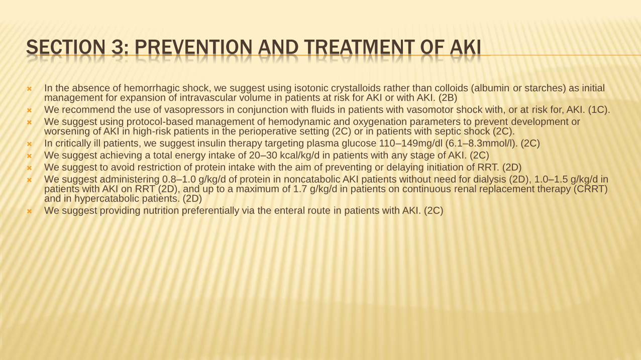
In the absence of hemorrhagic shock, we suggest using isotonic crystalloids rather than colloids (albumin or starches) as initial management for expansion of intravascular volume in patients at risk for AKI or with AKI. (2B)
We recommend the use of vasopressors in conjunction with fluids in patients with vasomotor shock with, or at risk for, AKI. (1C).
We suggest using protocol-based management of hemodynamic and oxygenation parameters to prevent development or worsening of AKI in high-risk patients in the perioperative setting (2C) or in patients with septic shock (2C).
In critically ill patients, we suggest insulin therapy targeting plasma glucose 110–149mg/dl (6.1–8.3mmol/l). (2C)
We suggest achieving a total energy intake of 20–30 kcal/kg/d in patients with any stage of AKI. (2C)
We suggest to avoid restriction of protein intake with the aim of preventing or delaying initiation of RRT. (2D)
We suggest administering 0.8–1.0 g/kg/d of protein in noncatabolic AKI patients without need for dialysis (2D), 1.0–1.5 g/kg/d in patients with AKI on RRT (2D), and up to a maximum of 1.7 g/kg/d in patients on continuous renal replacement therapy (CRRT) and in hypercatabolic patients. (2D)
We suggest providing nutrition preferentially via the enteral route in patients with AKI. (2C)

SECTION 3: PREVENTION AND TREATMENT OF AKI
We recommend not using diuretics to prevent AKI. (1B)
We suggest not using diuretics to treat AKI, except in the management of volume overload. (2C)
We recommend not using low-dose dopamine to prevent or treat AKI. (1A)
We suggest not using fenoldopam to prevent or treat AKI. (2C)
We suggest not using atrial natriuretic peptide (ANP) to prevent (2C) or treat (2B) AKI.
We recommend not using recombinant human (rh)IGF-1 to prevent or treat AKI. (1B)
We suggest that a single dose of theophylline may be given in neonates with severe perinatal asphyxia, who are at high risk of AKI. (2B)
We suggest not using aminoglycosides for the treatment of infections unless no suitable, less nephrotoxic,therapeutic alternatives are available. (2A)
We suggest that, in patients with normal kidney function in steady state, aminoglycosides are administered as a single dose daily rather than multiple-dose daily treatment regimens. (2B)
SECTION 3: PREVENTION AND TREATMENT OF AKI
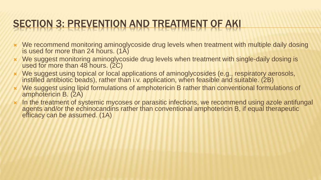
We recommend monitoring aminoglycoside drug levels when treatment with multiple daily dosing is used for more than 24 hours. (1A)
We suggest monitoring aminoglycoside drug levels when treatment with single-daily dosing is used for more than 48 hours. (2C)
We suggest using topical or local applications of aminoglycosides (e.g., respiratory aerosols, instilled antibiotic beads), rather than i.v. application, when feasible and suitable. (2B)
We suggest using lipid formulations of amphotericin B rather than conventional formulations of amphotericin B. (2A)
In the treatment of systemic mycoses or parasitic infections, we recommend using azole antifungal agents and/or the echinocandins rather than conventional amphotericin B, if equal therapeutic efficacy can be assumed. (1A)
SECTION 3: PREVENTION AND TREATMENT OF AKI
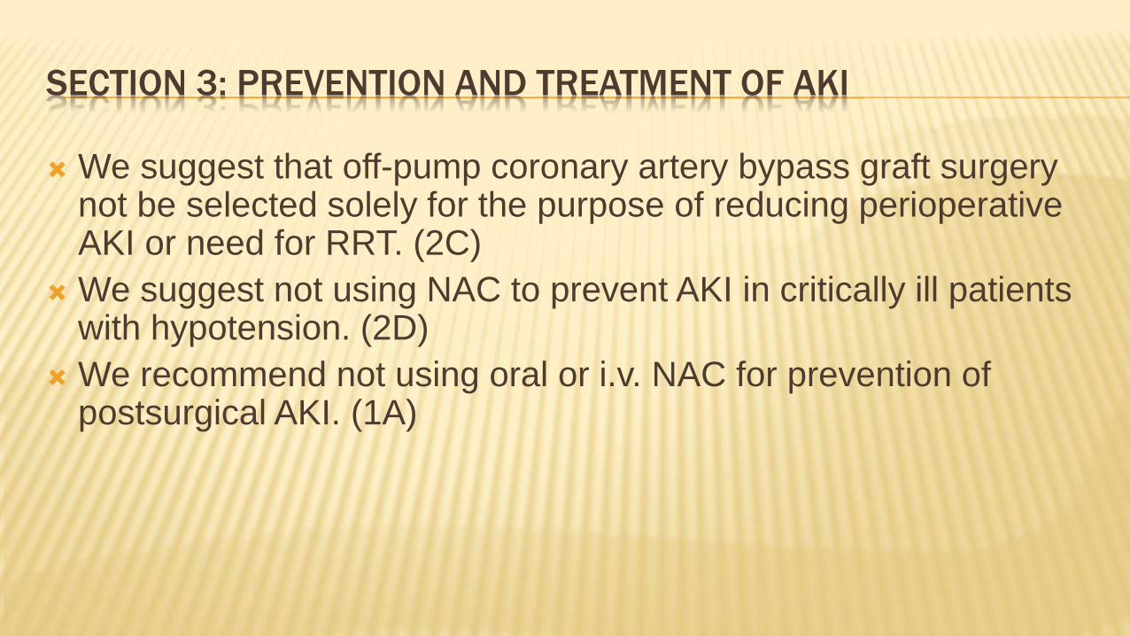
We suggest that off-pump coronary artery bypass graft surgery not be selected solely for the purpose of reducing perioperative AKI or need for RRT. (2C)
We suggest not using NAC to prevent AKI in critically ill patients with hypotension. (2D)
We recommend not using oral or i.v. NAC for prevention of postsurgical AKI. (1A)

SECTION 4: CONTRAST-INDUCED AKI
Define and stage AKI after administration of intravascular contrast media as per Recommendations.
In individuals who develop changes in kidney function after administration of intravascular contrast media, evaluate for CI-AKI as well as for other possible causes of AKI.
Assess the risk for CI-AKI and, in particular, screen for pre-existing impairment of kidney function in all patients who are considered for a procedure that requires intravascular (i.v. or i.a.) administration of iodinated contrast medium.
Consider alternative imaging methods in patients at increased risk for CI-AKI.
Use the lowest possible dose of contrast medium in patients at risk for CI-AKI.
We recommend using either iso-osmolar or low-osmolar iodinated contrast media, rather than high-osmolar iodinated contrast media in patients at increased risk of CI-AKI. (1B)
We recommend i.v. volume expansion with either isotonic sodium chloride or sodium bicarbonate solutions, rather than no i.v.volume expansion, in patients at increased risk for CI-AKI. (1A)
We recommend not using oral fluids alone in patients at increased risk of CI-AKI. (1C)
We suggest using oral NAC, together with i.v. isotonic crystalloids, in patients at increased risk of CI-AKI. (2D)
We suggest not using theophylline to prevent CI-AKI. (2C)
We recommend not using fenoldopam to prevent CI-AKI. (1B)
We suggest not using prophylactic intermittent hemodialysis (IHD) or hemofiltration (HF) for contrast-media
removal in patients at increased risk for CI-AKI. (2C)
SECTION 5: DIALYSIS INTERVENTIONS FOR TREATMENT OF AKI
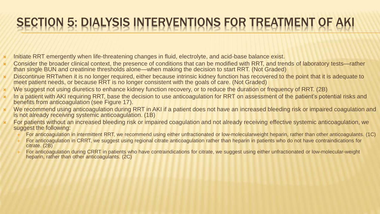
Initiate RRT emergently when life-threatening changes in fluid, electrolyte, and acid-base balance exist.
Consider the broader clinical context, the presence of conditions that can be modified with RRT, and trends of laboratory tests—rather than single BUN and creatinine thresholds alone—when making the decision to start RRT. (Not Graded)
Discontinue RRTwhen it is no longer required, either because intrinsic kidney function has recovered to the point that it is adequate to meet patient needs, or because RRT is no longer consistent with the goals of care. (Not Graded)
We suggest not using diuretics to enhance kidney function recovery, or to reduce the duration or frequency of RRT. (2B)
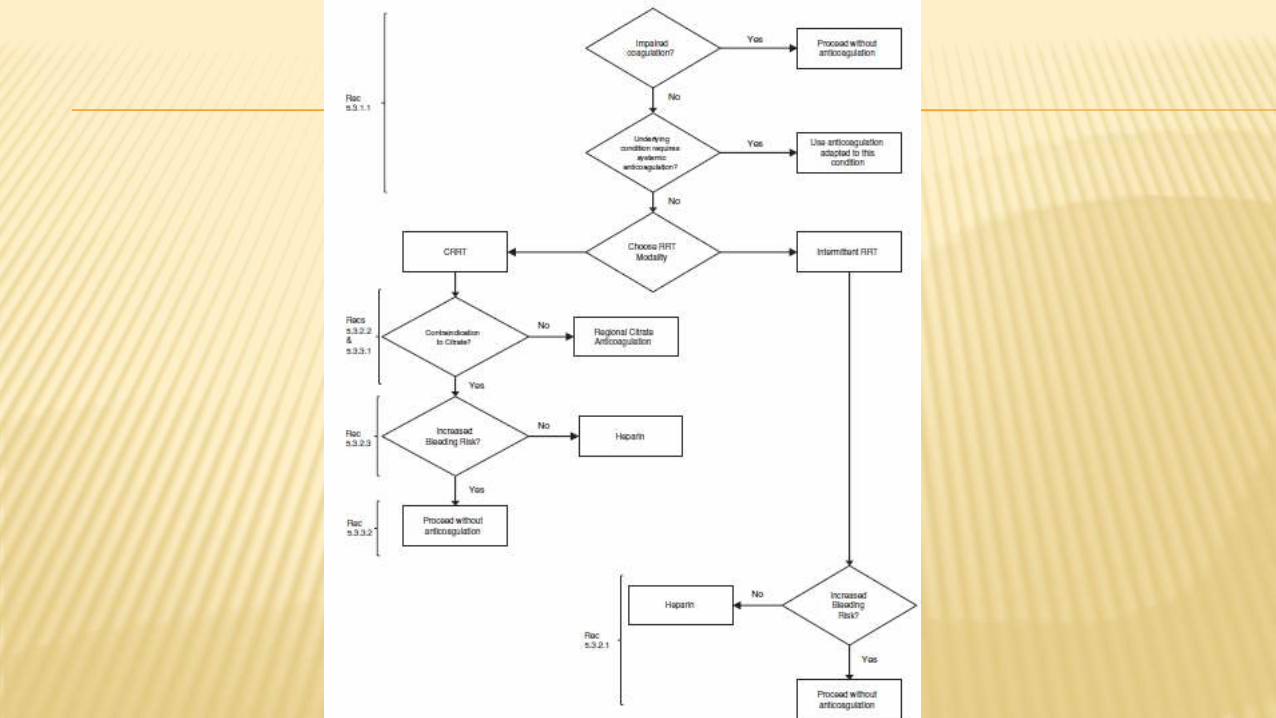
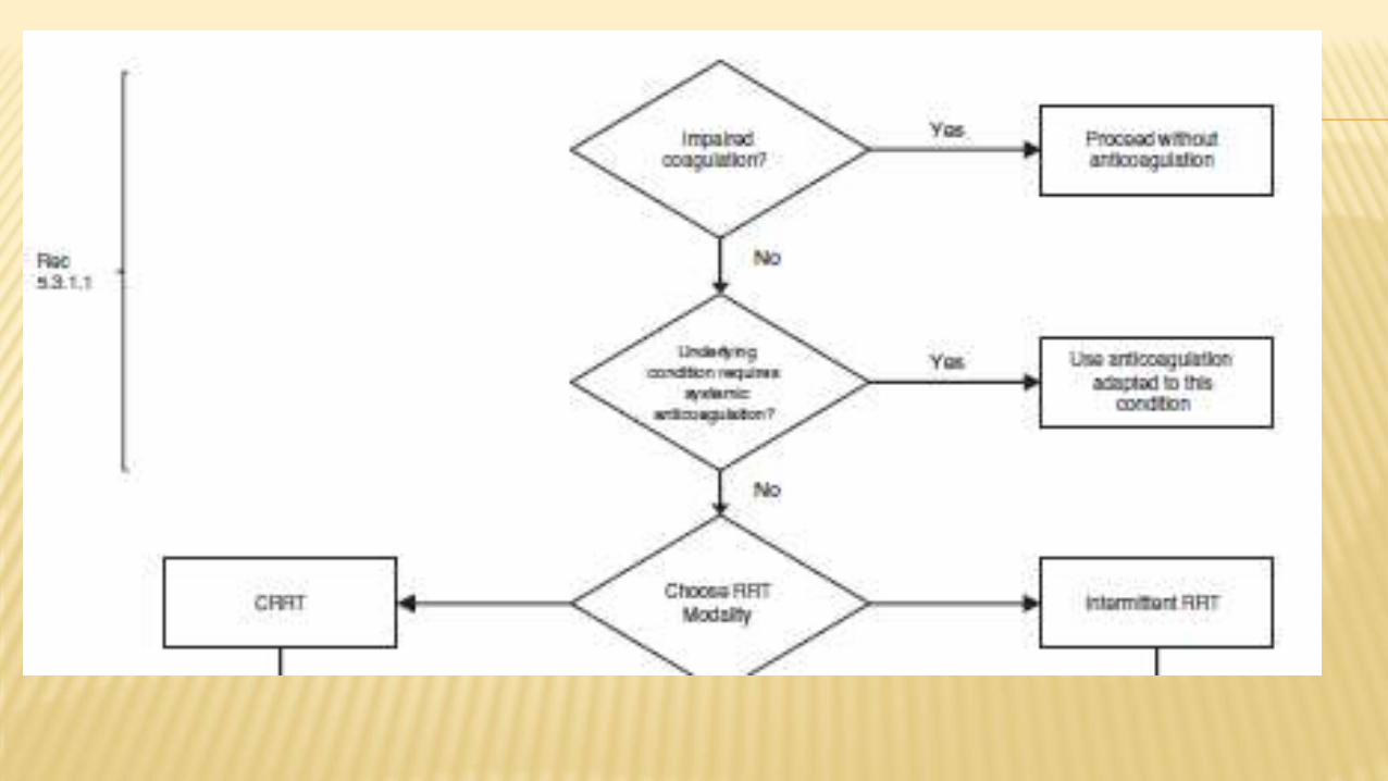
In a patient with AKI requiring RRT, base the decision to use anticoagulation for RRT on assessment of the patient’s potential risks and benefits from anticoagulation (see Figure 17).
We recommend using anticoagulation during RRT in AKI if a patient does not have an increased bleeding risk or impaired coagulation and is not already receiving systemic anticoagulation. (1B)
For patients without an increased bleeding risk or impaired coagulation and not already receiving effective systemic anticoagulation, we suggest the following: For anticoagulation in intermittent RRT, we recommend using either unfractionated or low-molecularweight heparin, rather than other anticoagulants. (1C)
For anticoagulation in CRRT, we suggest using regional citrate anticoagulation rather than heparin in patients who do not have contraindications for citrate. (2B)
For anticoagulation during CRRT in patients who have contraindications for citrate, we suggest using either unfractionated or low-molecular-weight heparin, rather than other anticoagulants. (2C)
SECTION 5: DIALYSIS INTERVENTIONS FOR
TREATMENT OF AKI
For patients with increased bleeding risk who are not receiving anticoagulation, we suggest the following for anticoagulation during RRT: We suggest using regional citrate anticoagulation, rather than no anticoagulation, during CRRT in a patient without contraindications for citrate. (2C)
We suggest avoiding regional heparinization during CRRT in a patient with increased risk of bleeding. (2C)
In a patient with heparin-induced thrombocytopenia (HIT), all heparin must be stopped and we recommend using direct thrombin inhibitors (such as argatroban) or Factor Xa inhibitors (such as danaparoid or
fondaparinux) rather than other or no anticoagulation during RRT. (1A)
In a patient with HIT who does not have severe liver failure, we suggest using argatroban rather than other thrombin or Factor Xa inhibitors during RRT. (2C)
We suggest initiating RRT in patients with AKI via an uncuffed nontunneled dialysis catheter, rather than a tunneled catheter. (2D)
When choosing a vein for insertion of a dialysis catheter in patients with AKI, consider these preferences
First choice: right jugular vein; Second choice: femoral vein; K Third choice: left jugular vein; Last choice: subclavian vein with preference for the dominant side.
We recommend using ultrasound guidance for dialysis catheter insertion. (1A)
We recommend obtaining a chest radiograph promptly after placement and before first use of an internal jugular or subclavian dialysis catheter. (1B)
We suggest not using topical antibiotics over the skin insertion site of a nontunneled dialysis catheter in ICU patients with AKI requiring RRT. (2C)
We suggest not using antibiotic locks for prevention of catheter-related infections of nontunneled dialysis catheters in AKI requiring RRT. (2C)

SECTION 5: DIALYSIS INTERVENTIONS FOR TREATMENT OF AKI
We suggest to use dialyzers with a biocompatible membrane for IHD and CRRT in patients with AKI. (2C)
Use continuous and intermittent RRT as complementary therapies in AKI patients. (Not Graded)
We suggest using CRRT, rather than standard intermittent RRT, for hemodynamicallyunstable patients. (2B)
We suggest using CRRT, rather than intermittent RRT, for AKI patients with acute brain injury or other causes of increased intracranial pressure or generalized brain edema. (2B)
We suggest using bicarbonate, rather than lactate, as a buffer in dialysate and replacement fluid for RRT in patients with AKI. (2C)
We recommend using bicarbonate, rather than lactate, as a buffer in dialysate and replacement fluid for RRT in patients with AKI and circulatory shock. (1B)
SECTION 5: DIALYSIS INTERVENTIONS FOR TREATMENT OF AKI
We suggest using bicarbonate, rather than lactate, as a buffer in dialysate and replacement fluid for RRT in patients with AKI and liver failure and/or lactic acidemia. (2B)
We recommend that dialysis fluids and replacement fluids in patients with AKI, at a minimum, comply with
American Association of Medical Instrumentation (AAMI) standards regarding contamination with bacteria and endotoxins. (1B)
The dose of RRT to be delivered should be prescribed before starting each session of RRT. (Not Graded) We recommend frequent assessment of the actual delivered dose in order to adjust the prescription. (1B)
Provide RRT to achieve the goals of electrolyte, acid-base, solute, and fluid balance that will meet the patient’s needs. (Not Graded)
We recommend delivering a Kt/V of 3.9 per week when using intermittent or extended RRT in AKI. (1A)
We recommend delivering an effluent volume of 20–25 ml/kg/h for CRRT in AKI (1A). This will usually require a higher prescription of effluent volume. (Not Graded)


SECTION 1: INTRODUCTION AND METHODOLOGY
Chapter 1.1: Introduction
Tasa de filtración Glomerular y creatinina sérica.
TFG es largamente aceptada como como el mejor índice de función renal
Recently, Chertow et al.1 que un amento de la CrS mayor de 0.3 mg/dl o mayor a 26.5 mmol/l era independientemente asociada
con mortalidad.
Similarly, Lassnigg et al.3 saw, pacientes sometidos a cirugía cardiac que tanto un amento de creatinine sérica por 0.5 mg/dl o
mayor a 44,2 nmol/l o un decrement de 0.3 mg/dl (mayor a 26.5 umol/l eran asociados con menor sobrevivencia.
Oliguria y anuria
Controvertidamente cuando los tubules estan dañados, la máxima habilidad de concentración es impar y el volume urinario podría
incluso ser normal (ej falla renal no oligúrica)
Una osmolaridad urinaria alta junto con un sodio urinario bajo en el context de oliguria y azotemia es una evidencia fuerte de
función tubular intacta.
Clasificaciones como azotemia inicial y suceso renal agudo no son consistentes con evidencia actual.
Oliguria severa e incluso anuria resultarían en un daño tubular renal., podrían ser casusadas tambien por obstrucción u occlusión
venosa. Estas condiciones resultarían en un irreversible y rápido daño para el riñón y require pronto reconocimiento y manejo.
.
SECTION 1: INTRODUCTION AND METHODOLOGY
Necrosis Tubular Aguda Cuando los riñones de los mamíferos son expuestos a isquemia caliente y luego
seguido por reperfusion hay extensive necrosis destruyendo los tubulos proximalesde las vecindades de la médula y los tubulos contorneados proximales se tornannecróticos tambien.
El término NTA es usado para describer una situación clínica en la cual hay adecuadaperfusión renal para mantener la integridad del tubulo pero no para mantener la filtración glomerular.
La verdadera NTA ocurre de hecho por ejemplo en pacientes con castástrofesarteriales (ruptura de aneurismas, descción aguda).
SECTION 1: INTRODUCTION AND METHODOLOGY
ARF ischuria renalis, was by William Heberden in 1802.
Durante la primera Guerra mundial se llamó nefritis de la guerra.
Fue Homer W. Smith a quien se le dió el crédito por la introducción del término“Falla Renal Aguda” es su capítulo ‘‘Acute renal failure related to traumatic injuries’’ in his textbook The kidney-structure and function in health and disease (1951).
Un studio reciente encontró 35 definiciones diferentes de falla renal aguda.

SECTION 1: INTRODUCTION AND METHODOLOGY
RIFLE criteria
Risk, Injury, and
Failure; and the two
outcome classes, Loss
and End-Stage Renal
Disease (ESRD)

SECTION 1: INTRODUCTION AND METHODOLOGY
AKI: acute kidney injury/impairment. Término propuesto para englobar el espectro entero del síndrome para cambios
mínimos en los marcadores de función renal para la terapia de reemplazo renal.
Aki no es ATN, tampoco es falla renal aguda, es en cambio un englobe de ambas e incluso incluye otras condiciones menos severas. En efecto un syndrome que incluyepacientes sin un daño actual al riñon pero con una función disminuida relative a la demanda fisiológica.
AKI sostenido lidera a una profunda alteración en fluidos, electrolitos, equilibrio ácidobase y regulación hormonal. AKI resulta en anormalidades en el nervio central, immune y sistemas de coagulación.
SECTION 1: INTRODUCTION AND METHODOLOGY
These criteria were formulated as follows: AKI is common.
AKI imposes a heavy burden of illness (morbidity and mortality).
The cost per person of managing AKI is high.
AKI is amenable to early detection and potential prevention.
There is considerable variability in practice to prevent, diagnose, treat, and achieve outcomes of AKI.
Clinical practice guidelines in the field have the potential to reduce variations, improve outcomes, and reduce costs.
Formal guidelines do not exist on this topic.

SECTION 2: AKI DEFINITION

CHAPTER 2.1: DEFINITION AND CLASSIFICATION OF AKI

CHAPTER 2.1: DEFINITION AND CLASSIFICATION OF AKI
CHAPTER 2.1: DEFINITION AND CLASSIFICATION OF AKI
Falla renal es definida como una Tasa de Filtración Glmerular
menor de 15 ml/min/1.73m2 supeficie de área corporal
TFG como mejor índice para evaluar la función renal.
CHAPTER 2.1: DEFINITION AND CLASSIFICATION OF AKI

CHAPTER 2.1: DEFINITION AND CLASSIFICATION OF AKI
CHAPTER 2.1: DEFINITION AND CLASSIFICATION OF AKI

CHAPTER 2.1: DEFINITION AND CLASSIFICATION OF AKI
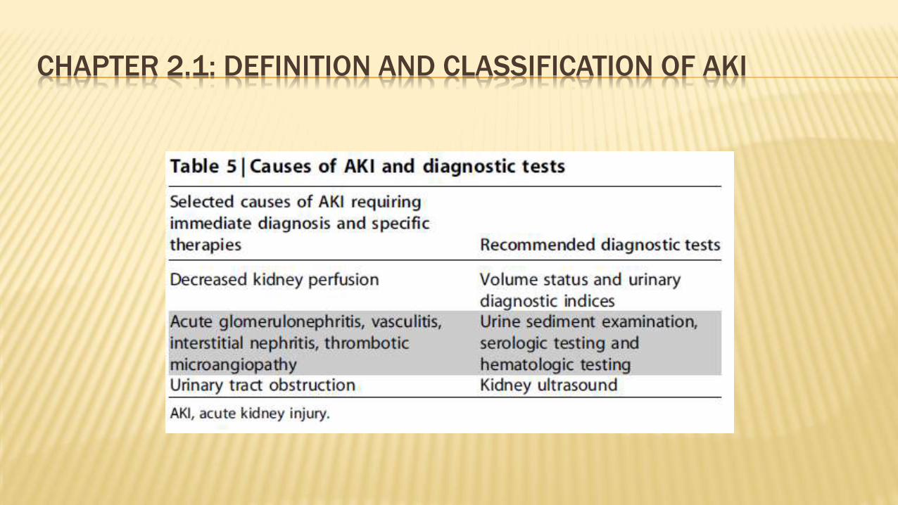
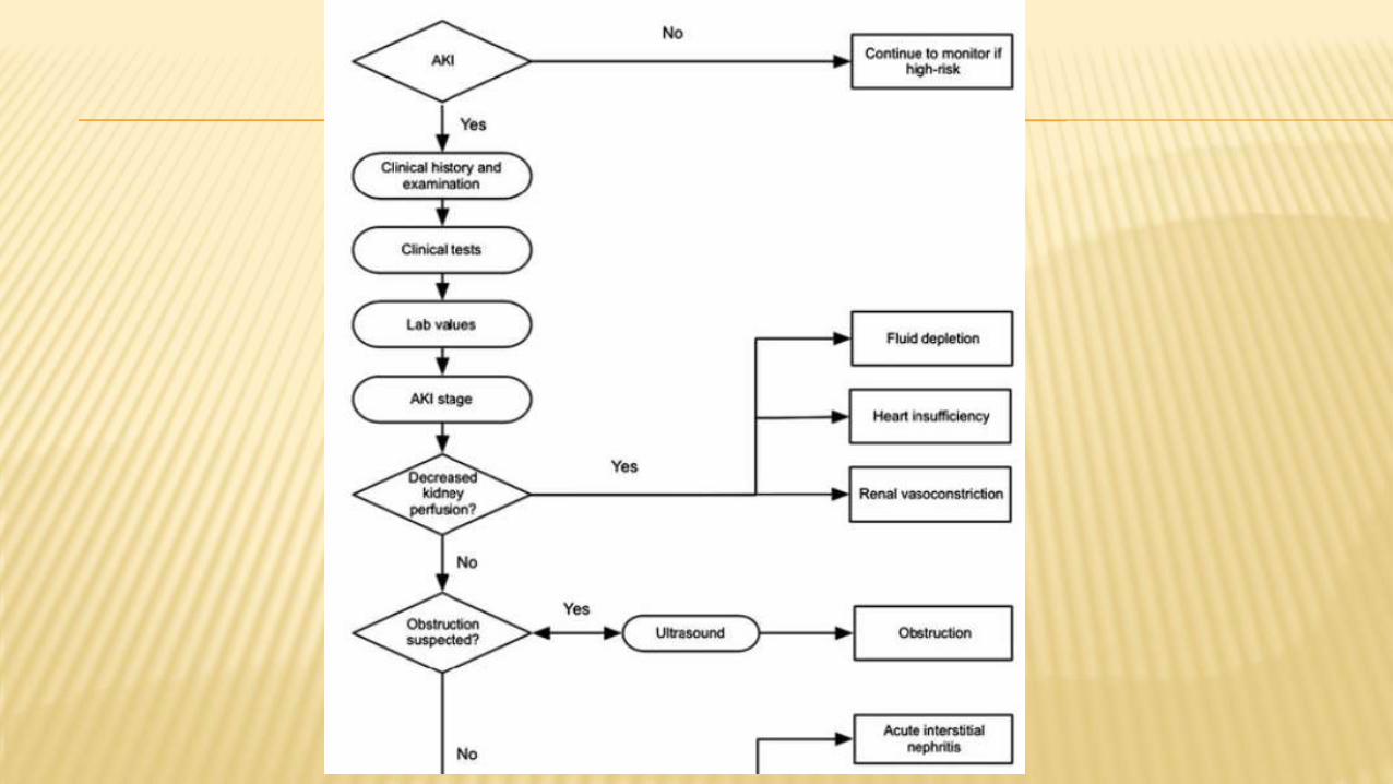
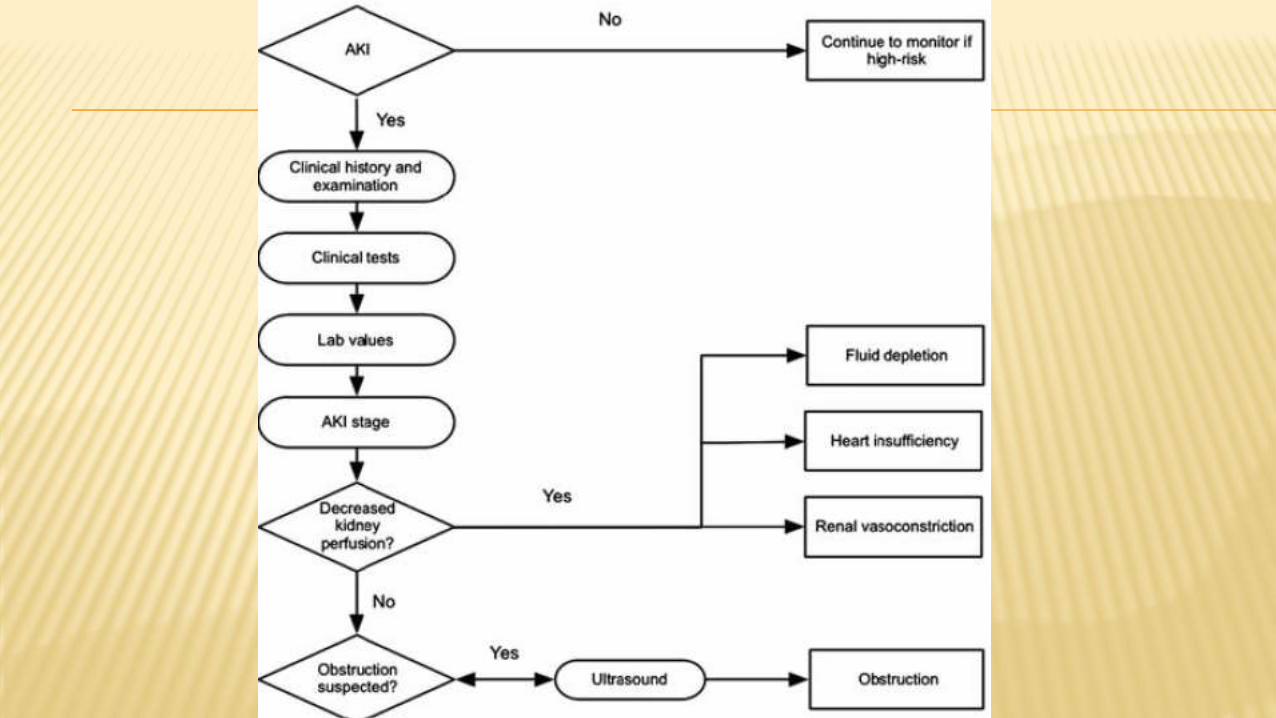
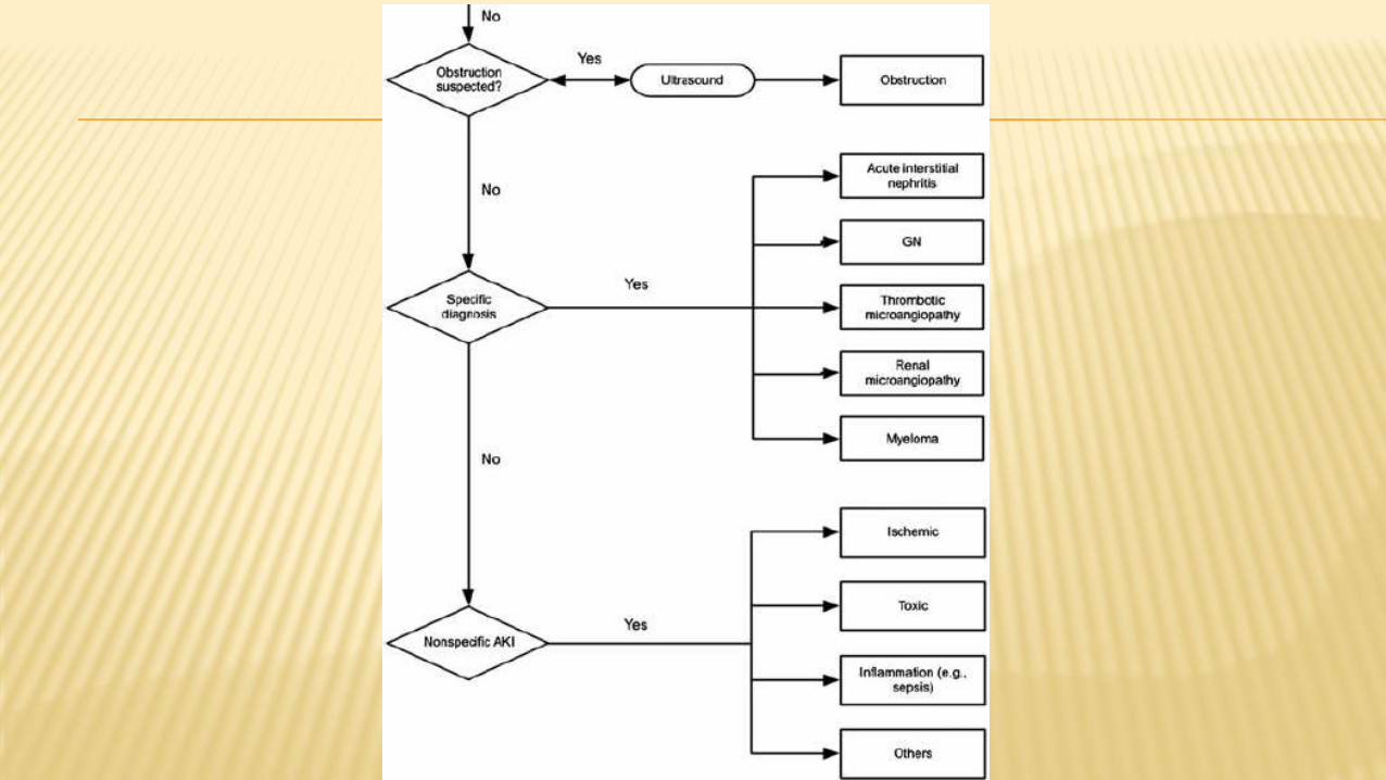
In particular, patients with decreased kidney perfusion, acute glomerulonephritis, vasculitis, interstitial nephritis, thromboticmicroangiopathy, and urinary tract obstruction require immediate diagnosis and specific therapeutic intervention, in addition to the general recommendations for AKI in the remainder of this guideline
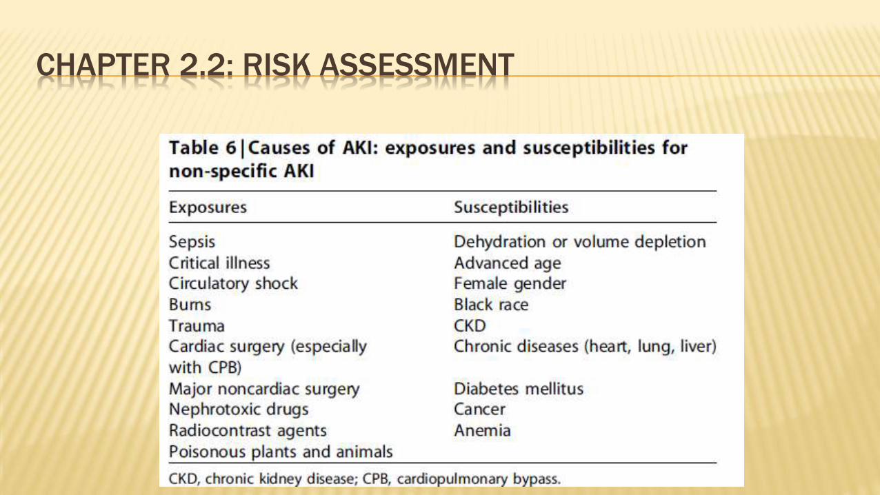
CHAPTER 2.2: RISK ASSESSMENT
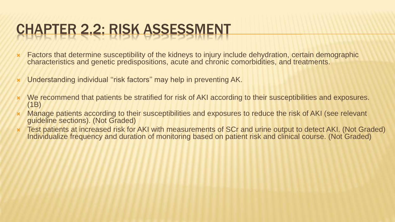
Factors that determine susceptibility of the kidneys to injury include dehydration, certain demographic characteristics and genetic predispositions, acute and chronic comorbidities, and treatments.
Understanding individual ‘‘risk factors’’ may help in preventing AK.
We recommend that patients be stratified for risk of AKI according to their susceptibilities and exposures. (1B)
Manage patients according to their susceptibilities and exposures to reduce the risk of AKI (see relevant guideline sections). (Not Graded)
Test patients at increased risk for AKI with measurements of SCr and urine output to detect AKI. (Not Graded) Individualize frequency and duration of monitoring based on patient risk and clinical course. (Not Graded)
CHAPTER 2.2: RISK ASSESSMENT
CHAPTER 2.3: EVALUATION AND GENERAL MANAGEMENT OF
PATIENTS WITH AND AT RISK FOR AKI
2.3.1: Evaluate patients with AKI promptly to determine the cause, with special attention to reversible causes. (Not Graded)
2.3.2: Monitor patients with AKI with measurements of SCr and urine output to stage the severity, according to Recommendation 2.1.2. (Not Graded)
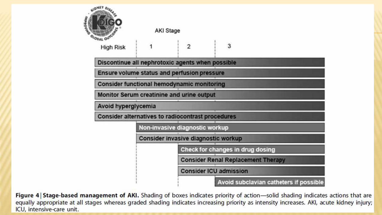
2.3.3: Manage patients with AKI according to the stage (see Figure 4) and cause. (Not Graded)
2.3.4: Evaluate patients 3 months after AKI for resolution, new onset, or worsening of pre-existing CKD. (Not Graded) If patients have CKD, manage these patients as detailed in the KDOQI CKD Guideline (Guidelines 7–15).
(Not Graded)
If patients do not have CKD, consider them to be at increased risk for CKD and care for them as detailed in the KDOQI CKD Guideline 3 for patients at increased risk for CKD. (Not Graded)

CHAPTER 2.3: EVALUATION AND GENERAL MANAGEMENT OF
PATIENTS WITH AND AT RISK FOR AKI
RACIONALIZACIÓN AKI is not a disease but rather a clinical syndrome with multiple etiologies.
Physicalexamination should include evaluation of fluid status, signs for acute and chronic heart failure, infection, and sepsis.
Laboratory parameters— including SCr, blood urea nitrogen (BUN), and electrolytes, complete blood count and differential—should be obtained.
- Urine analysis and microscopic examination as well as
urinary chemistries may be helpful in determining the
underlying cause of AKI.
- Stage is a predictor of the risk for mortality and decreased kidney function
Nephrotoxic drugs account for some part of AKI in 20–30% of patients. Often, agents like antimicrobials (e.g., aminoglycosides, amphotericin) and radiocontrast are used in patients that are already at high risk for AKI.
Static variables like central venous pressure are not nearly as useful as dynamic variables, such as pulse-pressure variation, inferior vena cava filling by ultrasound and echocardiographic appearance of the heart
CHAPTER 2.3: EVALUATION AND GENERAL MANAGEMENT OF
PATIENTS WITH AND AT RISK FOR AKI


CHAPTER 2.4: CLINICAL APPLICATIONS
Urine output vs. SCr
Atypical AKI: A complementary problem to pseudo-AKI isthe situation where a case of AKI fails to meet the definition. a patient might receive very large quantities of intravascular
fluids such that SCr is falsely lowered
Similarly, massive blood transfusions will result in the SCr more closely reflecting the kidney function of the blood donors than the patient
Changes in creatinine production are also well known in conditions such as muscle breakdown where production increases and in muscle wasting (including advanced liver disease) where production is decrease.
Creatinine production may also be decreased in sepsis66 possibly due to decreased muscle perfusion.
DIAGNOSTIC APPROACH TO ALTERATIONS IN
KIDNEY FUNCTION AND STRUCTURE
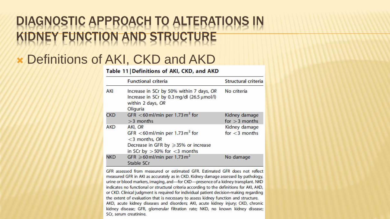
Definitions of AKI, CKD and AKD
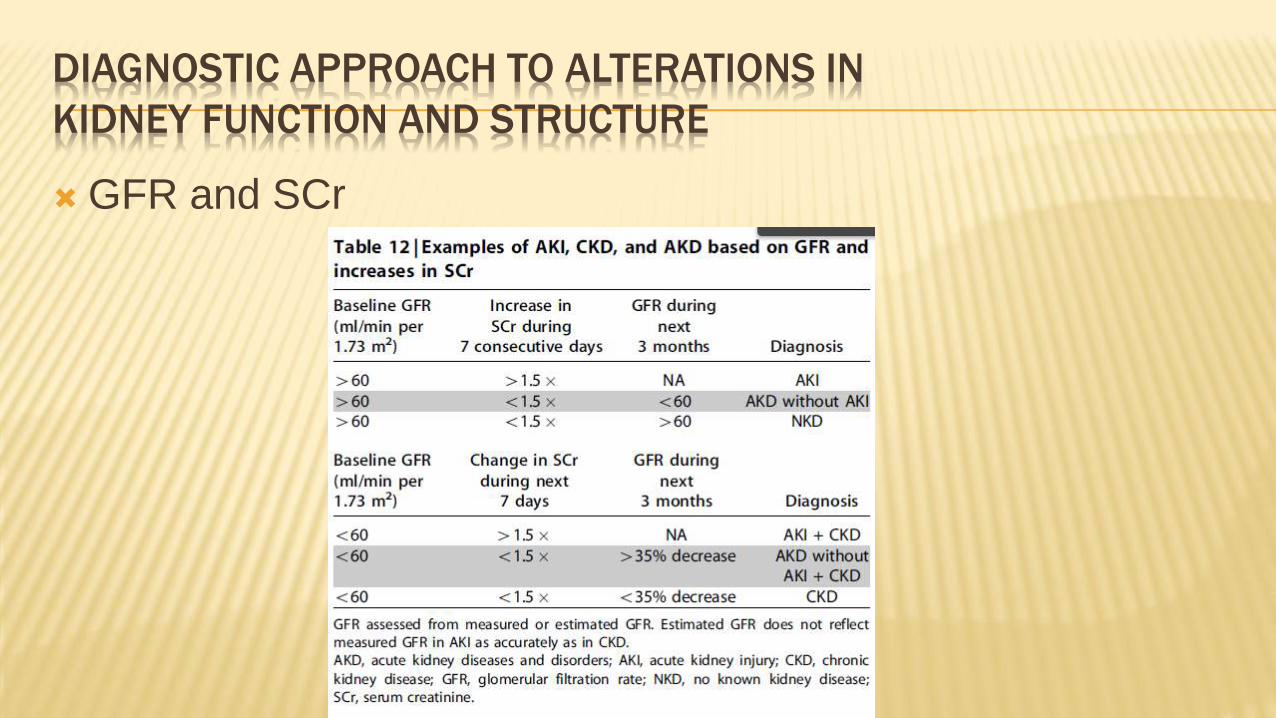
GFR and SCr
DIAGNOSTIC APPROACH TO ALTERATIONS IN
KIDNEY FUNCTION AND STRUCTURE

DIAGNOSTIC
APPROACH TO
ALTERATIONS IN
KIDNEY FUNCTION AND
STRUCTURE

Oliguria as a measure of kidney function 0.5 ml/kg/h would reflect reabsorption of
more than 99.5% of glomerular filtrate.
if GFR and SCr are normal and stable over an interval of 24 hours, it is generally not necessary to measure urine flow rate in order to assess kidney function.
Integrated approach to AKI, AKD, and CKD