hoon jai chun, md, phd, agaf, professor, diagnosis of
TRANSCRIPT
Diagnosis of gastric epithelial neoplasia: Dilemma for Korean pathologists
Joon Mee Kim, Mee-Yon Cho, Jin Hee Sohn, Dae Young Kang, Cheol Keun Park, Woo Ho Kim, So-Young Jin, Kyoung Mee Kim, Hee Kyung Chang, Eunsil Yu, Eun Sun Jung, Mee Soo Chang, Jong Eun Joo, Mee Joo, Youn Wha Kim, Do Youn Park, Yun Kyung Kang, Sun Hoo Park, Hye Seung Han, Young Bae Kim, Ho Sung Park, Yang Seok Chae, Kye Won Kwon, Hee Jin Chang, The Gastrointestinal Pathology Study Group of Korean Society of Pathologists
Joon Mee Kim, Department of Pathology, Inha University Hos-pital, Incheon 400-711, South KoreaMee-Yon Cho, Department of Pathology, Yonsei University Wonju College of Medicine Wonju Christian Hospital, Wonju, Kang won do 220-701, South KoreaJin Hee Sohn, Department of Pathology, Sungkyunkwan Uni-versity School of Medicine, Kangbuk Samsung Medical Center, Seoul 100-634, South KoreaDae Young Kang, Department of Pathology, Chungnam National University College of Medicine, Daejeon 301-747, South KoreaCheol Keun Park, Kyoung Mee Kim, Department of Pathology, Sungkyunkwan University School of Medicine, Samsung Medi-cal Center, Seoul 135-710, South KoreaWoo Ho Kim, Department of Pathology, Seoul National Univer-sity College of Medicine, Seoul 110-799, South KoreaSo-Young Jin, Department of Pathology, Soonchunhyang Uni-versity Hospital, Seoul 140-743, South KoreaHee Kyung Chang, Department of Pathology, Kosin University College of Medicine, Busan 602-702, South KoreaEunsil Yu, Department of Pathology, University of Ulsan College of Medicine, Asan Medical Center, Seoul 138-736, South KoreaEun Sun Jung, Department of Pathology, The Catholic University of Korea, Seoul St. Mary’s Hospital, Seoul 137-701, South KoreaMee Soo Chang, Department of Pathology, Seoul National Uni-versity Boramae Hospital, Seoul 156-707, South KoreaJong Eun Joo, Department of Pathology, Eulji General Hospital, Seoul 139-711, South KoreaMee Joo, Department of Pathology, Inje University Ilsan Paik Hospital, Goyang-si, Gyeonggi-do 411-706, South KoreaYoun Wha Kim, Department of Pathology, Kyunghee University College of Medicine, Seoul 130-701, South KoreaDo Youn Park, Department of Pathology, Pusan National Uni-versity College of Medicine, Busan 602-739, South KoreaYun Kyung Kang, Department of Pathology, Inje University Seoul Paik Hospital, Seoul 100-032, South KoreaSun Hoo Park, Department of Pathology, Korea Cancer Center
Hospital, Seoul 139-706, South KoreaHye Seung Han, Department of Pathology, Konkuk University Medical Center, Seoul 143-729, South KoreaYoung Bae Kim, Department of Pathology, Ajou University School of Medicine, Suwon, Gyeonggi-do 442-749, South KoreaHo Sung Park, Department of Pathology, Chonbuk National University Medical School, Jeonju 561-180, South KoreaYang Seok Chae, Department of Pathology, Korea University College of Medicine, Seoul 136-705, South KoreaKye Won Kwon, Department of Pathology, Bundang Jesaeng Hospital, Bundang-gu, Gyeonggi-do 463-050, South KoreaHee Jin Chang, Department of Pathology, National Cancer Cen-ter, Goyang, Gyeonggi-do 410-769, South KoreaThe Gastrointestinal Pathology Study Group of Korean Soci-ety of Pathologists, The Korean Society of Pathologists, 4F, The Korean Medical Association Building, Seoul 140-721, South KoreaAuthor contributions: Kim JM, Cho MY, Kang DY, Jin SY, Kim KM, Chang HK, Yu E, Jung ES, Chang MS, Joo JE, Kim YW, Park DY, Kang YK, Park SH, Han HS, Kwon KW and Chang HJ submitted slides; Kim JM, Cho MY, Sohn JH, Kim KM, Joo JE and Joo M selected slides; Kim JM, Cho MY, Sohn JH, Park CK, Kim WH, Jin SY, Chang HK, Yu E, Jung ES, Chang MS, Joo JE, Joo M, Park DY, Kang YK, Park SH, Han HS, Kim YB, Park HS and Chae YS attended workshop for discussion; members of The Gastrointestinal Pathology Study Group of Korean Society of Pathologists reviewed the circulating slides and contributed by voting; Kim JM wrote the paper.Supported by Korean Society of PathologistsCorrespondence to: Dr. Jin Hee Sohn, Department of Pathol-ogy, Sungkyunkwan University School of Medicine, Kangbuk Samsung Medical Center, 108, Pyeong-dong, Jongro-gu, Seoul 100-634, South Korea. [email protected]: +82-2-20012391 Fax: +82-2-20012398Received: June 29, 2010 Revised: September 9, 2010Accepted: September 16, 2010Published online: June 7, 2011
World J Gastroenterol 2011 June 7; 17(21): 2602-2610ISSN 1007-9327 (print) ISSN 2219-2840 (online)
© 2011 Baishideng. All rights reserved.
Online Submissions: http://www.wjgnet.com/[email protected]:10.3748/wjg.v17.i21.2602
2602 June 7, 2011|Volume 17|Issue 21|WJG|www.wjgnet.com
TOPIC HIGHLIGHT
Hoon Jai Chun, MD, PhD, AGAF, Professor, Series Editor

Kim JM et al . Pathologic diagnosis of gastric epithelial neoplasia
AbstractThe histopathological diagnosis of gastric mucosal bi-opsy and endoscopic mucosal resection/endoscopic submucosal dissection specimens is important, but the diagnostic criteria, terminology, and grading system are not the same in the East and West. A structurally inva-sive focus is necessary to diagnose carcinoma for most Western pathologists, but Japanese pathologists make a diagnosis of cancer based on severe dysplastic cytologic atypia irrespective of the presence of invasion. Although the Vienna classification was introduced to reduce diag-nostic discrepancies, it has been difficult to adopt due to different concepts for gastric epithelial neoplastic le-sions. Korean pathologists experience much difficulty making a diagnosis because we are influenced by Japa-nese pathologists as well as Western medicine. Japan is geographically close to Korea, and academic exchanges are active. Additionally, Korean doctors are familiar with Western style medical terminology. As a result, the ter-minology, definitions, and diagnostic criteria for gastric intraepithelial neoplasia are very heterogeneous in Ko-rea. To solve this problem, the Gastrointestinal Pathol-ogy Study Group of the Korean Society of Pathologists has made an effort and has suggested guidelines for differential diagnosis: (1) a diagnosis of carcinoma is based on invasion; (2) the most important character-istic of low grade dysplasia is the architectural pattern such as regular distribution of crypts without severe branching, budding, or marked glandular crowding; (3) if nuclear pseudostratification occupies more than the basal half of the cryptal cells in three or more adjacent crypts, the lesion is considered high grade dysplasia; (4) if severe cytologic atypia is present, careful inspec-tion for invasive foci is necessary, because the risk for invasion is very high; and (5) other structural or nuclear atypia should be evaluated to make a final decision such as cribriform pattern, papillae, ridges, vesicular nuclei, high nuclear/cytoplasmic ratio, loss of nuclear polarity, thick and irregular nuclear membrane, and nucleoli.
© 2011 Baishideng. All rights reserved.
Key words: Intraepithelial neoplasia; Stomach; Dyspla-sia; Adenoma; Carcinoma; Japanese; Western; Consen-sus; Vienna
Peer reviewers: Fabio Grizzi, PhD, Laboratories of Quantitative Medicine, Istituto Clinico Humanitas IRCCS, Via Manzoni 56, 20089 Rozzano, Milan, Italy; Vittorio Ricci, MD, PhD, Depart-ment of Physiology, Human Physiology Section, University of Pavia Medical School, Via Forlanini 6, Pavia, 27100, Italy
Kim JM, Cho MY, Sohn JH, Kang DY, Park CK, Kim WH, Jin SY, Kim KM, Chang HK, Yu E, Jung ES, Chang MS, Joo JE, Joo M, Kim YW, Park DY, Kang YK, Park SH, Han HS, Kim YB, Park HS, Chae YS, Kwon KW, Chang HJ, The Gastrointestinal Pathology Study Group of Korean Society of Pathologists. Diag-nosis of gastric epithelial neoplasia: Dilemma for Korean patholo-gists. World J Gastroenterol 2011; 17(21): 2602-2610 Available from: URL: http://www.wjgnet.com/1007-9327/full/v17/i21/2602.htm DOI: http://dx.doi.org/10.3748/wjg.v17.i21.2602
INTRODUCTIONTechniques for endoscopic mucosal resection (EMR) and endoscopic submucosal dissection (ESD) have been developing rapidly, and pathologists more commonly encounter specimens derived from endoscopic resec-tion. These procedures are sometimes performed for diagnostic purposes but mostly for therapeutic conve-nience compared with radical surgery. However, for both purposes, pathological diagnosis of gastric biopsies and EMR/ESD specimens is very important, because fur-ther treatment plans and a surveillance schedule must be established. The importance of a diagnosis is not only stressed from a clinical viewpoint, but also from an aca-demic perspective. Many studies regarding early gastric neoplastic lesions based on a histopathological diagnosis have been performed, and they focus on various clinical and pathological aspects such as survival, recurrence, sur-veillance programs, and molecular pathology. However, if the pathological diagnosis is different among patholo-gists, the results cannot be compared. Additionally, socio-economic problems are also important, because medical insurance is intimately associated with disease severity.
Although these issues are important, it is unfortu-nate that the definition, diagnostic criteria, and grading system for early stage gastric neoplastic lesions are not completely developed. In particular, it is well known that Eastern and Western pathologists use different criteria to make a gastric carcinoma diagnosis. A structurally in-vasive focus is necessary to diagnose carcinoma for most Western pathologists. The “Eastern” opinion is actually the “Japanese” concept, and diagnosis of gastric carcino-ma is based on the cytological findings. We have realized that Korean pathologists have different diagnostic criteria for gastric epithelial neoplasia than Japanese or Western pathologists. In this article, we discuss the current prob-lems for the pathological diagnosis of gastric neoplastic lesions, the Korean perspectives, and the path we should follow.
TERMINOLOGY: DYSPLASIA, ADENOMA, CARCINOMA IN SITU, AND INVASIVE CARCINOMA Initially “dysplasia” was used for inflammatory bowel disease, but, now it is used throughout the gastrointesti-nal tract as well as for other organs. Dysplasia means an unequivocally neoplastic but non-invasive lesion distin-guished from regenerative changes[1]. The term “gastric dysplasia” was used for the first time by Grundmann in 1975 to describe an exclusively precancerous gastric le-sion[2]. Shortly thereafter, a WHO committee published a definition that characterizes dysplasia as cellular atypia, abnormal differentiation, and disorganized architecture[3,4].
Most Western pathologists use the term “dysplasia” to describe a neoplastic premalignant abnormality[1,5-11]. How-ever, in Japan, the terminology for non-invasive neoplastic lesions is different. Since Nakamura et al[12] established
2603 June 7, 2011|Volume 17|Issue 21|WJG|www.wjgnet.com
specific histological atypical gastric epithelium criteria, which were classified as definitely benign, borderline, or carcinoma, the Japanese Society for Research on Gastric Cancer has recommended that gastric neoplastic lesions be subdivided into one of five categories: normal or be-nign without cellular atypia, benign with slight atypia, bor-derline, probable carcinoma, and obvious carcinoma[13,14]. “Atypia” has been used more frequently than “dysplasia” in Japan. Subsequently, Japanese authors suggested the definition of “group 3 or 4 lesions”[14-16] (Table 1).
In addition to the differences in terminology describ-ing premalignant gastric lesions, some confusion exists for the terms adenoma and dysplasia. Originally, adenoma was considered a raised circumscribed lesion, either ses-
sile or pedunculated, in contrast to dysplasia, which arose at flat or depressed mucosa. However, much confusing terminology has been introduced such as “flat adenoma”, “depressed adenoma”, “elevated dysplasia”, and “polypoid dysplasia”[17-31]. Thus, WHO defined adenoma as “a cir-cumscribed benign neoplasm composed of tubular and/or villous structures lined by dysplastic epithelium”. In 1998, Lewin et al[10] suggested nomenclature using both adenoma and dysplasia; the former meant neoplastic circumscribed benign lesions unassociated with underlying inflamma-tion whether pedunculated, sessile, flat or depressed, and the latter meant benign neoplastic lesions associated with underlying inflammation. Both were subdivided as low and high grade. Although there have been efforts to clarify the definition, a confusing situation still persists[9].
Another confusing concept is carcinoma in situ, which means carcinoma without invasion. However, a differ-ential diagnosis of high grade dysplasia/atypia and carci-noma in situ is problematic. When cytological atypia and architectural complexity is marked, the term “carcinoma in situ” is used by some pathologists, but others do not make a distinction between “high grade dysplasia” and “carci-noma in situ” because the behavior and management are the same[10]. In the Japanese classification, there is no dis-ease group describing adenocarcinoma in situ. The WHO International References Center for Histological/Clas-sification of Precancerous Lesions of the Stomach met in 1978 and developed a consensus statement that stated that presumed precancerous lesions of the stomach should be termed “dysplasia” and that the term “intramucosal carci-noma” should replace “in situ carcinoma” for lesions that have invasive malignant cells confined to the lamina pro-pria[3,4]. However, “adenocarcinoma in situ” is categorized in the AJCC and Vienna classifications as a non-invasive intraepithelial carcinoma, resulting in confusion.
The most important and surprising inconsistency be-tween Western and Japanese criteria is in the diagnosis of adenocarcinoma. Japanese pathologists make a diagnosis of cancer based on severe dysplastic cytologic atypia with enlarged vesicular oval nuclei and prominent nucleoli irrespective of the presence of invasion. But, Western pathologists believe there must be evidence of inva-sion into the lamina propria to make a cancer diagnosis. This inconsistency causes serious problems understand-ing “early” cancer. Many investigators have pointed out this discrepancy and made some efforts to reduce the confusion. The Vienna classification was developed for common world terminology of gastrointestinal epithelial neoplasia[32]. Some Western pathologists agreed with the Japanese criteria and changed their view points[33].
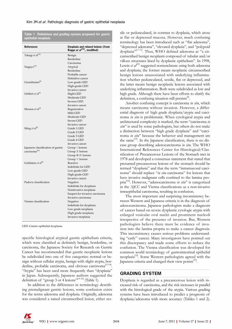
GRADING SYSTEMDysplasia is regarded as a precancerous lesion with in-creased risk of carcinoma, and the risk increases in parallel with the histological grade of the atypia. Various grading systems have been introduced to predict a prognosis of dysplasia/adenoma with more accuracy (Tables 1 and 2).
2604 June 7, 2011|Volume 17|Issue 21|WJG|www.wjgnet.com
Table 1 Definitions and grading systems proposed for gastric epithelial neoplasia
References Dysplasia and related lesions (from Rugge et al [40], modified)
Takagi et al[15] BenignBorderlineCarcinoma
Nagayo[14] AtypicalBorderlineProbable cancerDefinitive cancer
Grundmann[2] Low-grade GEDHigh-grade GEDInvasive cancer
Oehlert et al[5] Slight GEDModerate GEDSeverer GEDInvasive cancer
Morson et al[4] RegenerativeMild GEDModerate GEDSevere GEDInvasive cancer
Ming et al[7] Grade 1 GEDGrade 2 GEDGrade 3 GEDGrade 4 GEDInvasive cancer
Japanese classification of gastric carcinoma[16]
Group Ⅰ lesionsGroup Ⅱ lesionsGroup Ⅲ-Ⅳ lesionsGroup Ⅴ lesions
Goldstein et al[9] ReactiveIndefinite for GEDLow-grade GEDHigh-grade GEDInvasive cancer
Padova classification NegativeIndefinite for dysplasiaNoninvasive neoplasiaSuspect for invasive carcinoma
Invasive carcinomaVienna classification Negative
Indefinite for dysplasiaLow grade neoplasiaHigh grade neoplasiaInvasive neoplasia
GED: Gastric epithelial dysplasia.
Kim JM et al . Pathologic diagnosis of gastric epithelial neoplasia
The most popular grading system is the three-tiered (mild, moderate, and severe) or two-tiered (low and high) system; the latter shows better inter-observer agreement[1,33-37], and most management protocols are based on the two-tiered system. There is no distinctive management protocol ac-cording to the three-tiered system that is practically sig-nificant[1,34-37].
The morphological features of low grade dysplasia/adenoma are characterized by simple tubules with little branching, nuclear stratification below half of the cyto-plasm, tall columnar cells with dense spindle-shaped hy-perchromatic nuclei, ample amphophilic cytoplasm, and sparse mitotic figures. High-grade dysplasia/adenoma is composed of tubules with elongation and complex bud-ding, cribriform in the most extreme cases, greatly en-larged round to oval nuclei, markedly increased nuclear/cytoplasmic ratio, and loss of nuclear polarity (Table 2).
Although well-established low and high-grade dyspla-sia criteria are present, there are large scale interobserver or intraobserver discrepancies. Sometimes, regeneration causes serious confusion with carcinoma. The category of “indefinite for dysplasia” is maintained in the Vienna classification[32], and the histopathological finding of re-generation has been well described in many studies[9,10,38].
CURRENT STATUS OF PATHOLOGIC DIAGNOSIS OF GASTRIC EPITHELIAL NEOPLASTIC DISEASE IN KOREAKorea is geographically close to Japan, and academic exchanges are active. Korean endoscopists introduced the EMR/ESD technique from Japan, and many discus-sions and cooperation continues. In the pathology field, there are many conferences and collaborations between Japanese and Korean pathologists. Furthermore, Korea’s medical science is influenced by that in Western countries. Korean doctors are familiar with Western style medical terminology. As a result, the terminology, definition, and diagnostic criteria for gastric intraepithelial neoplasia are very heterogeneous in Korea.
To promote diagnostic consensus, The Gastrointes-tinal Pathology Study Group of the Korean Society of Pathologist (GIPS-KSP) established a grading system for gastric epithelial proliferative disease and produced a stan-dardized pathological report for gastric cancer[38,39]. The standard guidelines for grading gastric epithelial prolifera-tive disease are as follows: (1) proliferating gastric epi-thelium can be divided into hyperplastic and neoplastic; (2) the term “dysplasia” is reserved for the microscopic epithelial changes that are unequivocally neoplastic; (3) biopsy specimens are categorized as regenerative (negative for dysplasia), indefinite (questionable dysplasia), positive (positive for dysplasia) and overt carcinoma; and (4) the positive category is divided into two groups; high-grade dysplasia and low-grade dysplasia[40]. Another important criterion for the differential diagnosis of low and high-grade dysplasia is the extent of nuclear stratification; nuclear stratification below half of the cytoplasm is char-acteristic of low-grade dysplasia. If nuclear stratification above half of the cytoplasm is present at more than three contiguous glands, it is considered high-grade dysplasia[39]. This criterion is based on the definition of high-grade dysplasia associated with inflammatory bowel disease.
In Korea, most pathologists use the term “tubular/villous/villotubular adenoma with low/high grade dys-plasia” to describe intraepithelial precancerous disease. “Dysplasia” is used to describe atypia due to neoplastic etiology, excluding regenerative changes. This concept of dysplasia unassociated with adenoma (foveolar type dysplasia) is not well established and needs further study. The term “carcinoma in situ” is used by some patholo-gists to describe a highly anaplastic lesion without lamina propria invasion, but this is not widely accepted.
At the 8th Japan-Korea Pathologist Symposium in 2008 in Yokohama, Japan, there was a consensus confer-ence to discuss diagnostic differences in gastrointestinal neoplasia, and Japanese and Korean pathologists con-firmed their different viewpoints.
In 2009, GIPS-KSP began to establish new Korean diagnostic criteria for gastric epithelial proliferative disease, and the effort is ongoing. We gathered 117 cases of gastric
2605 June 7, 2011|Volume 17|Issue 21|WJG|www.wjgnet.com
Table 2 Differentiation of low and high grade dysplasia and gastric carcinomas
Histology Feature Low-grade dysplasia High-grade dysplsia Carcinoma
Structural atypia Gland size Uniform Variable VariableGland arrangement Regular Slightly irregular IrregularGlandular crowding Slight Moderate MarkedGlandular transition to surrounding mucosa No No NoGlandular branching/budding Focal Prominent ProminentGlandular cribriform No Yes YesSurface maturation No No No
Nuclear atypia Shape Elongated Elongated and/or irregular Oval/roundPseudostratification Basal 1/2 Over basal 1/2 IrregularMembrane Thin Thick UnevenHyperchromasia Hyperchromatic even Hyperchromatic irregular VesicularPleomorphism No Mild Moderate to markedProminent nucleoli Absent Present PresentLoss of polarity No No/yes Yes
Stroma Invasion No No Yes
Kim JM et al . Pathologic diagnosis of gastric epithelial neoplasia

biopsy specimens and ESD specimens from 14 institutes. Six pathologists screened the slides and selected 42 cases, which showed the difficulty of diagnosis. The selected cases were circulated and answers were gathered from 45 pathologists. The answer sheet was composed of five cat-egories of diagnosis; regenerative atypia, low grade dyspla-sia, high grade dysplasia, carcinoma in situ, and carcinoma. In most cases, there was a wide range of interobserver discrepancy. We tried to simplify the diagnostic criteria to enhance diagnostic consistency but realized that it was impossible because determining low-grade dysplasia, high-grade dysplasia, and carcinoma was a complex process based on many kinds of diagnostic criteria. A consensus conference was held eight times and the pathological findings of each case were discussed and voted on anony-mously. After the consensus conferences, the agreement rate increased (Table 3). Before the consensus conferences, only 10 cases among 42 showed a high agreement rate (more than 70%). After the conferences, the cases showing high agreement rate increased to 25 cases (Table 3). Al-though these data were not enough for a conclusion, it was suggested that there could be agreement for a pathologic diagnosis among Korean pathologists.
Many histological factors are helpful for the differen-tial diagnosis of low and high-grade dysplasia, but these factors sometimes conflicted with each other. We at-tempted to identify a more simple and reproducible way to determine the dysplasia grade. We propose guidelines for differential diagnosis: (1) a diagnosis of carcinoma is based on invasion; (2) the most important characteristic of low-grade dysplasia is a regular distribution of crypts without severe branching, budding, or marked glandular crowding; (3) if nuclear pseudostratification occupies more than the basal half of the cryptal cells in three or more adjacent crypts, the lesion is considered high-grade dysplasia (this rule was based according to the previously mentioned Korean Standard of Pathology Report of Gastric Cancer[39]); (4) if severe cytologic atypia is present, careful inspection for invasive foci is necessary, because the risk of invasion is very high; and (5) other structural or nuclear atypia should be evaluated to make a final deci-sion such as cribriform pattern, papillae, ridges, vesicular nuclei, high nuclear/cytoplasmic ratio, loss of nuclear po-larity, thick and irregular nuclear membrane, and nucleoli. Based on these principles, the consensus rate was mark-edly increased, although not in every case.
EXAMPLES OF CASES PRESENTED AT THE CONSENSUS CONFERENCE Case 1 An ESD specimen revealed a regular distribution of small proliferative glands without budding or branching (Figure 1A). The nuclei were elongated and stratified below half of the cytoplasm (Figure 1B). Hyperchromasia and mitoses were present; 57.8% and 100% of the pathologists agreed with a diagnosis of tubular adenoma with low grade dyspla-sia before and after the consensus conference, respectively.
Case 2 An ESD specimen revealed regular distribution of small proliferative glands without budding or branching (Figure 2A). Glandular crowding was mild. The nuclei were ovoid and vesicular with conspicuous nucleoli but nuclear strati-fication did not exceed the basal half of the cell (Figure 2B); 73.3% and 60.9% of the pathologists agreed with a diagnosis of tubular adenoma with low-grade dysplasia before and after the consensus conference, respectively.
Case 3An ESD specimen revealed compact small glandular pro-liferation with some variation in gland size (Figure 3A). Budding or branching was present. The nuclei were elon-gated and stratified with some ovoid nuclei. More than
2606 June 7, 2011|Volume 17|Issue 21|WJG|www.wjgnet.com
Table 3 Inter-observer agreement rate before and after con-sensus conferences
Agreement rate (%) Before (%) After (%)
0-50 16 (38.1) 1 (2.4)51-60 13 (31.0) 4 (9.5)61-70 3 (7.1) 8 (19.0)71-80 5 (11.9) 6 (14.3)81-90 3 (7.1) 6 (14.3)91-100 2 (4.8) 17 (40.5)Total 42 (100) 42 (100)
Kim JM et al . Pathologic diagnosis of gastric epithelial neoplasia
Figure 1 Consensus diagnosis of tubular adenoma with low grade dys-plasia. A: Regular distribution of small proliferative glands without budding or branching (HE, × 100); B: Elongated nuclei with stratification below half of the cytoplasm (HE, × 200).
B
A

three contiguous glands showed nuclear stratification above half of the cytoplasm (Figure 3B). Hyperchroma-sia and mitoses were present; 44.4% and 100% of the pathologists agreed with a diagnosis of tubular adenoma with high-grade dysplasia before and after the consensus conference, respectively.
Case 4An ESD specimen revealed glandular crowding with some variation in gland size and budding (Figure 4A).
The nuclei were elongated and stratified with some ovoid nuclei. Nuclear stratification above the basal half of the cytoplasm was present (Figure 4B). Hyperchromasia and mitoses were noted. Before the consensus conference, 44.4% of pathologists agreed with a diagnosis of tubular adenoma with high-grade dysplasia, which increased to 75% after the conference.
Case 5An ESD specimen revealed compact small glandular
2607 June 7, 2011|Volume 17|Issue 21|WJG|www.wjgnet.com
Kim JM et al . Pathologic diagnosis of gastric epithelial neoplasia
BA
Figure 2 Major consensus diagnosis of tubular adenoma with low-grade dysplasia. A: Regular distribution of small proliferative glands without budding or branching (HE, × 100); B: Ovoid and vesicular nuclei with conspicuous nucleoli and nuclear stratification not exceeding basal half of the cell (HE, × 400).
BA
Figure 3 Consensus diagnosis of tubular adenoma with high-grade dysplasia. A: Compact small glandular proliferation with some variation of gland size, bud-ding and branching (HE, × 40); B: Elongated or oval nuclei with stratification above half of the cytoplasm in more than three contiguous glands (HE, × 200).
BA
Figure 4 Major consensus diagnosis of tubular adenoma with high-grade dysplasia. A: Glandular crowding with some variation in gland size and budding (HE, × 100); B: Elongated or oval nuclei with stratification above basal half of the cytoplasm (HE, × 200).

2608 June 7, 2011|Volume 17|Issue 21|WJG|www.wjgnet.com
proliferation with variation in gland size, budding and branching (Figure 5A). The nuclei were elongated and stratified with some ovoid nuclei (Figure 5B). More than three contiguous glands showed nuclear stratification above the basal half of the cytoplasm. Hyperchromasia and mitoses were present. Glandular complexity was present but definite invasion was not identified; 42.5% and 62.5% of the pathologists agreed with a diagnosis of tubular adenoma with high-grade dysplasia before and after the consensus conference, respectively.
Case 6A mucosal biopsy specimen revealed compact small glandular proliferation without budding or branching (Figure 6A). Another section showed a villous configura-tion (Figure 6B). The glandular distribution was relatively regular but gland size was mildly variable. The nuclei were enlarged, oval to round, and pleomorphic. Nuclear stratifi-cation was not severe, but enlarged nuclei occupied more than the basal half of the cytoplasm. Hyperchromasia and mitoses were present. Invasion into the lamina propria was present (Figure 6C, arrow); 22.2% and 100% of the pathologists agreed with a diagnosis of adenocarcinoma before and after the consensus conference, respectively.
Case 7A mucosal biopsy specimen revealed compact small glan-dular proliferation with budding or branching (Figure 7A). Glandular size and distribution were irregular. The nuclei
were enlarged, oval to round, with vesicular chromatin. Severe nuclear stratification approaching the top of the cytoplasm in more than three contiguous glands was pres-ent. Marked hyperchromasia and mitoses were noted with invasion into the lamina propria (Figure 7B, arrow); 26.7% and 56.3% of the pathologists agreed with a diagnosis of adenocarcinoma before and after the consensus confer-ence, respectively.
PROBLEMS TO BE SOLVEDThe rate of agreement markedly increased after many consensus conferences (Table 3). However, this guideline has some limitations; (1) focal invasion into the lamina propria may not be detected on a biopsy specimen, which causes diagnostic discrepancy between a biopsy and re-section specimen; and (2) a gray zone due to overlapping or mismatching of diagnostic criteria lowers the agree-ment rate. We must conduct a further study to verify the hypothesis in expanded cases and to determine that the guideline lowers inter and intraobserver discrepancies and correlates with clinical outcome. If more reliable pathological findings suggesting possible invasion into an adjacent area could be found, it would be very useful for small biopsy specimens. We will attempt to define the pathological criteria in a more simple and subjective way, and we would like to develop a diagnostic algorithm. Education is also important. Symposiums, workshops, and publishing of articles will be helpful.
Kim JM et al . Pathologic diagnosis of gastric epithelial neoplasia
BA
Figure 5 Major diagnosis of tubular adenoma with high-grade dysplasia. A: Compact small glandular proliferation with variation in gland size, budding and branching (HE, × 100); B: Elongated or oval nuclei with stratification above basal half of the cytoplasm in more than three contiguous glands. Glandular complexity without definite invasion (HE, × 200).
CBA
Figure 6 Consensus diagnosis of adenocarcinoma. A: Compact small glandular proliferation without budding or branching. Relatively regular glandular distribution but enlarged, oval to round, and pleomorphic nuclei (HE, × 100); B: Another section showing villous configuration (HE, × 40); C: Hyperchromasia and mitoses with invasion into the lamina propria (arrow) (HE, × 400).

2609 June 7, 2011|Volume 17|Issue 21|WJG|www.wjgnet.com
B
A
Figure 7 Major diagnosis after the consensus conference of adenocarcinoma. A: Compact small glandular proliferation with budding and branching. Regular glan-dular size and distribution (HE, × 100); B: Severe nuclear stratification approaching the top of the cytoplasm in more than three contiguous glands. Marked hyperchro-masia and mitoses with invasion into the lamina propria (arrow) (HE, × 400).
We have additional problems to be solved, such as how to measure the invasion depth if submucosal invasion is present, the diagnostic policy for differentiation, judgment on lymphovascular invasion, and a fixation method for ESD specimens, which are all important decisions to de-velop a further treatment plan after EMR/ESD.
CONCLUSIONEastern and Western pathologists have different termi-nology and diagnostic criteria for gastric intraepithelial neoplasia. In Korea, pathologists experience much diffi-culty when making a diagnosis, and have made efforts to increase the interobserver agreement rate. As a result, we have achieved improved diagnostic consensus, although it is not yet perfect. We tentatively suggest the guidelines for differential diagnosis: (1) a diagnosis of carcinoma is based on invasion; (2) the most important characteristic of low grade dysplasia is the architectural pattern such as regular distribution of crypts without severe branching, budding, or marked glandular crowding; (3) if nuclear pseudostratification occupies more than the basal half of the cryptal cells in three or more adjacent crypts, the le-sion is considered high grade dysplasia; (4) if severe cyto-logic atypia is present, careful inspection for invasive foci is necessary, because the risk for invasion is very high; and (5) other structural or nuclear atypia should be evalu-ated to make a final decision such as cribriform pattern,
papillae, ridges, vesicular nuclei, high nuclear/cytoplasmic ratio, loss of nuclear polarity, thick and irregular nuclear membrane, and nucleoli. Further study on the pathologi-cal findings and clinicopathological correlations as well as a follow-up study are necessary to increase diagnostic accuracy.
ACKNOWLEDGMENTSWe express great gratitude to Dr. Shimoda T who gave us very informative academic lectures, and to Dr. Ajioka Y who helped us create a Japanese-Korean pathologist consensus conference in 2008.
REFERENCES1 Riddell RH, Goldman H, Ransohoff DF, Appelman HD,
Fenoglio CM, Haggitt RC, Ahren C, Correa P, Hamilton SR, Morson BC. Dysplasia in inflammatory bowel disease: stan-dardized classification with provisional clinical applications. Hum Pathol 1983; 14: 931-968
2 Grundmann E. Histologic types and possible initial stages in early gastric carcinoma. Beitr Pathol 1975; 154: 256-280
3 Serck-Hanssen A. Precancerous lesions of the stomach. Scand J Gastroenterol Suppl 1979; 54: 104-105
4 Morson BC, Sobin LH, Grundmann E, Johansen A, Nagayo T, Serck-Hanssen A. Precancerous conditions and epithelial dysplasia in the stomach. J Clin Pathol 1980; 33: 711-721
5 Oehlert W, Keller P, Henke M, Strauch M. [Gastric mucosal dysplasias: what is their clinical significance (author’s transl)]. Dtsch Med Wochenschr 1975; 100: 1950-1956
6 Jass JR. A classification of gastric dysplasia. Histopathology 1983; 7: 181-193
7 Ming SC, Bajtai A, Correa P, Elster K, Jarvi OH, Munoz N, Nagayo T, Stemmerman GN. Gastric dysplasia. Significance and pathologic criteria. Cancer 1984; 54: 1794-1801
8 Riddell RH. Premalignant and early malignant lesions in the gastrointestinal tract: definitions, terminology, and problems. Am J Gastroenterol 1996; 91: 864-872
9 Goldstein NS, Lewin KJ. Gastric epithelial dysplasia and ad-enoma: historical review and histological criteria for grading. Hum Pathol 1997; 28: 127-133
10 Lewin KJ. Nomenclature problems of gastrointestinal epithe-lial neoplasia. Am J Surg Pathol 1998; 22: 1043-1047
11 Lauwers GY, Riddell RH. Gastric epithelial dysplasia. Gut 1999; 45: 784-790
12 Nakamura K, Sugano H, Takagi K, Fuchigami A. Histopath-ological study on early carcinoma of the stomach: criteria for diagnosis of atypical epithelium. Gann 1966; 57: 613-620
13 Sugano H, Nakamura K, Takagi K. An atypical epithelium of the stomach: A clinico-pathological entity. Gann Monogr Can-cer Res 1971; 2: 257-269
14 Nagayo T. Histological diagnosis of biopsied gastric muco-sae with special reference to that of borderline lesions. Gann Monogr Cancer Res 1971; 11: 245-256
15 Takagi K, Kumakura K, Sugano H, Nakamura K. [Polypoid lesions of the stomach--with special reference to atypical epi-thelial lesions]. Gan No Rinsho 1967; 13: 809-817
16 Japanese Research Society for Gastric Cancer. Japanese clas-sification of gastric carcinoma. Tokyo: Kanehara & Co., Ltd., 1995
17 Schade ROK. The borderline between benign and malignant lesions of the stomach. In: Grundmann E, Grunze H, Witte S, editors. Early Gastric Cancer. New York: Springer Verlag, 1974: 45–53
18 Davaris P, Petraki K, Archimandritis A, Haritopoulos N, Papacharalampous N. Mucosal hyperplastic polyps of the
Kim JM et al . Pathologic diagnosis of gastric epithelial neoplasia

2610 June 7, 2011|Volume 17|Issue 21|WJG|www.wjgnet.com
stomach. Do they have any potential to malignancy? Pathol Res Pract 1986; 181: 385-389
19 Ming SC, Goldman H. Gastric polyps; A histogenetic classifi-cation and its relation to carcinoma. Cancer 1965; 18: 721-726
20 Nagayo T. Dysplasia of the gastric mucosa and its relation to the precancerous state. Gann 1981; 72: 813-823
21 Hattori T. Morphological range of hyperplastic polyps and carcinomas arising in hyperplastic polyps of the stomach. J Clin Pathol 1985; 38: 622-630
22 Nakamura T, Nakano G. Histopathological classification and malignant change in gastric polyps. J Clin Pathol 1985; 38: 754-764
23 Usha SD, Shukla HS, Singh RG, Khanna S, Gupta RM. Pre-cancerous lesions of stomach. Indian J Pathol Microbiol 1989; 32: 75-80
24 Ming SC. Adenocarcinoma and other malignant epithelial tumors of the stomach. In: Ming SC, Goldman H, editors. Pathology of the Gastrointestinal Tract. Philadelphia, PA: Saunders, 1992: 584-617
25 Ito H, Yasui W, Yoshida K, Nakayama H, Tahara E. Depressed tubular adenoma of the stomach: pathological and immuno-histochemical features. Histopathology 1990; 17: 419-426
26 Correa P. Clinical implications of recent developments in gas-tric cancer pathology and epidemiology. Semin Oncol 1985; 12: 2-10
27 Freeny PC, Vimont TR. Villous tumors of the stomach and small bowel. Arch Surg 1978; 113: 255-259
28 Xuan ZX, Ambe K, Enjoji M. Depressed adenoma of the stomach, revisited. Histologic, histochemical, and immuno-histochemical profiles. Cancer 1991; 67: 2382-2389
29 Nakamura K, Sakaguchi H, Enjoji M. Depressed adenoma of the stomach. Cancer 1988; 62: 2197-2202
30 Ito H, Yokozaki H, Ito M, Tahara E. Papillary adenoma of the stomach. Pathologic and immunohistochemical study. Arch Pathol Lab Med 1989; 113: 1030-1034
31 Tsujitani S, Furusawa M, Hayashi I. Morphological factors aid in therapeutic decisions concerning gastric adenomas.
Hepatogastroenterology 1992; 39: 56-5832 Schlemper RJ, Riddell RH, Kato Y, Borchard F, Cooper HS,
Dawsey SM, Dixon MF, Fenoglio-Preiser CM, Fléjou JF, Ge-boes K, Hattori T, Hirota T, Itabashi M, Iwafuchi M, Iwashita A, Kim YI, Kirchner T, Klimpfinger M, Koike M, Lauwers GY, Lewin KJ, Oberhuber G, Offner F, Price AB, Rubio CA, Shimizu M, Shimoda T, Sipponen P, Solcia E, Stolte M, Wata-nabe H, Yamabe H. The Vienna classification of gastrointesti-nal epithelial neoplasia. Gut 2000; 47: 251-255
33 Stolte M. Diagnosis of gastric carcinoma: Japanese fairy tales or Western deficiency? Virchows Arch 1999; 434: 279-280
34 de Dombal FT, Price AB, Thompson H, Williams GT, Mor-gan AG, Softley A, Clamp SE, Unwin BJ. The British Society of Gastroenterology early gastric cancer/dysplasia survey: an interim report. Gut 1990; 31: 115-120
35 Lewin KJ, Appleman HD. Carcinoma of the stomach. Tu-mors of the esophagus and stomach. In: Rosai J, Sobin LH, editors. Atlas of tumor pathology. Washington, DC: Armed Forces Institute of Pathology, 1996: 245-321
36 Tosi P, Baak JP, Luzi P, Miracco C, Lio R, Barbini P. Mor-phometric distinction of low- and high-grade dysplasias in gastric biopsies. Hum Pathol 1989; 20: 839-844
37 Burke AP, Sobin LH, Shekitka KM, Helwig EB. Dysplasia of the stomach and Barrett esophagus: a follow-up study. Mod Pathol 1991; 4: 336-341
38 Kim H, Jin SY, Jang JJ, Kim WH, Song SY, Kim KR, Yu ES, Shin HS, Kim HK, Sohn JH, Hong EK, Kim YW, Jeong JS, Kim CJ, Choi SE, Park IS, Park CI, Kim YI. Grading system for gastric epithelial proliferative diseases standardized guidelines proposed by Korean Study Group for Pathology of Digestive Diseases. Korean J Pathol 1997; 31: 389-400
39 Kim WH, Park CK, Kim YB, Kim YW, Kim HG, Bae HI, Song KS, Chang HK, Chang HJ, Chae YS. A standardized pathol-ogy report for gastric cancer. Korean J Pathol 2005; 39: 106- 113
40 Rugge M, Nitti D, Farinati F, di Mario F, Genta RM. Non-invasive neoplasia of the stomach. Eur J Gastroenterol Hepatol 2005; 17: 1191-1196
S- Editor Shi ZF L- Editor O’Neill M E- Editor Zheng XM
Kim JM et al . Pathologic diagnosis of gastric epithelial neoplasia