functional role of glycogen synthase kinase-3 on...
TRANSCRIPT
Functional Role of Glycogen synthase Kinase-3� on Glucocorticoid-mediated
signaling
Camila Rubio Patiño
ADVERTIMENT. La consulta d’aquesta tesi queda condicionada a l’acceptació de les següents condicions d'ús: La difusió d’aquesta tesi per mitjà del servei TDX (www.tdx.cat) i a través del Dipòsit Digital de la UB (diposit.ub.edu) ha estat autoritzada pels titulars dels drets de propietat intel·lectual únicament per a usos privats emmarcats en activitats d’investigació i docència. No s’autoritza la seva reproducció amb finalitats de lucre ni la seva difusió i posada a disposició des d’un lloc aliè al servei TDX ni al Dipòsit Digital de la UB. No s’autoritza la presentació del seu contingut en una finestrao marc aliè a TDX o al Dipòsit Digital de la UB (framing). Aquesta reserva de drets afecta tant al resum de presentació de la tesi com als seus continguts. En la utilització o cita de parts de la tesi és obligat indicar el nom de la persona autora.
ADVERTENCIA. La consulta de esta tesis queda condicionada a la aceptación de las siguientes condiciones de uso: La difusión de esta tesis por medio del servicio TDR (www.tdx.cat) y a través del Repositorio Digital de la UB (diposit.ub.edu) ha sido autorizada por los titulares de los derechos de propiedad intelectual únicamente para usos privados enmarcados en actividades de investigación y docencia. No se autoriza su reproducción con finalidades de lucro ni su difusión y puesta a disposición desde un sitio ajeno al servicio TDR o al Repositorio Digital de la UB. No se autoriza la presentación de su contenido en una ventana o marco ajeno a TDR o al Repositorio Digital de la UB (framing). Esta reserva de derechos afecta tanto al resumen de presentación de la tesis como a sus contenidos. En la utilización o cita de partes de la tesis es obligado indicar el nombre de la persona autora.
WARNING. On having consulted this thesis you’re accepting the following use conditions: Spreading this thesis by the TDX (www.tdx.cat) service and by the UB Digital Repository (diposit.ub.edu) has been authorized by the titular of the intellectual property rights only for private uses placed in investigation and teaching activities. Reproduction with lucrativeaims is not authorized nor its spreading and availability from a site foreign to the TDX service or to the UB Digital Repository. Introducing its content in a window or frame foreign to the TDX service or to the UB Digital Repository is not authorized (framing). Those rights affect to the presentation summary of the thesis as well as to its contents. In the using orcitation of parts of the thesis it’s obliged to indicate the name of the author.

�
�
�
�
�
�
�
�
�
�
�
�
�
�
�
�
�
�
�
�
�
�
�
�
�
�
�
�
�
�
�
�
�
�
�
�
�
�
�
�
�
�
�
�
�
�
�
�
�
�
�
�
�
BIOMEDICINE DOCTORAL PROGRAM
FUNCTIONAL ROLE OF GLYCOGEN SYNTHASE KINASE-3β ON GLUCOCORTICOID-MEDIATED
SIGNALING
This thesis has been conducted under the guidance of Dr. Gabriel Pons Irazazábal and Dr. Daniel Iglesias i Serret at the Biochemistry Unit of the Departament de Ciències
Fisiològiques II at the Universitat de Barcelona
Thesis directors
Camila Rubio Patiño Gabriel Pons Irazazábal Daniel Iglesias i Serret
Doctoral thesis submitted by Camila Rubio Patiño to obtain the PhD Degree by the Universitat de Barcelona


“Only one who devotes himself to a cause with his whole strength and soul
can be a true master. For this reason mastery demands all of a person.”
-Albert Einstein

�
�
Table of contents
I. Introduction ................................................................................................... 5
1. Apoptosis and Cancer ....................................................................................................... 7
1.1. Apoptotic phases .................................................................................................. 10
1.2. Caspases: The executioners of apoptosis ......................................................... 10
1.3. The HIAP family: endogenous caspase inhibitors .......................................... 13
1.4. Apoptotic pathways ............................................................................................. 14
1.4.1. Extrinsic apoptotic pathway ............................................................................ 14
1.4.2. Intrinsic apoptotic pathway ............................................................................. 16
1.5. BCL-2 family members ....................................................................................... 17
1.5.1. Interaction between the BCL-2 family members ......................................... 19
1.5.2. Interaction models of BCL-2 family members ............................................. 20
1.5.3. Activation of BH3-only proteins by different stimuli ................................. 21
2. Mechanisms of glucocorticoid signaling ......................................................................... 22
2.1. GR isoforms .......................................................................................................... 23
2.2. GC signaling through the GR ............................................................................ 26
2.3. GR translocation .................................................................................................. 27
2.4. Transactivation and transrepression .................................................................. 28
2.5. GR coactivators and corepressors ..................................................................... 30
2.6. GR phosphorylation ............................................................................................ 30
2.7. GSK3 ..................................................................................................................... 32
2.7.1. Regulation of apoptotic pathways by GSK3 ................................................ 34
2.8. GR regulation by GSK3 ...................................................................................... 36
2.9. Crosstalk between kinases and the GR ............................................................. 36
3. Mediators of glucocorticoid action .................................................................................. 39
3.1. Glucocorticoid-induced Leucine Zipper (GILZ) ............................................ 39
3.2. BIM ........................................................................................................................ 40
4. Glucocorticoids and Chronic Lymphocytic Leukemia (CLL)...................................... 41
II. Materials and methods .................................................................................. 45
1. Samples collection from CLL patients ............................................................................ 47

2. Mononuclear cell isolation from peripheral blood of CLL patients ........................... 47
3. Cell culture and cell lines ................................................................................................... 48
4. Freezing and thawing of cells ............................................................................................ 49
5. Reagents ............................................................................................................................... 50
6. Analysis of apoptosis and cell viability by flow cytometry ........................................... 51
7. Western blot analysis and antibodies ............................................................................... 52
8. RNA extraction ................................................................................................................... 54
9. Reverse Transcriptase Multiplex Ligation-dependent Probe Amplification (RT-
MLPA) ......................................................................................................................................
54
10. Quantitative PCR (RT-qPCR) analysis .......................................................................... 55
11. Bacterial cultures ............................................................................................................... 56
12. Preparation of thermocompetent bacteria .................................................................... 57
13. Transformation of competent bacteria and glycerol stock ........................................ 57
14. Plasmid obtention ............................................................................................................ 58
15. Reporter plasmids pSGF-T171A, pSGF-S424A, and Double mutant .................... 59
16. Transient transfection and reporter assays ................................................................... 60
17. Chromatin immunoprecipitation (ChIP) ...................................................................... 60
18. siRNA transfection ........................................................................................................... 62
19. Cellular Fractionation ....................................................................................................... 63
20. Confocal Laser Scanning Microscopy ........................................................................... 64
21. Statistical analysis .............................................................................................................. 64
22. Main buffers and solutions .............................................................................................. 65
III. Objectives .................................................................................................... 67
IV. Results .......................................................................................................... 71
1. Analysis of the effect of glucocorticoids in Jurkat Parental, GR wt and LS7 cells .. 73
1.1. Dexamethasone induces apoptosis in Jurkat GR wt and LS7 cells ............ 75
1.2 Dexamethasone’s effect over gene expression in Jurkat Parental, GR wt
and LS7 cells...................................................................................................................
76
1.3. BIM and GILZ are GC-induced early genes .................................................. 79
1.4. Analysis of BIM promoter constructs transcriptional activities in response
to GCs.............................................................................................................................
80
�
�
2. Analysis of the role of GSK3 on glucocorticoid-mediated signaling ......................... 83
2.1. Dexamethasone-induced apoptosis is reverted by GSK3 inhibition ........... 85
2.2. GSK3 inhibition alters GR-mediated gene expression at the
transcriptional level in Jurkat GR wt cells.................................................................
88
2.2.1. GSK3 inhibition does not affect GC-mediated transrepression in Jurkat
GR wt cells.....................................................................................................................
90
2.2.2. Akt and HDACs participate in BIM and GILZ transcriptional regulation
in Jurkat GR wt cells..................................................................................
91
2.2.3. GSK3 activity is important for GR transcriptional function in Jurkat
GR wt cells.....................................................................................................................
92
2.3. GSK3 inhibition does not affect GR-mediated gene expression at the
transcriptional level in Jurkat LS7 cells .....................................................................
93
2.4. GSK3 inhibition affects GR and RNA polymerase II recruitment to the
GILZ gene promoter ...................................................................................................
95
2.5. GSK3β gene silencing suppresses GC-stimulated gene expression in
HeLa cells ......................................................................................................................
96
2.6. GR transcriptional function is impaires in GSK3β null MEFs ..................... 99
2.7. GSK3 inhibition affects GR cellular distribution in response to GCs ......... 100
2.8. GSK3 inhibition alters transcriptional activity of GR phosphorylation
mutants............................................................................................................................
106
3. Role of GSK3 in glucocorticoid-induced apoptosis in CLL cells ............................... 109
3.1. GSK3 inhibition affects GC-induced apoptosis in CLL cells ....................... 111
3.2. GSK3 inhibition affects GC-dependent gene induction in CLL cells ......... 113
3.3. GSK3 inhibition affects GC-dependent protein induction in CLL cells ..... 114
V. General discussion and future perspectives .................................................. 117
VI. Conclusions .................................................................................................. 129
VII. References .................................................................................................. 133
VIII. Resumen en castellano …………………………………………………… 159

�
�
�
�
�
�
�
�
�
�
IX. Abbreviations ............................................................................................... 175
X. Publications ................................................................................................... 181

�
I. Introduction

�
� � Introduction
��
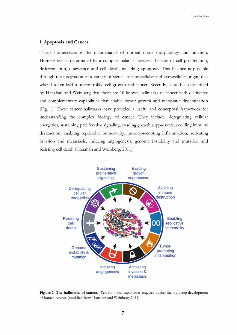
1. Apoptosis and Cancer
Tissue homeostasis is the maintenance of normal tissue morphology and function.
Homeostasis is determined by a complex balance between the rate of cell proliferation,
differentiation, quiescence and cell death, including apoptosis. This balance is possible
through the integration of a variety of signals of intracellular and extracellular origin, that
when broken lead to uncontrolled cell growth and cancer. Recently, it has been described
by Hanahan and Weinberg that there are 10 known hallmarks of cancer with distinctive
and complementary capabilities that enable tumor growth and metastatic dissemination
(Fig. 1). These cancer hallmarks have provided a useful and conceptual framework for
understanding the complex biology of cancer. They include: deregulating cellular
energetics, sustaining proliferative signaling, evading growth suppressors, avoiding immune
destruction, enabling replicative immortality, tumor-promoting inflammation, activating
invasion and metastasis, inducing angiogenesis, genome instability and mutation and
resisting cell death (Hanahan and Weinberg, 2011).
Figure 1. The hallmarks of cancer. Ten biological capabilities acquired during the multistep development of human tumors (modified from Hanahan and Weinberg, 2011).
Apoptosis and cancer
� ��
The concept that programmed cell death by apoptosis serves as a natural barrier to cancer
development has been established through functional studies conducted over the last
decades (Hanahan and Weinberg, 2011). Elucidation of the signaling pathways regulating
the apoptotic program has revealed how apoptosis is triggered in response to various
physiologic stresses that cancer cells experience during tumorigenesis or as a result of
anticancer therapy. Therefore, apoptosis plays a crucial role in the carcinogenic process
and is critical for the cell response to anticancer drugs. Besides apoptosis, tumor cells can
also die through non-apoptotic mechanisms, including autophagy, mitotic catastrophe, and
necrosis (Vangestel et al., 2009).
Apoptosis is the major type of programmed cell death in animals. This is a process that is
highly conserved throughout evolution and is essential for normal tissue development and
homeostasis (Kerr et al., 1972). Apoptosis is a highly controlled and energy-dependent
process that enables normal development and elimination of damaged and potentially
dangerous cells like cancer cells, cells infected with a virus or cells with highly damaged
DNA.
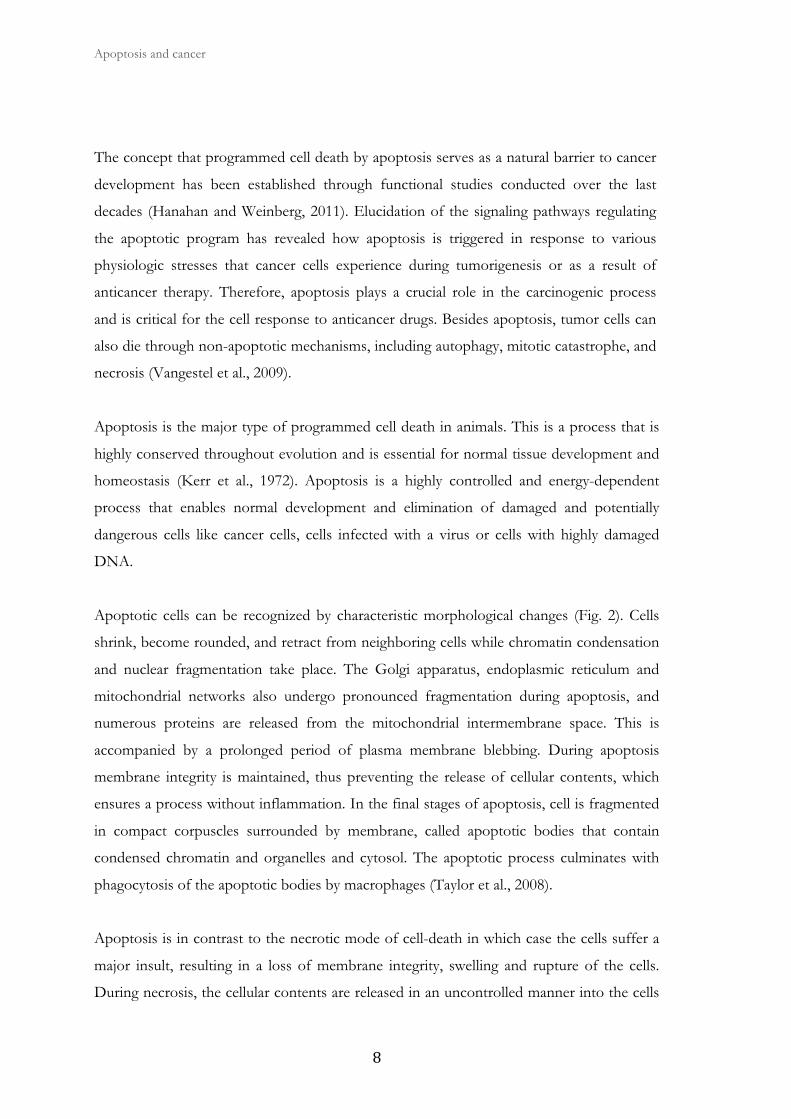
Apoptotic cells can be recognized by characteristic morphological changes (Fig. 2). Cells
shrink, become rounded, and retract from neighboring cells while chromatin condensation
and nuclear fragmentation take place. The Golgi apparatus, endoplasmic reticulum and
mitochondrial networks also undergo pronounced fragmentation during apoptosis, and
numerous proteins are released from the mitochondrial intermembrane space. This is
accompanied by a prolonged period of plasma membrane blebbing. During apoptosis
membrane integrity is maintained, thus preventing the release of cellular contents, which
ensures a process without inflammation. In the final stages of apoptosis, cell is fragmented
in compact corpuscles surrounded by membrane, called apoptotic bodies that contain
condensed chromatin and organelles and cytosol. The apoptotic process culminates with
phagocytosis of the apoptotic bodies by macrophages (Taylor et al., 2008).
Apoptosis is in contrast to the necrotic mode of cell-death in which case the cells suffer a
major insult, resulting in a loss of membrane integrity, swelling and rupture of the cells.
During necrosis, the cellular contents are released in an uncontrolled manner into the cells
� � Introduction
��
environment, which results in damage of surrounding cells and a strong inflammatory
response in the corresponding tissue (Leist and Jaattela, 2001).
Deregulation of apoptosis leads to pathological conditions such as autoimmune and
degenerative diseases, and cancer (Burz et al., 2009). Moreover, the anti-apoptotic
mechanisms regulating cell death have also been implicated in conferring drug resistance to
tumor cells (Fulda and Debatin, 2006). In mammalian cells, apoptosis occurs through two
distinct molecular pathways. The extrinsic apoptosis pathway receives signals through the
binding of extracellular protein death ligands to pro-apoptotic death receptors (DRs) with
subsequent activation of caspases, which are proteolytic enzymes that are closely involved
in the induction and execution phases of apoptosis. By contrast, the intrinsic or
mitochondrial pathway is activated by intracellular events and depends on the release of
pro-apoptotic factors from the mitochondria. Anti-apoptotic BCL-2 family members
preserve the integrity of the outer mitochondrial membrane whereas pro-apoptotic
members promote its permeabilization. Mitochondrial outer membrane permeabilization
(MOMP) allows the release into the cytosol of mitochondrial proteins like cytochrome c,
leading to caspase activation, which is essential in the execution of apoptosis. Other pro-
apoptotic proteins released by the mitochondria are EndoG, AIF, Omi/HtrA2, and
Smac/DIABLO (Fulda and Debatin, 2006) (Burz et al., 2009) (Pradelli et al., 2010) (Tait
and Green, 2010).
Figure 2. Typical features of apoptosis and necrosis. A particular mode of cell death may predominate, dependingon the injury and the type of cell (modified from Van Cruchten and Van Den Broeck, 2002; Hotchkiss et al., 2009).

Apoptosis and cancer
� ��
1.1 Apoptotic phases
Apoptosis is blocked in viable cells, but when they receive physiological and external
apoptotic signals, the apoptotic machinery is activated. The apoptotic process can be
divided in three stages (Vaux and Strasser, 1996):
1) Initiator phase: Includes a great variety of signaling pathways that mediate signals
from outside the cell, as well as others that originate inside the cell. There are three
main mechanisms for apoptosis activation, mainly through DRs, cell damage like
stress and radiation and through the action of T cytotoxic cells and natural killers.
2) Effector phase: Once signal transduction pathways have sent the apoptotic message
to the cell death effector machinery, this process is irreversible. At this point
caspases are activated.
3) Destruction phase: The activation of the cell death effector machinery leads to the
loss of cell integrity through biochemical and physiological changes that includes
chromatin condensation and DNA degradation.
1.2. Caspases: The executioners of apoptosis
The cell death effector machinery is constituted of a family of cysteine-dependent
aspartate-directed proteases (caspases). These enzymes proteolyse vital proteins to the cell,
as well as proteins that will contribute to the destruction of the cell, leading to the
morphological and biochemical changes that typically occur in apoptosis (Thornberry and
Lazebnik, 1998) (Danial and Korsmeyer, 2004). It is estimated that caspases have over 400
substrates, including protein kinases of the cytoskeleton, DNA repair enzymes, and
proteins involved in the processing of the mRNA (Meier and Vousden, 2007) (The
Caspase Substrate database http://bioinf.gen.tcd.ie/casbah/). These proteases share
similar domain structure including an N-terminal peptide or prodomain, and two subunits,
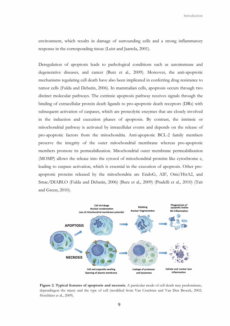
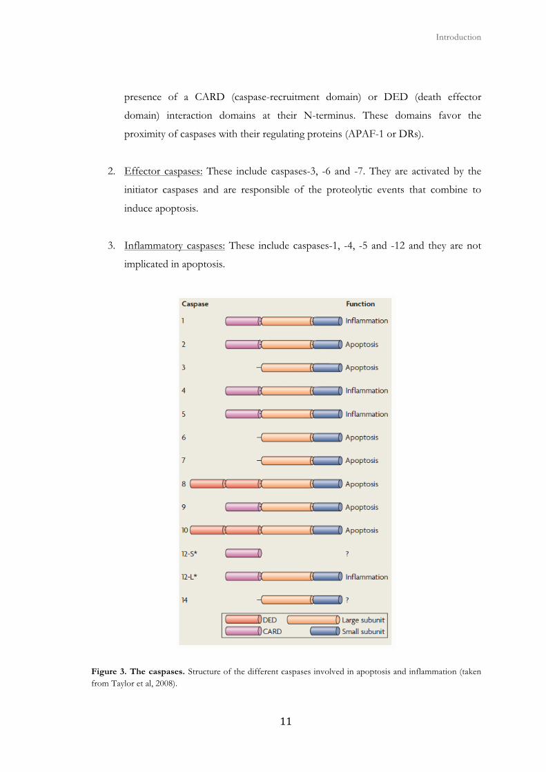
one large and one short, sometimes separated by a linker peptide (Fig. 3). There are three
groups of caspases (Fuentes-Prior and Salvesen, 2004) :
1. Initiator caspases: These include caspases-2, -8, -9 and -10. They are the first to be
activated after the apoptotic stimulus. Initiator caspases are characterized by the
� � Introduction
���
presence of a CARD (caspase-recruitment domain) or DED (death effector
domain) interaction domains at their N-terminus. These domains favor the
proximity of caspases with their regulating proteins (APAF-1 or DRs).
2. Effector caspases: These include caspases-3, -6 and -7. They are activated by the
initiator caspases and are responsible of the proteolytic events that combine to
induce apoptosis.
3. Inflammatory caspases: These include caspases-1, -4, -5 and -12 and they are not
implicated in apoptosis.
Figure 3. The caspases. Structure of the different caspases involved in apoptosis and inflammation (taken from Taylor et al, 2008).
Apoptosis and cancer
� ��
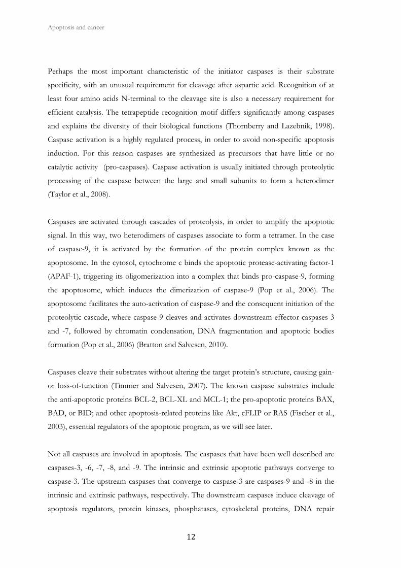
Perhaps the most important characteristic of the initiator caspases is their substrate
specificity, with an unusual requirement for cleavage after aspartic acid. Recognition of at
least four amino acids N-terminal to the cleavage site is also a necessary requirement for
efficient catalysis. The tetrapeptide recognition motif differs significantly among caspases
and explains the diversity of their biological functions (Thornberry and Lazebnik, 1998).
Caspase activation is a highly regulated process, in order to avoid non-specific apoptosis
induction. For this reason caspases are synthesized as precursors that have little or no
catalytic activity (pro-caspases). Caspase activation is usually initiated through proteolytic
processing of the caspase between the large and small subunits to form a heterodimer
(Taylor et al., 2008).
Caspases are activated through cascades of proteolysis, in order to amplify the apoptotic
signal. In this way, two heterodimers of caspases associate to form a tetramer. In the case
of caspase-9, it is activated by the formation of the protein complex known as the
apoptosome. In the cytosol, cytochrome c binds the apoptotic protease-activating factor-1
(APAF-1), triggering its oligomerization into a complex that binds pro-caspase-9, forming
the apoptosome, which induces the dimerization of caspase-9 (Pop et al., 2006). The
apoptosome facilitates the auto-activation of caspase-9 and the consequent initiation of the
proteolytic cascade, where caspase-9 cleaves and activates downstream effector caspases-3
and -7, followed by chromatin condensation, DNA fragmentation and apoptotic bodies
formation (Pop et al., 2006) (Bratton and Salvesen, 2010).
Caspases cleave their substrates without altering the target protein’s structure, causing gain-
or loss-of-function (Timmer and Salvesen, 2007). The known caspase substrates include
the anti-apoptotic proteins BCL-2, BCL-XL and MCL-1; the pro-apoptotic proteins BAX,
BAD, or BID; and other apoptosis-related proteins like Akt, cFLIP or RAS (Fischer et al.,
2003), essential regulators of the apoptotic program, as we will see later.
Not all caspases are involved in apoptosis. The caspases that have been well described are
caspases-3, -6, -7, -8, and -9. The intrinsic and extrinsic apoptotic pathways converge to
caspase-3. The upstream caspases that converge to caspase-3 are caspases-9 and -8 in the
intrinsic and extrinsic pathways, respectively. The downstream caspases induce cleavage of
apoptosis regulators, protein kinases, phosphatases, cytoskeletal proteins, DNA repair
� � Introduction
��
proteins, inhibitory subunits of endonucleaes, etc. Caspases also affect cytoskeletal
structure, cell cycle regulation, and signaling pathways, ultimately leading to the
morphologic manifestations of apoptosis, such as DNA condensation and fragmentation,
and membrane blebbing (Fulda and Debatin, 2006) (Tait and Green, 2010). In addition to
regulation by human IAPs (HIAPs), which are caspase inhibitors, this apoptotic signaling
pathway may be antagonized by the cFLIP family of proteins (FLICE inhibitory protein)
having structural homology and sequence of caspases-8 and -10 (Deveraux et al., 1997)
(Krueger et al., 2001).
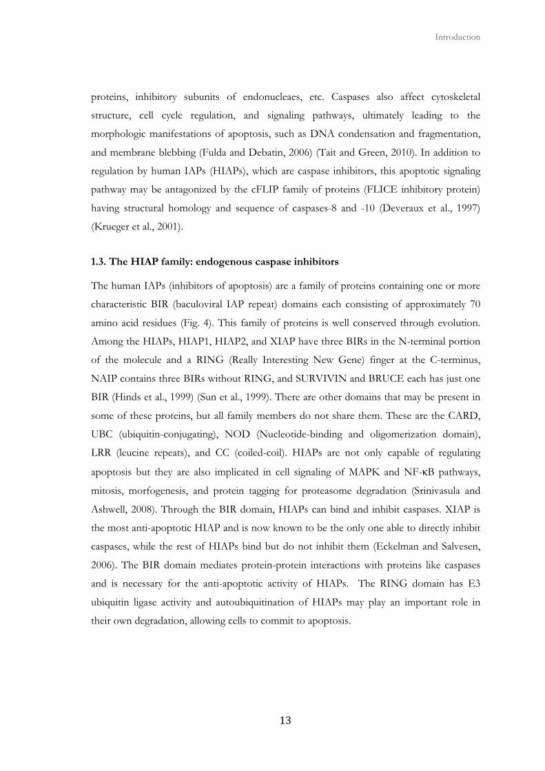
1.3. The HIAP family: endogenous caspase inhibitors
The human IAPs (inhibitors of apoptosis) are a family of proteins containing one or more
characteristic BIR (baculoviral IAP repeat) domains each consisting of approximately 70
amino acid residues (Fig. 4). This family of proteins is well conserved through evolution.
Among the HIAPs, HIAP1, HIAP2, and XIAP have three BIRs in the N-terminal portion
of the molecule and a RING (Really Interesting New Gene) finger at the C-terminus,
NAIP contains three BIRs without RING, and SURVIVIN and BRUCE each has just one
BIR (Hinds et al., 1999) (Sun et al., 1999). There are other domains that may be present in
some of these proteins, but all family members do not share them. These are the CARD,
UBC (ubiquitin-conjugating), NOD (Nucleotide-binding and oligomerization domain),
LRR (leucine repeats), and CC (coiled-coil). HIAPs are not only capable of regulating
apoptosis but they are also implicated in cell signaling of MAPK and NF-κB pathways,
mitosis, morfogenesis, and protein tagging for proteasome degradation (Srinivasula and
Ashwell, 2008). Through the BIR domain, HIAPs can bind and inhibit caspases. XIAP is
the most anti-apoptotic HIAP and is now known to be the only one able to directly inhibit
caspases, while the rest of HIAPs bind but do not inhibit them (Eckelman and Salvesen,
2006). The BIR domain mediates protein-protein interactions with proteins like caspases
and is necessary for the anti-apoptotic activity of HIAPs. The RING domain has E3
ubiquitin ligase activity and autoubiquitination of HIAPs may play an important role in
their own degradation, allowing cells to commit to apoptosis.
Apoptosis and cancer
� �
Figure 4. Structure of human IAPs. Schematic representation of the eight human IAPs with their functional domains. The common name and oficial name in BIRC nomenclature (baculoviral IAP repeat-containing) are shown (modified from Graaf et al., 2004; Srinivasula and Ashwell, 2008).
HIAP1 and HIAP2 are part of the cytoplasmic complex of TNFα where they interact
through the BIR1 domain with the adaptor protein TRAF2 (Rothe et al., 1995) (Shu et al.,
1996) and they activate the canonical NF-κB pathway (Chu et al., 1997) (Wang et al., 1998)
(Santoro et al., 2007) (Bertrand et al., 2008) (Mahoney et al., 2008), while they negatively
regulate the noncanonical pathway and spontaneous NF-κB activation (Varfolomeev et al.,
2007) (Vince et al., 2007).
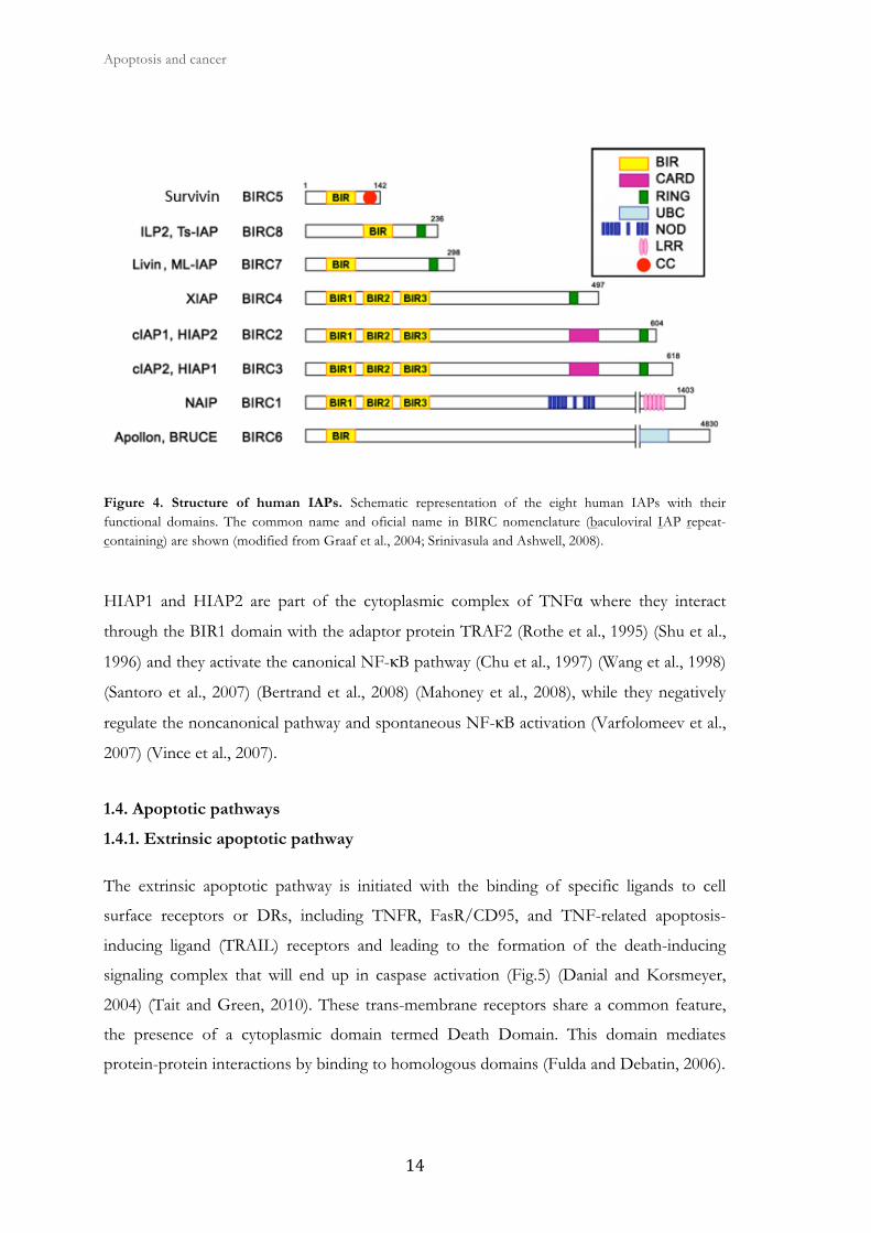
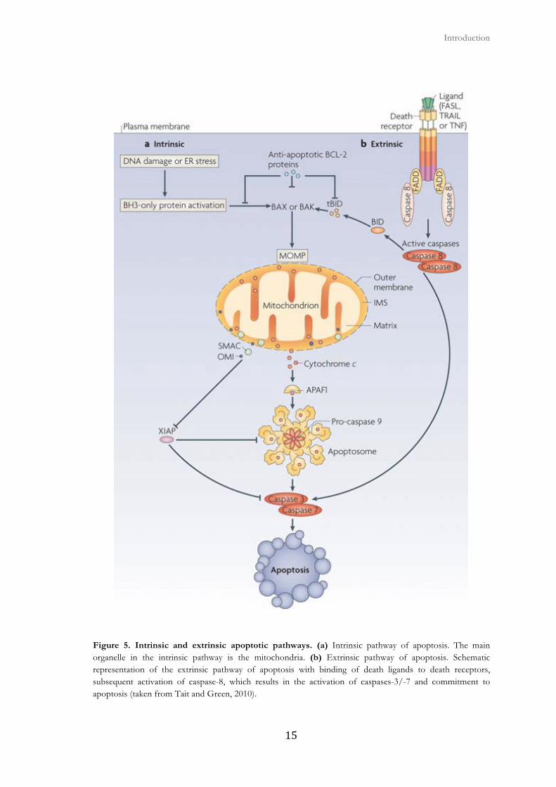
1.4. Apoptotic pathways
1.4.1. Extrinsic apoptotic pathway
The extrinsic apoptotic pathway is initiated with the binding of specific ligands to cell
surface receptors or DRs, including TNFR, FasR/CD95, and TNF-related apoptosis-
inducing ligand (TRAIL) receptors and leading to the formation of the death-inducing
signaling complex that will end up in caspase activation (Fig.5) (Danial and Korsmeyer,
2004) (Tait and Green, 2010). These trans-membrane receptors share a common feature,
the presence of a cytoplasmic domain termed Death Domain. This domain mediates
protein-protein interactions by binding to homologous domains (Fulda and Debatin, 2006).
� � Introduction
���
Figure 5. Intrinsic and extrinsic apoptotic pathways. (a) Intrinsic pathway of apoptosis. The main organelle in the intrinsic pathway is the mitochondria. (b) Extrinsic pathway of apoptosis. Schematic representation of the extrinsic pathway of apoptosis with binding of death ligands to death receptors, subsequent activation of caspase-8, which results in the activation of caspases-3/-7 and commitment to apoptosis (taken from Tait and Green, 2010).
Apoptosis and cancer
� ��
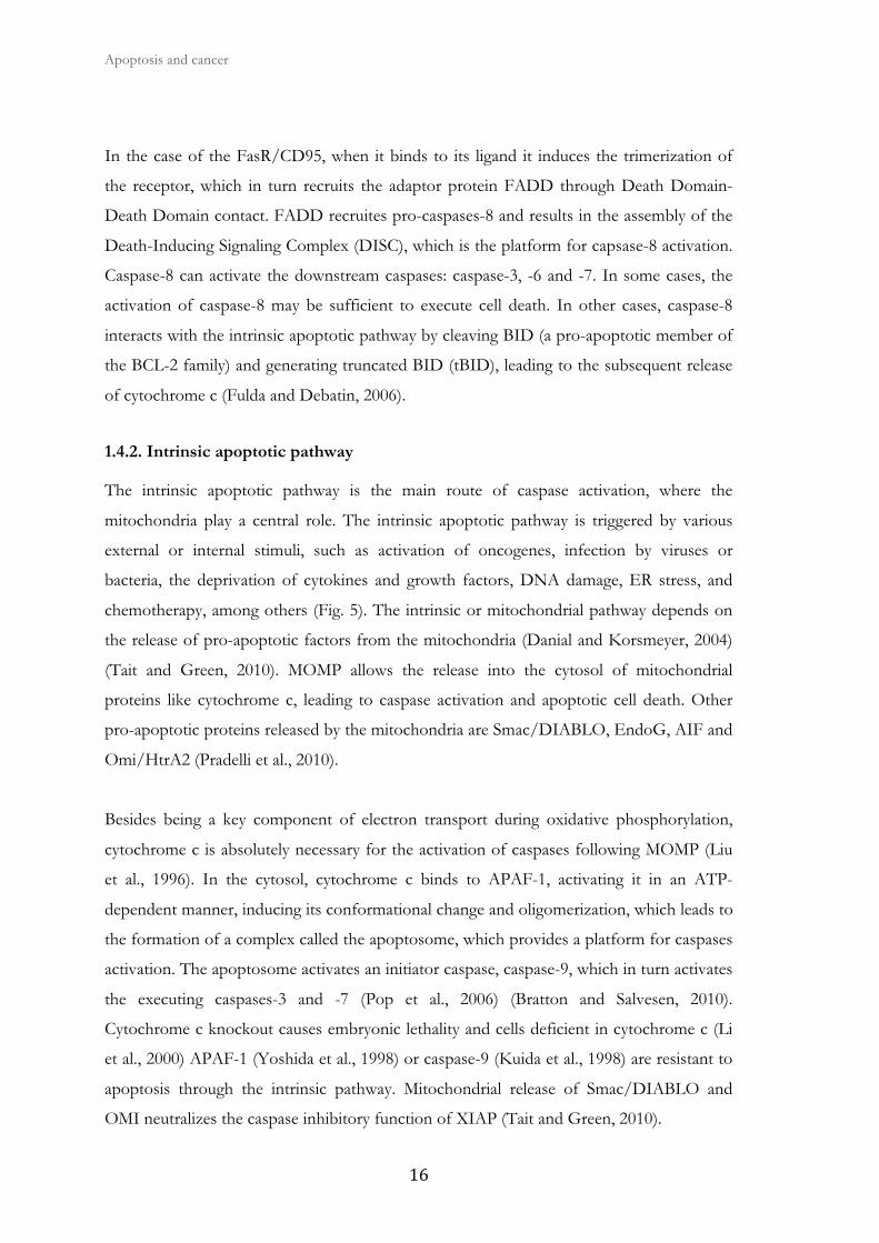
In the case of the FasR/CD95, when it binds to its ligand it induces the trimerization of
the receptor, which in turn recruits the adaptor protein FADD through Death Domain-
Death Domain contact. FADD recruites pro-caspases-8 and results in the assembly of the
Death-Inducing Signaling Complex (DISC), which is the platform for capsase-8 activation.
Caspase-8 can activate the downstream caspases: caspase-3, -6 and -7. In some cases, the
activation of caspase-8 may be sufficient to execute cell death. In other cases, caspase-8
interacts with the intrinsic apoptotic pathway by cleaving BID (a pro-apoptotic member of
the BCL-2 family) and generating truncated BID (tBID), leading to the subsequent release
of cytochrome c (Fulda and Debatin, 2006).
1.4.2. Intrinsic apoptotic pathway
The intrinsic apoptotic pathway is the main route of caspase activation, where the
mitochondria play a central role. The intrinsic apoptotic pathway is triggered by various
external or internal stimuli, such as activation of oncogenes, infection by viruses or
bacteria, the deprivation of cytokines and growth factors, DNA damage, ER stress, and
chemotherapy, among others (Fig. 5). The intrinsic or mitochondrial pathway depends on
the release of pro-apoptotic factors from the mitochondria (Danial and Korsmeyer, 2004)
(Tait and Green, 2010). MOMP allows the release into the cytosol of mitochondrial
proteins like cytochrome c, leading to caspase activation and apoptotic cell death. Other
pro-apoptotic proteins released by the mitochondria are Smac/DIABLO, EndoG, AIF and
Omi/HtrA2 (Pradelli et al., 2010).
Besides being a key component of electron transport during oxidative phosphorylation,
cytochrome c is absolutely necessary for the activation of caspases following MOMP (Liu
et al., 1996). In the cytosol, cytochrome c binds to APAF-1, activating it in an ATP-
dependent manner, inducing its conformational change and oligomerization, which leads to
the formation of a complex called the apoptosome, which provides a platform for caspases
activation. The apoptosome activates an initiator caspase, caspase-9, which in turn activates
the executing caspases-3 and -7 (Pop et al., 2006) (Bratton and Salvesen, 2010).
Cytochrome c knockout causes embryonic lethality and cells deficient in cytochrome c (Li
et al., 2000) APAF-1 (Yoshida et al., 1998) or caspase-9 (Kuida et al., 1998) are resistant to
apoptosis through the intrinsic pathway. Mitochondrial release of Smac/DIABLO and
OMI neutralizes the caspase inhibitory function of XIAP (Tait and Green, 2010).

� � Introduction
���
Mitochondrial integrity and the intrinsic pathway are controlled mainly by the
evolutionarily conserved BCL-2 family of proteins, which contains both pro-apoptotic and
anti-apoptotic members, which are able to respond to a variety of stimuli and stress stimuli
(Youle and Strasser, 2008) (Chipuk et al., 2010). Anti-apoptotic BCL-2 family members
preserve the integrity of the outer mitochondrial membrane whereas pro-apoptotic
members promote its permeabilization.
1.5. BCL-2 family members
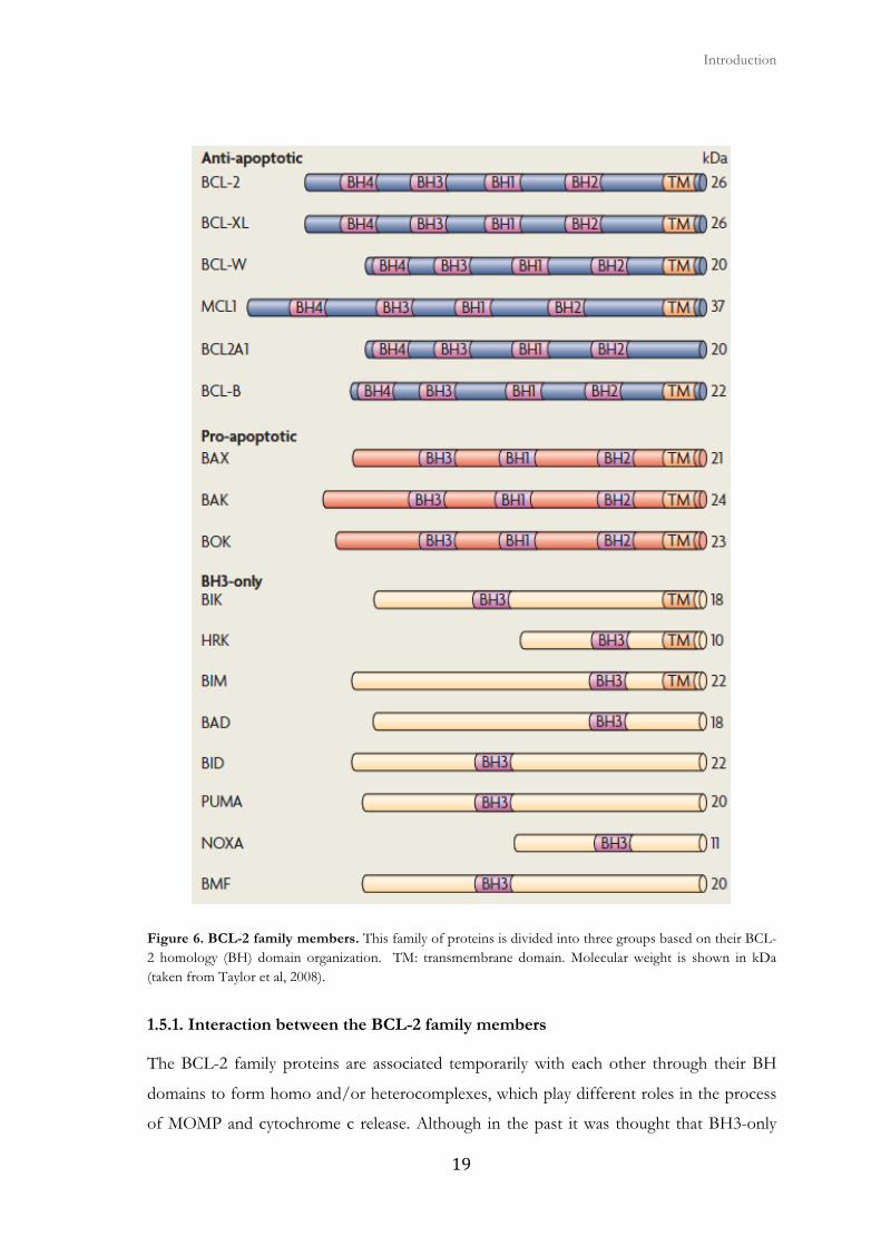
Members of the BCL-2 (B-cell lymphoma-2) family can be classified into three groups
according to their structure and function (Fig. 6):
Anti-apoptotic members (BCL-2-like)
This subfamily includes proteins that contain all four BH domains (BH1-4). A1 (BCL2A1
gene, or BFL-1), BCL-2, BCL-XL (BCL-2-related gene, long isoform), BCL-W, MCL-1
(myeloid cell leukemia 1), and BCL-B are the members of this subgroup and preserve the
integrity of the outer mitochondrial membrane to inhibit other pro-apoptotic proteins of
the family. The domains BH1, BH2 and BH3 are folded to form a hydrophobic pocket
that allows interaction with other pro-apoptotic members (Muchmore et al., 1996) (Sattler
et al., 1997). When overexpressed, each of these proteins protects cells in culture against a
variety of apoptotic stimuli. Some of these proteins are required for the survival of certain
cell types, such as BCL-2 and MCL-1, which are necessary to extend the life of mature B
and T lymphocytes (Veis et al., 1993) (Danial and Korsmeyer, 2004) (Strasser, 2005).
Pro-apoptotic multidomain members (BAX-like)
This group includes the effector members of apoptosis BAX (BCL-2-associated X protein)
and BAK (BCL-2-antagonist/killer-1). The members of this subfamily contain the
homology domains BH1-3 and induce apoptosis when overexpressed (Danial and
Korsmeyer, 2004). When activated, these proteins are supposed to promote apoptosis by
forming pores in the outer mitochondrial membrane and subsequent MOMP and
mitochondrial apoptogenic factors output. The protein BOK (BCL-2-related ovarian killer)
is also a potential effector protein, however, there is no biochemical evidence of a role
similar to that of BAX or BAK.

Apoptosis and cancer
� ��
Members with only the pro-apoptotic BH3 domain (BH3-only)
This subfamily is structurally diverse. Classically, the BH3-only proteins have been
identified to possess only the BH3 homology domain, which seems essential for its pro-
apoptotic function. However, recent sequence analysis indicate that, except BID, the
members of this subfamily differ in structure from the core members of the BCL-2 family
and is postulated to have acquired the BH3 motif by convergent evolution (Aouacheria et
al., 2005). These proteins interact with other family members to promote and regulate
apoptosis. The BH3 domain mediates these interactions. This group includes BAD, BIM,
BIK, BID/BOD, HRK/DP5, BMF, NOXA and PUMA although, it could include other
members as NIP3, BNIP3/NIX or MOAP-1.
There are other proteins homologous to BCL-2 like BCL-RAMBO, BCL-B/BOO/DIVA
or BCL-G that have not been deeply studied and that, nowadays, we cannot categorize.
There are other pro-apoptotic multidomain proteins identified as BFK (BCL-2-family kin)
and BCL-XS, which only have two BH domains, and whose role in apoptotic signaling is
unclear (Youle and Strasser, 2008). Furthermore, various forms of alternative splicing of
many of the family proteins have been described. For example, anti-apoptotic proteins
whose splicing variants are pro-apoptotic: BCL-XS or MCL-1S. Or conversely, BID-S, an
alternative splicing of BID-EL and BID-L, has an anti-apoptotic role since it has no BH3
domain (Renshaw et al., 2004). Interestingly, the BIM gene has 19 splicing variants (Akgul
et al., 2004). Many studies have achieved the knockout or overexpression mouse model of
the BCL-2 family members that allow the analysis of their physiological role, redundancy
and interactions in vivo and their contribution to the formation and progression of tumors
and resistance to therapy (Chipuk et al., 2010) (Youle and Strasser, 2008).
� � Introduction
���
Figure 6. BCL-2 family members. This family of proteins is divided into three groups based on their BCL-2 homology (BH) domain organization. TM: transmembrane domain. Molecular weight is shown in kDa (taken from Taylor et al, 2008).
1.5.1. Interaction between the BCL-2 family members
The BCL-2 family proteins are associated temporarily with each other through their BH
domains to form homo and/or heterocomplexes, which play different roles in the process
of MOMP and cytochrome c release. Although in the past it was thought that BH3-only
Apoptosis and cancer
� ��
proteins could join anti-apoptotic counterparts indiscriminately, currently the quantitative
assessment of the binding of BH3 peptides to BCL-2-like proteins revealed that the
affinities between different pairs varies up to 10,000 times (Chen et al., 2005) (Kuwana et
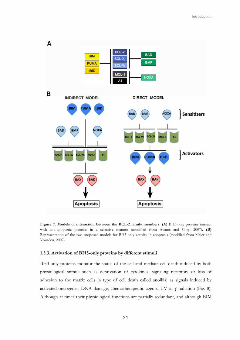
al., 2005) (Certo et al., 2006) (Kim et al., 2006). While BIM, PUMA and BID bind to all
anti-apoptotic family members, other BH3-only proteins bind only to a few. For example,
NOXA interacts only with MCL-1 and A1, and BAD only interacts with BCL-2, BCL-XL
and BCL-W (Fig. 7A). In addition, promiscuous members are much more potent as
inducers of apoptosis that members with restricted interactions. BH3-only proteins act
over BAX and BAK, which promote the activation of caspases through their effect on
mitochondrion. However, it is unknown how BH3-only proteins activate BAX and BAK.
1.5.2. Interaction models of BCL-2 family members
Two models have been proposed to explain this mechanism (Fig. 7B):
Indirect activation model (or neutralization). BAX and BAK are united in a constitutively
active state to the anti-apoptotic proteins of the family. Competitive interactions of BH3-
only proteins with anti-apoptotic proteins are sufficient to displace and liberate activated
BAX and BAK (Chen et al., 2005) (Willis and Adams, 2005) (Willis et al., 2007).
Direct activation (or derepression) model. BAX and BAK are activated following the
interaction with a subset of proteins called BH3-only activators. Anti-apoptotic proteins of
the BCL-2 family prevent MOMP by abducting these activating proteins or inhibiting
activated BAX and BAK. A second subset of BH3-only proteins called sensitizers, bind to
anti-apoptotic proteins displacing and freeing BH3-only proteins that activate BAX and
BAK (Letai et al., 2002) (Cartron et al., 2004) (Kuwana et al., 2005) (Certo et al., 2006).
Although different, both models agree on the fundamental basis: BH3-only proteins are
essential activators of apoptosis, among them BIM, BID, and PUMA are especially
powerful because they can join all anti-apoptotic proteins (indirect model), or they can bind
directly to BAX and BAK (direct model) (Meier and Vousden, 2007).
� � Introduction
���
Figure 7. Models of interaction between the BCL-2 family members. (A) BH3-only proteins interact with anti-apoptotic proteins in a selective manner (modified from Adams and Cory, 2007). (B) Representation of the two proposed models for BH3-only activity in apoptosis (modified from Meier and Vousden, 2007).
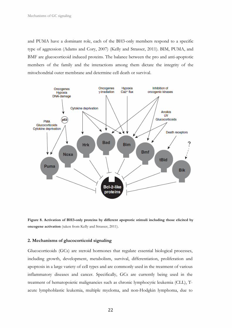
1.5.3. Activation of BH3-only proteins by different stimuli
BH3-only proteins monitor the status of the cell and mediate cell death induced by both
physiological stimuli such as deprivation of cytokines, signaling receptors or loss of
adhesion to the matrix cells (a type of cell death called anoikis) as signals induced by
activated oncogenes, DNA damage, chemotherapeutic agents, UV or γ radiation (Fig. 8).
Although at times their physiological functions are partially redundant, and although BIM
Mechanisms of GC signaling
� ��
and PUMA have a dominant role, each of the BH3-only members respond to a specific
type of aggression (Adams and Cory, 2007) (Kelly and Strasser, 2011). BIM, PUMA, and
BMF are glucocorticoid induced proteins. The balance between the pro and anti-apoptotic
members of the family and the interactions among them dictate the integrity of the
mitochondrial outer membrane and determine cell death or survival.
Figure 8. Activation of BH3-only proteins by different apoptotic stimuli including those elicited by
oncogene activation (taken from Kelly and Strasser, 2011).
2. Mechanisms of glucocorticoid signaling
Glucocorticoids (GCs) are steroid hormones that regulate essential biological processes,
including growth, development, metabolism, survival, differentiation, proliferation and
apoptosis in a large variety of cell types and are commonly used in the treatment of various
inflammatory diseases and cancer. Specifically, GCs are currently being used in the
treatment of hematopoietic malignancies such as chronic lymphocytic leukemia (CLL), T-
acute lymphoblastic leukemia, multiple myeloma, and non-Hodgkin lymphoma, due to

� � Introduction
��
their ability to induce intrinsic caspase-dependent apoptosis in these cell types (Kfir-
Erenfeld et al., 2010). These properties have made GCs one of the most frequently
prescribed drugs.
Steroid hormone research began in the late 1800s. In the early 1960s it was demonstrated
that a hormone can be taken up and retained by specific tissues, thus leading to the
identification of the estrogen receptor. It was not until 1966 that the glucocorticoid
receptor (GR) was first identified on rat thymic lymphocytes. The GR was cloned in 1985
(Hollenberg et al., 1985), starting an explosion of molecular studies on the GR and its
related family members, the steroid receptors. Members of this superfamily include the
GR, mineralocorticoid receptor (MR), progesterone receptor (PR), estrogen receptor
(ER), and androgen receptor (AR) (Heitzer et al., 2007).
GCs induce apoptosis in cells of the hematopoietic lineage, but also in non-hematologic
cells as described for osteoblasts (Herr et al., 2007). On the other hand, GCs support
survival in several non-hematologic tissues such as fibroblasts, liver, and ovary, among
others (Beck et al., 2011). Correspondingly, it seems that GCs acutely induce therapy
resistance in normal and transformed cells of epithelial origin, including the majority of
human solid malignant tumor cells like ovary, pancreas, brain, cervix and bladder. The
diverse steroid-mediated effects generate an unfavorable side-effect profile in chronic
GC-based therapy. These side effects result from overstimulation of normal physiological
GC-induced GR actions (Beck et al., 2011).
2.1. GR isoforms
Most of the actions of GCs are mediated through the GR. Ever since the cloning of the
GR (Hollenberg et al., 1985) much progress has been made in understanding the
mechanism of action of GCs (Beck et al., 2011). The GR is a member of the steroid
receptor superfamily (Zhou and Cidlowski, 2005) that is a class of transcription factors
regulated by small lipophilic ligands such as steroids, thyroid hormone retinoids, and
vitamin D3. Nuclear receptors are known for their ability to form homodimers (McKenna
and O'Malley, 2001) (McKenna and O'Malley, 2002). These receptors share a common
structural organization consisting of several modulatory domains very conserved
throughout evolution (Heitzer et al., 2007).

Mechanisms of GC signaling
� �
The GR gene (NR3C1) is located at chromosome 5q31-32 and consists of nine exons
highly conserved among species. The full length GR consists of an N-Terminal
transactivation domain (NTD) containing an activation function-1 (AF-1; aa 77-262). The
function of this region in transcriptional regulation can be ligand independent. Close to the
AF-1 region is the DNA-binding domain (DBD; aa 418-488) with two zinc fingers, a hinge
region, and a C-terminal ligand-binding domain (LBD; aa 526-777). The LBD is important
for receptor dimerization and contains sequences for protein-protein interactions with
proteins such as Hsp90. This interaction allows the folding of the receptor and prevents
the receptor to bind DNA in the absence of hormone. A second activation function (AF-2)
is embedded in the LBD and interacts with coregulators in a ligand-dependent manner,
facilitating the interaction of additional factors known as coactivators and corepressors.
AF-2 and AF-1 can act synergistically to mediate transcriptional activity (Heitzer et al.,
2007) (Oakley and Cidlowski, 2011).
Alternative splicing of the primary transcript generates several receptor isoforms (Fig. 9).
GRα and GRβ differ at their C-terminus. GRα (777 amino acid residues; 94 kDa) is
currently the main research isoform and results from the end of exon 8 being joined to the
beginning of exon 9. On the other hand, GRβ (742 amino acid residues; 90kDa), uses and
alternative splice acceptor site resulting in the union of the end of exon 8 to the
downstream sequences of exon 9. GRβ is unable to bind to GCs or activate GC-
responsive genes. It resides constitutively in the nucleus of cells and is not ubiquitously
expressed.
Several additional GR isoforms (GRϒ, GR-A, GR-P) arise from alternative splicing and
can affect GC signaling. GRϒ binds GCs and DNA in a similar way to GRα, but cannot
activate GC-responsive reporter constructs and exhibits a transcriptional profile distinct
from GRα on a subset of commonly regulated genes. GR-A and GR-P are non-hormone
binding variants due to the fact that they miss large regions of the LBD. GR-P appears to
be the predominant isoform in several GC-resistant cells (Oakley and Cidlowski, 2011).

� � Introduction
���
Figure 9. GR isoforms generated by alternative splicing. The human GR primary transcript is composed of nine exons, with exon 2 encoding most of the NTD, exons 3 and 4 encoding the DBD, and exons 5–9 encoding the hinge region (H) and LBD. The classic GRα protein results from splicing of exon 8 to the beginning of exon 9. GRβ is produced from an alternative splice acceptor site that links the end of exon 8 to downstream sequences in exon 9, encoding a variant with a unique 15-amino acid C terminus (positions 728–742). GRγ is generated by an alternative splice donor site in the intronic sequence separating exons 3 and 4, resulting in a protein with an arginine insertion (Arg452) between the two zinc fingers of the DBD. GR-A is produced from alternative splicing that joins exon 4 to exon 8, deleting the proximal 185 amino acids of the LBD (Ala490 –Ser674) encoded by exons 5–7. GR-P is formed by a failure to splice exon 7 to exon 8. The retained intronic sequence introduces a stop codon, resulting in a truncated receptor mutant missing the distal half of the LBD (taken from Oakley and Cidlowski, 2011).
It was also recently demonstrated that additional receptor proteins are generated by
alternative translation initiation from a single GR mRNA (Fig. 10). There are two well-
conserved AUG start codons derived from exon 2 and they produce eight GRα isoforms
with truncated N-terminus. They show no difference in their affinity for their ligand or
their capacity to bind GRE’s after GC exposure. Nevertheless, the GRα-D isoform resides
primarily in the nucleus and is constitutively binding to certain GRE-containing promoters.
GRα-C is the most active isoform and GRα-D is the less capable of enhancing GC-
dependent gene induction. Each of the other GR splice variants (GRβ, GRϒ, GR-A, GR-
P) are expected to give rise to similar translation variants (Oakley and Cidlowski, 2011).
Due to the fact that GRα is the most predominant isoform and our prime focus of
attention, it will be referred to as GR throughout this thesis.

Mechanisms of GC signaling
� ��
Figure 10. GRα isoforms that are generated by alternative translation initiation and sites of post-translational modification. Initiation of translation from eight different AUG start codons in a single GR mRNA generates receptor isoforms with progressively shorter N-terminal transactivation domains (NTDs). Asterisks designate approximate locations of the AUG start codons in the exon 2 sequences of the GR mRNA. Hinge region (H) (modified from Oakley and Cidlowski, 2011).
2.2. GC signaling through the GR
The unliganded GR resides mostly in the cytoplasm in an inactive state as part of a large
heat shock protein heterocomplex that includes various chaperone proteins, such as
Hsp90, Hsp70, Hsp40, and cochaperones Hsp90 binding protein p23 and Hoc among
others (Oakley and Cidlowski, 2011). Upon GC binding, the GR undergoes a
conformational change that results in its dissociation from the cytoplasmic chaperone
multiprotein complex and unmasking of the nuclear localization signal, leading to its
translocation to the nucleus. Nuclear translocation of the GR complex occurs within
minutes of cell exposure to GCs (Stahn and Buttgereit, 2008). Once in the nucleus, the
dimerized GR binds glucocorticoid response elements (GREs), usually located in the
promoter of GR-regulated genes (Beck et al., 2011).
� � Introduction
���
2.3. GR translocation
Even though the GR is thought to always reside in the cytoplasm in the absence of a
ligand, the GR continuously shuttles between the cytoplasm and the nucleus. Thus, the
subcellular localization of the GR is the result of the import and export rates of the
receptor through the nuclear pore complex (NPC) (Beck et al., 2011) (Vandevyver et al.,
2011). Previous studies suggest that GR dissociation form the chaperone complex does not
precede nuclear translocation. It has been observed that the Hsp90 chaperone multiprotein
complex is involved in GR fast and efficient trafficking (Vandevyver et al., 2011).
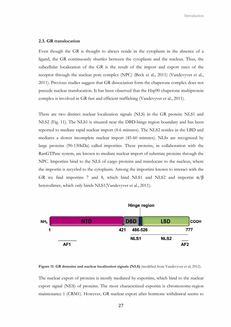
There are two distinct nuclear localization signals (NLS) in the GR protein: NLS1 and
NLS2 (Fig. 11). The NLS1 is situated near the DBD-hinge region boundary and has been
reported to mediate rapid nuclear import (4-6 minutes). The NLS2 resides in the LBD and
mediates a slower incomplete nuclear import (45-60 minutes). NLSs are recognized by
large proteins (90-130kDa) called importins. These proteins, in collaboration with the
RanGTPase system, are known to mediate nuclear import of substrate proteins through the
NPC. Importins bind to the NLS of cargo proteins and translocate to the nucleus, where
the importin is recycled to the cytoplasm. Among the importins known to interact with the
GR we find importins 7 and 8, which bind NLS1 and NLS2 and importin α/β
heterodimer, which only binds NLS1(Vandevyver et al., 2011).
Figure 11. GR domains and nuclear localization signals (NLS) (modified from Vandevyver et al, 2012).
The nuclear export of proteins is mostly mediated by exportins, which bind to the nuclear
export signal (NES) of proteins. The most characterized exportin is chromosome-region
maintenance 1 (CRM1). However, GR nuclear export after hormone withdrawal seems to
Mechanisms of GC signaling
� ��
be independent of CRM1-mediated transport as export appears not to be sensitive to the
CRM1 inhibitor leptomycin B (LMB) (Liu and DeFranco, 2000) (Holaska et al., 2001). In
this type of export it appears that there is a role for calreticulin (CRT), a calcium binding
protein localized to the lumen of the endoplasmic reticulum (Holaska et al., 2001). CRT
binds to the receptor’s DBD between the two zinc fingers, at a sequence that function as
NES. In fact, the redistribution of GR to the cytoplasm after hormone withdrawal is
compromised in CRT-deficient cells. Finally, GR shuttling between the cytoplasm and the
nucleus can influence GR nuclear signaling by altering the receptor’s turnover (Heitzer et
al., 2007).
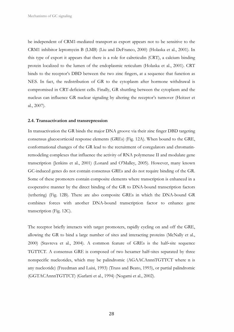
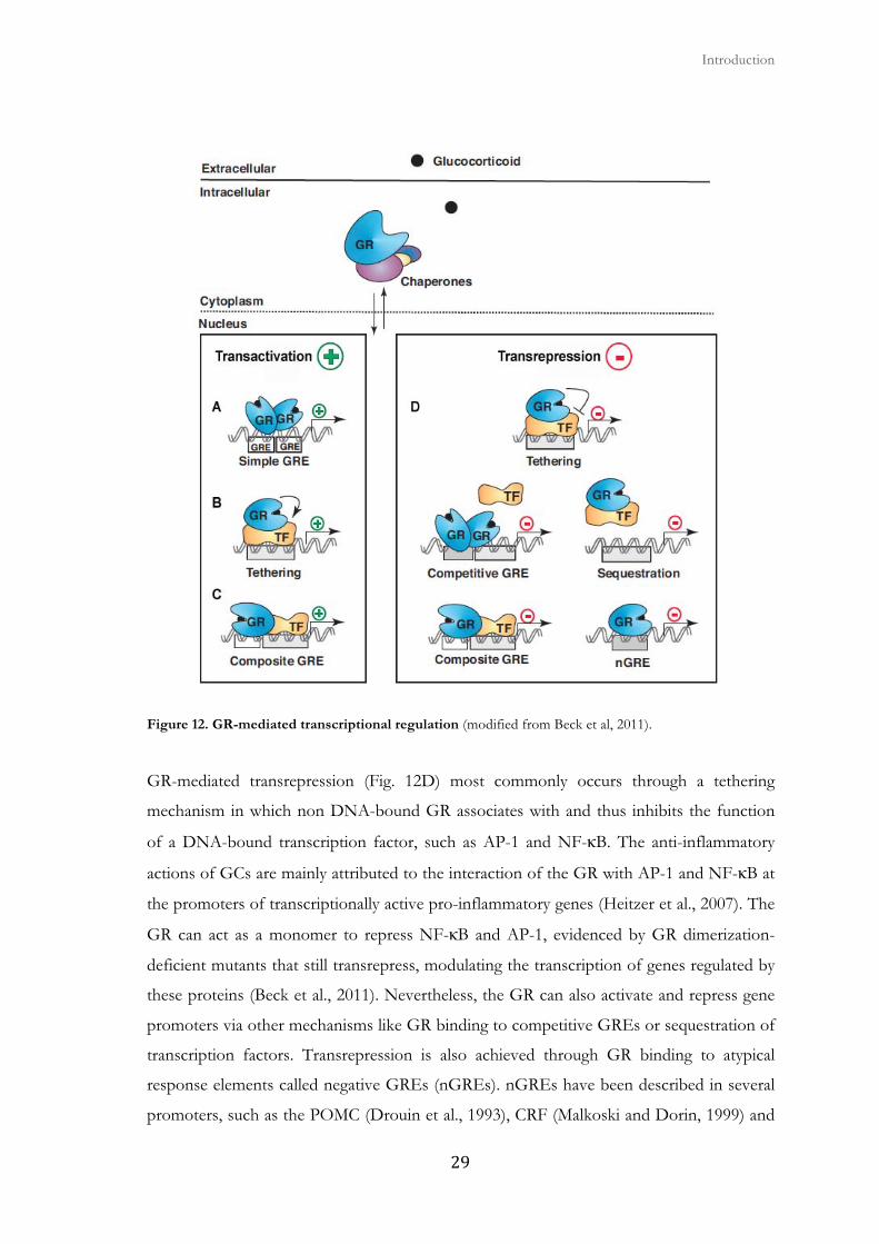
2.4. Transactivation and transrepression
In transactivation the GR binds the major DNA groove via their zinc finger DBD targeting
consensus glucocorticoid response elements (GREs) (Fig. 12A). When bound to the GRE,
conformational changes of the GR lead to the recruitment of coregulators and chromatin-
remodeling complexes that influence the activity of RNA polymerase II and modulate gene
transcription (Jenkins et al., 2001) (Lonard and O'Malley, 2005). However, many known
GC-induced genes do not contain consensus GREs and do not require binding of the GR.
Some of these promoters contain composite elements where transcription is enhanced in a
cooperative manner by the direct binding of the GR to DNA-bound transcription factors
(tethering) (Fig. 12B). There are also composite GREs in which the DNA-bound GR
combines forces with another DNA-bound transcription factor to enhance gene
transcription (Fig. 12C).
The receptor briefly interacts with target promoters, rapidly cycling on and off the GRE,
allowing the GR to bind a large number of sites and interacting proteins (McNally et al.,
2000) (Stavreva et al., 2004). A common feature of GREs is the half‐‐site sequence
TGTTCT. A consensus GRE is composed of two hexamer half‐sites separated by three
nonspecific nucleotides, which may be palindromic (AGAACAnnnTGTTCT where n is
any nucleotide) (Freedman and Luisi, 1993) (Truss and Beato, 1993), or partial palindromic
(GGTACAnnnTGTTCT) (Garlatti et al., 1994) (Nogami et al., 2002).
� � Introduction
���
Figure 12. GR-mediated transcriptional regulation (modified from Beck et al, 2011).
GR-mediated transrepression (Fig. 12D) most commonly occurs through a tethering
mechanism in which non DNA-bound GR associates with and thus inhibits the function
of a DNA-bound transcription factor, such as AP-1 and NF-κB. The anti-inflammatory
actions of GCs are mainly attributed to the interaction of the GR with AP-1 and NF-κB at
the promoters of transcriptionally active pro-inflammatory genes (Heitzer et al., 2007). The
GR can act as a monomer to repress NF-κB and AP-1, evidenced by GR dimerization-
deficient mutants that still transrepress, modulating the transcription of genes regulated by
these proteins (Beck et al., 2011). Nevertheless, the GR can also activate and repress gene
promoters via other mechanisms like GR binding to competitive GREs or sequestration of
transcription factors. Transrepression is also achieved through GR binding to atypical
response elements called negative GREs (nGREs). nGREs have been described in several
promoters, such as the POMC (Drouin et al., 1993), CRF (Malkoski and Dorin, 1999) and
Mechanisms of GC signaling
� �
osteocalcin (Meyer et al., 1997). Even though consequences of GR action may take hours,
some effects are observed within minutes. These are called non-genomic effects of GCs
and have been shown to involve protein kinases, phosphatases, and G-protein coupled
receptors (Heitzer et al., 2007).
2.5. GR coactivators and corepressors
The liganded GR can interact with components of the transcriptional machinery,
chromatin remodeling proteins, as well as RNA polymerase II and components of the
basal transcriptional machinery (Kumar and Thompson, 2005). The transcriptional
complex formed by the GR also includes the coactivators CREB-binding protein (CBP) or
its close homolog p300 (Chakravarti et al., 1996) and p160 family members known as
steroid receptor coactivators (SRCs) (Leo and Chen, 2000). Most coactivators bind to the
LBD of the GR and they enhance GR-dependent gene expression. Coactivator complexes
assemble to GR-bound promoters and stimulate GR transcriptional activation either
through direct interaction with the basal trancription machinery or by chromatin
remodeling through histone acetylation or methylation (Heitzer et al., 2007).
Besides coactivators, other GR interacting proteins have been identified and termed
corepressors. These corepressors include both nuclear receptor corepressor (NCoR) and
silencing mediator or retinoid and thyroid receptors (SMRT) (Szapary et al., 1999) (Schulz
et al., 2002). These corepressors repress transcription through the interaction with histone
deacetylases (HDACs) that are able to modify chromatin leading to a closed chromatin
structure (Watson et al., 2012).
2.6. GR phosphorylation
GR regulation is achieved by a combination of mechanisms involving ligand accessibility,
GR concentration, subcellular localization, and post-translational modifications of the GR
(Oakley and Cidlowski, 2011). Phosphorylation was the first identified modification of the
GR (Beck et al., 2009) and previous studies have highlighted the involvement of different
protein kinases in GC-mediated effects (Fig. 13) (Galliher-Beckley et al., 2008). It has been
shown that the modulation of GR phosphorylation cycle by phosphatases maintains
steady-state receptor phosphorylation at a low basal level in the absence of ligand, and GC-

� � Introduction
��
dependent GR phosphorylation ultimately affects transactivating and transrepressing
capacities of the GR (Wang et al., 2007).
Eight phosphorylated residues of the murine GR have been mapped: Ser122, 150, 212,
220, 234, 315, 412 and Thr159. Furthermore, Ser122, 150, 212, 220, 234, and 412 and the
surrounding sequences are conserved in the rat and human GR. Only Thr159 and Ser315
lacked homology to the human GR. Interestingly, all sites with the exception of Ser315
were constrained to the N-terminal transactivation domain of the GR, suggesting that
phosphorylation of the GR may function to modulate the transcription of target genes.
With the exception of Ser150 and Thr159, the majority of data suggest that the
phosphorylation of GR is induced by ligand binding to the receptor. Altogether, results
suggest that the GR can be phosphorylated at one or more residues and phosphorylation is
a dynamic process involving the dephosphorylation and phosphorylation of several
serine/threonine residues. Therefore, different patterns of GR phosphorylation could lead
to the alteration of the transcriptional activity of the GR (Galliher-Beckley and Cidlowski,
2009).
Figure 13. The known GR phosphorylation sites. Phosphorylation sites on the human, rat, and mouse receptor and the kinases implicated are shown (taken from Galliher-Beckley and Cidlowski, 2009).

Mechanisms of GC signaling
� �
The protein kinases involved in GR phosphorylation after GR exposure are still being
studied. The protein kinases that have been shown to phosphorylate the GR and modulate
its transcriptional activity include mitogen-activated protein kinases (MAPKs), glycogen
synthase kinase-3 (GSK3), and cyclin–dependent kinases (CDKs) (Fig. 13). The GR is also
a susbtrate for ubquitination, sumoylation and acetylation (Kfir-Erenfeld et al., 2010)
(Oakley and Cidlowski, 2011).
2.7. GSK3
GSK3 is one of the kinases known to phosphorylate and modulate the GR (Galliher-
Beckley and Cidlowski, 2009). GSK3 is a Serine/Threonine protein kinase highly
conserved from yeast to mammals (Beurel and Jope, 2006) (Forde and Dale, 2007)
(Rayasam et al., 2009). It was initially identified as a key regulator of insulin-dependent
glycogen synthesis, but it has been demonstrated that GSK3 is a multifunctional kinase
involved in cellular metabolism, signaling transduction, growth, differentiation, and cell fate
determination (Forde and Dale, 2007). There are two homologous mammalian GSK3
isoforms encoded by different genes, GSK3α and GSK3β. They share 98% identity within
their catalytic domain, but N- and C-terminal sequences diverge, making them structurally
similar but not functionally identical (Woodgett, 1990) (Forde and Dale, 2007).
GSK3α (483 amino acid residues; 52kDa) is larger than GSK3β (433 aminoacod residues;
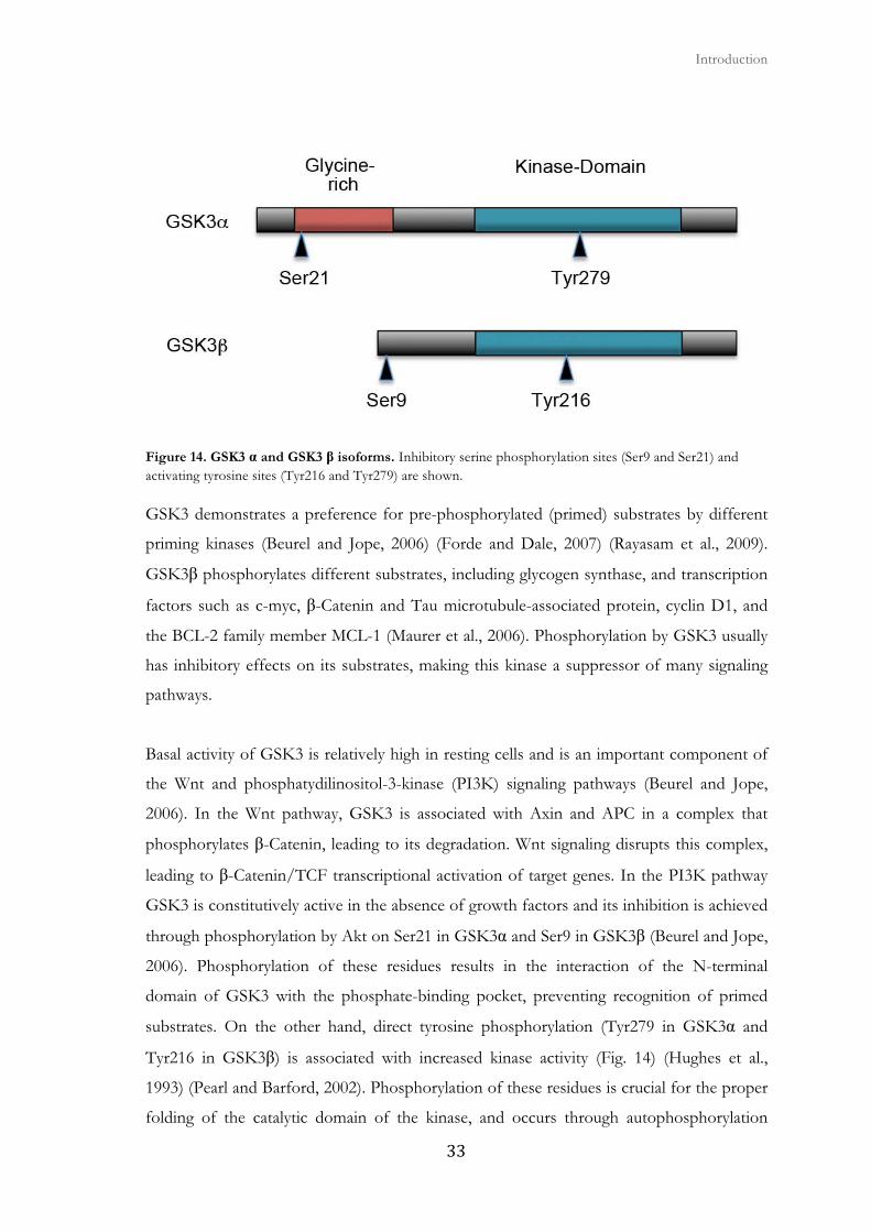
47 kDa) due to a glycine-rich extension at the N-terminus (Woodgett, 1990) (Fig. 14). The
GSK3β mRNA undergoes alternative splicing, producing at least two protein products
GSK3β1 and GSK3β2, the later being a brain specific isoform. GSK3α and GSK3β are
ubiquitously expressed, but GSK3β is the most predominant form in the brain (Sutherland,
2011). While GSK3α deficient mice are born fertile; GSK3β knockout causes embryonic
lethality due to severe liver degeneration and defects in embryonic cardiomyocyte
proliferation and differentiation resulting in heart failure and death (Force and Woodgett,
2009). The differential phenotypes between isoform deletions suggest nonredundant
functions of the GSK3 isoforms. Due to the fact that GSK3 inhibitors do not discriminate
between the two isoforms, many functions attributed in the literature to GSK3β are likely
shared by GSK3α.
� � Introduction
�
Figure 14. GSK3 α and GSK3 β isoforms. Inhibitory serine phosphorylation sites (Ser9 and Ser21) and activating tyrosine sites (Tyr216 and Tyr279) are shown.
GSK3 demonstrates a preference for pre-phosphorylated (primed) substrates by different
priming kinases (Beurel and Jope, 2006) (Forde and Dale, 2007) (Rayasam et al., 2009).
GSK3β phosphorylates different substrates, including glycogen synthase, and transcription
factors such as c-myc, β-Catenin and Tau microtubule-associated protein, cyclin D1, and
the BCL-2 family member MCL-1 (Maurer et al., 2006). Phosphorylation by GSK3 usually
has inhibitory effects on its substrates, making this kinase a suppressor of many signaling
pathways.
Basal activity of GSK3 is relatively high in resting cells and is an important component of
the Wnt and phosphatydilinositol-3-kinase (PI3K) signaling pathways (Beurel and Jope,
2006). In the Wnt pathway, GSK3 is associated with Axin and APC in a complex that
phosphorylates β-Catenin, leading to its degradation. Wnt signaling disrupts this complex,
leading to β-Catenin/TCF transcriptional activation of target genes. In the PI3K pathway
GSK3 is constitutively active in the absence of growth factors and its inhibition is achieved
through phosphorylation by Akt on Ser21 in GSK3α and Ser9 in GSK3β (Beurel and Jope,
2006). Phosphorylation of these residues results in the interaction of the N-terminal
domain of GSK3 with the phosphate-binding pocket, preventing recognition of primed
substrates. On the other hand, direct tyrosine phosphorylation (Tyr279 in GSK3α and
Tyr216 in GSK3β) is associated with increased kinase activity (Fig. 14) (Hughes et al.,
1993) (Pearl and Barford, 2002). Phosphorylation of these residues is crucial for the proper
folding of the catalytic domain of the kinase, and occurs through autophosphorylation
Mechanisms of GC signaling
�
during the synthesis of the GSK3 peptide. It appears that different pools of the kinase
function in Wnt and PI3-Kinase signaling, as regulation of GSK3 in Wnt signaling pathway
do not involve N-terminal or tyrosine phosphorylation. In this way, compartmentalization
of GSK3 allows differential upstream and downstream substrate phosphorylation.
Aditionally, ERK may phosphorylate Thr43 and p38 can phosphorylate Ser389 and Thr390
of GSK3β reducing its activity. Thr43 and Thr390 are not conserved among GSK3 α and β
isoforms, suggesting an isoform specific regulation. In both cases this phosphorylation may
favor Ser9 phosphorylation rather than promoting direct inhibition (Sutherland, 2011)
(Medina and Wandosell, 2011).
Additional mechanisms besides phosphorylation are employed to regulate GSK3 like the
control of its subcellular localization. GSK3β locates between the cytoplasm and nucleus in
a steady-state and GSK3α is mostly cytoplasmic. GSK3α accumulates in the nucleus via
activation of the calcium/calpain pathway or upon serum starvation. Additionally, the N-
terminal domain of GSK3α is responsible for GSK3α nuclear exclusion (Azoulay-Alfaguter
et al., 2011).
GSK3β is primarily cytoplasmic during G1 phase of the cell cycle and triggers the
proteolysis of some proteins that promote G1 to S transition. On the other hand, nuclear
levels of GSK3β are higher in the S phase were it phosphorylates cyclin D1 which is
subsequently degraded (Diehl et al., 1998). Additionally, GSK3β has been implicated in
regulating interphase microtubule dynamics and GSK3 inhibitors induce a delay in mitotic
entry and exit, mitotic spindle defects, and chromosome misalignment (Tighe et al., 2007).
2.7.1. Regulation of apoptotic pathways by GSK3
A) GSK3 facilitates the intrinsic apoptotic pathway
There is a well-established relationship between GSK3 activity and apoptosis. GSK3 seems
to prompt the intrinsic apoptosis-signaling pathway under a broad range of stimuli
including growth factor deprivation or inhibition of the PI3K/Akt signaling pathway,
DNA damage, hypoxia, ER-stress, and staurosporine treatment (Beurel and Jope, 2006).
These conditions that activate the intrinsic apoptotic signaling pathway cause the
disruption of mitochondrion, leading to cell destruction.

� � Introduction
��
Although direct intramitochondrial substrates of GSK3 involved in intrinsic apoptosis
pathway have not yet been identified, GSK3 targets several key proteins that regulate
signals leading to the disruption of mitochondrion. GSK3 can directly phosphorylate BAX
on Ser163, stimulating BAX translocation to the mitochondria, which leads to MOMP and
mitochondrial proteins release during apoptosis (Linseman et al., 2004). GSK3 is also
required for the stress-induced expression of BIM in cerebellar neurons (Hongisto et al.,
2003) and is able to target MCL-1 for ubiquitin-dependent degradation (Maurer et al.,
2006). Altogether, GSK3 regulates the expression of proteins that are key mitochondrial
components of the intrinsic apoptotic-signaling pathway and oppositely regulates anti-
apoptotic protein expression levels.
Importantly, GSK3 can modulate the activity of a great number of transcription factors
that encode apoptosis-regulating proteins that control gene expression. These proteins
include p53, β-Catenin, Myc, NFκB, cyclic AMP response element binding protein (CREB)
and Heat shock factor-1 (HSF-1). HSF-1 inhibition by GSK3 reduces the expression of
heat shock proteins, contributing to cell death. Besides regulating gene expression, GSK3
also regulates translation, and it was described that inhibition of protein synthesis, which
GSK3 achieves by phosphorylating and inhibiting eIF2B, contributes to GSK3-induced
apoptosis (Pap and Cooper, 2002). Thus, the role of GSK3 in intrinsic apoptotic signaling
is not that of an initiator but of a facilitator, promoting the signaling responses to insults
that initiate this pathway.
B) GSK3 inhibits the extrinsic apoptotic pathway
The involvement of GSK3 in the extrinsic apoptotic signaling pathway was first described
in mouse embryonic fibroblasts (MEFs), where knocking out GSK3β caused mouse
embryonic lethality due to TNF hypersensitivity in the liver (Hoeflich et al., 2000). This
provided the key insight that GSK3β inhibits TNF-induced apoptosis. This was supported
by the fact that lithium, a widely used GSK3 inhibitor, was shown to potentiate TNF-
induced cytotoxicity in MEFs from WT mice. The inhibitory effect of GSK3 on TNF-
induced apoptosis has been extended to other DRs. It is now clear that GSK3 inhibits
TNF-, TRAIL- or FAS-mediated apoptosis, demonstrating that this is a generalized action
regulating the extrinsic apoptotic pathway (Liao et al., 2003) (Song et al., 2004).
Mechanisms of GC signaling
� �
2.8. GR regulation by GSK3
Different interactions between GSK3 and the GR have been previously described. There is
a hormone-dependent GR phosphorylation of human Ser404 by GSK3β, which targets the
GR for nuclear export. This phosphorylation seems to play a role in GR protein stability
and turnover as an un-phosphorylatable mutant had an increase in half-life in the absence
of a ligand and when exposed to dexamethasone (Galliher-Beckley et al., 2008). Moreover,
a mutant that mimics both the size and negative charge of a phosphorylation serine residue
showed a decrease in half-life in the absence and presence of hormone. Additionally,
GSK3β mediated phosphorylation of rat GR Thr171 has been described (Rogatsky et al.,
1998).
Recently, a protein kinase screening in lymphoid cells showed that GSK3 has a role in GC-
induced apoptosis (Spokoini et al., 2010). In the absence of a ligand, GSK3α is bound to
the GR and exposure to GCs leads to its dissociation from the GR (Spokoini et al., 2010)
and then GSK3α and GSK3β interact with BIM, a BH3-only protein induced by GCs in
leukemia cells (Wang et al., 2003) (Zhang and Insel, 2004) (Iglesias-Serret et al., 2007).
Moreover, it has been described that the GR associates with GSK3β in the presence of
dexamethasone but not with GSK3α (Galliher-Beckley et al., 2008). Pharmacological
inhibition of GSK3 blocked GC-induced apoptosis in different hematopoietic cell lines
(Spokoini et al., 2010), and attenuated GC-induced upregulation of BIM (Nuutinen et al.,
2009). Thus, it seems that GSK3 isoforms regulate GR cellular response by using different
mechanisms.
2.9. Crosstalk between kinases and the GR
Some GC signaling events occur much faster and are of shorter duration than would be
expected on the basis of genomic signaling. It has been suggested that besides the genomic
mechanism, GCs could also act on diverse downstream targets, bypassing nuclear signaling.
This could be achieved by positively or negatively regulating kinase signaling. Several of
these target kinases have been identified, among which are intracellular proteins such as
kinases, including MAPKs, CDKs, PI3K/Akt, IKKs and protein kinase C (PKC) (Herr et
al., 2007) (Beck et al., 2009).
� � Introduction
��
MAPKs
Activated GR forms a complex regulatory loop with the MAPK signaling pathway. GC
induce DUSP1-mediated phosphorylation of JNK. The GR can also directly interact with
JNK, interfering with its activity (Bruna et al., 2003). JNK has been shown to
phosphorylate the rat GR at Ser246 and this phosphorylation attenuates GR transcriptional
activity. Additionally, homologous phosphorylation of human GR (Ser226) has a negative
effect on hormone signaling by enhancing nuclear export of the GR (Galliher-Beckley and
Cidlowski, 2009).
On the other hand, ERK can phosphorylate the rat GR on Ser224 and Ser232 and the
human GR on Ser203 and these phosphorylations attenuate the transcriptional activity of
the GR. GCs are known to inhibit ERK MAPK activation by inhibiting the interaction of
Raf-1 with its cochaperone Hsp90. In the case of p38 MAPK, there seems to be cell-
specific effects. GC exposure in lymphoid cells seems to activate p38 but in many cell lines
it seems to decrease its phosphorylation and activity. The phosphorylation of Ser134 of
the GR by p38 significantly increases the association of the GR with the 14-3-3 class of
signaling proteins on chromatin promoter regions, resulting in a blunted hormone-
dependent transcriptional response of specific genes (Galliher-Beckley et al., 2011). In
lymphoid cells, p38 MAPK-induced Ser211 phosphorylation of GR promoting GC
sensitivity while ERK/JNK activity decreased the amount of Ser211 phosphorylation
resulting in enhanced GC resistance.
CDKs
GCs can cell dependently repress gene expression of CDK4, CDK6 and their associating
cyclin D3. GCs can also induce the expression of the CDK inhibitor p21. CDK2 and
CDK4 activity are also inhibited by GCs. CDK1 and CDK2 were also described to be
activated by Cyclin O during GC-induced apoptosis in lymphoid cells (Roig et al., 2009).
On the other hand, several cyclin–CDK complexes (cyclin A-CDK2, cyclin A-CDC2,
cyclin B-CDK2, cyclin B-CDC2, and cyclin E-CDK2) can phosphorylate the rat GR on
Ser224 and Ser232 and this phosphorylation of GR by cyclin-CDKs is required for full
GR-mediated transcriptional activity. Additionally, CDK5 activity blunts GC signaling and
phosphorylates the GR at Ser226. However, it seems that Ser226 phosphorylated receptor
may still be transcriptionally active, and further research is required to determine the
Mechanisms of GC signaling
� �
precise role of human Ser226 phosphorylation of GR in modulating GC signaling. Unlike
most CDKs, CDK5 activity is restricted to cells of the nervous system. The data also show
that multiple serines within the human GR (Ser45, Ser203, Ser211, Ser226, Ser395) become
phosphorylated by CDK5 (Galliher-Beckley and Cidlowski, 2009).
PI3K/Akt
The PI3K/Akt signaling regulates important cellular functions such as proliferation and
survival. This pathway is activated in a wide variety of cancers, which results in apoptosis
resistance. Akt also known as protein kinase B (PKB) is rapidly activated upon GC
treatment through PI3K. PI3K is a heterodimer composed of a regulatory unit (p58) and
two catalytic α and β subunits. Although the activation of PI3K by GC requires the GR, no
GRE-mediated gene transcription is necessary for PI3K activation. Instead, the GR can
interact with the p85α subunit of PI3K under high doses of GC. PI3K/Akt signaling
seems to play a role in NF-κB mediated GC resistance. Additionally, transcriptional and
protein induction of serum and GC-regulated kinase-1 (SGK-1), a downstream effector of
PI3K signaling, is required for survival signaling induced upon GR activation (Beck et al.,
2009). Akt inhibition has been shown to enhance dexamethasone-induced apoptosis, as
PI3K/Akt protects and delays dexamethasone-induced apoptosis. Importantly, PI3K/Akt
signaling inhibits BIM expression by phosphorylating the FOXO3 transcription factor
(Nuutinen et al., 2006).
IKKα
IKKα and IKKβ can phosphorylate IκB leading to its proteosomal degradation. This leads
to NF-κB to translocate to the nucleus. GCs can induce IKKα in some cell lines mediating
apoptosis protection. This apoptosis protection is lost when the dominant negative form of
IκB is expressed. Thus, GC-induced resistance may require NF-κB activation (Beck et al.,
2009).
PKC
PKC regulates both positive and negative signal transduction pathways essential for the
initiation and homeostasis of immune responses. PKC isoforms constitute a family of
� � Introduction
��
serine/threonine kinases that depending on the cellular context regulate a wide variety of
cell responses including MAPKs (Spitaler and Cantrell, 2004). Glucocorticoids can both
increase (Kajita et al., 2001) (Maddali et al., 2005) (Cote-Velez et al., 2008) and decrease
(Jun et al., 1994) (Nguyen and Watts, 2006) PKC activity in several tissues or cells affecting
gene transcription.
3. Mediators of glucocorticoid action
3.1. Glucocorticoid-induced leucine zipper (GILZ)
GILZ was initially isolated as a dexamethasone-responsive gene from a thymus subtraction
DNA library (D'Adamio et al., 1997). Ever since, GILZ has been identified as a GC-
transactivated gene in various cell types. Most of the research done on GILZ has been
made in T cells. In T cells GILZ has been reported to inhibit transcription factors such as
NF-κB and AP-1 and the kinases RAF-1 and ERK (Beaulieu and Morand, 2011).
GILZ is a protein of 137 amino acids in humans and consists of three major domains: the
N-terminal, leucine zipper (LZ), and C-terminal domains. GILZ, also known as TSC22
domain family protein 3, also contains a tuberous sclerosis complex (TSC) domain
(Beaulieu and Morand, 2011). The LZ motif of GILZ is located in the central part of the
protein and mediates the homodimerization of GILZ required for many of its functions
(D'Adamio et al., 1997), while the other domains are responsible for protein-protein
interactions between GILZ and transcriptional and signaling molecules. The promoter of
GILZ contains 6 GREs and two functional forkhead-responsive elements. FOXO3
binding to these binding sites is necessary for maximal dexamethasone-induced expression
of GILZ in T lymphocytes (Asselin-Labat et al., 2004). The GILZ promoter also harbors
binding sites for signal transducer and activator of transcription 6 (STAT 6), nuclear factor
of activated T cells (NFAT), Octamer, and c-myc (Beaulieu and Morand, 2011). Activation
of Akt leads to the phosphorylation of transcription factors belonging to the FOXO
family, which results in the nuclear exclusion of the FOXO proteins and thus leads to the
inhibition of their cognate transcriptional targets. Thus, the inhibition of the PI3K/Akt
pathway results in the upregulation of GILZ expression (Grugan et al., 2008). In addition,
GILZ negatively feeds back to regulate the nuclear exclusion of FOXO3 in a CRM1
dependent manner (Latre de Late et al., 2010) (Fig. 15). Thus, GILZ not only inhibits its
own expression but also limits the effects of nuclear FOXO3.

Mediators of glucocorticoid action
� �
GILZ can also participate in the GC-mediated inhibition of AP-1 by directly binding to its
components c-Jun and c-Fos and by binding RAF-1, inhibiting its phosphorylation. The
resulting impairment of RAF-1 activation decreases ERK activity, which is important for
transcriptional activity of AP-1 (Beaulieu and Morand, 2011). Increased expression of
GILZ inhibits the activation, nuclear translocation and DNA binding activity of NF-κB
(Ayroldi et al., 2001). This inhibition of NF-κB is the result of direct association between
GILZ and both the p65 and p52 subunits of NF-κB, while the phosphorylation or
degradation of proteins belonging to the IκB family are not affected, which suggests that
GILZ acts directly as a glucocorticoid-induced corepressor of NF-κB. Altogether, it
appears that GILZ is an important mediator of GC signaling.
Figure 15. Interactions of GILZ with key signaling pathways. GILZ affects several major cell signaling
pathways involved in cell growth, cell differentiation, cell survival and inflammation (taken from Beaulieu et
al., 2011).
3.2. BIM
Among the GC responsive genes that have been described that can be involved in GC-
induced apoptosis is the pro-apoptotic BH3-only BCL-2 family protein BIM (Wang et al.,
2003) (Zhang and Insel, 2004). The role of BIM in GC-induced cell death was confirmed

� � Introduction
��
in BIM knockout thymocytes, which showed partial resistance to GCs (Bouillet et al.,
1999). Additionally, BIM down-regulation in malignant lymphoid cells confers resistance to
dexamethasone (Abrams et al., 2004) (Lu et al., 2006) (Rambal et al., 2009) (Lopez-Royuela
et al., 2010).
The three major isoforms BIM-EL, BIM-L, and BIM-S can all induce apoptosis, the latter
being the most potent inducer (Kfir-Erenfeld et al., 2010). Previous studies demonstrated
that the promoter of BIM does not contain GREs (Wang et al., 2003), suggesting a
mechanism of BIM regulation not mediated directly by the GR in response to GCs. In
accordance to this, it was recently demonstrated that GCs repress the expression of miR-
17-92, which results in elevated BIM protein expression (Molitoris et al., 2011). During the
writing of this thesis it was described that dexamethasone treatment induces transcription
factor RUNX2 and c-Jun in parallel with BIM induction. c-Jun binds to and activates the
AP-1-binding site at about 2.7 kb from the transcription start site of the BIM promoter
(Heidari et al., 2012).
4. GCs and Chronic Lymphocytic Leukemia (CLL)
Chronic lymphocytic leukemia (CLL) was first defined in 1966 as a disease characterized
for the monoclonal expansion of B lymphocytes with poor capacity to proliferate and long
lifespan (Galton, 1966) (Dameshek, 1967). These lymphocytes are small, with mature
appearance but functionally immature (immunoincompetents) due to deficient production
of antibodies, which favours the appearance of infectious diseases (Chiorazzi et al., 2005).
CLL is a disorder that is manifested by progressive accumulation of these resting cells in
the blood, bone marrow, lymphatic tissues and that ultimately becomes lethal (Dighiero
and Hamblin, 2008). In this lymphoid neoplasm, lymphocyte counts in the blood are
usually greater than or equal to 5x10⁹/L with a characteristic immunophenotype of CD5-
and CD23-positive B cells (Hallek et al., 2008) (Shanafelt et al., 2009).
CLL is a malignancy that results in failed apoptosis, as blood circulating CLL cells are non-
proliferating and are arrested in the G0/G1 phase of the cell cycle (Decker et al., 2003).
Cell division occurs in ‘proliferation centers’ in tissue microenvironments, accounting for
the rise in white blood cell counts in some patients. However, lack of apoptosis is
considered the prime cause for the loss of balance in B-cell homeostasis in this malignancy.
Glucocorticoids and Chronic Lymphocytic Leukemia
� �
Thus, clinically used chemotherapeutic agents act by inducing apoptosis in CLL
lymphocytes (Packham and Stevenson, 2005).
CLL is considered a clinically and biologically heterogeneous disease originated from
stimulated mature B cells and expressing in its surface the above mentioned markers and
low levels of IgM, IgD, and CD79. CLL cells may differ in activation, maturation state, or
cellular subgroup (Chiorazzi et al., 2005). CLL is the most common leukemia in western
countries (Europe and USA), where it represents approximately 30% of leukemias, 90% of
chronic lymphoid leukemia and 7% of non-Hodgkin lymphomas (Montserrat and Rozman,
1995). Each year 180,000 new cases are diagnosed in the world (Chen and McMillan, 2008)
(Gribben, 2010) (Pekarsky et al., 2010). The global incidence rate is 3 new cases per
100,000 inhabitants per year (Oscier et al., 2004). CLL is a disease of the elderly, with a
median age at diagnosis of 72 years and median age at death from CLL of 79 years. Almost
70% of CLL patients are older than 65 years at the time of diagnosis; less than 2%, younger
than 45; and the incidence increases gradually with age (Chen and McMillan, 2008)
(Gribben, 2008) (Gribben, 2010). The clinical course of CLL is heterogeneous as some
patients, have an aggressive disease and require treatment, most commonly with cytotoxic
therapy. On the other hand, there are patients that have a stable, non-progressive disease
that often requires no treatment. Relapses are common and CLL remains incurable
(Packham and Stevenson, 2005).
CLL is usually detected in routine medical check-ups or in response to a slight discomfort
of the patient, such as fatigue or respiratory problems during exercise. CLL cells are
monomorphic, small, round B lymphocytes, apparently mature, with condensed nuclear
chromatin, absence of nucleoli and poor cytoplasm (Fig. 16), characterized by a peculiar
fragility of the cell membrane that leads to frequent rupture of the leukemic cells while
preparing the blood film, creating the so-called “Gumprecht nuclear shadows” or “smudge
cells”. In addition, there are two morphologically atypical CLL forms: CLL/PL, containing
10-55% of prolymphocytes, and atypical or mixed CLL, containing >15% of population
with lymphoplasmocitoid features. These morphological variants have typical
immunophenotypic features of CLL (Gentile et al., 2005).

� � Introduction
�
Figure 16. Typical morphology of CLL cells in peripheral blood. The cells are small with condensed nuclear chromatin and scant pale cytoplasm (May Grumwald Giemsa staining taken from Schlette et al., 2003).
It is still unknown if the apoptotic resistance of CLL cells is due to genetic or epigenetic
alterations of apoptosis regulators within the cell, or are driven by environmental signals
that are received by cells in vivo (Malavasi et al., 2011). The microenvironment play and
important role in this apoptosis resistance, as there is propensity of CLL cells to undergo
apoptosis when cultured ex vivo (Collins et al., 1989). On the other hand, intrinsic cellular
alterations could also contribute to cell death evasion. Furthermore, there is considerable
patient-to-patient heterogeneity in CLL cell apoptosis induction ex vivo, suggesting that
intrinsic susceptibility to apoptosis may differ (Packham and Stevenson, 2005).
Current therapeutic options are varied and include treatment with antineoplastic drugs
(mainly analogues of purine and alkylating agents), monoclonal antibodies and bone
marrow transplantation (Table 1). The most important advances have been the
demonstration of improvement in the outcome in CLL with combination chemotherapy
and then further marked improvement with chemo-immunotherapy. One of the most
preferred treatments of choice (for patients with good performance status) is the
combination of fludarabine, cyclophosphamide and rituximab (FCR) (Gribben, 2010). In
addition, there are other combinations using all types of drugs summarized in Table 1.
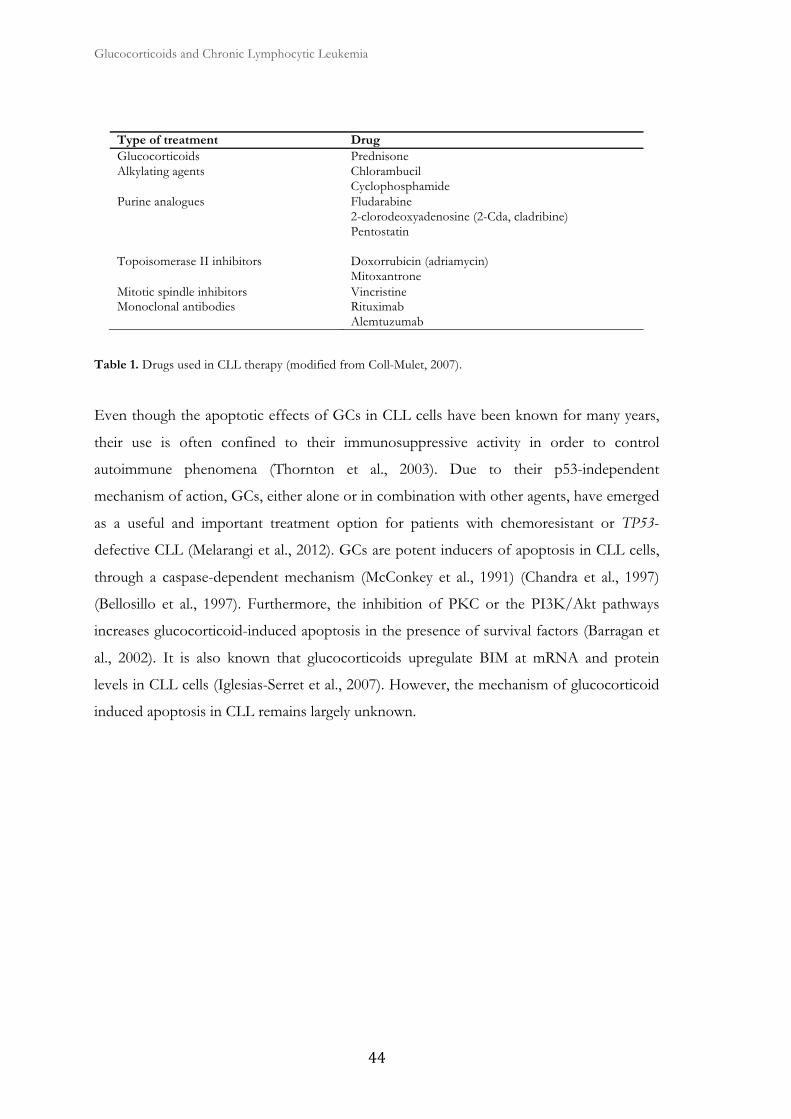
Glucocorticoids and Chronic Lymphocytic Leukemia
�
Type of treatment Drug Glucocorticoids Prednisone Alkylating agents Chlorambucil
Cyclophosphamide Purine analogues Fludarabine
2-clorodeoxyadenosine (2-Cda, cladribine) Pentostatin
Topoisomerase II inhibitors Doxorrubicin (adriamycin) Mitoxantrone
Mitotic spindle inhibitors Vincristine Monoclonal antibodies Rituximab
Alemtuzumab
Table 1. Drugs used in CLL therapy (modified from Coll-Mulet, 2007).
Even though the apoptotic effects of GCs in CLL cells have been known for many years,
their use is often confined to their immunosuppressive activity in order to control
autoimmune phenomena (Thornton et al., 2003). Due to their p53-independent
mechanism of action, GCs, either alone or in combination with other agents, have emerged
as a useful and important treatment option for patients with chemoresistant or TP53-
defective CLL (Melarangi et al., 2012). GCs are potent inducers of apoptosis in CLL cells,
through a caspase-dependent mechanism (McConkey et al., 1991) (Chandra et al., 1997)
(Bellosillo et al., 1997). Furthermore, the inhibition of PKC or the PI3K/Akt pathways
increases glucocorticoid-induced apoptosis in the presence of survival factors (Barragan et
al., 2002). It is also known that glucocorticoids upregulate BIM at mRNA and protein
levels in CLL cells (Iglesias-Serret et al., 2007). However, the mechanism of glucocorticoid
induced apoptosis in CLL remains largely unknown.

II. Materials and methods


Materials and methods
���
1. Samples collection from CLL patients.
Clinicians from the hospital diagnose all patients following clinical, morphological,
molecular and immunophenotypic criteria, as proposed by the World Health Organization.
Peripheral blood samples of CLL patients were collected in the Servei d'Hematologia of
Hospital Universitari de Bellvitge in tubes with anticoagulant EDTA. The information of
the sample was collected in an internal database which scored diagnosis, date of sample
removal, lymphocyte count and percentage of lymphocytes, as well as any information of
interest, such as genetic anomalies, aggressive disease, treatment or chemoresistance. Each
sample and each patient was assigned with an identification number, which was linked to
the clinical history number given by Institut Català de la Salut. Thus, in order to give
identification to each sample, we first refer to the patient number and then the sample
number; for instance, 143_17/12 corresponds to the patient 143 and the sample 17 of the
year 2012. In addition, the database informs whether there are cryopreserved samples and
allows obtaining historical plots. We do not include any personal patient information as it is
established by Universitat de Barcelona and Hospital de Bellvitge Ethical Committees.
Written informed consent was obtained from all patients in accordance with Hospital de
Bellvitge Ethical Committee.
2. Mononuclear cell isolation from peripheral blood of CLL patients
Mononuclear cells from peripheral blood samples were isolated by centrifugation on a
Ficoll-Hypaque (Seromed, Berlin, Germany) gradient (Fig. 17). Blood was diluted in
phosphate buffer saline (PBS) to a final volume of 8 mL. This mixture was added above 4
mL of Ficoll solution (density = 1,077 g/mL) with care to maintain the interface. The
preparation was centrifuged for 20 minutes at 850 g at room temperature. The centrifuge
stop was conducted without brake to avoid disrupting the gradient. In the resulting
separation, erythrocytes and polimorphonucleated leukocytes are at the bottom of the tube
due to their higher density. Above Ficoll solution and forming a ring are the mononuclear
cells and at the top are plasma and platelets diluted in PBS. We picked up the ring with a
glass Pasteur pipette. Mononuclear cells were deposited on 40 mL of PBS and a wash
centrifugation was performed 10 minutes to 480 g at room temperature and cell count was
performed. Pellets were resuspended with complete culture media when needed for
experiments or the sample was cryopreserved in liquid nitrogen in the presence of 10%
dimethyl sulfoxide (DMSO) (Sigma-Aldrich, Steinheim, Germany). Purified mononuclear
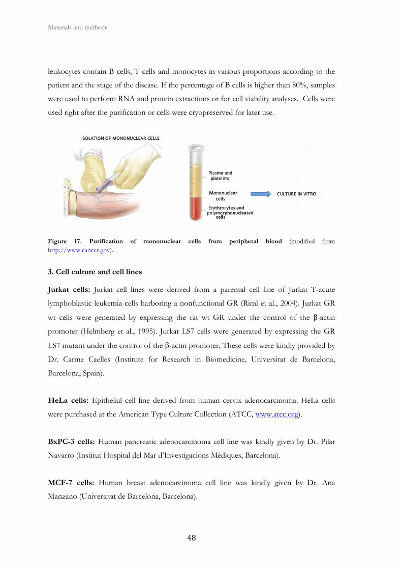
Materials and methods
� �
leukocytes contain B cells, T cells and monocytes in various proportions according to the
patient and the stage of the disease. If the percentage of B cells is higher than 80%, samples
were used to perform RNA and protein extractions or for cell viability analyses. Cells were
used right after the purification or cells were cryopreserved for later use.
Figure 17. Purification of mononuclear cells from peripheral blood (modified from http://www.cancer.gov).
3. Cell culture and cell lines
Jurkat cells: Jurkat cell lines were derived from a parental cell line of Jurkat T-acute
lymphoblastic leukemia cells harboring a nonfunctional GR (Riml et al., 2004). Jurkat GR
wt cells were generated by expressing the rat wt GR under the control of the β-actin
promoter (Helmberg et al., 1995). Jurkat LS7 cells were generated by expressing the GR
LS7 mutant under the control of the β-actin promoter. These cells were kindly provided by
Dr. Carme Caelles (Institute for Research in Biomedicine, Universitat de Barcelona,
Barcelona, Spain).
HeLa cells: Epithelial cell line derived from human cervix adenocarcinoma. HeLa cells
were purchased at the American Type Culture Collection (ATCC, www.atcc.org).
BxPC-3 cells: Human pancreatic adenocarcinoma cell line was kindly given by Dr. Pilar
Navarro (Institut Hospital del Mar d’Investigacions Mèdiques, Barcelona).
MCF-7 cells: Human breast adenocarcinoma cell line was kindly given by Dr. Ana
Manzano (Universitat de Barcelona, Barcelona).
Materials and methods
���
MC3T3 cells: Mouse preosteoblast cell line was kindly given by Dr. Francesc Ventura
(Universitat de Barcelon, Barcelona).
MEF cells: Mouse embryonic fibroblast cells. WT, GSK3α-/-, and GSK3β-/- MEF cells
were kindly given by Dr. J. Woodgett (Samuel Lunenfeld Research Institute, Toronto,
Canada).
Parental Jurkat, Jurkat GR WT, and BxPC-3 cells were grown in RPMI 1640 medium
(Biological Industries) containing 10% inactivated fetal bovine serum (FBS) (Biological
Industries), 2 mM L-glutamine, 100 μg/mL penicillin, and 100 mg/mL streptomycin at
37ºC in a humidified atmosphere at 5% carbon dioxide. HeLa, MC3T3, MCF-7 cells and
WT, GSK3α -/-, and GSK3β -/- MEF cells were maintained in DMEM (Biological
Industries) containing 10% FBS.
4. Freezing and thawing of cells.
Cryopreservation of CLL allows storage for later use. When CLL cells are thawed, they
maintain their characteristics and viability after years of cryopreservation. Regarding cell
lines, it is not recommended to keep them cycling for more than 1-3 months since many
lines easily mutate in culture, being genetically unstable and changing characteristics after
several passages. On the other hand, it is recommended to freeze cell lines right after
thawing a tube and expanding the line. The cryopreservation procedure is performed in
cold DMSO as a membrane stabilizer. Keep tubes at -80ºC for 12-24 hours in an
isopropanol freezer or in polystyrene so that temperature decreases gradually. At 12-24
hours, move the tubes into a liquid nitrogen tank.
Protocol of cryopreservation of primary CLL cells:
Resuspend the pellet of mononuclear cells in cold FBS (previously inactivated by heat for
30 minutes at 56°C) so that there are 20-30x106 cells/0.75 mL FBS/tube. Maintain the
suspension of cells in cold ice. Add slowly the previous suspension on an equal volume of
cryopreservation solution, prepared with RPMI 1640, inactivated FBS and DMSO in ratio
3:1:1, also cold. Finally, there will be 20-30x106 cells/1.5 mL/ tube and 10% DMSO.
Aliquote the sample in 2 mL cryopreservation tubes and cryopreserve in liquid nitrogen.
Materials and methods
� ��
Protocol of cryopreservation of Jurkat and BxPC-3 cell lines:
Collect cells in exponential growth phase. Wash by centrifugation for 5 minutes at 480 g.
The protocol is identical to CLL cells except that here we freeze 10x106 cells/tube.
Protocol of cryopreservation of MEF, HeLa, MCF7 and MC3T3 cells:
Collect cells in exponential growth phase. Wash by centrifugation for 5 minutes at 480 g.
On the basis of a 60 cm2 confluent plate we obtain 4 tubes. Resuspend the pellet of
mononuclear cells in cold FBS so that cells are in 0.75 mL FBS/tube. Maintain the
suspension of cells in cold ice. Slowly add the previous suspension on an equal volume of
cryopreservation solution, newly prepared in DMEM with 20% DMSO, also cold. Finally,
there will be 1.5 mL/tube. Aliquote the sample in 2 mL cryopreservation tubes and
cryopreserve in liquid nitrogen.
Protocol for thawing of primary CLL cells, Jurkat and BxPC-3 cell lines:
Place the cryopreserved tube in a bath at 37ºC unless its content is partially unfrozen. Pour
the tube content on 40 mL of RPMI 1640 medium supplemented with 20% of inactivated
FBS, previously heated at 37ºC, so that frozen cells thaw in a volume at least 10 times
higher. Clean cells from DMSO by centrifugation to 480 g for 5 minutes in the case of
leukemic cell lines or 10 minutes for CLL cells. Resuspend the pellet of cells in culture
medium.
Protocol for thawing of MEF, HeLa, MCF7 and MC3T3 cells:
Place the cryopreserved tube in a bath at 37ºC unless its content is partially unfrozen. Pour
the tube content on 40 mL of DMEM medium supplemented with 20% of inactivated
FBS, previously heated at 37ºC, so that frozen cells thaw in a volume at least 10 times
higher. Clean cells from DMSO by centrifugation to 480 g for 5 minutes. Resuspend the
pellet of cells in culture medium.
5. Reagents
Dexamethasone, actinomycin D, cycloheximide, lovastatin, wortmannin and SB216763
were purchased from Sigma- Aldrich (St. Louis, MO). Akt inhibitor VIII (Akti), SB203580,
U0126, LY294002, bisindolylmaleimide I (Bis I), and rapamycin were purchased from
Calbiochem (La Jolla, CA). SP600125, GSK650394, and KU0063794 were from Tocris

Materials and methods
����
Bioscience (Bristol, UK). Lithium chloride (LiCl), PP242, and MG-132 were from Sigma-
Aldrich, and LY333531 from Enzo Life Sciences. ABT-737 was purchased from Selleck
Chemicals LLC (Houston, TX). Roscovitine was kindly provided by Dr. Jacint Boix
(Universitat de Lleida, Lleida, Spain). Suberoylanilide hydroxamic acid (SAHA; vorinostat,
Zolinza) was obtained from Cayman Chemical (Ann Arbor, MI) and Kendine-92 (5-diaryl-
1H-pyrrole- 2-carboxamide derivates) was generously provided by Dr. Fernando Cossío
(Universidad del Pais Vasco, Bilbao, Spain). Annexin V allophycocyanin was purchased
from eBiosciences (San Diego, CA).
6. Analysis of apoptosis and cell viability by flow cytometry
In general, cell viability analysis was performed using FACSCalibur flow cytometer (Becton
Dickinson). The program used for the data acquisition and analysis was CellQuest Pro
(Becton Dickinson).
The foundation of the technique is very simple. The cell membrane is composed of a lipid
bilayer of asymmetrical distribution. Phosphatidylserine is a phospholipid entirely located
on the inner monolayer surface of the plasma membrane. When a cell undergoes apoptosis,
phosphatidylserine is no longer restricted to the cytosolic part of the membrane, but
becomes exposed on the surface of the cell, which plays an important role in the
recognition and removal of apoptotic cells by macrophages. The translocation of
phosphatidylserine to the outer layer is considered an initial event in the apoptotic process
and it has become a marker of apoptosis in mammalian cells. The protein annexin V binds
with great affinity to the lipid in a calcium-dependent manner (Fig. 18).
Figure 18. Staining of apoptotic cells with annexin V (modified from Coll-Mulet, 2007).
In this way, cell viability was determined by measuring phosphatidylserine exposure and
membrane integrity. This was determined by annexin V-FITC staining and PI. Cell viability

Materials and methods
� ��
was measured as the percentage of annexin V-FITC/PI-negative cell population, and it is
expressed as the percentage of nonapoptotic cells. In cases where cells were pre-incubated
with the GSK3 inhibitor SB216763, we used annexin V-APC to avoid interference. In this
case cell viability was measured as the percentage of annexin V-APC negative cell
population, and it is expressed as the percentage of nonapoptotic cells. In total, 2.5 x 10⁵
cells were incubated for 24 hours with the indicated factors. Cells were washed and
incubated with 150 μL annexin-binding buffer and 1.5 μL annexin V-FITC or -APC for 15
min in the dark. Cells were then analyzed by flow cytometry.
To analyze apoptosis in T cells and B cells from CLL patient samples, 0.5 x 10⁶ cells were
washed in annexin V-binding buffer, and incubated in 50 μL annexin V-binding buffer
with 2 μL allophycocyanin (APC)-conjugated anti-CD3 and 2 μL phycoerythrin (PE)-
conjugated anti-CD19, for 10 min in the dark. Cells were then diluted with annexin V-
binding buffer to a volume of 150 μL and incubated with 1 μL annexin V-FITC for 15 min
in the dark. Cells were then analyzed by flow cytometry.
7. Western blot analysis and antibodies
Lysing cells with reducing Laemmli sample buffer is quick, simple and reduces concerns
that the protein of interest has not been solubilized. The presence of 2% SDS ensures total
lysis of the nucleus, and results in the extraction of genomic DNA. This lysis procedure is
denaturing. Cells were incubated with the indicated factors for a specific period of time
that depends on each experiment. Cells were collected and washed with PBS. After
centrifugation the pellet was resuspended in 50-100 μL of Laemmli (Sample Buffer).
Depending on the cell number, samples were heated to 95°C for 10 minutes and the pellet
was vortexed. Efficiency of lysis was checked by various cycles of heating, vortex and
centrifugation to dissolve the pellets. Quantification of proteins was conducted with the kit
Micro BCA Protein Assay Reagent (Thermo Scientific), using a plate reader of 550 mm.
This kit is based on a colorimetric quantitative determination with bicinchoninic acid
(BCA) to detect Cu reduction due to the basic pH of the proteins. Equal amounts of
protein lysate were subjected to SDS-page-electrophoresis and electrophoretically
transferred to PVDF membranes Immobilon-P (Millipore, Bedford, MA). The membranes
were then blocked with 5% non-fat milk solution in TBS-T 0,1% for 1 hour at room
temperature and immunoblotted. Both incubations with primary and secondary antibodies

Materials and methods
���
(Table 2) were performed in 5% non-fat milk TBS-T solution for 90 minutes each. Some
antibodies were incubated in BSA. The membrane was placed in a cassette with ECL
solution (Amersham Biosciences) and a photographic film to detect the light emitted by the
membrane in the area where the secondary antibody is attached to the primary antibody
and detects the protein of interest. Finally, we developed the film by the traditional
methods of developer and fixer.
Antibody Source Application and dilution
Company
MCL-1 Rabbit WB (1:1000) Santa Cruz Biotechnology (S-19): sc-819
BIM Mouse WB (1:1000) Cell Signaling
GILZ Rabbit WB (1:200) Santa Cruz Biotechnology (FL-134): sc-819
Cleaved Caspase-9 Rabbit WB (1:1000) Cell Signaling
Pro-Caspase-3 Mouse WB (1:1000) BD Biosciences
β-Catenin Mouse WB (1:1000) BD Biosciences
GR Rabbit WB (1:1000) IP (1:1000)
Santa Cruz Biotechnology (H-300): sc-8992
GSK3α/β Mouse WB (1:1000) StressGen Biotechnologies
GSK3α Rabbit WB (1:1000) Cell Signaling
GSK3β Mouse WB (1:1000) Cell Signaling (27C10)
Cytochrome oxidase subunit II Mouse WB (1:1000) Molecular Probes, Inc.
Lamin A/C Mouse WB (1:1000) Cell Signaling
α-TUBULIN Rabbit WB (1:1000) Oncogene Research Products
ERK2 Mouse WB (1:500) Upstate Biotechnology
BCL-2 Mouse WB (1:1000) Dako (M0887)
RNA polymerase II Mouse IP (1:1000) Upstate Biotechnology
IgGs Rabbit IP (1:1000) Upstate Biotechnology
Anti-rabbit IgG, HRP linked whole antibody (from donkey)
WB (1:5000) GE Healthcare
Anti-mouse IgG, HRP linked whole antibody (from sheep)
WB (1:5000) GE Healthcare
Table 2. Sources, dilutions and companies of the antibodies used in this work. Western Blot (WB) and Immunoprecipitation (IP).
Materials and methods
� �
8. RNA extraction.
Total RNA was extracted from cells by using the RNeasy Micro Kit (QIAGEN, Germany).
2-5 x 10⁶ cells were collected for each condition. Cells were centrifuged for 10 minutes at
480 g and we proceeded according to the manufacturer's instructions while maintaining the
samples at room temperature (as opposed to other methods of RNA purification). Total
RNA samples were quantified in the Nanodrop® spectrophotometer. To obtain and work
with RNA solutions, all material used was RNAse free.
9. Reverse Transcriptase Multiplex Ligation-dependent Probe Amplification (RT-
MLPA)
RNA content was analyzed by RT-MLPA using SALSA MLPA KIT R011-C1 Apoptosis
mRNA from MRC-Holland for the simultaneous detection of 38 messenger RNA
molecules (Eldering et al., 2003). In brief, RNA samples (200 ng total RNA) were first
reverse transcribed using a gene-specific probe mix (Fig. 19). The resulting cDNA was
annealed overnight at 60°C to the MLPA probe mix. Annealed oligonucleotides were
ligated by adding Ligase-65 (MRC-Holland) and incubated at 54°C for 15 minutes. Ligation
products were amplified by PCR (35 cycles; 30 seconds at 95°C, 30 seconds at 60°C, and 1
minute at 72°C) with one unlabeled and one FAM-labeled primer. The final PCR
fragments amplified were separated by capillary electrophoresis on a 48-capillary ABI-
Prism 3730 Genetic Analyzer (Applied Biosystems). Peak area and height were measured
using GeneScan v3.0 analysis software (Applied Biosystems). The sum of all peak data was
set at 100% to normalize for fluctuations in total signal among samples, and individual
peaks were calculated relative to the 100% value. The mRNA levels of all the genes were
standardized to those of β-glucoronidase (GUS) for Jurkat cells and PARN for HeLa cells.
RNA content of wt, GSK3α-/- or GSK3β-/- MEF cells were analyzed by RT-MLPA using
SALSA KIT RM002-B1 Mouse Apoptosis mRNA from MRC-Holland. The mRNA levels
of all the genes were standardized to those of TATA box-binding protein (TBP).

Materials and methods
����
Figure 19. RT-MLPA steps (modified from www.mlpa.com).
10. Quantitative PCR (RT-qPCR) analysis
2 μg of total RNA were reverse-transcribed using a Ready-To-Go You-Prime First-Strand
Beads Kit (GE Healthcare) and Random Hexamers (Applied Biosystems). Quantitative
PCRs were carried out using ABI Prism 7900 HT Fast Real-Time PCR System and
Designed human TaqMan assays (Applied Biosystems) were used to quantify gene
expression of BIM (Hs00197982_m1), GILZ (Hs00608272_m1), GILZ (Mm00726417_s1),
HIAP1 (HS00154109_m1), BCL-3 (Hs00180403_m1), and MYC (Hs00153408_m1)
according to the manufacturer's guidelines. The housekeeping gene GUS
(Hs99999908_m1) or GAPDH (Mm99999915_g1) were used as a control for RNA quality,
and used for normalization. PCR data were captured and analyzed using the Sequence
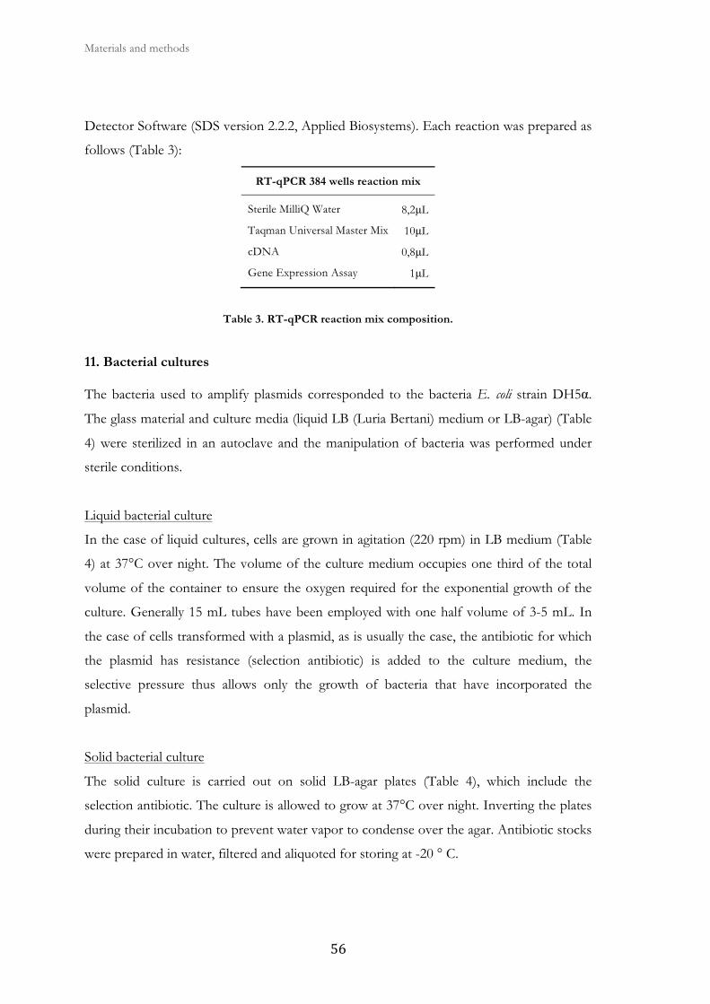
Materials and methods
� ��
Detector Software (SDS version 2.2.2, Applied Biosystems). Each reaction was prepared as
follows (Table 3):
Table 3. RT-qPCR reaction mix composition.
11. Bacterial cultures
The bacteria used to amplify plasmids corresponded to the bacteria E. coli strain DH5α.
The glass material and culture media (liquid LB (Luria Bertani) medium or LB-agar) (Table
4) were sterilized in an autoclave and the manipulation of bacteria was performed under
sterile conditions.
Liquid bacterial culture
In the case of liquid cultures, cells are grown in agitation (220 rpm) in LB medium (Table
4) at 37°C over night. The volume of the culture medium occupies one third of the total
volume of the container to ensure the oxygen required for the exponential growth of the
culture. Generally 15 mL tubes have been employed with one half volume of 3-5 mL. In
the case of cells transformed with a plasmid, as is usually the case, the antibiotic for which
the plasmid has resistance (selection antibiotic) is added to the culture medium, the
selective pressure thus allows only the growth of bacteria that have incorporated the
plasmid.
Solid bacterial culture
The solid culture is carried out on solid LB-agar plates (Table 4), which include the
selection antibiotic. The culture is allowed to grow at 37°C over night. Inverting the plates
during their incubation to prevent water vapor to condense over the agar. Antibiotic stocks
were prepared in water, filtered and aliquoted for storing at -20 ° C.
RT-qPCR 384 wells reaction mix
Sterile MilliQ Water 8,2μL
Taqman Universal Master Mix 10μL
cDNA 0,8μL
Gene Expression Assay 1μL
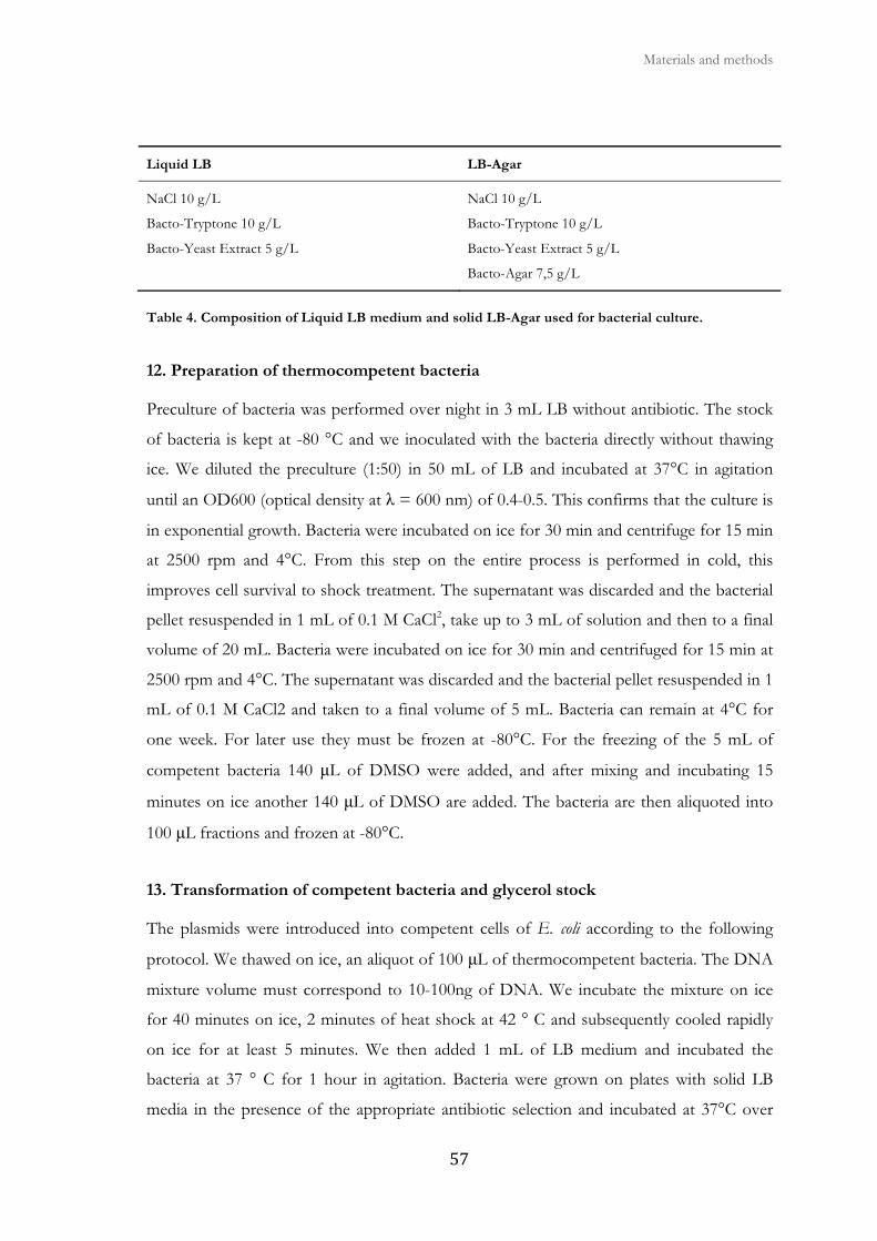
Materials and methods
����
Liquid LB LB-Agar
NaCl 10 g/L NaCl 10 g/L
Bacto-Tryptone 10 g/L Bacto-Tryptone 10 g/L
Bacto-Yeast Extract 5 g/L Bacto-Yeast Extract 5 g/L
Bacto-Agar 7,5 g/L
Table 4. Composition of Liquid LB medium and solid LB-Agar used for bacterial culture.
12. Preparation of thermocompetent bacteria
Preculture of bacteria was performed over night in 3 mL LB without antibiotic. The stock
of bacteria is kept at -80 °C and we inoculated with the bacteria directly without thawing
ice. We diluted the preculture (1:50) in 50 mL of LB and incubated at 37°C in agitation
until an OD600 (optical density at λ = 600 nm) of 0.4-0.5. This confirms that the culture is
in exponential growth. Bacteria were incubated on ice for 30 min and centrifuge for 15 min
at 2500 rpm and 4°C. From this step on the entire process is performed in cold, this
improves cell survival to shock treatment. The supernatant was discarded and the bacterial
pellet resuspended in 1 mL of 0.1 M CaCl2, take up to 3 mL of solution and then to a final
volume of 20 mL. Bacteria were incubated on ice for 30 min and centrifuged for 15 min at
2500 rpm and 4°C. The supernatant was discarded and the bacterial pellet resuspended in 1
mL of 0.1 M CaCl2 and taken to a final volume of 5 mL. Bacteria can remain at 4°C for
one week. For later use they must be frozen at -80°C. For the freezing of the 5 mL of
competent bacteria 140 μL of DMSO were added, and after mixing and incubating 15
minutes on ice another 140 μL of DMSO are added. The bacteria are then aliquoted into
100 μL fractions and frozen at -80°C.
13. Transformation of competent bacteria and glycerol stock
The plasmids were introduced into competent cells of E. coli according to the following
protocol. We thawed on ice, an aliquot of 100 μL of thermocompetent bacteria. The DNA
mixture volume must correspond to 10-100ng of DNA. We incubate the mixture on ice
for 40 minutes on ice, 2 minutes of heat shock at 42 ° C and subsequently cooled rapidly
on ice for at least 5 minutes. We then added 1 mL of LB medium and incubated the
bacteria at 37 ° C for 1 hour in agitation. Bacteria were grown on plates with solid LB
media in the presence of the appropriate antibiotic selection and incubated at 37°C over

Materials and methods
� ��
night. During the transformation negative controls were performed. Untransformed
Bacteria were grown to detect possible contamination. Control positive bacteria: in the case
of transforming a plasmid of unknown origin bacteria transformed with a known stock
plasmid were grown.
To verify that the clones contain the correct plasmid construct, we inoculated a mini
culture with 3 mL of LB culture and let it grow at 37°C in agitation over night.
Subsequently was extracted and analyzed the plasmid DNA by digestion and subsequent
agarose gel electrophoresis banding pattern. We kept a stock of the correct constructs for
further amplifications.
The stock of transformed bacteria was done by mixing glycerol to a final concentration of
15% with bacteria grown in LB medium, after which they were frozen at -80°C.
14. Plasmid obtention
During the course of this work, we used different plasmids, both for protein expression, as
in the case of plasmids used to normalize the number of transfected cells, and plasmids
carrying different promoters binding to the luciferase gene. Some of these plasmids have
been synthesized in our laboratory, while others have been kindly given by groups with
which we have collaborated (Table 5).
For DNA purification in a small-scale we started from a culture of 3 mL of liquid LB
previously inoculated and grown over night, following the kit instructions Wizard® Plus
Minipreps (Promega). These small-scale cultures were performed to confirm the
transformed plasmids by digestion and subsequent electrophoresis. To obtain large
amounts of plasmid DNA for its use in in vitro transfection experiments, we started from a
culture of 250 mL of liquid LB previously inoculated and grown over night and the kit used
Qiafilter Plasmid Maxi Kit (Qiagen) following the manufacturer’s instructions. This
purification provides performance of up to 500μg DNA. Quantification of the
concentration and purity of the plasmids were measured with the Nanodrop, and further
analysis on gels 1% agarose.
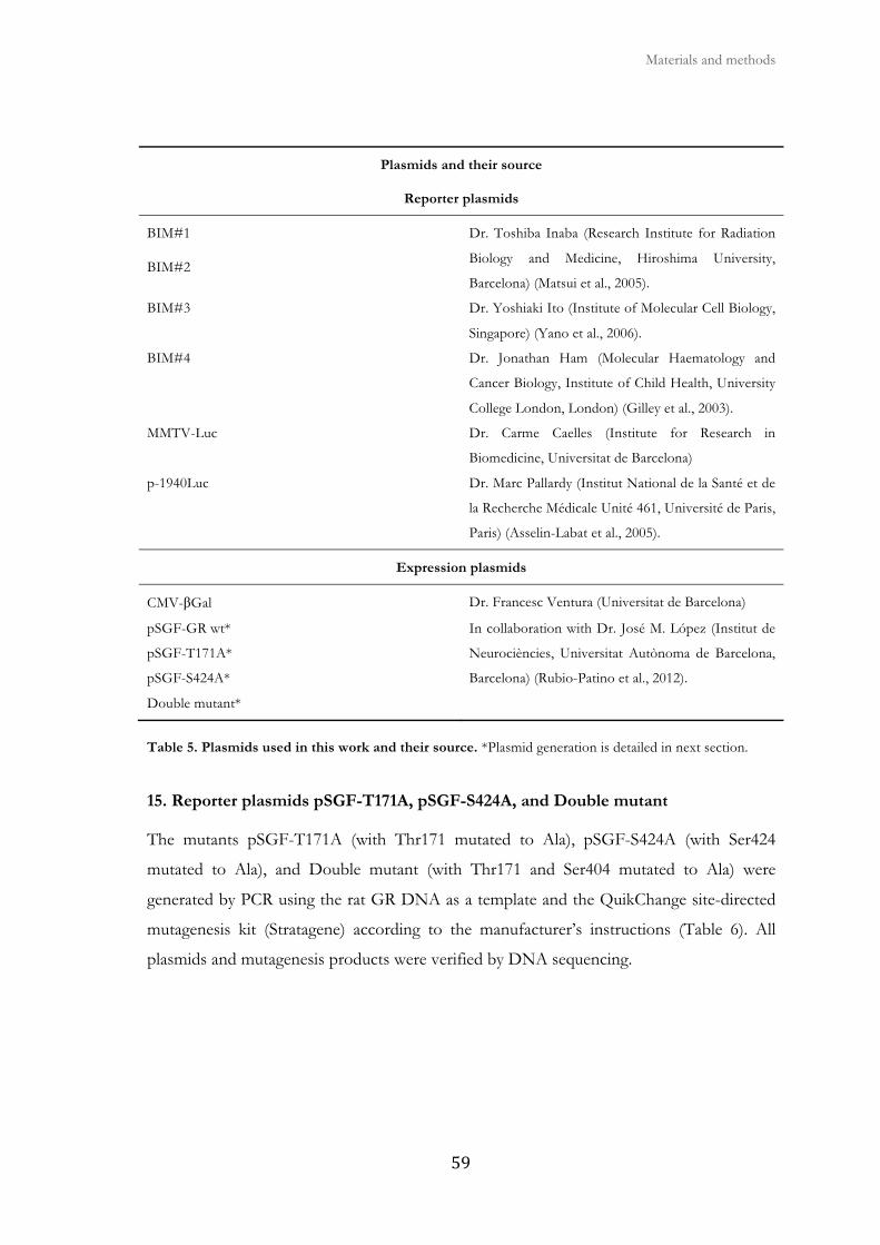
Materials and methods
����
Plasmids and their source
Reporter plasmids
BIM#1
BIM#2
Dr. Toshiba Inaba (Research Institute for Radiation
Biology and Medicine, Hiroshima University,
Barcelona) (Matsui et al., 2005).
BIM#3 Dr. Yoshiaki Ito (Institute of Molecular Cell Biology,
Singapore) (Yano et al., 2006).
BIM#4 Dr. Jonathan Ham (Molecular Haematology and
Cancer Biology, Institute of Child Health, University
College London, London) (Gilley et al., 2003).
MMTV-Luc Dr. Carme Caelles (Institute for Research in
Biomedicine, Universitat de Barcelona)
p-1940Luc Dr. Marc Pallardy (Institut National de la Santé et de
la Recherche Médicale Unité 461, Université de Paris,
Paris) (Asselin-Labat et al., 2005).
Expression plasmids
CMV-βGal Dr. Francesc Ventura (Universitat de Barcelona)
pSGF-GR wt*
pSGF-T171A*
pSGF-S424A*
Double mutant*
In collaboration with Dr. José M. López (Institut de
Neurociències, Universitat Autònoma de Barcelona,
Barcelona) (Rubio-Patino et al., 2012).
Table 5. Plasmids used in this work and their source. *Plasmid generation is detailed in next section.
15. Reporter plasmids pSGF-T171A, pSGF-S424A, and Double mutant
The mutants pSGF-T171A (with Thr171 mutated to Ala), pSGF-S424A (with Ser424
mutated to Ala), and Double mutant (with Thr171 and Ser404 mutated to Ala) were
generated by PCR using the rat GR DNA as a template and the QuikChange site-directed
mutagenesis kit (Stratagene) according to the manufacturer’s instructions (Table 6). All
plasmids and mutagenesis products were verified by DNA sequencing.
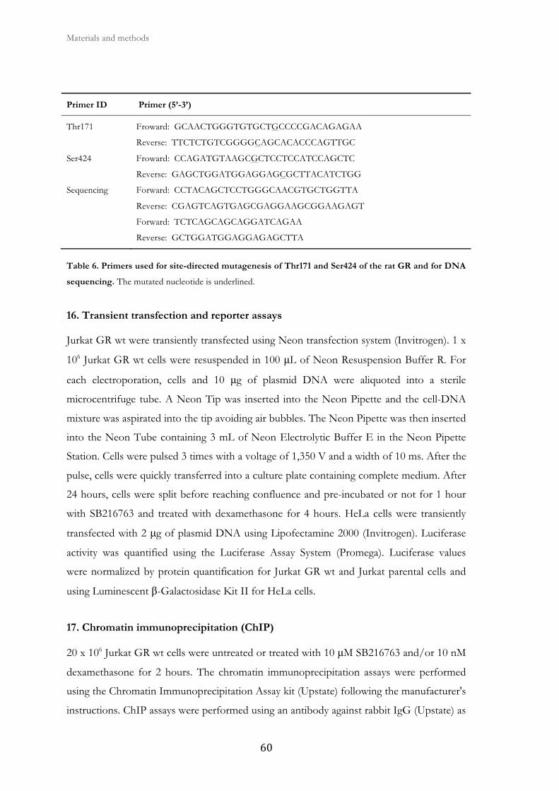
Materials and methods
� ��
Primer ID Primer (5’-3’)
Thr171 Froward: GCAACTGGGTGTGCTGCCCCGACAGAGAA
Reverse: TTCTCTGTCGGGGCAGCACACCCAGTTGC
Ser424 Froward: CCAGATGTAAGCGCTCCTCCATCCAGCTC
Reverse: GAGCTGGATGGAGGAGCGCTTACATCTGG
Sequencing Forward: CCTACAGCTCCTGGGCAACGTGCTGGTTA
Reverse: CGAGTCAGTGAGCGAGGAAGCGGAAGAGT
Forward: TCTCAGCAGCAGGATCAGAA
Reverse: GCTGGATGGAGGAGAGCTTA
Table 6. Primers used for site-directed mutagenesis of Thr171 and Ser424 of the rat GR and for DNA
sequencing. The mutated nucleotide is underlined.
16. Transient transfection and reporter assays
Jurkat GR wt were transiently transfected using Neon transfection system (Invitrogen). 1 x
106 Jurkat GR wt cells were resuspended in 100 μL of Neon Resuspension Buffer R. For
each electroporation, cells and 10 μg of plasmid DNA were aliquoted into a sterile
microcentrifuge tube. A Neon Tip was inserted into the Neon Pipette and the cell-DNA
mixture was aspirated into the tip avoiding air bubbles. The Neon Pipette was then inserted
into the Neon Tube containing 3 mL of Neon Electrolytic Buffer E in the Neon Pipette
Station. Cells were pulsed 3 times with a voltage of 1,350 V and a width of 10 ms. After the
pulse, cells were quickly transferred into a culture plate containing complete medium. After
24 hours, cells were split before reaching confluence and pre-incubated or not for 1 hour
with SB216763 and treated with dexamethasone for 4 hours. HeLa cells were transiently
transfected with 2 μg of plasmid DNA using Lipofectamine 2000 (Invitrogen). Luciferase
activity was quantified using the Luciferase Assay System (Promega). Luciferase values
were normalized by protein quantification for Jurkat GR wt and Jurkat parental cells and
using Luminescent β-Galactosidase Kit II for HeLa cells.
17. Chromatin immunoprecipitation (ChIP)
20 x 106 Jurkat GR wt cells were untreated or treated with 10 μM SB216763 and/or 10 nM
dexamethasone for 2 hours. The chromatin immunoprecipitation assays were performed
using the Chromatin Immunoprecipitation Assay kit (Upstate) following the manufacturer's
instructions. ChIP assays were performed using an antibody against rabbit IgG (Upstate) as
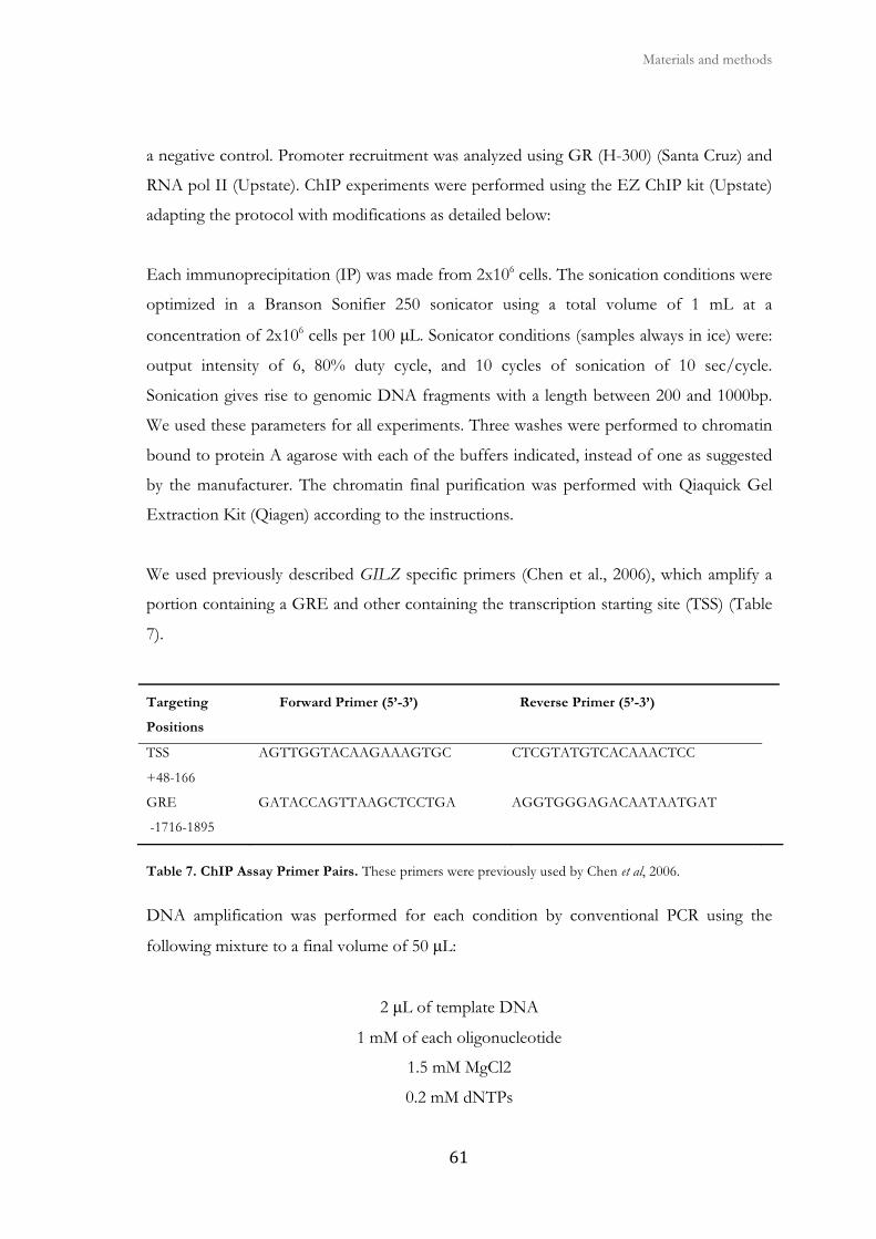
Materials and methods
����
a negative control. Promoter recruitment was analyzed using GR (H-300) (Santa Cruz) and
RNA pol II (Upstate). ChIP experiments were performed using the EZ ChIP kit (Upstate)
adapting the protocol with modifications as detailed below:
Each immunoprecipitation (IP) was made from 2x10⁶ cells. The sonication conditions were
optimized in a Branson Sonifier 250 sonicator using a total volume of 1 mL at a
concentration of 2x10⁶ cells per 100 μL. Sonicator conditions (samples always in ice) were:
output intensity of 6, 80% duty cycle, and 10 cycles of sonication of 10 sec/cycle.
Sonication gives rise to genomic DNA fragments with a length between 200 and 1000bp.
We used these parameters for all experiments. Three washes were performed to chromatin
bound to protein A agarose with each of the buffers indicated, instead of one as suggested
by the manufacturer. The chromatin final purification was performed with Qiaquick Gel
Extraction Kit (Qiagen) according to the instructions.
We used previously described GILZ specific primers (Chen et al., 2006), which amplify a
portion containing a GRE and other containing the transcription starting site (TSS) (Table
7).
Targeting
Positions
Forward Primer (5’-3’) Reverse Primer (5’-3’)
TSS
+48-166
AGTTGGTACAAGAAAGTGC CTCGTATGTCACAAACTCC
GRE
-1716-1895
GATACCAGTTAAGCTCCTGA AGGTGGGAGACAATAATGAT
Table 7. ChIP Assay Primer Pairs. These primers were previously used by Chen et al, 2006.
DNA amplification was performed for each condition by conventional PCR using the
following mixture to a final volume of 50 μL:
2 μL of template DNA
1 mM of each oligonucleotide
1.5 mM MgCl2
0.2 mM dNTPs
Materials and methods
� ��
Polymerase Buffer
0.5 μL of 5 U/μL EcoTaq (Ecogene)
To reduce the appearance of non-specific bands we performed a hot start for 5 min at 94 °
C before adding the EcoTaq. This ensures that all DNA strands are denatured before
amplification.
Thermocycler programming:
Initial dehybridization for 5 min at 94°C (hot start).
35 amplification cycles: 45 sec at 94°C, 45 seconds at 61,5ºC (annealing temperature of the
ChIP oligonucleotides), and the required elongation time at 72 ° C, assuming an average
elongation rate of 1 Kb EcoTaq per min.
Final elongation of 7 min at 72°C.
Maintaining the mixture at 4°C.
The PCR product was analyzed in a 1%-1,5% agarose gel. Densitometric scanning and
quantification of the intensities of PCR bands were carried out using Image J 1.44o
software based analysis (National Institute of Health).
18. siRNA transfection
HeLa cells were transfected with commercially available scramble siRNA, anti-GSK3α,
anti-GSK3β or both siRNAs (Invitrogen) (Table 8) at a concentration of 200 nM using
Lipofectamine 2000 transfection reagent (Invitrogen), following the formation of lipid-
DNA complexes for 20 minutes at room temperature in OptiMEM I medium (Gibco).
After 48 hours, cell populations at a density of 50-60% in 6-well plates were transfected
with 1-2 μg of MMTV-Luc plasmid DNA. Complexes were added directly to growing cells
in DMEM and incubated for 4-6 hours followed by washing with PBS buffer and addition
of fresh DMEM. Cells were used in experiments 72 hours following siRNA transfection.
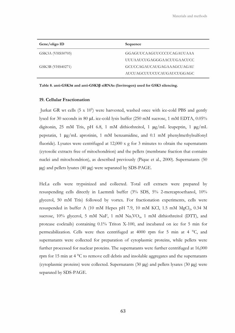
Materials and methods
���
Gene/oligo ID Sequence
GSK3A (VHS50705) GGAGUUCAAGUUCCCUCAGAUUAAA
UUUAAUCUGAGGGAACUUGAACUCC
GSK3B (VHS40271) GCUCCAGAUCAUGAGAAAGCUAGAU
AUCUAGCUUUCUCAUGAUCUGGAGC
Table 8. anti-GSK3α and anti-GSK3β siRNAs (Invitrogen) used for GSK3 silencing.
19. Cellular Fractionation
Jurkat GR wt cells (5 x 10⁶) were harvested, washed once with ice-cold PBS and gently
lysed for 30 seconds in 80 μL ice-cold lysis buffer (250 mM sucrose, 1 mM EDTA, 0.05%
digitonin, 25 mM Tris, pH 6.8, 1 mM dithiothreitol, 1 μg/mL leupeptin, 1 μg/mL
pepstatin, 1 μg/mL aprotinin, 1 mM benzamidine, and 0.1 mM phenylmethylsulfonyl
fluoride). Lysates were centrifuged at 12,000 x g for 3 minutes to obtain the supernatants
(cytosolic extracts free of mitochondrion) and the pellets (membrane fraction that contains
nuclei and mitochondrion), as described previously (Pique et al., 2000). Supernatants (50
μg) and pellets lysates (40 μg) were separated by SDS-PAGE.
HeLa cells were trypsinized and collected. Total cell extracts were prepared by
resuspending cells directly in Laemmli buffer (3% SDS, 5% 2-mercaptoethanol, 10%
glycerol, 50 mM Tris) followed by vortex. For fractionation experiments, cells were
resuspended in buffer A (10 mM Hepes pH 7.9, 10 mM KCl, 1.5 mM MgCl2, 0.34 M
sucrose, 10% glycerol, 5 mM NaF, 1 mM Na3VO4, 1 mM dithiothreitol (DTT), and
protease cocktails) containing 0.1% Triton X-100, and incubated on ice for 5 min for
permeabilization. Cells were then centrifuged at 4000 rpm for 5 min at 4 °C, and
supernatants were collected for preparation of cytoplasmic proteins, while pellets were
further processed for nuclear proteins. The supernatants were further centrifuged at 16,000
rpm for 15 min at 4 °C to remove cell debris and insoluble aggregates and the supernatants
(cytoplasmic proteins) were collected. Supernatants (30 μg) and pellets lysates (30 μg) were
separated by SDS-PAGE.
Materials and methods
� �
20. Confocal Laser Scanning Microscopy
Jurkat GR wt cells were collected after treatment with 10 μM SB216763 and/or 10 nM
dexamethasone for 2 hours, resuspended in PBS and incubated at room temperature for
30–60 minutes over poly-L-lysine coated coverslips (0.01% solution, Sigma-Aldrich). HeLa
cells were grown on sterilized glass coverslips and then treated with 100 nM
dexamethasone for 3 hours. Coverslips containing attached cells were fixed with 4%
paraformaldehyde, permeabilized with 0.25% Triton X-100 in PBS and incubated for 1
hour in 5% PBS-BSA to block nonspecific binding. Slides were incubated overnight at 4ºC
in a humidified chamber with rabbit polyclonal anti-GR (H-300) (1/100) primary antibody
(Santa Cruz). Afterwards, the slides were washed three times with PBS and further
incubated with Alexa Fluor 647 anti-rabbit secondary antibody (1/500; Invitrogen) for 1
hour. Nuclei were stained with Yoyo-1 iodide (Invitrogen). To validate the specificity of
the immunostaining, controls were performed by applying the same protocol but replacing
primary antibody with 5% PBS-BSA. Images were then obtained with a Spectral Confocal
Microscope (TCS-SL, Leica Microsystems) using a Plan-Apochromat 63×/1.4 N.A.
immersion oil objective (Leica Microsystems). We used HeNe Laser at 633 nm (Lasos Inc)
and pinhole of 114.54 μm for Alexa Fluor 647 GR staining and Argon Laser at 488 nm and
pinhole of 114.54 μm for Yoyo-1 nuclear staining. Images were captured using the
accompanying image processing software from Cytovision.
21. Statistical analysis
Results are shown as the mean ± standard error of the mean (SEM) of values obtained in
three or more independent experiments. Data were analyzed using SPSS 11.0 software
package. The paired Student t-test was used to compare the differences between paired
samples. ANOVA-Tukey was used to compare the differences between treatments.
Differences were considered significant at p values <0.05: *p<0.05, **p<0.01, and
***p<0.001 of dexamethasone treated cells compared to untreated cells and ✚ p<0.05
compared to treated cells, as indicated in the figure legends.
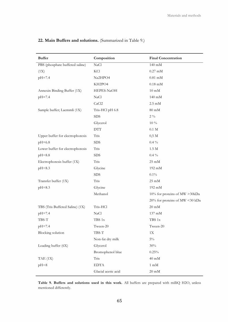
Materials and methods
����
22. Main Buffers and solutions. (Summarized in Table 9.)
Buffer Composition Final Concentration
PBS (phosphate buffered saline)
(1X)
pH=7.4
NaCl
KCl
Na2HPO4
KH2PO4
140 mM
0.27 mM
0.81 mM
0.18 mM
Annexin Binding Buffer (1X)
pH=7.4
HEPES-NaOH
NaCl
CaCl2
10 mM
140 mM
2.5 mM
Sample buffer; Laemmli (1X) Tris-HCl pH 6.8
SDS
Glycerol
DTT
80 mM
2 %
10 %
0.1 M
Upper buffer for electrophoresis
pH=6.8
Tris
SDS
0,5 M
0.4 %
Lower buffer for electrophoresis
pH=8.8
Tris
SDS
1.5 M
0.4 %
Electrophoresis buffer (1X)
pH=8.3
Tris
Glycine
SDS
25 mM
192 mM
0.1%
Transfer buffer (1X)
pH=8.3
Tris
Glycine
Methanol
25 mM
192 mM
10% for proteins of MW >30kDa
20% for proteins of MW <30 kDa
TBS (Tris Buffered Saline) (1X)
pH=7.4
Tris-HCl
NaCl
20 mM
137 mM
TBS-T
pH=7.4
TBS 1x
Tween-20
TBS 1x
Tween-20
Blocking solution TBS-T
Non-fat dry milk
1X
5%
Loading buffer (6X) Glycerol
Bromophenol blue
30%
0.25%
TAE (1X)
pH=8
Tris
EDTA
Glacial acetic acid
40 mM
1 mM
20 mM
Table 9. Buffers and solutions used in this work. All buffers are prepared with milliQ H2O, unless mentioned differently.


� � �
�
III. Objectives

�
�

� � � Objectives
����
GCs are steroid hormones that regulate important biological processes, including growth,
development, metabolism, survival, differentiation, proliferation, and apoptosis. GCs
induce cell cycle arrest and apoptosis in different cell types and therefore are widely used to
treat a variety of diseases including autoimmune disorders and cancer. The effect of GCs is
mediated by the GR, a member of the steroid receptor superfamily and a ligand-activated
transcription factor that translocates into the nucleus where it modulates transcription of
target genes in a promoter-specific manner. Previous studies have described the role of
different protein kinases in GC-mediated effects and one of them is Glycogen synthase
kinase-3 (GSK3). The aim of this thesis has been to study the mechanism of regulation of
GC-induced apoptosis by GSK3. The specific objectives of this thesis are:
1. Analyze the effect of GCs in Jurkat Parental, GR wt and LS7 cells.
2. Analyze the role of GSK3 on GC-mediated signaling.
3. Study of the role of GSK3 in GC-induced apoptosis in CLL cells.

�
�

IV. Results

�
�

1. Analysis of the effect of glucocorticoids
in Jurkat Parental, GR wt and LS7 cells

�
�

� � � Results
���
1.1. Dexamethasone induces apoptosis in Jurkat GR wt and LS7 cells
Our group is focused on the regulation of BCL-2 family members in the apoptosis induced
by several drugs, like GCs, in Chronic Lymphocytic Leukemia (CLL) cells. Several studies
in different cell models have demonstrated that BIM plays and important role in GC-
induced apoptosis (Kfir-Erenfeld et al., 2010). Previous results from our group showed
that BIM protein and mRNA levels are induced during dexamethasone-induced apoptosis
in CLL cells (Iglesias-Serret et al., 2007). Unfortunately, we have been unable to
successfully transfect CLL cells and maintain cell viability after transfection. For this
reason, we searched for an alternative cell type that would allow us to continue studying
GC-induced apoptosis at the molecular level.
To further elucidate the mechanism of GC-induced apoptosis, we used the T-acute
lymphoblastic leukemia Jurkat parental cell line, which harbor a function-impairing point
mutation (R477H) in one of their GR alleles that causes GC resistance (Riml et al., 2004).
These cells were either stably transfected with a rat GR expression vector under the control
of the β-actin promoter, to ensure constant GR protein expression (Jurkat GR wt) or with
the LS7 mutant (Jurkat LS7) (Helmberg et al., 1995). The later contains two adjacent amino
acid substitutions in the Hinge Region adjacent to the DBD of the GR, replacing Proline
493 and Alanine 494 by Arginine and Serine, respectively (Fig. 20). The LS7 mutant was
reported to have little or no transactivating potential, while retaining transrepression
activity.
Figure 20. Primary structure of the Zn-finger region of wt and LS7 GR. Numbers represent the amino acid sequence. (Modified from Helmberg et al., 1995).
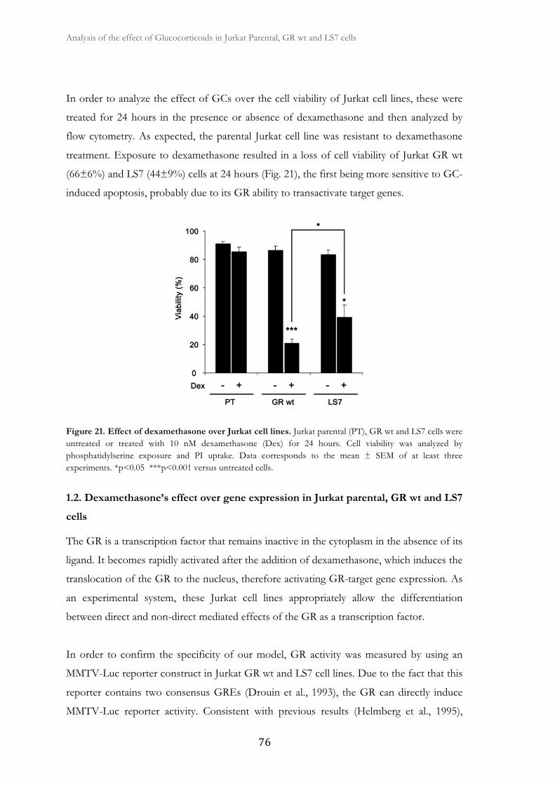
Analysis of the effect of Glucocorticoids in Jurkat Parental, GR wt and LS7 cells �
� ��
In order to analyze the effect of GCs over the cell viability of Jurkat cell lines, these were
treated for 24 hours in the presence or absence of dexamethasone and then analyzed by
flow cytometry. As expected, the parental Jurkat cell line was resistant to dexamethasone
treatment. Exposure to dexamethasone resulted in a loss of cell viability of Jurkat GR wt
(66±6%) and LS7 (44±9%) cells at 24 hours (Fig. 21), the first being more sensitive to GC-
induced apoptosis, probably due to its GR ability to transactivate target genes.
Figure 21. Effect of dexamethasone over Jurkat cell lines. Jurkat parental (PT), GR wt and LS7 cells were untreated or treated with 10 nM dexamethasone (Dex) for 24 hours. Cell viability was analyzed by phosphatidylserine exposure and PI uptake. Data corresponds to the mean ± SEM of at least three experiments. *p<0.05 ***p<0.001 versus untreated cells.
1.2. Dexamethasone’s effect over gene expression in Jurkat parental, GR wt and LS7
cells
The GR is a transcription factor that remains inactive in the cytoplasm in the absence of its
ligand. It becomes rapidly activated after the addition of dexamethasone, which induces the
translocation of the GR to the nucleus, therefore activating GR-target gene expression. As
an experimental system, these Jurkat cell lines appropriately allow the differentiation
between direct and non-direct mediated effects of the GR as a transcription factor.
In order to confirm the specificity of our model, GR activity was measured by using an
MMTV-Luc reporter construct in Jurkat GR wt and LS7 cell lines. Due to the fact that this
reporter contains two consensus GREs (Drouin et al., 1993), the GR can directly induce
MMTV-Luc reporter activity. Consistent with previous results (Helmberg et al., 1995),

� � � Results
���
Jurkat GR wt cells exhibited GC-induced transcriptional activation of the MMTV-Luc
reporter construct, whereas the LS7 mutant exhibited minimal transactivating potential
(Fig. 22).
Figure 22. Transcriptional activation by GR in Jurkat GR wt and LS7 cells. Jurkat GR wt and LS7 cells were transfected with MMTV-Luc vector and untreated or treated with 10 nM dexamethasone (Dex) for 24 hours. Luciferase activity was measured and expressed relative to untreated cells basal activity.
Taking advantage of these cell specific characteristics, these cell lines were then used to
analyze the effect of GC treatment over the expression of the pro-apoptotic protein BIM.
As shown in Figure 23, Western Blot analysis showed that dexamethasone induced the
expression of BIM-EL, BIM-L and BIM-S isoforms in a dose-dependent manner in Jurkat
GR wt and LS7 cells. Jurkat LS7 cells were equally capable of inducing BIM protein levels.
These results demonstrate that BIM regulation by GCs is independent of GR
transactivation, as the LS7 GR mutant has no transactivation potential.
We also analyzed the effect of GCs over the expression of the anti-apoptotic BCL-2 family
member protein, MCL-1. It has been described that MCL-1 is a sensor of protein synthesis
inhibition and that the decrease of MCL-1 protein is necessary to induce apoptosis
(Nijhawan et al., 2003). We observed that MCL-1 protein levels were downregulated in
both Jurkat GR wt and LS7 cell lines in response to dexamethasone, therefore facilitating
cell death (Fig. 23). As expected, the levels of BIM and MCL-1 were not affected by GC
treatment in Jurkat parental cells, as they harbor a non-functional GR.
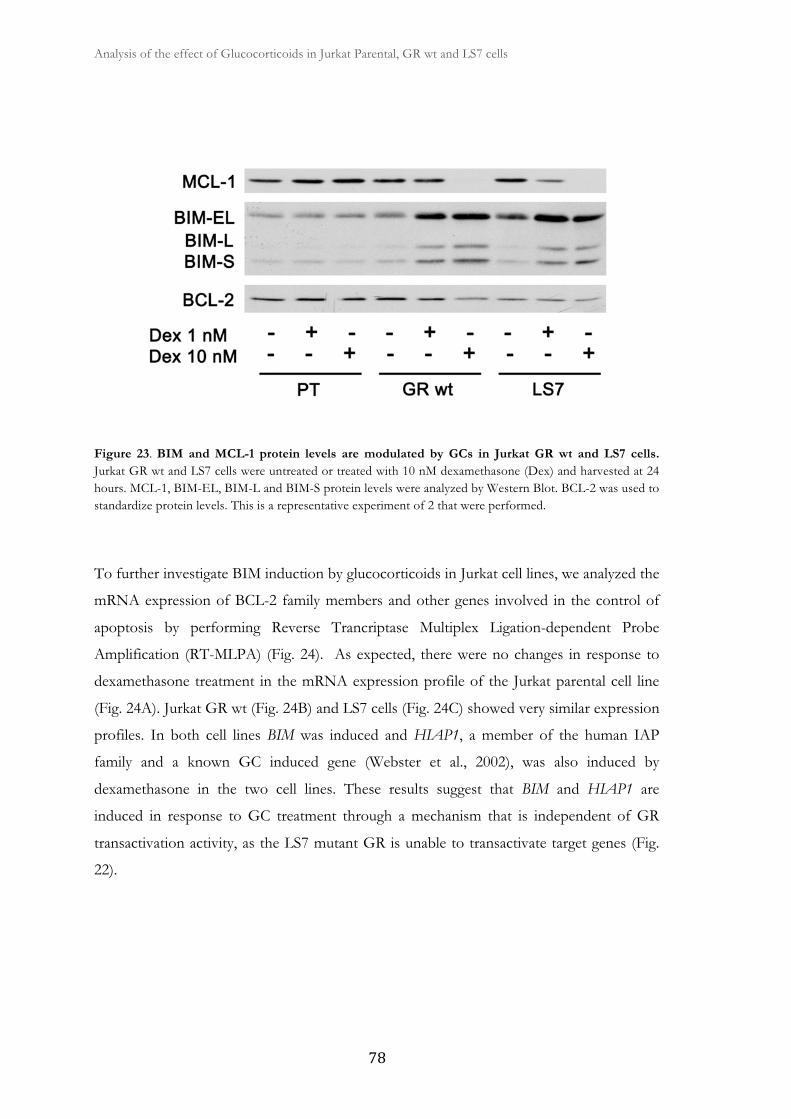
Analysis of the effect of Glucocorticoids in Jurkat Parental, GR wt and LS7 cells �
� ��
Figure 23. BIM and MCL-1 protein levels are modulated by GCs in Jurkat GR wt and LS7 cells. Jurkat GR wt and LS7 cells were untreated or treated with 10 nM dexamethasone (Dex) and harvested at 24 hours. MCL-1, BIM-EL, BIM-L and BIM-S protein levels were analyzed by Western Blot. BCL-2 was used to standardize protein levels. This is a representative experiment of 2 that were performed.
To further investigate BIM induction by glucocorticoids in Jurkat cell lines, we analyzed the
mRNA expression of BCL-2 family members and other genes involved in the control of
apoptosis by performing Reverse Trancriptase Multiplex Ligation-dependent Probe
Amplification (RT-MLPA) (Fig. 24). As expected, there were no changes in response to
dexamethasone treatment in the mRNA expression profile of the Jurkat parental cell line
(Fig. 24A). Jurkat GR wt (Fig. 24B) and LS7 cells (Fig. 24C) showed very similar expression
profiles. In both cell lines BIM was induced and HIAP1, a member of the human IAP
family and a known GC induced gene (Webster et al., 2002), was also induced by
dexamethasone in the two cell lines. These results suggest that BIM and HIAP1 are
induced in response to GC treatment through a mechanism that is independent of GR
transactivation activity, as the LS7 mutant GR is unable to transactivate target genes (Fig.
22).

� � � Results
���
Figure 24. Dexamethasone affects gene expression profiles of Jurkat GR wt and LS7 cells. RT-MLPA gene expression profile induced by GCs. Jurkat parental (PT) (A), GR wt (B) and LS7 cells (C) were untreated (white bars) or treated (black bars) with 10 nM dexamethasone (Dex) for 24 hours. Cells were lysed and the expression of apoptotic-related genes was analyzed by RT-MLPA as described in Materials and methods. The mRNA levels of all genes were normalized with respect to those of β-glucoronidase (GUS). The results are shown as the mean ± SD of two representative experiments.
1.3. BBIM and GILZ are GC-induced early genes
To further study dexamethasone-mediated gene induction we decided to choose genes
whose induction is dependent or independent of GR transcriptional activity in Jurkat cell
lines. We used GILZ, a well-known GC-induced gene, as a direct target of the GR and BIM
as a gene that does not have GREs in its promoter. Jurkat GR wt cells were treated for 1
hour with dexamethasone and BIM and GILZ mRNA levels were analyzed by RT-qPCR.
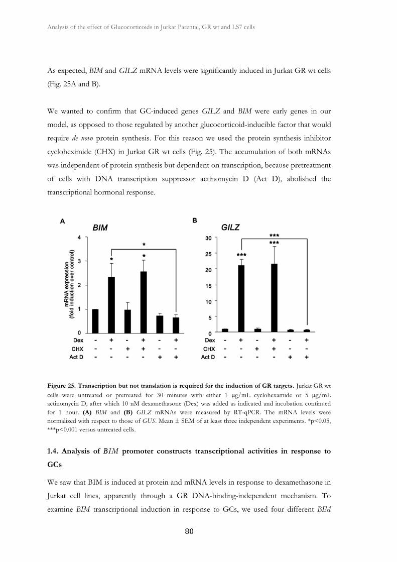
Analysis of the effect of Glucocorticoids in Jurkat Parental, GR wt and LS7 cells �
� ��
As expected, BIM and GILZ mRNA levels were significantly induced in Jurkat GR wt cells
(Fig. 25A and B).
We wanted to confirm that GC-induced genes GILZ and BIM were early genes in our
model, as opposed to those regulated by another glucocorticoid-inducible factor that would
require de novo protein synthesis. For this reason we used the protein synthesis inhibitor
cycloheximide (CHX) in Jurkat GR wt cells (Fig. 25). The accumulation of both mRNAs
was independent of protein synthesis but dependent on transcription, because pretreatment
of cells with DNA transcription suppressor actinomycin D (Act D), abolished the
transcriptional hormonal response.
Figure 25. Transcription but not translation is required for the induction of GR targets. Jurkat GR wt cells were untreated or pretreated for 30 minutes with either 1 μg/mL cyclohexamide or 5 μg/mL actinomycin D, after which 10 nM dexamethasone (Dex) was added as indicated and incubation continued for 1 hour. (A) BIM and (B) GILZ mRNAs were measured by RT-qPCR. The mRNA levels were normalized with respect to those of GUS. Mean ± SEM of at least three independent experiments. *p<0.05, ***p<0.001 versus untreated cells.
1.4. Analysis of BBIM promoter constructs transcriptional activities in response to
GCs
We saw that BIM is induced at protein and mRNA levels in response to dexamethasone in
Jurkat cell lines, apparently through a GR DNA-binding-independent mechanism. To
examine BIM transcriptional induction in response to GCs, we used four different BIM
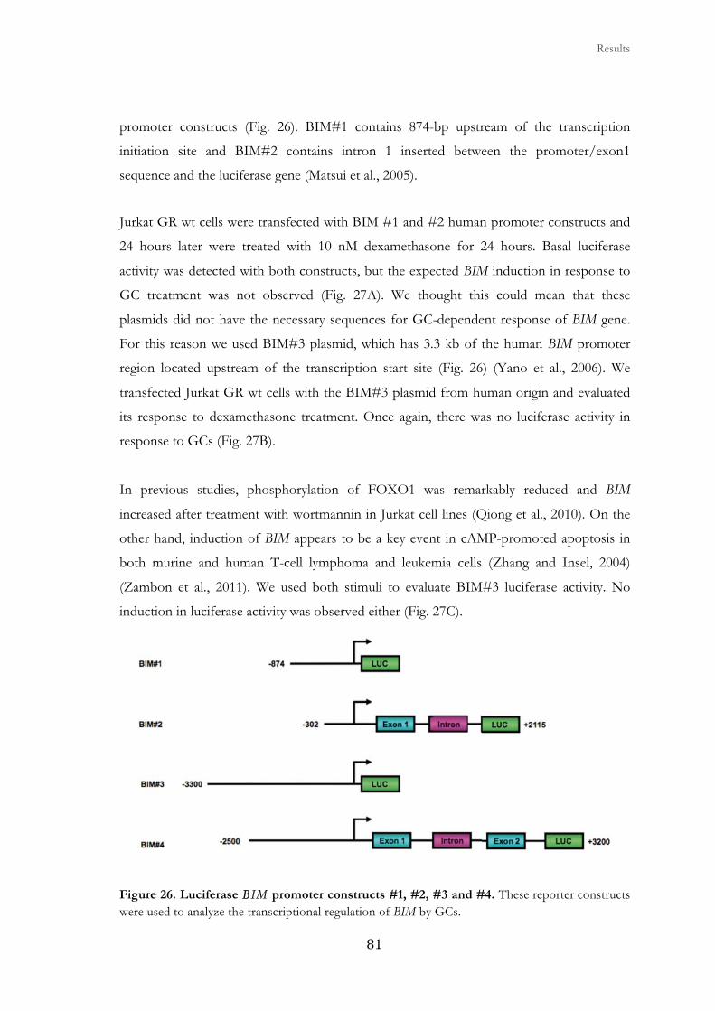
� � � Results
���
promoter constructs (Fig. 26). BIM#1 contains 874-bp upstream of the transcription
initiation site and BIM#2 contains intron 1 inserted between the promoter/exon1
sequence and the luciferase gene (Matsui et al., 2005).
Jurkat GR wt cells were transfected with BIM #1 and #2 human promoter constructs and
24 hours later were treated with 10 nM dexamethasone for 24 hours. Basal luciferase
activity was detected with both constructs, but the expected BIM induction in response to
GC treatment was not observed (Fig. 27A). We thought this could mean that these
plasmids did not have the necessary sequences for GC-dependent response of BIM gene.
For this reason we used BIM#3 plasmid, which has 3.3 kb of the human BIM promoter
region located upstream of the transcription start site (Fig. 26) (Yano et al., 2006). We
transfected Jurkat GR wt cells with the BIM#3 plasmid from human origin and evaluated
its response to dexamethasone treatment. Once again, there was no luciferase activity in
response to GCs (Fig. 27B).
In previous studies, phosphorylation of FOXO1 was remarkably reduced and BIM
increased after treatment with wortmannin in Jurkat cell lines (Qiong et al., 2010). On the
other hand, induction of BIM appears to be a key event in cAMP-promoted apoptosis in
both murine and human T-cell lymphoma and leukemia cells (Zhang and Insel, 2004)
(Zambon et al., 2011). We used both stimuli to evaluate BIM#3 luciferase activity. No
induction in luciferase activity was observed either (Fig. 27C).
Figure 26. Luciferase BBIM promoter constructs #1, #2, #3 and #4. These reporter constructs were used to analyze the transcriptional regulation of BIM by GCs.
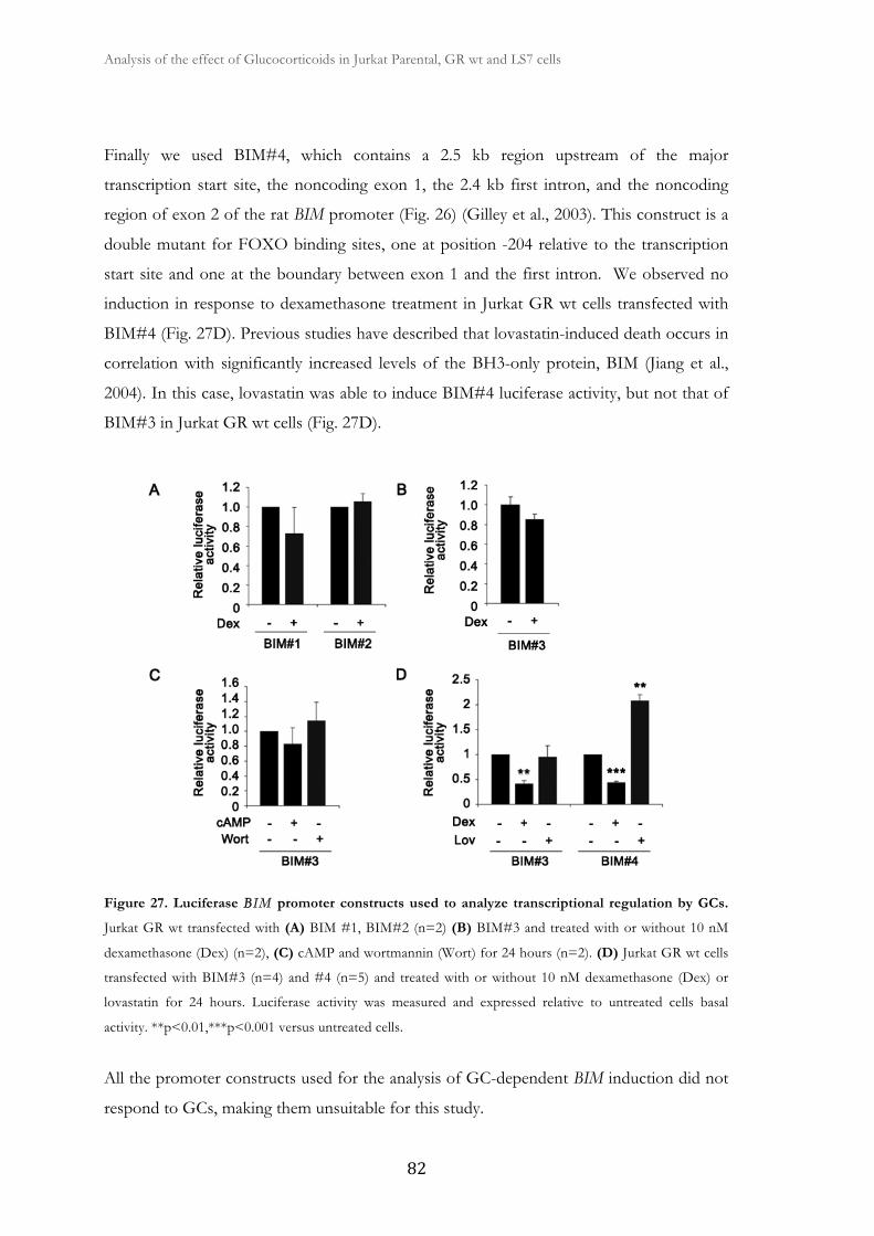
Analysis of the effect of Glucocorticoids in Jurkat Parental, GR wt and LS7 cells �
� ��
Finally we used BIM#4, which contains a 2.5 kb region upstream of the major
transcription start site, the noncoding exon 1, the 2.4 kb first intron, and the noncoding
region of exon 2 of the rat BIM promoter (Fig. 26) (Gilley et al., 2003). This construct is a
double mutant for FOXO binding sites, one at position -204 relative to the transcription
start site and one at the boundary between exon 1 and the first intron. We observed no
induction in response to dexamethasone treatment in Jurkat GR wt cells transfected with
BIM#4 (Fig. 27D). Previous studies have described that lovastatin-induced death occurs in
correlation with significantly increased levels of the BH3-only protein, BIM (Jiang et al.,
2004). In this case, lovastatin was able to induce BIM#4 luciferase activity, but not that of
BIM#3 in Jurkat GR wt cells (Fig. 27D).
Figure 27. Luciferase BBIM promoter constructs used to analyze transcriptional regulation by GCs.
Jurkat GR wt transfected with (A) BIM #1, BIM#2 (n=2) (B) BIM#3 and treated with or without 10 nM
dexamethasone (Dex) (n=2), (C) cAMP and wortmannin (Wort) for 24 hours (n=2). (D) Jurkat GR wt cells
transfected with BIM#3 (n=4) and #4 (n=5) and treated with or without 10 nM dexamethasone (Dex) or
lovastatin for 24 hours. Luciferase activity was measured and expressed relative to untreated cells basal
activity. **p<0.01,***p<0.001 versus untreated cells.
All the promoter constructs used for the analysis of GC-dependent BIM induction did not
respond to GCs, making them unsuitable for this study.

2. Analysis of the role of GSK3 on
glucocorticoid-mediated signaling

�
�

� � � Results
���
2.1. Dexamethasone-induced apoptosis is reverted by GSK3 inhibition
The GR is known to be rapidly phosphorylated after exposure to GCs (Ismaili and
Garabedian, 2004) and there is growing evidence that GCs can affect the cell’s kinome
(Kfir-Erenfeld et al., 2010). GCs are also known to regulate kinases such as MAPKs,
CDKs and Akt (Kfir-Erenfeld et al., 2010).
In order to study the possible signaling pathways involved in GC-induced apoptosis, we
examined cell viability upon treatment with dexamethasone in combination with different
protein kinases inhibitors, as well as HDAC inhibitors, BCL-2 inhibitor and proteasome
inhibitor (Table 10). Exposure to dexamethasone resulted in a significant loss of cell
viability in Jurkat GR wt cells after 24 hours of treatment (Fig. 28). Dexamethasone
decreased cell viability from 90% to 28%. These results are in agreement with previous
reports that showed increased cell death upon GC stimulation in primary CLL cells
(Bellosillo et al., 1997) (Iglesias-Serret et al., 2007) and other leukemic cells (Spokoini et al.,
2010). Pre-incubation with the selective GSK3 inhibitor SB216763 significantly decreased
this effect, in agreement with a previous observation where the pharmacological inhibition
of GSK3 reduced GC-induced apoptosis in hematopoietic cell lines (Spokoini et al., 2010).
Other inhibitors did not show inhibition of dexamethasone-induced apoptosis, included
the less specific inhibitor of GSK3 lithium chloride (LiCl) (Fig. 28). This suggests that
GSK3 plays an important role in GC-mediated apoptotic signaling pathways in leukemic
cells. Additionally, both the Akt inhibitor Akti and the proteasome inhibitor MG-132 alone
induced apoptotic cell death in these cells at the doses used.
We next evaluated the effect of SB216763 treatment on the expression of GC-induced
proteins BIM and GILZ. The expression of these proteins was induced by dexamethasone
treatment, and pre-incubation with SB216763 reduced this upregulation, as shown by
Western Blot analysis (Fig. 29A-B). These data establish for the first time that GILZ, a GR
direct target gene, requires GSK3 activity for its GC-dependent protein induction.

Analysis of the role of GSK3 on glucocorticoid-mediated signaling �
� ��
Figure 28. Effect of GSK3 inhibition on GC-induced apoptosis. Jurkat GR wt cells were untreated or
pre-incubated for 30 minutes with inhibitors (opened bars) and/or treated with 10 nM dexamethasone for 24
hours (filled bars). Cell viability was analyzed by phosphatidylserine exposure. Data corresponds to the mean
± SEM of at least three representative experiments. ***p<0.001.
Inhibitor Doses Target
SB216763 10 μM GSK3
LiCl 10 mM GSK3
SB203580 10 μM P38
SP600125 10 μM JNK
U0126 10 μM MEK
Akt inhibitor VIII (Akti) 10 μM Akt
LY294002 20 μM PI3K
PP242 50 nM mTOR
Roscovitine 10 μM CDKs
Rapamycin 10 nM mTOR
BIS I 50 nM PKC
LY333531 50 nM PKCβ
SAHA 0.5 μM HDACs
Kendine-92 0.5 μM HDACs
GSK650394 1 μM SGK1
KU0063794 5 μM mTOR
ABT-737 5 μM BCL-2 and BCL-XL
MG-132 1 μM Proteasome
Table 10. Inhibitors used for cell viability analysis.

� � � Results
���
It has been previously reported that the decrease of MCL-1 protein is necessary to induce
apoptosis (Nijhawan et al., 2003) and its phosphorylation by GSK3 leads to its
ubiquitinylation and degradation (Maurer et al., 2006). According to this, pro-survival
protein MCL-1 was downregulated after 24 hours of dexamethasone treatment and this
downregulation was also blocked by GSK3 inhibition (Fig. 29A-B). To further understand
the role of GSK3 over GC-induced apoptosis, we examined the activation of caspases
under GSK3 inhibition. For this purpose we analyzed caspase-9 activation, determined by
the appearance of the intermediate cleavage product of 37 kDa, and pro-caspase-3
disappearance as a parameter of caspase activation. Dexamethasone treatment clearly
induced caspase-9 and caspase-3 activation. We observed that SB216763 treatment partially
prevented caspase-3 and caspase-9 activation contributing to the blockade of
dexamethasone-induced cell death (Fig. 29A).
Figure 29. Effect of GSK3 inhibition on GC-regulated proteins. (A) Jurkat GR wt cells were untreated or
pre-incubated for 30 minutes with 10 μM SB216763 (SB) in the absence or presence of 10 nM
dexamethasone (Dex) and harvested at 24 hours. Analysis of MCL-1, BIM-EL, BIM-L, GILZ, caspase-9 and
pro-caspase 3 protein levels were analyzed by Western Blot. α-TUBULIN was used as a loading control. (B)
MCL-1, BIM-EL and GILZ were quantified by densitometric analysis and corrected by α-TUBULIN levels
by using ImageJ software (NIH). Mean ± SEM of at least three independent experiments. *p<0.05, *p<0.01,
***p<0.001.
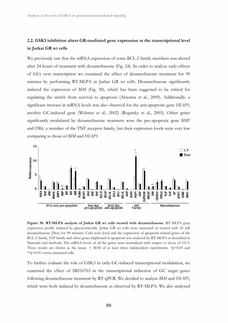
Analysis of the role of GSK3 on glucocorticoid-mediated signaling �
� ��
2.2. GSK3 inhibition alters GR-mediated gene expression at the transcriptional level
in Jurkat GR wt cells
We previously saw that the mRNA expression of some BCL-2 family members was altered
after 24 hours of treatment with dexamethasone (Fig. 24). In order to analyze early effects
of GCs over transcription we examined the effect of dexamethasone treatment for 90
minutes by performing RT-MLPA in Jurkat GR wt cells. Dexamethasone significantly
induced the expression of BIM (Fig. 30), which has been suggested to be critical for
regulating the switch from survival to apoptosis (Akiyama et al., 2009). Additionally, a
significant increase in mRNA levels was also observed for the anti-apoptotic gene HIAP1,
another GC-induced gene (Webster et al., 2002) (Rogatsky et al., 2003). Other genes
significantly modulated by dexamethasone treatment were the pro-apoptotic gene BMF
and DR6, a member of the TNF receptor family, but their expression levels were very low
comparing to those of BIM and HIAP1.
Figure 30. RT-MLPA analysis of Jurkat GR wt cells treated with dexamethasone. RT-MLPA gene expression profile induced by glucocorticoids. Jurkat GR wt cells were untreated or treated with 10 nM dexamethasone (Dex) for 90 minutes. Cells were lysed and the expression of apoptotic-related genes of the BCL-2 family, IAP family and other genes implicated in apoptosis was analyzed by RT-MLPA as described in Materials and methods. The mRNA levels of all the genes were normalized with respect to those of GUS. These results are shown as the mean ± SEM of at least three independent experiments. *p<0.05 and **p<0.01 versus untreated cells.
To further evaluate the role of GSK3 in early GC-induced transcriptional modulation, we
examined the effect of SB216763 in the transcriptional induction of GC target genes
following dexamethasone treatment by RT-qPCR. We decided to analyze BIM and HIAP1,
which were both induced by dexamethasone as observed by RT-MLPA. We also analyzed
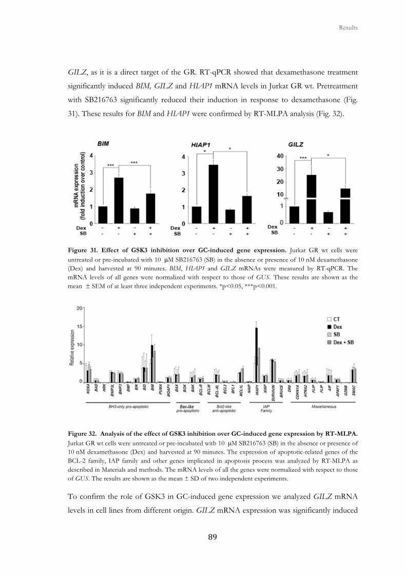
� � � Results
���
GILZ, as it is a direct target of the GR. RT-qPCR showed that dexamethasone treatment
significantly induced BIM, GILZ and HIAP1 mRNA levels in Jurkat GR wt. Pretreatment
with SB216763 significantly reduced their induction in response to dexamethasone (Fig.
31). These results for BIM and HIAP1 were confirmed by RT-MLPA analysis (Fig. 32).
Figure 31. Effect of GSK3 inhibition over GC-induced gene expression. Jurkat GR wt cells were untreated or pre-incubated with 10 μM SB216763 (SB) in the absence or presence of 10 nM dexamethasone (Dex) and harvested at 90 minutes. BIM, HIAP1 and GILZ mRNAs were measured by RT-qPCR. The mRNA levels of all genes were normalized with respect to those of GUS. These results are shown as the mean ± SEM of at least three independent experiments. *p<0.05, ***p<0.001.
Figure 32. Analysis of the effect of GSK3 inhibition over GC-induced gene expression by RT-MLPA. Jurkat GR wt cells were untreated or pre-incubated with 10 μM SB216763 (SB) in the absence or presence of 10 nM dexamethasone (Dex) and harvested at 90 minutes. The expression of apoptotic-related genes of the BCL-2 family, IAP family and other genes implicated in apoptosis process was analyzed by RT-MLPA as described in Materials and methods. The mRNA levels of all the genes were normalized with respect to those of GUS. The results are shown as the mean ± SD of two independent experiments.
To confirm the role of GSK3 in GC-induced gene expression we analyzed GILZ mRNA
levels in cell lines from different origin. GILZ mRNA expression was significantly induced
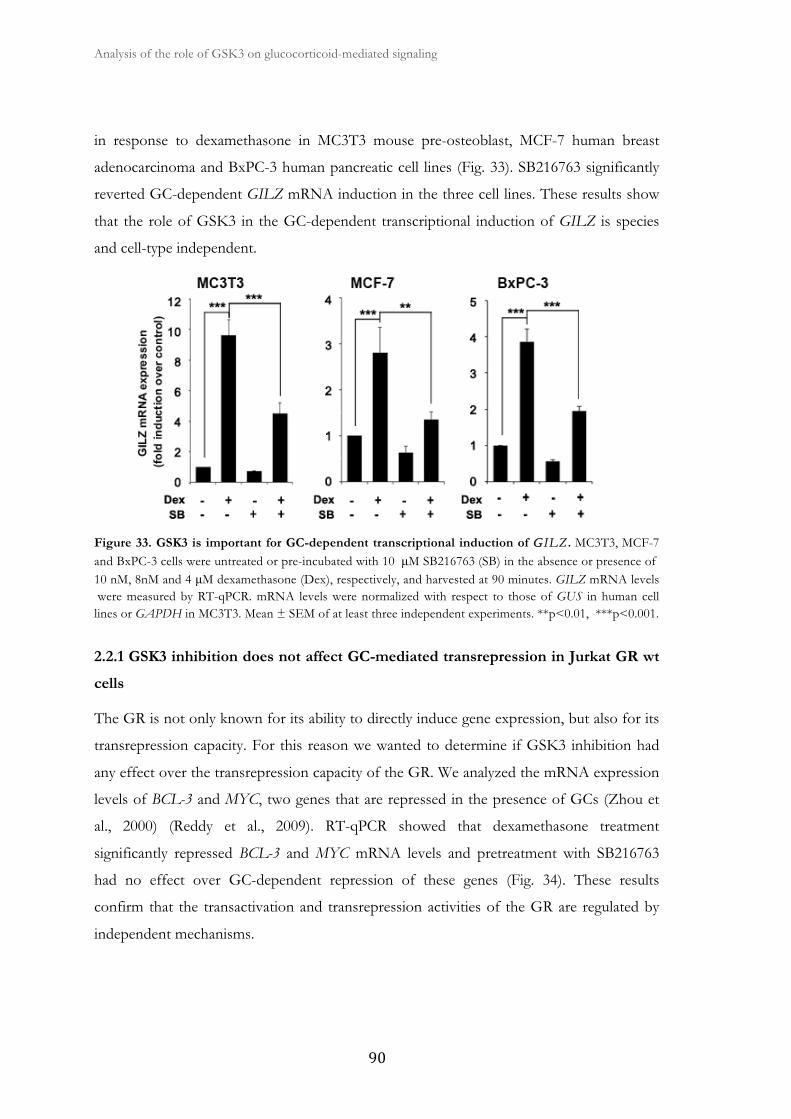
Analysis of the role of GSK3 on glucocorticoid-mediated signaling �
� ��
in response to dexamethasone in MC3T3 mouse pre-osteoblast, MCF-7 human breast
adenocarcinoma and BxPC-3 human pancreatic cell lines (Fig. 33). SB216763 significantly
reverted GC-dependent GILZ mRNA induction in the three cell lines. These results show
that the role of GSK3 in the GC-dependent transcriptional induction of GILZ is species
and cell-type independent.
Figure 33. GSK3 is important for GC-dependent transcriptional induction of GGILZ . MC3T3, MCF-7 and BxPC-3 cells were untreated or pre-incubated with 10 μM SB216763 (SB) in the absence or presence of 10 nM, 8nM and 4 μM dexamethasone (Dex), respectively, and harvested at 90 minutes. GILZ mRNA levels were measured by RT-qPCR. mRNA levels were normalized with respect to those of GUS in human cell lines or GAPDH in MC3T3. Mean ± SEM of at least three independent experiments. **p<0.01, ***p<0.001.
2.2.1 GSK3 inhibition does not affect GC-mediated transrepression in Jurkat GR wt
cells
The GR is not only known for its ability to directly induce gene expression, but also for its
transrepression capacity. For this reason we wanted to determine if GSK3 inhibition had
any effect over the transrepression capacity of the GR. We analyzed the mRNA expression
levels of BCL-3 and MYC, two genes that are repressed in the presence of GCs (Zhou et
al., 2000) (Reddy et al., 2009). RT-qPCR showed that dexamethasone treatment
significantly repressed BCL-3 and MYC mRNA levels and pretreatment with SB216763
had no effect over GC-dependent repression of these genes (Fig. 34). These results
confirm that the transactivation and transrepression activities of the GR are regulated by
independent mechanisms.
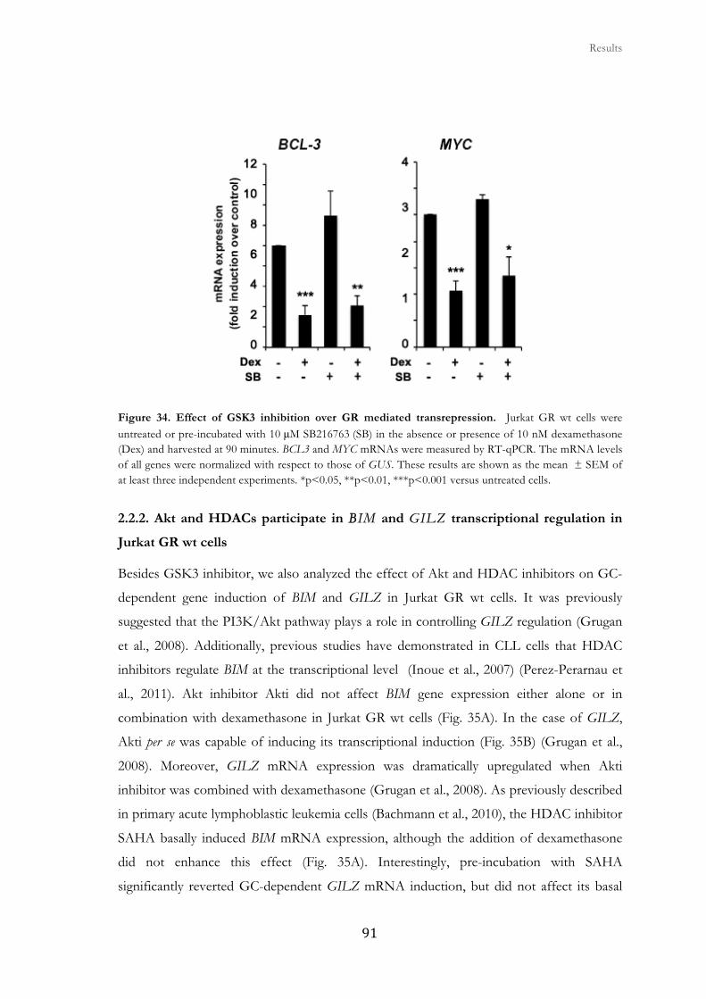
� � � Results
���
Figure 34. Effect of GSK3 inhibition over GR mediated transrepression. Jurkat GR wt cells were untreated or pre-incubated with 10 μM SB216763 (SB) in the absence or presence of 10 nM dexamethasone (Dex) and harvested at 90 minutes. BCL3 and MYC mRNAs were measured by RT-qPCR. The mRNA levels of all genes were normalized with respect to those of GUS. These results are shown as the mean ± SEM of at least three independent experiments. *p<0.05, **p<0.01, ***p<0.001 versus untreated cells.
2.2.2. Akt and HDACs participate in BBIM and GILZ transcriptional regulation in
Jurkat GR wt cells
Besides GSK3 inhibitor, we also analyzed the effect of Akt and HDAC inhibitors on GC-
dependent gene induction of BIM and GILZ in Jurkat GR wt cells. It was previously
suggested that the PI3K/Akt pathway plays a role in controlling GILZ regulation (Grugan
et al., 2008). Additionally, previous studies have demonstrated in CLL cells that HDAC
inhibitors regulate BIM at the transcriptional level (Inoue et al., 2007) (Perez-Perarnau et
al., 2011). Akt inhibitor Akti did not affect BIM gene expression either alone or in
combination with dexamethasone in Jurkat GR wt cells (Fig. 35A). In the case of GILZ,
Akti per se was capable of inducing its transcriptional induction (Fig. 35B) (Grugan et al.,
2008). Moreover, GILZ mRNA expression was dramatically upregulated when Akti
inhibitor was combined with dexamethasone (Grugan et al., 2008). As previously described
in primary acute lymphoblastic leukemia cells (Bachmann et al., 2010), the HDAC inhibitor
SAHA basally induced BIM mRNA expression, although the addition of dexamethasone
did not enhance this effect (Fig. 35A). Interestingly, pre-incubation with SAHA
significantly reverted GC-dependent GILZ mRNA induction, but did not affect its basal
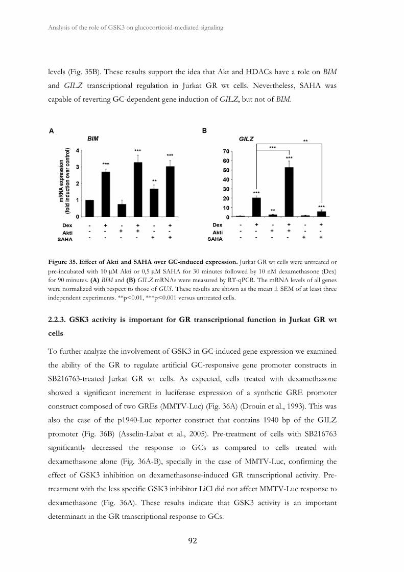
Analysis of the role of GSK3 on glucocorticoid-mediated signaling �
� ��
levels (Fig. 35B). These results support the idea that Akt and HDACs have a role on BIM
and GILZ transcriptional regulation in Jurkat GR wt cells. Nevertheless, SAHA was
capable of reverting GC-dependent gene induction of GILZ, but not of BIM.
Figure 35. Effect of Akti and SAHA over GC-induced expression. Jurkat GR wt cells were untreated or pre-incubated with 10 μM Akti or 0,5 μM SAHA for 30 minutes followed by 10 nM dexamethasone (Dex) for 90 minutes. (A) BIM and (B) GILZ mRNAs were measured by RT-qPCR. The mRNA levels of all genes were normalized with respect to those of GUS. These results are shown as the mean ± SEM of at least three independent experiments. **p<0.01, ***p<0.001 versus untreated cells.
2.2.3. GSK3 activity is important for GR transcriptional function in Jurkat GR wt
cells
To further analyze the involvement of GSK3 in GC-induced gene expression we examined
the ability of the GR to regulate artificial GC-responsive gene promoter constructs in
SB216763-treated Jurkat GR wt cells. As expected, cells treated with dexamethasone
showed a significant increment in luciferase expression of a synthetic GRE promoter
construct composed of two GREs (MMTV-Luc) (Fig. 36A) (Drouin et al., 1993). This was
also the case of the p1940-Luc reporter construct that contains 1940 bp of the GILZ
promoter (Fig. 36B) (Asselin-Labat et al., 2005). Pre-treatment of cells with SB216763
significantly decreased the response to GCs as compared to cells treated with
dexamethasone alone (Fig. 36A-B), specially in the case of MMTV-Luc, confirming the
effect of GSK3 inhibition on dexamethasonse-induced GR transcriptional activity. Pre-
treatment with the less specific GSK3 inhibitor LiCl did not affect MMTV-Luc response to
dexamethasone (Fig. 36A). These results indicate that GSK3 activity is an important
determinant in the GR transcriptional response to GCs.
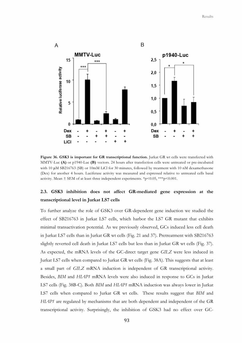
� � � Results
��
Figure 36. GSK3 is important for GR transcriptional function. Jurkat GR wt cells were transfected with MMTV-Luc (A) or p1940-Luc (B) vectors. 24 hours after transfection cells were untreated or pre-incubated with 10 μM SB216763 (SB) or 10mM LiCl for 30 minutes, followed by treatment with 10 nM dexamethasone (Dex) for another 4 hours. Luciferase activity was measured and expressed relative to untreated cells basal activity. Mean ± SEM of at least three independent experiments. *p<0.05, ***p<0.001.
2.3. GSK3 inhibition does not affect GR-mediated gene expression at the
transcriptional level in Jurkat LS7 cells
To further analyze the role of GSK3 over GR-dependent gene induction we studied the
effect of SB216763 in Jurkat LS7 cells, which harbor the LS7 GR mutant that exhibits
minimal transactivation potential. As we previously observed, GCs induced less cell death
in Jurkat LS7 cells than in Jurkat GR wt cells (Fig. 21 and 37). Pretreatment with SB216763
slightly reverted cell death in Jurkat LS7 cells but less than in Jurkat GR wt cells (Fig. 37).
As expected, the mRNA levels of the GC-direct target gene GILZ were less induced in
Jurkat LS7 cells when compared to Jurkat GR wt cells (Fig. 38A). This suggests that at least
a small part of GILZ mRNA induction is independent of GR transcriptional activity.
Besides, BIM and HIAP1 mRNA levels were also induced in response to GCs in Jurkat
LS7 cells (Fig. 38B-C). Both BIM and HIAP1 mRNA induction was always lower in Jurkat
LS7 cells when compared to Jurkat GR wt cells. These results suggest that BIM and
HIAP1 are regulated by mechanisms that are both dependent and independent of the GR
transcriptional activity. Surprisingly, the inhibition of GSK3 had no effect over GC-
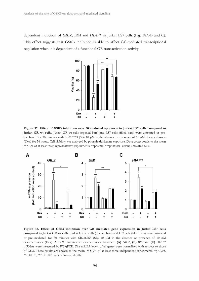
Analysis of the role of GSK3 on glucocorticoid-mediated signaling �
� �
dependent induction of GILZ, BIM and HIAP1 in Jurkat LS7 cells (Fig. 38A-B and C).
This effect suggests that GSK3 inhibition is able to affect GC-mediated transcriptional
regulation when it is dependent of a functional GR transactivation activity.
Figure 37. Effect of GSK3 inhibition over GC-induced apoptosis in Jurkat LS7 cells compared to Jurkat GR wt cells. Jurkat GR wt cells (opened bars) and LS7 cells (filled bars) were untreated or pre-incubated for 30 minutes with SB216763 (SB) 10 μM in the absence or presence of 10 nM dexamethasone (Dex) for 24 hours. Cell viability was analyzed by phosphatidylserine exposure. Data corresponds to the mean ± SEM of at least three representative experiments. **p<0.01, ***p<0.001 versus untreated cells.
Figure 38. Effect of GSK3 inhibition over GR mediated gene expression in Jurkat LS7 cells compared to Jurkat GR wt cells. Jurkat GR wt cells (opened bars) and LS7 cells (filled bars) were untreated or pre-incubated for 30 minutes with SB216763 (SB) 10 μM in the absence or presence of 10 nM dexamethasone (Dex). After 90 minutes of dexamethasone treatment (A) GILZ, (B) BIM and (C) HIAP1 mRNAs were measured by RT-qPCR. The mRNA levels of all genes were normalized with respect to those of GUS. These results are shown as the mean ± SEM of at least three independent experiments. *p<0.05, **p<0.01, ***p<0.001 versus untreated cells.
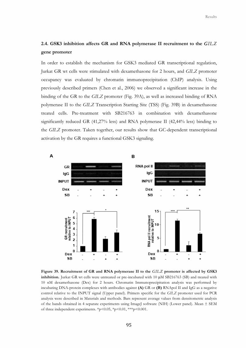
� � � Results
���
2.4. GSK3 inhibition affects GR and RNA polymerase II recruitment to the GGILZ
gene promoter
In order to establish the mechanism for GSK3 mediated GR transcriptional regulation,
Jurkat GR wt cells were stimulated with dexamethasone for 2 hours, and GILZ promoter
occupancy was evaluated by chromatin immunoprecipitation (ChIP) analysis. Using
previously described primers (Chen et al., 2006) we observed a significant increase in the
binding of the GR to the GILZ promoter (Fig. 39A), as well as increased binding of RNA
polymerase II to the GILZ Transcription Starting Site (TSS) (Fig. 39B) in dexamethasone
treated cells. Pre-treatment with SB216763 in combination with dexamethasone
significantly reduced GR (41,27% less) and RNA polymerase II (42,44% less) binding to
the GILZ promoter. Taken together, our results show that GC-dependent transcriptional
activation by the GR requires a functional GSK3 signaling.
Figure 39. Recruitment of GR and RNA polymerase II to the GILZ promoter is affected by GSK3
inhibition. Jurkat GR wt cells were untreated or pre-incubated with 10 μM SB216763 (SB) and treated with 10 nM dexamethasone (Dex) for 2 hours. Chromatin Immunoprecipitation analysis was performed by incubating DNA-protein complexes with antibodies against (A) GR or (B) RNApol II and IgG as a negative control relative to the INPUT signal (Upper panel). Primers specific for the GILZ promoter used for PCR analysis were described in Materials and methods. Bars represent average values from densitometric analysis of the bands obtained in 4 separate experiments using ImageJ software (NIH) (Lower panel). Mean ± SEM of three independent experiments. *p<0.05, *p<0.01, ***p<0.001.
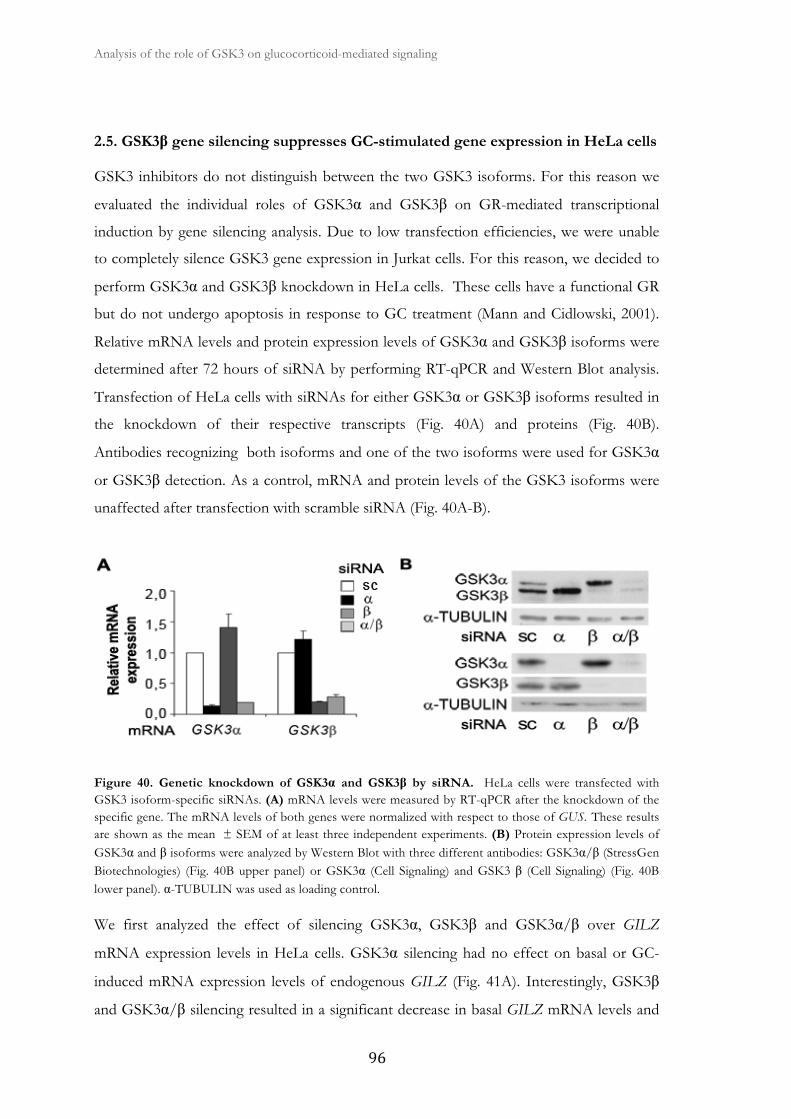
Analysis of the role of GSK3 on glucocorticoid-mediated signaling �
� ��
2.5. GSK3β gene silencing suppresses GC-stimulated gene expression in HeLa cells
GSK3 inhibitors do not distinguish between the two GSK3 isoforms. For this reason we
evaluated the individual roles of GSK3α and GSK3β on GR-mediated transcriptional
induction by gene silencing analysis. Due to low transfection efficiencies, we were unable
to completely silence GSK3 gene expression in Jurkat cells. For this reason, we decided to
perform GSK3α and GSK3β knockdown in HeLa cells. These cells have a functional GR
but do not undergo apoptosis in response to GC treatment (Mann and Cidlowski, 2001).
Relative mRNA levels and protein expression levels of GSK3α and GSK3β isoforms were
determined after 72 hours of siRNA by performing RT-qPCR and Western Blot analysis.
Transfection of HeLa cells with siRNAs for either GSK3α or GSK3β isoforms resulted in
the knockdown of their respective transcripts (Fig. 40A) and proteins (Fig. 40B).
Antibodies recognizing both isoforms and one of the two isoforms were used for GSK3α
or GSK3β detection. As a control, mRNA and protein levels of the GSK3 isoforms were
unaffected after transfection with scramble siRNA (Fig. 40A-B).
Figure 40. Genetic knockdown of GSK3α and GSK3β by siRNA. HeLa cells were transfected with GSK3 isoform-specific siRNAs. (A) mRNA levels were measured by RT-qPCR after the knockdown of the specific gene. The mRNA levels of both genes were normalized with respect to those of GUS. These results are shown as the mean ± SEM of at least three independent experiments. (B) Protein expression levels of GSK3α and β isoforms were analyzed by Western Blot with three different antibodies: GSK3α/β (StressGen Biotechnologies) (Fig. 40B upper panel) or GSK3α (Cell Signaling) and GSK3 β (Cell Signaling) (Fig. 40B lower panel). α-TUBULIN was used as loading control.
We first analyzed the effect of silencing GSK3α, GSK3β and GSK3α/β over GILZ
mRNA expression levels in HeLa cells. GSK3α silencing had no effect on basal or GC-
induced mRNA expression levels of endogenous GILZ (Fig. 41A). Interestingly, GSK3β
and GSK3α/β silencing resulted in a significant decrease in basal GILZ mRNA levels and
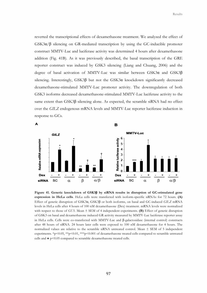
� � � Results
���
reverted the transcriptional effects of dexamethasone treatment. We analyzed the effect of
GSK3α/β silencing on GR-mediated transcription by using the GC-inducible promoter
construct MMTV-Luc and luciferase activity was determined 4 hours after dexamethasone
addition (Fig. 41B). As it was previously described, the basal transcription of the GRE
reporter construct was induced by GSK3 silencing (Liang and Chuang, 2006) and the
degree of basal activation of MMTV-Luc was similar between GSK3α and GSK3β
silencing. Interestingly, GSK3β but not the GSK3α knockdown significantly decreased
dexamethasone-stimulated MMTV-Luc promoter activity. The downregulation of both
GSK3 isoforms decreased dexamethasone-stimulated MMTV-Luc luciferase activity to the
same extent than GSK3β silencing alone. As expected, the scramble siRNA had no effect
over the GILZ endogenous mRNA levels and MMTV-Luc reporter luciferase induction in
response to GCs.
Figure 41. Genetic knockdown of GSK3β by siRNA results in disruption of GC-stimulated gene expression in HeLa cells. HeLa cells were transfected with isoform-specific siRNAs for 72 hours. (A) Effect of genetic disruption of GSK3α, GSK3β or both isoforms, on basal and GC-induced GILZ mRNA levels in HeLa cells after 4 hours of 100 nM dexamethasone (Dex) treatment. mRNA levels were normalized with respect to those of GUS. Mean ± SEM of 4 independent experiments. (B) Effect of genetic disruption of GSK3 on basal and dexamethasone induced GR activity measured by MMTV-Luc luciferase reporter assay in HeLa cells. Cells were co-transfected with MMTV-Luc and β-galactosidase (internal control) constructs after 48 hours of siRNA. 24 hours later cells were exposed to 100 nM dexamethasone for 4 hours. The normalized values are relative to the scramble siRNA untreated control. Mean ± SEM of 5 independent experiments. *p<0.05, **p<0.01, ***p<0.001 of dexamethasone treated cells compared to scramble untreated cells and ✚ p<0.05 compared to scramble dexamethasone treated cells.

Analysis of the role of GSK3 on glucocorticoid-mediated signaling �
� ��
We further used RT-MLPA to analyze changes in response to GSK3α/β silencing in HeLa
cells (Fig. 42A). BMF, BCL-XL, MCL-1, and HIAP1 were significantly induced by
dexamethasone (Fig. 42B). GSK3β or GSK3α/β silencing resulted in downregulation of
GC-dependent BMF, BCL-XL and MCL-1 mRNA induction. HIAP1 was still induced by
dexamethasone treatment even in the absence of GSK3α, GSK3β or both.
Figure 42. RT-MLPA analysis of GSK3α, GSK3β and GSK3α/β silenced HeLa cells. Cells were transfected with scramble or GSK3 isoform-specific siRNAs (α, β or α/β). 72 hours later cells were untreated (CT) or treated with 100 nM dexamethasone (Dex) for 4 hours. (A) Cells were lysed and the expression of apoptotic-related genes of the BCL-2 family, IAP family and other genes implicated in apoptosis process was analyzed by RT-MLPA as described in Materials and methods. The mRNA levels of all the genes were normalized with respect to those of PARN. (B) Genes significantly modulated by dexamethasone treatment are shown. The results are shown as the mean ± SEM of three independent experiments. *p<0.05, **p<0.01, ***p<0.001 compared to scramble untreated cells.

� � � Results
���
2.6. GR transcriptional function is impaired in GSK3β null MEFs
To further confirm our results obtained in GSK3 knockdown experiments in HeLa cells,
we used WT, and GSK3α or β null MEFs to analyze endogenous GILZ mRNA induction
in response to GC treatment. In accordance with our previous results in HeLa cells, we
found that MEFs deficient for GSK3β expression did not induce GILZ mRNA levels in
response to dexamethasone treatment, while wt and GSK3α null MEFs were able to
significantly induce GILZ mRNA upon GC treatment (Fig. 43A). To further confirm our
results, we transfected MEFs with the MMTV-Luc promoter construct. GSK3β null MEFs
did not induce MMTV-Luc promoter activity in response to dexamethasone, while wt and
GSK3α null MEFs significantly induced luciferase activity in response to GCs (Fig. 43B).
Figure 43. GR transcriptional activity is impaired in GSK3β null MEFs. (A) Wt, GSK3α and β null MEFs were untreated or treated with 100 nM dexamethasone for 4 hours. GILZ mRNA levels were measured by RT-qPCR and normalized with respect to GAPDH. Mean ± SEM of 4 independent experiments. **p<0.01 of dexamethasone treated cells compared to WT untreated cells. (B) MEFs were transfected with MMTV-Luc reporter and were treated with 100 nM dexamethasone (Dex) for 4 hours. Luciferase activity was measured. The normalized values are relative to the untreated control. Mean ± SEM of at least three independent experiments. *p<0.05, **p<0.01 compared to untreated cells.
On the other hand, even though there were differences in basal gene expression between
MEF cell lines, RT-MLPA analysis showed no significant changes in MEF cells gene
expression profile in response to GCs (Fig. 44). It is important to note that MEF cells do
not undergo apoptosis under GC treatment, but instead GCs have shown to inhibit cell
growth in this type of cells (Roca et al., 2003). If we had analyzed the growth control gene
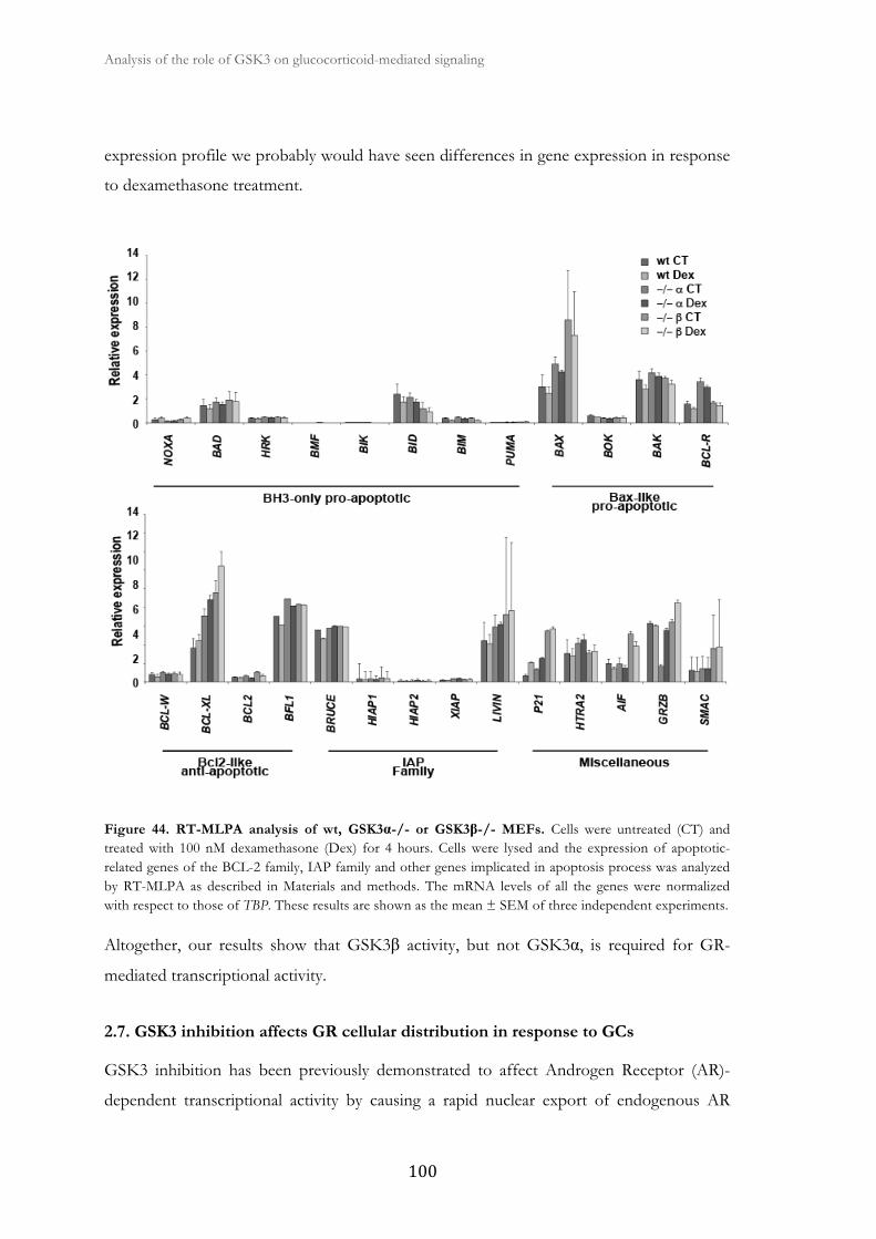
Analysis of the role of GSK3 on glucocorticoid-mediated signaling �
� ���
expression profile we probably would have seen differences in gene expression in response
to dexamethasone treatment.
Figure 44. RT-MLPA analysis of wt, GSK3α-/- or GSK3β-/- MEFs. Cells were untreated (CT) and treated with 100 nM dexamethasone (Dex) for 4 hours. Cells were lysed and the expression of apoptotic-related genes of the BCL-2 family, IAP family and other genes implicated in apoptosis process was analyzed by RT-MLPA as described in Materials and methods. The mRNA levels of all the genes were normalized with respect to those of TBP. These results are shown as the mean ± SEM of three independent experiments.
Altogether, our results show that GSK3β activity, but not GSK3α, is required for GR-
mediated transcriptional activity.
2.7. GSK3 inhibition affects GR cellular distribution in response to GCs
GSK3 inhibition has been previously demonstrated to affect Androgen Receptor (AR)-
dependent transcriptional activity by causing a rapid nuclear export of endogenous AR

� � � Results
����
(Mazor et al., 2004) (Rinnab et al., 2008) (Schutz et al., 2010). GSK3 inhibition also affects
Estrogen Receptor (ER) transcriptional activity (Medunjanin et al., 2005) (Grisouard and
Mayer, 2009). To examine the role of GSK3 in the regulation of GR subcellular localization
we analyzed cytosolic and membrane (which contains nuclei and mitochondrion) fractions
from Jurkat GR wt cells treated with dexamethasone. In non-stimulated cells, the GR was
detected mostly in the cytosol, while dexamethasone treatment induced its translocation to
the nucleus (Fig. 45A). Dexamethasone-dependent GR nuclear protein localization was
significantly reduced when cells were pretreated with SB216763 (Fig. 45A-B). GSK3α and
GSK3β levels were mainly observed in the cytoplasm and the nucleus respectively,
although under SB216763 treatment GSK3α levels where reduced in the nucleus.
Additionally, there was an increase in β-CATENIN protein levels in the membrane
fraction when cells were treated with SB216763, confirming GSK3 inhibition.
We wanted to determine if the partial reduction in GR nuclear translocation by SB216763
in dexamethasone treated cells was a result of a chromosome region maintenance 1
(CRM1)-dependent export, as CRM1 exportin has been suggested to play an important role
in the early nuclear export of the GR (Itoh et al., 2002). For this purpose, nuclear
translocation experiments were performed in the presence of leptomycin B (LMB), which
blocks specifically the CRM1-dependent nuclear export (Kudo et al., 1999). A significant
increase in nuclear GR localization was observed when cells were treated with LMB,
suggesting that there might be a CRM1-dependent nuclear export of the GR in response to
GSK3 inhibition (Fig 45A-B).
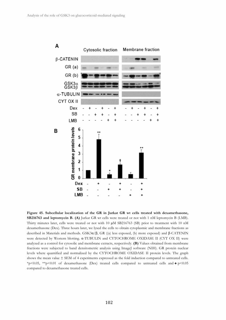
Analysis of the role of GSK3 on glucocorticoid-mediated signaling �
� ���
Figure 45. Subcellular localization of the GR in Jurkat GR wt cells treated with dexamethasone, SB216763 and leptomycin B. (A) Jurkat GR wt cells were treated or not with 1 nM leptomycin B (LMB). Thirty minutes later, cells were treated or not with 10 μM SB216763 (SB) prior to treatment with 10 nM dexamethasone (Dex). Three hours later, we lysed the cells to obtain cytoplasmic and membrane fractions as described in Materials and methods. GSK3α/β, GR ((a) less exposed, (b) more exposed) and β-CATENIN were detected by Western blotting. α-TUBULIN and CYTOCHROME OXIDASE II (CYT OX II) were analyzed as a control for cytosolic and membrane extracts, respectively. (B) Values obtained from membrane fractions were subjected to band densitometric analysis using ImageJ software (NIH). GR protein nuclear levels where quantified and normalized by the CYTOCHROME OXIDASE II protein levels. The graph shows the mean value ± SEM of 4 experiments expressed as the fold induction compared to untreated cells. *p<0.05, **p<0.01 of dexamethasone (Dex) treated cells compared to untreated cells and ✚ p<0.05 compared to dexamethasone treated cells.

� � � Results
���
We further confirmed the subcellular localization of the GR through immunofluorescent
staining visualized by confocal microscopy. In control Jurkat GR wt cells specific GR
staining was mainly observed in the cytoplasm (Fig. 46) and treatment with dexamethasone
for 2 hours induced GR nuclear translocation. Interestingly, cells pre-incubated with
SB216763 in combination with dexamethasone presented a decrease in the GR protein
levels in the nucleus compared with dexamethasone treatment alone. Moreover, SB216763
per se led to a slight increase in GR expression levels. Altogether, these results indicate that
short-term GSK3 inhibition is able to affect GR protein localization by decreasing early
GR nuclear levels in dexamethasone-treated Jurkat GR wt cells finally affecting GC-
stimulated gene expression.
Figure 46. Immunofluorescent staining of the GR in Jurkat GR wt cells. Cells were pre-treated or not with 10 μM SB216763 (SB) prior to treatment with 10 nM dexamethasone (Dex) for 2 hours. Yoyo-1 iodide (green) stains nuclear DNA (DNA) and Alexa Fluor 647 (red) stains GR (GR). Merged images are shown for comparison. This is a representative experiment of 3 that were performed.

Analysis of the role of GSK3 on glucocorticoid-mediated signaling �
� ��
Next, we analyzed the effect of long-term silencing of GSK3α and GSK3β isoforms over
the GR subcellular distribution induced by dexamethasone using isoform specific
knockdown in HeLa cells. We determined GR subcellular distribution by analyzing
cytoplasmic and nuclear fractions. Surprisingly, treatment with dexamethasone always
resulted in GR translocation to the nucleus even when GSK3α, GSK3β, or GSK3α/β were
silenced (Fig. 47). This was also observed through immunofluorescent staining visualized
by confocal microscopy (Fig. 48). Moreover, depletion of GSK3β, or GSK3α/β proteins
by siRNA led to an increase in general GR expression levels (Fig. 48).
Figure 47. Subcellular localization of the GR using isoform specific knockdown in HeLa cells
treated with dexamethasone. HeLa cells were transfected with GSK3 isoform-specific siRNAs. Cells were
untreated (CT) or treated with 100 nM dexamethasone (Dex). Three hours later, we lysed the cells to obtain
cytoplasmic and nuclear fractions as described in Materials and methods. GR was detected by Western blot.
α-TUBULIN and LAMIN A/C were analyzed as a control for cytosolic (C) and nuclear (N) extracts
respectively.
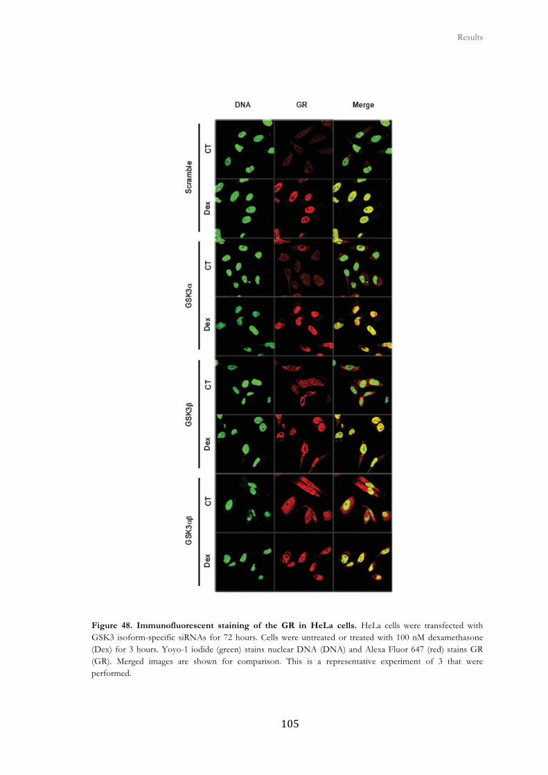
� � � Results
����
Figure 48. Immunofluorescent staining of the GR in HeLa cells. HeLa cells were transfected with GSK3 isoform-specific siRNAs for 72 hours. Cells were untreated or treated with 100 nM dexamethasone (Dex) for 3 hours. Yoyo-1 iodide (green) stains nuclear DNA (DNA) and Alexa Fluor 647 (red) stains GR (GR). Merged images are shown for comparison. This is a representative experiment of 3 that were performed.

Analysis of the role of GSK3 on glucocorticoid-mediated signaling �
� ���
2.8. GSK3 inhibition alters transcriptional activity of GR phosphorylation mutants
Besides phosphorylation of human GR at Ser404, GSK3β has also been described to
phosphorylate rat GR at Thr171 (Rogatsky et al., 1998). As Jurkat GR wt cells harbor a rat
GR, we mutated both Thr171 and Ser424 (corresponding to human Ser404 (Galliher-
Beckley and Cidlowski, 2009)) residues in the rat GR in order to analyze mutant
transactivation capacity under GSK3 inhibition. Both residues were replaced for Alanine
making them unphosphorylatable by GSK3, which is a Serine/Threonine kinase. Figure 49
shows the two known residues phosphorylated by GSK3 and the changes made in the rat
GR and the alignment with the human GR sequence. Note that Thr171 is not present in
the human GR sequence.
Figure 49. Phosphorylation sites of the GR by GSK3β. Schematic representation of the two known residues of rat GR that are phosphorylated by GSK3β, Thr171 and Ser424. The localization of each phosphorylation site of the rat and human receptor are shown. The framed sequences indicate the amino acids conserved between the two species. The nucleotide changes made by site directed mutagenesis to alter amino acid sequence are indicated.
GR wt and GR mutants were expressed in the Jurkat parental cell line, which harbor a
function-impairing point mutation (R477H) in one of their GR alleles that causes GC
resistance (Riml et al., 2004). We used the GC-inducible promoter MMTV-Luc to evaluate
the effect of GSK3 inhibition over the GC-induced transactivation activity of single and
double mutants. Cells were treated with dexamethasone for 4 hours in the presence or
absence of SB216763. According to previous results in Jurkat GR wt cells, pre-treatment of
Jurkat cells with SB216763 significantly inhibits GR wt hormone-dependent transcriptional
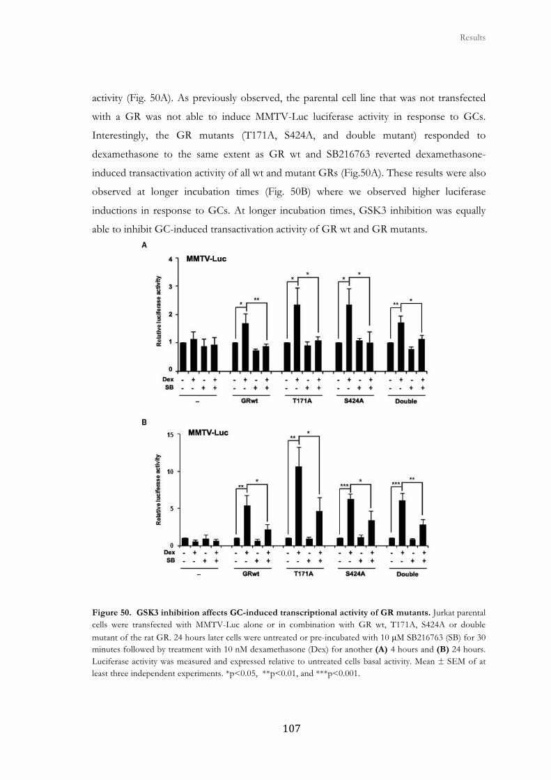
� � � Results
����
activity (Fig. 50A). As previously observed, the parental cell line that was not transfected
with a GR was not able to induce MMTV-Luc luciferase activity in response to GCs.
Interestingly, the GR mutants (T171A, S424A, and double mutant) responded to
dexamethasone to the same extent as GR wt and SB216763 reverted dexamethasone-
induced transactivation activity of all wt and mutant GRs (Fig.50A). These results were also
observed at longer incubation times (Fig. 50B) where we observed higher luciferase
inductions in response to GCs. At longer incubation times, GSK3 inhibition was equally
able to inhibit GC-induced transactivation activity of GR wt and GR mutants.
Figure 50. GSK3 inhibition affects GC-induced transcriptional activity of GR mutants. Jurkat parental cells were transfected with MMTV-Luc alone or in combination with GR wt, T171A, S424A or double mutant of the rat GR. 24 hours later cells were untreated or pre-incubated with 10 μM SB216763 (SB) for 30 minutes followed by treatment with 10 nM dexamethasone (Dex) for another (A) 4 hours and (B) 24 hours. Luciferase activity was measured and expressed relative to untreated cells basal activity. Mean ± SEM of at least three independent experiments. *p<0.05, **p<0.01, and ***p<0.001.
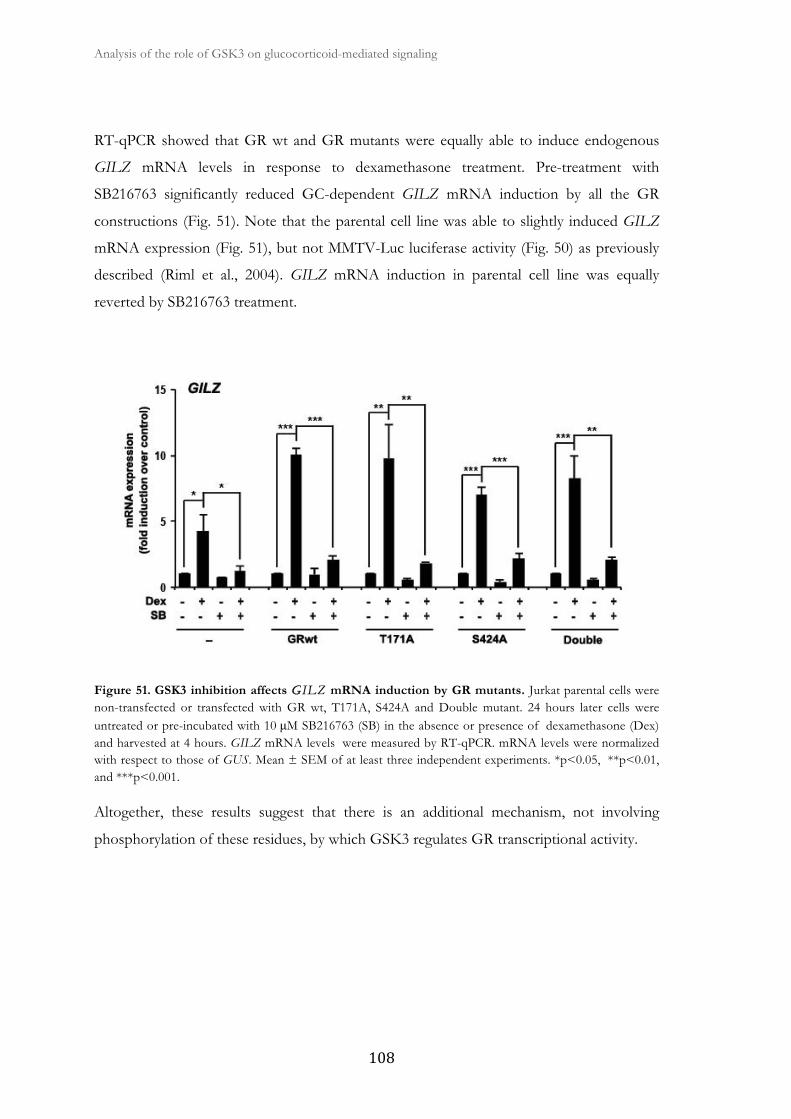
Analysis of the role of GSK3 on glucocorticoid-mediated signaling �
� ���
RT-qPCR showed that GR wt and GR mutants were equally able to induce endogenous
GILZ mRNA levels in response to dexamethasone treatment. Pre-treatment with
SB216763 significantly reduced GC-dependent GILZ mRNA induction by all the GR
constructions (Fig. 51). Note that the parental cell line was able to slightly induced GILZ
mRNA expression (Fig. 51), but not MMTV-Luc luciferase activity (Fig. 50) as previously
described (Riml et al., 2004). GILZ mRNA induction in parental cell line was equally
reverted by SB216763 treatment.
Figure 51. GSK3 inhibition affects GGILZ mRNA induction by GR mutants. Jurkat parental cells were non-transfected or transfected with GR wt, T171A, S424A and Double mutant. 24 hours later cells were untreated or pre-incubated with 10 μM SB216763 (SB) in the absence or presence of dexamethasone (Dex) and harvested at 4 hours. GILZ mRNA levels were measured by RT-qPCR. mRNA levels were normalized with respect to those of GUS. Mean ± SEM of at least three independent experiments. *p<0.05, **p<0.01, and ***p<0.001.
Altogether, these results suggest that there is an additional mechanism, not involving
phosphorylation of these residues, by which GSK3 regulates GR transcriptional activity.

3. Role of GSK3 in glucocorticoid-induced
apoptosis in CLL cells

�
�
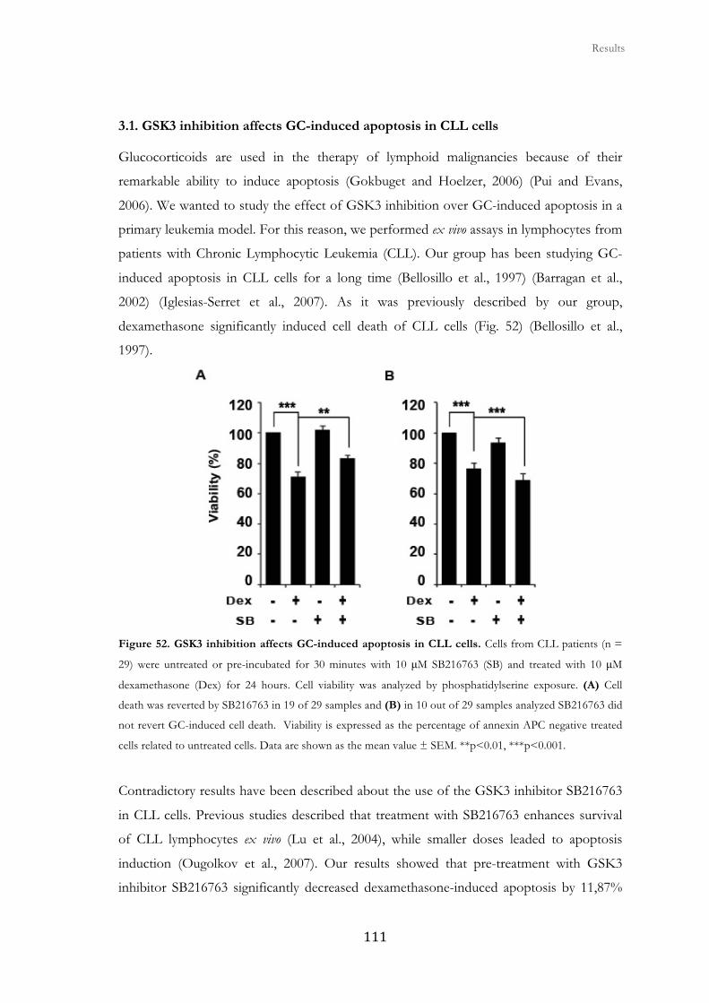
� � � Results
����
3.1. GSK3 inhibition affects GC-induced apoptosis in CLL cells
Glucocorticoids are used in the therapy of lymphoid malignancies because of their
remarkable ability to induce apoptosis (Gokbuget and Hoelzer, 2006) (Pui and Evans,
2006). We wanted to study the effect of GSK3 inhibition over GC-induced apoptosis in a
primary leukemia model. For this reason, we performed ex vivo assays in lymphocytes from
patients with Chronic Lymphocytic Leukemia (CLL). Our group has been studying GC-
induced apoptosis in CLL cells for a long time (Bellosillo et al., 1997) (Barragan et al.,
2002) (Iglesias-Serret et al., 2007). As it was previously described by our group,
dexamethasone significantly induced cell death of CLL cells (Fig. 52) (Bellosillo et al.,
1997).
�
Figure 52. GSK3 inhibition affects GC-induced apoptosis in CLL cells. Cells from CLL patients (n =
29) were untreated or pre-incubated for 30 minutes with 10 μM SB216763 (SB) and treated with 10 μM
dexamethasone (Dex) for 24 hours. Cell viability was analyzed by phosphatidylserine exposure. (A) Cell
death was reverted by SB216763 in 19 of 29 samples and (B) in 10 out of 29 samples analyzed SB216763 did
not revert GC-induced cell death. Viability is expressed as the percentage of annexin APC negative treated
cells related to untreated cells. Data are shown as the mean value ± SEM. **p<0.01, ***p<0.001.
Contradictory results have been described about the use of the GSK3 inhibitor SB216763
in CLL cells. Previous studies described that treatment with SB216763 enhances survival
of CLL lymphocytes ex vivo (Lu et al., 2004), while smaller doses leaded to apoptosis
induction (Ougolkov et al., 2007). Our results showed that pre-treatment with GSK3
inhibitor SB216763 significantly decreased dexamethasone-induced apoptosis by 11,87%
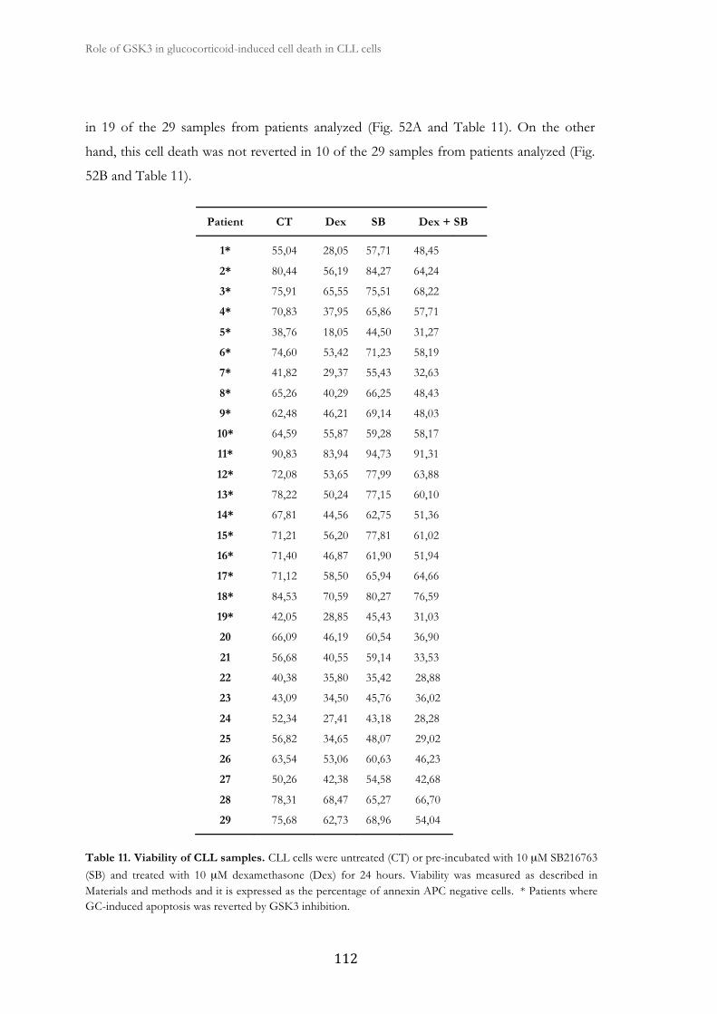
Role of GSK3 in glucocorticoid-induced cell death in CLL cells �
� ���
in 19 of the 29 samples from patients analyzed (Fig. 52A and Table 11). On the other
hand, this cell death was not reverted in 10 of the 29 samples from patients analyzed (Fig.
52B and Table 11).
Patient CT Dex SB Dex + SB
1* 55,04 28,05 57,71 48,45
2* 80,44 56,19 84,27 64,24
3* 75,91 65,55 75,51 68,22
4* 70,83 37,95 65,86 57,71
5* 38,76 18,05 44,50 31,27
6* 74,60 53,42 71,23 58,19
7* 41,82 29,37 55,43 32,63
8* 65,26 40,29 66,25 48,43
9* 62,48 46,21 69,14 48,03
10* 64,59 55,87 59,28 58,17
11* 90,83 83,94 94,73 91,31
12* 72,08 53,65 77,99 63,88
13* 78,22 50,24 77,15 60,10
14* 67,81 44,56 62,75 51,36
15* 71,21 56,20 77,81 61,02
16* 71,40 46,87 61,90 51,94
17* 71,12 58,50 65,94 64,66
18* 84,53 70,59 80,27 76,59
19* 42,05 28,85 45,43 31,03
20 66,09 46,19 60,54 36,90
21 56,68 40,55 59,14 33,53
22 40,38 35,80 35,42 28,88
23 43,09 34,50 45,76 36,02
24 52,34 27,41 43,18 28,28
25 56,82 34,65 48,07 29,02
26 63,54 53,06 60,63 46,23
27 50,26 42,38 54,58 42,68
28 78,31 68,47 65,27 66,70
29 75,68 62,73 68,96 54,04
Table 11. Viability of CLL samples. CLL cells were untreated (CT) or pre-incubated with 10 μM SB216763 (SB) and treated with 10 μM dexamethasone (Dex) for 24 hours. Viability was measured as described in Materials and methods and it is expressed as the percentage of annexin APC negative cells. * Patients where GC-induced apoptosis was reverted by GSK3 inhibition.
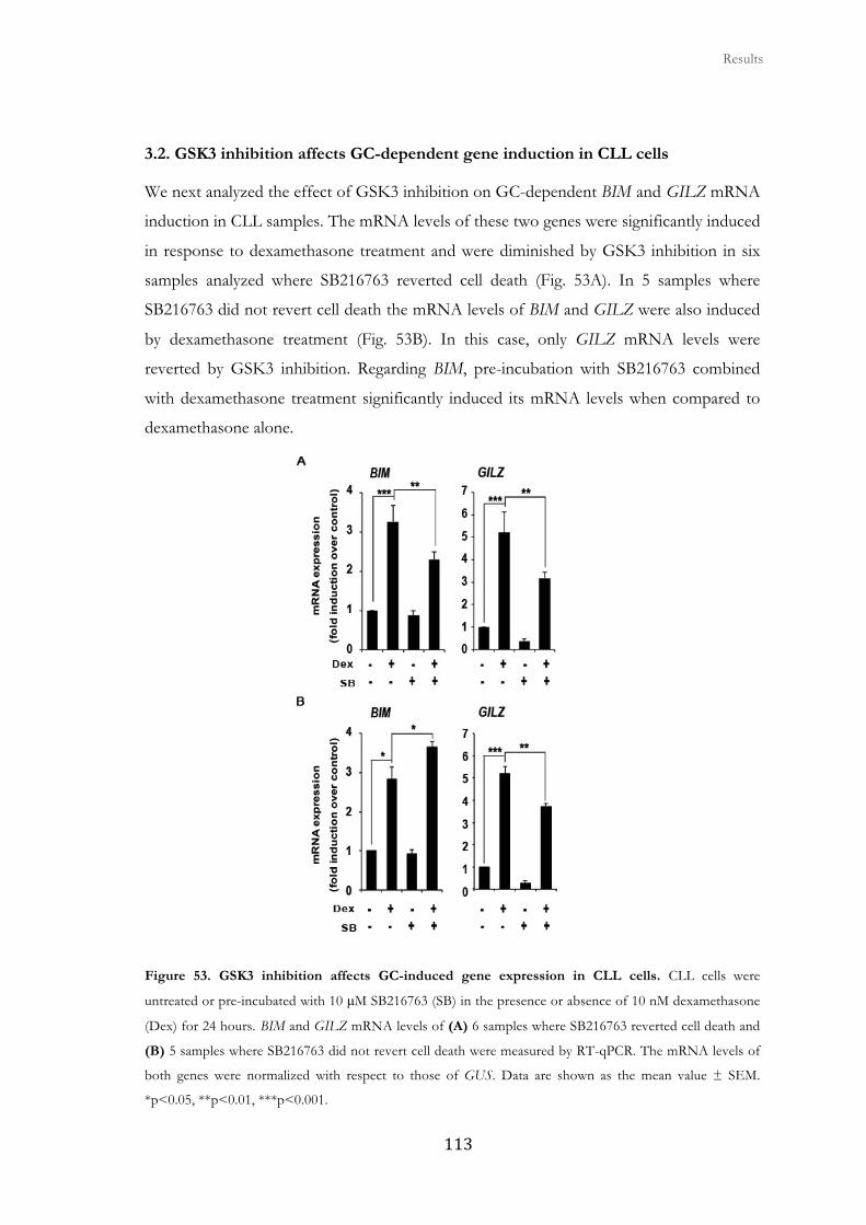
� � � Results
���
3.2. GSK3 inhibition affects GC-dependent gene induction in CLL cells
We next analyzed the effect of GSK3 inhibition on GC-dependent BIM and GILZ mRNA
induction in CLL samples. The mRNA levels of these two genes were significantly induced
in response to dexamethasone treatment and were diminished by GSK3 inhibition in six
samples analyzed where SB216763 reverted cell death (Fig. 53A). In 5 samples where
SB216763 did not revert cell death the mRNA levels of BIM and GILZ were also induced
by dexamethasone treatment (Fig. 53B). In this case, only GILZ mRNA levels were
reverted by GSK3 inhibition. Regarding BIM, pre-incubation with SB216763 combined
with dexamethasone treatment significantly induced its mRNA levels when compared to
dexamethasone alone.
Figure 53. GSK3 inhibition affects GC-induced gene expression in CLL cells. CLL cells were
untreated or pre-incubated with 10 μM SB216763 (SB) in the presence or absence of 10 nM dexamethasone
(Dex) for 24 hours. BIM and GILZ mRNA levels of (A) 6 samples where SB216763 reverted cell death and
(B) 5 samples where SB216763 did not revert cell death were measured by RT-qPCR. The mRNA levels of
both genes were normalized with respect to those of GUS. Data are shown as the mean value ± SEM.
*p<0.05, **p<0.01, ***p<0.001.
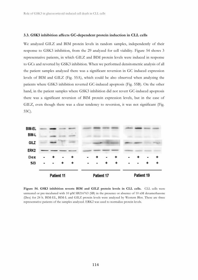
Role of GSK3 in glucocorticoid-induced cell death in CLL cells �
� ��
3.3. GSK3 inhibition affects GC-dependent protein induction in CLL cells
We analyzed GILZ and BIM protein levels in random samples, independently of their
response to GSK3 inhibition, from the 29 analyzed for cell viability. Figure 54 shows 3
representative patients, in which GILZ and BIM protein levels were induced in response
to GCs and reverted by GSK3 inhibition. When we performed densitometric analysis of all
the patient samples analyzed there was a significant reversion in GC induced expression
levels of BIM and GILZ (Fig. 55A), which could be also observed when analyzing the
patients where GSK3 inhibition reverted GC-induced apoptosis (Fig. 55B). On the other
hand, in the patient samples where GSK3 inhibition did not revert GC-induced apoptosis
there was a significant reversion of BIM protein expression levels, but in the case of
GILZ, even though there was a clear tendency to reversion, it was not significant (Fig.
55C).
Figure 54. GSK3 inhibition reverts BIM and GILZ protein levels in CLL cells. CLL cells were untreated or pre-incubated with 10 μM SB216763 (SB) in the presence or absence of 10 nM dexamethasone (Dex) for 24 h. BIM-EL, BIM-L and GILZ protein levels were analyzed by Western Blot. These are three representative patients of the samples analyzed. ERK2 was used to normalize protein levels.

� � � Results
����
Figure 55. GSK3 inhibition affects GC-induced BIM and GILZ protein levels in CLL cells. BIM and GILZ protein levels from Western Blots from CLL patients were quantified by densitometric analysis and corrected by ERK2 levels by using ImageJ software (NIH). Results are represented as the mean ± SEM of (A) all the patients analyzed (BIM: 14 and GILZ: 11), (B) patients where GSK3 inhibition reverted GC-induced apoptosis (BIM: 6 and GILZ: 4), and (C) patients where GSK3 inhibition did not revert GC-induced apoptosis (BIM: 8 and GILZ: 7).
Collectively, these data indicate that GSK3 inhibition reduces sensitivity to GC-induced
apoptosis in CLL cells and impairs GC-dependent gene and protein induction.


�
�
V. General discussion and future perspectives

�
�

General discussion and future perspectives
�����
Role of GSK3 on GC-mediated signaling and GR transcriptional regulation.
Effect of GCs in apoptosis induction
GCs are steroidal ligands for the GR, a ligand-activated transcription factor. In addition to
their developmental and homeostatic roles, GCs also regulate the functions of the immune
system (Glass and Ogawa, 2006) (Chinenov and Rogatsky, 2007) (Beck et al., 2009). For
this reason, GCs are the first line of treatment in several inflammatory diseases (Glass and
Saijo, 2010). Furthermore, GCs induce apoptosis in different cell types including leukemia
cells, in a complex process regulated by multiple signaling pathways that alter gene
expression profiles through GR-mediated transactivation and transrepression (Kfir-
Erenfeld et al., 2010). Our group previously described that GCs induce apoptosis in CLL
cells (Bellosillo et al., 1997) through a mechanism that regulates the pro-apoptotic BCL-2
family member BIM at the transcriptional and protein level (Iglesias-Serret et al., 2007).
Unfortunately, CLL cells are not easy to transfect making them an unsuitable model for
molecular analysis. In order to analyze GC-mediated signaling at the molecular level, we
used three Jurkat T-ALL cell lines harboring different types of GR. The Jurkat parental cell
line carries one wt and one mutated GR allele. This function-impairing point mutation
(R477H) affects transactivation, transrepression, but not ligand binding or translocation of
the GR (Riml et al., 2004). As expected, this cell line was completely resistant to GC-
induced apoptosis. We used the Jurkat GR wt cell line, which is stably transfected with a wt
rat GR expression vector (Helmberg et al., 1995), suffered GC-induced apoptosis after
dexamethasone treatment. Additionally, we used the Jurkat LS7 cell line, which expresses
the LS7 mutant that contains two adjacent amino acid mutations in the second half of the
second zinc finger (Helmberg et al., 1995). The LS7 GR mutant was previously reported to
have little or no transactivating potential, while retaining transrepression capacity. These
cells were also sensitive to GC-induced apoptosis, although to a less extent than Jurkat GR
wt cells.
When we analyzed the mRNA expression profile of the BCL-2 family members and other
apoptosis related genes in Jurkat cell lines we observed that in both Jurkat GR wt and LS7
cell lines, dexamethasone induced the mRNA levels of two well known GC-induced genes,
BIM (Wang et al., 2003) (Zhang and Insel, 2004) and HIAP1 (Webster et al., 2002)
General discussion and future perspectives �
����
(Rogatsky et al., 2003). These results show that the induction of these genes is independent
of the transactivation capacity of the GR as the LS7 mutant is aunable to transactivate
target genes. GCs equally induced BIM protein expression. We analyzed MCL-1 protein
expression levels, as its overexpression is known to be important for cancer cells to evade
cell death (Quinn et al., 2011) by sequestering BAX- or BAK-activating BH3-only proteins
BIM and BID (Maurer et al., 2006). MCL-1 expression levels were downregulated in Jurkat
GR wt and LS7 cell lines in the presence of dexamethasone. These results where BIM is
induced and MCL-1 is downregulated confirm the importance of the balance between pro-
apoptotic and anti-apoptotic BCL-2 family members in the regulation and determination of
cell death or survival (Kelly and Strasser, 2011). As expected, in the parental cell line
dexamethasone treatment did not induce changes in the mRNA expression profile or the
protein expression of BIM and MCL-1. Besides analyzing the apoptotic profile in Jurkat
cell lines we also analyzed the transcriptional regulation of GILZ, a direct GR-target gene
(Beaulieu and Morand, 2011), and BIM, a gene regulated independently of GR direct
binding to DNA (Molitoris et al., 2011) (Heidari et al., 2012), in Jurkat GR wt cells. We
confirmed that ongoing transcription but not translation is required for the GC-dependent
induction of GILZ and BIM in Jurkat GR wt cells. Both genes were shown to be primary
GC targets, as opposed to those regulated by another glucocorticoid-inducible factor that
would require de novo protein synthesis.
Role of GSK3 in GC-induced apoptosis
Previous studies have shown the involvement of different protein kinases in GC-
mechanism of action (Galliher-Beckley et al., 2008). It has been previously described that in
the absence of a ligand, GSK3α is bound to the GR and exposure to GCs or GSK3
inhibitor lead to the disruption of this interaction (Spokoini et al., 2010). It has also been
described that the GR associates with GSK3β in the presence of dexamethasone but not
with GSK3α (Galliher-Beckley et al., 2008). Consistent with previous studies in other cell
lines, we observed a critical role for GSK3 in GC-dependent cell death (Nuutinen et al.,
2009) (Spokoini et al., 2010). Inhibition of GSK3 by the specific inhibitor SB216763
significantly reverted GC-induced apoptosis in Jurkat GR wt cells. In Jurkat LS7 cells there
was a less pronounced inhibitory effect of GSK3 inhibition over GC-induced apoptosis.
The anti-apoptotic protein MCL-1 contains a conserved consensus site for GSK3
phosphorylation, which targets it for ubiquitin-dependent degradation (Maurer et al., 2006).

General discussion and future perspectives
�����
In Jurkat GR wt cells we observed that GC-dependent downregulation of MCL-1 protein
levels and caspase activation was prevented by GSK3 inhibition. In accordance with a
previous study, GSK3 inhibition reverted dexamethasone-induced upregulation of BIM
(Nuutinen et al., 2009). Interestingly, this was also the case of GC-dependent GILZ protein
induction, which was significantly reverted by GSK3 inhibition. To the best of our
knowledge, there are no previous studies demonstrating that GSK3 is important in the
GC-dependent upregulation of GILZ.
Role of GSK3 in GR-mediated transcriptional regulation
Pharmacological inhibition of GSK3 resulted in a reduced induction of BIM, HIAP1 and
GILZ endogenous mRNA levels in response to GCs in Jurkat GR wt cells. This effect over
GILZ mRNA levels was confirmed in several cell lines form different species and tissue
origin, ruling out cell type specific effects. GSK3 inhibition by SB216763, but not by LiCl,
resulted in the reduced induction of GC-responsive promoter constructs. In Jurkat LS7
cells dexamethasone induced BIM and HIAP1 mRNA levels, but to a less extent than in
Jurkat GR wt cells. GILZ was only slightly induced in Jurkat LS7 cells as it is a GR-direct
target gene and in agreement with the low transactivating potential of the LS7 mutant.
Interestingly, there was no reversion in GC-induced mRNA levels of target genes in
response to GSK3 inhibition in this cell line.
ChIP assays in Jurkat GR wt cells showed that GSK3 inhibition reduced GR and RNA
polymerase II recruitment to the GILZ promoter after dexamethasone treatment. These
results indicate that GC-dependent transcriptional activation requires a functional GSK3
signaling and show for the first time that GSK3 could be required by the GR for its
activation as a transcription factor and for RNA polymerase II recruitment. These results
are in the line with previous studies in which GSK3 inhibition represses other steroid
receptors transcriptional activity in various cell types (Liao et al., 2004) (Mazor et al., 2004)
(Medunjanin et al., 2005) (Grisouard and Mayer, 2009).
In contrast to transcriptional activation there are mechanisms by which the GR can
transrepress gene expression. Transrepression typically involves indirect association
(tethering) of the receptor with target genes rather than direct sequence-specific DNA

General discussion and future perspectives �
����
binding (Glass and Saijo, 2010). For example, GCs can interact with and inhibit AP1 and
NF-κB family members, suppressing the expression of their target genes (De Bosscher et
al., 2003). We wanted to determine if GSK3 inhibition is able to affect GC-induced
transcriptional transrepression. We analyzed MYC and BCL-3, two genes that are
transrepressed by GCs and whose promoters harbor AP1 and NF-κB binding sites (Duyao
et al., 1990) (Ge et al., 2003) (Iavarone et al., 2003) (Vartanian et al., 2011) (Wang et al.,
2011). As expected, these genes were significantly repressed by dexamethasone treatment
in Jurkat GR wt cells. Interestingly, pretreatment with SB216763 had no significant effect
over the GC-induced transrepression of these genes. These results suggest that GSK3
inhibition can affect GC-induced transactivation but not transrepression. These findings
are of clinical interest, as several side effects of GC therapy are thought to be
predominantly mediated via transactivation (Glass and Saijo, 2010). Thus, approaches, like
GSK3 inhibition, that preferentially do not affect transrepression and suppress
transactivation activity of the GR could result in fewer undesirable effects of therapy.
Interaction networks between the GR and different signaling pathways
Signaling pathways including protein kinase networks are known to regulate glucocorticoid-
induced apoptosis (Kfir-Erenfeld et al., 2010). We observed that pre-incubation with
protein kinase inhibitors, HDAC inhibitors, BCL-2 inhibitor, and proteasome inhibitor
could not prevent apoptosis induction by dexamethasone.
We further analyzed the effect of SAHA over GC induced gene expression, as our group
has recently demonstrated in CLL cells that BIM regulation is one of the most critical
molecular events explaining the apoptotic effect of the HDAC inhibitors Kendine 92 and
SAHA (Perez-Perarnau et al., 2011). HDAC inhibitors are promising chemotherapeutic
agents that exert a range of antitumor activities through gene regulation, cell cycle arrest
and apoptosis. In the case of SAHA, BIM upregulation is mediated by E2F recruitment to
its promoter (Zhao et al., 2005). On the other hand, GILZ has been previously described
to be dramatically upregulated when PI3K and Akt inhibitors were combined with
dexamethasone (Grugan et al., 2008). In order to see if Akti inhibitor could shed a light on
GC-mediated transcriptional regulation we analyzed its effect over BIM and GILZ mRNA
induction in response to dexamethasone. We confirmed the previously described positive
effect of SAHA over basal BIM mRNA expression (Bachmann et al., 2010). Nevertheless,
General discussion and future perspectives
����
SAHA had no effect over GC-induced BIM mRNA levels. Interestingly, SAHA clearly
reverted GILZ induction by GCs. Even though, acetylation of histones is thought to allow
the transcriptional machinery to function (Hong et al., 1993), it was previously
demonstrated that HDAC inhibitors are able to repress GC-induced MMTV-Luc
transcriptional activity (Mulholland et al., 2003). This happens through chromatin
remodeling independent mechanisms suggesting that the primary target of HDAC
inhibition at the MMTV promoter is a nonhistone protein that is involved in an essential
step of basal transcription. It is likely that the mechanism by which HDAC inhibition
represses GC-dependent induction of GILZ mRNA levels is similar to the mechanism by
which it represses MMTV-Luc activity.
As previously described, Akt inhibition affected basal levels and potentiated
dexamethasone-induced GILZ mRNA expression (Grugan et al., 2008) probably through
FOXO3 binding to the GILZ promoter (Asselin-Labat et al., 2004). As previously
described by our group for CLL cells (de Frias et al., 2009), Akt inhibition had no effect
over BIM mRNA levels in Jurkat GR wt cells. These data confirm a role for Akt in GILZ
transcriptional regulation and of HDACs in the transcriptional regulation of BIM and
GILZ.
Analysis of GSK3 isoforms and their implication in GR regulation
Furthermore, we analyzed which was the GSK3 isoform involved in the regulation of GR
transcriptional activation in response to GCs. As inhibitors do not distinguish between the
two isoforms, we performed isoform specific gene silencing analysis. As it was previously
described, GSK3α and GSK3β silencing in HeLa cells, resulted in the activation of a GRE-
promoter construct at a basal level (Liang and Chuang, 2006). Interestingly, only GSK3β
silencing effectively reverted GC-dependent transcriptional activation. Knockdown of
GSK3β and double knockdown of α and β isoforms reduced both basal and GC-induced
mRNA expression of endogenous GILZ. This reversion in transcriptional activation was
not observed after GSK3α silencing, indicating that GSK3β is the isoform that regulates
basal and GC-induced gene transcription.
RT-MLPA analysis in HeLa cells revealed differences in apoptosis mRNA expression
profile when compared to Jurkat GR wt cells. These results are coherent with the fact that

General discussion and future perspectives �
���
HeLa cells do not undergo apoptosis in response to dexamethasone (Mann and Cidlowski,
2001). We demonstrated that GSK3β and α/β silencing in HeLa cells resulted in the
reduction of GC-dependent induction of BMF, BCL-XL and MCL-1, previously described
GC-regulates genes (Scoltock et al., 2007) (Ploner et al., 2008) (Lynch et al., 2010) (Xu et
al., 2011). Nevertheless, HIAP1 was induced by dexamethasone even when GSK3β was
silenced, suggesting a different mechanism of GC-dependent induction.
Moreover, we confirmed our results obtained from isoform silencing analysis in WT,
GSK3α and GSK3β null MEF cells. Dexamethasone significantly induced GILZ mRNA
levels and MMTV-Luc promoter construct luciferase activity in WT and GSK3α null MEF
cells. Interestingly, GSK3β null MEF cells were significantly unable to induce GILZ
mRNA levels and MMTV-Luc promoter construct luciferase activity in response to
dexamethasone treatment.
GSK3 affects GR cellular distribution
The balance between nuclear import and export determines the subcellular localization of
the GR. The mechanisms of nuclear transport have been extensively studied and even
though the mechanism responsible for the nuclear import of steroid receptors is well
documented, the mechanisms of GR export are still largely unknown (Vandevyver et al.,
2011). It has been suggested that CRM1 plays an important role in the early nuclear export
of the GR (Itoh et al., 2002). However, contradictory results have been reported on
whether GR nuclear export is CRM1-dependent (Carrigan et al., 2007) or CRM1-
independent (Liu and DeFranco, 2000) (Kumar et al., 2006). Our results demonstrate that
short-term pharmacological inhibition of GSK3 activity in combination with
dexamethasone treatment in Jurkat GR wt cells targets the GR for a rapid export from the
nucleus. This nuclear export could collaborate in part with the observed down-regulation
of early GR transcriptional activity. This was also described for the androgen receptor (AR)
in prostate cancer cell lines, where GSK3 inhibition reverted AR-dependent transcriptional
activity and caused a rapid nuclear export of endogenous AR (Rinnab et al., 2008) (Schutz
et al., 2010). Interestingly, we observed that SB216763-induced nuclear export was partially
inhibited by Leptomycin B, an inhibitor of CRM1-dependent export, suggesting there
might be a CRM1-dependent nuclear export of the GR in response to GSK3 inhibition at

General discussion and future perspectives
�����
short incubation times (3 hours). However, other participating export mechanisms like
Ca2+-dependent Calreticulin-based mechanism cannot be completely ruled out (Holaska et
al., 2002) (Kumar et al., 2004) (Beck et al., 2011).
One would expect that if GSK3β silencing in HeLa cells was responsible for the
downregulation of GR transcriptional activity, we would obtain the same enhanced GR
nuclear export observed in Jurkat GR wt cells, at least when both isoforms were silenced in
HeLa cells at longer times. Nonetheless, dexamethasone treatment always resulted in GR
translocation to the nucleus even after GSK3α, GSK3β and GSK3α/β were silenced, when
analyzed by cytoplasm and nuclear fractionation. Interestingly, we observed by
immunofluorescent staining in GSK3β silenced cells that there were higher basal GR
expression levels and presence of the GR in the nucleus in response to GC treatment. This
effect was not observed when it was analyzed by western blot. This is probably due to the
fact that in western blots we were only analyzing GRα levels by focusing in the 94 kDa
isoform. On the other side, in the immunofluorescent analysis, we were indiscriminately
seeing all GR isoforms recognized by the antibody. Further GR isoform-specific cell
fractioning analysis could help to better understand GSK3β silencing effects over each one
of these isoforms. The increase in GR expression levels in GSK3β silenced cells is in
agreement with previously described phosphorylation of Ser404 by GSK3β. This
phosphorylation seems to be important for GR turnover by favoring GR protein export
from the nucleus and downregulation by proteasome degradation (Galliher-Beckley et al.,
2008). It is important to note that short pre-incubation times with SB216763 in Jurkat GR
wt cells gives little time frame for changes to occur in the expression levels of GSK3
targets. When we used siRNAs to silence GSK3 isoforms there was 72 hours of GSK3
inhibition, which may significantly affect expression levels of GSK3 target proteins. For
this reason, further analysis with longer incubation times should be performed in Jurkat
GR wt cells in order to analyze more profoundly the effect of GSK3 inhibition over GR
expression levels.
It was previously described that GSK3 phosphorylates the rat GR at Thr171 and that
GSK3 overexpression inhibits GR transcriptional activity through this phosphorylation
(Rogatsky et al., 1998). This phosphorylation site is not present in the human GR sequence,
indicating that GSK3 mediated regulation of this residue is likely species-specific (Galliher-
Beckley and Cidlowski, 2009). We wanted to know if GSK3-mediated GR phosphorylation
General discussion and future perspectives �
����
status affects the GR transcriptional function. For this reason we tested the effect of
Thr171 and Ser424 (human Ser404) mutations, the two sites known to be phosphorylated
by GSK3 on the rat GR, over GC-dependent transcriptional activation of the MMTV-Luc
reporter construct and endogenous GILZ mRNA levels. Our results with rat GR mutants
of these residues phosphorylated by GSK3 in Jurkat parental cells show that these
mutations do not abrogate the ability of the GR to translocate to the nucleus and
transactivate a GRE containing reporter construct. In the same line, all the mutants were
able to induce endogenous GILZ mRNA levels. Surprisingly, GSK3 inhibition reverted the
induction of luciferase activity of MMTV-Luc reporter construct and endogenous GILZ
mRNA levels mediated by all mutants. These results indicate that there could be an
additional mechanism, not involving the phosphorylation of these residues, by which
GSK3 regulates GR transcriptional activity. Interestingly, the Jurkat parental cell line was
able to slightly induce GILZ mRNA expression levels, but the MMTV-Luc reporter
construct was not induced by dexamethasone treatment in these cells. The effect over the
MMTV-Luc reposrter construct could be due to the previously described function-
impairing point mutation (R477H) of Jurkat parental cell lines, which impairs GR
transactivation and transrepression. These results are unclear, even though it was
previously described (Riml et al., 2004) and they might be reflecting promoter specific
differences or effects that are independent of the GR transcriptional activity over the GILZ
promoter (Kfir-Erenfeld et al., 2010). Our results indicate that GSK3 isoforms regulate GR
cellular response by using different mechanisms besides GSK3 mediated phosphorylation
of the GR.
Role of GSK3 in GC-induced apoptosis in CLL cells
In order to elucidate the role of GSK3 in a more physiological context and model, we
performed experiments with primary lymphocyte cells of CLL patients. We evaluated the
effect of GSK3 inhibition over GC-induced apoptosis. SB216763 pre-treatment reverted
GC-induced cell death in 19 of 29 samples and augmented it in 10 of 29 samples analyzed.
Additionally, GC dependent BIM and GILZ mRNA levels were reverted in the patients
analyzed where GSK3 inhibition reverted GC-dependent cell death. In the samples from
patients where SB216763 increased GC-induced apoptosis, only GILZ mRNA levels were
reverted by GSK3 inhibition. In these samples BIM mRNA levels were significantly
induced when compared to dexamethasone treatment alone. These results may be

General discussion and future perspectives
�����
reflecting differences among patients, as CLL patients may harbor different genetic
alterations that determine the response to GC-treatment. The fact that GILZ mRNA levels
were always reverted suggests that GSK3 inhibition affects GR-transcriptional activity in
CLL cells. Nevertheless, in the case of BIM its expression was always in accordance with
the apoptotic outcome, suggesting that there are additional factors playing a role in its GC-
dependent induction. Additionally, GC-dependent BIM and GILZ protein induction was
reverted by GSK3 inhibition in most of the samples analyzed.
In summary, our results demonstrate that GSK3β is involved on GC-dependent gene
transcriptional induction through the regulation of GR and RNA polymerase II
recruitment to target gene sequences and by affecting GR protein subcellular localization.
Our results suggest that GSK3 plays an important role in regulating GC mechanism of
action. The fact that this regulation is exerted independently of known phosphorylation
sites of the GR by GSK3β raises the possibility that other GR-GSK3β interactions could
exist and have not been explored. One hypothesis would be that GSK3β could have a
structural role making part of transcription complexes. This hypothesis is highly probable,
as there is growing evidence on chromatin-associated protein kinases that regulate gene
expression. Previous studies in yeast have shown that kinases translocate to the nucleus and
associate with the promoter of genes to regulate expression (Pascual-Ahuir et al., 2006)
(Pokholok et al., 2006). These chromatin-tethered kinases have been shown to make part
of transcription complexes, as well as phosphorylating their target proteins (Edmunds and
Mahadevan, 2006) (de Nadal and Posas, 2010). p38 is recruited to the chromatin of muscle-
specific genes and targets the SWI-SNF chromatin remodeling complex (Simone et al.,
2004). It has also been described that phosphorylated ERK can accumulate in the nucleus
and with RUNX2 it specifically associates with osteoblast-specific genes inducing their
expression (Li et al., 2010). Moreover, PKC-theta physically associates with the regulatory
regions of inducible immune response genes in human T cells (Sutcliffe et al., 2011). This
data indicates that further studies on the mechanism of GR-dependent transcriptional
regulation by GSK3 are necessary for better understanding how this kinase affects GR
transactivating potential.
Our results show that GSK3 plays an important role in regulating GC mechanism of
action, suggesting that keeping GSK3 in an active state could improve GC therapy.

General discussion and future perspectives �
����
Therefore, additional analyses of the involvement of GSK3 activity in GC treatment of
lymphoma and leukemia malignancies may help gain insight into the molecular basis of
these disorders.

�
�
VI. Conclusions

�
�
������ � � Conclusions
����
• BIM and GILZ are GC-induced early genes in Jurkat GR wt cells, as ongoing
transcription but not translation is required for their GC-dependent induction.
• GSK3 inhibition reverts GC-induced apoptosis in Jurkat GR wt cells by
preventing GC-dependent MCL-1 downregulation and caspase-3 and -9 activation.
• GSK3 inhibition results in the reduction of GC-induced protein and mRNA
expression in Jurkat GR wt and HeLa cells and does not affect GC-induced
transrepression in Jurkat GR wt cells.
• In HeLa cells where GSK3β has been silenced and in GSK3β null MEF cells there
is a reduction of the GC-induced mRNA expression and GR-dependent reporter
gene activity.
• GSK3 inhibition impairs dexamethasone-mediated binding of the GR and RNA
polymerase II to endogenous GILZ promoter in Jurkat GR wt cells by a
mechanism that affects GR protein localization by decreasing early GR nuclear
levels.
• GSK3 regulates rat GR transcriptional activity through a mechanism not involving
phosphorylation of known GSK3β phosphorylation sites, serine 424 and threonine
171.
• In CLL primary cells GSK3 inhibition results in the reduction of GC-induced
apoptosis and impairs GC-induced protein and mRNA levels of BIM and GILZ in
most of the samples analyzed.

�
�

VII. References

�
�

� � � �References
����
A • Abrams, M.T., Robertson, N.M., Yoon, K., and Wickstrom, E. (2004). Inhibition of
glucocorticoid-induced apoptosis by targeting the major splice variants of BIM mRNA
with small interfering RNA and short hairpin RNA. J. Biol. Chem. 279, 55809-55817.
• Adams, J.M., and Cory, S. (2007). The Bcl-2 apoptotic switch in cancer development
and therapy. Oncogene 26, 1324-1337.
• Akgul, C., Moulding, D.A., and Edwards, S.W. (2004). Alternative splicing of Bcl-2-
related genes: functional consequences and potential therapeutic applications. Cell
Mol. Life Sci. 61, 2189-2199.
• Akiyama, T., Dass, C.R., and Choong, P.F. (2009). Bim-targeted cancer therapy: a link
between drug action and underlying molecular changes. Mol. Cancer. Ther. 8, 3173-
3180.
• Aouacheria, A., Brunet, F., and Gouy, M. (2005). Phylogenomics of life-or-death
switches in multicellular animals: Bcl-2, BH3-Only, and BNip families of apoptotic
regulators. Mol. Biol. Evol. 22, 2395-2416.
• Asselin-Labat, M.L., Biola-Vidamment, A., Kerbrat, S., Lombes, M., Bertoglio, J., and
Pallardy, M. (2005). FoxO3 mediates antagonistic effects of glucocorticoids and
interleukin-2 on glucocorticoid-induced leucine zipper expression. Mol. Endocrinol.
19, 1752-1764.
• Asselin-Labat, M.L., David, M., Biola-Vidamment, A., Lecoeuche, D., Zennaro, M.C.,
Bertoglio, J., and Pallardy, M. (2004). GILZ, a new target for the transcription factor
FoxO3, protects T lymphocytes from interleukin-2 withdrawal-induced apoptosis.
Blood 104, 215-223.
• Ayroldi, E., Migliorati, G., Bruscoli, S., Marchetti, C., Zollo, O., Cannarile, L.,
D'Adamio, F., and Riccardi, C. (2001). Modulation of T-cell activation by the
glucocorticoid-induced leucine zipper factor via inhibition of nuclear factor kappaB.
Blood 98, 743-753.
• Azoulay-Alfaguter, I., Yaffe, Y., Licht-Murava, A., Urbanska, M., Jaworski, J.,
Pietrokovski, S., Hirschberg, K., and Eldar-Finkelman, H. (2011). Distinct molecular
regulation of glycogen synthase kinase-3alpha isozyme controlled by its N-terminal
region: functional role in calcium/calpain signaling. J. Biol. Chem. 286, 13470-13480.
References
���
B
• Bachmann, P.S., Piazza, R.G., Janes, M.E., Wong, N.C., Davies, C., Mogavero, A.,
Bhadri, V.A., Szymanska, B., Geninson, G., Magistroni, V., et al. (2010). Epigenetic
silencing of BIM in glucocorticoid poor-responsive pediatric acute lymphoblastic
leukemia, and its reversal by histone deacetylase inhibition. Blood 116, 3013-3022.
• Barragan, M., Bellosillo, B., Campas, C., Colomer, D., Pons, G., and Gil, J. (2002).
Involvement of protein kinase C and phosphatidylinositol 3-kinase pathways in the
survival of B-cell chronic lymphocytic leukemia cells.99, 2969-2976.
• Beaulieu, E., and Morand, E.F. (2011). Role of GILZ in immune regulation,
glucocorticoid actions and rheumatoid arthritis. Nat. Rev. Rheumatol. 7, 340-348.
• Beck, I.M., De Bosscher, K., and Haegeman, G. (2011). Glucocorticoid receptor
mutants: man-made tools for functional research. Trends Endocrinol. Metab. 22, 295-
310.
• Beck, I.M., Vanden Berghe, W., Vermeulen, L., Yamamoto, K.R., Haegeman, G., and
De Bosscher, K. (2009). Crosstalk in inflammation: the interplay of glucocorticoid
receptor-based mechanisms and kinases and phosphatases. Endocr. Rev. 30, 830-882.
• Bellosillo, B., Dalmau, M., Colomer, D., and Gil, J. (1997). Involvement of CED-
3/ICE proteases in the apoptosis of B-chronic lymphocytic leukemia cells. Blood 89,
3378-3384.
• Bertrand, M.J., Milutinovic, S., Dickson, K.M., Ho, W.C., Boudreault, A., Durkin, J.,
Gillard, J.W., Jaquith, J.B., Morris, S.J., and Barker, P.A. (2008). cIAP1 and cIAP2
facilitate cancer cell survival by functioning as E3 ligases that promote RIP1
ubiquitination. Mol. Cell 30, 689-700.
• Beurel, E., and Jope, R.S. (2006). The paradoxical pro- and anti-apoptotic actions of
GSK3 in the intrinsic and extrinsic apoptosis signaling pathways. Prog. Neurobiol. 79,
173-189.
• Bouillet, P., Metcalf, D., Huang, D.C., Tarlinton, D.M., Kay, T.W., Kontgen, F.,
Adams, J.M., and Strasser, A. (1999). Proapoptotic Bcl-2 relative Bim required for
certain apoptotic responses, leukocyte homeostasis, and to preclude autoimmunity.
Science 286, 1735-1738.
• Bratton, S.B., and Salvesen, G.S. (2010). Regulation of the Apaf-1-caspase-9
apoptosome. J. Cell. Sci. 123, 3209-3214.
� � � �References
����
• Bruna, A., Nicolas, M., Munoz, A., Kyriakis, J.M., and Caelles, C. (2003).
Glucocorticoid receptor-JNK interaction mediates inhibition of the JNK pathway by
glucocorticoids. EMBO J. 22, 6035-6044.
• Burz, C., Berindan-Neagoe, I., Balacescu, O., and Irimie, A. (2009). Apoptosis in
cancer: key molecular signaling pathways and therapy targets. Acta Oncol. 48, 811-821.
C
• Carrigan, A., Walther, R.F., Salem, H.A., Wu, D., Atlas, E., Lefebvre, Y.A., and Hache,
R.J. (2007). An active nuclear retention signal in the glucocorticoid receptor functions
as a strong inducer of transcriptional activation. J. Biol. Chem. 282, 10963-10971.
• Cartron, P.F., Gallenne, T., Bougras, G., Gautier, F., Manero, F., Vusio, P., Meflah, K.,
Vallette, F.M., and Juin, P. (2004). The first alpha helix of Bax plays a necessary role in
its ligand-induced activation by the BH3-only proteins Bid and PUMA. Mol. Cell 16,
807-818.
• Certo, M., Del Gaizo Moore, V., Nishino, M., Wei, G., Korsmeyer, S., Armstrong,
S.A., and Letai, A. (2006). Mitochondria primed by death signals determine cellular
addiction to antiapoptotic BCL-2 family members. Cancer. Cell. 9, 351-365.
• Chakravarti, D., LaMorte, V.J., Nelson, M.C., Nakajima, T., Schulman, I.G., Juguilon,
H., Montminy, M., and Evans, R.M. (1996). Role of CBP/P300 in nuclear receptor
signalling. Nature 383, 99-103.
• Chandra, J., Gilbreath, J., Freireich, E.J., Kliche, K.O., Andreeff, M., Keating, M., and
McConkey, D.J. (1997). Protease activation is required for glucocorticoid-induced
apoptosis in chronic lymphocytic leukemic lymphocytes. Blood 90, 3673-3681.
• Chen, J., and McMillan, N.A. (2008). Molecular basis of pathogenesis, prognosis and
therapy in chronic lymphocytic leukaemia. Cancer. Biol. Ther. 7, 174-179.
• Chen, L., Willis, S.N., Wei, A., Smith, B.J., Fletcher, J.I., Hinds, M.G., Colman, P.M.,
Day, C.L., Adams, J.M., and Huang, D.C. (2005). Differential targeting of prosurvival
Bcl-2 proteins by their BH3-only ligands allows complementary apoptotic function.
Mol. Cell 17, 393-403.
• Chen, W., Rogatsky, I., and Garabedian, M.J. (2006). MED14 and MED1 differentially
regulate target-specific gene activation by the glucocorticoid receptor. Mol.
Endocrinol. 20, 560-572.
References
���
• Chinenov, Y., and Rogatsky, I. (2007). Glucocorticoids and the innate immune system:
crosstalk with the toll-like receptor signaling network. Mol. Cell. Endocrinol. 275, 30-
42.
• Chiorazzi, N., Rai, K.R., and Ferrarini, M. (2005). Chronic lymphocytic leukemia. N.
Engl. J. Med. 352, 804-815.
• Chipuk, J.E., Moldoveanu, T., Llambi, F., Parsons, M.J., and Green, D.R. (2010). The
BCL-2 family reunion. Mol. Cell 37, 299-310.
• Chu, Z.L., McKinsey, T.A., Liu, L., Gentry, J.J., Malim, M.H., and Ballard, D.W.
(1997). Suppression of tumor necrosis factor-induced cell death by inhibitor of
apoptosis c-IAP2 is under NF-kappaB control. Proc. Natl. Acad. Sci. U. S. A. 94,
10057-10062.
• Collins, R.J., Verschuer, L.A., Harmon, B.V., Prentice, R.L., Pope, J.H., and Kerr, J.F.
(1989). Spontaneous programmed death (apoptosis) of B-chronic lymphocytic
leukaemia cells following their culture in vitro. Br. J. Haematol. 71, 343-350.
• Coll-Mulet, L. (2007). Doctoral Thesis.
• Cote-Velez, A., Perez-Martinez, L., Charli, J.L., and Joseph-Bravo, P. (2008). The PKC
and ERK/MAPK pathways regulate glucocorticoid action on TRH transcription.
Neurochem. Res. 33, 1582-1591.
D
• D'Adamio, F., Zollo, O., Moraca, R., Ayroldi, E., Bruscoli, S., Bartoli, A., Cannarile,
L., Migliorati, G., and Riccardi, C. (1997). A new dexamethasone-induced gene of the
leucine zipper family protects T lymphocytes from TCR/CD3-activated cell death.
Immunity 7, 803-812.
• Dameshek, W. (1967). Chronic lymphocytic leukemia--an accumulative disease of
immunolgically incompetent lymphocytes. Blood 29, Suppl:566-84.
• Danial, N.N., and Korsmeyer, S.J. (2004). Cell death: critical control points. Cell 116,
205-219.
• De Bosscher, K., Vanden Berghe, W., and Haegeman, G. (2003). The interplay
between the glucocorticoid receptor and nuclear factor-kappaB or activator protein-1:
molecular mechanisms for gene repression. Endocr. Rev. 24, 488-522.
� � � �References
����
• de Frias, M., Iglesias-Serret, D., Cosialls, A.M., Coll-Mulet, L., Santidrian, A.F.,
Gonzalez-Girones, D.M., de la Banda, E., Pons, G., and Gil, J. (2009). Akt inhibitors
induce apoptosis in chronic lymphocytic leukemia cells. Haematologica 94, 1698-1707.
• de Nadal, E., and Posas, F. (2010). Multilayered control of gene expression by stress-
activated protein kinases. EMBO J. 29, 4-13.
• Decker, T., Hipp, S., Ringshausen, I., Bogner, C., Oelsner, M., Schneller, F., and
Peschel, C. (2003). Rapamycin-induced G1 arrest in cycling B-CLL cells is associated
with reduced expression of cyclin D3, cyclin E, cyclin A, and survivin. Blood 101, 278-
285.
• Deveraux, Q.L., Takahashi, R., Salvesen, G.S., and Reed, J.C. (1997). X-linked IAP is a
direct inhibitor of cell-death proteases. Nature 388, 300-304.
• Diehl, J.A., Cheng, M., Roussel, M.F., and Sherr, C.J. (1998). Glycogen synthase
kinase-3beta regulates cyclin D1 proteolysis and subcellular localization. Genes Dev.
12, 3499-3511
• Dighiero, G., and Hamblin, T.J. (2008). Chronic lymphocytic leukaemia. Lancet 371,
1017-1029.
• Drouin, J., Sun, Y.L., Chamberland, M., Gauthier, Y., De Lean, A., Nemer, M., and
Schmidt, T.J. (1993). Novel glucocorticoid receptor complex with DNA element of
the hormone-repressed POMC gene. EMBO J. 12, 145-156.
• Duyao, M.P., Buckler, A.J., and Sonenshein, G.E. (1990). Interaction of an NF-kappa
B-like factor with a site upstream of the c-myc promoter. Proc. Natl. Acad. Sci. U. S.
A. 87, 4727-4731.
E
• Eckelman, B.P., and Salvesen, G.S. (2006). The human anti-apoptotic proteins cIAP1
and cIAP2 bind but do not inhibit caspases. J. Biol. Chem. 281, 3254-3260.
• Edmunds, J.W., and Mahadevan, L.C. (2006). Cell signaling. Protein kinases seek close
encounters with active genes. Science 313, 449-451.
• Eldering, E., Spek, C.A., Aberson, H.L., Grummels, A., Derks, I.A., de Vos, A.F.,
McElgunn, C.J., and Schouten, J.P. (2003). Expression profiling via novel multiplex
assay allows rapid assessment of gene regulation in defined signalling pathways.31,
e153.

References
���
F
• Fischer, U., Janicke, R.U., and Schulze-Osthoff, K. (2003). Many cuts to ruin: a
comprehensive update of caspase substrates. Cell Death Differ. 10, 76-100.
• Force, T., and Woodgett, J.R. (2009). Unique and overlapping functions of GSK-3
isoforms in cell differentiation and proliferation and cardiovascular development. J.
Biol. Chem. 284, 9643-9647.
• Forde, J.E., and Dale, T.C. (2007). Glycogen synthase kinase 3: a key regulator of
cellular fate.64, 1930-1944.
• Freedman, L.P., and Luisi, B.F. (1993). On the mechanism of DNA binding by nuclear
hormone receptors: a structural and functional perspective. J. Cell. Biochem. 51, 140-
150.
• Fuentes-Prior, P., and Salvesen, G.S. (2004). The protein structures that shape caspase
activity, specificity, activation and inhibition. Biochem. J. 384, 201-232.
• Fulda, S., and Debatin, K.M. (2006). Extrinsic versus intrinsic apoptosis pathways in
anticancer chemotherapy. Oncogene 25, 4798-4811.
G
• Galliher-Beckley, A.J., Williams, J.G., Collins, J.B., and Cidlowski, J.A. (2008).
Glycogen synthase kinase 3beta-mediated serine phosphorylation of the human
glucocorticoid receptor redirects gene expression profiles.28, 7309-7322.
• Galliher-Beckley, A.J., and Cidlowski, J.A. (2009). Emerging roles of glucocorticoid
receptor phosphorylation in modulating glucocorticoid hormone action in health and
disease. IUBMB Life 61, 979-986.
• Galliher-Beckley, A.J., Williams, J.G., and Cidlowski, J.A. (2011). Ligand-independent
phosphorylation of the glucocorticoid receptor integrates cellular stress pathways with
nuclear receptor signaling. Mol. Cell. Biol. 31, 4663-4675.
• Galton, D.A. (1966). The pathogenesis of chronic lymphocytic leukemia. Can. Med.
Assoc. J. 94, 1005-1010.
• Garlatti, M., Daheshia, M., Slater, E., Bouguet, J., Hanoune, J., Beato, M., and Barouki,
R. (1994). A functional glucocorticoid-responsive unit composed of two overlapping
inactive receptor-binding sites: evidence for formation of a receptor tetramer. Mol.
Cell. Biol. 14, 8007-8017.

� � � �References
����
• Ge, B., Li, O., Wilder, P., Rizzino, A., and McKeithan, T.W. (2003). NF-kappa B
regulates BCL3 transcription in T lymphocytes through an intronic enhancer. J.
Immunol. 171, 4210-4218.
• Gentile, M., Mauro, F.R., Guarini, A., and Foa, R. (2005). New developments in the
diagnosis, prognosis and treatment of chronic lymphocytic leukemia. Curr. Opin.
Oncol. 17, 597-604.
• Gilley, J., Coffer, P.J., and Ham, J. (2003). FOXO transcription factors directly activate
bim gene expression and promote apoptosis in sympathetic neurons. J. Cell Biol. 162,
613-622.
• Glass, C.K., and Ogawa, S. (2006). Combinatorial roles of nuclear receptors in
inflammation and immunity. Nat. Rev. Immunol. 6, 44-55.
• Glass, C.K., and Saijo, K. (2010). Nuclear receptor transrepression pathways that
regulate inflammation in macrophages and T cells. Nat. Rev. Immunol. 10, 365-376.
• Gokbuget, N., and Hoelzer, D. (2006). Treatment of adult acute lymphoblastic
leukemia. Hematology Am. Soc. Hematol. Educ. Program. 133-141.
• Gribben, J.G. (2010). How I treat CLL up front. Blood 115, 187-197.
• Gribben, J.G. (2008). Molecular profiling in CLL. Hematology Am. Soc. Hematol.
Educ. Program. 444-449.
• Grisouard, J., and Mayer, D. (2009). Specific involvement of glycogen synthase kinase-
3 in the function and activity of sex steroid hormone receptors reveals the complexity
of their regulation. J. Steroid Biochem. Mol. Biol. 117, 87-92.
• Grugan, K.D., Ma, C., Singhal, S., Krett, N.L., and Rosen, S.T. (2008). Dual regulation
of glucocorticoid-induced leucine zipper (GILZ) by the glucocorticoid receptor and
the PI3-kinase/AKT pathways in multiple myeloma. J. Steroid Biochem. Mol. Biol.
110, 244-254.
H
• Hallek, M., Cheson, B.D., Catovsky, D., Caligaris-Cappio, F., Dighiero, G., Dohner,
H., Hillmen, P., Keating, M.J., Montserrat, E., Rai, K.R., and Kipps, T.J. International
Workshop on Chronic Lymphocytic Leukemia. (2008). Guidelines for the diagnosis
and treatment of chronic lymphocytic leukemia: a report from the International
References
���
Workshop on Chronic Lymphocytic Leukemia updating the National Cancer Institute-
Working Group 1996 guidelines. Blood 111, 5446-5456.
• Hanahan, D., and Weinberg, R.A. (2011). Hallmarks of cancer: the next generation.
Cell 144, 646-674.
• Heidari, N., Miller, A.V., Hicks, M.A., Marking, C.B., and Harada, H. (2012).
Glucocorticoid-mediated BIM induction and apoptosis are regulated by Runx2 and c-
Jun in leukemia cells. Cell. Death Dis. 3, e349.
• Heitzer, M.D., Wolf, I.M., Sanchez, E.R., Witchel, S.F., and DeFranco, D.B. (2007).
Glucocorticoid receptor physiology. Rev. Endocr Metab. Disord. 8, 321-330.
• Helmberg, A., Auphan, N., Caelles, C., and Karin, M. (1995). Glucocorticoid-induced
apoptosis of human leukemic cells is caused by the repressive function of the
glucocorticoid receptor.14, 452-460.
• Herr, I., Gassler, N., Friess, H., and Buchler, M.W. (2007). Regulation of differential
pro- and anti-apoptotic signaling by glucocorticoids. Apoptosis 12, 271-291.
• Hinds, M.G., Norton, R.S., Vaux, D.L., and Day, C.L. (1999). Solution structure of a
baculoviral inhibitor of apoptosis (IAP) repeat. Nat. Struct. Biol. 6, 648-651.
• Hoeflich, K.P., Luo, J., Rubie, E.A., Tsao, M.S., Jin, O., and Woodgett, J.R. (2000).
Requirement for glycogen synthase kinase-3beta in cell survival and NF-kappaB
activation.406, 86-90.
• Holaska, J.M., Black, B.E., Love, D.C., Hanover, J.A., Leszyk, J., and Paschal, B.M.
(2001). Calreticulin Is a receptor for nuclear export. J. Cell Biol. 152, 127-140.
• Holaska, J.M., Black, B.E., Rastinejad, F., and Paschal, B.M. (2002). Ca2+-dependent
nuclear export mediated by calreticulin. Mol. Cell. Biol. 22, 6286-6297.
• Hollenberg, S.M., Weinberger, C., Ong, E.S., Cerelli, G., Oro, A., Lebo, R.,
Thompson, E.B., Rosenfeld, M.G., and Evans, R.M. (1985). Primary structure and
expression of a functional human glucocorticoid receptor cDNA. Nature 318, 635-
641.
• Hong, L., Schroth, G.P., Matthews, H.R., Yau, P., and Bradbury, E.M. (1993). Studies
of the DNA binding properties of histone H4 amino terminus. Thermal denaturation
studies reveal that acetylation markedly reduces the binding constant of the H4 "tail"
to DNA. J. Biol. Chem. 268, 305-314.
� � � �References
���
• Hongisto, V., Smeds, N., Brecht, S., Herdegen, T., Courtney, M.J., and Coffey, E.T.
(2003). Lithium blocks the c-Jun stress response and protects neurons via its action on
glycogen synthase kinase 3. Mol. Cell. Biol. 23, 6027-6036.
• Hotchkiss, R.S., Strasser, A., McDunn, J.E., and Swanson, P.E. (2009). Cell death. N.
Engl. J. Med. 361, 1570-1583.
• Hughes, K., Nikolakaki, E., Plyte, S.E., Totty, N.F., and Woodgett, J.R. (1993).
Modulation of the glycogen synthase kinase-3 family by tyrosine phosphorylation.
EMBO J. 12, 803-808.
I
• Iavarone, C., Catania, A., Marinissen, M.J., Visconti, R., Acunzo, M., Tarantino, C.,
Carlomagno, M.S., Bruni, C.B., Gutkind, J.S., and Chiariello, M. (2003). The platelet-
derived growth factor controls c-myc expression through a JNK- and AP-1-dependent
signaling pathway. J. Biol. Chem. 278, 50024-50030.
• Iglesias-Serret, D., de Frias, M., Santidrian, A.F., Coll-Mulet, L., Cosialls, A.M.,
Barragan, M., Domingo, A., Gil, J., and Pons, G. (2007). Regulation of the
proapoptotic BH3-only protein BIM by glucocorticoids, survival signals and
proteasome in chronic lymphocytic leukemia cells.21, 281-287.
• Inoue, S., Riley, J., Gant, T.W., Dyer, M.J., and Cohen, G.M. (2007). Apoptosis
induced by histone deacetylase inhibitors in leukemic cells is mediated by Bim and
Noxa. Leukemia 21, 1773-1782.
• Ismaili, N., and Garabedian, M.J. (2004). Modulation of glucocorticoid receptor
function via phosphorylation. Ann. N. Y. Acad. Sci. 1024, 86-101.
• Itoh, M., Adachi, M., Yasui, H., Takekawa, M., Tanaka, H., and Imai, K. (2002).
Nuclear export of glucocorticoid receptor is enhanced by c-Jun N-terminal kinase-
mediated phosphorylation. Mol. Endocrinol. 16, 2382-2392.
J
• Jenkins, B.D., Pullen, C.B., and Darimont, B.D. (2001). Novel glucocorticoid receptor
coactivator effector mechanisms. Trends Endocrinol. Metab. 12, 122-126.

References
��
• Jiang, Z., Zheng, X., Lytle, R.A., Higashikubo, R., and Rich, K.M. (2004). Lovastatin-
induced up-regulation of the BH3-only protein, Bim, and cell death in glioblastoma
cells. J. Neurochem. 89, 168-178.
• Jun, C.D., Hoon-Ryu, Um, J.Y., Kim, T.Y., Kim, J.M., Kang, S.S., Kim, H.M., and
Chung, H.T. (1994). Involvement of protein kinase C in the inhibition of nitric oxide
production from murine microglial cells by glucocorticoid. Biochem. Biophys. Res.
Commun. 199, 633-638.
K
• Kajita, K., Ishizuka, T., Miura, A., Kanoh, Y., Ishizawa, M., Kimura, M., Muto, N., and
Yasuda, K. (2001). Glucocorticoid-induced insulin resistance associates with activation
of protein kinase C isoforms. Cell. Signal. 13, 169-175.
• Kelly, P.N., and Strasser, A. (2011). The role of Bcl-2 and its pro-survival relatives in
tumourigenesis and cancer therapy. Cell Death Differ. 18, 1414-1424.
• Kerr, J.F., Wyllie, A.H., and Currie, A.R. (1972). Apoptosis: a basic biological
phenomenon with wide-ranging implications in tissue kinetics. Br. J. Cancer 26, 239-
257.
• Kfir-Erenfeld, S., Sionov, R.V., Spokoini, R., Cohen, O., and Yefenof, E. (2010).
Protein kinase networks regulating glucocorticoid-induced apoptosis of hematopoietic
cancer cells: fundamental aspects and practical considerations.51, 1968-2005.
• Kim, H., Rafiuddin-Shah, M., Tu, H.C., Jeffers, J.R., Zambetti, G.P., Hsieh, J.J., and
Cheng, E.H. (2006). Hierarchical regulation of mitochondrion-dependent apoptosis by
BCL-2 subfamilies. Nat. Cell Biol. 8, 1348-1358.
• Krueger, A., Baumann, S., Krammer, P.H., and Kirchhoff, S. (2001). FLICE-inhibitory
proteins: regulators of death receptor-mediated apoptosis. Mol. Cell. Biol. 21, 8247-
8254.
• Kudo, N., Matsumori, N., Taoka, H., Fujiwara, D., Schreiner, E.P., Wolff, B., Yoshida,
M., and Horinouchi, S. (1999). Leptomycin B inactivates CRM1/exportin 1 by
covalent modification at a cysteine residue in the central conserved region. Proc. Natl.
Acad. Sci. U. S. A. 96, 9112-9117.
� � � �References
����
• Kuida, K., Haydar, T.F., Kuan, C.Y., Gu, Y., Taya, C., Karasuyama, H., Su, M.S.,
Rakic, P., and Flavell, R.A. (1998). Reduced apoptosis and cytochrome c-mediated
caspase activation in mice lacking caspase 9. Cell 94, 325-337.
• Kumar, R., and Thompson, E.B. (2005). Gene regulation by the glucocorticoid
receptor: structure:function relationship. J. Steroid Biochem. Mol. Biol. 94, 383-394.
• Kumar, S., Chaturvedi, N.K., Nishi, M., Kawata, M., and Tyagi, R.K. (2004). Shuttling
components of nuclear import machinery involved in nuclear translocation of steroid
receptors exit nucleus via exportin-1/CRM-1* independent pathway. Biochim.
Biophys. Acta 1691, 73-77.
• Kumar, S., Saradhi, M., Chaturvedi, N.K., and Tyagi, R.K. (2006). Intracellular
localization and nucleocytoplasmic trafficking of steroid receptors: an overview. Mol.
Cell. Endocrinol. 246, 147-156.
• Kuwana, T., Bouchier-Hayes, L., Chipuk, J.E., Bonzon, C., Sullivan, B.A., Green,
D.R., and Newmeyer, D.D. (2005). BH3 domains of BH3-only proteins differentially
regulate Bax-mediated mitochondrial membrane permeabilization both directly and
indirectly. Mol. Cell 17, 525-535.
L
• Latre de Late, P., Pepin, A., Assaf-Vandecasteele, H., Espinasse, C., Nicolas, V.,
Asselin-Labat, M.L., Bertoglio, J., Pallardy, M., and Biola-Vidamment, A. (2010).
Glucocorticoid-induced leucine zipper (GILZ) promotes the nuclear exclusion of
FOXO3 in a Crm1-dependent manner. J. Biol. Chem. 285, 5594-5605.
• Leist, M., and Jaattela, M. (2001). Four deaths and a funeral: from caspases to
alternative mechanisms. Nat. Rev. Mol. Cell Biol. 2, 589-598.
• Leo, C., and Chen, J.D. (2000). The SRC family of nuclear receptor coactivators. Gene
245, 1-11.
• Letai, A., Bassik, M.C., Walensky, L.D., Sorcinelli, M.D., Weiler, S., and Korsmeyer,
S.J. (2002). Distinct BH3 domains either sensitize or activate mitochondrial apoptosis,
serving as prototype cancer therapeutics. Cancer. Cell. 2, 183-192.
• Li, K., Li, Y., Shelton, J.M., Richardson, J.A., Spencer, E., Chen, Z.J., Wang, X., and
Williams, R.S. (2000). Cytochrome c deficiency causes embryonic lethality and
attenuates stress-induced apoptosis. Cell 101, 389-399.

References
���
• Li, Y., Ge, C., and Franceschi, R.T. (2010). Differentiation-dependent association of
phosphorylated extracellular signal-regulated kinase with the chromatin of osteoblast-
related genes. J. Bone Miner. Res. 25, 154-163.
• Liang, M.H., and Chuang, D.M. (2006). Differential roles of glycogen synthase kinase-
3 isoforms in the regulation of transcriptional activation.281, 30479-30484.
• Liao, X., Thrasher, J.B., Holzbeierlein, J., Stanley, S., and Li, B. (2004). Glycogen
synthase kinase-3beta activity is required for androgen-stimulated gene expression in
prostate cancer.145, 2941-2949.
• Liao, X., Zhang, L., Thrasher, J.B., Du, J., and Li, B. (2003). Glycogen synthase kinase-
3beta suppression eliminates tumor necrosis factor-related apoptosis-inducing ligand
resistance in prostate cancer. Mol. Cancer. Ther. 2, 1215-1222.
• Linseman, D.A., Butts, B.D., Precht, T.A., Phelps, R.A., Le, S.S., Laessig, T.A.,
Bouchard, R.J., Florez-McClure, M.L., and Heidenreich, K.A. (2004). Glycogen
synthase kinase-3beta phosphorylates Bax and promotes its mitochondrial localization
during neuronal apoptosis. J. Neurosci. 24, 9993-10002.
• Liu, J., and DeFranco, D.B. (2000). Protracted nuclear export of glucocorticoid
receptor limits its turnover and does not require the exportin 1/CRM1-directed
nuclear export pathway. Mol. Endocrinol. 14, 40-51.
• Liu, X., Kim, C.N., Yang, J., Jemmerson, R., and Wang, X. (1996). Induction of
apoptotic program in cell-free extracts: requirement for dATP and cytochrome c. Cell
86, 147-157.
• Lonard, D.M., and O'Malley, B.W. (2005). Expanding functional diversity of the
coactivators. Trends Biochem. Sci. 30, 126-132.
• Lopez-Royuela, N., Balsas, P., Galan-Malo, P., Anel, A., Marzo, I., and Naval, J.
(2010). Bim is the key mediator of glucocorticoid-induced apoptosis and of its
potentiation by rapamycin in human myeloma cells. Biochim. Biophys. Acta 1803,
311-322.
• Lu, D., Zhao, Y., Tawatao, R., Cottam, H.B., Sen, M., Leoni, L.M., Kipps, T.J., Corr,
M., and Carson, D.A. (2004). Activation of the Wnt signaling pathway in chronic
lymphocytic leukemia. Proc. Natl. Acad. Sci. U. S. A. 101, 3118-3123.
� � � �References
����
• Lu, J., Quearry, B., and Harada, H. (2006). p38-MAP kinase activation followed by
BIM induction is essential for glucocorticoid-induced apoptosis in lymphoblastic
leukemia cells.580, 3539-3544.
• Lynch, J.T., Rajendran, R., Xenaki, G., Berrou, I., Demonacos, C., and Krstic-
Demonacos, M. (2010). The role of glucocorticoid receptor phosphorylation in Mcl-1
and NOXA gene expression. Mol. Cancer. 9, 38.
M
• Maddali, K.K., Korzick, D.H., Turk, J.R., and Bowles, D.K. (2005). Isoform-specific
modulation of coronary artery PKC by glucocorticoids. Vascul Pharmacol. 42, 153-
162.
• Mahoney, D.J., Cheung, H.H., Mrad, R.L., Plenchette, S., Simard, C., Enwere, E.,
Arora, V., Mak, T.W., Lacasse, E.C., Waring, J., and Korneluk, R.G. (2008). Both
cIAP1 and cIAP2 regulate TNFalpha-mediated NF-kappaB activation. Proc. Natl.
Acad. Sci. U. S. A. 105, 11778-11783.
• Malavasi, F., Deaglio, S., Damle, R., Cutrona, G., Ferrarini, M., and Chiorazzi, N.
(2011). CD38 and chronic lymphocytic leukemia: a decade later. Blood 118, 3470-
3478.
• Malkoski, S.P., and Dorin, R.I. (1999). Composite glucocorticoid regulation at a
functionally defined negative glucocorticoid response element of the human
corticotropin-releasing hormone gene. Mol. Endocrinol. 13, 1629-1644.
• Mann, C.L., and Cidlowski, J.A. (2001). Glucocorticoids regulate plasma membrane
potential during rat thymocyte apoptosis in vivo and in vitro. Endocrinology 142, 421-
429.
• Matsui, H., Shinjyo, T., and Inaba, T. (2005). Structure of the human Bim gene and its
transcriptional regulation in Baf-3, interleukin-3-dependent hematopoietic cells. Mol.
Biol. Rep. 32, 79-85.
• Maurer, U., Charvet, C., Wagman, A.S., Dejardin, E., and Green, D.R. (2006).
Glycogen synthase kinase-3 regulates mitochondrial outer membrane permeabilization
and apoptosis by destabilization of MCL-1.21, 749-760.

References
���
• Mazor, M., Kawano, Y., Zhu, H., Waxman, J., and Kypta, R.M. (2004). Inhibition of
glycogen synthase kinase-3 represses androgen receptor activity and prostate cancer
cell growth. Oncogene 23, 7882-7892.
• McConkey, D.J., Aguilar-Santelises, M., Hartzell, P., Eriksson, I., Mellstedt, H.,
Orrenius, S., and Jondal, M. (1991). Induction of DNA fragmentation in chronic B-
lymphocytic leukemia cells. J. Immunol. 146, 1072-1076.
• McKenna, N.J., and O'Malley, B.W. (2002). Minireview: nuclear receptor coactivators-
-an update. Endocrinology 143, 2461-2465.
• McKenna, N.J., and O'Malley, B.W. (2001). Nuclear receptors, coregulators, ligands,
and selective receptor modulators: making sense of the patchwork quilt. Ann. N. Y.
Acad. Sci. 949, 3-5.
• McNally, J.G., Muller, W.G., Walker, D., Wolford, R., and Hager, G.L. (2000). The
glucocorticoid receptor: rapid exchange with regulatory sites in living cells. Science
287, 1262-1265.
• Medina, M., and Wandosell, F. (2011). Deconstructing GSK-3: The Fine Regulation of
Its Activity. Int. J. Alzheimers Dis. 2011, 479249.
• Medunjanin, S., Hermani, A., De Servi, B., Grisouard, J., Rincke, G., and Mayer, D.
(2005). Glycogen synthase kinase-3 interacts with and phosphorylates estrogen
receptor alpha and is involved in the regulation of receptor activity. J. Biol. Chem. 280,
33006-33014.
• Meier, P., and Vousden, K.H. (2007). Lucifer's labyrinth--ten years of path finding in
cell death. Mol. Cell 28, 746-754.
• Melarangi, T., Zhuang, J., Lin, K., Rockliffe, N., Bosanquet, A.G., Oates, M., Slupsky,
J.R., and Pettitt, A.R. (2012). Glucocorticoid resistance in chronic lymphocytic
leukaemia is associated with a failure of upregulated Bim/Bcl-2 complexes to activate
Bax and Bak. Cell. Death Dis. 3, e372.
• Meyer, T., Gustafsson, J.A., and Carlstedt-Duke, J. (1997). Glucocorticoid-dependent
transcriptional repression of the osteocalcin gene by competitive binding at the TATA
box. DNA Cell Biol. 16, 919-927.
• Molitoris, J.K., McColl, K.S., and Distelhorst, C.W. (2011). Glucocorticoid-mediated
repression of the oncogenic microRNA cluster miR-17~92 contributes to the
induction of Bim and initiation of apoptosis. Mol. Endocrinol. 25, 409-420.
� � � �References
����
• Montserrat, E., and Rozman, C. (1995). Chronic lymphocytic leukemia: present status.
Ann. Oncol. 6, 219-235.
• Muchmore, S.W., Sattler, M., Liang, H., Meadows, R.P., Harlan, J.E., Yoon, H.S.,
Nettesheim, D., Chang, B.S., Thompson, C.B., Wong, S.L., Ng, S.L., and Fesik, S.W.
(1996). X-ray and NMR structure of human Bcl-xL, an inhibitor of programmed cell
death. Nature 381, 335-341.
• Mulholland, N.M., Soeth, E., and Smith, C.L. (2003). Inhibition of MMTV
transcription by HDAC inhibitors occurs independent of changes in chromatin
remodeling and increased histone acetylation. Oncogene 22, 4807-4818.
N
• Nguyen, C.H., and Watts, V.J. (2006). Dexamethasone-induced Ras protein 1
negatively regulates protein kinase C delta: implications for adenylyl cyclase 2 signaling.
Mol. Pharmacol. 69, 1763-1771.
• Nijhawan, D., Fang, M., Traer, E., Zhong, Q., Gao, W., Du, F., and Wang, X. (2003).
Elimination of Mcl-1 is required for the initiation of apoptosis following ultraviolet
irradiation. Genes Dev. 17, 1475-1486.
• Nogami, H., Hiraoka, Y., Matsubara, M., Nonobe, E., Harigaya, T., Katayama, M.,
Hemmi, N., Kobayashi, S., Mogi, K., Aiso, S., Kawamura, K., and Hisano, S. (2002). A
composite hormone response element regulates transcription of the rat GHRH
receptor gene. Endocrinology 143, 1318-1326.
• Nuutinen, U., Postila, V., Matto, M., Eeva, J., Ropponen, A., Eray, M., Riikonen, P.,
and Pelkonen, J. (2006). Inhibition of PI3-kinase-Akt pathway enhances
dexamethasone-induced apoptosis in a human follicular lymphoma cell line. Exp. Cell
Res. 312, 322-330.
• Nuutinen, U., Ropponen, A., Suoranta, S., Eeva, J., Eray, M., Pellinen, R., Wahlfors, J.,
and Pelkonen, J. (2009). Dexamethasone-induced apoptosis and up-regulation of Bim
is dependent on glycogen synthase kinase-3.33, 1714-1717.

References
����
O
• Oakley, R.H., and Cidlowski, J.A. (2011). Cellular processing of the glucocorticoid
receptor gene and protein: new mechanisms for generating tissue-specific actions of
glucocorticoids.286, 3177-3184.
• Oscier, D., Fegan, C., Hillmen, P., Illidge, T., Johnson, S., Maguire, P., Matutes, E.,
Milligan, D., and Guidelines Working Group of the UK CLL Forum. British
Committee for Standards in Haematology. (2004). Guidelines on the diagnosis and
management of chronic lymphocytic leukaemia. Br. J. Haematol. 125, 294-317.
• Ougolkov, A.V., Bone, N.D., Fernandez-Zapico, M.E., Kay, N.E., and Billadeau, D.D.
(2007). Inhibition of glycogen synthase kinase-3 activity leads to epigenetic silencing of
nuclear factor kappaB target genes and induction of apoptosis in chronic lymphocytic
leukemia B cells. Blood 110, 735-742.
P
• Packham, G., and Stevenson, F.K. (2005). Bodyguards and assassins: Bcl-2 family
proteins and apoptosis control in chronic lymphocytic leukaemia. Immunology 114,
441-449.
• Pap, M., and Cooper, G.M. (2002). Role of translation initiation factor 2B in control of
cell survival by the phosphatidylinositol 3-kinase/Akt/glycogen synthase kinase 3beta
signaling pathway. Mol. Cell. Biol. 22, 578-586.
• Pascual-Ahuir, A., Struhl, K., and Proft, M. (2006). Genome-wide location analysis of
the stress-activated MAP kinase Hog1 in yeast. Methods 40, 272-278.
• Pearl, L.H., and Barford, D. (2002). Regulation of protein kinases in insulin, growth
factor and Wnt signalling. Curr. Opin. Struct. Biol. 12, 761-767.
• Pekarsky, Y., Zanesi, N., and Croce, C.M. (2010). Molecular basis of CLL. Semin.
Cancer Biol. 20, 370-376.
• Perez-Perarnau, A., Coll-Mulet, L., Rubio-Patino, C., Iglesias-Serret, D., Cosialls, A.M.,
Gonzalez-Girones, D.M., de Frias, M., de Sevilla, A.F., de la Banda, E., Pons, G., and
Gil, J. (2011). Analysis of apoptosis regulatory genes altered by histone deacetylase
inhibitors in chronic lymphocytic leukemia cells. Epigenetics 6, 1228-1235.
� � � �References
�����
• Pique, M., Barragan, M., Dalmau, M., Bellosillo, B., Pons, G., and Gil, J. (2000).
Aspirin induces apoptosis through mitochondrial cytochrome c release. FEBS Lett.
480, 193-196.
• Ploner, C., Rainer, J., Niederegger, H., Eduardoff, M., Villunger, A., Geley, S., and
Kofler, R. (2008). The BCL2 rheostat in glucocorticoid-induced apoptosis of acute
lymphoblastic leukemia. Leukemia 22, 370-377.
• Pokholok, D.K., Zeitlinger, J., Hannett, N.M., Reynolds, D.B., and Young, R.A.
(2006). Activated signal transduction kinases frequently occupy target genes. Science
313, 533-536.
• Pop, C., Timmer, J., Sperandio, S., and Salvesen, G.S. (2006). The apoptosome
activates caspase-9 by dimerization. Mol. Cell 22, 269-275.
• Pradelli, L.A., Beneteau, M., and Ricci, J.E. (2010). Mitochondrial control of caspase-
dependent and -independent cell death. Cell Mol. Life Sci. 67, 1589-1597.
• Pui, C.H., and Evans, W.E. (2006). Treatment of acute lymphoblastic leukemia. N.
Engl. J. Med. 354, 166-178.
Q
• Qiong, Z., Ruofan, H., Xiaohua, L., Xinli, Z., Jingwei, J., and Zhaohui, C. (2010). Role
of dephosphorylation of FOXO1 on apoptosis induced by wortmannin for non-
Hodgkin's lymphoma cells. Mol. Biol. Rep. 37, 2397-2402.
• Quinn, B.A., Dash, R., Azab, B., Sarkar, S., Das, S.K., Kumar, S., Oyesanya, R.A.,
Dasgupta, S., Dent, P., Grant, S., et al. (2011). Targeting Mcl-1 for the therapy of
cancer. Expert Opin. Investig. Drugs 20, 1397-1411.
R
• Rambal, A.A., Panaguiton, Z.L., Kramer, L., Grant, S., and Harada, H. (2009). MEK
inhibitors potentiate dexamethasone lethality in acute lymphoblastic leukemia cells
through the pro-apoptotic molecule BIM. Leukemia 23, 1744-1754.
• Rayasam, G.V., Tulasi, V.K., Sodhi, R., Davis, J.A., and Ray, A. (2009). Glycogen
synthase kinase 3: more than a namesake. Br. J. Pharmacol. 156, 885-898.
References
����
• Reddy, T.E., Pauli, F., Sprouse, R.O., Neff, N.F., Newberry, K.M., Garabedian, M.J.,
and Myers, R.M. (2009). Genomic determination of the glucocorticoid response
reveals unexpected mechanisms of gene regulation. Genome Res. 19, 2163-2171.
• Renshaw, S.A., Dempsey, C.E., Barnes, F.A., Bagstaff, S.M., Dower, S.K., Bingle,
C.D., and Whyte, M.K. (2004). Three novel Bid proteins generated by alternative
splicing of the human Bid gene. J. Biol. Chem. 279, 2846-2855.
• Riml, S., Schmidt, S., Ausserlechner, M.J., Geley, S., and Kofler, R. (2004).
Glucocorticoid receptor heterozygosity combined with lack of receptor auto-induction
causes glucocorticoid resistance in Jurkat acute lymphoblastic leukemia cells. Cell
Death Differ. 11 Suppl 1, S65-72.
• Rinnab, L., Schutz, S.V., Diesch, J., Schmid, E., Kufer, R., Hautmann, R.E., Spindler,
K.D., and Cronauer, M.V. (2008). Inhibition of glycogen synthase kinase-3 in
androgen-responsive prostate cancer cell lines: are GSK inhibitors therapeutically
useful? Neoplasia 10, 624-634.
• Roca, R., Kypta, R.M., and Vivanco, M.M. (2003). Loss of p16INK4a results in
increased glucocorticoid receptor activity during fibrosarcoma development. Proc.
Natl. Acad. Sci. U. S. A. 100, 3113-3118.
• Rogatsky, I., Waase, C.L., and Garabedian, M.J. (1998). Phosphorylation and inhibition
of rat glucocorticoid receptor transcriptional activation by glycogen synthase kinase-3
(GSK-3). Species-specific differences between human and rat glucocorticoid receptor
signaling as revealed through GSK-3 phosphorylation.273, 14315-14321.
• Rogatsky, I., Wang, J.C., Derynck, M.K., Nonaka, D.F., Khodabakhsh, D.B., Haqq,
C.M., Darimont, B.D., Garabedian, M.J., and Yamamoto, K.R. (2003). Target-specific
utilization of transcriptional regulatory surfaces by the glucocorticoid receptor. Proc.
Natl. Acad. Sci. U. S. A. 100, 13845-13850.
• Roig, M.B., Roset, R., Ortet, L., Balsiger, N.A., Anfosso, A., Cabellos, L., Garrido, M.,
Alameda, F., Brady, H.J., and Gil-Gomez, G. (2009). Identification of a novel cyclin
required for the intrinsic apoptosis pathway in lymphoid cells. Cell Death Differ. 16,
230-243.
• Rothe, M., Pan, M.G., Henzel, W.J., Ayres, T.M., and Goeddel, D.V. (1995). The
TNFR2-TRAF signaling complex contains two novel proteins related to baculoviral
inhibitor of apoptosis proteins. Cell 83, 1243-1252.

� � � �References
����
• Rubio-Patino, C., Palmeri, C.M., Perez-Perarnau, A., Cosialls, A.M., Moncunill-
Massaguer, C., Gonzalez-Girones, D.M., Pons-Hernandez, L., Lopez, J.M., Ventura,
F., Gil, J., Pons, G., and Iglesias-Serret, D. (2012). Glycogen synthase kinase-3beta is
involved in ligand-dependent activation of transcription and cellular localization of the
glucocorticoid receptor. Mol. Endocrinol. 26, 1508-1520.
S
• Santoro, M.M., Samuel, T., Mitchell, T., Reed, J.C., and Stainier, D.Y. (2007). Birc2
(cIap1) regulates endothelial cell integrity and blood vessel homeostasis. Nat. Genet.
39, 1397-1402.
• Sattler, M., Liang, H., Nettesheim, D., Meadows, R.P., Harlan, J.E., Eberstadt, M.,
Yoon, H.S., Shuker, S.B., Chang, B.S., Minn, A.J., Thompson, C.B., and Fesik, S.W.
(1997). Structure of Bcl-xL-Bak peptide complex: recognition between regulators of
apoptosis. Science 275, 983-986.
• Schlette, E., Medeiros, L.J., Keating, M., and Lai, R. (2003). CD79b expression in
chronic lymphocytic leukemia. Association with trisomy 12 and atypical
immunophenotype. Arch. Pathol. Lab. Med. 127, 561-566.
• Schulz, M., Eggert, M., Baniahmad, A., Dostert, A., Heinzel, T., and Renkawitz, R.
(2002). RU486-induced glucocorticoid receptor agonism is controlled by the receptor
N terminus and by corepressor binding. J. Biol. Chem. 277, 26238-26243.
• Schutz, S.V., Cronauer, M.V., and Rinnab, L. (2010). Inhibition of glycogen synthase
kinase-3beta promotes nuclear export of the androgen receptor through a CRM1-
dependent mechanism in prostate cancer cell lines. J. Cell. Biochem. 109, 1192-1200.
• Scoltock, A.B., Heimlich, G., and Cidlowski, J.A. (2007). Glucocorticoids inhibit the
apoptotic actions of UV-C but not Fas ligand in hepatoma cells: direct evidence for a
critical role of Bcl-xL. Cell Death Differ. 14, 840-850.
• Shanafelt, T.D., Kay, N.E., Jenkins, G., Call, T.G., Zent, C.S., Jelinek, D.F., Morice,
W.G., Boysen, J., Zakko, L., Schwager, S., Slager, S.L., and Hanson, C.A. (2009). B-cell
count and survival: differentiating chronic lymphocytic leukemia from monoclonal B-
cell lymphocytosis based on clinical outcome. Blood 113, 4188-4196.
References
���
• Shu, H.B., Takeuchi, M., and Goeddel, D.V. (1996). The tumor necrosis factor
receptor 2 signal transducers TRAF2 and c-IAP1 are components of the tumor
necrosis factor receptor 1 signaling complex. Proc. Natl. Acad. Sci. U. S. A. 93, 13973-
13978.
• Simone, C., Forcales, S.V., Hill, D.A., Imbalzano, A.N., Latella, L., and Puri, P.L.
(2004). p38 pathway targets SWI-SNF chromatin-remodeling complex to muscle-
specific loci. Nat. Genet. 36, 738-743.
• Song, L., Zhou, T., and Jope, R.S. (2004). Lithium facilitates apoptotic signaling
induced by activation of the Fas death domain-containing receptor. BMC Neurosci. 5,
20.
• Spitaler, M., and Cantrell, D.A. (2004). Protein kinase C and beyond. Nat. Immunol. 5,
785-790.
• Spokoini, R., Kfir-Erenfeld, S., Yefenof, E., and Sionov, R.V. (2010). Glycogen
synthase kinase-3 plays a central role in mediating glucocorticoid-induced
apoptosis.24, 1136-1150.
• Srinivasula, S.M., and Ashwell, J.D. (2008). IAPs: what's in a name? Mol. Cell 30, 123-
135.
• Stahn, C., and Buttgereit, F. (2008). Genomic and nongenomic effects of
glucocorticoids. Nat. Clin. Pract. Rheumatol. 4, 525-533.
• Stavreva, D.A., Muller, W.G., Hager, G.L., Smith, C.L., and McNally, J.G. (2004).
Rapid glucocorticoid receptor exchange at a promoter is coupled to transcription and
regulated by chaperones and proteasomes. Mol. Cell. Biol. 24, 2682-2697.
• Strasser, A. (2005). The role of BH3-only proteins in the immune system. Nat. Rev.
Immunol. 5, 189-200.
• Sun, C., Cai, M., Gunasekera, A.H., Meadows, R.P., Wang, H., Chen, J., Zhang, H.,
Wu, W., Xu, N., Ng, S.C., and Fesik, S.W. (1999). NMR structure and mutagenesis of
the inhibitor-of-apoptosis protein XIAP. Nature 401, 818-822.
• Sun, M., Meares, G., Song, L., and Jope, R.S. (2009). XIAP associates with GSK3 and
inhibits the promotion of intrinsic apoptotic signaling by GSK3. Cell. Signal. 21, 1857-
1865.
� � � �References
�����
• Sutcliffe, E.L., Bunting, K.L., He, Y.Q., Li, J., Phetsouphanh, C., Seddiki, N., Zafar,
A., Hindmarsh, E.J., Parish, C.R., Kelleher, A.D., et al. (2011). Chromatin-associated
protein kinase C-theta regulates an inducible gene expression program and
microRNAs in human T lymphocytes. Mol. Cell 41, 704-719.
• Sutherland, C. (2011). What Are the bona fide GSK3 Substrates? Int. J. Alzheimers
Dis. 2011, 505607.
• Szapary, D., Huang, Y., and Simons, S.S.,Jr. (1999). Opposing effects of corepressor
and coactivators in determining the dose-response curve of agonists, and residual
agonist activity of antagonists, for glucocorticoid receptor-regulated gene expression.
Mol. Endocrinol. 13, 2108-2121.
T
• Tait, S.W., and Green, D.R. (2010). Mitochondria and cell death: outer membrane
permeabilization and beyond. Nat. Rev. Mol. Cell Biol. 11, 621-632.
• Taylor, R.C., Cullen, S.P., and Martin, S.J. (2008). Apoptosis: controlled demolition at
the cellular level. Nat. Rev. Mol. Cell Biol. 9, 231-241.
• Thornberry, N.A., and Lazebnik, Y. (1998). Caspases: enemies within. Science 281,
1312-1316.
• Thornton, P.D., Matutes, E., Bosanquet, A.G., Lakhani, A.K., Grech, H., Ropner, J.E.,
Joshi, R., Mackie, P.H., Douglas, I.D., Bowcock, S.J., and Catovsky, D. (2003). High
dose methylprednisolone can induce remissions in CLL patients with p53
abnormalities. Ann. Hematol. 82, 759-765.
• Tighe, A., Ray-Sinha, A., Staples, O.D., and Taylor, S.S. (2007). GSK-3 inhibitors
induce chromosome instability. BMC Cell Biol. 8, 34.
• Timmer, J.C., and Salvesen, G.S. (2007). Caspase substrates. Cell Death Differ. 14, 66-
72.
• Truss, M., and Beato, M. (1993). Steroid hormone receptors: interaction with
deoxyribonucleic acid and transcription factors. Endocr. Rev. 14, 459-479.
V
• Van Cruchten, S., and Van Den Broeck, W. (2002). Morphological and biochemical
aspects of apoptosis, oncosis and necrosis. Anat. Histol. Embryol. 31, 214-223.
References
����
• Vandevyver, S., Dejager, L., and Libert, C. (2011). On the Trail of the Glucocorticoid
Receptor: Into the Nucleus and Back. Traffic
• Vangestel, C., Van de Wiele, C., Mees, G., and Peeters, M. (2009). Forcing cancer cells
to commit suicide. Cancer Biother. Radiopharm. 24, 395-407.
• Varfolomeev, E., Blankenship, J.W., Wayson, S.M., Fedorova, A.V., Kayagaki, N.,
Garg, P., Zobel, K., Dynek, J.N., Elliott, L.O., Wallweber, H.J., et al. (2007). IAP
antagonists induce autoubiquitination of c-IAPs, NF-kappaB activation, and
TNFalpha-dependent apoptosis. Cell 131, 669-681.
• Vartanian, R., Masri, J., Martin, J., Cloninger, C., Holmes, B., Artinian, N., Funk, A.,
Ruegg, T., and Gera, J. (2011). AP-1 regulates cyclin D1 and c-MYC transcription in
an AKT-dependent manner in response to mTOR inhibition: role of AIP4/Itch-
mediated JUNB degradation. Mol. Cancer. Res. 9, 115-130.
• Vaux, D.L., and Strasser, A. (1996). The molecular biology of apoptosis. Proc. Natl.
Acad. Sci. U. S. A. 93, 2239-2244.
• Veis, D.J., Sorenson, C.M., Shutter, J.R., and Korsmeyer, S.J. (1993). Bcl-2-deficient
mice demonstrate fulminant lymphoid apoptosis, polycystic kidneys, and
hypopigmented hair. Cell 75, 229-240.
• Vince, J.E., Wong, W.W., Khan, N., Feltham, R., Chau, D., Ahmed, A.U., Benetatos,
C.A., Chunduru, S.K., Condon, S.M., McKinlay, M., et al. (2007). IAP antagonists
target cIAP1 to induce TNFalpha-dependent apoptosis. Cell 131, 682-693.
W
• Wang, C.Y., Mayo, M.W., Korneluk, R.G., Goeddel, D.V., and Baldwin, A.S.,Jr.
(1998). NF-kappaB antiapoptosis: induction of TRAF1 and TRAF2 and c-IAP1 and c-
IAP2 to suppress caspase-8 activation. Science 281, 1680-1683.
• Wang, H., Song, W., Hu, T., Zhang, N., Miao, S., Zong, S., and Wang, L. (2011).
Fank1 interacts with Jab1 and regulates cell apoptosis via the AP-1 pathway. Cell Mol.
Life Sci. 68, 2129-2139.
• Wang, Z., Chen, W., Kono, E., Dang, T., and Garabedian, M.J. (2007). Modulation of
glucocorticoid receptor phosphorylation and transcriptional activity by a C-terminal-
associated protein phosphatase. Mol. Endocrinol. 21, 625-634.

� � � �References
�����
• Wang, Z., Malone, M.H., He, H., McColl, K.S., and Distelhorst, C.W. (2003).
Microarray analysis uncovers the induction of the proapoptotic BH3-only protein Bim
in multiple models of glucocorticoid-induced apoptosis.278, 23861-23867.
• Watson, P.J., Fairall, L., and Schwabe, J.W. (2012). Nuclear hormone receptor co-
repressors: structure and function. Mol. Cell. Endocrinol. 348, 440-449.
• Webster, J.C., Huber, R.M., Hanson, R.L., Collier, P.M., Haws, T.F., Mills, J.K., Burn,
T.C., and Allegretto, E.A. (2002). Dexamethasone and tumor necrosis factor-alpha act
together to induce the cellular inhibitor of apoptosis-2 gene and prevent apoptosis in a
variety of cell types. Endocrinology 143, 3866-3874.
• Willis, S.N., and Adams, J.M. (2005). Life in the balance: how BH3-only proteins
induce apoptosis. Curr. Opin. Cell Biol. 17, 617-625.
• Willis, S.N., Fletcher, J.I., Kaufmann, T., van Delft, M.F., Chen, L., Czabotar, P.E.,
Ierino, H., Lee, E.F., Fairlie, W.D., Bouillet, P., et al. (2007). Apoptosis initiated when
BH3 ligands engage multiple Bcl-2 homologs, not Bax or Bak. Science 315, 856-859.
• Woodgett, J.R. (1990). Molecular cloning and expression of glycogen synthase kinase-
3/factor A.9, 2431-2438.
X
• Xu, B., Strom, J., and Chen, Q.M. (2011). Dexamethasone induces transcriptional
activation of Bcl-xL gene and inhibits cardiac injury by myocardial ischemia. Eur. J.
Pharmacol. 668, 194-200.
Y
• Yano, T., Ito, K., Fukamachi, H., Chi, X.Z., Wee, H.J., Inoue, K., Ida, H., Bouillet, P.,
Strasser, A., Bae, S.C., and Ito, Y. (2006). The RUNX3 tumor suppressor upregulates
Bim in gastric epithelial cells undergoing transforming growth factor beta-induced
apoptosis. Mol. Cell. Biol. 26, 4474-4488.
• Yoshida, H., Kong, Y.Y., Yoshida, R., Elia, A.J., Hakem, A., Hakem, R., Penninger,
J.M., and Mak, T.W. (1998). Apaf1 is required for mitochondrial pathways of
apoptosis and brain development. Cell 94, 739-750.
• Youle, R.J., and Strasser, A. (2008). The BCL-2 protein family: opposing activities that
mediate cell death. Nat. Rev. Mol. Cell Biol. 9, 47-59.

References
����
Z
• Zambon, A.C., Wilderman, A., Ho, A., and Insel, P.A. (2011). Increased expression of
the pro-apoptotic protein BIM, a mechanism for cAMP/protein kinase A (PKA)-
induced apoptosis of immature T cells. J. Biol. Chem. 286, 33260-33267.
• Zhang, L., and Insel, P.A. (2004). The pro-apoptotic protein Bim is a convergence
point for cAMP/protein kinase A- and glucocorticoid-promoted apoptosis of
lymphoid cells. J. Biol. Chem. 279, 20858-20865.
• Zhao, Y., Tan, J., Zhuang, L., Jiang, X., Liu, E.T., and Yu, Q. (2005). Inhibitors of
histone deacetylases target the Rb-E2F1 pathway for apoptosis induction through
activation of proapoptotic protein Bim. Proc. Natl. Acad. Sci. U. S. A. 102, 16090-
16095.
• Zhou, F., Medh, R.D., and Thompson, E.B. (2000). Glucocorticoid mediated
transcriptional repression of c-myc in apoptotic human leukemic CEM cells. J. Steroid
Biochem. Mol. Biol. 73, 195-202.
• Zhou, J., and Cidlowski, J.A. (2005). The human glucocorticoid receptor: one gene,
multiple proteins and diverse responses. Steroids 70, 407-417.

VIII. Resumen en castellano

�
�

Resumen en castellano
�����
Introducción
Apoptosis
La apoptosis es un proceso de muerte celular programada altamente conservado en la
evolución (Kerr et al., 1972). En células de mamíferos, la apoptosis se produce a través de
dos vías moleculares distintas. La vía extrínseca de la apoptosis recibe señales a través de la
unión de proteínas extracelulares que sirven como ligandos de muerte a los receptores de
muerte con la posterior activación de las caspasas. Por otro lado, la vía intrínseca o
mitocondrial es activada por eventos intracelulares y depende de la liberación de factores
pro-apoptóticos de la mitocondria. Los miembros anti-apoptóticos de la familia de BCL-2
preservan la integridad de la membrana mitocondrial externa, mientras que los miembros
pro-apoptóticos promueven su permeabilización. La permeabilización de la membrana
mitocondrial externa (MOMP) permite la liberación al citosol de proteínas mitocondriales
tales como el citocromo c, lo que conduce a la activación de las caspasas, un paso esencial
en la ejecución de la apoptosis. Otras proteínas pro-apoptóticas liberadas por la
mitocondrias son EndoG, AIF, Omi/HtrA2 y Smac/DIABLO (Pradelli et al., 2010).
Glucocorticoides
Los glucocorticoides (GCs) son hormonas esteroidales que regulan procesos esenciales
como el crecimiento, el desarrollo, el metabolismo, la supervivencia, la diferenciación, la
proliferación y la apoptosis. Son comúnmente utilizados en el tratamiento de enfermedades
hematopoyéticas como las leucemias, debido a su capacidad de inducir apoptosis en células
cancerosas y a su actividad antiinflamatoria e inmunosupresora (Kfir-Erenfeld et al., 2010).
La mayoría de los efectos de los GCs están mediados a través del receptor de
glucocorticoides (GR), un miembro de la superfamilia de receptores esteroidales.
Después de que el ligando se une al GR, éste sufre un cambio conformacional que lleva a
su homodimerización (Stahn and Buttgereit, 2008). El dímero del GR se transloca al núcleo
para regular la expresión génica por medio de su unión a los elementos de respuesta a GCs
(GREs). Al unirse a los GREs, el GR sufre un cambio conformacional que le permite
reclutar coactivadores y corepresores y complejos de remodelación de la cromatina,
influenciando la actividad de la RNA polimerasa II y modulando la transcripción génica
por medio de transactivación o transrepresión (Beck et al., 2011).

Resumen en castellano
����
Regulación del GR por la GSK3
La GSK3 es una Serina/Treonina quinasa que fue inicialmente identificada como
reguladora de la síntesis de glicógeno dependiente de insulina, pero se ha demostrado que
es una quinasa multifuncional relacionada con el metabolismo celular, la transducción de
señales, el crecimiento, la diferenciación, y la muerte celular (Forde and Dale, 2007).
Existen dos isoformas de la GSK3 y estas son la GSK3α y la GSK3β. La GSK3 actúa
sobre una gran variedad de substratos, incluyendo la glicógeno sintasa, Tau, c-Myc, β-
Catenina, MCL-1 y la ciclina D. Esta quinasa facilita la vía intrínseca e inhibe la vía
extrínseca de la apoptosis (Beurel and Jope, 2006)
Previamente se ha descrito la fosforilación del GR humano por la GSK3β en la serina 404
de manera dependiente de hormona y esta fosforilación es importante para la estabilidad de
la proteína, regulando la expresión génica (Galliher-Beckley et al., 2008). Por otro lado, la
GSK3β fosforila al GR de rata en la treonina 171. Adicionalmente, se ha descrito que en
ausencia de ligando, la GSK3α se encuentra unida al GR (Spokoini et al., 2010). Por último,
se ha descrito que el GR interactúa con la GSK3β en presencia de GCs (Galliher-Beckley
et al., 2008).
Leucemia Linfocítica Crónica
La Leucemia Linfocítica Crónica (LLC) es la leucemia mas común en países occidentales,
representando aproximadamente el 30% de las leucemias, un 90% de las leucemias
linfoides crónicas y un 7% de los linfomas no-Hodkin (Montserrat and Rozman, 1995). La
LLC es una patología caracterizada por la expansión monoclonal de linfocitos B de
pequeño tamaño y con apariencia madura aunque funcionalmente inmaduros (Chiorazzi et
al., 2005). La LLC se manifiesta por linfocitosis absoluta permanente en sangre periférica,
acompañada de infiltración linfocitaria en médula ósea y tejidos linfáticos (Dighiero and
Hamblin, 2008). Actualmente se considera que la LLC es una enfermedad causada por una
apoptosis inapropiada, ya que las células circulantes de LLC no proliferan y se encuentran
paradas en la fase G0/G1 del ciclo celular (Decker et al., 2003).
Los GCs son potentes inductores de la apoptosis en células de LLC, a través de un
mecanismo dependiente de caspasas (McConkey et al., 1991) (Chandra et al., 1997).
Resumen en castellano
����
También se sabe que los GCs regulan la inducción de BIM a nivel de proteína y ARNm en
células de LLC (Iglesias-Serret et al., 2007). Sin embargo, el mecanismo de la apoptosis
inducida por GCs en la LLC sigue siendo desconocido. A pesar de que el efecto pro-
apoptótico de los GCs en células de LLC se conoce hace muchos años, su uso se limita a
menudo a su actividad inmunosupresora con el fin de controlar fenómenos autoinmunes.
Los GCs son de interés en el tratamiento de la LLC, debido a que pueden inducir apoptosis
de manera independiente de p53 y los defectos en p53 están fuertemente asociados a la
quimioresistencia (Thornton et al., 2003).
Objetivos
1. Analizar el efecto de los GCs en células Jurkat parentales, GR wt y LS7.
2. Analizar el papel de la actividad de la GSK3 en la vía de señalización mediada por GCs.
3. Estudiar el papel de la actividad de la GSK3 en la apoptosis inducida por GCs en células
de LLC.
Resultados
1. Análisis del efecto de los GCs en células Jurkat Parentales, GR wt y LS7.
La dexametasona induce apoptosis en células Jurkat GR wt y LS7.
Nuestro grupo tiene una amplia experiencia en el estudio de los miembros de la familia de
BCL-2 y su regulación en la apoptosis inducida por diferentes drogas, como los GCs, en
células de LLC. Las células primarias de pacientes con LLC son difíciles de transfectar de
manera exitosa sin afectar la viabilidad celular. Por esta razón, utilizamos una línea Jurkat
parental (PT) que expresa un GR no funcional (Riml et al., 2004). Este GR tiene una
mutación de pérdida de función en uno de los alelos (R477H). Esta mutación afecta la
capacidad transactivadora y transrepresora del GR, pero no su unión al ligando o su
translocación al núcleo. Estas células PT se transfectaron de manera estable con el GR de
rata bajo el control del promotor de la β-actina para asegurar su expresión constante (Jurkat
GR wt) y con el mutante LS7 (Jurkat LS7) (Helmberg et al., 1995). Este mutante tiene dos
mutaciones cerca del dominio de unión al ADN del GR (Fig. 20) y por lo tanto presenta un

Resumen en castellano
���
potencial transactivador muy bajo o nulo, mientras que conserva su potencial transrepresor.
Después de tratar estas células con dexametasona durante 24 horas observamos que la línea
Jurkat PT fueron resistentes al tratamiento con GCs, mientras que las líneas Jurkat GR wt y
LS7 fueron sensibles a la dexametasona (Fig.21). La línea Jurkat GR wt fue más sensible al
tratamiento con GCs que las células Jurkat LS7, probablemente debido a que su receptor
mantiene su capacidad transactivadora de genes.
Efecto de la dexametasona sobre la expresión génica en células Jurkat PT, GR wt y
LS7.
Para confirmar la especificidad de nuestro modelo, la actividad del GR se midió utilizando
una construcción MMTV-Luc en células Jurkat GR wt y LS7. Esta construcción reportera
tiene dos sitios de unión consensus para el GR o GREs (Drouin et al., 1993). De acuerdo
con estudios previos (Helmberg et al., 1995) el GR wt indujo la actividad luciferasa del
MMTV-Luc y el mutante LS7 mostró un potencial de transactivación mínimo (Fig. 22).
Estas líneas celulares se utilizaron para analizar el efecto de los GCs sobre la expresión de
la proteína pro-apoptótica BIM. La dexametasona indujo la expresión de las tres isoformas
de BIM (BIM-EL, BIM-L y BIM-S) de manera dosis dependiente en células Jurkat GR wt y
LS7 (Fig. 23). También analizamos el efecto de los GCs sobre la expresión de la proteína
anti-apoptótica MCL-1 y observamos que los niveles de ésta disminuyen en ambas líneas
celulares, facilitando la muerte celular por apoptosis. Al analizar por RT-MLPA los niveles
de mensajero de los miembros de la familia de BCL-2 y otros genes relacionados con la
apoptosis observamos que las células Jurkat PT no sufrieron ningún cambio a nivel
transcripcional en respuesta al tratamiento con dexametasona. Las líneas Jurkat GR wt y
LS7 mostraron perfiles de expresión parecidos, al inducirse tanto BIM como HIAP1 en
respuesta a los GCs en ambas líneas (Fig. 24).
BBIM y GILZ son genes de inducción temprana por GCs.
Analizamos a tiempos cortos a GILZ como un gen regulado por el GR y a BIM como un
gen que no tiene GREs en su promotor. Se trataron las células Jurkat GR wt con
dexametasona por 1 hora y se analizaron los niveles de ARNm de los dos genes. BIM y
GILZ se indujeron de manera significativa (Fig. 25) en respuesta al tratamiento con GCs.
Pre-incubamos las células con un inhibidor de la síntesis proteica, la cicloheximida (CHX),
Resumen en castellano
�����
o con un inhibidor de la transcripción, la actinomicina D (Act D). La inducción
transcripcional de BIM (Fig. 25A) y GILZ (Fig. 25B) en respuesta al tratamiento con
dexametasona es independiente de síntesis proteica, pero dependiente de transcripción ya
que el tratamiento con Act D abolió la respuesta hormonal.
Análisis de la actividad transcripcional de diferentes construcciones del promotor
de BBIM en respuesta a los GCs.
Buscando analizar la inducción transcripcional de BIM en respuesta al tratamiento con
GCs, utilizamos 4 construcciones diferentes del promotor de BIM (Fig. 26). BIM#1
contiene 874 pb por encima del sitio de inicio de transcripción del promotor humano de
BIM. BIM#2 contiene el intrón 1 insertado entre la secuencia del promotor/exon1
humano y el gen de la luciferasa. BIM#3 contiene 3.3 kb por encima del sitio de inicio de
transcripción del promotor humano de BIM. BIM#4 contiene 2.5 kb por encima del sitio
de inicio de transcripción, el exón 1, el primer intrón y la región no codificante del exón 2
del promotor de rata de BIM. Esta última construcción tiene mutados dos sitios de unión
de FOXOs. Transfectamos las células Jurkat GR wt con estas construcciones y tratamos
con dexametasona durante 24 horas (Fig. 27). En ninguno de los cuatro casos observamos
actividad luciferasa en respuesta al tratamiento con dexametasona, wortmanina o cAMP,
otros inductores de BIM (Jiang et al., 2004) (Zhang and Insel, 2004) (Zambon et al., 2011).
Solo la lovastatina fue capaz de inducir la actividad luciferasa de BIM#4.
2. Análisis del papel de la actividad de la GSK3 en la vía de señalización mediada
por GCs.
La apoptosis inducida por la dexametasona es revertida por la inhibición de la
GSK3.
Quisimos analizar las posibles vías de señalización relacionadas con la apoptosis inducida
por GCs. Analizamos el efecto de diferentes inhibidores de quinasas, de HDACs, de BCL-
2 y del proteasoma sobre la viabilidad celular de células Jurkat GR wt (Table 10). Como era
de esperar, el tratamiento con dexametasona por 24 horas produjo una disminución
significativa de la viabilidad celular (Fig. 28). Sólo el SB216763, un inhibidor de la GSK3,
revirtió significativamente la muerte inducida por GCs. Esto sugiere un papel importante
de la GSK3 en la vías de señalización de la apoptosis inducida por GCs. La inhibición de la

Resumen en castellano
����
GSK3 revirtió significativamente la inducción de BIM y GILZ a nivel de proteína (Fig. 29).
Estos resultados establecieron por primera vez que GILZ, un gen regulado por GCs,
requiere la actividad de la GSK3 para su inducción proteica dependiente de GCs. La
inhibición de la GSK3 también revirtió la disminución en los niveles proteicos de MCL-1
causada por los GCs (Fig. 29). Además de esto, revirtió la inducción de las caspasas-9 y -3,
contribuyendo al bloqueo de la muerte inducida por el tratamiento con dexametasona (Fig.
29).
La inhibición de la GSK3 altera la expresión génica mediada por GCs a nivel
transcripcional en células Jurkat GR wt.
Se analizó el perfil de RT-MLPA de las células Jurkat GR wt a tiempos cortos de
tratamiento (90 minutos) con GCs en células Jurkat GR wt (Fig. 30). El tratamiento con
dexametasona indujo significativamente la expresión de BIM y HIAP1. Utilizamos al
SB216763 para evaluar el papel de la GSK3 en la regulación transcripcional temprana por
los GCs. Por medio de RT-q PCR observamos que la inhibición de la GSK3 revirtió la
inducción transcripcional de BIM, GILZ y HIAP1 en repuesta a los GCs (Fig. 31). Los
efectos sobre BIM y HIAP1 se confirmaron por medio de la técnica RT-MLPA (Fig. 32).
Este efecto sobre los niveles de ARNm de GILZ fueron confirmados en otras líneas
celulares de origen distinto (Fig. 33). El SB216763 no tuvo ningún efecto sobre la
transrepresión mediada por GCs en células Jurkat GR wt, tal como se observó con BCL-3
y MYC, dos genes que son transreprimidos en repuesta al tratamiento con dexametasona
(Fig. 34).
Adicionalmente, analizamos el efecto de la inhibición de Akt y de las HDACs en la
regulación transcripcional de BIM y GILZ. Nuestros resultados apoyan la idea de que Akt
y las HDACs cumplen un papel importante en la regulación transcripcional de estos dos
genes. La inhibición de Akt potenció la inducción de GILZ en respuesta a los GCs sin
afectar los niveles de BIM (Fig. 35A). La inhibición de las HDACs con SAHA afectó los
niveles basales de BIM (Fig. 35A) y fue capaz de revertir la inducción transcripcional
dependiente de GCs de GILZ (Fig. 35B).
La inhibición de la GSK3 fue capaz de revertir la actividad luciferasa del MMTV-Luc
inducida por GCs (Fig. 36A). Éste fue también el caso de la construcción p1940-Luc del

Resumen en castellano
�����
promotor de GILZ (Fig. 36B). Estos resultados sugieren que la actividad de la GSK3 puede
ser importante para determinar la respuesta transcripcional del GR al tratamiento con GCs.
La inhibición de la GSK3 no altera la expresión génica mediada por GCs a nivel
transcripcional en células Jurkat LS7.
Las células Jurkat LS7 respondieron de manera diferente a la inhibición de la GSK3 en
comparación con las células Jurkat GR wt. Estas células mueren menos que las Jurkat GR
wt en presencia de GCs y el SB216763 revirtió la apoptosis pero en menor porcentaje (Fig.
37). GILZ se indujo muy poco en estas células de acuerdo con la poca capacidad
transactivadora del mutante LS7 (Fig. 38A). BIM y HIAP1 también se indujeron
transcripcionalmente en respuesta a los GCs, pero menos que en las Jurkat GR wt (Fig.
38B-C). La inhibición de la GSK3 no tuvo ningún efecto sobre la inducción transcripcional
de GILZ, BIM y HIAP1. Este efecto sugiere que la inhibición de la GSK3 la regulación
transcripcional mediada por GCs cuando ésta depende de la actividad transactivadora del
GR.
La inhibición de la GSK3 afecta el reclutamiento del GR y la RNA polimerasa II al
promotor de GGILZ .
Quisimos determinar el mecanismo mediante el cual la GSK3 regula al GR. Tratamos las
células Jurkat GR wt con dexametasona por 2 horas y evaluamos la ocupación del
promotor de GILZ por medio de la técnica de inmunoprecipitación de la cromatina
(ChIP). Observamos un aumento significativo en el reclutamiento del GR (Fig. 39A) y la
RNA polimerasa II (Fig. 39B) al promotor de GILZ en respuesta al tratamiento con
dexametasona. La inhibición de la GSK3 con el SB216763 redujo significativamente la
unión de estas dos proteínas al promotor de GILZ. Esto demuestra que la activación
transcripcional dependiente de GCs del GR requiere que la vía de la GSK3 esté activa.
La silenciación génica de la GSK3β suprime la expresión génica estimulada por
GCs.
Debido a que los inhibidores de la GSK3 no distinguen entre sus dos isoformas,
analizamos el efecto de la silenciación de la GSK3α y la GSK3β en la regulación
transcripcional mediada por GCs. Transfectamos células HeLa con siRNAs para las dos
Resumen en castellano
����
isoformas de la GSK3 (Fig. 40) y observamos que el silenciamiento de la GSK3β o de
GSK3α/β resultó en una disminución significativa de los niveles basales de GILZ y de su
inducción en respuesta al tratamiento con dexametasona (Fig. 41A). Otros genes inducidos
por GCs siguieron el mismo comportamiento, tales como BMF, MCL-1 y BCL-XL (Fig.
42). Este no fue el caso de HIAP1. En el caso de la construcción MMTV-Luc la inhibición
de las dos isoformas de la GSK3 produjo un incremento de la actividad luciferasa basal
(Fig. 42B). Sin embargo, el silenciamiento de la GSK3β o de GSK3α/β resultó en la
inhibición de la actividad luciferasa inducida por GCs.
La función transcripcional del GR está bloqueada en MEFs deficientes para la
GSK3β
De acuerdo con los resultados obtenidos en células HeLa, las células MEF deficientes para
la GSK3β no indujeron los niveles de ARNm de GILZ en respuesta al tratamiento con
GCs, a diferencia de las células WT o deficientes para la GSK3α que sí lo hicieron (Fig.
43A). Éste fue también el caso de la construcción MMTV-Luc donde los MEFs deficientes
para la GSK3β no indujeron la actividad luciferasa de esta construcción (Fig. 43B). Debido
a que las células MEF no mueren por apoptosis, no se observaron cambios en el perfil de
RT-MLPA de estas células, aunque sí se observaron diferencias en los niveles basales de
expresión génica entre las líneas celulares (Fig. 44). Estos resultados sugieren que la
GSK3β, pero no la GSK3α, es necesaria para la actividad transcripcional mediada por el
GR.
La inhibición de la GSK3 afecta la distribución celular del GR en respuesta a GCs.
Analizamos el papel de la GSK3 en la regulación de la localización subcelular del GR en las
células Jurkat GR wt por medio de la técnica de fraccionamiento celular (Fig. 45 A-B). El
GR se mostró principalmente citoplasmático en células sin estimular y el tratamiento con
dexametasona indujo su translocación al núcleo. La localización del GR se redujo
significativamente cuando las células eran pre-incubadas con SB216763. Este resultado se
confirmó por inmunocitoquímica (Fig. 46). Este efecto sobre la localización del GR fue
parcialmente inhibido por la pre-incubación con leptomicina B (LMB), tal y como se
observó por medio de la técnica de fraccionamiento celular (Fig. 45A-B). Estos resultados
sugieren un posible mecanismo de exporte nuclear del GR dependiente de CRM1 en

Resumen en castellano
�����
respuesta a la inhibición de la GSK3. Estos resultados también indican que la inhibición de
la GSK3 a tiempos cortos es capaz de afectar la localización del GR, disminuyendo sus
niveles nucleares en células tratadas con dexametasona. En el caso de las células HeLa, el
tratamiento con GCs siempre resultó en la translocación del GR al núcleo incluso cuando
la GSK3β o GSK3α/β estaban silenciadas (Fig. 47-48). Adicionalmente, la silenciación de
la GSK3β o GSK3α/β, resultó en un aumento en los niveles de expresión del GR (Fig. 48).
La inhibición de la GSK3 altera la actividad transcripcional de los mutantes de
fosforilación del GR.
Analizamos la actividad transcripcional de los mutantes para los residuos descritos que son
fosforilados por la GSK3. Estos son mutantes de la treonina 171 y la serina 424 del
receptor de rata. Ambos aminoácidos fueron substituidos por alanina, de manera que no
pudieran ser fosforilados por la GSK3 (Fig. 49). Tanto la construcción WT como las
mutantes fueron expresadas en células Jurkat PT y evaluamos la actividad luciferasa de la
construcción MMTV-Luc (Fig. 50) y la inducción de GILZ a nivel endógeno (Fig. 51).
Ambos se indujeron en respuesta al tratamiento con dexametasona y ambos fueron
revertidos por el SB216763 en las células transfectadas con todas las construcciones. Estos
resultados sugieren que existe un mecanismo adicional que no implica la fosforilación de
estos residuos, por medio del cual la GSK3 regula la actividad transcripcional del GR.
3. Estudio del papel de la GSK3 en la apoptosis inducida por GCs en células de
LLC
La inhibición de la GSK3 afecta la apoptosis inducida por GCs y la inducción
proteica y génica dependiente de GCs en células de LLC.
Quisimos estudiar el efecto de la inhibición de la GSK3 sobre la apoptosis inducida por
GCs en células de pacientes con LLC. Estas células murieron por apoptosis en respuesta al
tratamiento con dexametasona y la pre-incubación con SB216763 redujo significativamente
el porcentaje de muerte en la mayoría de las muestras analizadas (Fig. 52A and Table 11).
En estas muestras la inhibición de la GSK3 también revirtió la inducción transcripcional de
BIM y GILZ (Fig. 53A). En aquellas muestras en las que no se observó reversión de la
muerte celular por la inhibición de la GSK3 (Fig. 52B and Table 11) tampoco se observó
reversión en los niveles de ARNm de BIM en respuesta a GCs (Fig. 53B). En el caso de

Resumen en castellano
����
GILZ, sí se observó reversión en sus niveles de ARNm en respuesta a la inhibición de la
GSK3 en estas muestras (Fig. 53). A nivel de proteína tanto BIM como GILZ se indujeron
en respuesta al tratamiento con GCs y se observó reversión con el SB216763 en la mayoría
de las muestras analizadas (Fig. 54).
Discusión
Los GCs inducen apoptosis en diferentes tipos celulares, incluyendo células de leucemia, en
un proceso regulado por múltiples vías de señalización que alteran el perfil de expresión
génica a través de la transactivación y la transrepresión mediada por el GR (Kfir-Erenfeld
et al., 2010). Nuestro grupo demostró previamente que los GCs inducen apoptosis en las
células de LLC (Bellosillo et al., 1997) por medio de un mecanismo que regula la proteína
pro-apoptótica BIM a nivel proteico y transcripcional (Iglesias-Serret et al., 2007). Sin
embargo, las células primarias de LLC son difíciles de transfectar sin afectar la viabilidad
celular, por lo que utilizamos tres líneas celulares Jurkat. La línea PT expresa un receptor
no funcional del GR. Esto es debido a una mutación de pérdida de función (R477H) en
uno de los alelos del GR (Riml et al., 2004). Esta línea fue totalmente resistente al
tratamiento con GCs. La línea PT se transfectó de manera estable con el GR de rata bajo
el control del promotor de la β-actina para asegurar su expresión constante (Jurkat GR wt)
y con el mutante LS7 (Jurkat LS7) (Helmberg et al., 1995). Este GR tiene dos mutaciones
cerca del DBD (Fig. 20) y por lo tanto tiene un potencial transactivador muy bajo o nulo,
mientras que conserva su potencial transrepresor. Estas dos líneas fueron sensibles al
tratamiento con GCs. Adicionalmente, confirmamos que BIM y GILZ son genes de
inducción temprana por GCs, ya que su inducción es independiente de síntesis proteica y
dependiente de transcripción.
En esta tesis se demuestra que la GSK3 regula la actividad transcripcional del GR
afectando su localización y su reclutamiento a promotores de genes diana, afectando su
expresión. Se observó un papel crítico para la GSK3 en la muerte celular dependiente de
GCs ya que su inhibición con el inhibidor específico SB216763 revirtió la apoptosis
inducida por GCs de manera consistente con estudios previos (Nuutinen et al., 2009) (Sun
et al., 2009) (Spokoini et al., 2010). MCL-1 contiene un sitio consenso conservado de
fosforilación por la GSK3, que regula su degradación por la vía del proteasoma (Maurer et
al., 2006). Hemos observado que la inhibición de MCL-1 dependiente de GCs fue revertida

Resumen en castellano
�����
por la inhibición de la GSK3. Esto fue acompañado por la inhibición de la activación de la
caspasa-9 y -3 y por la reducción de la inducción de BIM (Nuutinen et al., 2009) y de
GILZ. A nuestro conocimiento, no existen estudios previos que demuestren la
participación de la GSK3 en la regulación de la inducción dependiente de GCs de GILZ.
También se evaluó el efecto de la inhibición de la GSK3 sobre la apoptosis inducida por
GCs en células primarias de LLC. El pre-tratamiento con SB216763 revertió la muerte
celular inducida por GCs en la mayoría de los pacientes analizados. Además, la inducción
de proteína y ARNm dependiente de GC de BIM y GILZ fue revertida por la inhibición de
la GSK3 en la mayoría de las muestras analizadas.
La inhibición farmacológica de la GSK3 resultó en una inducción reducida de los niveles
de ARNm de BIM, GILZ y HIAP1 y de construcciones sensibles a los GCs en células
Jurkat GR wt. Este efecto sobre los niveles de ARNm endógeno de GILZ se confirmó en
varias líneas celulares de origen diferente. En las células Jurkat LS7 la dexametasona indujo
los niveles de ARNm de BIM y HIAP1, pero en menor porcentaje que en las Jurkat GR wt.
GILZ se indujo levemente en las Jurkat LS7 debido a la baja capacidad transactivadora de
genes de este GR. La pre-incubación con SB216763 no tuvo ningún efecto sobre la
inducción de los genes de respuesta a GCs en estas células. Esto sugiere que el SB216763
tiene un efecto sobre la capacidad transactivadora del GR.
Los ensayos de ChIP mostraron que la inhibición de la GSK3 reduce el reclutamiento del
GR y la ARN polimerasa II al promotor de GILZ después del tratamiento con
dexametasona. Estos resultados indican que la activación transcripcional dependiente de
GCs requiere una señalización funcional de la GSK3 y muestra por primera vez que la
GSK3 podría ser requerida por el GR para su activación como un factor de transcripción y
para el reclutamiento de la RNA polimerasa II. Adicionalmente, demostramos que la
inhibición de la GSK3 no afecta la transrepresión mediada por GCs.
El presente estudio demuestra que la GSK3β es la isoforma involucrada en la regulación de
la activación transcripcional del GR en respuesta a los GCs. El silenciamiento de la GSK3α
y la GSK3β en células HeLa resultó en la activación del MMTV-Luc a nivel basal, pero
sólo el silenciamiento de la GSK3ß fue eficaz en la reversión de su activación
transcripcional dependiente de GCs. El silenciamiento de la GSK3β y de las dos isoformas
Resumen en castellano
����
de la GSK3 redujo la expresión tanto basal como inducida por GCs del ARNm de GILZ.
El silenciamiento de la GSK3β y de la las dos isoformas de la GSK3 produjo una reversión
en la inducción de genes dependiente de GCs. Sin embargo, la HIAP1 fue inducida por el
tratamiento con dexametasona, incluso cuando la GSK3β fue silenciada. Además, las
células MEF deficientes para la GSK3β fueron incapaces de inducir los niveles de ARNm
de GILZ y la actividad luciferasa de la construcción promotora MMTV-Luc en respuesta al
tratamiento con dexametasona.
Demostramos que la inhibición de la GSK3 en combinación con los GCs en células Jurkat
GR wt causa una exportación rápida del GR del núcleo, causando una bajada en su
actividad transcripcional temprana. La exportación nuclear inducido por el SB216763 fue
parcialmente inhibida por el tratamiento con LMB, lo que sugiere que podría haber una
exportación nuclear del GR dependiente de CRM1 en respuesta a la inhibición de la GSK3.
Se demostró que el silenciamiento de la GSK3β y de la GSK3α/β en células HeLa resulta
en la reducción de la inducción dependiente de GCs de BMF, BCL-XL y MCL-1, genes
previamente descritos como diana de GCs (Scoltock et al., 2007) (Ploner et al., 2008)
(Lynch et al., 2010) (Xu et al., 2011). Sin embargo, observamos que en estas condiciones el
tratamiento con GCs siempre llevó a una acumulación de GR en el núcleo y un aumento
en los niveles de expresión del GR en respuesta al tratamiento con GCs. El aumento en los
niveles de expresión del GR en respuesta al silenciamiento de la GSK3β está de acuerdo
con la fosforilación de la serina 404 por la GSK3β, que previamente había sido descrita y
que favorece la regulación negativa de la proteína del GR por medio de su degradación
proteasomal (Galliher-Beckley et al., 2008).
La GSK3 también fosforila la Thr171 del GR de rata (Rogatsky et al., 1998). Este sitio de
fosforilación no está presente en la secuencia humana del GR, lo cual indica que la
regulación de este residuo por la GSK3 es específica de la especie (Galliher-Beckley and
Cidlowski, 2009). Nuestros resultados con mutantes del GR de rata de los residuos que son
fosforilados por la GSK3 muestran que estas mutaciones no anulan la capacidad del GR de
trasladarse al núcleo y transactivar genes diana. Todos los mutantes fueron capaces de
inducir los niveles endógenos de ARNm de GILZ. La inhibición de la GSK3 revirtió la
inducción de la actividad luciferasa de la construcción reportera MMTV-Luc y del ARNm

Resumen en castellano
����
de GILZ en todos los mutantes. Estos resultados indican que podría existir un mecanismo
adicional, que no implica la fosforilación de estos residuos, por medio del cual la GSK3
regula la actividad transcripcional del GR. La línea celular Jurkat PT fue capaz de inducir
ligeramente los niveles de expresión de ARNm de GILZ, mientras que fue incapaz de
inducir la actividad lucifera de la construcción MMTV-Luc. Estos resultados no están
claros, aunque se han descrito previamente (Riml et al., 2004) y pueden ser el reflejo de las
diferencias específicas de promotor o de efectos que son independientes de la actividad
transcripcional del GR sobre el promotor de GILZ (Kfir-Erenfeld et al., 2010).
Se ha descrito previamente que, en ausencia de ligando, la GSK3α está unida al GR y que la
exposición a los GCs o al inhibidor de la GSK3 inhibe esta interacción (Spokoini et al.,
2010). También se ha descrito que el GR se asocia con la GSK3β en presencia de
dexametasona, pero no con la GSK3α (Galliher-Beckley et al., 2008). Nuestros resultados
indican que las isoformas de la GSK3 regulan la respuesta celular del GR por medio de
diferentes mecanismos, además de la fosforilación del GR mediada por la GSK3.
En resumen, el presente estudio demuestra la participación de la GSK3β en la inducción
transcripcional de genes por GCs a través de la regulación del reclutamiento del GR y la
ARN polimerasa II a genes diana y afectando la localización subcelular de la proteína del
GR.
Conclusiones
• BIM y GILZ son genes de inducción temprana por GCs en células Jurkat GR wt,
ya que su inducción es independiente de síntesis proteica e independiente de
transcripción.
• La inhibición de la GSK3 revirte la apoptosis inducida por GCs en células Jurkat
GR wt por medio de la prevención de la disminución regulada de MCL-1 y la
activación de las caspasas-9 y -3 dependientes de GCs.
• La inhibición de la GSK3 resulta en la reducción de la expresión proteica y de
ARNm inducida por GCs en células Jurkat GR wt y HeLa y no afecta la
transrepresión inducida por GCs en células Jurkat GR wt.
Resumen en castellano
���
• En células HeLa donde la GSK3β ha sido silenciada y en MEFs deficientes para la
GSK3β hay una reducción en la expresión de ARNm inducida por GCs y de la
actividad reportera de construcciones de respuesta a GCs.
• La inhibición de la GSK3 perjudica la unión dependiente de GCs del GR y la RNA
polimerasa II al promotor endógeno de GILZ en células Jurkat GR wt por medio
de un mecanismo que afecta la localización de la proteína del GR y que disminuye
los niveles nucleares tempranos del GR.
• La GSK3 regula la actividad transcripcional del GR de rata por medio de un
mecanismo que no incluye la fosforilación de residuos descritos como fosforilables
por la GSK3, como la serina 424 y la treonina 171.
• En células primarias de LLC la inhibición de la GSK3 resulta en la reducción de la
apoptosis inducida por GCs y perjudica la inducción mediada por GCs de la
proteína y el ARNm de BIM y GILZ en la mayoría de las muestras analizadas.

IX. Abbreviations

�
�
Abbreviations
�����
Act D: actinomycin D AF-1: activation function-1 AF-2: activation function-2 Akti: Akt inhibitor VIII APAF-1: apoptotic protease-activating factor-1 APC: allophycocyanin AR: androgen receptor ATCC: American Type Culture Collection BAK: BCL-2 antagonis/killer-1 BAX: BCL-2 associated X protein BCA: bicinchoninic acid BCL-2: B-cell lymphoma-2 BCL-XL: BCL-2-related gene BFK: BCL-2-family kin BH: BCL-2 homology BIR: baculoviral IAP repeat BIRC: baculoviral IAP repeat-containing Bis I: bisindolylmaleimide I BOK: BCL-2 related ovarian killer CARD: caspase-recruitment domain Caspases: cysteine-dependent-aspartate-directed proteases CBP: CREB-binding protein CC: coiled-coil CDKs: cyclin-dependent kinases cFLIP: FLICE inhibitory protein ChIP: Chromatin inmunoprecipitation CLL: Chronic Lymphocytic Leukemia CREB: Cyclic AMP response element binding protein CRM1: chromosome-region maintenance 1 CRT: calreticulin CHX: Cycloheximide DBD: DNA-binding domain DED: death effector domain Dex: dexamethasone DISC: Death-Inducing Signaling Complex DR: death receptors DMSO: dimethyl sulfoxide ER: estrogen receptor FBS: fetal bovine serum FCR: combination of fludarabine, cyclophosphamide and rituximab GCs: Glucocorticoids GILZ: Glucocorticoid-induced Leucine Zipper GR: glucocorticoid receptor GREs: glucocorticoid response elements GSK3: glycogen synthase kinase-3 GUS: β-glucoronidase H: hinge región HDACs: histone deacetylases HIAPs: human inhibitors of apoptosis

Abbreviations
����
HSF1: Heat shock factor 1 HSP: Heat shock protein IAPs: inhibitors of apoptosis IP: Immunoprecipitation LiCl: Lithium chloride LB: Luria Bertani LBD: ligand-binding domain LMB: leptomycin B LRR: leaucine repeats LZ: leucine zipper MAPKs: mitogen activated protein kinases MCL-1: myeloid cell leukemia 1 MEFs: Mouse embryonic fibroblasts MOMP: Mitochondrial outer membrane permeabilization MR: mineralocorticoid receptor nGREs: negative GREs NCoR: nuclear receptor corepressor NES: Nuclear export signal NFAT: nuclear factor of activated T cells NLS: nuclear localization signals NOD: Nucleotide-binding and oligomerization domain NPC: nuclear pore complex NTD: N-terminal transactivation domain PBS: phosphate saline buffer PE: phycoerythrin PI3K: phosphatydilinositol-3-kinase PKB: protein kinase B PKC: protein kinase C PR: progesterone receptor PT: parental RING: Really Interesting New Gene RNA pol II: RNA polymerase II RT-qPCR: Reverse Transcriptase quantitative PCR RT-MLPA: Reverse Transcriptase Multiplex Ligation-dependent Probe Amplification SAHA: Suberoylanilide hydroxamic acid SB: SB216763 SD: standard deviation SEM: standard error of the mean SGK-1: serum and GC-regulated kinase-1 SMRT: silencing mediator or retinoid and thyroid receptors SRCs: steroid receptor coactivators STAT6: signal transducer and activator of transcription 6 tBID: truncated BID TBP: TATA box-binding protein TBS: tris buffered saline TRAIL: TNF-related apoptosis-inducing ligand TSC: tuberous sclerosis complex TSS: Transcription Starting Site UBC: ubiquitin-conjugating

Abbreviations
�����
WB: Western Blot Wort: Wortmannin


� � � �
�
X. Publications

�
�
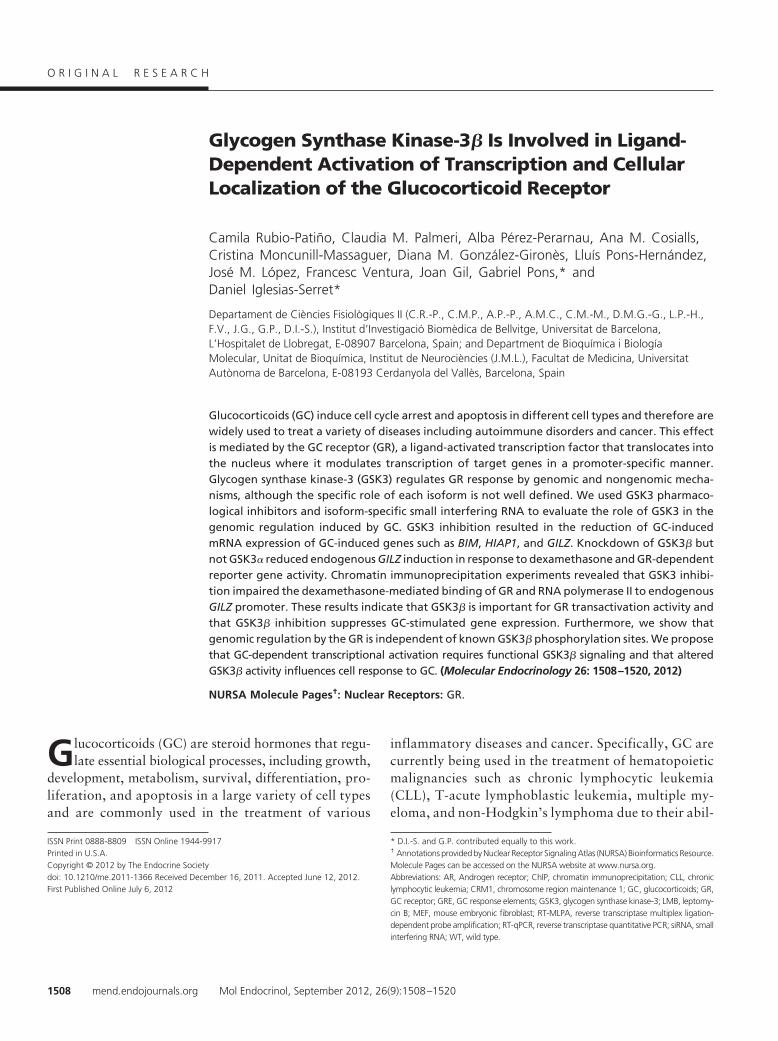
Glycogen Synthase Kinase-3� Is Involved in Ligand-Dependent Activation of Transcription and CellularLocalization of the Glucocorticoid Receptor
Camila Rubio-Patiño, Claudia M. Palmeri, Alba Pérez-Perarnau, Ana M. Cosialls,Cristina Moncunill-Massaguer, Diana M. González-Gironès, Lluís Pons-Hernández,José M. López, Francesc Ventura, Joan Gil, Gabriel Pons,* andDaniel Iglesias-Serret*
Departament de Ciències Fisiològiques II (C.R.-P., C.M.P., A.P.-P., A.M.C., C.M.-M., D.M.G.-G., L.P.-H.,F.V., J.G., G.P., D.I.-S.), Institut d’Investigació Biomèdica de Bellvitge, Universitat de Barcelona,L’Hospitalet de Llobregat, E-08907 Barcelona, Spain; and Department de Bioquímica i BiologíaMolecular, Unitat de Bioquímica, Institut de Neurociències (J.M.L.), Facultat de Medicina, UniversitatAutònoma de Barcelona, E-08193 Cerdanyola del Vallès, Barcelona, Spain
Glucocorticoids (GC) induce cell cycle arrest and apoptosis in different cell types and therefore arewidely used to treat a variety of diseases including autoimmune disorders and cancer. This effectis mediated by the GC receptor (GR), a ligand-activated transcription factor that translocates intothe nucleus where it modulates transcription of target genes in a promoter-specific manner.Glycogen synthase kinase-3 (GSK3) regulates GR response by genomic and nongenomic mecha-nisms, although the specific role of each isoform is not well defined. We used GSK3 pharmaco-logical inhibitors and isoform-specific small interfering RNA to evaluate the role of GSK3 in thegenomic regulation induced by GC. GSK3 inhibition resulted in the reduction of GC-inducedmRNA expression of GC-induced genes such as BIM, HIAP1, and GILZ. Knockdown of GSK3� butnot GSK3� reduced endogenous GILZ induction in response to dexamethasone and GR-dependentreporter gene activity. Chromatin immunoprecipitation experiments revealed that GSK3 inhibi-tion impaired the dexamethasone-mediated binding of GR and RNA polymerase II to endogenousGILZ promoter. These results indicate that GSK3� is important for GR transactivation activity andthat GSK3� inhibition suppresses GC-stimulated gene expression. Furthermore, we show thatgenomic regulation by the GR is independent of known GSK3� phosphorylation sites. We proposethat GC-dependent transcriptional activation requires functional GSK3� signaling and that alteredGSK3� activity influences cell response to GC. (Molecular Endocrinology 26: 1508–1520, 2012)
NURSA Molecule Pages†: Nuclear Receptors: GR.
Glucocorticoids (GC) are steroid hormones that regu-late essential biological processes, including growth,
development, metabolism, survival, differentiation, pro-liferation, and apoptosis in a large variety of cell typesand are commonly used in the treatment of various
inflammatory diseases and cancer. Specifically, GC arecurrently being used in the treatment of hematopoieticmalignancies such as chronic lymphocytic leukemia(CLL), T-acute lymphoblastic leukemia, multiple my-eloma, and non-Hodgkin’s lymphoma due to their abil-
ISSN Print 0888-8809 ISSN Online 1944-9917Printed in U.S.A.Copyright © 2012 by The Endocrine Societydoi: 10.1210/me.2011-1366 Received December 16, 2011. Accepted June 12, 2012.First Published Online July 6, 2012
* D.I.-S. and G.P. contributed equally to this work.† Annotations providedbyNuclear Receptor SignalingAtlas (NURSA) Bioinformatics Resource.Molecule Pages can be accessed on the NURSA website at www.nursa.org.Abbreviations: AR, Androgen receptor; ChIP, chromatin immunoprecipitation; CLL, chroniclymphocytic leukemia; CRM1, chromosome region maintenance 1; GC, glucocorticoids; GR,GC receptor; GRE, GC response elements; GSK3, glycogen synthase kinase-3; LMB, leptomy-cin B; MEF, mouse embryonic fibroblast; RT-MLPA, reverse transcriptase multiplex ligation-dependent probe amplification; RT-qPCR, reverse transcriptase quantitative PCR; siRNA, smallinterfering RNA; WT, wild type.
O R I G I N A L R E S E A R C H
1508 mend.endojournals.org Mol Endocrinol, September 2012, 26(9):1508–1520

ity to induce intrinsic caspase-dependent apoptosis inthese cell types (1).
Most of the actions of GC are mediated through theGC receptor (GR), a member of the steroid receptor su-perfamily (2). The unliganded GR resides primarily in thecytoplasm in an inactive state as part of a large heat-shockprotein heterocomplex that includes various chaperoneproteins, such as heat-shock protein 90 (3). Upon GCbinding, the GR undergoes a conformational change thatresults in its dissociation from the cytoplasmic chaperonemultiprotein complex and unmasking of the nuclear lo-calization signal, leading to its translocation to the nu-cleus. Once in the nucleus, the dimerized GR binds GCresponse elements (GRE), usually located in the promoterof GR-regulated genes, resulting in gene transactivationor transrepression (4).
It has been shown that the modulation of the GR phos-phorylation cycle by phosphatases maintains steady-statereceptor phosphorylation at a low basal level in the ab-sence of ligand, and GC-dependent GR phosphorylationaffects GR target gene expression (5). Previous studieshave highlighted the involvement of different protein ki-nases in GC-mediated effects (6). Recently, a protein ki-nase screening in lymphoid cells showed that glycogensynthase kinase-3 (GSK3) has a role in GC-induced apo-ptosis (7). Pharmacological inhibition of GSK3 blockedGC-induced apoptosis in different hematopoietic celllines (7), and attenuated GC-induced up-regulation ofBIM (8), a Bcl-2 homology domain-3-only protein in-volved in GC-induced apoptosis in leukemia cells (9–11).
GSK3 is a serine/threonine protein kinase highly con-served from yeast to mammals (12–14). It was initiallyidentified as a key regulator of insulin-dependent glyco-gen synthesis, but it has been demonstrated that GSK3 isa multifunctional kinase involved in cellular metabolism,signaling transduction, growth, differentiation, and cellfate determination (13). There are two homologous mam-malian GSK3 isoforms encoded by different genes,GSK3� and GSK3�. They share 98% identity withintheir catalytic domain, but N- and C-terminal sequencesdiverge, making them structurally similar but not func-tionally identical (13, 15).
GSK3 demonstrates a preference for prephosphory-lated (primed) substrates by different priming kinases(12–14). GSK3� phosphorylates different substrates, in-cluding glycogen synthase, and transcription factors suchas c-myc, �-catenin, and Tau-microtubule-associatedprotein (12). There is a hormone-dependent GR phos-phorylation on human serine 404 (Ser404) by GSK3�,which plays an important role in GR protein stability andregulates GR-dependent gene expression (6). Addition-ally, GSK3�-mediated phosphorylation of rat GR threo-
nine 171 (Thr171) has been described (16). Different in-teractions between GSK3 and the GR have beenpreviously described. In the absence of a ligand, GSK3� isbound to the GR, and exposure to GC leads to its disso-ciation from the GR (7). Moreover, it has been describedthat the GR associates with GSK3� in the presence ofdexamethasone but not with GSK3� (6). Thus, it seemsthat GSK3 isoforms regulate GR cellular response by us-ing different mechanisms.
In the present study, we have used pharmacologicalinhibitors and GSK3 isoform-specific small interferingRNA (siRNA) to analyze the role of GSK3 isoforms in theregulation of GR-mediated transcriptional activation.
Materials and Methods
CLL samples and cell isolationBlood samples from CLL patients were obtained from the
Hospital de Bellvitge, L’Hospitalet de Llobregat, Spain. CLLwas diagnosed according to standard clinical and laboratorycriteria. Written informed consent was obtained from all pa-tients in accordance with the Hospital de Bellvitge Ethical Com-mittee. Peripheral blood mononuclear cells were isolated by cen-trifugation on a Ficoll-Hypaque (Seromed, Berlin, Germany)gradient. Human lymphocytes were cultured immediately afterthawing or isolation at a concentration of 0.5–3 � 106 cells/mlin RPMI 1640 culture medium (Biological Industries) supple-mented with 10% heat-inactivated fetal bovine serum (Biolog-ical Industries, Kibbutz Beit Haemek, Israel), 2 mM L-glutamine,100 U/ml penicillin, and 100 �g/ml streptomycin at 37 C in ahumidified atmosphere containing 5% carbon dioxide.
Cell cultureJurkat GR wild-type (WT) cells, derived from a parental cell
line of Jurkat T-acute lymphoblastic leukemia cells harboring anonfunctional GR were generated by expressing the rat GRunder control of the �-actin promoter (17, 18). These cells werekindly provided by Dr. Carme Caelles (Institute for Research inBiomedicine, Universitat de Barcelona, Barcelona, Spain). Pa-rental Jurkat, Jurkat GR WT, and BxPC-3 cells were grown inRPMI 1640 medium (Biological Industries), 2 mM L-glutamine,100 �g/ml penicillin, and 100 mg/ml streptomycin at 37 C in ahumidified atmosphere at 5% carbon dioxide. WT, GSK3��/�,and GSK3��/� mouse embryonic fibroblast (MEF) cells werekindly given by Dr. J. Woodgett (Samuel Lunenfeld ResearchInstitute, Toronto, Canada). HeLa, MC3T3, and MCF-7 cellsand WT, GSK3��/�, and GSK3��/� MEF cells were main-tained in DMEM (Biological Industries) containing 10% fetalbovine serum (Biological Industries).
Plasmids and reagentsDexamethasone and SB216763 were purchased from Sigma-
Aldrich (St. Louis, MO). Akt inhibitor VIII, SB203580, U0126,LY294002, bisindolylmaleimide I, and rapamycin were pur-chased from Calbiochem (La Jolla, CA). SP600125,GSK650394, and KU0063794 were from Tocris Bioscience
Mol Endocrinol, September 2012, 26(9):1508–1520 mend.endojournals.org 1509
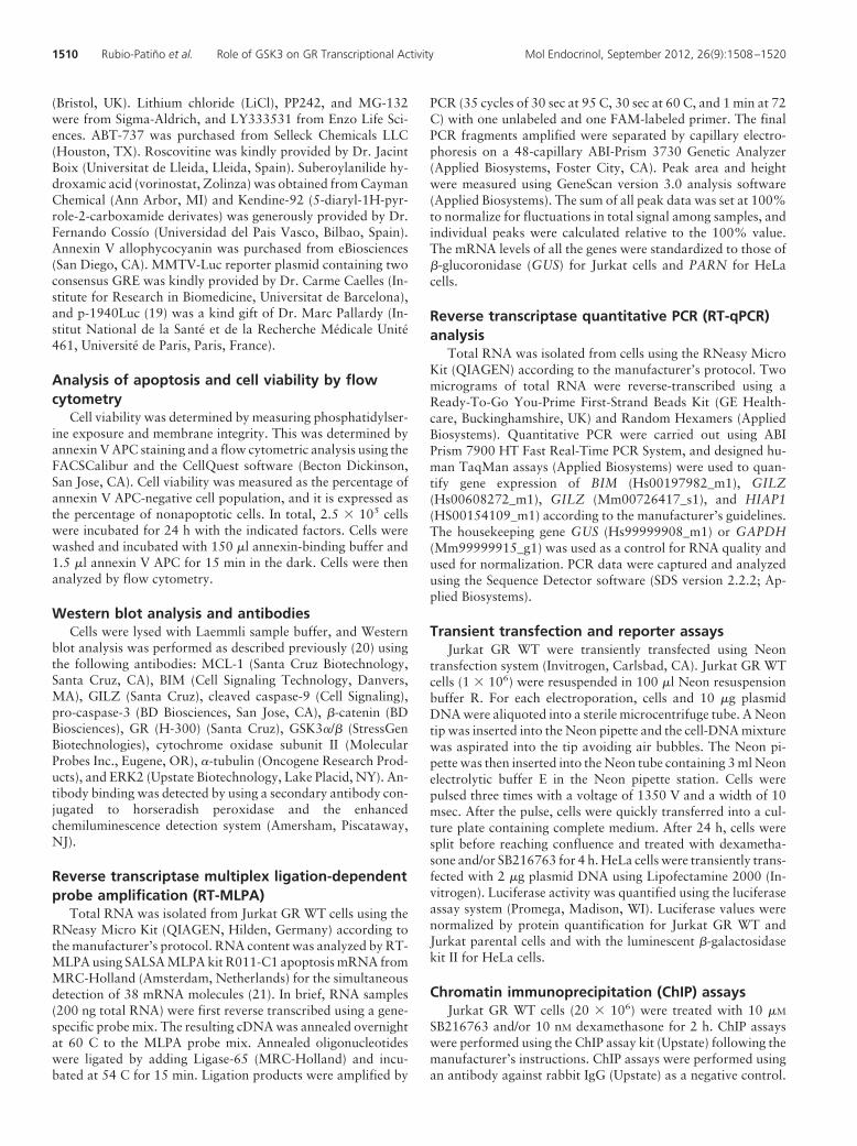
(Bristol, UK). Lithium chloride (LiCl), PP242, and MG-132were from Sigma-Aldrich, and LY333531 from Enzo Life Sci-ences. ABT-737 was purchased from Selleck Chemicals LLC(Houston, TX). Roscovitine was kindly provided by Dr. JacintBoix (Universitat de Lleida, Lleida, Spain). Suberoylanilide hy-droxamic acid (vorinostat, Zolinza) was obtained from CaymanChemical (Ann Arbor, MI) and Kendine-92 (5-diaryl-1H-pyr-role-2-carboxamide derivates) was generously provided by Dr.Fernando Cossío (Universidad del Pais Vasco, Bilbao, Spain).Annexin V allophycocyanin was purchased from eBiosciences(San Diego, CA). MMTV-Luc reporter plasmid containing twoconsensus GRE was kindly provided by Dr. Carme Caelles (In-stitute for Research in Biomedicine, Universitat de Barcelona),and p-1940Luc (19) was a kind gift of Dr. Marc Pallardy (In-stitut National de la Santé et de la Recherche Médicale Unité461, Université de Paris, Paris, France).
Analysis of apoptosis and cell viability by flowcytometry
Cell viability was determined by measuring phosphatidylser-ine exposure and membrane integrity. This was determined byannexin V APC staining and a flow cytometric analysis using theFACSCalibur and the CellQuest software (Becton Dickinson,San Jose, CA). Cell viability was measured as the percentage ofannexin V APC-negative cell population, and it is expressed asthe percentage of nonapoptotic cells. In total, 2.5 � 105 cellswere incubated for 24 h with the indicated factors. Cells werewashed and incubated with 150 �l annexin-binding buffer and1.5 �l annexin V APC for 15 min in the dark. Cells were thenanalyzed by flow cytometry.
Western blot analysis and antibodiesCells were lysed with Laemmli sample buffer, and Western
blot analysis was performed as described previously (20) usingthe following antibodies: MCL-1 (Santa Cruz Biotechnology,Santa Cruz, CA), BIM (Cell Signaling Technology, Danvers,MA), GILZ (Santa Cruz), cleaved caspase-9 (Cell Signaling),pro-caspase-3 (BD Biosciences, San Jose, CA), �-catenin (BDBiosciences), GR (H-300) (Santa Cruz), GSK3�/� (StressGenBiotechnologies), cytochrome oxidase subunit II (MolecularProbes Inc., Eugene, OR), �-tubulin (Oncogene Research Prod-ucts), and ERK2 (Upstate Biotechnology, Lake Placid, NY). An-tibody binding was detected by using a secondary antibody con-jugated to horseradish peroxidase and the enhancedchemiluminescence detection system (Amersham, Piscataway,NJ).
Reverse transcriptase multiplex ligation-dependentprobe amplification (RT-MLPA)
Total RNA was isolated from Jurkat GR WT cells using theRNeasy Micro Kit (QIAGEN, Hilden, Germany) according tothe manufacturer’s protocol. RNA content was analyzed by RT-MLPA using SALSA MLPA kit R011-C1 apoptosis mRNA fromMRC-Holland (Amsterdam, Netherlands) for the simultaneousdetection of 38 mRNA molecules (21). In brief, RNA samples(200 ng total RNA) were first reverse transcribed using a gene-specific probe mix. The resulting cDNA was annealed overnightat 60 C to the MLPA probe mix. Annealed oligonucleotideswere ligated by adding Ligase-65 (MRC-Holland) and incu-bated at 54 C for 15 min. Ligation products were amplified by
PCR (35 cycles of 30 sec at 95 C, 30 sec at 60 C, and 1 min at 72C) with one unlabeled and one FAM-labeled primer. The finalPCR fragments amplified were separated by capillary electro-phoresis on a 48-capillary ABI-Prism 3730 Genetic Analyzer(Applied Biosystems, Foster City, CA). Peak area and heightwere measured using GeneScan version 3.0 analysis software(Applied Biosystems). The sum of all peak data was set at 100%to normalize for fluctuations in total signal among samples, andindividual peaks were calculated relative to the 100% value.The mRNA levels of all the genes were standardized to those of�-glucoronidase (GUS) for Jurkat cells and PARN for HeLacells.
Reverse transcriptase quantitative PCR (RT-qPCR)analysis
Total RNA was isolated from cells using the RNeasy MicroKit (QIAGEN) according to the manufacturer’s protocol. Twomicrograms of total RNA were reverse-transcribed using aReady-To-Go You-Prime First-Strand Beads Kit (GE Health-care, Buckinghamshire, UK) and Random Hexamers (AppliedBiosystems). Quantitative PCR were carried out using ABIPrism 7900 HT Fast Real-Time PCR System, and designed hu-man TaqMan assays (Applied Biosystems) were used to quan-tify gene expression of BIM (Hs00197982_m1), GILZ(Hs00608272_m1), GILZ (Mm00726417_s1), and HIAP1(HS00154109_m1) according to the manufacturer’s guidelines.The housekeeping gene GUS (Hs99999908_m1) or GAPDH(Mm99999915_g1) was used as a control for RNA quality andused for normalization. PCR data were captured and analyzedusing the Sequence Detector software (SDS version 2.2.2; Ap-plied Biosystems).
Transient transfection and reporter assaysJurkat GR WT were transiently transfected using Neon
transfection system (Invitrogen, Carlsbad, CA). Jurkat GR WTcells (1 � 106) were resuspended in 100 �l Neon resuspensionbuffer R. For each electroporation, cells and 10 �g plasmidDNA were aliquoted into a sterile microcentrifuge tube. A Neontip was inserted into the Neon pipette and the cell-DNA mixturewas aspirated into the tip avoiding air bubbles. The Neon pi-pette was then inserted into the Neon tube containing 3 ml Neonelectrolytic buffer E in the Neon pipette station. Cells werepulsed three times with a voltage of 1350 V and a width of 10msec. After the pulse, cells were quickly transferred into a cul-ture plate containing complete medium. After 24 h, cells weresplit before reaching confluence and treated with dexametha-sone and/or SB216763 for 4 h. HeLa cells were transiently trans-fected with 2 �g plasmid DNA using Lipofectamine 2000 (In-vitrogen). Luciferase activity was quantified using the luciferaseassay system (Promega, Madison, WI). Luciferase values werenormalized by protein quantification for Jurkat GR WT andJurkat parental cells and with the luminescent �-galactosidasekit II for HeLa cells.
Chromatin immunoprecipitation (ChIP) assaysJurkat GR WT cells (20 � 106) were treated with 10 �M
SB216763 and/or 10 nM dexamethasone for 2 h. ChIP assayswere performed using the ChIP assay kit (Upstate) following themanufacturer’s instructions. ChIP assays were performed usingan antibody against rabbit IgG (Upstate) as a negative control.
1510 Rubio-Patiño et al. Role of GSK3 on GR Transcriptional Activity Mol Endocrinol, September 2012, 26(9):1508–1520
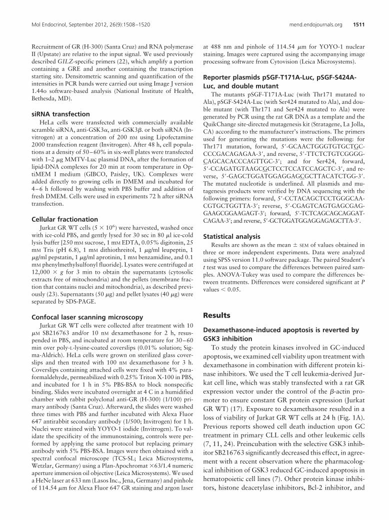
Recruitment of GR (H-300) (Santa Cruz) and RNA polymeraseII (Upstate) are relative to the input signal. We used previouslydescribed GILZ-specific primers (22), which amplify a portioncontaining a GRE and another containing the transcriptionstarting site. Densitometric scanning and quantification of theintensities in PCR bands were carried out using Image J version1.44o software-based analysis (National Institute of Health,Bethesda, MD).
siRNA transfectionHeLa cells were transfected with commercially available
scramble siRNA, anti-GSK3�, anti-GSK3�. or both siRNA (In-vitrogen) at a concentration of 200 nM using Lipofectamine2000 transfection reagent (Invitrogen). After 48 h, cell popula-tions at a density of 50–60% in six-well plates were transfectedwith 1–2 �g MMTV-Luc plasmid DNA, after the formation oflipid-DNA complexes for 20 min at room temperature in Op-tiMEM I medium (GIBCO, Paisley, UK). Complexes wereadded directly to growing cells in DMEM and incubated for4–6 h followed by washing with PBS buffer and addition offresh DMEM. Cells were used in experiments 72 h after siRNAtransfection.
Cellular fractionationJurkat GR WT cells (5 � 106) were harvested, washed once
with ice-cold PBS, and gently lysed for 30 sec in 80 �l ice-coldlysis buffer [250 mM sucrose, 1 mM EDTA, 0.05% digitonin, 25mM Tris (pH 6.8), 1 mM dithiothreitol, 1 �g/ml leupeptin, 1�g/ml pepstatin, 1 �g/ml aprotinin, 1 mM benzamidine, and 0.1mM phenylmethylsulfonyl fluoride]. Lysates were centrifuged at12,000 � g for 3 min to obtain the supernatants (cytosolicextracts free of mitochondria) and the pellets (membrane frac-tion that contains nuclei and mitochondria), as described previ-ously (23). Supernatants (50 �g) and pellet lysates (40 �g) wereseparated by SDS-PAGE.
Confocal laser scanning microscopyJurkat GR WT cells were collected after treatment with 10
�M SB216763 and/or 10 nM dexamethasone for 2 h, resus-pended in PBS, and incubated at room temperature for 30–60min over poly-L-lysine-coated coverslips (0.01% solution; Sig-ma-Aldrich). HeLa cells were grown on sterilized glass cover-slips and then treated with 100 nM dexamethasone for 3 h.Coverslips containing attached cells were fixed with 4% para-formaldehyde, permeabilized with 0.25% Triton X-100 in PBS,and incubated for 1 h in 5% PBS-BSA to block nonspecificbinding. Slides were incubated overnight at 4 C in a humidifiedchamber with rabbit polyclonal anti-GR (H-300) (1/100) pri-mary antibody (Santa Cruz). Afterward, the slides were washedthree times with PBS and further incubated with Alexa Fluor647 antirabbit secondary antibody (1/500; Invitrogen) for 1 h.Nuclei were stained with YOYO-1 iodide (Invitrogen). To val-idate the specificity of the immunostaining, controls were per-formed by applying the same protocol but replacing primaryantibody with 5% PBS-BSA. Images were then obtained with aspectral confocal microscope (TCS-SL; Leica Microsystems,Wetzlar, Germany) using a Plan-Apochromat �63/1.4 numericaperture immersion oil objective (Leica Microsystems). We useda HeNe laser at 633 nm (Lasos Inc., Jena, Germany) and pinholeof 114.54 �m for Alexa Fluor 647 GR staining and argon laser
at 488 nm and pinhole of 114.54 �m for YOYO-1 nuclearstaining. Images were captured using the accompanying imageprocessing software from Cytovision (Leica Microsystems).
Reporter plasmids pSGF-T171A-Luc, pSGF-S424A-Luc, and double mutant
The mutants pSGF-T171A-Luc (with Thr171 mutated toAla), pSGF-S424A-Luc (with Ser424 mutated to Ala), and dou-ble mutant (with Thr171 and Ser424 mutated to Ala) weregenerated by PCR using the rat GR DNA as a template and theQuikChange site-directed mutagenesis kit (Stratagene, La Jolla,CA) according to the manufacturer’s instructions. The primersused for generating the mutations were the following: forThr171 mutation, forward, 5�-GCAACTGGGTGTGCTGC-CCCGACAGAGAA-3�, and reverse, 5�-TTCTCTGTCGGGG-CAGCACACCCAGTTGC-3�; and for Ser424, forward,5�-CCAGATGTAAGCGCTCCTCCATCCAGCTC-3�, and re-verse, 5�-GAGCTGGATGGAGGAGCGCTTACATCTGG-3�.The mutated nucleotide is underlined. All plasmids and mu-tagenesis products were verified by DNA sequencing with thefollowing primers: forward, 5�-CCTACAGCTCCTGGGCAA-CGTGCTGGTTA-3�; reverse, 5�-CGAGTCAGTGAGCGAG-GAAGCGGAAGAGT-3�; forward, 5�-TCTCAGCAGCAGGAT-CAGAA-3�; and reverse, 5�-GCTGGATGGAGGAGAGCTTA-3�.
Statistical analysisResults are shown as the mean � SEM of values obtained in
three or more independent experiments. Data were analyzedusing SPSS version 11.0 software package. The paired Student’st test was used to compare the differences between paired sam-ples. ANOVA-Tukey was used to compare the differences be-tween treatments. Differences were considered significant at Pvalues � 0.05.
Results
Dexamethasone-induced apoptosis is reverted byGSK3 inhibition
To study the protein kinases involved in GC-inducedapoptosis, we examined cell viability upon treatment withdexamethasone in combination with different protein ki-nase inhibitors. We used the T cell leukemia-derived Jur-kat cell line, which was stably transfected with a rat GRexpression vector under the control of the �-actin pro-moter to ensure constant GR protein expression (JurkatGR WT) (17). Exposure to dexamethasone resulted in aloss of viability of Jurkat GR WT cells at 24 h (Fig. 1A).Previous reports showed cell death induction upon GCtreatment in primary CLL cells and other leukemic cells(7, 11, 24). Preincubation with the selective GSK3 inhib-itor SB216763 significantly decreased this effect, in agree-ment with a recent observation where the pharmacolog-ical inhibition of GSK3 reduced GC-induced apoptosis inhematopoietic cell lines (7). Other protein kinase inhibi-tors, histone deacetylase inhibitors, Bcl-2 inhibitor, and
Mol Endocrinol, September 2012, 26(9):1508–1520 mend.endojournals.org 1511
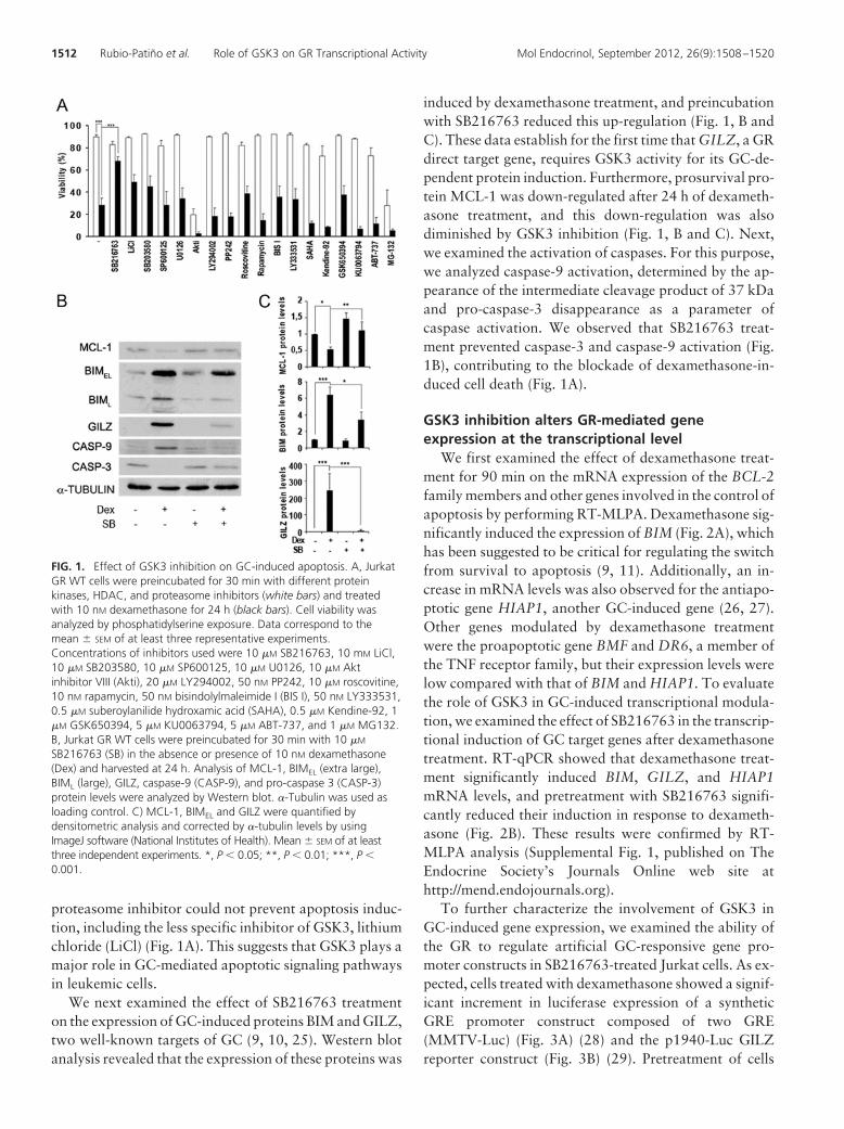
proteasome inhibitor could not prevent apoptosis induc-tion, including the less specific inhibitor of GSK3, lithiumchloride (LiCl) (Fig. 1A). This suggests that GSK3 plays amajor role in GC-mediated apoptotic signaling pathwaysin leukemic cells.
We next examined the effect of SB216763 treatmenton the expression of GC-induced proteins BIM and GILZ,two well-known targets of GC (9, 10, 25). Western blotanalysis revealed that the expression of these proteins was
induced by dexamethasone treatment, and preincubationwith SB216763 reduced this up-regulation (Fig. 1, B andC). These data establish for the first time that GILZ, a GRdirect target gene, requires GSK3 activity for its GC-de-pendent protein induction. Furthermore, prosurvival pro-tein MCL-1 was down-regulated after 24 h of dexameth-asone treatment, and this down-regulation was alsodiminished by GSK3 inhibition (Fig. 1, B and C). Next,we examined the activation of caspases. For this purpose,we analyzed caspase-9 activation, determined by the ap-pearance of the intermediate cleavage product of 37 kDaand pro-caspase-3 disappearance as a parameter ofcaspase activation. We observed that SB216763 treat-ment prevented caspase-3 and caspase-9 activation (Fig.1B), contributing to the blockade of dexamethasone-in-duced cell death (Fig. 1A).
GSK3 inhibition alters GR-mediated geneexpression at the transcriptional level
We first examined the effect of dexamethasone treat-ment for 90 min on the mRNA expression of the BCL-2family members and other genes involved in the control ofapoptosis by performing RT-MLPA. Dexamethasone sig-nificantly induced the expression of BIM (Fig. 2A), whichhas been suggested to be critical for regulating the switchfrom survival to apoptosis (9, 11). Additionally, an in-crease in mRNA levels was also observed for the antiapo-ptotic gene HIAP1, another GC-induced gene (26, 27).Other genes modulated by dexamethasone treatmentwere the proapoptotic gene BMF and DR6, a member ofthe TNF receptor family, but their expression levels werelow compared with that of BIM and HIAP1. To evaluatethe role of GSK3 in GC-induced transcriptional modula-tion, we examined the effect of SB216763 in the transcrip-tional induction of GC target genes after dexamethasonetreatment. RT-qPCR showed that dexamethasone treat-ment significantly induced BIM, GILZ, and HIAP1mRNA levels, and pretreatment with SB216763 signifi-cantly reduced their induction in response to dexameth-asone (Fig. 2B). These results were confirmed by RT-MLPA analysis (Supplemental Fig. 1, published on TheEndocrine Society’s Journals Online web site athttp://mend.endojournals.org).
To further characterize the involvement of GSK3 inGC-induced gene expression, we examined the ability ofthe GR to regulate artificial GC-responsive gene pro-moter constructs in SB216763-treated Jurkat cells. As ex-pected, cells treated with dexamethasone showed a signif-icant increment in luciferase expression of a syntheticGRE promoter construct composed of two GRE(MMTV-Luc) (Fig. 3A) (28) and the p1940-Luc GILZreporter construct (Fig. 3B) (29). Pretreatment of cells
FIG. 1. Effect of GSK3 inhibition on GC-induced apoptosis. A, JurkatGR WT cells were preincubated for 30 min with different proteinkinases, HDAC, and proteasome inhibitors (white bars) and treatedwith 10 nM dexamethasone for 24 h (black bars). Cell viability wasanalyzed by phosphatidylserine exposure. Data correspond to themean � SEM of at least three representative experiments.Concentrations of inhibitors used were 10 �M SB216763, 10 mM LiCl,10 �M SB203580, 10 �M SP600125, 10 �M U0126, 10 �M Aktinhibitor VIII (Akti), 20 �M LY294002, 50 nM PP242, 10 �M roscovitine,10 nM rapamycin, 50 nM bisindolylmaleimide I (BIS I), 50 nM LY333531,0.5 �M suberoylanilide hydroxamic acid (SAHA), 0.5 �M Kendine-92, 1�M GSK650394, 5 �M KU0063794, 5 �M ABT-737, and 1 �M MG132.B, Jurkat GR WT cells were preincubated for 30 min with 10 �MSB216763 (SB) in the absence or presence of 10 nM dexamethasone(Dex) and harvested at 24 h. Analysis of MCL-1, BIMEL (extra large),BIML (large), GILZ, caspase-9 (CASP-9), and pro-caspase 3 (CASP-3)protein levels were analyzed by Western blot. �-Tubulin was used asloading control. C) MCL-1, BIMEL and GILZ were quantified bydensitometric analysis and corrected by �-tubulin levels by usingImageJ software (National Institutes of Health). Mean� SEM of at leastthree independent experiments. *, P � 0.05; **, P � 0.01; ***, P �0.001.
1512 Rubio-Patiño et al. Role of GSK3 on GR Transcriptional Activity Mol Endocrinol, September 2012, 26(9):1508–1520

with SB216763 significantly decreased the response toGC as compared with cells treated with dexamethasonealone, especially in the case of MMTV-Luc, confirmingthe role of GSK3 on dexamethasone-induced GR tran-scriptional activity.
Moreover, we analyzed GILZ mRNA levels in differ-ent cell types. GILZ expression was also induced by GCtreatment in MC3T3 mouse preosteoblast, MCF-7 hu-man breast adenocarcinoma, and BxPC-3 human pancre-atic adenocarcinoma cell lines and significantly revertedby SB216763 pretreatment (Fig. 3C). These results indi-cate that GSK3 is an important determinant in the GRtranscriptional response to GC in a species- and cell type-independent manner.
GSK3 inhibition affects GC-induced apoptosis andGC-dependent gene induction in CLL cells
GC are used in the therapy of lymphoid malignanciesbecause of their remarkable ability to induce apoptosis(30, 31). To study the effect of GSK3 inhibition overGC-induced apoptosis in a primary leukemia model, weemployed B lymphocytes from patients with CLL. As wedescribed previously, dexamethasone significantly in-
duced cell death of CLL cells (24).Moreover, pretreatment with GSK3 in-hibitor SB216763 significantly de-creased dexamethasone-induced apo-ptosis by 11.87% in 19 of the 29patients analyzed (Fig. 4A). Supple-mental Table 1 shows the viability ofthe full cohort of patients analyzed. Wenext analyzed the effect of GSK3 inhi-bition on GC-dependent GILZ andBIM induction. mRNA levels were in-duced in response to dexamethasonetreatment and reverted by GSK3 inhi-bition in the six patients analyzed (Fig.4B). GILZ and BIM protein levels werealso induced in response to GC and re-verted by GSK3 inhibition in nine of 12samples for GILZ and 11 of 13 samplesfor BIM (Fig. 4C). Collectively, thesedata indicate that GSK3 inhibition re-duces sensitivity to GC-induced apo-ptosis in CLL cells and impairs GC-dependent gene and protein induction.
GSK3 inhibition affects GR andRNA polymerase II recruitment tothe GILZ gene promoter
To establish the mechanism forGSK3-mediated GR transcriptionalregulation, Jurkat GR WT cells were
stimulated with dexamethasone for 2 h, and GILZ pro-moter occupancy was evaluated by ChIP analysis. Usingpreviously described primers (22), we observed a signifi-cant increase in binding of the GR to the GILZ promoter(Fig. 5A) as well as increased binding of RNA polymeraseII to the GILZ transcription start site (Fig. 5B) in dexa-methasone-treated cells. Pretreatment with SB216763significantly reduced GR and RNA polymerase II bindingto the GILZ promoter induced by dexamethasone. Takentogether, our results show that GC-dependent transcrip-tional activation requires a functional GSK3 signaling.
GSK3� gene silencing suppresses GC-stimulatedgene expression
GSK3 inhibitors do not distinguish between the twoGSK3 isoforms, so we next evaluated the individual rolesof GSK3� and GSK3� on GR-mediated transcriptionalactivation by gene silencing analysis. Because we wereunable to completely silence GSK3 gene expression inJurkat cells, we performed GSK3� and GSK3� knock-down in HeLa cells. Relative mRNA levels and proteinexpression levels of GSK3� and GSK3� were determined
FIG. 2. GR ability to regulate GC-induced gene expression is affected by GSK3 inhibition. A,RT-MLPA gene expression profile induced by GC. Jurkat GR WT cells were untreated [control(CT)] or treated with 10 nM dexamethasone (Dex) for 90 min. Cells were lysed and theexpression of apoptotic-related genes of the Bcl-2 family, IAP (inhibitors of apoptosis) family,and other genes implicated in apoptosis was analyzed by RT-MLPA as described in Materialsand Methods. The mRNA levels of all the genes were normalized with respect to those ofGUS. B, Jurkat GR WT cells were preincubated with 10 �M SB216763 (SB) in the absence orpresence of 10 nM dexamethasone (Dex) and harvested at 90 min. BIM, GILZ, and HIAP1mRNA were measured by RT-qPCR. The mRNA levels of all genes were normalized withrespect to those of GUS. These results are shown as the mean � SEM of at least threeindependent experiments. *, P � 0.05; **, P � 0.01; ***, P � 0.001 vs. untreated cells.
Mol Endocrinol, September 2012, 26(9):1508–1520 mend.endojournals.org 1513
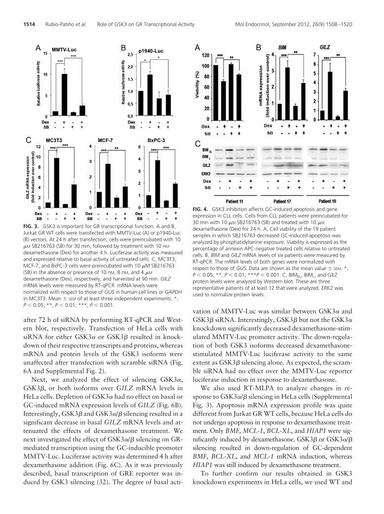
after 72 h of siRNA by performing RT-qPCR and West-ern blot, respectively. Transfection of HeLa cells withsiRNA for either GSK3� or GSK3� resulted in knock-down of their respective transcripts and proteins, whereasmRNA and protein levels of the GSK3 isoforms wereunaffected after transfection with scramble siRNA (Fig.6A and Supplemental Fig. 2).
Next, we analyzed the effect of silencing GSK3�,GSK3�, or both isoforms over GILZ mRNA levels inHeLa cells. Depletion of GSK3� had no effect on basal orGC-induced mRNA expression levels of GILZ (Fig. 6B).Interestingly, GSK3� and GSK3�/� silencing resulted in asignificant decrease in basal GILZ mRNA levels and at-tenuated the effects of dexamethasone treatment. Wenext investigated the effect of GSK3�/� silencing on GR-mediated transcription using the GC-inducible promoterMMTV-Luc. Luciferase activity was determined 4 h afterdexamethasone addition (Fig. 6C). As it was previouslydescribed, basal transcription of GRE reporter was in-duced by GSK3 silencing (32). The degree of basal acti-
vation of MMTV-Luc was similar between GSK3� andGSK3� siRNA. Interestingly, GSK3� but not the GSK3�
knockdown significantly decreased dexamethasone-stim-ulated MMTV-Luc promoter activity. The down-regula-tion of both GSK3 isoforms decreased dexamethasone-stimulated MMTV-Luc luciferase activity to the sameextent as GSK3� silencing alone. As expected, the scram-ble siRNA had no effect over the MMTV-Luc reporterluciferase induction in response to dexamethasone.
We also used RT-MLPA to analyze changes in re-sponse to GSK3�/� silencing in HeLa cells (SupplementalFig. 3). Apoptosis mRNA expression profile was quitedifferent from Jurkat GR WT cells, because HeLa cells donot undergo apoptosis in response to dexamethasone treat-ment. Only BMF, MCL-1, BCL-XL, and HIAP1 were sig-nificantly induced by dexamethasone. GSK3� or GSK3�/�silencing resulted in down-regulation of GC-dependentBMF, BCL-XL, and MCL-1 mRNA induction, whereasHIAP1 was still induced by dexamethasone treatment.
To further confirm our results obtained in GSK3knockdown experiments in HeLa cells, we used WT and
FIG. 3. GSK3 is important for GR transcriptional function. A and B,Jurkat GR WT cells were transfected with MMTV-Luc (A) or p1940-Luc(B) vectors. At 24 h after transfection, cells were preincubated with 10�M SB216763 (SB) for 30 min, followed by treatment with 10 nMdexamethasone (Dex) for another 4 h. Luciferase activity was measuredand expressed relative to basal activity of untreated cells. C, MC3T3,MCF-7, and BxPC-3 cells were preincubated with 10 �M SB216763(SB) in the absence or presence of 10 nM, 8 nM, and 4 �Mdexamethasone (Dex), respectively, and harvested at 90 min. GILZmRNA levels were measured by RT-qPCR. mRNA levels werenormalized with respect to those of GUS in human cell lines or GAPDHin MC3T3. Mean � SEM of at least three independent experiments. *,P � 0.05; **, P � 0.01; ***, P � 0.001.
FIG. 4. GSK3 inhibition affects GC-induced apoptosis and geneexpression in CLL cells. Cells from CLL patients were preincubated for30 min with 10 �M SB216763 (SB) and treated with 10 �Mdexamethasone (Dex) for 24 h. A, Cell viability of the 19 patientsamples in which SB216763 decreased GC-induced apoptosis wasanalyzed by phosphatidylserine exposure. Viability is expressed as thepercentage of annexin APC-negative treated cells relative to untreatedcells. B, BIM and GILZ mRNA levels of six patients were measured byRT-qPCR. The mRNA levels of both genes were normalized withrespect to those of GUS. Data are shown as the mean value � SEM. *,P � 0.05; **, P � 0.01; ***P � 0.001. C, BIMEL, BIML, and GILZprotein levels were analyzed by Western blot. These are threerepresentative patients of at least 12 that were analyzed. ERK2 wasused to normalize protein levels.
1514 Rubio-Patiño et al. Role of GSK3 on GR Transcriptional Activity Mol Endocrinol, September 2012, 26(9):1508–1520

GSK3�- or GSK3�-null MEF cells to analyze endogenousGILZ mRNA induction (Fig. 6D) in response to GC treat-ment. Moreover, we found that MEF cells deficient forGSK3� expression did not induce GILZ mRNA levels inresponse to dexamethasone, whereas WT and GSK3�-null MEF cells were able to significantly induce GILZmRNA upon GC treatment. Additionally, we showedthat MEF cells deficient for GSK3� did not exhibit dexa-methasone-induced luciferase activity, whereas WT andGSK3�-null MEF cells significantly induced MMTV-Lucpromoter activity in response to dexamethasone (Supple-mental Fig. 4). On the other hand, RT-MLPA analysisshowed no significant changes in MEF cells gene expres-sion profile in response to GC, even though there weredifferences in basal gene expression between cell lines(Supplemental Fig. 5). Together, these results show thatGSK3� activity, but not GSK3�, is required for the tran-scriptional GR-mediated activity.
GSK3 inhibition affects GR cellular distribution inresponse to GC
To examine the role of GSK3 in the regulation of GRsubcellular localization, we analyzed cytosolic and mem-brane (which contains nuclei and mitochondria) fractionsfrom Jurkat GR WT cells. In nonstimulated cells, the GRwas detected mostly in the cytosolic fractions, whereasdexamethasone treatment induced its translocation to themembrane fractions (Fig. 7A). Dexamethasone-depen-
dent GR nuclear protein localization was significantlyreduced by SB216763 (Fig. 7, A and B). GSK3� andGSK3� levels were mainly observed in the cytoplasm andthe nucleus, respectively. Under SB216763 treatment,GSK3� levels were reduced in the nucleus, and there wasan increase in �-catenin protein levels in the membranefraction, confirming GSK3 inhibition.
Taking into account that chromosome region mainte-nance 1 (CRM1) exportin has been suggested to play apivotal role in the early nuclear export of the GR (33), wewanted to determine whether the partial reduction of theGR nuclear translocation by SB216763 in dexametha-sone-treated cells is a result of a CRM1-dependent ex-port. For this purpose, nuclear translocation experimentswere performed in the presence of leptomycin B (LMB),which blocks specifically the CRM1-dependent nuclearexport. A significant increase in nuclear GR localizationwas observed when cells were treated with LMB, suggest-ing that there might be a CRM1-dependent nuclear ex-port of the GR in response to GSK3 inhibition (Fig. 7, Aand B).
We further confirmed the subcellular localization ofthe GR through immunofluorescent staining visualizedby confocal microscopy. In control Jurkat GR WT cells,specific staining of GR was mainly observed in the cyto-plasmic compartment (Fig. 7C). As expected, treatmentwith dexamethasone for 2 h induced GR translocation tothe nuclear compartment predominantly to regions whereeuchromatin was present. Interestingly, when cells werepreincubated with SB216763 in combination with dexa-methasone, a decrease in the GR protein levels in thenucleus was observed compared with dexamethasonetreatment alone. Altogether, these results indicate thatGSK3 inhibition is able to affect GR protein localizationand decreases early GR nuclear levels in dexamethasone-treated Jurkat GR WT cells, affecting GC-stimulated geneexpression.
Next, we analyzed the contribution of GSK3� andGSK3� to GR subcellular distribution induced by dexa-methasone using isoform-specific knockdown in HeLacells. Treatment with dexamethasone always resulted inGR translocation to the nucleus even when GSK3� orGSK3�/� was silenced. Moreover, depletion of GSK3� orGSK3�/� proteins by siRNA led to an increase in generalGR expression levels (Supplemental Fig. 6). These resultsare also in agreement with a previous report where mu-tation of human Ser404 (a residue phosphorylated byGSK3�) leads to the inability of the GR to exit the nu-cleus, making it inaccessible to the proteasome degrada-tion machinery (6).
FIG. 5. Recruitment of GR and RNA polymerase II to the GILZ gene isaffected by GSK3 inhibition. Jurkat GR WT cells were preincubatedwith 10 �M SB216763 (SB) and treated with 10 nM dexamethasone(Dex) for 2 h. ChIP analysis was performed by incubating DNA-proteincomplexes with antibodies against GR (A) or RNA polymerase II (RNApol II) (B) and IgG as a negative control relative to the input signal.Primers specific for the GILZ promoter used for PCR analysis weredescribed in Materials and Methods. Bars represent average valuesfrom densitometric analysis of the bands obtained in four separateexperiments using ImageJ software (National Institutes of Health).Mean � SEM of at least three independent experiments. *, P � 0.05;**, P � 0.01; ***, P � 0.001.
Mol Endocrinol, September 2012, 26(9):1508–1520 mend.endojournals.org 1515
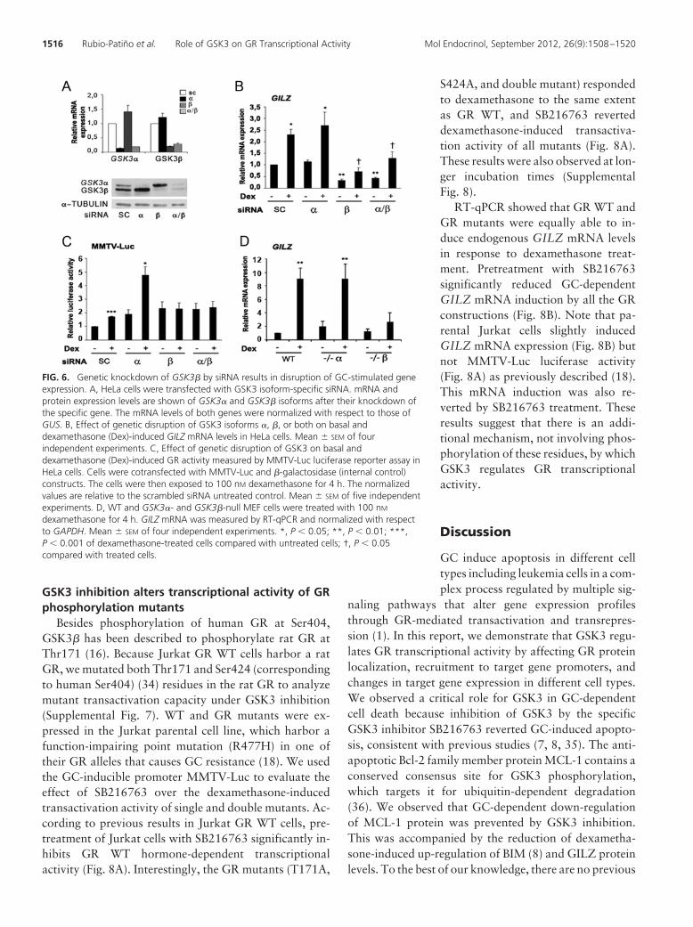
GSK3 inhibition alters transcriptional activity of GRphosphorylation mutants
Besides phosphorylation of human GR at Ser404,GSK3� has been described to phosphorylate rat GR atThr171 (16). Because Jurkat GR WT cells harbor a ratGR, we mutated both Thr171 and Ser424 (correspondingto human Ser404) (34) residues in the rat GR to analyzemutant transactivation capacity under GSK3 inhibition(Supplemental Fig. 7). WT and GR mutants were ex-pressed in the Jurkat parental cell line, which harbor afunction-impairing point mutation (R477H) in one oftheir GR alleles that causes GC resistance (18). We usedthe GC-inducible promoter MMTV-Luc to evaluate theeffect of SB216763 over the dexamethasone-inducedtransactivation activity of single and double mutants. Ac-cording to previous results in Jurkat GR WT cells, pre-treatment of Jurkat cells with SB216763 significantly in-hibits GR WT hormone-dependent transcriptionalactivity (Fig. 8A). Interestingly, the GR mutants (T171A,
S424A, and double mutant) respondedto dexamethasone to the same extentas GR WT, and SB216763 reverteddexamethasone-induced transactiva-tion activity of all mutants (Fig. 8A).These results were also observed at lon-ger incubation times (SupplementalFig. 8).
RT-qPCR showed that GR WT andGR mutants were equally able to in-duce endogenous GILZ mRNA levelsin response to dexamethasone treat-ment. Pretreatment with SB216763significantly reduced GC-dependentGILZ mRNA induction by all the GRconstructions (Fig. 8B). Note that pa-rental Jurkat cells slightly inducedGILZ mRNA expression (Fig. 8B) butnot MMTV-Luc luciferase activity(Fig. 8A) as previously described (18).This mRNA induction was also re-verted by SB216763 treatment. Theseresults suggest that there is an addi-tional mechanism, not involving phos-phorylation of these residues, by whichGSK3 regulates GR transcriptionalactivity.
Discussion
GC induce apoptosis in different celltypes including leukemia cells in a com-plex process regulated by multiple sig-
naling pathways that alter gene expression profilesthrough GR-mediated transactivation and transrepres-sion (1). In this report, we demonstrate that GSK3 regu-lates GR transcriptional activity by affecting GR proteinlocalization, recruitment to target gene promoters, andchanges in target gene expression in different cell types.We observed a critical role for GSK3 in GC-dependentcell death because inhibition of GSK3 by the specificGSK3 inhibitor SB216763 reverted GC-induced apopto-sis, consistent with previous studies (7, 8, 35). The anti-apoptotic Bcl-2 family member protein MCL-1 contains aconserved consensus site for GSK3 phosphorylation,which targets it for ubiquitin-dependent degradation(36). We observed that GC-dependent down-regulationof MCL-1 protein was prevented by GSK3 inhibition.This was accompanied by the reduction of dexametha-sone-induced up-regulation of BIM (8) and GILZ proteinlevels. To the best of our knowledge, there are no previous
FIG. 6. Genetic knockdown of GSK3� by siRNA results in disruption of GC-stimulated geneexpression. A, HeLa cells were transfected with GSK3 isoform-specific siRNA. mRNA andprotein expression levels are shown of GSK3� and GSK3� isoforms after their knockdown ofthe specific gene. The mRNA levels of both genes were normalized with respect to those ofGUS. B, Effect of genetic disruption of GSK3 isoforms �, �, or both on basal anddexamethasone (Dex)-induced GILZ mRNA levels in HeLa cells. Mean � SEM of fourindependent experiments. C, Effect of genetic disruption of GSK3 on basal anddexamethasone (Dex)-induced GR activity measured by MMTV-Luc luciferase reporter assay inHeLa cells. Cells were cotransfected with MMTV-Luc and �-galactosidase (internal control)constructs. The cells were then exposed to 100 nM dexamethasone for 4 h. The normalizedvalues are relative to the scrambled siRNA untreated control. Mean � SEM of five independentexperiments. D, WT and GSK3�- and GSK3�-null MEF cells were treated with 100 nMdexamethasone for 4 h. GILZ mRNA was measured by RT-qPCR and normalized with respectto GAPDH. Mean � SEM of four independent experiments. *, P � 0.05; **, P � 0.01; ***,P � 0.001 of dexamethasone-treated cells compared with untreated cells; †, P � 0.05compared with treated cells.
1516 Rubio-Patiño et al. Role of GSK3 on GR Transcriptional Activity Mol Endocrinol, September 2012, 26(9):1508–1520
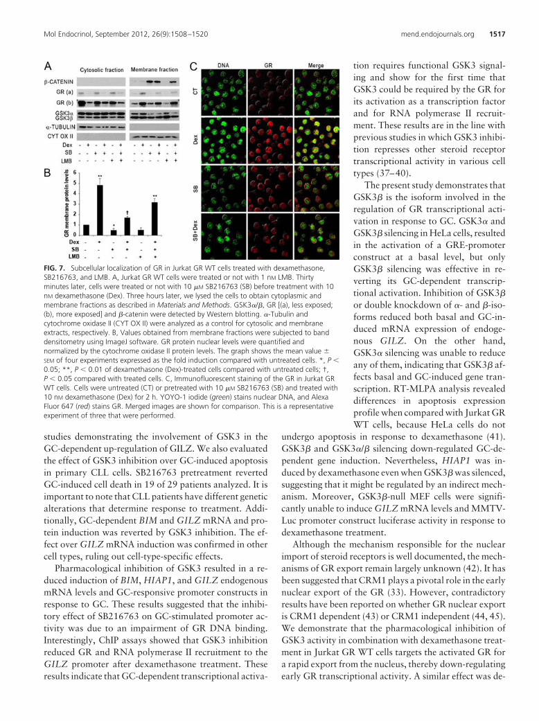
studies demonstrating the involvement of GSK3 in theGC-dependent up-regulation of GILZ. We also evaluatedthe effect of GSK3 inhibition over GC-induced apoptosisin primary CLL cells. SB216763 pretreatment revertedGC-induced cell death in 19 of 29 patients analyzed. It isimportant to note that CLL patients have different geneticalterations that determine response to treatment. Addi-tionally, GC-dependent BIM and GILZ mRNA and pro-tein induction was reverted by GSK3 inhibition. The ef-fect over GILZ mRNA induction was confirmed in othercell types, ruling out cell-type-specific effects.
Pharmacological inhibition of GSK3 resulted in a re-duced induction of BIM, HIAP1, and GILZ endogenousmRNA levels and GC-responsive promoter constructs inresponse to GC. These results suggested that the inhibi-tory effect of SB216763 on GC-stimulated promoter ac-tivity was due to an impairment of GR DNA binding.Interestingly, ChIP assays showed that GSK3 inhibitionreduced GR and RNA polymerase II recruitment to theGILZ promoter after dexamethasone treatment. Theseresults indicate that GC-dependent transcriptional activa-
tion requires functional GSK3 signal-ing and show for the first time thatGSK3 could be required by the GR forits activation as a transcription factorand for RNA polymerase II recruit-ment. These results are in the line withprevious studies in which GSK3 inhibi-tion represses other steroid receptortranscriptional activity in various celltypes (37–40).
The present study demonstrates thatGSK3� is the isoform involved in theregulation of GR transcriptional acti-vation in response to GC. GSK3� andGSK3� silencing in HeLa cells, resultedin the activation of a GRE-promoterconstruct at a basal level, but onlyGSK3� silencing was effective in re-verting its GC-dependent transcrip-tional activation. Inhibition of GSK3�
or double knockdown of �- and �-iso-forms reduced both basal and GC-in-duced mRNA expression of endoge-nous GILZ. On the other hand,GSK3� silencing was unable to reduceany of them, indicating that GSK3� af-fects basal and GC-induced gene tran-scription. RT-MLPA analysis revealeddifferences in apoptosis expressionprofile when compared with Jurkat GRWT cells, because HeLa cells do not
undergo apoptosis in response to dexamethasone (41).GSK3� and GSK3�/� silencing down-regulated GC-de-pendent gene induction. Nevertheless, HIAP1 was in-duced by dexamethasone even when GSK3� was silenced,suggesting that it might be regulated by an indirect mech-anism. Moreover, GSK3�-null MEF cells were signifi-cantly unable to induce GILZ mRNA levels and MMTV-Luc promoter construct luciferase activity in response todexamethasone treatment.
Although the mechanism responsible for the nuclearimport of steroid receptors is well documented, the mech-anisms of GR export remain largely unknown (42). It hasbeen suggested that CRM1 plays a pivotal role in the earlynuclear export of the GR (33). However, contradictoryresults have been reported on whether GR nuclear exportis CRM1 dependent (43) or CRM1 independent (44, 45).We demonstrate that the pharmacological inhibition ofGSK3 activity in combination with dexamethasone treat-ment in Jurkat GR WT cells targets the activated GR fora rapid export from the nucleus, thereby down-regulatingearly GR transcriptional activity. A similar effect was de-
FIG. 7. Subcellular localization of GR in Jurkat GR WT cells treated with dexamethasone,SB216763, and LMB. A, Jurkat GR WT cells were treated or not with 1 nM LMB. Thirtyminutes later, cells were treated or not with 10 �M SB216763 (SB) before treatment with 10nM dexamethasone (Dex). Three hours later, we lysed the cells to obtain cytoplasmic andmembrane fractions as described in Materials and Methods. GSK3�/�, GR [(a), less exposed;(b), more exposed] and �-catenin were detected by Western blotting. �-Tubulin andcytochrome oxidase II (CYT OX II) were analyzed as a control for cytosolic and membraneextracts, respectively. B, Values obtained from membrane fractions were subjected to banddensitometry using ImageJ software. GR protein nuclear levels were quantified andnormalized by the cytochrome oxidase II protein levels. The graph shows the mean value �SEM of four experiments expressed as the fold induction compared with untreated cells. *, P �0.05; **, P � 0.01 of dexamethasone (Dex)-treated cells compared with untreated cells; †,P � 0.05 compared with treated cells. C, Immunofluorescent staining of the GR in Jurkat GRWT cells. Cells were untreated (CT) or pretreated with 10 �M SB216763 (SB) and treated with10 nM dexamethasone (Dex) for 2 h. YOYO-1 iodide (green) stains nuclear DNA, and AlexaFluor 647 (red) stains GR. Merged images are shown for comparison. This is a representativeexperiment of three that were performed.
Mol Endocrinol, September 2012, 26(9):1508–1520 mend.endojournals.org 1517
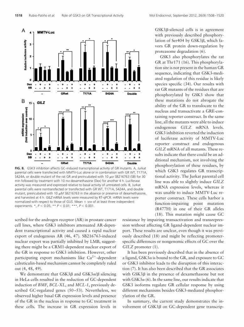
scribed for the androgen receptor (AR) in prostate cancercell lines, where GSK3 inhibitors attenuated AR-depen-dent transcriptional activity and caused a rapid nuclearexport of endogenous AR (46, 47). SB216763-inducednuclear export was partially inhibited by LMB, suggest-ing there might be a CRM1-dependent nuclear export ofthe GR in response to GSK3 inhibition. However, otherparticipating export mechanisms like Ca2�-dependentcalreticulin-based mechanism cannot be completely ruledout (4, 48, 49).
We demonstrate that GSK3� and GSK3�/� silencingin HeLa cells resulted in the reduction of GC-dependentinduction of BMF, BCL-XL, and MCL-1, previously de-scribed GC-regulated genes (50–53). Nevertheless, weobserved higher basal GR expression levels and presenceof the GR in the nucleus in response to GC treatment inthese cells. The increase in GR expression levels in
GSK3�-silenced cells is in agreementwith previously described phosphory-lation of Ser404 by GSK3�, which fa-vors GR protein down-regulation byproteasome degradation (6).
GSK3 also phosphorylates the ratGR at Thr171 (16). This phosphoryla-tion site is not present in the human GRsequence, indicating that GSK3-medi-ated regulation of this residue is likelyspecies specific (34). Our results withrat GR mutants of the residues that arephosphorylated by GSK3 show thatthese mutations do not abrogate theability of the GR to translocate to thenucleus and transactivate a GRE-con-taining reporter construct. In the sameline, all the mutants were able to induceendogenous GILZ mRNA levels.GSK3 inhibition reverted the inductionof luciferase activity of MMTV-Lucreporter construct and endogenousGILZ mRNA of all mutants. These re-sults indicate that there could be an ad-ditional mechanism, not involving thephosphorylation of these residues, bywhich GSK3 regulates GR transcrip-tional activity. The Jurkat parental cellline was able to slightly induce GILZmRNA expression levels, whereas itwas unable to induce MMTV-Luc re-porter construct. These cells harbor afunction-impairing point mutation(R477H) in one of their GR alleles(18). This mutation might cause GC
resistance by impairing transactivation and transrepres-sion without affecting GR ligand-dependent nuclear im-port. These results are unclear, even though it was previ-ously described (18) and might be reflecting promoter-specific differences or nongenomic effects of GC over theGILZ promoter (1).
It has been previously described that in the absence ofa ligand, GSK3� is bound to the GR, and exposure to GCor GSK3 inhibitor leads to the disruption of this interac-tion (7). It has also been described that the GR associateswith GSK3� in the presence of dexamethasone but notwith GSK3� (6). In the same line, our results indicate thatGSK3 isoforms regulate GR cellular response by usingdifferent mechanisms besides GSK3-mediated phosphor-ylation of the GR.
In summary, the current study demonstrates the in-volvement of GSK3� on GC-dependent gene transcrip-
FIG. 8. GSK3 inhibition affects GC-induced transcriptional activity of GR mutants. A, Jurkatparental cells were transfected with MMTV-Luc alone or in combination with GR WT, T171A,S424A, or double mutant of the rat GR and preincubated with 10 �M SB216763 (SB) for 30min followed by treatment with 10 nM dexamethasone (Dex) for another 4 h. Luciferaseactivity was measured and expressed relative to basal activity of untreated cells. B, Jurkatparental cells were nontransfected or transfected with GR WT, T171A, S424A, and doublemutant, preincubated with 10 �M SB216763 in the absence or presence of dexamethasone,and harvested at 4 h. GILZ mRNA levels were measured by RT-qPCR. mRNA levels werenormalized with respect to those of GUS. Mean � SEM of at least three independentexperiments. *, P � 0.05; ** P � 0.01; ***, P � 0.001.
1518 Rubio-Patiño et al. Role of GSK3 on GR Transcriptional Activity Mol Endocrinol, September 2012, 26(9):1508–1520
tional induction through the regulation of GR and RNApolymerase II recruitment to target gene sequences and byaffecting GR protein subcellular localization. Our resultssuggest that GSK3 plays an important role in regulatingGC mechanism of action, suggesting that keeping GSK3in an active state could improve GC therapy. Therefore,additional analyses of the involvement of GSK3 activity inGC treatment of lymphoma and leukemia malignanciesmay help gain insight into the molecular basis of thesedisorders.
Acknowledgments
We thank Dr. Jose Luis Rosa, Edgardo Rodríguez-Carballo, andMiguel Peña-Rico for helpful discussions and suggestions. Wealso thank Adriana Forero for English language editing. More-over, we thank the Scientific-Technical Services of the Unitat deBellvitge at the Universitat de Barcelona for their technical sup-port. We thank Dr. Carme Caelles for kindly providing JurkatGR WT cells and MMTV-Luc construct and Dr. Marc Pallardyfor providing p-1940-Luc construct. We also thank Dr. J.Woodgett for providing WT, GSK3��/�, and GSK3��/� MEFcells.
Address all correspondence and requests for reprints to: Ga-briel Pons, Departament de Ciències Fisiològiques II, Universitatde Barcelona, Institut d’Investigació Biomèdica de Bellvitge,C/Feixa Llarga s/n, L’Hospitalet de Llobregat, E-08907 Barce-lona, Spain. E-mail: [email protected]
This study was supported by grants from the Ministerio deEconomía y Competitividad and FEDER (SAF2010-20519), theInstituto de Salud Carlos III (RTICC RD06/0020/0097), and theAGAUR-Generalitat de Catalunya (AGAUR- 2009SGR395).C.R.-P., A.P.-P., C.M.-M., and D.M.G.-G. are recipients of re-search fellowships from the Ministerio de Economía y Competi-tividad. D.I.-S. has a postdoctoral contract from FundacióBosch i Gimpera.
Disclosure Summary: The authors have nothing to disclose.
References
1. Kfir-Erenfeld S, Sionov RV, Spokoini R, Cohen O, Yefenof E 2010Protein kinase networks regulating glucocorticoid-induced apopto-sis of hematopoietic cancer cells: fundamental aspects and practicalconsiderations. Leuk Lymphoma 51:1968–2005
2. Zhou J, Cidlowski JA 2005 The human glucocorticoid receptor:one gene, multiple proteins and diverse responses. Steroids 70:407–417
3. Oakley RH, Cidlowski JA 2011 Cellular processing of the gluco-corticoid receptor gene and protein: new mechanisms for generatingtissue-specific actions of glucocorticoids. J Biol Chem 286:3177–3184
4. Beck IM, De Bosscher K, Haegeman G 2011 Glucocorticoid recep-tor mutants: man-made tools for functional research. Trends Endo-crinol Metab 22:295–310
5. Wang Z, Chen W, Kono E, Dang T, Garabedian MJ 2007 Modu-lation of glucocorticoid receptor phosphorylation and transcrip-
tional activity by a C-terminal-associated protein phosphatase. MolEndocrinol 21:625–634
6. Galliher-Beckley AJ, Williams JG, Collins JB, Cidlowski JA 2008Glycogen synthase kinase 3�-mediated serine phosphorylation ofthe human glucocorticoid receptor redirects gene expression pro-files. Mol Cell Biol 28:7309–7322
7. Spokoini R, Kfir-Erenfeld S, Yefenof E, Sionov RV 2010 Glycogensynthase kinase-3 plays a central role in mediating glucocorticoid-induced apoptosis. Mol Endocrinol 24:1136–1150
8. Nuutinen U, Ropponen A, Suoranta S, Eeva J, Eray M, Pellinen R,Wahlfors J, Pelkonen J 2009 Dexamethasone-induced apoptosisand up-regulation of Bim is dependent on glycogen synthase ki-nase-3. Leuk Res 33:1714–1717
9. Wang Z, Malone MH, He H, McColl KS, Distelhorst CW 2003Microarray analysis uncovers the induction of the proapoptoticBH3-only protein Bim in multiple models of glucocorticoid-inducedapoptosis. J Biol Chem 278:23861–23867
10. Zhang L, Insel PA 2004 The pro-apoptotic protein Bim is a conver-gence point for cAMP/protein kinase A- and glucocorticoid-pro-moted apoptosis of lymphoid cells. J Biol Chem 279:20858–20865
11. Iglesias-Serret D, de Frias M, Santidrián AF, Coll-Mulet L, CosiallsAM, BarragánM,DomingoA,Gil J, PonsG 2007 Regulation of theproapoptotic BH3-only protein BIM by glucocorticoids, survivalsignals and proteasome in chronic lymphocytic leukemia cells. Leu-kemia 21:281–287
12. Beurel E, Jope RS 2006 The paradoxical pro- and anti-apoptoticactions of GSK3 in the intrinsic and extrinsic apoptosis signalingpathways. Prog Neurobiol 79:173–189
13. Forde JE, Dale TC 2007 Glycogen synthase kinase 3: a key regula-tor of cellular fate. Cell Mol Life Sci 64:1930–1944
14. RayasamGV, Tulasi VK, Sodhi R, Davis JA, Ray A 2009 Glycogensynthase kinase 3: more than a namesake. Br J Pharmacol 156:885–898
15. Woodgett JR 1990 Molecular cloning and expression of glycogensynthase kinase-3/factor A. EMBO J 9:2431–2438
16. Rogatsky I, Waase CL, Garabedian MJ 1998 Phosphorylation andinhibition of rat glucocorticoid receptor transcriptional activationby glycogen synthase kinase-3 (GSK-3). Species-specific differencesbetween human and rat glucocorticoid receptor signaling as re-vealed through GSK-3 phosphorylation. J Biol Chem 273:14315–14321
17. Helmberg A, Auphan N, Caelles C, Karin M 1995 Glucocorticoid-induced apoptosis of human leukemic cells is caused by the repres-sive function of the glucocorticoid receptor. EMBO J 14:452–460
18. Riml S, Schmidt S, Ausserlechner MJ, Geley S, Kofler R 2004 Glu-cocorticoid receptor heterozygosity combined with lack of receptorauto-induction causes glucocorticoid resistance in Jurkat acute lym-phoblastic leukemia cells. Cell Death Differ 11(Suppl 1):S65–S72
19. Asselin-Labat ML, David M, Biola-Vidamment A, Lecoeuche D,Zennaro MC, Bertoglio J, Pallardy M 2004 GILZ, a new target forthe transcription factor FoxO3, protects T lymphocytes from inter-leukin-2 withdrawal-induced apoptosis. Blood 104:215–223
20. Barragán M, Bellosillo B, Campàs C, Colomer D, Pons G, Gil J2002 Involvement of protein kinase C and phosphatidylinositol3-kinase pathways in the survival of B-cell chronic lymphocyticleukemia cells. Blood 99:2969–2976
21. Eldering E, Spek CA, Aberson HL, Grummels A, Derks IA, de VosAF,McElgunn CJ, Schouten JP 2003 Expression profiling via novelmultiplex assay allows rapid assessment of gene regulation in de-fined signalling pathways. Nucleic Acids Res 31:e153
22. Chen W, Rogatsky I, Garabedian MJ 2006 MED14 and MED1differentially regulate target-specific gene activation by the gluco-corticoid receptor. Mol Endocrinol 20:560–572
23. Piqué M, Barragán M, Dalmau M, Bellosillo B, Pons G, Gil J 2000Aspirin induces apoptosis through mitochondrial cytochrome c re-lease. FEBS Lett 480:193–196
24. Bellosillo B, Dalmau M, Colomer D, Gil J 1997 Involvement of
Mol Endocrinol, September 2012, 26(9):1508–1520 mend.endojournals.org 1519

CED-3/ICE proteases in the apoptosis of B-chronic lymphocyticleukemia cells. Blood 89:3378–3384
25. D’Adamio F, Zollo O, Moraca R, Ayroldi E, Bruscoli S, Bartoli A,Cannarile L, Migliorati G, Riccardi C 1997 A new dexamethasone-induced gene of the leucine zipper family protects T lymphocytesfrom TCR/CD3-activated cell death. Immunity 7:803–812
26. Webster JC, Huber RM, Hanson RL, Collier PM, Haws TF, MillsJK, Burn TC, Allegretto EA 2002 Dexamethasone and tumor ne-crosis factor-� act together to induce the cellular inhibitor of apo-ptosis-2 gene and prevent apoptosis in a variety of cell types. En-docrinology 143:3866–3874
27. Rogatsky I, Wang JC, Derynck MK, Nonaka DF, KhodabakhshDB, Haqq CM, Darimont BD, Garabedian MJ, Yamamoto KR2003 Target-specific utilization of transcriptional regulatory sur-faces by the glucocorticoid receptor. Proc Natl Acad Sci USA 100:13845–13850
28. Drouin J, Sun YL, ChamberlandM, Gauthier Y, De Léan A, NemerM, Schmidt TJ 1993 Novel glucocorticoid receptor complex withDNA element of the hormone-repressed POMC gene. EMBO J12:145–156
29. Asselin-LabatML, Biola-Vidamment A, Kerbrat S, LombèsM, Ber-toglio J, Pallardy M 2005 FoxO3 mediates antagonistic effects ofglucocorticoids and interleukin-2 on glucocorticoid-induced leu-cine zipper expression. Mol Endocrinol 19:1752–1764
30. Gokbuget N, Hoelzer D 2006 Treatment of adult acute lympho-blastic leukemia. Hematology Am Soc Hematol Educ Program2006:133–141
31. Pui CH, Evans WE 2006 Treatment of acute lymphoblastic leuke-mia. N Engl J Med 354:166–178
32. Liang MH, Chuang DM 2006 Differential roles of glycogen syn-thase kinase-3 isoforms in the regulation of transcriptional activa-tion. J Biol Chem 281:30479–30484
33. ItohM, AdachiM, Yasui H, TakekawaM, TanakaH, Imai K 2002Nuclear export of glucocorticoid receptor is enhanced by c-JunN-terminal kinase-mediated phosphorylation. Mol Endocrinol 16:2382–2392
34. Galliher-Beckley AJ, Cidlowski JA 2009 Emerging roles of gluco-corticoid receptor phosphorylation in modulating glucocorticoidhormone action in health and disease. IUBMB Life 61:979–986
35. Sun M, Meares G, Song L, Jope RS 2009 XIAP associates withGSK3 and inhibits the promotion of intrinsic apoptotic signaling byGSK3. Cell Signal 21:1857–1865
36. Maurer U, Charvet C, Wagman AS, Dejardin E, Green DR 2006Glycogen synthase kinase-3 regulates mitochondrial outer mem-brane permeabilization and apoptosis by destabilization of MCL-1.Mol Cell 21:749–760
37. LiaoX, Thrasher JB,Holzbeierlein J, Stanley S, Li B 2004 Glycogensynthase kinase-3� activity is required for androgen-stimulatedgene expression in prostate cancer. Endocrinology 145:2941–2949
38. Mazor M, Kawano Y, Zhu H, Waxman J, Kypta RM 2004 Inhibi-tion of glycogen synthase kinase-3 represses androgen receptor ac-tivity and prostate cancer cell growth. Oncogene 23:7882–7892
39. Medunjanin S, Hermani A, De Servi B, Grisouard J, Rincke G,Mayer D 2005 Glycogen synthase kinase-3 interacts with and phos-phorylates estrogen receptor � and is involved in the regulation ofreceptor activity. J Biol Chem 280:33006–33014
40. Grisouard J, Mayer D 2009 Specific involvement of glycogen syn-thase kinase-3 in the function and activity of sex steroid hormonereceptors reveals the complexity of their regulation. J SteroidBiochem Mol Biol 117:87–92
41. Mann CL, Cidlowski JA 2001 Glucocorticoids regulate plasmamembrane potential during rat thymocyte apoptosis in vivo and invitro. Endocrinology 142:421–429
42. Vandevyver S, Dejager L, Libert C 2012 On the trail of the gluco-corticoid receptor: into the nucleus and back. Traffic 13:364–374
43. Carrigan A, Walther RF, Salem HA, Wu D, Atlas E, Lefebvre YA,Haché RJ 2007 An active nuclear retention signal in the glucocor-ticoid receptor functions as a strong inducer of transcriptional ac-tivation. J Biol Chem 282:10963–10971
44. Liu J, DeFranco DB 2000 Protracted nuclear export of glucocorti-coid receptor limits its turnover and does not require the exportin1/CRM1-directed nuclear export pathway. Mol Endocrinol 14:40–51
45. Kumar S, Saradhi M, Chaturvedi NK, Tyagi RK 2006 Intracellularlocalization and nucleocytoplasmic trafficking of steroid receptors:an overview. Mol Cell Endocrinol 246:147–156
46. Rinnab L, Schütz SV, Diesch J, Schmid E, Küfer R, Hautmann RE,Spindler KD, Cronauer MV 2008 Inhibition of glycogen synthasekinase-3 in androgen-responsive prostate cancer cell lines: are GSKinhibitors therapeutically useful? Neoplasia 10:624–634
47. Schütz SV, Cronauer MV, Rinnab L 2010 Inhibition of glycogensynthase kinase-3� promotes nuclear export of the androgen recep-tor through a CRM1-dependent mechanism in prostate cancer celllines. J Cell Biochem 109:1192–1200
48. Holaska JM, Black BE, Rastinejad F, Paschal BM 2002 Ca2�-dependent nuclear export mediated by calreticulin. Mol Cell Biol22:6286–6297
49. Kumar S, Chaturvedi NK, Nishi M, Kawata M, Tyagi RK 2004Shuttling components of nuclear import machinery involved in nu-clear translocation of steroid receptors exit nucleus via exportin-1/CRM-1* independent pathway. Biochim Biophys Acta 1691:73–77
50. Ploner C, Rainer J, Niederegger H, Eduardoff M, Villunger A, Ge-ley S, Kofler R 2008 The BCL2 rheostat in glucocorticoid-inducedapoptosis of acute lymphoblastic leukemia. Leukemia 22:370–377
51. Scoltock AB, Heimlich G, Cidlowski JA 2007 Glucocorticoids in-hibit the apoptotic actions of UV-C but not Fas ligand in hepatomacells: direct evidence for a critical role of Bcl-xL. Cell Death Differ14:840–850
52. Xu B, Strom J, Chen QM 2011 Dexamethasone induces transcrip-tional activation of Bcl-xL gene and inhibits cardiac injury by myo-cardial ischemia. Eur J Pharmacol 668:194–200
53. Lynch JT, Rajendran R, Xenaki G, Berrou I, Demonacos C, Krstic-Demonacos M 2010 The role of glucocorticoid receptor phosphor-ylation in Mcl-1 and NOXA gene expression. Mol Cancer 9:38
1520 Rubio-Patiño et al. Role of GSK3 on GR Transcriptional Activity Mol Endocrinol, September 2012, 26(9):1508–1520
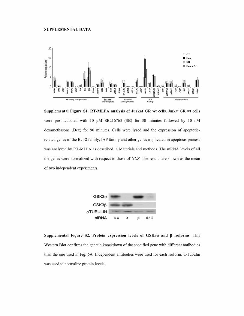
���������� ����
������������ ������ ��� ������ �����!�! "# $��%�� &� '� (���!� ��� ����� �������
���� �������� ��� �� �� ��� � � !"#�$%$&� '!"�� ()� &�� *�� ��� ()��)���� �+� ��� � �
��-�*� ���)��� '.�-�� ()� /�� *�� ��0� 1����� ���� �+���� ���� ��� �-�����)�� )(� ��)� ) ���
��� ���2�����)(� ���"���#�(�*��+3�456�(�*��+�����) ���2������*����� ��������)� )�����)�����
��������+7����+��8� 965���������������� � ����������*� �)��0�8���*�:5���;����)(�����
���2����������)*���7����� ������� � )� �)���)(�GUS0�8������ �������)������ ���*����
)(� �)����������� ��-���*�� �0�
�
�
������������ ������ �)� ��"���� �*���!!�"� ��+��! "# &�,-� ��. / �!"#"��!0� 8����
<�� ���"�) ��)�(�*�� ���2��� �����)���)���)(� ��������(����2������ ����((��� ��� ��)�����
���� ���)����������=�20�$50�4��������� ��� ��)�������������()��������)()*0���8�����
�������� )��)*���7���) ������;���0�
�
�
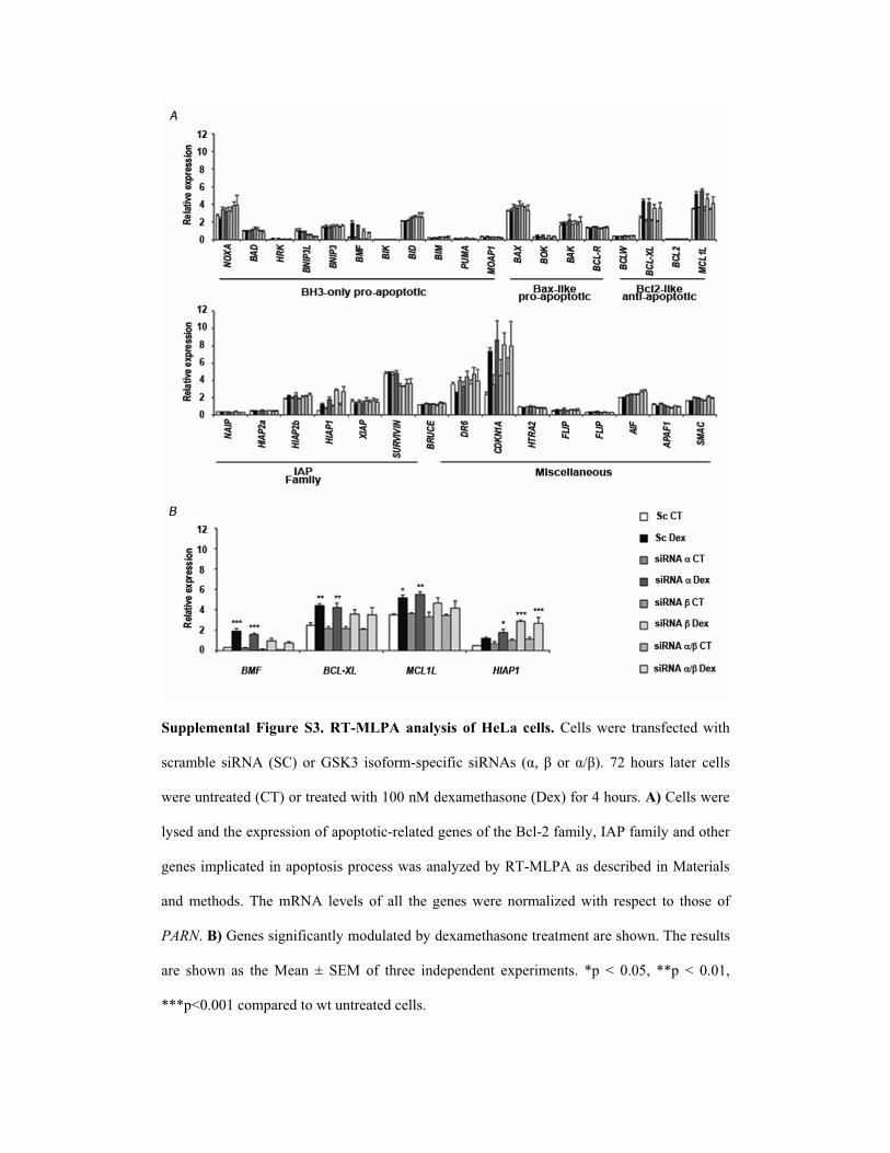
�
������������ ������ �-� ������ �����!�! "# 0��� (���!� 1��������� ���(�� ����� ��
���*���� ���:5� '!1�� )��!>&� ��)()*������(��� ���:5�� '�3��� )��?��0 %#��)�� �� � ������
����� �� ���'18��)� �� ����� ������� ���-�*� ���)���'.�-��()�@��)�0����1���������
�+�������� ����-�����)��)(���)� ) ������ ���2�����)(� ���"���#�(�*��+3�456�(�*��+�����) ���
2������*����� ��������)� )�����)�������������+7����+��8� 965���������������� � ������
����*� �)��0� 8���*�:5� ��;���� )(� ���� ��� 2��������� �)*���7����� �� ����� � )� �)��� )(�
PARN0�1����������2��(���� �+�*)��� ����+���-�*� ���)��� �� *�� ������)��0�8������ ��
��� ��)��� ��� ��� ���� A� !B � )(� ���� ���������� � �-���*�� �0� C�� D� �0�E3� CC�� D� �0��3�
CCC�D�0�����)*����� )�� �� �� ��������0��
�
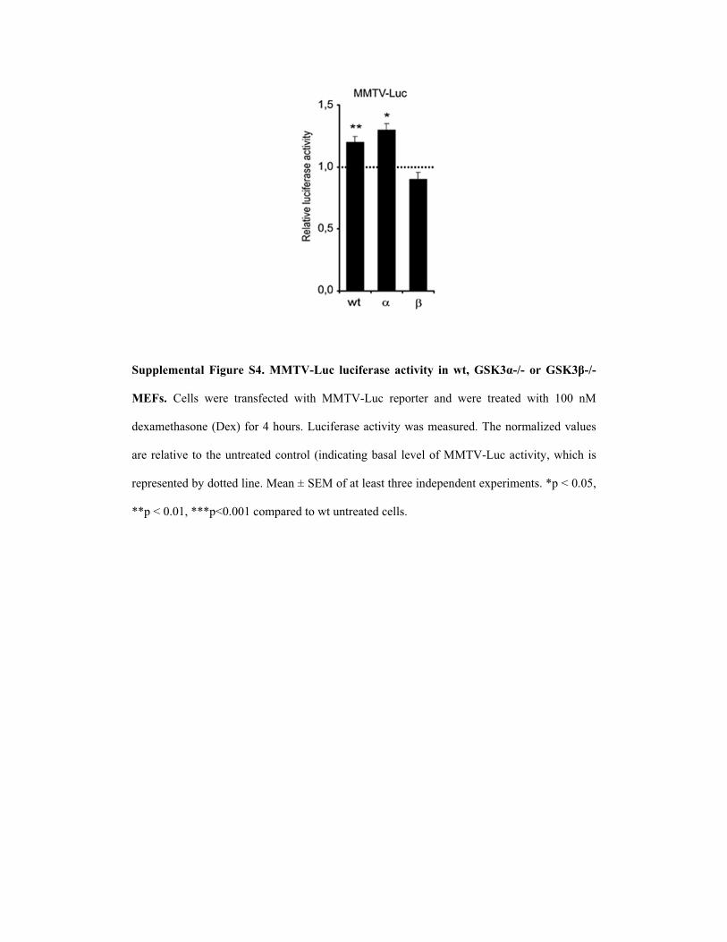
�
�
������������ ������ �2� �3���( ��(�#���!� �(��+��� �� '�4 &�,-��5� "� &�,-/�5�
��!� 1����� ���� ���(�� ��� �� �� 8F�9�� ��) �� ���� ���� �� ��� �� �� ���� � �
��-�*� ���)���'.�-��()�@��)�0�9��(������� �;� +�����*�����0�8����)*���7���;�����
��� ��� �;�� )� ���� �� ����)� )��'������ ��2������� ��;���)(� 8F�9���� �;� +3������� ���
������ ����+��) �������0� ����A�!B �)(�� ����� � �������������� ��-���*�� �0�C��D��0�E3�
CC��D��0��3�CCC�D�0�����)*����� )�� �� �� ��������0��
�
�
�
�
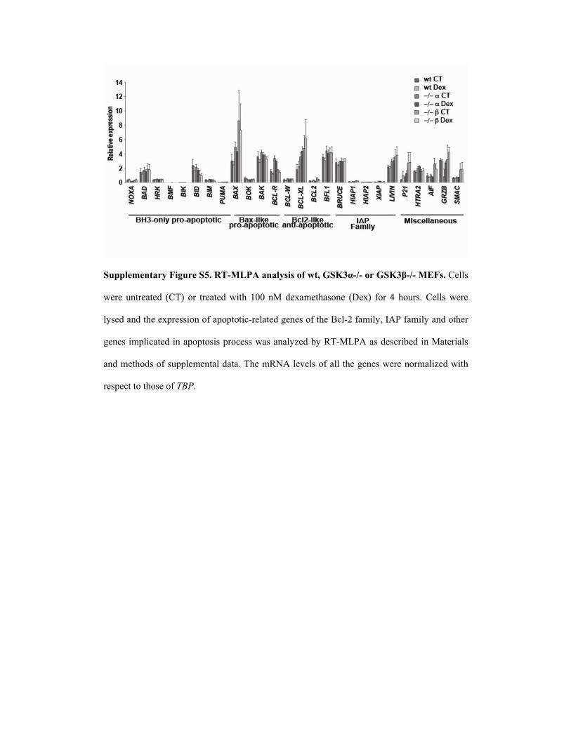
�
������������� ������ �6� ������ �����!�! "# '�4 &�,-��5� "� &�,-/�5� ��!� 1�����
���� � �� ��� '18�� )� �� ����� �� ����� ���-�*� ���)��� '.�-�� ()� @� �)�0�1���������
�+�������� ����-�����)��)(���)� ) ������ ���2�����)(� ���"���#�(�*��+3�456�(�*��+�����) ���
2������*����� ��������)� )�����)�������������+7����+��8� 965���������������� � ������
����*� �)���)(������*�� ����� �0�8���*�:5���;����)(����� ���2����������)*���7����� ��
����� � )� �)���)(�TBP0
�
�
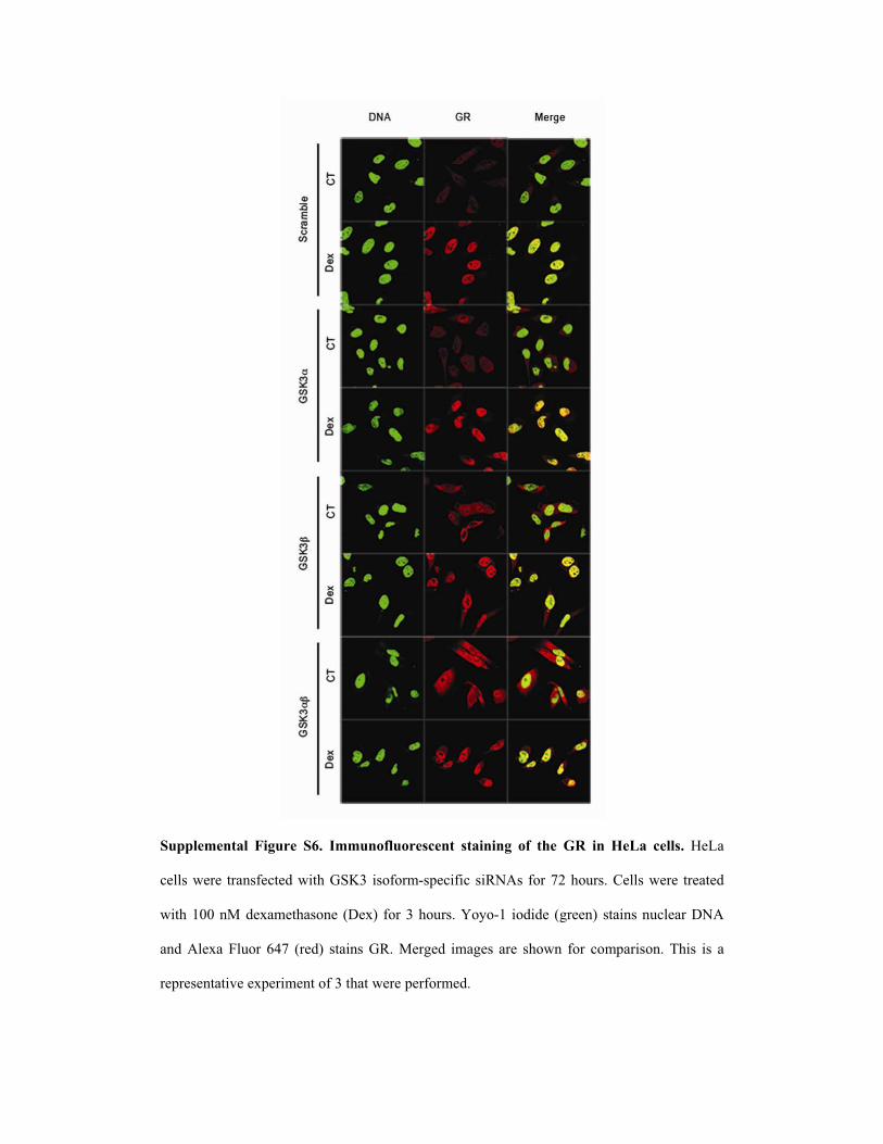
�
������������ ������ �7� 8����"#��"��!(��� !������� "# �9� &� �� 0��� (���!� G�9��
���������� ���(�� ����� ���!>&� ��)()*������(������:5��()�%#��)�0�1��������� �� ���
�� ������� ���-�*� ���)��� '.�-�� ()�&��)�0�H)+)��� �)����� '2����� � �����������.:5�
����5��-��=�)� $@%� '���� � �������0� �2��� �*�2��� ��� ��)��� ()� �)*����)�0�8���� ��� ��
������ � �;���-���*�� �)(�&� �� �������()*��0�

������������ ������ �:� �9"!�9"������"� !���! "# �9� &� ;� &�,-� !���*� ���
������ � �)�� )(� ��� �)� ��)��� ������� )(� � ������)���)+�� ��� �+��!>&3�8��%�� ����
!�@#@0� 8��� �)����7� �)�� )(� ����� ��)���)+�� �)�� �� �� )(� ��� � � ���� �*��� ���� )� ���
��)��0�8��� (�*�����I������ ������ �� ����*��)��������)���;����� ����� ��� �)��������0�
8�������) ��������2���*�����+��� ������ ���* �2������� )��� ���*��)��������I��������
������ ��0�
�
�
�
�
�
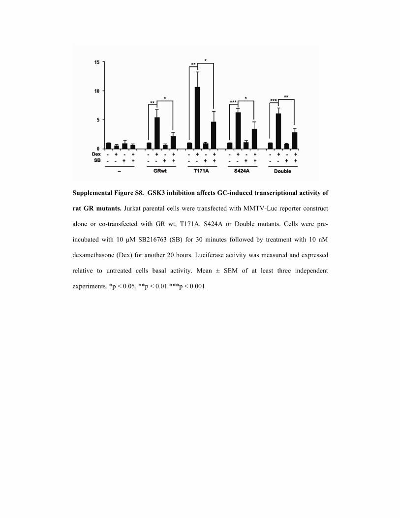
������������ ������ �<� &�,- ��9�;���"� �##�(�! &=���.�(�. ����!(�����"��� �(��+��� "#
��� &� ������!� ��� ����� ������������� ���(�� ����� �� 8F�9����) ���)�� � �
��)��� )� �)� ���(�� ��� �� �� ��� � 3� 8�%�53� !@#@5� )� .)���� * �� �0� 1����� ���� ���
����� ��� �� �� ��� � � !"#�$%$&� '!"�� ()� &��*�� ��� ()��)���� �+� �� *�� � �� �� ��� � �
��-�*� ���)���'.�-��()���) ���#���)�0�9��(������� �;� +�����*�����������-�������
��� �;�� )� � �� ��� ������ ������ �� �;� +0� ���� A� !B � )(� � � ���� � ���� ���������� �
�-���*�� �0�C��D��0�E3���CC��D��0�� ��CCC��D��0���0�

Patient CT Dex SB Dex + SB
1* 55,04 28,05 57,71 48,45 2* 80,44 56,19 84,27 64,24 3* 75,91 65,55 75,51 68,22 4* 70,83 37,95 65,86 57,71 5* 38,76 18,05 44,50 31,27 6* 74,60 53,42 71,23 58,19 7* 41,82 29,37 55,43 32,63 8* 65,26 40,29 66,25 48,43 9* 62,48 46,21 69,14 48,03
10* 64,59 55,87 59,28 58,17 11* 90,83 83,94 94,73 91,31 12* 72,08 53,65 77,99 63,88 13* 78,22 50,24 77,15 60,10 14* 67,81 44,56 62,75 51,36 15* 71,21 56,20 77,81 61,02 16* 71,40 46,87 61,90 51,94 17* 71,12 58,50 65,94 64,66 18* 84,53 70,59 80,27 76,59 19* 42,05 28,85 45,43 31,03 20 66,09 46,19 60,54 36,90 21 56,68 40,55 59,14 33,53 22 40,38 35,80 35,42 28,88 23 43,09 34,50 45,76 36,02 24 52,34 27,41 43,18 28,28 25 56,82 34,65 48,07 29,02 26 63,54 53,06 60,63 46,23 27 50,26 42,38 54,58 42,68 28 78,31 68,47 65,27 66,70 29 75,68 62,73 68,96 54,04
�
������������ ��;�� ��� 3��;����� "# =�� !�����!� 199������������ �� ���'18��)����
����� ����� ������ �!"#�$%$&�'!"������ �� ����� ������ ���-�*� ���)���'.�-��()�#@�
�)�0�F������ +�����*��������������������� � ����������*� �)�������� � ����-����������
�������� �2��)(�����-���561���2� �;�������0��
C�6� ��� �������1����������)� )���������;� ����+��!>&������� �)�0�
��������� ����8��� �� ��0>��
�������! ��. ����;".��!
�!>&�������!>&��'#%1������ ��)���������()*�1����!�2�����20�
������ #"� �"�!� !�����!
�:5��)� �� � )(�� 3��!>&��?�� )��!>&��?�� B=����������� ����+7����+��8� 965����2�
!59!5�>48�� ��#�"�� )���5�)� )����*�:5�()*� �1�G)�����0�8���*�:5���;����)(�
���� �����2���������� ������7��� )� �)���)(�TBP.�
�