
UNIVERSIDAD DE MURCIA FACULTAD DE MEDICINA
Departamento de Bioquímica y Biología Molecular B e Inmunología
CARACTERIZACIÓN DE ODCp COMO UNA NUEVA PROTEÍNA INHIBIDORA DE ANTIZIMAS (AZIN2). ASPECTOS ESTRUCTURALES Y FUNCIONALES.
Memoria presentada por Andrés Joaquín López Contreras
para optar al grado de Doctor por la Universidad de Murcia
Murcia, Julio 2008

El trabajo experimental presentado en esta Tesis Doctoral ha sido financiado
por el Ministerio de Educación y Ciencia, y por la Fundación Séneca (Comunidad
Autónoma Región de Murcia), con cargo a los proyectos BFU2005-09378-C02 y
00466/PI/04, respectivamente.
Andrés Joaquín López Contreras ha disfrutado de una beca del Programa de
Formación de Personal Investigador de la Fundación Séneca (2004-2008). La
Fundación Séneca ha financiado, además, la realización de dos estancias en el
instituto “The Wellcome Trust Sanger Institute” (Cambridge, UK) y una estancia en el
Departamento de Biología Molecular y Celular de la Universidad PennState (USA).

AGRADECIMIENTOS
A mis directores de Tesis, Asunción y Rafa, gracias por darme la
oportunidad de investigar con vosotros y hacerme sentir vuestro compañero,
por vuestra dedicación, por el gran trabajo que habéis realizado en el desarrollo
de esta tesis, por vuestro cariño y por muchas cosas más.
A tod@s l@s compañer@s de laboratorio que he tenido durante estos
años, por todos los buenos momentos que hemos pasado, y por la ayuda y
apoyo que me habéis prestado siempre que la he necesitado.
A todos los miembros del departamento, gracias por vuestra buena
disposición y apoyo en todo momento.
A mis amig@s, simplemente por eso, por vuestra amistad, que me hace
sentirme muy afortunado y que hace que todo tenga sentido, entre muchas
cosas, y aunque quizá la menos importante, esta tesis.
A todos l@s buen@s maestr@s que he tenido a lo largo de mi vida,
porque ellos, entre los que se encuentran mis padres, son los que me han
enseñado con gran dedicación casi todo lo poco que sé. Especialmente a Rafa,
por brindarme la oportunidad de investigar, como siempre había deseado. Si
consigo llegar a ser un buen científico será gracias a todo lo que me has
enseñado y apoyado para ello.
A toda mi familia. A mis padres y a mi hermana. Gracias por vuestro
amor incondicional. Gracias por estar siempre ahí.

Índice
ÍNDICE

Índice

Índice
ABREVIATURAS 1
INTRODUCCIÓN 7
1. POLIAMINAS: NATURALEZA, MECANISMOS DE ACCIÓN,
METABOLISMO Y TRANSPORTE. 9
1.1. Generalidades de las poliaminas. 9
1.2. Interacciones y mecanismos de acción de las poliaminas. 13
1.2.1. Interacciones electrostáticas. 15
1.2.2. Interacciones covalentes: hipusinación. 17
1.2.3. Secuestro de radicales libres. 17
1.2.4. Formación de productos citotóxicos. 17
1.3. Metabolismo de las poliaminas en mamíferos. 18
1.3.1. Ruta biosintética. 18
1.3.2. Ruta de retroconversión. 21
1.3.3. Catabolismo de poliaminas. 22
1.3.4. Ruta de la arginina descarboxilasa. 23
1.4. Transporte de poliaminas. 24
2. REGULACIÓN DE LOS NIVELES INTRACELULARES DE POLIAMINAS. 27
2.1. Generalidades. 27
2.2. Ornitina descarboxilasa. 30
2.2.1. Estructura y actividad de ODC. 30
2.2.2. Gen y ARNm de ODC. 31
2.2.3. Regulación de ODC. 32
2.2.3.1. Regulación transcripcional. 32
2.2.3.2. Regulación traduccional. 33
2.2.3.3. Regulación post-traduccional. Degradación de
ODC. 35

Índice
2.3. Antizimas (AZs) e inhibidor de antizimas. 37
2.3.1. Síntesis y degradación de AZs. 38
2.3.2. Acción de AZ sobre ODC y el transporte de poliaminas. 40
2.3.3. Expresión y función de las diferentes isoformas de AZs. 41
2.3.4. Nuevas dianas de las antizimas. 41
2.3.5. Inhibidor de antizima (AZIN) y otras proteínas
homólogas a ODC. 42
3. FUNCIONES FISIOLÓGICAS DEL SISTEMA ODC/POLIAMINAS. 44
3.1. Crecimiento celular. 44
3.2. Muerte celular y apoptosis. 45
3.3. Poliaminas y procesos dediferenciación celular. Espermatogénesis. 46
4. IMPLICACIONES FISIOPATOLÓGICAS DE LAS POLIAMINAS. MODELOS TRANSGÉNICOS Y ENFERMEDADES HUMANAS. 48
ANTECEDENTES Y OBJETIVOS. 53
MATERIALES Y MÉTODOS. 59
1. Reactivos y Equipamiento. 61
2. Líneas y Cultivos Celulares. 62
3. Tranfecciones transitorias. 63
4. Electroforesis analítica de proteínas. 64
5. Transferencia Western. 64
6. Experimentos de Inmunoprecipitación. 64
7. Clonación y Construcciones Génicas. 66
8. Microscopía Confocal. 68
9. Animales. Manipulación y Obtención de Tejidos. 70
10. Determinación de la actividad ODC y ADC. 70

Índice
11. Ensayo de captación de poliaminas. 71
12. Determinación de poliaminas. 71
13. Extracción, Purificación y Cuantificación de ARN. 72
14. Síntesis de ADNc, Amplificación por RT-PCR y Análisis de los productos
obtenidos. 73
15. PCR cuantitativa o PCR a tiempo real. 73
16. Inmunohistoquímica. 74
17. Hibridación in situ de ARN. 74
RESULTADOS. 77
Capitulo 1.
El gen ornitina descarboxilasa-like (ODCp) murino codifica una proteína inhibidora de antizimas (AZIN2) carente de actividad ornitina y arginina descarboxilasa. 79
Capítulo 2.
El inhibidor de antizimas 2 (AZIN2) estimula la captación de poliaminas en células de mamífero. 107
Capítulo 3.
La expresión del inhibidor de antizimas 2 (AZIN2) en células haploides germinales masculinas sugiere su papel en la espermiogénesis. 129
Capítulo 4.
Localización subcelular del inhibidor de antizimas 2 (AZIN2) y de sus proteínas parálogas ODC y AZIN1 en células de mamífero. 151
DISCUSIÓN GENERAL. 169
CONCLUSIONES. 185
BIBLIOGRAFÍA. 189

Índice
ENGLISH SUMMARY. 219
APÉNDICE. 237

Abreviaturas
ABREVIATURAS
1

Abreviaturas
2

Abreviaturas
β-Act: β-Actina
ADC: arginina decarboxilasa
ADN: ácido desoxirribonucléico
ADNc: ADN complementario
Agm: Agmatina
AMPc: adenosina 3´,5´-monofosfato cíclico
ARN: ácido ribonucléico
ARNm: ARN mensajero
ARNt: ARN de transferencia
ATP: adenosin 5´-trifosfato
AZ: antizima
AZIN: inhibidor de antizimas
BSA: albúmina de suero bovino
14C: carbono 14
CRE: elemento de respuesta al AMPc
DAO: diamino oxidasa
DFMO: α-difluorometilornitina
DFMA: α-difluorometilarginina
DMEM: medio mínimo esencial de Dulbecco
dNTP: desoxinucleótidos trifosfato
dpm: desintegraciones por minuto
DTT: ditiotreitol
EC: extracto crudo
EDTA: ácido etilendiamino tetraacético
GABA: ácido γ-aminobutírico
GFP: proteína de fluorescencia verde
Golgi: aparato de Golgi
HPLC: cromatografía líquida de alta resolución
3

Abreviaturas
Kb: kilobase
KDa: kilodalton
Km: constante de Michaelis-Menten
β-ME: β-mercaptoetanol
NMDA: N-metil-D-aspartato
ODC: ornitina decarboxilasa
ORF: pauta de lectura abierta
PA: poliaminas
PAO: poliamino oxidasa
PBS: tampón fosfato salino
pb: pares de bases
PKA: proteín-quinasa A
PMF-1: factor modulado por poliaminas
POPOP: 2´,2´p-fenil bis (4-metil-5feniloxazol)
PPO: 2,5 difeniloxazol
PRE: elemento de respuesta a poliaminas
Put: putrescina
qPCR: PCR cuantitativa
RE: retículo endoplasmático
ROS: especies de oxígeno reactivas
rpm: revoluciones por minuto
RT-PCR: reacción en cadena de la polimerasa acoplada a trancripción reversa
S12: fracción soluble tras centrifugar a 12000 rpm 20 minuntos
SAMDC: S-adenosil L-metionina decarboxilasa
SDS: dodecil sulfato sódico
SEM: error estandar
SNC: sistema nervioso central
SNP: polimorfismo de un solo nucleótido
4

Abreviaturas
Spd: espermidina
SpdST: spermidina sintetasa
Spn: espermina
SpnOx: espermina oxidasa
SpnST: espermina sintetasa
SSAT: espermidina, espermina N-acetiltransferasa
TAE: tampón acético-EDTA
TEMED: N,N,N,N'-tetrametilnediamina
Tm: temperatura de fusión
TRIS: trizma base Tris (hidroximetil) aminometano
UTR: regiones no traducidas
Vmax: velocidad máxima
5

Abreviaturas
6

Introducción
INTRODUCCIÓN
7

Introducción
8

Introducción
1. POLIAMINAS: NATURALEZA, MECANISMO DE ACCIÓN, METABOLISMO Y TRANSPORTE.
1. Generalidades de las poliaminas.
Las poliaminas putrescina, espermidina y espermina son aminas alifáticas
imprescindibles para la vida en casi todos los organismos conocidos. Son moléculas
sencillas de un tamaño similar a los aminoácidos y que se encuentran presentes en la
mayoría de nuestras células a unas concentraciones importantes (en el rango mM),
regulando múltiples procesos vitales para el mantenimiento y el crecimiento celular. A
pesar de su indudable importancia y quizá por la falta de un conocimiento más
detallado sobre sus mecanismos de acción, las poliaminas son aún “grandes
desconocidas” en bioquímica y biología celular. A pesar de ello existen miles de
trabajos científicos que han ido aportando un gran conocimiento sobre aspectos
concretos de las poliaminas, las proteínas implicadas en su regulación y diversas
patologías que se encuentran relacionadas con las alteraciones de sus niveles.
La estructura química de estas moléculas se puede observar en la figura 1. A
pH fisiológico poseen cargas positivas, responsables de sus interacciones con
múltiples proteínas, ácidos nucleícos y otros componentes celulares cargados
negativamente, de forma “inespecífica” en algunos casos o totalmente específica en
otros. Mediante estas interacciones las poliaminas ejercen muchas de sus funciones
regulando, por ejemplo, la actividad de muchas proteínas o la expresión de
determinados genes.
La concentración de poliaminas, y la proporción entre ellas (putrescina,
espermidina y espermina) determinará de qué forma regulan cada proceso en el que
están implicadas. En este sentido se conocen más de 20 proteínas distintas que en su
conjunto determinan los niveles de poliaminas, regulando su síntesis, degradación,
interconversión y procesos de transporte. El sistema de regulación de los niveles de
poliaminas es uno de los más complejos presentes en células eucariotas, presentando
mecanismos muy exquisitos y, en algunos casos, únicos. La gran complejidad de la
regulación de los niveles de poliaminas nos informa sobre la importancia de estas
moléculas y de un control riguroso de su concentración.
9

Introducción
H2NCH2CH2CH2CH2NH2
Putrescina(1,4-diaminobutano)
H2NCH2CH2CH2CH2NHCH2CH2CH2NH2
NH2H3N NH3
+ ++
Espermidina(1,8-diamino-4azaoctano)
NH3H3N NH2H2N
+ ++
+
NH2CH2CH2CH2HNCH2CH2CH2CH2NHCH2CH2CH2NH2
Espermina(1,12-diamino-4,9-diazaoctano)
NH3H3N
++
H2NCH2CH2CH2CH2NH2
Putrescina(1,4-diaminobutano)
H2NCH2CH2CH2CH2NHCH2CH2CH2NH2
NH2H3N NH3
+ ++
Espermidina(1,8-diamino-4azaoctano)
NH3H3N NH2H2N
+ ++
+
NH2CH2CH2CH2HNCH2CH2CH2CH2NHCH2CH2CH2NH2
Espermina(1,12-diamino-4,9-diazaoctano)
NH3H3N
++NH3
H3N+
+
Figura 1. Fórmula y estructura de las poliaminas mayoritarias.
Las principales y más abundantes poliaminas en organismos superiores son tres,
putrescina, espermidina y espermina. Como se puede observar en la figura 1 son
aminas alifáticas de tamaño sucesivamente mayor, que poseen 2, 3 y 4 grupos aminos
respectivamente. Putrescina (1,4-diaminobutano) normalmente es el precursor de las
poliaminas mayores, espermidina (1,8-diamino-4azaoctano) y espermina (1,12-
diamino-4,9-diazaoctano). Sin embargo también se han encontrado otras poliaminas
minoritarias como diaminopropano, cadaverina o agmatina tanto en procariotas como
en eucariotas. Especial interés tiene la presencia de agmatina en mamíferos, cuya
existencia y su capacidad de modular distintos procesos parece clara, pero su origen,
endógeno o exógeno, sigue siendo incierto y su estudio forma parte del presente
trabajo. También se conocen otras poliaminas más complejas como termina o
termoespermina, encontradas especialmente en bacterias termófilas, y ciertas
moléculas derivadas de poliaminas, como formas conjugadas con compuestos
fenólicos o alcaloides, en plantas o formas acetiladas de las poliaminas en mamíferos
(figura 2).
10

Introducción
H2NCH 2CH 2CH 2CH 2CH 2NH 2
Figura 2. Algunas poliaminas minoritarias.
Nuestro trabajo de investigación se ha llevado a cabo principalmente en
modelos de ratón y en líneas celulares de mamífero, estudiando genes de ratón
implicados en la regulación de los niveles de poliaminas, de modo que esta
introducción versará preferentemente sobre los diversos aspectos del sistema de
poliaminas en mamíferos.
En general los tejidos con escaso crecimiento poseen niveles inferiores de
poliaminas, mientras que aquellos con gran potencial proliferativo poseen una mayor
concentración de poliaminas (Jänne et al., 1978). Por ejemplo, en células
NHHN
NH3
+NH3
+
NH2C(NH)NHCH 2CH 2CH 2CH2NH2
NH3H3N+ +
Cadaverina(1,5 -diaminopentano)
Agmatina(1-amino ,4 -guanidinobutano)
NH 2CH 2CH 2CH2HNCH 2CH 2CH 2NHCH 2CH 2CH 2NH 2
NHCH 2CH 2CH 2HNCH 2CH 2CH2CH 2NH 2
NH2CH 2CH2CH 2HNCH 2CH2CH 2NHCH 2CH 2CH 2CH2NH 2
NHCH 2CH 2CH 2HNCH 2CH2CH 2CH 2NHCH 2CH 2CH 2NH 2
Termina (3,3,3)
Termoespermina (3,3,4)
N1-Acetilespermidina
N1-Acetilespermina
H2NCH 2CH 2CH 2CH 2CH 2NH 2 NH3H3N+ +
CH3CO
CH 3CO
NHHN
NH3
+NH3
+
NHHN
NH3
+NH3
+
NH2C(NH)NHCH 2CH 2CH 2CH2NH2
NH3H3N+ +
Cadaverina(1,5 -diaminopentano)
Agmatina(1-amino ,4 -guanidinobutano)
NH 2CH 2CH 2CH2HNCH 2CH 2CH 2NHCH 2CH 2CH 2NH 2
NHCH 2CH 2CH 2HNCH 2CH 2CH2CH 2NH 2
NH2CH 2CH2CH 2HNCH 2CH2CH 2NHCH 2CH 2CH 2CH2NH 2
NHCH 2CH 2CH 2HNCH 2CH2CH 2CH 2NHCH 2CH 2CH 2NH 2
Termina (3,3,3)
Termoespermina (3,3,4)
N1-Acetilespermidina
N1-Acetilespermina
H2NCH 2CH 2CH 2CH 2CH 2NH 2
CH3CO
CH 3CO
NHHN
NH3
+NH3
+
NH2C(NH)NHCH 2CH 2CH 2CH2NH2
NH3H3N+ +
Cadaverina(1,5 -diaminopentano)
Agmatina(1-amino ,4 -guanidinobutano)
NH 2CH 2CH 2CH2HNCH 2CH 2CH 2NHCH 2CH 2CH 2NH 2
NHCH 2CH 2CH 2HNCH 2CH 2CH2CH 2NH 2
NH2CH 2CH2CH 2HNCH 2CH2CH 2NHCH 2CH 2CH 2CH2NH 2
NHCH 2CH 2CH 2HNCH 2CH2CH 2CH 2NHCH 2CH 2CH 2NH 2
Termina (3,3,3)
Termoespermina (3,3,4)
N1-Acetilespermidina
N1-Acetilespermina
H2NCH 2CH 2CH 2CH 2CH 2NH 2
CH3CO
CH 3CO
NHHN
H2NCH 2CH 2CH 2CH 2CH 2NH 2
NHHN
NH3
+NH3
+
NH2C(NH)NHCH 2CH 2CH 2CH2NH2
NH3H3N+ +
Cadaverina(1,5 -diaminopentano)
Agmatina(1-amino ,4 -guanidinobutano)
NH 2CH 2CH 2CH2HNCH 2CH 2CH 2NHCH 2CH 2CH 2NH 2
NHCH 2CH 2CH 2HNCH 2CH 2CH2CH 2NH 2
NH2CH 2CH2CH 2HNCH 2CH2CH 2NHCH 2CH 2CH 2CH2NH 2
NHCH 2CH 2CH 2HNCH 2CH2CH 2CH 2NHCH 2CH 2CH 2NH 2
Termina (3,3,3)
Termoespermina (3,3,4)
N1-Acetilespermidina
N1-Acetilespermina
H2NCH 2CH 2CH 2CH 2CH 2NH 2 NH3H3N+ +
NHHN
NH3
+NH3
+
NHHN
NH3
+NH3
+
NH2C(NH)NHCH 2CH 2CH 2CH2NH2
NH3H3N+ +
Cadaverina(1,5 -diaminopentano)
Agmatina(1-amino ,4 -guanidinobutano)
CH3CO
CH 3CO
NH 2CH 2CH 2CH2HNCH 2CH 2CH 2NHCH 2CH 2CH 2NH 2
NHCH 2CH 2CH 2HNCH 2CH 2CH2CH 2NH 2
NH2CH 2CH2CH 2HNCH 2CH2CH 2NHCH 2CH 2CH 2CH2NH 2
NHCH 2CH 2CH 2HNCH 2CH2CH 2CH 2NHCH 2CH 2CH 2NH 2
Termina (3,3,3)
Termoespermina (3,3,4)
N1-Acetilespermidina
N1-Acetilespermina
CH3CO
CH 3CO
11

Introducción
transformadas, con crecimiento descontrolado y en la mayoría de los crecimientos
neoplásicos se ha encontrado una abundante presencia de poliaminas. Por ello, desde
hace varias décadas la depleción de los niveles de poliaminas ha sido un objetivo en el
estudio de nuevas terapias antineoplásicas. Se ha comprobado que el uso de
inhibidores de la síntesis de poliaminas produce un importante descenso del
crecimiento celular (Pegg, 1988; Bardocz et al., 1995). Hasta el momento las
estrategias empleadas para deplecionar los niveles de poliaminas no han producido
resultados clínicos satisfactorios, aunque actualmente siguen siendo objeto de estudio
en diversos grupos de investigación e incluso existen varios ensayos clínicos
relacionados con este campo, que comentaremos más adelante.
Además de la función que ejercen las poliaminas sobre la tasa de crecimiento
celular, diversos experimentos han demostrado que estas moléculas también están
implicadas en diversos procesos de diferenciación celular (Oka y Borellini, 1989) así
como en muerte celular por apoptosis (Seiler y Raul, 2005).
Una muestra del interés actual por las poliaminas dentro del campo de las
ciencias moleculares viene reflejado en el importante número de revisiones publicadas
desde al año 2000 sobre este tema (Coffino, 2000; Hoet & Nemery, 2000; Igarashi &
Kashiwagi, 2000; Loser, 2000; Murakami et al., 2000; Oliver et al., 2000; Schipper et
al., 2000; Seiler, 2000; Bachrach et al., 2001; Coffino, 2001a,b; Deloyer et al., 2001;
Igarashi et al., 2001; Milovic, 2001; Thomas & Thomas, 2001; McCormack & Johnson,
2001; Urdiales et al., 2001; Wallace & Caslake, 2001; Bacchi & Yarlett, 2002;
Bachrach & Wang, 2002; Binda et al., 2002; Thomas et al., 2002; Casero et al., 2003;
Childs et al., 2003; Heby et al., 2003; Hillary & Pegg, 2003; Medina et al., 2003; Milovic
& Turchanowa, 2003; Oredsson, 2003; Pegg et al., 2003; Satriano, 2003; Schipper et
al., 2003; Wallace, 2003, Wallace et al., 2003; Bachrach, 2004; Gerner & Meyskens,
2004; Grillo & Colombatto, 2004; Gugliucci, 2004; Jänne et al., 2004; Lentini et al.,
2004; Pignatti et al., 2004; Salvi & Toninello, 2004; Seiler, 2004 a,b; Wallace & Fraser,
2004; Bachrach, 2005; Casero et al., 2005;Huang et al., 2005; Jänne et al., 2005;
Medina et al., 2005; Mangold, 2005; Moinard et al., 2005; Reguera et al., 2005; Seiler
& Raul, 2005; Wang, 2005; D’Agostino et al., 2006; Igarashi & Kashiwagi, 2006;
Mangold, 2006; Morris, 2006; Pegg, 2006; Wang & Casero, 2006; Igarashi, 2006;
Jänne et al., 2006; Agostinelli & Seiler, 2007; Babbar et al., 2007; Casero & Marton,
2007; Hebby et al., 2007; Ivanov & Atkins, 2007; Kahana, 2007; Kashiwagi et al., 2007;
Marra et al., 2007; Mitchell et al., 2007; Montañez et al., 2007; Seiler & Raul, 2007;
Shantz & Levin, 2007; Wallace, 2007; Wallace & Niiranen, 2007; Wolff et al., 2007;
Grillo & Colombatto, 2008).
12

Introducción
1.2. Interacciones moleculares y mecanismos de acción de las poliaminas.
A pH fisiológico los grupos amino de las poliaminas se encuentran protonados
y por tanto estas moléculas se encuentran cargadas positivamente. A mayor número
de grupos amino la carga neta de la molécula será mayor, así putrescina tiene dos
cargas positivas, espermidina tres y espermina cuatro. Este hecho hace que las
poliaminas interaccionen electrostáticamente con diversos componentes celulares que
tienen cargas negativas. Por otro lado las cadenas carbonadas de estas moléculas
permiten que también se establezcan interacciones hidrofóbicas, así como que exista
cierta flexibilidad conformacional, que en conjunto permiten a putrescina, espermidina
y espermina establecer interacciones más fuertes y específicas con diversas
biomoléculas de las que podrían tener cationes inorgánicos (Marton & Pegg 1995;
Thomas & Thomas 2001).
Estas propiedades químicas de las poliaminas hacen que sean moléculas con
una gran promiscuidad en sus interacciones, lo que conlleva que en la célula la mayor
parte de ellas se encuentren unidas a diversas biomoléculas (ADN, ARN, proteínas y
fosfolípidos), mientras que la proporción en forma libre (no unidas a ningún
componente celular) se cree que es minoritaria. Algunos estudios describen que la
concentración de poliaminas libres tanto en bacterias como en células eucariotas es
inferior al 10%, mientras que la mayoría se encontrarían unidas a ARN (Watanabe et
al., 1991). En cualquier caso las técnicas utilizadas para realizar estas estimaciones,
determinados fraccionamientos celulares y técnicas de filtración molecular, pueden
inevitablemente producir artefactos, y más aún teniendo en cuenta la gran
“promiscuidad” de estas moléculas (también in vitro), de modo que la proporción real
de poliaminas libres y unidas no se conoce con exactitud.
Esta incógnita sobre la concentración libre de poliaminas no es un asunto banal,
ya que la concentración de poliaminas es crítica en múltiples procesos celulares, y por
tanto, la determinación de esas concentraciones o su posible manipulación
experimental o farmacológica, siempre está sujeta a la incertidumbre de la proporción
de poliaminas libres. Ya que, en teoría, la variación de la concentración de poliaminas
en forma libre es la que puede modular diversos procesos en los que están implicadas.
En cualquier caso, indudablemente debe existir un equilibrio entre las poliaminas
unidas y libres, y además las poliaminas unidas a biomoléculas también pueden estar
ejerciendo importantes funciones como veremos a continuación.
Otro aspecto importante que puede determinar la acción llevada a cabo por las
poliaminas, es la proporción que existe entre cada una de ellas, la cual varía entre
13

Introducción
distintos tejidos. En la mayoría de las células estudiadas las concentraciones de
espermidina y espermina son del orden de mM, mientras que las de putrescina suelen
ser un orden de magnitud menor (Cohen, 1998). Los mecanismos que regulan los
niveles intracelulares de poliaminas están mediados por múltiples proteínas y
regulados por diversos estímulos y factores externos, determinando diferentes
concentraciones según el tejido estudiado y su estado proliferativo.
Los principales mecanismos de acción por los que las poliaminas ejercen sus
distintas funciones se pueden resumir en cuatro (figura 3), que comprenden:
1) Interacciones electrostáticas con moléculas cargadas negativamente,
2) Uniones covalentes catalizadas por enzimas específicas.
3) Secuestro de radicales libres.
4) Producción de aldehídos citotóxicos y especies reactivas oxigenadas.
POLIAMINAS
Interaccioneselectrostáticas
Formación deenlaces covalentes
Secuestro de
radicales
Formación de
productos citotóxicos
Cambios conformacionalesADN, ARN y cromatina
Cambios conformacionalesproteínas
Protección frente
a irradiación y
especies reactivas
oxigenadas (ROS)
Oxidación de
lípidos, membranas,
proteínas y cromatina
Función de ADN, ARN y cromatina
Actividad enzimática, función de receptores, factores de transcripción, canales iónicos,
oncoproteínas, etc.
Hipusina (eFI 5A)
Tripanotiona Entrecruzamiento de proteínas
Figura 3. Interacciones y funciones de las poliaminas.
14

Introducción
1.2.1. Interacciones electrostáticas
Las interacciones electrostáticas de las poliaminas se ejercen
fundamentalmente sobre los elementos cargados negativamente de los ácidos
nucléicos y las proteínas, regulando así múltiples procesos vitales en la célula y
relacionados con el crecimiento celular.
Las interacciones de poliaminas con el ADN se han demostrado mediante
múltiples experimentos. Estas interacciones son independientes de la secuencia del
ADN, ya que las cargas positivas de los grupos amino de las poliaminas interaccionan
con las cargas negativas de los grupos fosfatos presentes en las cadenas
polinucleotídicas. Se ha descrito que las poliaminas pueden acoplarse en el surco
menor de la doble hélice del ADN (Feuerstein et al., 1990; Feuerstein et al., 1991;
Tippin & Sundaralingam 1997; Deng et al., 2000), incrementando la temperatura de
fusión del ADN (Thomas & Bloomfield, 1984), por lo que pueden tener una importante
función de estabilizar el ADN in vivo. Por otro lado, también se ha descrito que las
poliaminas pueden interaccionar con el surco mayor del ADN e inducir curvaturas que
podrían regular la transcripción de genes (Feuerstein et al., 1986; Feuerstein et al.,
1989; Rouzina & Bloomfield, 1998; Childs et al., 2003). También se ha demostrado
que las poliaminas pueden condensar moléculas de ADN de alto peso molecular
(Arscott et al., 1990), inducir cambios conformacionales que determinan transiciones
entre las formas ADN-B y ADN-Z (Hasan et al., 1995), estabilizar ADN de triple
cadena (ADN triplex) (Hampel et al., 1991) o formas alteradas del ADN duplex como
apareamientos incorrectos, etc. (Hou et al., 2001). Igualmente, las poliaminas
participan en la estabilización de los nucleosomas y de niveles estructurales
superiores de la cromatina (Morgan et al., 1987; Matthews, 1993). Estas interacciones
con el ADN pueden modificar su compactación y por tanto la accesibilidad de las
múltiples proteínas necesarias para la replicación y transcripción. Además, su acción
sobre diferentes proteínas o enzimas que participan en dichos procesos de replicación,
transcripción o remodelado de la cromatina, puede contribuir a su regulación.
Más concretamente, se ha demostrado que una variación de la concentración
de poliaminas es capaz de modificar la afinidad de varios factores de transcripción por
su sitio de unión al ADN (Thomas & Thomas, 1993; Panagiotidis et al., 1995; Desiderio
et al., 1999). Por ejemplo, las poliaminas son capaces de modificar la conformación del
elemento de respuesta a estrógenos, al que se une el receptor de estrógenos (Thomas
et al., 1993).
15

Introducción
De manera similar, las poliaminas son capaces de interaccionar con los
diversos tipos de ARN. De hecho, algunos investigadores sostienen que la mayor
parte de la poliaminas presentes en la célula se encuentran unidas a ARN (Igarashi &
Kashigawi, 2000). Existen diversos trabajos que describen las interacciones de estas
moléculas con ARNm, ARNr y ARNt (Quigley et al., 1978; Igarashi & Kashigawi, 2000;
Igarashi & Kashigawi, 2006), modulando su plegamiento o estabilidad.
Además se conocen varios ejemplos tanto en bacterias como en eucariotas
donde las poliaminas son capaces de interaccionar específicamente con secuencias
nucleotídicas muy concretas, estimulando la traducción de determinadas moléculas de
ARNm. Por ejemplo mediante la inducción del frameshifting en el ARN mensajero de
antizimas, proceso del que hablaremos en apartados posteriores
Por otro lado, las poliaminas también interaccionan de forma electrostática con
proteínas, en muchos casos quizá meramente compensando sus cargas negativas,
pero en otros múltiples casos conocidos afectando de forma concreta la actividad o la
función de esas proteínas. Se ha observado, que tanto espermidina como espermina
regulan la actividad de diversas enzimas implicadas en el metabolismo del ADN,
activando ADN polimerasas, topoisomerasas, transcriptasa inversa, ADNasas,
ARNasas o inhibiendo otras enzimas como ciertas endonucleasas de restricción o
metilasas de ADN (Matthews, 1993; Ruiz-Herrera et al., 1997; Alm et al., 1999).
También se conocen ejemplos donde las poliaminas son capaces de modular la
fosforilación de determinados factores de transcripción, regulando su capacidad de
unión al ADN (Wang et al., 1999; Pfeffer et al., 2000). En muchos casos la acción
activadora o inhibidora depende de la concentración de la poliamina.
También existe un amplio conocimiento acerca del papel que ejercen las
poliaminas en la modulación de determinadas proteínas integrales de membrana,
determinados receptores y canales iónicos, tanto desde el espacio extracelular como
del intracelular. Algunos de estos canales se expresan abundantemente en el cerebro,
lo que ha incrementado el interés sobre el metabolismo de poliaminas en el SNC. Se
conoce que las poliaminas extracelulares actúan regulando el receptor ionotrópico
NMDA (N-metil-D-aspartato) de glutamato en el SNC (Ransom & Stec, 1988; Williams
et al., 1989). Por su parte las poliaminas intracelulares en el SNC son responsables de
la activación periódica y la rectificación de los canales rectificadores de K+ (Kir) (Fakler
et al., 1994; Ficker et al., 1994; Lopatin et al., 1994), del receptor AMPA (Pellegrini-
Giampietro, 2003) y del canal nicotínico de acetilcolina (Haghighi y Cooper, 1998;
Haghighi y Cooper, 2000; Bixel et al., 2001).
16

Introducción
1.2.2 Interacciones covalentes: hipusinación.
Quizá el mecanismo molecular de acción mejor caracterizado de las poliaminas
es la modificación covalente de un residuo de lisina, denominada hipusinación, del
factor de iniciación 5A (eFI5A). Esta proteína es la única conocida en eucariotas que
contiene el aminoácido hipusina, residuo que se forma mediante una modificación
post-traduccional de una lisina de su secuencia aminoacídica. Para ello interviene una
enzima específica denominada deoxihipusina sintasa que cataliza la adicción del
grupo butilamina de la espermidina al grupo amino de la lisina (Park et al., 1981). Este
mecanismo de hipusinación del factor eFI5A es esencial para la viabilidad celular
(Schnier et al., 1991) y supone otro mecanismo por el cual las poliaminas son capaces
de regular la síntesis protéica, ya que este factor está implicado en la traducción de
determinados ARNm (Childs et al., 2003).
1.2.3. Secuestro de radicales libres.
Como se puede observar en el esquema de la figura 3, otro de los principales
mecanismos de acción de estas moléculas está relacionado con su capacidad de
secuestrar radicales libres. Los radicales libres o especies reactivas del oxígeno son
sustancias tóxicas generadas en el metabolismo celular que pueden dañar distintas
biomoléculas, destancando su potencial mutagénico sobre el ADN. Se sabe que las
poliaminas son capaces de proteger el ADN frente a estas sustancias reactivas,
especialmente el ADN bacteriano. Las poliaminas pueden realizar esta acción
protectora mediante su unión al ADN, compactándolo y reduciendo así la accesibilidad
de las especies reactivas, o mediante el secuestro de los radicales libres al reaccionar
directamente con ellos (Douki et al., 2000; Spotheim-Maurizot et al., 1995; Ha et al.,
1998). Paradójicamente en algunos casos se ha demostrado que espermina es capaz
de potenciar la acción oxidante de los radicales libres sobre el ADN, al menos in vitro
(Pedreño et al., 2005).
1.2.4. Formación de productos citotóxicos.
En otros casos las poliaminas pueden tener una acción pro-oxidante (Lovaas,
1997), y es que además de su posible efecto potenciador de especies reactivas del
oxígeno en determinadas condiciones (Pedreño et al., 2005), las poliaminas pueden
generar H2O2 cuando son utilizadas como sustratos por las poliamino-oxidasas. Por
17

Introducción
ello, niveles elevados de poliaminas tanto intracelulares, como extracelulares pueden
resultar citotóxicos y producir la muerte celular, mediada en gran medida por los
aminoaldehídos generados y los productos derivados de los mismos (Seiler, 2004a).
Los principales procesos fisiológicos en los que las poliaminas juegan un papel
importante, como el crecimiento y diferenciación celular, o la apoptosis, se comentarán
más adelante después de describir las proteínas implicadas en el metabolismo y
regulación de las poliaminas.
1.3. Metabolismo de las poliaminas en mamíferos.
Los niveles intracelulares de poliaminas vienen determinados por el balance
entre los procesos de síntesis, degradación, interconversión y transporte de las
mismas.
1.3.1. Ruta biosintética.
A continuación describiremos la ruta biosintética en células de mamíferos, que
en gran medida es parecida a la de otros organismos inferiores, aunque en algunos de
ellos existen ciertas poliaminas y enzimas diferentes.
Putrescina, espermidina y espermina son sintetizadas “de novo” por la célula a
partir de los aminoácidos L-arginina y L-metionina, por medio de seis reacciones
catalizadas por enzimas específicas (figura 4), y que se encuentran en algunos casos
sometidas a una gran regulación.
La ruta de biosíntesis se inicia con la transformación de L-arginina en L-ornitina
y urea, catalizada por la enzima arginasa (EC 3.5.3.1). En mamíferos existen dos
isoenzimas de arginasa codificadas por genes diferentes: arginasa I, que es
fundamentalmente hepática, citosólica y forma parte del ciclo de la urea, y la arginasa
II, de distribución mitocondrial, más ubicua e implicada en el metabolismo de
poliaminas (Jenkinson et al., 1996). A continuación, L-ornitina sufre una
descarboxilación mediada por ornitina descarboxilasa (EC 4.1.1.17, ODC) transformándose en putrescina. La poliamina putrescina será precursora de las
poliaminas mayores, en dos etapas sucesivas. Estas etapas consisten en la
transferencia de grupos aminopropilo a ambos extremos de la molécula de putrescina.
En primer lugar ocurre la condensación de un aminopropilo con un grupo amino de la
putrescina, catalizado por la espermidina sintasa (EC 2.5.1.16, SpdST), prduciéndose
espermidina. Y a continuación espermina sintasa (EC 2.5.1.22, SpmST) cataliza la
18

Introducción
condensación de un segundo aminopropilo, específicamente con el grupo amino del
otro extremo de la molécula, produciendo la espermina.
H3N
NH3+
+
NH2+
+NH3+
H3N
NH2
++
NH3+
NH2
H3N+
H3N
NH3+
+
COO-
NH
NH3+
COO-2N
NH2+
L-Arginina
L-Ornitina
Putrescina
Espermidina
Espermina
Urea
CO2S-adenosilmetionina
S-adenosilmetioninadescarboxilada
CO2
Figura 4. Ruta biosintética de las poliaminas.
Arginasa
Ornitina descarboxilasa(ODC)
Espermidina sintasa
S-adenosilmetioninadescarboxilasa (SAMDC)
(SpdST)
L-metioninaMetionina adenosiltransferasa (MAT) ATP
H3N
NH3+
+
NH2+
+NH3+
H3N
NH2
++
NH3+
NH2
H3N+
H3N
NH3+
+
COO-
NH
NH3+
COO-
NH2+
L-Arginina
L-Ornitina
Putrescina
Espermidina
Espermina
2N
Urea
CO2S-adenosilmetionina
S-adenosilmetioninadescarboxilada
CO2
Arginasa
Ornitina descarboxilasa(ODC)
Espermidina sintasa
S-adenosilmetioninadescarboxilasa (SAMDC)
(SpdST)
L-metioninaMetionina adenosiltransferasa (MAT) ATP
Espermina sintasa(SpnST)
Espermina sintasa(SpnST)
H3N
NH3+
+
NH2+
+NH3+
H3N
NH2
++
NH3+
NH2
H3N+
H3N
NH3+
+
COO-
NH
NH3+
COO-
NH2+
L-Arginina
L-Ornitina
Putrescina
Espermidina
Espermina
2N
Urea
CO2S-adenosilmetionina
S-adenosilmetioninadescarboxilada
CO2
Arginasa
Ornitina descarboxilasa(ODC)
Espermidina sintasa
S-adenosilmetioninadescarboxilasa (SAMDC)
(SpdST)
L-metioninaMetionina adenosiltransferasa (MAT) ATP
H3N
NH3+
+
NH2+
+NH3+
H3N
NH2
++
NH3+
NH2
H3N+
H3N
NH3+
+
COO-
NH
NH3+
COO-
NH2+
L-Arginina
L-Ornitina
Putrescina
Espermidina
Espermina
2N
Urea
CO2S-adenosilmetionina
S-adenosilmetioninadescarboxilada
CO2
Arginasa
Ornitina descarboxilasa(ODC)
Espermidina sintasa
S-adenosilmetioninadescarboxilasa (SAMDC)
(SpdST)
L-metioninaMetionina adenosiltransferasa (MAT) ATP
Espermina sintasa(SpnST)
Espermina sintasa(SpnST)
Espermina sintasa(SpnST)
Espermina sintasa(SpnST)
19

Introducción
Los grupos aminopropilo necesarios para la síntesis de las poliaminas
mayores,proceden de S-adenosilmetionina (SAM) descarboxilada. La descarboxilación
de SAM es catalizada por la enzima S-adenosilmetionina descarboxilasa (EC 4.1.1.50,
SAMDC). A su vez, SAM es formada a partir de L-metionina y ATP por medio de
metionina adenosil transferasa (EC 2.5.2.6, MAT).
Existe una ruta alternativa, al menos en plantas y bacterias, para la síntesis de
poliaminas, también a partir de L-arginina, pero en la que participan dos enzimas
diferentes, arginina descarboxilasa y agmatinasa, que describiremos con detalle en el
siguiente apartado.
La síntesis de cadaverina (una poliamina minoritaria) en mamíferos parece estar
también catalizada por ODC, aunque en este caso la enzima descarboxila L-lisina en
lugar de L-ornitina.
Como comentaremos a continuación ODC y SAMDC son las enzimas clave de
esta ruta biosintética, ya que se encuentran sometidas a una compleja regulación. Por
otro lado, es de interés comentar que ratones knock-out para ODC o SAMDC mueren
en fases tempranas del desarrollo embrionario (Pendeville et al., 2001; Nishimura et al.,
2002), lo que indica que son enzimas esenciales para la vida. Otros animales
transgénicos que sobreexpresan o que carecen de otras enzimas de la ruta
biosintética también han mostrado importantes alteraciones fenotípicas (Pegg et al.,
2003; Jänne et al., 2004; Jänne et al., 2006). Sin embargo hay que destacar que
ratones transgénicos que carecen de la enzima espermina sintasa, en los que
consecuentemente no existe espermina, son viables, lo que sugiere que la espermina
no es una poliaminas esencial para la supervivencia en mamíferos (Seiler, 2004a).
20

Introducción
1.3.2. Ruta de retroconversión.
Existe una ruta inversa que transforma las diferentes poliaminas en sus
precursoras inmediatas. Es la denominada ruta de retroconversión (figura 5).
H3NNH3
++
NH2++
NH3+
H3N
NH2
++
NH3+
NH2
H3N+
Figura 5. Ruta de retroconversión de las poliaminas.
En primer lugar las poliaminas mayores, espermina o espermidina son
acetiladas, mediante la espermidina/espermina N1-acetiltransferasa (EC 2.3.1.57,
SSAT), enzima que acetila un grupo amino primario de la espermidina o espermina a
partir de acetilCoA, dando lugar a la formación de N1-acetil espermidina o N1-
acetilespermina, respectivamente (Casero & Pegg, 1993). Una vez acetiladas, las
poliaminas pueden ser convertidas en su precursor inmediato, mediante la acción de
poliamino oxidasa (EC 1.5.3.11, PAO), una flavoproteína de localización peroxisomal.
Esta enzima cataliza la escisión de la poliamina acetilada por el grupo amino
secundario dando lugar a la poliamina menor y liberando H2O2 y 3-acetamidopropanal
(Holtta, 1977; Bolkenius y Seiler, 1981). Los niveles de poliaminas acetiladas en las
células normales son muy bajos, acumulándose por el contrario en células cancerosas
(Kingsnorth y Wallace, 1985). Recientemente se ha caracterizado una nueva enzima
poliamino oxidasa que presenta gran afinidad por espermina no acetilada, que ha sido
Espermidina
Espermina
NH2
++ N
HNH2
H3N+
O
AcCoA
CoA
O2,H2O
H2O2 AcCoA
CoA
H2O2 O2,H2O
N1-acetilespermidina
N1-acetilespermina
O
Putrescina
SSAT
PAO
SSAT
PAO
H2O2
NH
SMO
NH2++
H3N
3-Acetamidopropanal
3-Acetamidopropanal
3-aminopropanal
H3NNH3
++
NH2++
NH3+
H3N
NH2
++
NH3+
NH2
H3N+
ina
Espermina
Espermid
NH2
++ N
HNH2
H3N+
O
AcCoA
CoA
O2,H2O
H2O2 AcCoA
CoA
H2O2 O2,H2O
N1-acetilespermidina
N1-acetilespermina
O
Putrescina
H2O2
NH
NH2++
H3N
NH
NH2++
H3N
3-Acetamidopropanal
3-Acetamidopropanal
3-aminopropanal
H3NNH3
++
NH2++
NH3+
H3N
NH2
++
NH3+
NH2
H3N+
ina
Espermina
Espermid
NH2
++ N
HNH2
H3N+
O
AcCoA
CoA
O2,H2O
H2O2 AcCoA
CoA
H2O2 O2,H2O
N1-acetilespermidina
N1-acetilespermina
O
Putrescina
SSAT
PAO
SSAT
PAO
H2O2
NH
SMO
NH2++
H3N
NH
NH2++
H3N
3-Acetamidopropanal
3-Acetamidopropanal
3-aminopropanal
H3NNH3
++
NH2++
NH3+
H3N
NH2
++
NH3+
NH2
H3N+
ina
Espermina
Espermid
NH2
++ N
HNH2
H3N+
O
AcCoA
CoA
O2,H2O
H2O2 AcCoA
CoA
H2O2 O2,H2O
N1-acetilespermidina
N1-acetilespermina
O
Putrescina
H2O2
NH
NH2++
H3N
NH
NH2++
H3N
3-Acetamidopropanal
3-Acetamidopropanal
3-aminopropanal
21

Introducción
denominada espermina oxidasa (SMO) (Vujcic et al., 2002) y que ha sido implicada
tanto en los efectos citotóxicos de los análogos de poliaminas (Murray-Stewart et al.,
2008), como en el desarrollo de tumores de próstata (Goodwin et al., 2008).
Esta ruta de retroconversión contribuye al mantenimiento de unos niveles
adecuados de cada una de las poliaminas. Por otro lado la producción de peróxido de
hidrógeno se ha relacionado con la capacidad de las poliaminas de inducir apoptosis
en determinadas condiciones (Parchment, 1993). En cualquier caso, los efectos
deseables o indeseables que la inducción de la vía de retroconversión dependen del
nivel de inducción y duración de estos procesos en un determinado tipo celular (Wang
& Casero, 2006; Babbar et al, 2007).
1.3.3.Catabolismo de poliaminas.
En mamíferos se conocen una serie de enzimas que participan en el
catabolismo de poliaminas. Una de estas enzimas es la diamino oxidasa (EC 1.4.3.6,
DAO), también conocida como histaminasa. DAO es una cupro-oxidasa que tiene
como principales sustratos a putrescina e histamina, aunque también cataliza la
desaminación oxidativa de otras aminas primarias, liberando H2O2 y los
correspondientes aminoaldehídos (Seiler et al., 1983). Además de una función
meramente catabólica, en algunos casos, se cree que los productos formados pueden
tener ciertas funciones fisiológicas. En algunos tejidos, como en cerebro, la oxidación
de putrescina por DAO puede dar lugar a la formación de ácido γ-aminobutírico
(GABA), cuya función mejor conocida es la de actuar como neurotransmisor inhibitorio,
aunque también se ha postulado que en determinados tejidos este compuesto podría
desempeñar funciones relacionadas con el crecimiento celular (Seiler, 2004b). Por otro
lado, en la mucosa intestinal existe una alta actividad diamino oxidasa, y se piensa que
puede tener un significado fisiológico protector, evitando la absorción de diaminas
presentes en la dieta o generadas por la flora intestinal (Chayen et al., 1985). Otras
enzimas implicadas en el catabolismo de poliaminas son las amino oxidasas séricas, y
transferasas como espermidina N8-acetiltransferasa y N8-acetilespermidina
desacetilasa (Seiler, 2004b), que han sido implicadas en procesos tales como la
movilización de depósitos de espermidina del núcleo (Seiler, 1987), la inhibición de la
apoptosis (Berry, 1999) o la acetilación de histonas (Desiderio et al., 1992).
22

Introducción
1.3.4. Vía de la arginina descarboxilasa/agmatinasa.
Agmatina es una poliamina presente en plantas y bacterias, que se sintetiza por
descarboxilación de L-arginina mediante arginina descarboxilasa (EC 4.1.1.19, ADC),
que puede ser transformada en putrescina y urea por agmatinasa (EC 3.5.3.11),
constituyendo una vía alternativa a la producción de poliaminas (figura 6).
H3N
NH3+
+
H3N
NH3+
+
COO-
NH
NH3+
COO-H2N
NH2+
L-Arginina
L-Ornitina
Putrescina
Urea
CO2
Figura 6. Ruta biosintética de poliaminas alternativa.
Existen fundadas evidencias que apoyan la existencia de agmatina en tejidos de
mamíferos (Li et al., 1994; Raasch et al., 1995; Lortie et al., 1996) y además en
múltiples trabajos se han descrito funciones fisiológicas y farmacológicas para esta
poliamina, especialmente como neurotransmisor (Li et al., 1994; Reis & Regunathan,
1999; Reis & Regunathan, 2000; Raasch et al., 2001; Bence et al, 2003). Sin embargo,
la existencia de una auténtica ADC en mamíferos ha estado sujeta a una gran
controversia. Y es que la agmatina encontrada en células de mamífero podría
proceder de un aporte externo, de la dieta o la flora microbiana. En cambio, sí ha sido
demostrada la existencia de la enzima agmatinasa, habiéndose clonado el gen
humano que la codifica y comprobado su actividad en sistemas de expresión (Iyer et
al., 2002; Mistry et al., 2002).
Existen diversos trabajos en los que se ha detectado actividad ADC en
diferentes tejidos de rata, especialmente en cerebro e hígado, mediante ensayos
Arginasa
Ornitina descarboxilasa(ODC)
NH
NH3+
H2N
NH2+
CO2
Urea
Agmatina
Arginina descarboxilasa(ADC)
Agmatinasa
H3N
NH3+
+
H3N
NH3+
+
COO-
NH
NH3+
COO-
NH2+
L-Arginina
L-Ornitina
Putrescina
H2N
Urea
CO2
Arginasa
Ornitina descarboxilasa(ODC)
NH
NH3+
H2N
NH2+
CO2
Urea
Agmatina
Arginina descarboxilasa(ADC)
Arginina descarboxilasa(ADC)
Agmatinasa
H3N
NH3+
+
H3N
NH3+
+
COO-
NH
NH3+
COO-
NH2+
L-Arginina
L-Ornitina
Putrescina
H2N
Urea
CO2
Arginasa
Ornitina descarboxilasa(ODC)
NH
NH3+
H2N
NH2+
CO2
Urea
Agmatina
Arginina descarboxilasa(ADC)
Arginina descarboxilasa(ADC)
Agmatinasa
H3N
NH3+
+
H3N
NH3+
+
COO-
NH
NH3+
COO-
NH2+
L-Arginina
L-Ornitina
Putrescina
H2N
Urea
CO2
Arginasa
Ornitina descarboxilasa(ODC)
NH
NH3+
NH2+
CO2
H2N
Urea
Agmatina
Arginina descarboxilasa(ADC)
Arginina descarboxilasa(ADC)
Agmatinasa
23

Introducción
radioactivos que determinaban la liberación de 14CO2 partiendo de 14C-L-arginina (Li et
al., 1994; Lortie et al., 1996; Sastre et al., 1998; Regunathan & Reis, 2000). Pero estas
medidas de actividad ADC podrían ser erróneas, y el 14CO2 determinado ser el
resultado de dos reacciones sucesivas mediadas por arginasa y ODC (Gilad et al.,
1996). Y es que aunque algunos investigadores han descrito la formación del producto
de dicha reacción, agmatina, (Lortie et al., 1996; Horyn et al., 2005), en otros grupos,
incluído el nuestro, pese a muchos esfuerzos no se ha podido determinar la
producción de agmatina partiendo de arginina ni en tejidos de ratón (Peñafiel et al.,
1998; Ruzafa, 2001; Ruzafa et al., 2003) ni de rata (Coleman et al., 1994).
Esta controversia sobre la existencia o no de una auténtica arginina
descarboxilasa en mamíferos quedaría zanjada si se encontrase un gen que codificara
una proteína con dicha actividad. En este sentido hace unos años se se publicó la
clonación parcial de un gen homólogo a otras ADC conocidas (bacterianas) en riñón
de rata (Morrissey et al.,1995), aunque su correspondencia en el genoma no ha sido
confirmada, y probablemente fue el resultado de una amplificación inespecífica de
algún ADN bacteriano contaminante. Recientemente, se describió la clonación de un
gen humano que presentaba actividad ADC al ser tranfectado en células COS7 (Zhu et
al., 2004). Este gen no tiene ninguna homología con las ADC conocidas en bacterias o
plantas, y en cambio, es altamente homólogo al gen de ODC. De hecho, este gen fue
previamente clonado y descrito como ODC-like u ODCp, una proteína altamente
homóloga a ODC pero que carecía de actividad descarboxilante de ornitina (Pitkanen
et al., 2001).
Este gen, que aparece actualmente en diversas bases de datos como ADC,
vendría a confirmar la existencia de una auténtica ADC en mamíferos. Sin embargo un
análisis de su secuencia aminoacídica sugiere que difícilmente esta proteína podría
tener actividad descarboxilante, ya que carece de varios de los residuos críticos para
ello. De hecho, posteriormente Pegg y colaboradores no pudieron detectar actividad
descarboxilante en la proteína codificada por este gen (Coleman et al., 2004), si bien
es cierto que utilizaron un sistema de expresión bacteriano, que podría impedir un
correcto procesamiento post-traduccional de la proteína.
1.4. Transporte de poliaminas.
Se conocen distintos mecanismos de transporte de poliaminas que contribuyen
de manera importante a establecer los niveles intracelulares de poliaminas. Se ha
comprobado que la mayoría de las células de mamíferos son capaces de captar
24

Introducción
poliaminas extracelulares, procedentes de la dieta, la flora microbiana o de otras
células del propio organismo. Los procesos de excreción de poliaminas parecen más
limitados, aunque el conocimiento sobre estos es menor.
Los sistemas de transporte de poliaminas han sido descritos y caracterizados
molecularmente en procariotas, donde los genes PotD, PotE y PotF codifican para
proteínas transportadoras en E. coli (Igarashi y Kashiwagi ,1999; Tomitori et al., 1999;
Igarashi et al., 2001) y, recientemente, en eucariotas unicelulares, donde se han
descrito LmPOT1 en L. major, y DUR3 y SAM3 en S. cerevisiae (Hasne & Ullman,
2005; Uemura et al., 2007). Sin embargo, en células de mamífero se desconocen por
completo las proteínas equivalentes u otras proteínas que formen parte de los
transportadores.
A pesar de ello existen numerosos trabajos sobre el transporte de poliaminas
en células de mamíferos que describen diversas características cinéticas o de su
regulación. Se sabe que la mayoría de las células son capaces de captar poliaminas,
siendo este sistema generalmente específico, dependiente de energía, saturable y que
está mediado por un transportador tipo “carrier” (Seiler y Dezeure, 1990; Seiler et al.,
1996; Reguera et al., 2005). Se ha descrito que el transporte de putrescina y
espermidina es dependiente de cationes divalentes como Ca2+, Mg2+ y Mn2+ (Brachet
el al., 1995; Poulin et al., 1998) y puede ser modulado por sodio (Morgan, 1999).
Además se conoce una familia de proteínas denominadas antizimas (AZs), que son
capaces de inhibir la captación de poliaminas.
Recientemente se han postulado otros mecanismos para la captación de
poliaminas, mediados por endocitosis (Soulet et al.,2002; Belting et al., 1999), a través
de la interacción con determinados proteoglicanos (Belting et al., 1999) o a través de
un mecanismo dual consistente en un transportador tipo “carrier” y posterior
acumulación en “vesículas secuestradoras de poliaminas” (Soulet et al., 2004).
La mayoría de las células de mamíferos parecen poseer un transportador único
y común para putrescina, espermidina y espermina, con valores de Km del orden de
micromolar, normalmente con mayor afinidad para espermina y espermidina que para
putrescina (Seiler et al., 1996). En cambio, en determinados tipos de células se han
descrito diferentes transportadores con afinidad muy específica para cada poliamina
(Morgan, 1992; Bogle et al., 1994). Por ejemplo en las células humanas endoteliales
de la vena umbilical se ha descrito la existencia de un transportador común para las
tres poliaminas y otro específico para espermidina y espermina (Morgan, 1992).
25

Introducción
Por otro lado, la especificidad de estos sistemas transportadores por las
poliaminas fisiológicas no parece ser absoluta, ya que otras poliaminas como agmatina
(Satriano et al., 2001) y varios derivados alquilados de las poliaminas (Porter et al.,
1987) pueden ser transportados compitiendo con la captación de poliaminas. Incluso
compuestos con escaso parecido estructural con las poliaminas como el MGBG
(metilglioxal-bis-guanilhidrazona), inhibidor de SAMDC (Alhonen-Hongisto et al., 1984)
o el herbicida paraquat (Byers et al., 1987) parecen utilizar el mismo sistema de
transporte que las poliaminas.
Los mecanismos de transporte de poliaminas incluyen también procesos de
excreción. Estos sistemas de excreción parecen estar implicados principalmente en la
salida de las poliaminas acetiladas al exterior de la célula (Seiler et al., 1996). Existen
diversos trabajos que describen una importante excreción urinaria de poliaminas y sus
derivados acetilados (Heffner et al., 1995; O'Brien et al., 1995; Hyltander et al., 1998;
Langen et al., 2000). En determinados tipos de células se ha identificado la existencia
de un exportador de diaminas (Xie et al., 1997), no pudiéndose descartar que exista
una secreción de poliaminas celulares por medio de mecanismos distintos al uso de
transportadores. El hecho de que líneas celulares mutantes incapaces de captar
poliaminas puedan, en cambio, excretarlas sugiere que la captación y la liberación
funcionan por medio de sistemas transportadores diferentes (Byers et al., 1994;
Hyvönen et al., 1994). Por otro lado, algunos autores han descrito que las antizimas
también pueden afectar la excreción de poliaminas, en este caso potenciándola
(Sakata el al., 2000).
Por último, comentaremos que en células de rápida proliferación los procesos
de biosíntesis y de captación de poliaminas están especialmente activados,
consiguiendo unos niveles mayores de poliaminas necesarias para su crecimiento
(Pegg, 1988; Gerner & Meyskens, 2004; Casero & Marton, 2007). En concreto las
células tumorales presentan una tasa de captación de poliaminas muy superior a otras
células, y además cuando se inhibe farmacológicamente la síntesis de poliaminas se
produce un aumento del transporte de poliaminas de forma compensatoria (Seiler et
al., 1996). Se piensa que esta es una de las principales limitaciones de las terapias
que han intentado deplecionar los niveles de poliaminas para tratar diferentes
patologías, como ciertos tipos de cáncer. De modo que el bloqueo del transporte de
poliaminas ha sido y es una diana terapéutica atractiva para luchar contra
determinados tumores. Se ha intentado bloquear dicho transporte mediante distintas
estrategias, como la administración de ciertos análogos de poliaminas, pero hasta el
momento no se han conseguido resultados esperanzadores. El hecho de que no se
26

Introducción
conozcan las proteínas que forman parte de los sistemas de captación de poliaminas
limita el abordaje experimental que pueda conducir a un eficiente bloqueo de este
proceso.
2. REGULACIÓN DE LOS NIVELES INTRACELULARES DE POLIAMINAS.
2.1. Generalidades.
Los niveles intracelulares de poliaminas están regulados de forma muy estricta
mediante el control de los diferentes procesos explicados anteriormente: biosíntesis,
retroconversión, degradación y transporte. El control de las concentraciones de las
distintas poliaminas en el rango adecuado es fundamental, ya que son necesarios
unos niveles mínimos para el mantenimiento celular y, sin embargo, un aumento
excesivo puede resultar tóxico para la célula. Los niveles adecuados de poliaminas
varían de un tejido a otro y especialmente con el estado de crecimiento celular.
Además, en determinados procesos de diferenciación, como por ejemplo la
luteinización (Bastida el al., 2002), la melanogénesis (Sánchez-Mas et al., 2002) o la
espermiogénesis (Quian et al., 1985; Alcivar et al., 1989), los niveles de poliaminas
parecen estar sometidos a cambios críticos para el correcto funcionamiento dichos
procesos.
Clásicamente se ha considerado que las etapas reguladas de forma más
importante son las catalizadas por las enzimas ODC y SAMDC, de la ruta de
biosíntesis, y SSAT, de la ruta de retroconversión (Cohen, 1998, Wallace, 2003). Si
bien, en los últimos años se ha atribuido una importancia creciente a los procesos de
transporte de poliaminas y a la degradación catalizado por DAO (Vujcic et al., 2003).
Sin duda, ODC es la enzima más estudiada en el campo de las poliaminas y
posee múltiples e interesantes mecanismos de regulación, que serán comentados con
detalle en apartados posteriores. Básicamente ODC está regulada a todos los niveles
posibles: transcripcional, post-transcripcional, traduccional y post-traduccional. Resulta
muy llamativa la gran inestabilidad metabólica de la proteína ODC, con una vida media
inferior a 30 minutos en muchos casos, siendo una de las proteínas conocidas con
menor vida media (Seely et al., 1982a). Este hecho permite que ODC pueda responder
rápidamente a estímulos, aumentando o disminuyendo su actividad, contribuyendo a
una regulación rápida y precisa. Se conocen múltiples estímulos capaces de regular a
ODC, factores de crecimiento, hormonas, neurotransmisores, etc. (Russell, 1985).
Además existe un complejo mecanismo autoregulatorio de la homeostasis de
poliaminas que comprende la existencia de unas proteínas inhibidoras de ODC,
27

Introducción
denominadas antizimas (AZs) y que se inducen cuando los niveles de poliaminas son
elevados.
SAMDC es la enzima limitante para la síntesis de espermidina y espermina.
SAMDC se sintetiza en forma de proenzima con un tamaño de 37 Kda. La proenzima
es digerida específicamente entre los aminoácidos Glu67 y Ser68, dando lugar a las
subunidades α y β, y la Ser68 es modificada convirtiéndose en un grupo prostético
piruvato necesario para la actividad de la enzima (Shirahata y Pegg, 1986; Stanley y
col., 1989). Al igual que ODC, su actividad puede ser regulada rápidamente en
respuesta a diferentes estímulos, debido a que también posee un recambio metabólico
muy rápido, entre 30 y 60 minutos (Pegg, 1979; Shirata & Pegg, 1985). La regulación
de SAMDC también se ejerce a distintos niveles. Se ha comprobado que diversos
factores de crecimiento producen un aumento de actividad SAMDC, presumiblemente
debido a cambios en la transcripción de su gen (Shirahata & Pegg 1986, Pajunen et
al.,1988; Jänne et al., 1991). También la hormona insulina es capaz de inducir los
niveles de ARNm de SAMDC, e incluso se ha encontrado un elemento de respuesta a
insulina en el promotor de SAMDC en el gen de rata (Soininen et al., 1996).
Por otro lado SAMDC también está sometida a una importante regulación en
etapas posteriores a su transcripción, lo que explica el hecho de que frecuentemente
cuando existe una inducción de SAMDC la elevación de los niveles de la proteína es
proporcionalmente mayor a la elevación del ARNm (Pajunen et al.,1988 Sjernborg et
al., 1993; Svensson et al., 1997). Al igual que para ODC las poliaminas son
importantes reguladores de los niveles de SAMDC. Se conocen diversos mecanismos
por los que pueden afectarla. Por un lado unos niveles altos de espermidina y
espermina reprimen la traducción del ARNm (Kameji & Pegg, 1987a). Por otro lado
unos niveles altos de putrescina incrementan el procesado de la proenzima a la forma
activa y potencian la actividad enzimática (Kameji & Pegg, 1987b; Pegg et al., 1988).
Por último, la degradación de la proteína SAMDC también se encuentra
regulada, así se han detectado variaciones en su estabilidad en respuesta a
poliaminas y otros factores (Shirahata & Pegg, 1985; Autelli et al., 1991; Svensson et
al., 1997). En resumen, la enzima SAMDC, al igual que ODC, se encuentra regulada
de una forma muy importante por los niveles de poliaminas, y además posee una vida
media muy corta.
SSAT es la enzima clave en la ruta de retroconversión de las poliaminas. SSAT
es una proteína de 20 Kda que al igual que las enzimas vistas anteriormente posee
una vida media muy corta y una importante regulación (Butcher et al., 2007; Pegg,
28

Introducción
2008). Se cree que su actividad es muy baja en la mayoría de tejidos en condiciones
normales (Casero & Pegg, 1993; Wang & Casero, 2006), mientras que se ha
comprobado que altos niveles de su actividad pueden inducir una mayor
susceptibilidad a padecer ciertos tumores de piel (Wang et al., 2007). Putrescina,
espermidina y espermina estabilizan la proteína, aumentando ligeramente su actividad,
mientras que análogos alquilados de las poliaminas producen una gran inducción de
SSAT, actuando a nivel transcripcional y traduccional (Fogel-Petrovic et al., 1993;
Wang & Casero, 2006). Se ha comprobado que la traducción del ARNm de SSAT es
altamente dependiente de los niveles de poliaminas (Alhonen et al., 1998). En células
estimuladas para proliferar se produce una importante inducción de SSAT, que
conlleva una disminución de espermina y espermidina, y un aumento de putrescina,
aunque en muchos casos parte de las poliaminas acetiladas por SSAT son excretadas
y se produce una disminución global de las poliaminas intracelulares (Coleman &
Wallace, 1990).
Se conocen varios aspectos concretos acerca de la regulación transcripcional
del gen SSAT. Como hecho más destacado, se ha comprobado la existencia de un
elemento de respuesta a poliaminas (PRE). Cuando los niveles de poliaminas son
altos el factor de transcripción modulado por poliaminas (PMF-1) se une al factor de
transcripción Nrf-2, formando un complejo que activa la transcripción de SSAT y otros
genes que poseen el PRE en su promotor (Wang et al, 1998; Wang et al., 1999; Wang
y Casero, 2006).
Otro proceso importante para la regulación de este sistema, al menos en
determinadas circunstancias, es el transporte de poliaminas. La captación de
poliaminas es especialmente importante en células de rápido crecimiento, en tumores
y en situaciones donde la capacidad biosintética de poliaminas está comprometida
(Seiler & Dezeure, 1990; Seiler et al., 1996). En muchos casos, tanto la síntesis como
la captación de poliaminas son activados simultáneamente, como se ha observado en
cultivos celulares transfectados con oncogénes o sometidos a distintos factores
estimulantes de la proliferación (Pakala et al., 1988; Stuber et al., 1993). Por otro lado,
en condiciones donde la síntesis de novo de poliaminas se encuentra inhibida, por
ejemplo en determinadas terapias farmacológicas con DFMO, se produce un
mecanismo compensatorio induciéndose la captación de poliaminas (Byers & Pegg,
1989; Byers et al., 1994), lo cual limita la efectividad de estos tratamientos. Por último,
la excreción de poliaminas también depende del estado proliferativo de la célula, así
se ha demostrado en cultivos celulares que una inhibición del crecimiento produce un
aumento de excreción (Wallace et al., 2003).
29

Introducción
En apartados siguientes comentaremos con más detalle los mecanismos de
regulación de ODC, con especial atención en las AZs, y describiremos modelos de
animales transgénicos con alteraciones en algunos de estos genes.
2.2. Ornitina descarboxilasa.
La enzima ODC es sin duda la proteína más estudiada en el complejo campo
de las poliaminas, y una de las enzimas más importantes de la ruta biosintética. Es
una enzima fundamental para la viabilidad celular en casi todas las especies
conocidas, prueba de ello es que el gen ODC está presente en toda la escala
filogenética desde bacterias hasta el hombre. Existen además claras evidencias de su
importancia en mamíferos, principalmente el hecho de que ratones knock-out que
carecen del gen ODC no son viables (Pendeville et al., 2001).
2.2.1. Estructura y actividad de ODC.
ODC cataliza la descarboxilación del aminoácido no proteíco L-ornitina,
covirtiéndolo en putrescina y liberando CO2. Todas las enzimas ODC eucariotas
conocidas requieren la participación del cofactor fosfato de piridoxal (PLP). En
mamíferos la enzima ODC tiene una alta especificidad por el sustrato L-ornitina, con
una Km del orden de μM (Pegg & McGill, 1979; Seely et al., 1982b; Coleman et al.,
1993; Osterman et al., 1994).
Ya en los años 80 se demostró mediante técnicas cromatográficas que la forma
activa de esta enzima en mamíferos es un homodímero (Solano et al., 1985),
confirmándose posteriormente mediante otras aproximaciones (Coleman et al., 1993;
Tobias & Kahana, 1993; Kern et al.,1999). El dímero de ODC está formado por dos
subunidades de 51 kDa. La estuctura tridimensional de cada monómero comprende
dos dominios, un barril α/β en la parte amino terminal de la proteína y un dominio
formado por hojas β en el extremo carboxilo. Existen dos sitios activos en cada dímero,
formados cada uno de ellos en la interfase entre el barril α/β de un monómero y el
dominio de hojas β del otro (Tobias & Kahana,1993; Osterman et al., 1995a; Osterman
et al., 1995b; Kern et al., 1999; Almrud et al., 2000), de modo que la forma
monomérica no puede ser enzimáticamente activa. Una característica del dímero de
ODC es que la unión entre sus dos dominios es débil y en condiciones fisiológicas
existe un equilibrio entre la forma monomérica y el dímero, en el que L-ornitina
favorece la formación del dímero activo (Solano et al., 1985).
30

Introducción
Mediante diferentes estudios de mutagénesis dirigida se han podido identificar
varios residuos críticos de la enzima, entre los que cabe destacar la Lys69 y la Cys360.
La Lys69 es necesaria para la unión de PLP mediante la formación de una base de
Schiff (Poulin et al., 1992) y parece ser importante para la correcta colocación del
sustrato en el centro activo (Osterman et al., 1999). Por su parte la Cys360 es otro
residuo esencial para la actividad de la enzima, parece participar como donador de
protones en la catálisis (Kern et al., 1999; Jackson et al., 2000) y es necesaria para la
especificidad de sustrato. Se ha comprobado que la sustitución de esta cisteína por
alanina o serina produce una importante pérdida de la actividad (Coleman et al., 1994).
Además el inhibidor irreversible y análogo de sustrato di-fluoro-metil-ornitina (DFMO)
se une covalentemente a ODC formando un aducto con este residuo Cys360 (McCann
& Pegg, 1992). Otros residuos que pueden participar en la catálisis enzimática son
Asp88, Glu94, Asp223, Glu274, Lys115 y Lys169 ya que se ha demostrado que la
actividad ODC disminuye cuando son mutados (Lu et al., 1991; Tsirka & Coffino 1992;
Osterman et al., 1995b; Kern et al., 1999). Además la Lys169 y la Gly387 parecen
residuos importantes para la dimerización de ODC (Tobias et al., 1993;Kern et al.,
1999).
El mecanismo catalítico de ODC es similar al de otras descarboxilasas
dependientes de PLP. En primer lugar la ornitina reacciona con el cofactor PLP
mediante una reacción de transaldiminación formando una aldimina externa.
Posteriormente se elimina CO2, formándose un intermedio quinoide que es protonado
para formar una nueva aldimina externa, que se trata de la putrescina unida a PLP
mediante una base de Schiff. Por último la putrescina se libera del centro activo y PLP
vuelve a unirse a la lisina 69, como en el estado inicial.
2.2.2. Gen y ARNm de ODC.
En mamíferos, el gen ODC está formado por 12 exones. En ratón se encuentra
localizado en el cromosoma 12, mientras que en humano se localiza en el cromosoma
2. Es un gen relativamente pequeño de unas 6 a 8 kb de tamaño. La región codificante,
altamente conservada, se encuentra comprendida entre los exones 3 y 12. No se
conoce ningún splicing alternativo para ODC, en cambio, sí se han encontrado dos
posibles lugares de poliadenilación existentes en el exon 12 que pueden dar lugar a
dos especies de ARNm de 2.4 kb y 2.0 kb (Heby & Persson, 1990). Por otra parte se
han descrito elementos reguladores en el gen de ODC aguas arriba del inicio de la
transcripción y en el intrón 1, responsables del control transcripcional de ODC, que
31

Introducción
veremos en siguientes apartados. Además el transcrito de ODC posee un 5’ UTR
especialmente largo, que ha sido implicado en el control traduccional de ODC
(Lovkvist et al., 1993; Wallstrom & Persson, 1999; Lowkvist et al., 2001), ya que
contiene regiones ricas en GC que le hacen formar estructuras secundarias con alta
energía libre y estabilidad (Brabant et al., 1988; Katz & Kahana 1988; Wen et al.,1989).
2.2.3. Regulación de ODC.
2.2.3.1. Regulación transcripcional.
ODC es una de las enzimas conocidas más reguladas en organismos
eucariotas. Su actividad puede aumentar o disminuir varios órdenes de magnitud en
respuesta a distintos estímulos en muy poco tiempo. Sin embargo conviene mencionar
que ODC es una enzima ubicua y se expresa, al menos a niveles basales, en casi
todos los tejidos de mamíferos. Se conocen diferentes factores que estimulan el
crecimiento y la proliferación celular que son capaces de inducir la actividad de ODC.
Las variaciones de actividad ODC se han encontrado en muchos casos asociadas a
cambios en sus niveles de ARNm, aunque frecuentemente este mecanismo no es
suficiente para explicar la magnitud de los cambios de actividad detectados.
Diversos factores de crecimiento producen una elevación importante de ODC a
expensas de una inducción de la transcripción (Feinstein et al., 1985; Greenberg et al.,
1985). Se conocen otros muchos factores que inducen la actividad ODC, actuando en
muchos casos específicamente en determinados tejidos, por ejemplo, la testosterona
en el riñón (Sánchez-Capelo et al., 1994; Tovar et al., 1995; Sánchez-Capelo et al.,
1999) o agonistas β-adrenérgicos en el corazón (Shantz et al., 2001). En cambio, los
mecanismos de transducción de estas señales que llevan hasta la activación de la
transcripción de ODC no son conocidos.
Estudios in silico e in vivo han permitido caracterizar diversos posibles
elementos reguladores de la expresión de ODC. En primer lugar, el promotor de ODC
posee regiones típicas de genes de expresión constitutiva, como las cajas TATA y
CAAT. También se han encontrado regiones ricas en GC que son potenciales sitios de
los factores de transcripción de la familia Sp/Krüppel-like (Black et al., 2001),
implicados también en la expresión de diversos genes constitutivos. De hecho se ha
comprobado que diversos miembros de esta familia se unen al promotor y regulan la
expresión de ODC (Li et al., 1994; Kumar & Butler, 1997; Law et al., 1998).
Por otro lado, es reseñable que el gen ODC contiene elementos de respuesta
al proto-oncogén c-myc, y de hecho, ODC fue la primera diana conocida de c-myc y se
32

Introducción
cree que parte de sus efectos son mediados por la inducción de ODC (Packham &
Cleveland, 1997; Nilsson et al., 2004). El gen de ODC posee dos cajas “E” con la
secuencia “CACGTG” a las que se une el complejo de transcripción Myc/Max, formado
cuando los niveles de c-myc están elevados (Packham & Cleveland, 1997; Nilsson et
al., 2004). Estos elementos de respuesta se encuentran localizados en el primer intrón
del gen, donde existe un polimorfismo de una base (SNP) entre ambas cajas “E” que
se ha relacionado con una variable afinidad por c-myc y que posiblemente conlleva
una diferente susceptibilidad a padecer cáncer de próstata y colon (Martínez et al.,
2003). Además de c-myc, se conoce otro oncogén, ras, capaz de regular ODC a nivel
transcripcional (Shantz, 2004). La activación transcripcional parece producirse por la
activación de la vía Raf/MEK/ERK (Origanti & Shantz, 2007).
Otros factores que se han propuesto como reguladores de la transcripción de
ODC son el supresor de tumores WT1 (Moshier et al.,1996; Li et al., 1999), CREB-
binding factors, que responden a aumentos de AMPc (Palvimo et al., 1991;
Abrahamsen et al., 1992; Palvimo et al., 1996) o el receptor de andrógenos, ya que se
ha observado en diferentes estudios que ODC puede estar regulada por testosterona
(Bai et al., 1998; Bastida et al., 2007), aunque no se han encontrado secuencias de
respuesta a andrógenos en el promotor de ODC.
2.2.3.2. Regulación traduccional.
Existe un amplio conocimiento sobre la regulación de la traducción del ARNm
de ODC. La región 5’ no traducida (UTR) o líder del ARNm de ODC es especialmente
larga, aproximadamente de 300 nucleótidos, e interviene en la mayoría de los
mecanismos de regulación traduccional. El 5’ UTR contiene dos regiones
diferenciadas, una región con un contenido rico en GC, y que forma estructuras
secundarias importantes, y otra región más desestructurada (Brabant et al., 1988; Katz
& Kahana, 1988; Wen et al., 1989). La mayoría de mensajeros poseen el extremo 5’
UTR desestructurado, mientras que muchos genes relacionados con proliferación
celular y oncogénes tienen 5’UTRs largos que pueden formar estructuras secundarias
(Gray & Henze 1994).
Experimentos de expresión in vitro e in vivo de ODC han mostrado que la
presencia del extremo 5’ UTR reduce de forma importante la eficiencia o la velocidad
de la traducción (Ito et al., 1990; Grens & Scheffler, 1990), debido aparentemente a la
región proximal rica en GC (Manzella & Blackshear, 1990; Shantz et al., 1996).
33

Introducción
Por otro lado la participación del extremo 3’ UTR en la regulación traduccional
de ODC no está tan clara. Algunos trabajos exponen que el 3’ UTR podría
interaccionar con el extremo 5’ UTR disminuyendo el efecto inhibitorio de este sobre la
traducción (Grens & Scheffler, 1990; Lorenzini & Scheffler, 1997).
Otro elemento que podría estar implicado en la regulación traduccional de ODC
es la existencia de un pequeño ORF en el 5’ UTR, altamente conservado en
mamíferos, que podría codificar un péptido de 10 aminoácidos. A pesar de que no se
ha podido detectar la expresión de ese péptido, sí se ha demostrado que la
eliminación de este pequeño ORF o ciertas mutaciones puntuales aumentan la eficacia
de la traducción del mensajero de ODC (Grens & Scheffler, 1990; Manzella &
Blackshear, 1990; Shantz et al., 1996).
El factor de iniciación 4F (eIF4F) es importante para la traducción del ARNm de
ODC. Parece que modula las estructuras secundarias presentes en el 5’ UTR,
facilitando la unión de los ribosomas al extremo 5’ del ARNm, para que ocurra una
traducción clásica dependiente de la caperuza (Gingras et al., 1999; Pestova et al.,
2001). Por otro laso se cree que el mensajero de ODC puede ser traducido por un
mecanismo independiente de la interacción con la caperuza, ya que se han descrito
sitio internos de unión a ribosomas (IREs) en el 5’ UTR.
El oncogén ras además de regular ODC a nivel transcripcional, es capaz de
inducir su traducción (Shantz, 2004), posiblemente debido a la fosforilación del eIF-4E
y su proteína de unión, mediada por la vía del inositol trifosfato kinasa y señalización
por Raf/MEK/ERK (Origanti & Shantz, 2007).
Por último, las propias poliaminas son también importantes moduladores de la
traducción del ARNm de ODC. De hecho, como comentamos anteriormente, las
poliaminas pueden modular la traducción de cualquier ARNm. En general unos niveles
bajos de poliaminas son necesarios para la traducción de cualquier mensajero y unos
niveles elevados pueden inhibirla. La traducción del ARNm de ODC se afecta en el
mismo sentido por los niveles de poliaminas, aunque parece que de una forma
especialmente sensible (Kameji & Pegg 1987b; Persson et al., 1988; Ito et al., 1990).
Existen además indicios de la existencia de elementos de respuesta a poliaminas en el
5 ‘UTR del ARNm de ODC (Shantz & Pegg, 1999).
34

Introducción
2.2.3.3. Regulación post-traduccional. Degradación de ODC.
A pesar de que se han descrito algunas modificaciones post-traduccionales de
ODC, como la fosforilación de la Ser303 en determinadas líneas celulares (Rosenberg-
Hasson et al., 1991), no se ha podido establecer que dicha fosforilación afecte la
actividad de la enzima y menos aún que tenga un papel fisiológico. En cambio la
regulación post-traduccional de ODC en cuanto a su degradación e inhibición mediada
por antizimas es un mecanismo de gran relevancia y complejidad.
ODC posee un rápido recambio metabólico, siendo una de las proteínas de
menor vida media conocidas (Hayashi & Murakami, 1995). La mayoría de las proteínas
conocidas de vida media muy corta son degradadas por el proteasoma tras ser
ubiquitinadas. ODC es también una proteína degradada por el proteasoma 26 S, pero
interesantemente, sin mediar su ubiquitinación previa (Coffino, 2001). ODC fue la
primera proteína en la que se determinó su degradación por el proteasoma sin
necesidad de estar ubiquitinada, sin embargo en los últimos años se han ido
conociendo un número creciente de proteínas con esta característica.
La degradación de ODC por el proteasoma, independiente de ubiquitinación, se
ha demostrado mediante diversos experimentos. Añadiendo ODC purificada a
preparados de proteasomas en ausencia de ubiquitina se ha observado la degradación
de la proteína (Bercovich et al., 1989). También se ha comprobado en experimentos in
vivo, por ejemplo utilizando líneas celulares con mecanismos de ubiquitinación
deficientes, en las que ODC se degrada normalmente, mientras que las proteínas que
necesitan ser ubiquitinadas para su degradación son acumuladas (Rosenberg-Hasson
et al., 1989).
El extremo C-terminal de la proteína ODC es esencial para su degradación por
el proteasoma (Ghoda et al., 1989; Rosenberg-Hasson et al., 1991; Zhang et al., 2003).
De hecho, la delección de los 37 residuos finales de ODC hace que se convierta en
una proteína estable (Ghoda et al., 1989; Rosenberg-Hasson et al., 1991), y así, la
ODC de T. brucei, que carece de esta región, es muy estable (Ghoda et al., 1990). Y
además construcciones quiméricas de proteínas estables unidas a ese fragmento de
37 aminóacidos son eficientemente degradadas por el proteasoma (Zhang et al., 2003).
La presencia de secuencias PEST (ricas en prolina, glutamato, serina y treonina) en el
extremo C-terminal parece ser fundamental para desestabilizar la proteína. Se cree
que el extremo carboxilo de ODC posee una estructura desorganizada que es capaz
de interaccionar con el proteasoma de una forma similar a la que lo hacen las
moléculas de ubiquitina. Además se ha determinado que los residuos Cys441 y Ala442
35

Introducción
son fundamentales para la interacción con el proteasoma 26S (Hayashi & Murakami,
1995; Zhang et al., 2003; Takeuchi et al., 2008).
Por otro lado, existen unas proteínas, denominadas antizimas (AZs), que se
unen a ODC de forma no covalente y facilitan su degradación por el proteasoma (14-
17). Se cree que AZ une a ODC y la conduce al proteasoma, donde ODC es
degradada.
Recientemente se ha descrito un mecanismo alternativo para la degradación de
ODC durante el estrés oxidativo. Según algunos autores ODC puede ser degradada
por el proteasoma 20 S por un mecanismo independiente de ubiquitina y de AZ, y en el
que no participa el extremo carboxilo de la proteína. Este mecanismo está regulado
por NAD(P)H quinona oxidoreductasa (NQO1) (Asher et al., 2005; Kahana et al., 2005).
Su relevancia fisiológica para el mantenimiento de los niveles de ODC no es conocida,
aunque podría estar relacionada con la degradación de proteínas de nueva síntesis
aún no correctamente plegadas, ya que se cree que el proteasoma 20 S sólo es capaz
de degradar proteínas no plegadas.
AZ
AZ-IAZ
AZ
IAZ
AZ
ODC
ODCODC dímero
Proteasoma26S
Síntesis de
Figura 7. Regulación y degradación de ODC por AZ.
poliaminas
Transportador de poliaminas
Poliaminas (PA)
+
PA
AZ
AZ-IAZ
AZ
IAZ
AZ
ODC
ODCODC dímero
Proteasoma26S
Síntesis de poliaminas
Transportador de poliaminas
Poliaminas (PA)
+
PA
36
Introducción
2.3. Antizimas e inhibidor de antizimas.
El sistema ODC/poliaminas pofigurqsee un exclusivo sistema de regulación que
comprende la existencia de unas pequeñas proteínas, las antizimas (AZs) que son
capaces de inhibir y degradar ODC, y además inhiben la captación de poliaminas
extracelulares. Cuando los niveles de poliaminas son altos inducen la síntesis de AZs
mediante un mecanismo de control traduccional único en eucariotas, denominado
frameshifting (Matsufuji et al., 1995). Se conocen tres isoformas diferentes de AZs en
mamíferos. Además existe otra proteína, inhibidor de antizimas (AZIN), homóloga a
ODC pero carente de actividad enzimática, que tiene gran afinidad por antizima y
amortigua sus efectos sobre ODC o el transportador de poliaminas. La proteína
antizima se puede considerar el regulador central de la homeostasis de poliaminas y
está presente a lo largo de toda la escala filogenética (Ivanov & Atkins, 2007).
La existencia de una proteína inhibidora de ODC fue demostrada en la década
de los 70 por el grupo de Canellakis, denominándola antizima de ODC (Fong et al.,
1976; Heller et al., 1976). Posteriormente se demostró que esta proteína no sólo era
capaz de inhibir ODC sino también de potenciar su degradación (Seely & Pegg, 1983).
La antizima pudo ser purificada a partir de hígado de rata, y se determinó que tenía un
peso molecular de unos 22 kDa (Kitani & Fusijawa, 1989). El gen codificante para
antizima fue caracterizado posteriormente (Miyazaki et al., 1992). Dicho gen se
encuentra en todos los vertebrados estudiados, mostrando un alto grado de
conservación entre distintas especies. Además posteriormente se descubrieron otros
genes ortólogos en mamíferos que codifican para otras isoformas de antizima, que se
denominaron AZ2 y AZ3.
Antizima 1 se considera un factor regulador negativo del crecimiento celular y
se sugerido su posible función como supresor tumoral (Mangold, 2005). Se ha
demostrado que la sobreexpresión de AZ1 conlleva la inhibición de la proliferación
celular y un bloqueo del ciclo celular (Iwata et al., 1999). Además la sobreexpresión de
AZ1 disminuye el desarrollo de tumores en varios modelos animales de cáncer (Feith
et al., 2001; Fong et al., 2003).
A continuación describiremos algunos aspectos generales de las antizimas,
centrándonos principalmente en la AZ1, la mejor caracterizada de las tres antizimas
conocidas en mamíferos. Posteriormente veremos características concretas de las
distintas isoformas de AZs.
37

Introducción
2.3.1. Síntesis y degradación de AZs. Las síntesis de AZs está regulada por un mecanismo denominado frameshifting
o frameshift +1, que consiste en el cambio en la pauta de lectura del ARNm de AZ en
un nucleótido. Este mecanismo es inducido cuando los niveles de poliaminas son altos
(Rom & Kahana, 1994; Matsufuji et al., 1995). El ARNm de las AZs posee un codon
stop prematuro que da lugar a una proteína truncada no funcional. Cuando los niveles
de poliaminas son elevados se produce el mecanismo de frameshifting +1 y el
ribosoma ignora el primer codón stop, cambia la pauta de lectura en un nucleótido y
continua la síntesis proteica hasta llegar a un segundo codon stop, sintetizando la
forma completa y funcional de AZ. Por tanto, para la síntesis de AZ funcional se tienen
que leer dos ORFs distintos, el ORF1 que es más pequeño y el ORF2 que esta en
fase +1 con respecto al ORF1
AUG ……… .……… .. UGC UCC
Figura 8. Esquema del mecanismo de frameshifting inducido por poliaminas.
Este mecanismo de control traduccional de AZ está presente en animales,
hongos y bacterias (Ivanov & Atkins, 2007). A pesar de la conservación del mecanismo
general, se encuentran notables diferencias en la secuencia que flanquea al codón
UGA
UGA
UGU CCC UCA CCC ACC C …………Cys Ser Stop
AUG …… ..……… .. UGC UCC U GUC CCU CAC CCA CCC …………
ORF1
Poliaminas
Poliaminas
Antizima truncado
Cys Ser
Asp Val Pro His Pro Pro
ORF1
a)
b) +1ARNm AZ
ARNm AZ
ORF2
AUG ……… .……… .. UGC UCC UGU CCC UCA CCC ACC C …………Cys Ser Stop
AUG …… ..……… .. UGC UCC U GUC CCU CAC CCA CCC …………
ORF1
Antizima funcional
Poliaminas
Poliaminas
UGA
UGA
Antizima truncado
Cys Ser
Asp Val Pro His Pro Pro
ORF1
a)
b) +1ARNm AZ
ARNm AZ
ORF2
AUG ……… .……… .. UGC UCC UGU CCC UCA CCC ACC C …………Cys Ser Stop
AUG …… ..……… .. UGC UCC U GUC CCU CAC CCA CCC …………
ORF1UGA
UGA
Poliaminas
Poliaminas
Antizima truncado
Cys Ser
Asp Val Pro His Pro Pro
ORF1
a)
b) +1ARNm AZ
ARNm AZ
ORF2
AUG ……… .……… .. UGC UCC UGU CCC UCA CCC ACC C …………Cys Ser Stop
AUG …… ..……… .. UGC UCC U GUC CCU CAC CCA CCC …………
ORF1
Antizima funcional
Poliaminas
Poliaminas
a)
b)
UGA
UGA
Antizima truncado
Cys Ser
Asp Val Pro His Pro Pro
ORF1+1
ARNm AZ
ARNm AZ
ORF2
38

Introducción
stop temprano y en los eventos moleculares necesarios para el frameshift: (Ivanov &
Atkins, 2007).
En mamíferos, el sitio del frameshifting posee la secuencia UCCUGA, de modo
que el “cuadruplete” UCCU se lee como un si fuera un codón y ocurre el cambio en la
pauta de lectura, justo antes de leer el codón stop UGA (Matsufuji et al., 1995). Las
secuencia de la región 3’ inmediata al sitio de frameshifting permite formar estructuras
secundarias concretas en el entorno del ribosoma que facilitarían el deslizamiento de
este en la pauta de lectura del mensajero, ignorando el codon stop y prosiguiendo con
la traducción del ORF2 del ARNm de AZ (Matsufuji et al., 1995).
Por otro lado, no se ha caracterizado ningún elemento cis en la secuencia del
ARNm de AZ que parezca ser el responsable de la inducción específica llevada a cabo
por las poliaminas. Se cree, en cambio, que las poliaminas inducen el frameshift
interaccionando con la maquinaria traduccional (Matsufuji et al., 1995).
Espermidina y espermina son más eficientes que putrescina a la hora de
estimular la traducción de la forma completa de AZ (Matsufuji et al., 1995). Las
concentraciones óptimas de espermina y espermidina para inducir el frameshift se
encuentran en rangos fisiológicos, y se ha determinado en sistemas in vivo que
pueden conseguir una eficiencia de hasta el 30% en la traducción de AZ completa. Por
otro lado, agmatina y ciertos análogos de poliaminas han demostrado su capacidad de
inducir este mecanismo de frameshifting +1 (Mitchell et al., 2002; Satriano et al., 1998).
Se ha descrito que la disminución de los niveles de poliaminas por medio de
DFMO produce un descenso en los niveles del ARNm de AZs, demostrando la posible
existencia de un control transcripcional (Nilsson, 1997). En cualquier caso se cree que
el control traduccional mediado por el mecanismo de frameshifting es el principal
mecanismo de control de los niveles de AZ.
Por otro lado, AZ1 es también una proteína de rápido recambio metabólico,
pero es degradada por el proteasoma 26S por un mecanismo dependiente de
ubiquitinación (Gandre et al., 2002; Kahana, 2007). Mientras que la interacción de AZ
con ODC acelera la degradación de esta última, la velocidad de degradación de AZ no
se afecta por ODC. Por ello se ha postulado que AZ no se degrada junto a ODC, sino
que es reciclada tras conducir a ODC al proteasoma 26S (Gandre et al., 2002).
39

Introducción
2.3.2. Acción de AZ sobre ODC y el transporte de poliaminas.
En primer lugar AZ fue descubierta como una proteína inhibitoria de la actividad
ODC y que era estimulada por las poliaminas (Heller et al., 1976). Se cree que esta
inhibición se debe a la interacción de AZ con el monómero de ODC, evitando así la
formación del heterodímero activo. Posteriormente se demostró que AZ además de
inhibir la actividad ODC, también es capaz de inducir la degradación de ODC (Hayashi
& Murakami , 1995; Coffino, 2001a; Mangold, 2005).
Se han diferenciado dos segmentos de AZ con diferentes funciones. La región
que comprende los residuos 104 a 183, es responsable de su interacción con ODC y
la inactivación de esta, mientras que la región entre los aminoácidos 70 y 103 es
necesaria para la inducción de la degradación proteasomal de ODC (Mamroud-Kidron
et al., 1994).
También es conocida la secuencia de ODC a la que se une AZ, denominada
elemento de unión a AZ (AZBE), y que comprende los residuos de 117 a140. El AZBE
está localizado en la superficie de las hélices del barril α/β de ODC y es una región
rica en cargas positivas. Por su parte la región de la AZ que interacciona con ODC
posee varios aminoácidos cargados positivamente muy conservados (Glu 161, Glu
164 y Glu 165), que pueden ser esenciales para la interacción con ODC (Hoffman et
al., 2005). Se ha comprobado que la eliminación de la secuencia del AZBE de ODC
estabiliza la proteína (Li & Coffino, 1992; Mamroud-Kidron et al.,1994a).
Como hemos comentado en apartados anteriores el transportador de
poliaminas no está caracterizado molecularmente en organismos superiores. Sin
embargo, es bien conocido que AZ es capaz de inhibir la captación de poliaminas
(Mitchell et al., 1994; Suzuki, et al., 1994). Además existen ciertas evidencias de que
AZ es capaz de estimular la excreción de poliaminas (Sakata et al., 2000). En este
caso no se conocen los mecanismos moleculares ni las regiones de AZ implicadas en
este mecanismo adicional de regulación de los niveles de poliaminas.
40
Introducción
2.3.3. Expresión y función de las diferentes isoformas de AZs.
En los mamíferos cuyo genoma está secuenciado se conocen tres genes que
codifican para tres proteínas antizimas, AZ1, AZ2 y AZ3.
AZ1 fue la primera descubierta y es la mejor caracterizada hasta la fecha. Se
puede considerar que es un gen de expresión ubicua, aunque como hemos visto la
expresión de la proteína dependerá de los niveles de poliaminas. AZ2 también posee
una expresión ubicua, pero sus niveles de expresión son inferiores a AZ1 (Zhu et al.,
1999). Ambas proteínas, que poseen una gran homología, son capaces de unir a ODC,
inhibiendo su actividad (Mangold, 2005) y además son capaces de inhibir la captación
de poliaminas (Mitchell et al., 1994; Suzuki, et al., 1994). Sin embargo existen ciertas
dudas acerca de si AZ2 es capaz de inducir la degradación de ODC por el proteasoma
(Chen et al., 2002).
Por su parte, AZ3 tiene importantes diferencias respecto a las otras 2 AZs. Por
un lado su expresión, lejos de ser ubicua como las de sus ortólogos, es altamente
específica, habiéndose encontrado únicamente en testículo (Ivanov et al., 2000;
Tosaka et al., 2000). AZ3 se expresa específicamente en células haploides germinales,
por lo que se ha sugerido que puede jugar un papel importante durante el proceso de
espermiogénesis (Ivanov et al., 2000). Por otro lado mientras AZ1 y AZ2 son altamente
homólogas, AZ3 posee mayores diferencias de secuencia. De hecho sólo existe
homología en la región correspondiente al ORF2 (Ivanov et al., 2000). Las regiones
más conservadas son la zona responsable del frameshifting y la zona necesaria para
la unión e inhibición de ODC. Sin embargo, la zona implicada en la desestabilización
de la proteína ODC está poco conservada. Se ha demostrado que AZ3 es capaz de
unir a ODC e inhibirla (Ivanov et al., 2000; Tosaka et al., 2000), pero su capacidad de
conducir a ODC al proteasoma para su degradación o de inhibir el transporte de
poliaminas no han sido determinadas.
2.3.4. Nuevas dianas de las antizimas.
En los últimos años han ido apareciendo varios trabajos que ponen de
manifiesto que las AZs podría tener otras funciones diferentes de la de regular los
niveles de poliaminas. Así se ha demostrado que AZ además de interaccionar con
ODC e inhibirla o de bloquear el transportador de poliaminas, es también capaz de
interaccionar con otras proteínas no relacionadas con la homeostasis de las
poliaminas.
41

Introducción
Se ha descrito que AZ puede interaccionar con el regulador transcripcional
Smad1 (Gruendler et al., 2001); Snip1, un represor de CBP/p300, (Lin et al., 2002);
ciclina D, importante modulador del ciclo celular, (Newman et al., 2004), gametogenina,
una proteína específica de células germinales, (Zhang et al., 2005); el oncogén Aurora
A kinasa (Lim et al 2007)). Algunas de estas interacciones parecen ser específicas
para una determinada isoforma de AZ, por ejemplo gametogenina con AZ3 y la
proteína CDR2 con AZ2.
La interacción de las AZs con estas proteínas se ha determinado en muchos
casos con técnicas indirectas como el sistema doble híbrido de levadura. Queda por
dilucidar si dichas interacciones ocurren realmente en condiciones fisiológicas y si
tienen repercusión en el funcionamiento celular. En algunos casos se ha sugerido que
AZ es capaz de unir a estas proteínas y dirigirlas al proteasoma para su degradación
de forma independiente de ubiquitinación. De ser así, éste podría ser un nuevo
mecanismo de acción mediado de las poliaminas, afectando la inducción de las AZs,
las cuales podrían ejercer diversas funciones regulando proteínas concretas.
2.3.5. Inhibidor de antizima (AZIN) y otras proteínas homólogas a ODC.
Años después de la caracterización de AZ, se descubrió una nueva proteína
implicada en este sistema de regulación de los niveles de poliaminas. Se trata de una
proteína capaz de desplazar a ODC del complejo ODC-AZ, recuperando la actividad la
enzima, por lo que fue denominada inhibidor de antizima (AZIN) (Fujita et al., 1982).
AZIN es una proteína homóloga a ODC pero sin actividad enzimática ya que carece de
los residuos críticos para ello (Murakami et al., 1996). AZIN, al igual que ODC, posee
el elemento de unión a antizima (AZBE), y es capaz de formar heteródimeros AZ-AZIN,
en este caso con mayor afinidad que ODC, por lo que puede liberar a ODC de su
unión con AZ (Kitani & Fujisawa, 1989; Murakami et al., 1989). Debido a su gran
afinidad por AZ, AZIN puede también inducir la captación de poliaminas (Mitchell et al.,
2004; Keren-Paz et al., 2006). Recientemente se ha comprobado que AZIN tiene la
capacidad de interaccionar con las tres AZs conocidas (Mangold & Leberer, 2005), sin
embargo estos experimentos sólo demuestran una interacción física mediante
experimentos de doble híbrido de levadura y no que AZIN pueda contrarrestar los
efectos funcionales de las distintas isoformas de AZ.
A pesar de la homología existente entre AZIN y ODC existen notables
diferencias entre ambas proteínas. En primer lugar, como hemos comentado AZIN no
posee actividad enzimática. Además AZIN no es capaz de formar homodímeros ni
42

Introducción
heterodímeros con ODC (Albeck et al., 2008). Al igual que ODC, AZIN posee un
recambio metabólico rápido, pero esta proteína es degrada por el proteasoma 26S por
un mecanismo clásico mediado por ubiquitinación (Gandre et al., 2002; Bercovich &
Kahana, 2004; Kahana, 2007). Además la interacción de AZ con AZIN no sólo no
aumenta la velocidad de degradación de AZIN sino que la estabiliza, ya que evita su
ubiquitinación (Bercovich & Kahana, 2004).
Por otro lado, hay ciertos estudios que afirman que AZIN puede ejercer
funciones relacionadas con el control del ciclo celular independientemente de su
interacción con AZs (Keren-Paz et al., 2006). De hecho la sobreexpresión de AZIN en
distintos tipos celulares aumenta su tasa de crecimiento e incluso induce su
transformación (Keren-Paz et al., 2006; Kim et al., 2006).
Paralelamente a ODC, la expresión de AZIN parece estar relacionada con el
crecimiento celular, así por ejemplo su expresión se induce rápidamente en
fibroblastos estimulados para proliferar (Nilsson et al., 2000), y se encuentra
sobreexpresado en tumores humanos (Jung et al., 2000). También se ha detectado
expresión del mensajero de AZIN en tejidos normales como hígado, riñón, pulmón y
corazón (Murakami et al., 1996), donde presumiblemente contribuye a la homeostasis
normal de las poliaminas.
Más recientemente fue clonado un nuevo gen humano homólogo a ODC y
AZIN, denominado ODC-like u ODCp que se expresa específicamente en testículo y
en cerebro (Pitkänen et al., 2001). La secuencia proteica de ODC-like es un 54%
idéntica a ODC y un 45% idéntica a AZIN. El grupo de Pitkänen comprobó que ODC-
like no posee actividad descarboxilante de ornitina al ser expresada en un sistema in
vitro de reticulocitos, mientras que producía un pequeño aumento de actividad ODC al
ser transfectada en células CHO. Posteriormente, el grupo de investigación de
Regunathan afirmó que este gen codificaba en realidad la enzima arginina
descarboxilasa (ADC) (Zhu et al., 2004), cuya existencia en mamíferos era y sigue
siendo objeto de controversia.
La caracterización de este gen ODC-like, que se encuentra altamente
conservado en mamíferos, es el objetivo fundamental de la presente tesis doctoral.
Tras la secuenciación completa de varios genomas, se han encontrado otros
posibles genes pertenecientes a la familia de ODC. En el genoma humano se localizan
tres secuencias relacionadas con ODC y nueve en el genoma de ratón. Sin embargo,
aparentemente muchas de ellas son en realidad pseudogenes, que en algunos casos
43
Introducción
codifican para proteínas truncadas, sólo ODC, AZIN y ODC-like están conservadas
entre distintas especies.
3. FUNCIONES FISIOLÓGICAS DEL SISTEMA ODC/POLIAMINAS.
Muchas de las funciones de las poliaminas están sin duda mediadas por su
capacidad de establecer diferentes interacciones con las biomoléculas, y algunas de
estas funciones las hemos visto al principio de esta introducción, como la protección
frente a radicales libres, la hipusinación del factor eFI5A, o la regulación de ciertas
proteínas. Ahora, una vez descritas las principales proteínas que regulan los niveles
de poliaminas, comentaremos una serie de procesos fisiológicos más generales en los
que este sistema juega un papel muy importante
3.1. Crecimiento celular.
El crecimiento celular está principalmente controlado por unas proteínas
reguladas cíclicamente, las ciclinas y las kinasas dependientes de ciclinas (cdks). La
activación de estas cdks y sus correspondientes ciclinas determina la apropiada
progresión del ciclo celular a sus diferentes fases (G1, S, G2 y mitosis). Los niveles de
ODC y poliaminas también varían específicamente a lo largo del ciclo celular (Heby,
1981; Bettuzzi et al., 1999). AZ y SSAT también varían a lo largo del ciclo celular,
contribuyendo a determinar los niveles de poliaminas. Aunque la relación entre las
poliaminas y las ciclinas no está clara, se ha sugerido que las poliaminas podrían
regular la expresión o la degradación de las ciclinas (Thomas & Thomas, 2001), y
concretamente la ciclina D parece ser una diana de AZ (Newman et al., 2004). Dichas
variaciones en la expresión de algunas ciclinas podría estar mediada por el factor NF-
κB, el cual es rápidamente activado tras una disminución de los niveles de poliaminas
(Pfeffer et al., 2001).
La necesidad de las poliaminas para el crecimiento celular se pone de
manifiesto en experimentos en los que se induce una depleción de los niveles de estas
moléculas, provocada por ejemplo mediante el inhibidor de ODC, DFMO, provocando
la detención del ciclo celular y por tanto del crecimiento, que puede ser restablecido al
adicionar poliaminas exógenas al medio de cultivo (Marton & Pegg, 1995). Se ha
sugerido que putrescina es esencial para que la célula no entre en la fase S de
síntesis de ADN. Y es que las poliaminas son esenciales tanto para la síntesis proteica
como para la síntesis de ADN (Fredlund & Oredsson, 1996a; Fredlund & Oredsson
1996b).
44

Introducción
Además existen diversas evidencias de que las poliaminas pueden regular
numerosos genes relacionados con el crecimiento y la proliferación celular. Y existe
además una clara relación entre el incremento de los niveles de poliaminas o la
sobreexpresión de genes como ODC y la aparición o progresión de determinados tipos
de cáncer.
3.2. Muerte celular y apoptosis.
Clásicamente se ha pensado que la función fundamental de las poliaminas era
aumentar la tasa de crecimiento celular, actuando como factores de crecimiento
intracelulares (Wallace et al., 2003). Sin embargo, se han encontrado múltiples
evidencias de que las poliaminas pueden también inducir la muerte celular o apoptosis
(Nitta et al., 2002).
En condiciones normales el complejo circuito autoregulatorio explicado
anteriormente es capaz de mantener los niveles de poliaminas en el rango adecuado
para el mantenimiento o la proliferación celular. Pero en ciertas circunstancias, los
niveles de poliaminas se pueden desregular provocando la muerte celular (Brunton et
al, 1990; Brunton et al, 1991) o en otros casos la transformación celular (Auvinen et al.,
1992).
Las vías de señalización que regulan los procesos de apoptosis o muerte
celular y proliferación celular se entrecruzan y poseen muchos componentes comunes.
Por tanto, no resulta extraño que las poliaminas puedan participar en ambos procesos,
aparentemente opuestos.
Por un lado, es bien conocido que un exceso de poliaminas, por ejemplo un
aporte exógeno en medios de cultivo, puede resultar tóxico para las células y causar la
muerte celular (Allen et al., 1979; Gahl & Pilot, 1979; He et al., 1993; Poulin et al.,
1993). En algunos casos promoviendo la apoptosis celular (Tome et al., 1994; Tobias
& Kahana, 1995; Poulin et al., 1995; Xie et al., 1997). El mecanismo por el que las
poliaminas pueden producir la apoptosis puede ser directo (Brunton et al. 1991,
Mitchell et al. 1992, Alhonen et al. 2000) o mediado por productos derivados de su
oxidación (Parchment & Pierce 1989, Ha et al., 1997).
Paradójicamente las poliaminas pueden promover la apoptosis, pero en
algunas circunstancias también pueden actuar como agentes protectores frente a ella
(Schipper et al., 2000). Así se ha comprobado que espermidina y espermina tienen
efectos protectores frente a la apoptosis en diferentes tipos celulares como neuronas o
linfoma de células B (Harada & Sugimoto, 1997).
45

Introducción
Para mayor complejidad no sólo unos niveles elevados de poliaminas pueden
resultar tóxicos e inducir apoptosis, sino que se ha demostrado que en algunos casos
la depleción de estas moléculas, especialmente espermidina y espermina, también
puede inducir una muerte celular programada (Tome et al., 1997; Penning et al., 1998;
Moffatt et al., 2000; Nitta et al. 2001). Este efecto se ha achacado a la alteración de los
reguladores del ciclo celular p53, p21 y p27 (Kramer et al., 1999; Ray et al., 1999) o
también a la importancia de las poliaminas en la estabilización de componentes
celulares como las membranas o la cromatina.
En resumen, tanto unos niveles elevados como unos niveles insuficientes de
poliaminas pueden inducir la muerte celular o apoptosis. De ahí la importancia del
complejo sistema que garantiza su homeostasis. Aunque los mecanismos que
conducen a la célula a sufrir apoptosis pueden ser múltiples y no se conocen con
detalle, parece que los efectos mediados por las poliaminas residen principalmente en
el control del ciclo celular (Schipper et al., 2000).
Por su parte, en muchos casos, la actividad ODC aumenta rápida y
marcadamente tras la inducción de la apoptosis (Donato et al., 1991; Grassilli et al.,
1991; Manchester et al., 1993; Desiderio et al., 1995; Penning et al., 1998; Lindsay &
Wallace, 1999). El papel que puede jugar ODC en el mecanismo de apoptosis no está
claro. Por un lado puede inducir la apoptosis al aumentar los niveles de poliaminas,
como demuestran algunos modelos en los que tratamientos con DFMO inhiben la
muerte celular (Piacentini et al., 1991; Manchester et al., 1993; Monti et al., 1998;
Penning et al., 1998; Ray et al., 2000). Y por otro lado, se ha sugerido que ODC puede
ser un mediador directo de la apoptosis inducida por el proto-oncogén c-myc (Bello-
Fernandez et al., 1993; Packham & Cleveland, 1994).
3.3. Poliaminas y procesos de diferenciación celular. Espermatogénesis.
Se conocen numerosos ejemplos de procesos de diferenciación en los que las
poliaminas están implicadas. Un fino control en el contenido de las diferentes
poliaminas espacial y temporalmente puede ser crítico para la diferenciación de un
determinado tejido. Durante los últimos años nuestro grupo de investigación ha
abordado el estudio de las poliaminas en diferentes procesos de diferenciación en
tejidos de mamífero. Determinando, por ejemplo, que ODC es un importante mediador
de algunos efectos mediados por LH en el desarrollo folicular y la luteinización
(Bastida et al., 2005), y puede tener un papel crítico en el proceso de esteroidogénesis
y secreción de progesterona por el cuerpo lúteo (Bastida et al., 2002). Otro proceso
46

Introducción
crucial en el que las poliaminas y ODC son reguladores esenciales, es el desarrollo
embrionario y placentario, tanto en vertebrados como invertebrados (Lowkvist et al.,
1980; Oka & Borellini, 1989; Zhao et al., 2008; López-García et al., 2008). Como lo
demuestra el hecho de que la administración de DFMO, un inhibidor irreversible de
ODC, durante el periodo pre-implantacional resulta contragestacional (Fozard et al.,
1980a; Fozard et al. 1980b; Galliani et al., 1983; López-García et al., 2008).
Especial interés tiene el estudio del sistema de poliaminas en el proceso de
espermatogénesis, donde los mecanismos reguladores de la homeostasis de
poliaminas parecen estar especialmente regulados por proteínas específicas, que no
se expresan apreciablemente en otros tejidos, como AZ3 y ODC-like.
La espermatogénesis es el proceso a través del cual se producen
espermatozoides viables a partir de espermatogonias precursoras en los tubos
seminíferos de los testículos (Clermont, 1972; Oakberg, 1956). En el testículo se
pueden distinguir dos compartimentos: los túbulos seminíferos, donde se encuentran
las células germinales en sus distintos estadíos y las células de Sertoli; y el intersticio,
donde exsiten varios tipos celulares incluyendo las células de Leydig, productoras de
testosterona.
En mamíferos, la espermatogénesis es un proceso continuo de diferenciación
celular en el que se distinguen tres fases principales: renovación y proliferación de
espermatogonias, meiosis, y espermiogénesis. La espermiogénesis es la fase final
donde las espermátidas se diferencian hasta espermatozoides.
Diversos estudios han descrito una importante presencia de poliaminas y una
distribución característica de ODC y poliaminas en los distintos tipos celulares en
testículo (MacIndoe & Turkington, 1973; Qian et al., 1985; Shubhada et al., 1989; Tsai
& Lin, 1985). Se han determinado importantes variaciones en los niveles de actividad y
ARNm de ODC durante el proceso de espermatogénesis tanto en rata como en ratón,
encontrándose un pico de actividad durante la meiosis que disminuye posteriormente
en la fase de espermiogénesis (Alcívar et al., 1989; Kaipia et al., 1990; Shubhada et al.,
1989; Weiner et al., 1992). Por otro lado, ratones transgénicos que sobreexpresan
ODC en testículo, produciendo una gran acumulación de putrescina, han mostrado
tener una fertilidad reducida (Halmekyto et al., 1991), demostrando la importancia de
una homeostasis adecuada de las poliaminas en el proceso de espermatogénesis.
La importancia de la homeostasis de poliaminas en el testículo adquirió mayor
consistencia tras el descubrimiento de una antizima específica testicular, la AZ3
(Ivanov et al., 2000; Tosaka et al., 2000). Se comprobó que AZ3 se expresa
47

Introducción
específicamente en células haploides germinales, alcanzando sus niveles máximos de
expresión durante la espermiogénesis (Ivanov et al., 2000).
La idea general más aceptada es que se requieren unos niveles altos de
actividad ODC y poliaminas en las primeras fases de la espermatogénesis, y
posteriormente, durante la espermiogénesis, es necesario que ocurra una drástica
disminución, que podría estar mediada por la expresión de AZ3.
Como hemos comentado anteriormente, recientemente se ha encontrado la
expresión un nuevo gen relacionado con las poliaminas, ODC-like, en cerebro y
testículo (Pitkanen et al., 2001). Su patrón de expresión temporal y espacial en los
distintos tipos celulares de testículo, así como su posible relación con el proceso de
espermatogénesis, no eran conocidos, y por ello su estudio fue uno de los objetivos
que nos planteamos para la realización de esta tesis doctoral.
4. IMPLICACIONES FISIOPATOLÓGICAS DE LAS POLIAMINAS. MODELOS TRANSGÉNICOS Y ENFERMEDADES HUMANAS.
Múltiples modelos de animales transgénicos que sobreexpresan o bloquean la
expresión de genes implicados en la biosíntesis y la regulación de los niveles de
poliaminas han aportado mucha información sobre las implicaciones fisiopatológicas
de estas moléculas. En muchos casos estas manipulaciones génicas causan fenotipos
muy similares a ciertas enfermedades humanas. Las poliaminas parecen estar
implicadas, entre otros procesos, en tumorigénesis, metabolismo lipídico, fertilidad
masculina y femenina, integridad pancreática, regeneración hepática, patologías del
sistema nervioso central y embriogénesis.
Numerosas evidencias demuestran que las poliaminas y ODC juegan un papel
importante en el desarrollo de varios tipos de tumores. Como hemos comentado
anteriormente, se ha demostrado que la transfección de ODC o de AZIN en células en
cultivo es suficiente para que tenga lugar la transformación maligna (Auvinen et al.,
1992; Nilsson et al., 2000) y además son numerosos los estudios que han encontrado
niveles elevados tanto de poliaminas como de ODC, AZIN y otros genes relacionados
en diversos tumores (Auvinen et al., 1997; Jung et al., 2000). Además, son muchos los
modelos de animales transgénicos que corroboran esta asociación entre poliaminas y
cáncer. Así, ratones que sobreexpresan ODC han mostrado una mayor predisposición
a padecer tumores. En muchos casos la mera sobreexpresión de ODC no es suficiente
para la aparición de tumores espontáneos (Alhonen et al., 1995), sin embargo, cuando
han sido sometidos a factores pro-cancerígenos adicionales, como carcinógenos
48

Introducción
químicos o la sobreexpresión de otros proto-oncogenes se ha puesto de manifiesto un
aumento de la predisposición a sufrir diversos tipos de tumores (Megosh et al., 1995;
O’Brien et al., 1997; Smith et al., 1998). Además, en determinados modelos de
sobreexpresión de ODC dirigida a la glándula mamaria aumenta la incidencida de
tumores de forma espontánea (Kilpeläinen et al., 2001.
Por otro lado, la reducción de los niveles de poliaminas, conseguida mediante
administración de DFMO, o mediante modelos trangénicos, como ratones
haploinsuficientes para ODC (+/-) o ratones que sobreexpresan AZ1, se ha encontrado
una menor susceptibilidad a la carcinogénesis, en modelos de piel (Feith et al., 2001;
Coleman et al., 2002; Guo et al., 2005), estómago (Fong et al., 2003) o linfomas
(Nilsson et al., 2005).
La sobreexpresión de SSAT, enzima implicada en el catabolismo de las
poliaminas, tiene una mayor repercusión en la alteración de la homeostasis de
poliaminas que la sobreexpresión de ODC, y también produce en ciertos modelos
transgénicos una mayor sensibilidad a la tumorigénesis en piel (Coleman et al., 2002)
o en modelos de cáncer de colon (Pietilä et al., 1997). Parece existir una especial
relación entre el cáncer de colon y SSAT. Así cuando se cruzan ratones MIN (un
modelo en el que se desarrollan adenomas intestinales debido a la mutación del gen
supresor de tumores Apc) con ratones knock-out para SSAT se reduce la
susceptibilidad de padecer dichos adenomas intestinales (Tucker et al., 2005).
Por todo lo expuesto, el sistema de las poliaminas y en concreto genes como
ODC, SAMDC o SSAT se han considerado como una importante diana terapéutica en
la terapia antitumoral. Por ello, desde hace varias décadas se han desarrollado
múltiples fármacos inhibidores de estas enzimas, habiéndose llevado a cabo con
algunos de ellos diversos ensayos clínicos. Hasta la fecha no se ha encontrado ningún
resultado completamente satisfactorio, aunque sí ciertas evidencias de reducción en la
progresión tumoral. Actualmente siguen desarrollándose nuevos ensayos clínicos,
basados especialmente en la administración de análogos de poliaminas, que pueden
inhibir la biosíntesis y la captación de las poliaminas naturales mediante la inducción
de AZs o por otros mecanismos. Podemos encontrar una revisión completa y
actualizada sobre estos aspectos en el trabajo de Casero & Marton, 2007. Quizá una
de las mayores limitaciones en las estrategias empleadas para deplecionar los niveles
de poliaminas es el desconocimiento del transportador de poliaminas a nivel molecular
y la no disponibilidad de inhibidores adecuados para la captación de poliaminas
exógenas.
49

Introducción
Como hemos comentado anteriormente, existen numerosos estudios sobre las
implicaciones de las poliaminas en diversos procesos fisiológicos en cerebro, en base,
fundamentalmente, a su capacidad de regular diversos canales iónicos y receptores de
membrana. Además, la acumulación de putrescina, y otras poliaminas, en el SNC se
ha relacionado con diversos tipos de patologías. Modelos transgénicos que
sobreexpresan ODC no producen ninguna alteración aparente en el SNC (Alhonen et
al., 1995), sin embargo estos animales parecen tener una mayor resistencia a
isquemia cerebral provocada en modelos de oclusión y reperfusión (Lukkarinen et al.,
1995; Lukkarinen et al., 1997; Lukkarinen et al., 1999). Por otro lado, la sobreexpresión
de SSAT en el cerebro causa ciertas alteraciones, como hipomotricidad o retraso en el
aprendizaje (Kaasinen et al., 2004). Se piensa que la causa es la disminución de
espermidina y espermina, que son importantes agonistas de los receptores NMDA
(Williams et al., 1989; Williams et al., 1990), implicados en diversas funciones
cerebrales, como la potenciación a largo plazo o la memoria.
El páncreas tiene un especial interés en el campo de las poliaminas, ya que es
el tejido de mamíferos con mayor contenido en poliaminas, en concreto en
espermidina y espermina (Hämäläinen, 1947). La función de este alto contenido en
poliaminas no es conocida, pero quizá tiene que ver con la gran actividad de síntesis
proteica que tiene lugar en este órgano. Ratas transgénicas que sobreexpresan SSAT
en este tejido sufren una pancreatitis necrotizante (Alhonen et al., 2000). De modo que
parece que unos altos niveles de poliaminas en páncreas son necesarios para su
viabilidad y funcionamiento normal.
Aunque es bien conocido que tras una hepactectomía parcial, se produce una
regeneración hepática en la que es característico la inducción temprana de ODC y
elevación de poliaminas (Jänne, 1967), no se tiene una idea definitiva de las funciones
específica de las poliaminas en hígado. Ratas transgénicas que sobreexpresan SSAT
en hígado, reduciendo los niveles de espermidina y espermina, tienen afectado el
proceso de regeneración hepática de forma importante (Räsänen et al., 2002),
posiblemente por la limitación de la hipusinación de eIF5A, factor necesario para la
proliferación celular (Park et al., 1993).
A pesar de que no existen muchos datos sobre el efecto de las poliaminas
sobre las enzimas del metabolismo lipídico, los estudios en modelos trangénicos que
sobreexpresan SSAT han mostrado manifestaciones fenotípicas muy evidentes, como
una muy llamativa pérdida de depósitos grasos subcutáneos y viscerales. (Pietilä et
50

Introducción
al., 1997; Kee et al., 2004). Se cree que esta atrofia lipídica es consecuencia del gran
incremento del flujo metabólico hacia la ruta de retroconversión de poliaminas que
provoca la depleción de los niveles de acetil-CoA (Kee et al., 2004; Kramer et al.,
2008), precursor de los ácidos grasos. Estas alteraciones pueden conllevar desde
resistencia a insulina hasta una diabetes manifiesta.
La regulación de los niveles de poliaminas a lo largo de la gestación es muy
compleja y crítica para el correcto desarrollo de la embriogénesis (Lowkvist et al., 1980;
Oka & Borellini, 1989; Zhao et al., 2008; López-García et al., 2008). Pruebas
indiscutibles de ello es que embriones deficientes en ODC o SAMDC mueren en el
estado de blastocisto, justo antes de la implantación (Pendeville et al., 2001; Nishimura
et al., 2002).
Como vimos anteriormente durante el proceso de espermatogénesis los niveles
de poliaminas se encuentran finamente regulados, y una gran sobreexpresión de ODC
en testículo, con la consiguiente acumulación de poliaminas, causa infertilidad
masculina (Halmekytö et al., 1991). Por otro lado en ratones transgénicos que
sobreexpresan SSAT se ha visto alterada la fertilidad femenina, ya que se produce
una hipoplasia uterina y no se forma el cuerpo lúteo en el ovario (Pietilä et al., 1997).
Otros estudios refuerzan la idea de que la regulación de los niveles de poliaminas son
importantes en procesos necesarios para la fertilidad femenina (Bastida et al, 2002;
Bastida et al., 2005).
A pesar de todo lo comentado anteriormente, hasta la fecha se conocen pocas
patologías en humanos que estén claramente relacionadas con alteraciones en la
homeostasis de poliaminas. En este sentido hay que destacar que posibles
mutaciones críticas en genes relacionados con el metabolismo de poliaminas, por un
lado podrían ser incompatibles con la vida y por otro lado podrían causar infertilidad y
por tanto no ser transmitidas a la descendencia (Jänne et al., 2005).
No obstante, se han descrito algunos casos, como ciertas mutaciones en el gen
de ODC, en que aumenta la frecuencia de padecer hepatomas (Tamori et al., 1995), o
un polimorfismo de una sola base (SNP), en concreto A316G, en el gen de ODC,
asociado a la disminución del riesgo de recurrencia del cáncer de colon, protección
que se potencia sinérgicamente con la administración crónica de antiinflamatorios no
esteriodeos, como aspirina (Martínez et al., 2003).
Otro ejemplo conocido, es el síndrome de Snyder-Robinson, ligado al
cromosoma X y caracterizado por retraso mental, hipotonía y anormalidades óseas.
51

Introducción
52
Este síndrome se asocia a una mutación en el gen de la espermina sintasa, que
reduce su actividad drásticamente, y por tanto se reducen los niveles de espermina
(Cason et al., 2003). Espermidina puede compensar en gran medida la función de
espermina, y quizá por ello son compatibles con la vida. Sin embargo, se piensa que
los síntomas de esta enfermedad se deben a la falta de modulación de los canales
rectificadores de potasio, por la deficiencia en espermina que no sería adecuadamente
sustituida por espermidina (Cason et al., 2003).

Antecedentes y Objetivos
ANTECEDENTES Y OBJETIVOS
53

Antecedentes y Objetivos
54

Antecedentes y Objetivos
Como se comentó anteriormente, las poliaminas son moléculas esenciales para
la vida en toda la escala filogenética, ya que participan en múltiples procesos
necesarios para la supervivencia y la proliferación celular, diferenciación o apoptosis,
entre otros. El interés de su estudio radica además, en la importante asociación
existente entre la desregulación de la homeostasis de poliaminas y el desarrollo de
diversos tipos de cánceres y otras patologías.
El control de la homeostasis de poliaminas es ejercido a distintos niveles de
una forma muy elaborada y rigurosa, ya que intervienen decenas de proteínas,
muchas de ellas con actividad catalítica, pero otras muchas con acciones reguladoras
muy específicas y propiedades muy singulares.
En mamíferos la biosíntesis de poliaminas está ampliamente estudiada y se
conoce con certeza que la ruta desde el precursor arginina, incluye las etapas
catalizadas por las enzimas arginasa y ornitina descarboxilasa (ODC) que van a
producir putrescina, la diamina precursora de las poliaminas mayores espermidina y
espermina. En el grupo de investigación “Bioquímica y Farmacología de Poliaminas y
Aminoácidos” se ha venido estudiando desde hace años tanto los procesos de síntesis
de poliaminas como su regulación, así como la participación de esta vía en la
utilización de arginina en diferentes tejidos de mamíferos.
Desde la década de los noventa, diversos estudios demostraron la existencia de
agmatina en tejidos animales (Ras et al., 1995; Regunathan et al., 1995; Lortie et al.,
1996; Otake et al., 1998; Satriano, 2000), e incluso se postuló la existencia de una ruta
alternativa para la producción de putrescina independiente de ODC, y mediada por las
enzimas arginina descarboxilasa (ADC) y agmatinasa (Li et al., 1994; Li et al., 1995;
Lortie et al., 1996; Sastre et al., 1998). Nuestros estudios sobre este tema pusieron de
manifiesto que aunque numerosos extractos celulares, procedentes de diferentes
tejidos de rata y ratón eran capaces de liberar 14CO2 a partir de arginina marcada con 14C, en ningún caso pudo demostrarse la formación del producto agmatina (Peñafiel et
al., 1998; Ruzafa, 2001; Ruzafa et al., 2003). Estudios complementarios utilizando
difluorometilarginina (DFMA), un inhibidor específico de ADC de origen vegetal y
difluorometilornitina (DFMO), inhibidor específico de ODC, así como otros inhibidores
de enzimas del metabolismo de arginina y ornitina vinieron a concluir que la formación
de 14CO2 a partir de 14C-arginina era el resultado de la acción concertada de arginasa
con ODC u ornitinaaminotransferasa, más que de la acción directa de una auténtica
ADC existente en mamíferos (Peñafiel et al., 1998; Ruzafa, 2001; Ruzafa et al., 2003;
Coleman et al., 2004). Durante una década, ni nuestros estudios, ni los realizados por
55

Antecedentes y Objetivos
otros grupos fueron capaces de corroborar la existencia de ADC en mamíferos, ni de
validar la expresión de ARNm de un cDNA correspondiente a ADC obtenido a partir de
ARNm de riñón de rata (Morrissey et al., 1995), y que tenía una alta homología de
secuencia con ADC de E. coli.
La mayoría de los estudios sobre ADC de mamíferos, que fueron llevados a
cabo fundamentalmente por el mismo grupo de investigación, estaban sustentados
básicamente en el ensayo de actividad ADC por el método radiométrico que utiliza [1-14C]-arginina como sustrato. Con dicho método se detectó actividad ADC en cerebro,
hígado, riñón, endotelio vascular y macrófagos, fundamentalmente (Li et al., 1994;
Lortie et al., 1996; Regunathan et al., 1996; Sastre et al., 1998; Regunathan & Reis,
2000). En base a estos estudios ADC de mamíferos se caracterizó como una enzima
mitocondrial, bastante inestable, y con unos parámetros cinéticos poco eficientes para
transformar arginina en agmatina, a las concentraciones fisiológicas de arginina
(Regunathan & Reis, 2000). Aunque la existencia de esta enzima serviría para explicar
las concentraciones de agmatina halladas en los tejidos animales, había que tener en
cuenta que la presencia de agmatina en tejidos animales podría tener su origen en la
agmatina presente en la dieta o en la flora bacteriana intestinal (Bardocz, 1993;
Nishimura et al., 2006).
Las técnicas de clonación y secuenciación individual de muchos genes de
mamíferos y la publicación de los genomas humano y de ratón en los primeros años
del siglo XXI, han proporcionado poderosas herramientas para abordar determinadas
controversias bioquímicas, como la comentada anteriormente, desde una perspectiva
menos especulativa. En el año 2001 se caracterizó un nuevo gen humano, homólogo a
ODC, que fue denominado ODC-like u ODCp, el cual se expresaba fundamentalmente
en cerebro y testículo, pero sin llegar a caracterizarse el producto codificado por dicho
gen (Pitkanen et al., 2001). La constatación posterior de la existencia de un gen
ortólogo a ODCp en el genoma del ratón (NM_172875, GenBank), abría la posibilidad
de poder estudiar la expresión y función de este nuevo gen en tejidos de ratón, y de
testar su posible influencia en el metabolismo de poliaminas. Por ello, el primer
objetivo planteado dentro de esta tesis doctoral fue el estudio de la expresión de
ODCp en ratón por medio de experimentos de RT-PCR, y una vez demostrada su
expresión, lograr la clonación del gen murino y determinar, a través de su expresión en
células de mamíferos mediante experimentos de transfección transitoria, su posible
función y sus propiedades más importantes.
Al inicio de nuestro trabajo, el grupo de Regunathan y col. describió la
clonación y caracterización de un gen humano que al expresarlo en cultivos celulares
56

Antecedentes y Objetivos
presentaba actividad arginina descarboxilasa, por lo que fue denominado ADC (Zhou
et al., 2004). Sorprendentemente, dicho gen era totalmente idéntico al gen ODC-like
humano, anteriormente caracterizado (Pitkanen et al., 2001), y no presentaba
homología de secuencia con las ADC conocidas en bacterias y plantas. Además,
aunque ODC-like posee una alta homología de secuencia con ODC, carece de varios
residuos críticos para la actividad catalítica de las descarboxilasas de los aminoácidos
básicos conocidas (Kidron et al., 2007). Estos hallazgos sobre la actividad ADC del
gen ODC-like humano, no pudieron ser confirmados por otros (Coleman et al., 2004),
al expresarlo en E. coli.
Ante este estado de cosas, otro de los objetivos de nuestro trabajo se centró en
determinar si entre las posibles funciones a estudiar de ODCp de ratón se encontraba
la de actuar como descarboxilasa de arginina.
En nuestra opinión, otra de las posibles funciones de ODCp, a tenor de su
homología de secuencia con el inhibidor de la antizima de ODC, sería la de poder
actuar como proteína inhibidora de antizimas.
Con estos antecedentes, los objetivos del trabajo planteados fueron:
1) Estudio de la expresión del gen ODCp y otros genes relacionados con la regulación
de los niveles de poliaminas (ODC, AZ1, AZ2, AZ3 y AZIN1) en tejidos de ratón.
2) Clonación y secuenciación del gen ODCp murino y análisis de su posible capacidad
enzimática para descarboxilar ornitina o arginina.
Una vez que comprobamos que ODCp no poseía actividad enzimática, nos
propusimos testar su capacidad de actuar como una proteína inhibidora de las
antizimas de ODC y sus posibles implicaciones fisiológicas:
3) Análisis de la capacidad de ODCp de interaccionar con las diferentes isoformas de
AZs y revertir sus efectos inhibitorios sobre la actividad ODC y la degración de dicha
enzima.
4) Estudio de la influencia de ODCp en el sistema de captación de poliaminas en
células de mamíferos.
5) Estudio de la localización subcelular de ODCp en células de mamífero y en
comparación con la localización de sus proteínas parálogas ODC y AZIN.
6) Estudio de la expresión celular y temporal de ODCp y otros genes relacionados en
relación al proceso de espermatogénesis en testículo de ratón.
57

Antecedentes y Objetivos
58

Materiales y Métodos
MATERIALES Y MÉTODOS
59

Materiales y Métodos
60

Materiales y Métodos
1. Reactivos y Equipamiento.
Los reactivos utilizados en este trabajo fueron de grado analítico. Las
disoluciones fueron preparadas con agua bidestilada tratada con un sistema de
desionización MilliQ-Plus de Water, hasta alcanzar una resistividad final de 18.2
MΩ.cm.
Cada una de estas empresas suministró los siguientes materiales, reactivos o
kits:
AGFA (Leverkusen, Alemania): revelador y fijador fotográfico.
Ambion (Austin, TX): hODC siRNA, control negativo siRNA, RNAse Zap,
RNAlater.
American Radiolabeled Chemicals Inc. (St. Louis, MO, USA): L-[U-14C]-
Arginine (6,4 mCi/mmol).
Amersham Pharmacia/Ge Healthcare (Little Chalfont, UK): ECL Plus Western
Botting Detection System, Hyperfilm ECL, reactivos de revelado y películas para
western-blot, 14C-Putrescine (107 mCi/mmol), 14C-Spermidine (112 mCi/mmol), 14C-
Spermine (113 mCi/mmol).
AppliChem GmbH (Darmstad, Alemania): TRIS, EDTA, SDS.
Applied Biosystems (Warrington,UK): SYBR Green PCR Master
Mix, 96-well PCR plates and plastic covers.
Bio-Rad (Richmond, VA, USA): Extra Thick Blot Paper Protean XL Size,
Precision Protein Dual Color Standars, acrilamida-bisacrilamida, TEMED, Gel Doc
1000, reactivo de Bradford.
Biotools (Madrid, Spain): Pfu ADN polimerasa.
Calbiochem (Darmstad, Alemania): lactacistina.
DAKO Corporation (Carpinteria, USA): Immunofluorescence mounting
medium.
Fermentas (Vilnius, Lituania): Endonucleasas de restricción y estándares de
peso molecular para ADN.
Ilex Products Inc. (San Antonio, TX): 2-difluorometilornitina (DFMO).
Invitrogen Corporation (Carlsbad, CA, USA): DMEM, suero bovino fetal,
glutamina, penicilina/estreptomicina, tripsina/EDTA, Lipofectamina 2000, Library
61

Materiales y Métodos
Efficiency DH5α chemically competent cells, Alexa 488-AntiRabbit conjugate, Alexa
568-AntiMouse conjugate, T4 ADN ligasa, pcADN3, Mitotracker.
Merck (Darmstadt, Alemania): sacarosa, ácido acético, etanol, isopropanol.
Millipore (Billerica, MA, USA): Immobilon-P.
Movarek Biochemicals Inc (Brea, CA, USA): L-[1-14C]-Ornitina (4,7 mCi/mmol).
National Diagnostics (Atlanta, GA): Líquido de centelleo Ecoscint-H.
Pronadisa (Madrid, España): Agarosa, Medio LB agar, Medio SOB.
Quiagen (Hilden, Alemania): Quiaquick Gel Extraction Kit, QIAquick PCR
Purification Kit.
Santa Cruz Biotechnology (St Cruz, CA, USA): anti- ratón IgG conjugado con
peroxidasa, anti Erk2 policlonal, anticalnexina de conejo.
Sigma-Aldrich Co. (St Louis, MO, USA): Ampicilina, kanamicina, monoclonal
de ratón anti HA, monoclonal de ratón anti HA conjugado con peroxidad, monoclonal
de ratón anti Flag M2, anti Flag M2 conjugado con peroxidasa, Igepal CA-630,
βmercaptoetanol (βME), albúmina de suero bovino, gel de afinidad EZview Red AntiHA,
GenElute HP Plasmid Miniprep Kit, GenElute HP Plasmid Maxiprep Kit, GenElute
mammalian total RNA Miniprep Kit, persulfato de amonio, paraformaldehido, MMLV
transcriptasa reversa, Taq DNA polimerasa, mezcla de indibidores de proteasas,
poliaminas no radioactivas, cicloheximida, ADC de E. coli.
Sigma Genosys (Cambridge, UK): cebadores.
Stratagene (La Jolla, CA, USA): QuickChange Site-directed mutagenesis kit.
TPP (Trasadingen, Suiza): Materiales de plástico para cultivos celulares.
Los productos no descritos aquí, se especifican en los apartados
correspondientes y la composición de tampones que no se detallan en esta sección y
que se subrayan en el texto, se encuentra recogida en el Apéndice I. Asimismo, la
secuencia de los cebadores utilizados para RT-PCR, PCR a tiempo real y para
generar las diversas clonaciones y modificaciones génicas, se pueden encontrar en los
apartados correspondientes de los resultados.
2. Líneas y Cultivos Celulares.
El medio de cultivo celular empleado en todos los casos fue DMEM
(Dulbecco´s Eagle Medium) enriquecido con un 10% de suero bovino fetal (SBF), 2
62

Materiales y Métodos
mM de glutamina, 100 U/ml de penicilina, y 100 µg/ml de estreptomicina. (DMEM
Completo).
Los cultivos celulares se realizaron en distintos tipos de botellas o placas según
lo más idóneo en cada experimento y el crecimiento de las células tuvo lugar en un
Incubador ThermoQuest a 37о C, en una atmósfera saturada de humedad con un 5 %
de CO2 en todos los casos.
Las líneas celulares empleadas en este trabajo, fueron las células embrionarias
humanas, HEK293T, y las células renales de mono, COS7, proporcionadas por el
Servicio de Cultivo de Tejidos (SACE, Universidad de Murcia).
Para recoger las células primero se lavaron con PBS y posteriormente se
despegaron con un tratamiento de tripsina/EDTA de 5 minutos. Por último se
centrifugaron a 4000g durante 10 min.
Para la extracción de proteínas, las células lavadas fueron resuspendidas en
tampón de solubilización celular. El volumen utilizado de tampón fue de 200 µl para
106 células. Tras la resuspensión, las muestras se incubaron con agitación vigorosa en
oscuridad, a 4о C durante 1h. Posteriormente se centrifugaron en una centrífuga tipo
Eppendorf, durante 20 min a 12000g y 4о C. Los sobrenadantes se recogieron para
determinar el contenido de proteína de los lisados celulares mediante el método de
Bradford. Como patrón para obtener la recta de calibrado se utilizó BSA. En otros
casos, las células se resuspendieron en el tampón isotónico (tampón A), se
homogenizaron en un Polytron CH-6010 y posteriormente se centrifugaron durante 20
min a 12000g y 4ºC, obteniendo un sobrenadante citosólico (S12) y un pellet
membranoso-mitocondrial (P12).
3. Transfecciones transitorias.
Los experimentos de transfección transitoria se realizaron en células HEK293T,
que expresan el antígeno T del virus SV40, lo que permite que los vectores elegidos
se repliquen episomalmente. El reactivo de transfección utilizado fue la Lipofectamina
2000.
Habitualmente, se usaron placas de 6, 12 o 24 pocillos. Para placas de 12
pocillos y cuando las células estaban confluentes a un 80-90 % se añadieron 0.3 µg de
ADN plásmido/pocillo y 1-2 µl de lipofectamina, para los otros tipos de placas se
usaron cantidades proporcionales. Tras añadir la mezcla de transfección e incubar
63

Materiales y Métodos
durante 6 h, el medio se sustituyó por 0.5 ml de medio completo, para que las células
se recuperaran al menos durante 16 h.
4. Electroforesis analítica de proteínas.
Una vez solubilizadas las células y cuantificada la proteína las muestras fueron
diluidas en tampón de muestra (3x), con β-mercaptoetanol. La electroforesis en geles
de poliacrilamida en presencia de SDS (SDS-PAGE) se realizó mediante el sistema
discontinuo descrito por Laemmli (Laemmli, 1970) con algunas modificaciones, con
una cubeta Mini Protean 2 o 3 y una fuente de alimentación 3000 Xi, ambos de BioRad.
El gel hacinador fue de aproximadamente 1 cm de altura, con una concentración final
de acrilamida del 4 %, y el gel separador era de unos 6 cm de altura, con un
porcentaje de acrilamida del 10 %. Los geles polimerizan por la acción del TEMED y
del persulfato amónico sobre una disolución de acrilamida y bisacrilamida. El volumen
máximo de muestra aplicado en cada calle fue de 28 μl, con una cantidad de proteína
celular comprendida entre 5 y 30 μg. El tampón de recorrido se describe en el
Apéndice I.
La intensidad de corriente aplicada durante la electroforesis fue 15 mA/gel
hasta que la muestra entró en el gel separador y posteriormente de 25 mA/gel. A
continuación, se detuvo la corriente, y se procedió a la detección inmunoquímica de
las proteínas separadas por peso molecular, mediante Transferencia Western
(Western-Blot)
5. Transferencia Western.
La presencia y cantidad de determinadas proteínas en células previamente
transfectadas, se analizó mediante transferencia Western utilizando anticuerpos
específicos (Tabla 1). Una vez finalizada la electroforesis en condiciones reductoras, el
gel fue incubado en tampón de transferencia durante 10 min y transferido a una
membrana de PVDF de 0.45 μm de tamaño de poro, previamente tratada según las
instrucciones de la casa proveedora. Para ello utilizamos una unidad de transferencia
semi-seca de Bio-Rad. Tras 1 h de transferencia a 22 V, la membrana se bloqueó
durante 1 h a temperatura ambiente en agitación orbital suave, con leche desnatada
en polvo al 5 % en PBS. Posteriormente, se incubó durante toda la noche a 4ºC en
agitación orbital suave con el anticuerpo primario en el tampón de unión más idóneo
(Tabla 1). Finalizada la incubación, la membrana fue lavada tres veces con Tampón de
64

Materiales y Métodos
lavado y a continuación, cuando fue necesario, se incubó a temperatura ambiente
durante 1 h con el correspondiente anticuerpo secundario utilizando el mismo tampón
de unión que en la incubación anterior (Tabla 1).
Antes del revelado, la membrana fue lavada de nuevo tres veces. Para
revelarla se empleó el kit de quimioluminescencia ECL Plus siguiendo las
instrucciones del fabricante. La emisión se registró en una película de autorradiografía
Hyperfilm ECL en una cámara oscura. Para su revelado se emplearon líquidos de
revelado y fijación. La cuantificación se realizó con el programa ImageJ (National
Institutes of Health, Bethesda, MA, USA) previa captación y digitalización de la imagen.
La homogeneidad de la carga total de proteínas en las distintas calles del gel
se comprobó mediante la incubación de la membrana con el anticuerpo α-Erk2, que se
une específicamente a la MAP quinasa Erk2. Para ello, a la membrana se le realizó un
tratamiento para despegar el primer y segundo anticuerpo utilizados anteriormente.
Éste consiste en un lavado de 10 min con NaOH 0.5 M y otro lavado de 10 min con
agua destilada, ambos en agitación vigorosa. Una vez terminados los lavados, se
repite el bloqueo y la incubación con los anticuerpos y posterior revelado.
Anticuerpo Primario
Dilución
Anticuerpo Secundario
(Santa Cruz)
Dilución
Tampón de Unión
anti FLAG-HRP
(Sigma-Aldrich)
1:5000 ---------- ---------- 0.1% Tween 20
en PBS
anti HA-HRP
(Sigma-Aldrich)
1:20000 ---------- ---------- 0.1% Tween 20
en PBS
anti-AZIN2
(Sigma-Aldrich)
1:500 α-conejo-HRP 1:10000 0.1% Tween 20
en TBS
α-Erk2
(Santa Cruz)
1:7500 α-conejo-HRP 1:10000 0.1% Tween 20
en PBS
Tabla 1. Anticuerpos utilizados en ensayos de Western. Se especifica para cada anticuerpo primario, la casa comercial, la dilución empleada, el anticuerpo secundario empleado (si fuera necesario) y su dilución empleada, así como el tampón de unión en el que se incuba, que es el mismo utilizado para su anticuerpo secundario respectivo.
65

Materiales y Métodos
6. Experimentos de Inmunoprecipitación.
Para los experimentos de inmunoprecipitación, se utilizaron aproximadamente
0.5 X 10⁶ células que se lavaron dos veces con PBS y se lisaron en 100 μl de tampón
de solubilización .
Las inmunoprecipitaciones se llevaron a cabo con EZview Red Anti HA Affinity
Gel (Anti HA affinity gel). En esta resina de afinidad, el anticuerpo anti-HA está unido
covalentemente a las partículas de agarosa que además están coloreadas en rojo.
Previo a su utilización en cualquiera de las etapas de la inmunoprecipitación,
las suspensiones de Anti HA affinity gel, se lavaron tres veces con tampón TBS y se
resuspendieron en el mismo volumen del tampón que había inicialmente en dicha
suspensión. Las incubaciones descritas a continuación se realizaron siempre en
cámara fría y con agitación orbital.
Los extractos celulares solubles se incubaron directamente con 20 μl de la
resina de afinidad durante al menos 1h. Transcurrido ese tiempo, las muestras se
centrifugaron 1 min a 8200g y el precipitado resultante se lavó 5 veces con 500 μl de
TBS frío.
Para eluir la proteína inmunoprecipitada, las muestras se resuspendieron en 25
μl de tampón de muestra 3x con βME y se incubaron 30 min a temperatura ambiente.
Finalmente, se centrifugaron las muestras y se recogió el sobrenadante, se cargó en
un gel para electroforesis SDS-PAGE y se realizó la transferencia Western.
7. Clonación y Construcciones Génicas.
7.1. Clonación de ODCp, ODC, AZIN, AZ1, AZ2 y AZ3.
Las regiones codificantes (ORFs) de los genes ODCp, ODC, AZ1, AZ2, AZ3 y
AZIN murinos fueron clonadas en el plásmido de expresión pcDNA3 mediante
protocolos habituales (Sambrook et al., 1989). Los ORF completos fueron amplificados
a partir de ADNc de diversos tejidos mediante cebadores específicos, diseñados para
contener dianas de enzimas de restricción en sus extremos, adecuadas para su
clonación en el polylinker del pcDNA3. Los productos de amplificación (obtenidos
mediante la polimerasa de alta fidelidad Pfu) fueron purificados mediante QIAquick
PCR Purification Kit (Quiagen) o extraídos de un gel de agarosa mediante Quiaquick
Gel Extraction Kit (Quiagen) y posteriormente digeridos con las enzimas de restricción
correspondientes (Fermentas), bajo las condiciones recomendadas por el fabricante. A
continuación se realizaron las ligaciones del inserto y el vector digeridos, usando la T4
66

Materiales y Métodos
ADN ligasa (Invitrogen) a 17ºC durante toda la noche. En la ligación se utilizó una
relación molar de inserto/vector 5:1. Aproximadamente 10 ng de la ligación fueron
transformados en la células competentes DH5α E. coli (Invitrogen) mediante un
choque térmico de 45 segundos a 42ºC. Las bacterias fueron sembradas en placas de
agar con ampicilina y las colonias resultantes fueron crecidas en medio LB para
posteriormente extraer el plásmido recombinante mediante el GenElute HP Plasmid
Miniprep Kit (Sigma). Los clones obtenidos fueron secuenciados mediante el
analizador genético ABI PRISM 310 en el Servicio de Apoyo a las Ciencias
Experimentales de la Universidad de Murcia.
7.2. Generación de construcciones génicas de ODCp, ODC y AZIN con el epítopo FLAG.
Los clones de ODC, ODCp y AZIN fueron fusionados a la secuencia que
codifica para el epítopo FLAG (péptido DYKDDDDK), para su adecuada detección
mediante anticuerpos comerciales. Para ello diseñamos un oligonucleótido sintético de
doble cadena, que codificara para este epítopo con los extremos cohesivos para poder
ser insertado en el lugar adecuado de los clones de interés digeridos con enzimas de
restricción. Primero, se realizó el “annealing” de dos oligonucleótidos monocatenarios
(“forward” y “reverse”), mezclando 1 nmol de cada uno en 100 μL de agua,
incubándolos a 95ºC durante 5 min y dejándolos enfriar a temperatura ambiente. A
continuación, 1 μL de la mezcla fue insertado en los clones de ODC, ODCp o AZIN
adecuadamente digeridos mediante ligación y los posteriores pasos descritos
anteriormente, de forma que el epítopo FLAG se quedó fusionado al extremo N-
terminal o C-terminal de las proteínas.
7.3. Mutagénsis dirigida. Generación de AZs con la eliminación del codón stop prematuro.
Para conseguir la expresión de las formas completas y funcionales de las
antizimas sin necesidad de que ocurra el mecanismote frameshift dependiente de la
concentración de poliaminas, generamos formas mutadas de los clones de AZs (MAZ1,
MAZ2 y MAZ3) con la deleción del codón stop prematuro, mediante el diseño de
cebadores adecuados y usando el kit de mutagénesis dirigida QuickChange XL.
67
Materiales y Métodos
7.4. Generación de construcciones génicas de MAZs con el epítopo HA.
Los clones de las antizimas mutadas (MAZs) fueron fusionados al epítopo HA
(hemaglutinina), en este caso se diseñaron cebadores para amplificar de nuevo el
ORF de las distintas MAZs que contenían la secuencia que codifica para el epítopo HA.
Los fragmentos amplificados, que contenían el ORF, la secuencia del epítopo HA y las
secuencias diana de las enzimas de restricción apropiadas, fueron clonados como se
ha descrito anteriormente.
Otras construcciones con fusión a otros epítopos o a la proteína GFP, así como
diversas delecciones del clon de ODCp (o AZIN2) fueron generados mediante
procedimientos similares. Las estrategias concretas y los cebadores utilizados en cada
caso están descritos en los apartados correspondientes en los capítulos de resultados.
8. Microscopía Confocal.
8.1. Tinción inmunoquímica.
En todos los casos se utilizaron células sembradas en monocapa sobre
cubreobjetos colocados en placas de 24 pocillos. La tinción se realizó tanto en células
HEK293T y COS7 transfectadas transitoriamente.
Las células se lavaron con PBS, previamente a su fijación con 200 µl de
paraformaldehído al 4% en PBS, durante 10 min. Después de 2 lavados con PBS, se
incubaron al menos 20 minutos con glicina 20 mM en PBS para bloquear los grupos
amino libres. Para la tinción de proteínas intracelulares, las células se permeabilizaron
con Igepal CA630 al 0.5% en PBS, a temperatura ambiente durante 15 min,
previamente a la incubación con el anticuerpo primario correspondiente (Tabla 2). Tras
30 min de incubación, se lavaron las células con PBS-BSA 1%, 3 veces durante 5 min
y se añadió el anticuerpo secundario correspondiente, durante otros 30 min. Tras la
incubación, las preparaciones se lavaron de nuevo 3 veces con PBS-BSA 1%, y tras
retirar el exceso de líquido, los cubreobjetos se montaron sobre portaobjetos, para ello
se usaron 3 µl de medio de montaje para fluorescencia. Tras el secado del medio
sobrante, se sellaron los cubreobjetos con laca de uñas, quedando preparados para su
observación.
Para el marcaje mitocondrial las células se incubaron con Mitotracker
(Invitrogen) a una concentración de 500 nM en el medio de cultivo durante 30 min
antes de ser fijadas.
68

Materiales y Métodos
8.2. Adquisición de Imágenes.
Las imágenes fueron captadas mediante un microscopio de fluorescencia
confocal True Confocal Scanner TCS-SP2 de Leica. Las imágenes representan
múltiples series de cortes transversales en el eje Z, adquiridos a intervalos de entre 0.1
y 0.5 µm, desde el polo superior hasta el inferior de la célula, o bien imágenes de una
sola sección en diferentes niveles del eje Z. Las secciones se obtuvieron a través de
un objetivo de 63X en aceite de inmersión, con una apertura numérica nunca inferior a
0.9 ni superior a 1.2. La correcta adquisición de imágenes para una posterior
colocalización requirió que la selección de los filtros fuera la idónea y que la emisión
espectral de los fluorocromos usados estuviera suficientemente separada para evitar
el fenómeno de cruce de señales o “crosstalking”. Otro de los requisitos importantes
fue que la intensidad de fluorescencia del fondo fuera mínima y la intensidad de
fluorescencia de la muestra no estuviera saturada. Para los estudios de colocalización,
la toma de las imágenes de una misma sección marcada con diferentes colores se
realizó de manera secuencial para cada color y ambas imágenes fueron adquiridas en
condiciones espaciales idénticas.
Anticuerpo primario Dilución Anticuerpo secundario
(Invitrogen)
Dilución
anti FLAG
(Sigma-Aldrich)
1: 7000 α-ratón conjugado con
Alexa 568
1:400
anti HA
(Sigma-Aldrich)
1: 7000 α-ratón conjugado con
Alexa 568
1:400
α-calnexina
(Santa Cruz)
1:1500 α-conejo conjugado con
Alexa 488
1:400
Tabla 2. Anticuerpos utilizados en los ensayos de inmunofluorescencia. Se especifica para cada anticuerpo primario, su procedencia, la dilución empleada, el anticuerpo secundario empleado y su dilución. Todas las incubaciones con los anticuerpos tanto primario como secundario se realizaron en PBS-BSA 1%.
69

Materiales y Métodos
9. Animales. Manipulación y Obtención de Tejidos.
Los ratones Swiss CD1 fueron proporcionadas por el Servicio de Animales de
Laboratorio de la Universidad de Murcia. Todas las manipulaciones de animales se
realizaron siguiendo las normativas RD 223/1988 y 1201/2005.
Los animales permanecieron en habitaciones termostatizadas a 22ºC, con una
humedad del 50% y ciclos de luz-oscuridad de 12 horas, con libre acceso al agua y
recibiendo ad libitum alimentación estándar de laboratorio excepto los animales con
dietas especiales.
Los animales fueron sacrificados mediante dislocación cervical. Los tejidos
destinados a la extracción de ARN fueron extraídos con material quirúrgico tratado con
RNAse Zap de Ambion.
10. Determinación de la actividad ODC y ADC.
Para la determinación de la actividad ODC se utilizó el método de captura de
CO2 producido por la descarboxilación del sustrato L-[1-14C]-ornitina, (Russell &
Snyder, 1968). Las células fueron homogeneizados por medio de Polytron CH-6010
(Kriens-Luzern, Suiza) en Tampón A. El extracto crudo obtenido se centrifugó a
12000xg en una centrífuga Biofuge A (Heraeus Sepatech GmbH, Alemania). La
fracción citosólica (S12) y el pellet membranoso-mitocondrial (P12) fueron usados para
la determinación de la actividad enzimática.
La reacción enzimática se llevó a cabo en tubos de vidrio de 10 cm. de longitud
en los cuales se introdujeron 50 μl de S12 o del P12 resuspendido en un volumen
proporcional de tampón A. La reacción se inició añadiendo 12.5 μl de la dilución de
sustrato radiactivo. El tubo se cerró con un tapón de goma a cuya parte inferior se
había fijado un alfiler con dos discos de papel Whatman 3MM de 0.6 cm. de diámetro
impregnados con 20 μl de una disolución 1:1 de cloruro de benzetonio y metanol. La
reacción enzimática se incubó a 37ºC durante 30 min y se detuvo con la adición de
400 μl de ácido cítrico 2M a través del tapón de goma con una jeringa sin tocar los
discos de papel. Los discos se introdujeron en tubos que contenían disolución de
centelleo (15 g de PPO y 50 mg de dimetil-POPOP por litro de tolueno) y la
radiactividad se midió en contador Tri-Carb 2900TR de centelleo líquido (Perkin Elmer,
USA). La actividad ODC se expresó en nmoles de CO2 por hora y mg de proteína.
70

Materiales y Métodos
La actividad ADC fue determinada mediante un protocolo similar, usando como
sustrato radioactivo L-[U-14C]-arginina y un tampón de distinta composición, el Tampón
B.
11. Ensayo de captación de poliamina.
Las células fueron sembradas en placas de 24 pocillos y crecidas durante dos
días hasta alcanzar aproximadamente el 80% de confluencia. Entonces, las células
fueron transfectadas o ensayadas directamente para la captación de poliaminas. Se
lavaron con DMEM solo (sin suero), para eliminar las posibles trazas de suero, que
pueden inhibir la captación de poliaminas, posteriormente se inició el ensayo de
captación mediante la adición de poliaminas radioactivas (14C-putrescina, espermidina
o espermina) a una concentración de 2 μM en DMEM solo. Se realizaron incubaciones
durante diferentes periodos de tiempo (normalmente 30 min) a 37ºC y posteriormente
las células fueron lavadas 3 veces con PBS frío. A continuación se les añadió tripsina
y se incuburan 30 min a 37ºC para conseguir la lisis celular. Por último a los lisados se
les añadió 3 mL de solución de centelleo Ecoscint-H, se agitaron y la radioactividad fue
medida en el analizador Tri-Carb 2900 TR (Perkin Elmer).
La posible acumulación no específica de poliaminas fue determinada mediante
incubaciones similares a 4ºC, valores que fueron restados a la acumulación total para
calcular la captación específica de poliaminas.
12. Determinación de Poliaminas. Para la determinación de poliaminas se usó el método de Seiler (Seiler, 1983),
con ciertas modificaciones (Valcarcel, 1988). El método consta de varias etapas:
extracción, desproteinización, dansilación, separación y cuantificación de las
poliaminas dansiladas por HPLC. Las células se homogeneizaron en ácido perclórico
0.4M en proporción 1:2 a 1:4 mediante Polytron CH-6010. El extracto obtenido se
centrifugó durante 10 min a 12000 rpm y el sobrenadante se utilizó para la
determinación de poliaminas totales. A 200 μl del sobrenadante se le añadieron 50 μl
de una disolución de hexanodiamina 0.2 mM como patrón interno. A 100 μl de esta
mezcla se le añadieron 200 μl de disolución de carbonato sódico saturada y 400 μl de
una disolución de cloruro de dansilo en acetona (10 mg/ml), dejando transcurrir la
reacción toda la noche en la oscuridad. Pasado este tiempo, los derivados dansilados
se extrajeron añadiendo a la mezcla de reacción 1.5 ml de tolueno, agitando en Vortex
y centrifugando para separar las dos fases, tras lo cual se separo 1 ml de fase
71

Materiales y Métodos
orgánica y llevó a sequedad mediante evaporación en vacío con Speedvac
Concentrator (Savant Instruments Inc. Farmingdale, NY, USA). El residuo se redisolvió
en 100 μl de acetonitrilo/metanol (3:2).
La separación de poliaminas dansiladas se realizó mediante cromatografía
líquida de alta resolución (HPLC), usando un cromatógrafo Waters 510 (Millipore
Corporation, Milford, Mass., USA). Se utilizó una columna de fase reversa de
octadecilsilano (10RP-C18 Licrosorb, Merck) de 4.6x 250 mm, y como fase móvil un
gradiente lineal de acetonitrilo:metanol (3:2) y agua (relación inicial 57:43, relación final
100:0 a los 40 minutos). El tiempo de análisis fue 40 minutos y la temperatura de la
columna de 32ºC. La detección de las poliaminas dansiladas se realizó mediante un
detector de fluorescencia Waters 420-AC (Millipore).
Los cálculos de las concentraciones se realizaron dividiendo el área de los
picos del cromatograma entre el área del patrón interno y entre el factor de respuesta
de cada poliamina y teniendo en cuenta la dilución del patrón y de la muestra utilizada.
13. Extracción, Purificación y Cuantificación de ARN.
La extracción de ARN se realizó mediante el GenElute mammalian total RNA
Miniprep Kit (Sigma), de acuerdo a las instrucciones del fabricante. Básicamente, los
tejidos frescos o conservados en RNAlater se homogeneizaron mediante un Polytron
CH-6010 en 500 μl de solución de lisis y 5 μl de mercaptoetanol. El extracto se
centrifugó en columnas de filtración durante 2 minutos, tras lo cual se recogió el filtrado
y se diluyó con etanol 70% en proporción 1:1. Esta mezcla se pasó a columnas de
retención de sílice y se centrifugó durante 15 segundos. Se eliminó el filtrado y se pasó
por el filtro 500 μl de disolución de lavado 1 centrifugando durante 15 segundos. La
misma operación se repitió dos veces con disolución de lavado 2 (la segunda vez se
centrifugó durante 2 minutos). Para la elución del ARN retenido en el filtro se pasaron
50 μl de disolución de elución y se centrifugó durante 1 minuto. La disolución de ARN
se congeló a –70ºC tras la determinación de la concentración de ARN.
Para la determinación de la concentración de ARN se diluyó una alícuota del
extracto de ARN 50 veces en agua y se determinó la concentración mediante la lectura
de la absorbancia a 260 nm con un espectrofotómetro Gene Quant (Pharmacia
Biotech, Cambridge, Reino Unido). La pureza del ARN se determinó mediante la
relación entre las absorbancias a 260 y 280 nm.
72

Materiales y Métodos
14. Síntesis de ADNc, Amplificación por RT-PCR y Análisis de los productos obtenidos.
La síntesis de ADN complementario se realizó en un volumen total de 20 μl. La
reacción se realizó sobre un molde de 5 μg de ARN al que se le añadió 1μl de Oligo
dT 100 μM, 1 μl de dNTP Mix 10 mM y el agua libre de nucleasas requerida. La
mezcla resultante se incubó durante 10 min a 75ºC para la desnaturalización del ARN.
A continuación se enfrió a temperatura ambiente y a la mezcla anterior se le añadieron
2 μl de tampón (x10) y 1μl de transcriptasa reversa MMLV. La mezcla de reacción se
incubó a 37ºC durante una hora, tras lo cual, se desnaturalizó la transcriptasa reversa
calentando a 90ºC durante 10 min.
Para la amplificación de fragmentos específicos de ADNc se utilizaron
cebadores diseñados específicamente para cada gen a estudiar. La PCR se realizó
con 0.5 μl de ADNc al que se le añadieron 0.8 μl de Taq ADN polimerasa, 0.8 μl de
dNTP Mix (2.5 mM), 2 μl de tampón (10x), 0.6 μl de una mezcla de los cebadores
“forward” y “reverse” 100 μM para cada uno y agua libre de nucleasas hasta un
volumen de 20 μl. Se usó un termociclador Mastercycler (Eppendorf, Hamburgo,
Alemania) con un programa de 25 ciclos que constaron de una fase de
desnaturalización de 1 min a 90 ºC, una fase de “annealing” de 1.5 minutos a
temperatura variable dependiendo de la Tm de los cebadores y una fase de
elongación de 2 min a 72 ºC. Al final del proceso de realizó una fase de elongación
durante 10 min para completar la síntesis de posibles fragmentos incompletos.
El análisis de los productos obtenidos por RT-PCR se realizó mediante
electroforesis en gel de agarosa al 2% en tampón TAE (TRIS base 0.04M, ácido
acético 0.04M, EDTA 0.001M, pH 8). A la mezcla de reacción se le añadieron 4 μl de
tampón de carga (5x). La electroforesis se realizó durante 35 minutos a 110 V. El gel
se reveló con bromuro de etidio (2 μg/ml) durante 15 minutos tras lo cual se introdujo
en un transiluminador ultravioleta para el análisis y cuantificación de las bandas
obtenidas mediante la aplicación Gel Doc (BioRad).
15. PCR cuantitativa o PCR a tiempo real.
El análisis de la expresión génica mediante PCR a tiempo real se realizó con el
mismo ADNc utilizado para PCR semicuantitativa. Los cebadores usados se diseñaron
para amplificar secuencias de 100 pb aproximadamente situadas entre dos exones,
hibridando el cebador “forward” en la zona de unión de dos exones para evitar la
amplificación de un posible ADN genómico contaminante. La reacción se realizó en
73

Materiales y Métodos
placas de PCR de 96 pocillos con 12.5 µl de la mezcla SYBR Green PCR Master Mix
en un volumen total de 25 µl. El volumen total se completó con 2.25 µl de la mezcla de
cebadores a una concentración de 100 µM, 0,5 µl de ADNc y agua libre de nucleasas.
La PCR en tiempo real se realizó en un equipo 7500 Real Time PCR de
Applied Biosystems (Warrington, UK). El protocolo usado para la PCR fue el siguiente:
1 ciclo a 95ºC durante 10 minutos, 40 ciclos a 95ºC durante 15 segundos y 60ºC
durante 1 minuto. La recogida de datos de fluorescencia se realizó en cada ciclo y el
análisis de estos datos se efectuó con la aplicación 7500 SDS Software de Applied
Biosystems. La cuantificación de la expresión génica se realizó mediante el cálculo del
ciclo de corte (Ct) para un umbral de fluorescencia dentro de la fase exponencial y su
relación con el Ct de un gen control (β-actina). Para descartar posibles amplificaciones
múltiples con las mismas parejas de cebadores se realizó una curva de disociación. La
existencia de un solo pico en esta curva excluye la posibilidad de dos productos en la
mezcla de reacción con distinta temperatura de fusión.
16. Inmunohistoquímica.
Los testículos de ratones adultos fueron extraídos y fijados en formalina al 10%
en PBS durante 10 horas. Después de varios lavados en PBS, las muestras fueron
procesadas, incluidas en parafina y cortadas en secciones transversales de 5 μm. Las
secciones fueron sometidas a una técnica de dos pasos. Fueron desparafinizadas en
xileno e hidratadas en series graduales de etanol. La actividad peroxidasa endógena
fue bloqueada mediante el tratamiento previo con peróxido de hidrógeno al 0,5% en
metanol durante 20 min. Las secciones se lavaron 3 veces en PBS durante 5 min y a
continuación fueron incubadas con suero porcino (1:100) durante 20 min, y después se
incubaron con el anticuerpo policlonal anti-AZIN2 (1:100) a 4ºC toda la noche.
Después de tres lavados en PBS, las secciones fueron incubadas con inmunoglobulina
anti-conejo conjugada con biotina (DAKO North America Inc., Carpinteria, CA) (1:250)
durante 20 min. Después de otros 3 lavados con PBS, se añadió el complejo avidina-
biotina-peroxidasa (ABC Kit VECTOR, Vector Laboratorios Inc., Burlingame, CA) y se
incubó durante 20 min. El revelado se realizó con 3,3 diaminobenzidina al 0.05% y
peróxido de hidrógeno al 0.015%.
17. Hibridación in situ de ARN.
En primer lugar se tiene que llevar a cabo la obtención de sondas de ARN
marcadas con digoxigenina. Las sondas para ODC y AZIN2 se diseñaron, debido a la
74

Materiales y Métodos
alta homología de estos genes, frente a las secuencias 3’ no codificantes (3’ UTR),
zonas con menor conservación entre genes homólogos. Se amplificaros mediante
PCR con cebadores específicos las secuencias seleccionadas, que contenían dianas
de restricción para EcoRI y XbaI, y fueron clonadas en el vector pcDNA3. El vector fue
linearizado mediante digestión con EcoRI y la ribosonda antisentido fue sintetizada
usando la enzima SP6 (Promega, CA, USA), en presencia de 11-UTP-digoxigenina y
posteriormente purificada mediante columnas de purificación de ARN (Roche,
Indianápolis, USA). La sonda para AZ3 se obtuvo del mismo modo, pero en este caso
se dirigió frente a toda la secuencia codificante del gen.
Los animales fueron anestesiados y posteriormente perfundidos con
paraformaldehído al 4% tamponado en PBS. Los testículos fueron fijados en
paraformaldehído al 4% a 4ºC toda la noche. Después fueron deshidratados en series
de metanol (25-100%), incluidos en un bloque de parafina y se realizaron secciones
transversales de 100 μm con el vibratomo. Las secciones fueron rehidratadas en
series de etanol (100-25%) El protocolo de la hibridación con la ribosonda marcada se
encuentra detallado en el apéndice.
75

Materiales y Métodos
76

Resultados
RESULTADOS
77

Resultados
78

Capítulo 1
CAPÍTULO 1
El gen ornitina descarboxilasa-like (ODCp) murino codifica una proteína inhibidora de antizimas (AZIN2) carente de actividad ornitina y arginina descarboxilasa.
RESUMEN.
Ornitina descarboxilasa (ODC), enzima limitante de la síntesis de poliaminas,
es una proteína lábil que es regulada mediante su interacción con las antizimas (AZs),
una familia de proteínas que se inducen por poliaminas. Recientemente, se clonó un
nuevo gen humano homólogo a ODC, llamado ODC-like u ODC-parálogo (ODCp), sin
embargo estudios que trataban de determinar su función han aportado resultados
contradictorios. En nuestro laboratorio, hemos clonado el gen de ratón ortólogo al gen
ODCp humano, y hemos estudiado su expresión y posible función. El ARNm de ODCp
murino fue detectado en cerebro y testículo, mostrando un patrón de expresión
conservado respecto al gen humano. La transfección del gen ODCp murino en células
HEK 293T produjo un aumento de la actividad ODC, pero en ningún caso se detectó
actividad arginina descarboxilasa. Por otra parte, mientras la proteína ODCp se
localizó principalmente en la fracción membranosa-mitocondrial, la actividad ODC se
encontró en la fracción citosólica y, además, siARN dirigido a ODC humana redujo
marcadamente dicha actividad. Experimentos de cotransfección con combinaciones de
los genes ODC, AZ1, AZ2, AZ3, inhibidor de antizima (AZIN) y ODCp mostraron que
ODCp posee la misma función que AZIN, rescatando ODC de los efectos de AZs y
previniendo su degradación por el proteasoma. La interacción directa entre ODCp y
AZs se detectó mediante experimentos de inmunoprecipitación. En conclusión, ODCp
murina no posee actividad descarboxilasa intrínseca, y en cambio, actúa como una
nueva proteína inhibidora de las antizimas (AZIN2).
79

Capítulo 1
ABSTRACT.
Ornithine decarboxylase (ODC), a key enzyme in the biosynthesis of
polyamines, is a labile protein that is regulated by interacting with antizymes (AZs), a
family of polyamine-induced proteins. Recently, a novel human gene highly
homologous to ODC, termed ODC-like or ODC-paralogue (ODCp), was cloned, but the
studies aimed to determine its function rendered contradictory results. We have cloned
the mouse orthologue of human ODCp and studied its expression and possible function.
mRNA of mouse Odcp was found in the brain and testes, showing a conserved
expression pattern with regard to the human gene. Transfection of mouse Odcp in HEK
293T cells elicited an increase in ODC activity, but no signs of arginine decarboxylase
activity were evident. On the other hand, whereas the ODCp protein was mainly
localized in the mitochondrial/membrane fraction, ODC activity was found in the
cytosolic fraction and was markedly decreased by small interfering RNA against human
ODC. Co-transfection experiments with combinations of Odc, Az1, Az2, Az3, antizyme
inhibitor (Azi), and Odcp genes showed that ODCp mimics the action of AZI, rescuing
ODC from the effects of AZs and prevented ODC degradation by the proteasome. A
direct interaction between ODCp and AZs was detected by immunoprecipitation
experiments. We conclude that mouse ODCp has no intrinsic decarboxylase activity,
but it acts as a novel antizyme inhibitory protein (AZI2).
INTRODUCTION.
The polyamines spermidine and spermine and their precursor putrescine are
ubiquitous polycations implicated in the growth, differentiation, and death of eukaryotic
cells (1-4). Intracellular levels of polyamines are tightly regulated through multiple
mechanisms affecting their biosynthesis, catabolism, and transport (5-9). In mammalian
cells, putrescine synthesis, the first step in the polyamine biosynthetic pathway, is
mediated by ornithine decarboxylase (ODC) (EC 4.1.1.17) through the decarboxylation
of L-ornithine. This enzyme is subject to a complex regulation by transcriptional,
translational, and post-translational mechanisms (10-16). At the post-translational level,
ODC is finely regulated by a family of inhibitory proteins called antizymes (AZ) (15, 17,
18). AZ1, the first described member of the family, binds to ODC monomers preventing
the formation of active ODC homodimers and promoting the degradation of ODC
through the 26 S proteasome in a ubiquitin-independent manner (19-21). Synthesis of
AZ is influenced by polyamines through the stimulation of ribosomal frame-shifting (22,
23). Moreover, the action of AZ on ODC function is also mediated by a protein called
80

Capítulo 1
antizyme inhibitor (AZI). This protein, having a sequence highly similar to that of ODC,
is devoid of ornithine decarboxylating activity; however, it can activate ODC by
competing for AZ, because AZI binds to AZ with high affinity preventing or decreasing
the formation of the ODC-AZ complex (24, 25). In addition, AZ1 and AZ2 not only
decrease polyamine biosynthesis but also prevent the accumulation of excess
polyamines by inhibiting or suppressing polyamine transport (26-28). Transcription of a
third member of the family (AZ3) appears to be restricted to human testis, where it
could participate in spermatogenesis (29).
Although there is ample evidence supporting that elevated polyamine levels and
ODC overexpression are connected with cell transformation (30, 31), less is known
about the relevance of AZ and AZI in abnormal cell growth. Transgenic mice over-
expressing AZ showed reduced tumorigenesis (32, 33), and AZI has been found to be
elevated in some tumors (34) and stimulated by growth-promoting stimuli (35). Recent
reports have revealed that AZ may also interact with proteins other than ODC, related
with signal transduction such as Smad-1 (36) or cell cycle progression such as cyclin
D1 (37), increasing the interest on the potential role of these ODC-related proteins in
the control of cell growth. Furthermore, the demonstration during the last decade of the
presence of agmatine in mammalian cells (38-40) and the existence in the human
genome of the agmatinase gene (41, 42), coding for the enzyme forming putrescine
from agmatine, raised the possibility that this alternate pathway to putrescine synthesis
could be relevant in polyamine metabolism in mammalian cells, in particular in
therapeutic interventions based on the inhibition of ODC.
Although there is sound evidence supporting that agmatine is present in
mammalian tissues (38-40), the existence of authentic arginine decarboxylase (ADC),
the enzyme forming agmatine from L-arginine, in mammalian tissues is subject to some
controversy. Although it is clear that different rat tissues are able to produce 14CO2 from 14C-labeled L-arginine (38, 40, 43, 44), and in some cases agmatine formation has
been claimed (40, 45), in other studies agmatine production from arginine could not be
found either in mouse (46, 47) or in rat tissues (48). Moreover, although it was
suggested that arginine decarboxylation reaction in the rodent brain may be catalyzed
by ODC (49), later studies reported that rat ADC was able to decarboxylate both
arginine and ornithine, this enzyme being distinct from ODC (44). In this regard, a viral
gene, a close homologue of eukaryotic ODC, has been shown to code for an enzyme
capable of decarboxylating L-arginine preferentially to L-ornithine (50). Recently,
Regunathan and co-workers (51) have identified a human cDNA clone that exhibits
ADC activity when expressed in COS-7 cells. The deduced amino acid sequence of this
81

Capítulo 1
protein is not related to bacterial or plant ADC, but it is identical to a previously
identified human ODC-like protein, a human ODC paralogue named ODCp (52).
However, the facts that in vitro translated human ODCp did not decarboxylate L-
ornithine (52) and that Escherichia coli extracts expressing recombinant human ODCp
lacked ODC or ADC activity (48) raise doubts on its possible ADC activity, leaving
unsettled the function of human ODCp.
In our previous studies, in spite of the fact that we did not find agmatine
synthesis in mouse extracts using HPLC and electro-phoretic techniques, we could not
rule out the possibility that a minor part of the 14CO2 released from mouse tissue
extracts incubated with 14C-labeled arginine could be the result of the existence of ADC
in mammalian cells (46, 47). In this work, we have taken advantage of the published
sequence of the murine orthologue gene of human ODCp to study its expression in
mouse tissues. We cloned and sequenced this gene and studied the activity of its
product by means of transient transfection experiments of human HEK 293T cells. The
results obtained support the view that the product of the murine Odcp gene acts as an
antizyme inhibitor protein that is devoid of substantial intrinsic ODC or ADC activity.
EXPERIMENTAL PROCEDURES. Materials.
L-[1-14C]Ornithine was purchased from Moravek Biochemicals Inc. (Brea, CA).
L-[U-14C]arginine (specific activity from different lots ranged from 240-320 mCi/mmol)
was supplied by American Radiolabeled Chemicals Inc. (St. Louis, MO). Moloney
murine leukemia virus reverse transcriptase, Taq DNA polymerase, anti-FLAG M2
monoclonal antibody peroxidase conjugate, anti-HA monoclonal antibody peroxidase
conjugate, anti-HA affinity gel beads, protease inhibitor mixture (4-(2-
aminoethyl)benzenesulfonyl fluoride, EDTA, bestatin, E-64, leupeptin, aprotinin), Igepal
CA-630, and E. coli ADC were purchased from Sigma. Pfu DNA polymerase was
obtained from Biotools (Madrid, Spain). Restriction endo-nucleases EcoRI, XbaI, and
BamHI were from Fermentas Life Sciences (Vilnius, Lithuania). Lipofectamine 2000
Transfection Reagent was purchased from Invitrogen. QuikChange site-directed
mutagenesis kit was from Stratagene (La Jolla, CA). 2-Difluoromethylornithine (DFMO)
was obtained from Ilex Products Inc. (San Antonio, TX). 2-Difluoromethylarginine was a
gift from Merrell-Dow Pharmaceuticals.
82

Capítulo 1
RT-PCR. Total RNA was extracted from tissues with GenElute mammalian total RNA
Miniprep kit (Sigma) following the manufacturer's instructions. Total RNA was reverse-
transcribed using oligo(dT)18 as primer and Moloney murine leukemia virus reverse
transcriptase. Products were amplified by means of Taq polymerase using specific
primer pairs within the linear range for each gene product. Amplified products were
resolved by electrophoresis in 2% agarose gel containing 40 mM Tris acetate and 1
mM EDTA (pH 8.0) in a horizontal slab gel apparatus using the same Tris
acetate/EDTA buffer. The gel was stained with ethidium bromide (0.2 µg/ml for 15 min)
and photographed by UV transillumination using a Gel Doc system camera (Bio-Rad).
The bands were quantified using the Multi-Analyst PC software from Bio-Rad. Primers
(Sigma Genosys) were as follows: mouse -actin (forward, 5'-
TGCGTCTGGACCTGGCTG; reverse, 5'-CTGCTGGAAGGTGGACAG); mouse ODC
(forward, 5'-TGGAGTGAGAATCATAGCTG; reverse, 5'-
TTGGCCTCTGGAACCCATTG); mouse ODCp (forward, 5'-
GTGAATCGGACTTTGTGATGGT; reverse, 5'-GGGTAGCAATGCACAGAACC);
mouse AZ1 (forward, 5'-ACGCAGCGCCACGCTTCACGC; reverse, 5'-
TTCGGAGTAGGGCGGCTCTGT); mouse AZ2 (forward, 5'-
AAGTGTCCCCAGCTCCAGTGCT; reverse, 5'-CGAGTCAACTCCGAGAACACAATG);
mouse AZ3 (forward, 5'-TCCAGTGCTCCTGAGTCCCTA; reverse, 5'-
CACATACTCCAGTGTTGCTG).
Cloning of Mouse ODCp, ODC, AZ1, AZ2, AZ3, and AZI.
Total RNA was extracted from brain and testis of Swiss CD1 adult mice, and
cDNA was obtained as described above. The complete mouse ODCp coding sequence
was amplified by PCR using Pfu polymerase with the forward primer 5'-
CTGGAATTCATGGCTGGCTATCTGAGTG and the reverse primer 5'-
ACATCTAGACTCACATGATGCTTGCTGG derived from the mouse ODCp cDNA
sequence (GenBankTM accession number NM_172875). Thirty PCR rounds
(denaturation for 1 min at 95 °C, annealing for 2 min at 64 °C, and extension for 2 min
at 72 °C, followed by a final 10-min extension at 72 °C) were performed using brain
cDNA as template, 5 µM of each primer, 200 µM of each dNTP, and 1.5 units of the
proofreading Pfu polymerase. The amplification product was purified, digested, and
inserted in the expression vector pcDNA3 (Invitrogen) by the added restriction sites
(underlined) and used to transform competent DH5α E. coli cells following standard
procedures (53). Mouse ODC, AZ1, AZ2, AZ3, and AZI coding sequences were cloned
83
Capítulo 1
by a similar procedure using the following primers: ODC (forward, 5'-
ACAGAATTCACCATGAGCAGCTTTAC; reverse 5'-
AACTTCTAGACAAGAGCTACAAGAATG); AZ1 (forward, 5'-
TGGGAATTCACCCCAGCGGCCGGATGG; reverse 5'-
ACGTCTAGAGGACTAACCCAGGAGAGGG); AZ2 (forward, 5'-
CCGGAATTCAAGTGTCCCCAGCTCC; reverse, 5'-
GCTTCTAGACAAAGCATCCTCTGTGAC); AZ3 (forward, 5'-
GCGGAATTCCTCTACTGTTACAAATAC; reverse 5'-
TTATCTAGACTCACTGGCCAGGGTGGCC); and AZI (forward, 5'-
ACGGAATTCATGAAAGGATTTATTGACGATG; reverse, 5'-
ACTTCTAGACTAAGGAAGCGTTAATGCC). The identity of the clones was
ascertained by DNA sequencing of the cloned inserts by means of an ABI PRISM® 310
Genetic Analyzer (Applied Biosystems) at the Servicio de Apoyo a las Ciencias
Experimentales facilities, University of Murcia.
Odc-FLAG, Odcp-FLAG, and Azi-FLAG Plasmid Construction.
The pcDNA3 plasmids containing ODC, ODCp, or AZI coding sequences
inserted between the restriction sites EcoRI and XbaI of the polylinker were opened by
digestion with BamHI and EcoRI enzymes. The FLAG epitope DYKDDDDK was added
to the N terminus of ODC by inserting a double-stranded synthetic oligonucleotide
encoding the epitope (in boldface) and designed for annealing with the cohesive ends
of the open vector (54). The sense 33-oligomer had the sequence 5'-
GATCCATGGACTATAAGGACGATGATGACAAGG and the antisense 33-oligomer
had the sequence 5'-AATTCCTTGTCATCATCGTCCTTATAGTCCATG. One nmol of
each oligonucleotide was diluted in 100 µl of water, and the mixture was incubated at
95 °C for 5 min and then left for annealing at room temperature. One µl of the annealed
mixture and 10 ng of the digested plasmid were ligated and used to transform
competent DH5α E. coli cells following standard procedures. The sequences of the
constructs were verified as described above.
Mutagenesis and Az-HA Construction. To express full-length functional antizymes, the anti-zyme-specific stop codon of
the ribosomal frame-shifting site was deleted by site-directed mutagenesis by means of
the QuickChange site-directed mutagenesis kit, according to the manufacturer's
protocol and the following mutagenic primers: ∆AZ1 (forward, 5'-
CGGTGGTGCTCCGATGTCCCTCACCC; reverse, 5'-
GGGTGAGGGACATCGGAGCACCACCG); ∆AZ2 (forward, 5'-
84

Capítulo 1
CCTCTGTGGTGCTCCGATGCCCCTCACC; reverse, 5'-
GGTGAGGGGCATCGGAGCACCACAGAGG); and ∆AZ3 (forward, 5'-
CTCCAGTGCTCCGAGTCCCTAGGAG; reverse, 5'-
CTCCTAGGGACTCGGAGCACTGGAG). Deletions were introduced by incorporating
the appropriate nucleotide changes into the primers. The N-terminal HA (hemagglutinin)
epitope was introduced into the three antizyme clones by PCR. The sequence for the
HA epitope (in boldface) was added in front of the following sense primers: HA-AZ1 (5'-
CGGGAATTCATGTACCCATACGATGTCCCAGATTACGCTATGGTGAAATCCTCCC
TGCAGCGG); HA-AZ2 (5'-
CTGGAATTCATGTACCCATACGATGTCCCAGATTACGCTATGCCCCTCACCCACT
G); and HA-AZ3 (5'-
AAGGAATTCATGTACCCATACGATGTCCCAGATACGCTATGCTGCCTTGTTGTTA
C). HA-tagged antizymes were cloned into the EcoRI and XbaI sites of pcDNA3. The
sequences of the constructs were verified as described above.
Cell Culture and Transient Transfections.
Human embryonic HEK 293T cells obtained from the ATCC were cultured in
RPMI 1640, containing 10% fetal calf serum, 100 units/ml penicillin, and 100 µg/ml
streptomycin sulfate, in a humidified atmosphere containing 5% CO2 at 37 °C. Cells
were grown to 90% confluence. Transient transfections were carried out with
Lipofectamine 2000 transfection reagent with 1.5 or 0.75 µg of pcDNA3 plasmid per
well for 6- or 12-well plates, respectively. After 6 h of incubation the transfection
medium was removed, and fresh complete medium was added, and cells were grown
for 16 h. The cells were then collected for ODC/ADC assays, RNA isolation, and
Western blot. In co-transfection experiments, 0.75 µg of DNA per well was used, with
the mixtures containing equimolecular amounts of each construct. The plasmid pcDNA3
without gene insertion was used as negative control.
siRNA Assay. Specific siRNAs targeting human ODC were selected using siRNA Target
Finder, and the SilencerTM predesigned siRNA matching exon 4 (sense, 5'-
GGAUGCCUUCUAUGUGGAtt; antisense 5'-UGCCACAUAGAAGGCAUCCtt) was
obtained from Ambion Inc. (Austin, TX). This siRNA was used to co-transfect HEK
293T cells with pcDNA3 plasmid containing ODCp or AZI cDNA, as described above.
siRNA concentration in the transfection medium ranged from 15 to 100 nM. Silencer
85

Capítulo 1
Negative Control 1 siRNA (Ambion), having no significant similarity to any known gene
sequences from mouse, rat, or human, was used as negative control.
Enzyme Measurements.
Protocol I—Except when specifically indicated, ODC activity was assayed in the
soluble fraction of HEK 293T cells lysed in a media containing the detergent Igepal. In
brief, after transfection the medium was aspirated, and cells were washed with ice-cold
phosphate-buffered saline (PBS) and pelleted. Then cells were resuspended and lysed
in PBS containing 1% phenyl-methylsulfonyl fluoride, 1% Igepal, and 2 mM dithiothreitol.
The extract was centrifuged at 20,000 x g for 20 min, and ODC activity was determined
in the supernatant. ODC activity was assayed basically as described elsewhere (55) by
measuring 14CO2 release from L-[1-14C]ornithine. The incubation mixture contained 20
mM Tris (pH 7.2), 0.1 mM pyridoxal phosphate, 0.1 mM EDTA, 2 mM dithiothreitol, and
0.4 mM L-[1-14C]ornithine (specific activity, 4.7 mCi/mmol) in a total volume of 62.5 µl.
The reaction was performed in glass tubes with tightly closed rubber stopper. The
samples were incubated at 37 °C for 30 min, and the reaction was stopped by adding
0.5 ml of 2 M citric acid. 14CO2 was trapped in two disks of filter paper wetted in 0.5 M
benzethonium hydroxide dissolved in methanol. The filter paper disks were transferred
to scintillation vials and counted by liquid scintillation. Activity was expressed as
nanomoles of 14CO2 produced per h and per mg of protein. 1% Igepal did not
significantly affect ODC activity that was inhibited more than 95% by 1 mM DFMO.
Protocol II—ODC and putative ADC measurements in homogenates and
fractionated extracts of HEK 293T cells were carried out as follows. Cells collected and
washed in PBS were homogenized in ice-cold Tris/sucrose buffer using a Polytron
homogenizer. The composition of the homogenizing buffer (buffer A) was as follows: 10
mM Tris-HCl (pH 7.2), 0.1 mM pyridoxal phosphate, 0.2 mM EDTA, 1 mM di-thiothreitol,
0.25 M sucrose, 200 µM 4-(2-aminoethyl)benzenesulfonyl fluoride, 13 µM bestatin, 1.4
µM E-64, 100 µM leupeptin, 30 nM aprotinin. The cell homogenate was centrifuged at
500 x g for 10 min to obtain a post-nuclear supernatant that was centrifuged at 12,000 x
g for 20 min to collect a post-mitochondrial supernatant (S12) and a crude
mitochondrial pellet (P12). The P12 pellet was resuspended either in buffer A (for ODC
determination) or in buffer B (for ADC determination) consisting of 10 mM Tris-HCl (pH
8.2), 0.1 mM pyridoxal phosphate, 1 mM dithiothreitol, 0.2 mM EDTA, 1 mM MgSO4,
and protease inhibitors as in buffer A. ODC activity in the different cell fractions
obtained was determined basically as described above. ADC activity was measured in
buffer B containing 0.06 M sucrose and 0.25 mM L-[(U)-14C]arginine (specific activity
86

Capítulo 1
6.4 mCi/mmol) at 30 °C for 1 h, and 14CO2 was trapped and counted as described in
the ODC assay. The protein content was determined by the method of Bradford (56)
using bovine serum albumin as standard.
Polyamine Analysis.
Cells were extracted with 0.4 M perchloric acid, and the supernatant obtained
after centrifugation at 10,000 x g for 10 min was used for polyamine determination. In
brief, polyamines underwent dansylation according to the method described by Seiler
(57), and the dansylated polyamines were separated by HPLC using a Lichrosorb 10-
RP-18 column (4.6 x 250 mm; Merck) and acetonitrile/methanol/water mixtures
(running from 42:28:30 to 58:38:4 ratio during 40 min of analysis) as mobile phase and
at a flow rate of 1 ml/min. 1,6-Hexanediamine was used as internal standard, and
standard solutions of agmatine, putrescine, spermidine, and spermine were used to
calibrate the column. Detection of the derivatives was achieved using a fluorescence
detector, with a 340-nm excitation filter and a 435-nm emission filter. In addition, after
ADC assays using L-[U-14C] arginine, the putative radioactive polyamines underwent
dansylation and were separated by HPLC as described above. 1-ml fractions were
collected, and aliquots were counted by liquid scintillation using EcoscintTM H
scintillation solution (National Diagnostics, Atlanta, GA). Agmatine formation was also
estimated by analyzing the radioactive products formed after ADC assay by paper
electrophoresis in 500 mM pyridine/acetic acid buffer (pH 6.1) (300 V, 1 h). The wet
paper was dried, and radioactive spots were detected by PhosphorImaging using a Bio-
Rad Molecular Imagen System and a β-imaging screen-CS (Bio-Rad). As a positive
control of agmatine formation, ADC from E. coli (0.4 units/ml) was incubated with L-[U-14C]arginine under the conditions for assaying ADC activity described above.
Western Blot Analysis.
Cells were solubilized in 50 mM Tris-HCl (pH 8), 1% Igepal, 1 mM EDTA, and
0.1 mM phenylmethylsulfonyl fluoride and centrifuged at 12000 x g for 20 min.
Reducing SDS-PAGE was performed in 10% polyacrylamide gels. Gels were
transferred to polyvinylidene difluoride membranes, blocked with 5% nonfat dry milk in
PBS, and incubated overnight at 4 °C with the anti-FLAG antibody peroxidase-labeled
(1:5000) or anti-HA antibody (1:20,000). Immunoreactive bands were detected by using
ECL+ detection reagent (Amersham Biosciences) and commercial developing reagents
87

Capítulo 1
and films (Amersham Biosciences). In loading controls, Erk2 was determined by means
of polyclonal anti-Erk2 antibody (Santa Cruz Biotechnology, Inc., Santa Cruz, CA).
Immunoprecipitation.
HEK 293T cells were transfected with ODCp-FLAG, alone or in combination
with HA epitope-labeled AZ variants. Approximately 2 x 10⁶ cells were washed twice
with phosphate-buffered saline and solubilized in 200 µl of solubilization buffer (50 mM
Tris-HCl (pH 8), 1% Igepal, 1 mM EDTA, and 0.1 mM phenylmethylsulfonyl fluoride).
The sample was centrifuged (12,000 x g, 20 min), and the supernatant was
immunoprecipitated by adding anti-HA affinity gel beads. After a 3h incubation, the
sample was centrifuged, and the pellet was washed five times with the same
solubilization buffer. Elution was performed in 20 µl of 2% SDS for 30 min at room
temperature. Eluted supernatants were mixed in a ratio of 2:1 with an electrophoresis
sample buffer (180 mM Tris-HCl (pH 6.8), 15% glycerol, 9% SDS, 0.075% bromphenol
blue, and 7.5% β-mercaptoethanol). Electrophoresis and Western blotting were
performed as described above.
RESULTS.
Expression of ODCp in Murine Tissues.
The expression of ODCp in different mouse tissues was studied by RT-PCR
using primers derived from mouse ODCp sequence (GenBankTM accession number
NM_172875 [GenBank]). Fig. 1 shows that among the different tissues studied, mouse
ODCp expression was restricted to brain and testes, in contrast to ODC, AZ1, and AZ2
that were expressed in all tissues tested. ODCp expression appears to be conserved
because previous studies revealed that human ODCp expression was found in the
central nervous system and testes (52). In agreement with previous findings, AZ3 was
mainly expressed in testes (29), although we also found expression of this antizyme
isoform in the brain.
88

Capítulo 1
Figure 1. Expression of ODC, ODCp, AZ1, AZ2, and AZ3 in different mouse tissues. Total RNA was extracted from tissues of three male and two female mice and analyzed by semiquantitative RT-PCR using specific primers as described under "Experimental Procedures." β-Actin was used as control.
Cloning of Mouse ODCp and Comparison of Its Protein Sequence with Those of ODC and AZI.
The coding region of mouse ODCp, deduced after the cloning and sequencing
of cDNA obtained from ODCp mRNA isolated from mouse testes, is shown in Fig. 2.
This sequence was identical to GenBankTM accession number NM_172875 [GenBank]
corresponding to a murine ornithine decarboxylase-like protein, except that we found
His instead of Tyr at position 317. His residue is also present at this position in human
ODCp (ENSG00000142920), Gallus gallus ODCp (ENSGALG00000003614), and
Canis familiaris ODCp (ENSCAFG00000010369). The comparison of the protein
sequences of murine ODCp, ODC, and AZI showed that there is 48% identity and 69%
similarity between ODCp and ODC. The corresponding values for ODCp and AZI were
44 and 66%, respectively. Higher dissimilarities were observed in the N- and C-terminal
regions, whereas in the putative region of interaction with AZ (58), ODCp showed a
high percentage of similarity (86%) with regard to the sequences in ODC and AZI.
Important residues for ODC activity such as Lys-69, Asp-88, and Cys-360 (59-63) are
not conserved in mouse ODCp, whereas conservative substitutions in other residues
such as Lys-169, Asp-361, and Phe-400, associated with the catalytic activity (63, 64),
can be observed. Gly-387 and Asp-364, residues implicated in ODC dimerization (63,
65) are conserved in ODCp.
89

Capítulo 1
The sequence of mouse ODCp protein exhibits 86% identity and 92% similarity
with respect to that reported for human ODCp (52). To note that the residue Lys-69 that
in ODC is involved in the binding of pyridoxal 5'-phosphate is conserved in human
ODCp, but it is substituted by glycine in murine ODCp.
Decarboxylating Activity of Mouse ODCp in Transfected HEK 293T Cells.
To test the possible ODC or ADC activity of mouse ODC-like protein, ODCp was
transiently expressed in HEK 293T cells, and its capacity to decarboxylate ornithine or
arginine was measured in the cell homogenates, and cell fractions were obtained.
These activities were also determined in homogenates from cells transfected with the
empty vector and from cells transfected with mouse ODC cDNA. Table 1 shows, as
expected, that ODC activity in ODC-transfected cells was remarkably high when
compared with control cells, whereas in ODCp-transfected cells there was a moderate
increase in the decarboxylation of ornithine with respect to control cells, but the activity
was lower than 2% that of ODC-transfected cells. In both cases, ODC activity was
mainly found in the post-mitochondrial fraction. This activity was inhibited more than
95% by 1 mM DFMO (results not shown), a specific inhibitor of mammalian ODC (66).
To analyze whether the large differences observed in the decarboxylating activities of
the extracts of ODC and ODCp could be due to gross variations in protein expression,
HEK 293T cells were transfected with different constructs encoding ODC-FLAG,
ODCp-FLAG, or AZI-FLAG fusion proteins, and expressed proteins were detected by
an anti-FLAG antibody. Fig. 3A shows similar expression levels of fusion proteins. The
decarboxylating activity of the fusion proteins appeared not to be significantly altered
(data not shown).
ODC activity (nmol 14CO2/h/mg protein)
CH S12 P12 Vector-transfected 0.15±0.12 0.13±0.07 0.05±0.03 ODC-transfected 106.8±3.4 293.6±11.6 7.65±0.4 ODCp-transfected 1.55±0.43 3.51±0.33 0.15±0.05
Table 1. ODC activity of cell homogenates obtained from ODC or ODCp transfected HEK 293T cells. HEK 293T cells were transfected as described in Materials and Methods. Cell fractionation and ODC determination were performed as described in protocol II. CH, crude cell homogenate; S12, supernatant centrifugation 12,000xg, 20 min; P12, crude mitochondrial pellet resuspended in buffer A. Results are the mean±SD.
90

Capítulo 1
Figure 2. Comparison of the amino acid sequences of mouse ODCp, ODC, and AZI using ClustalW program. Black background indicates amino acid identity, and gray background indicates amino acid similarity between at least two proteins. Asterisks represent changes in ODCp residues corresponding to residues associated with the catalytic activity of ODC. The putative AZ-binding site is underlined.
91

Capítulo 1
Figure 3.Expression of ODC, ODCp, and AZI in HEK 293T-transfected cells. A, Western blot of ODC-, ODCp-, and AZI-FLAG fusion proteins expressed in HEK 293T cells and detected by anti-FLAG antibody. Crude extracts were obtained as described under "Protocol I" (see under "Experimental Procedures"). B, Western blot analysis of ODC- and ODCp-tagged with the FLAG epitope in the cytosolic fraction (S12) and in the mitochondrial membrane pellet (P12) was from transient transfected HEK 293T cells. Cells were homogenized and fractions obtained as described under "Protocol II" (described under "Experimental Procedures").
To test whether the ornithine decarboxylating activity measured in ODCp-
transfected cells is the result of authentic ODC activity of mouse ODCp protein or the
consequence of increased endogenous ODC activity of HEK 293T cells, as result of a
possible interaction of mouse ODCp protein with AZ, we examined ODC activity in
ODCp-transfected cells that were co-transfected with human ODC-siRNA, specific for
interfering human ODC mRNA but not for human or mouse ODCp mRNA. As shown in
Fig. 4, ODC activity was markedly decreased in ODC-siRNA transfectants compared
with control cells or control-siRNA transfectants. Fig. 4 also shows that in cells
transfected with AZI, a rise in ODC activity similar to the one observed in the ODCp
transfectants took place and that this activity was also remarkably reduced by 30 nM
human ODC siRNA. It can be seen that human ODC siRNA did not affect the levels of
ODCp protein (Fig. 4B). Furthermore, the analysis of protein localization of ODC and
ODCp-tagged proteins with the FLAG epitope in the transient transfected HEK 293T
cells showed that although ODC protein was mainly found in the cytosolic fraction, as it
is widely accepted, ODCp protein was preferentially found in the mitochondrial pellet
obtained after centrifuging the cell extracts at 12,000 x g (Fig. 3B). The fact that in the
ODCp-transfected cells the enhancement of ODC activity was mainly found in the
92

Capítulo 1
cytosolic fraction, whereas ODCp protein was fundamentally located at the
mitochondrial/membranes fraction, also corroborates the view that ODCp lacks intrinsic
ODC activity. Overall, these results suggest that ODCp protein may function as an AZ
inhibitory protein.
Figure 4. Effect of siRNA-mediated ODC silencing on ODC activity. A, HEK 293T cells were co-transfected with mouse ODCp cDNA inserted in the expression vector pcDNA3 and variable amounts of human ODC-siRNA or control siRNA ranging from 15 to 60 nM. Sixteen hours after transfection cells were homogenized as described under "Protocol I" (see "Experimental Procedures"), and ODC activity was assayed in the supernatant fraction. On the right, effect of 30 nM human ODC (hODC) siRNA on cells transfected with mouse AZI cDNA inserted in pcDNA3 on ODC activity. Bars represent mean values of duplicated transfection experiments. B, expression of ODCp-FLAG protein in HEK 293T cells co-transfected with ODCp-FLAG cDNA inserted in the expression vector pcDNA3 and control or human ODC siRNA.
The arginine decarboxylating activity of ODCp and ODC-transfected cell
extracts was negligible in most experiments when recently purchased [14C]arginine was
used. However, in some cases, cell extracts from ODCp and ODC-transfected cells
were able to release 14CO2 from 14C-labeled L-arginine (Table 2). Even in these cases,
the release of 14CO2 from arginine was remarkably lower than from ornithine, when the
same homogenate was assayed with these amino acids. Moreover, this activity was
much lower in ODCp than in ODC-transfected cells, and in both cases it was the lowest
in the mitochondrial fraction. In all these homogenates, decarboxylation of arginine was
not significantly affected by 1 mM difluoromethylarginine, an effective inhibitor of
93

Capítulo 1
bacterial and plant ADC (67), but it was markedly reduced (more than 95%) by 1 mM
DFMO, whereas NG-hydroxyarginine, a potent inhibitor of arginase (68, 69), also
decreased the release of 14CO2 from L-arginine, about 50% inhibition at 1 mM (data not
shown). Although these results suggested that the 14CO2 release from L-arginine by
extracts from the transfected cells incubated with [14C]arginine may be the result of the
concerted action of arginase and ODC activities that may be present in these extracts,
or from trace amounts of labeled ornithine formed by chemical decomposition of
arginine, we also tested the formation of agmatine and putrescine, the direct products
of ADC and ODC, respectively, to check this possibility. The analysis by HPLC of the
radioactive polyamines that could be generated by incubation of cell extracts with [U-14C]arginine is shown in Fig. 5. Although in cells transfected with ODC a peak with a
retention time identical to that of putrescine was seen, evidence for agmatine formation
could not be found (Fig. 5B). In extracts from ODCp-transfected cells, we could not
detect the formation of agmatine (Fig. 5C). In contrast, a peak with the same retention
time as agmatine was clearly seen when incubates of bacterial ADC with labeled
arginine were analyzed (Fig. 5A). When the products were separated according to their
positive charge by means of paper electrophoresis at pH 6.1 and detected by
PhosphorImager, the results were similar to those found by HPLC analysis (Fig. 5D).
Similar results were obtained when Chinese hamster ovary and COS-7 cells were
transfected with ODCp (results not shown).
Arginine decarboxylating activity (nmol 14CO2/h/mg protein)
CH S12 P12 Vector-transfected <0.05 <0.05 <0.05 ODC-transfected 4.85±0.12 14.64±0.45 4.58±0.15 ODCp-transfected 0.51±0.06 1.55±0.10 0.08±0.02
Table 2. Arginine decarboxylating activity of cell homogenates obtained from ODC or ODCp transfected HEK 293T cells. HEK 293T cells were transfected as described in Materials and Methods. Cell fractionation and ADC determination were performed as described in protocol II. CH, crude cell homogenate; S12, supernatant after centrifugation at 12,000xg, 20 min; P12, crude mitochondrial pellet resuspended in buffer B. Results are the mean±SD.
Functional Analysis of the Possible Antizyme Inhibitory Capacity of ODCp by Co-transfection Experiments in HEK 293T Cells.
To investigate the possibility that ODCp protein may function as an antizyme
inhibitor, as commented above, we also cloned cDNA corresponding to mouse ODC,
AZ1, AZ2, AZ3, and AZI in the expression vector pcDNA3, and HEK 293T cells were
transiently co-transfected with several combinations of the different recombinant
94

Capítulo 1
plasmids. ODC activity was measured in the cytosolic fraction of the co-transfected
cells and compared with the values of ODC-transfected cells. Fig. 6 shows that the
three AZ tested down-regulated ODC activity, with AZ1 having an apparently stronger
effect than AZ2 or AZ3. This figure also shows that both AZI and ODCp were able to
rescue ODC from the inhibitory effect produced by AZ. The analysis of polyamines
revealed that in the ODC-transfected cells there was a marked increase of putrescine
(about 15-fold) and a moderate rise of spermidine ( 50%), whereas spermine remained
unchanged (Table 3). These results suggest that the levels in polyamine concentration
reached by the ODC-transfected cells seem to be sufficient to induce ribosomal frame-
shifting of AZ mRNA (22, 23) in the co-transfected cells, and they indicate that the
product of the Odcp gene behaves similarly to that of Azi. Moreover, to assess the role
of ODCp protein on ODC degradation in HEK cells, we studied the effect of AZ, AZI,
and ODCp on the levels of ODC-tagged protein with the FLAG epitope, using a specific
antibody directed to this sequence and Western blot analysis. As shown by Fig. 7A,
there was a parallelism between the changes in ODC activity and ODC protein, with AZ
decreasing ODC activity and promoting the degradation of ODC, and ODCp and AZI
preventing this effect. In fact, preliminary results on the half-life of ODC indicated that
transfection with ODCp increased the t1/2 of ODC. It must be noted that AZ2 and AZ3
also appear to promote ODC degradation as reported previously for AZ1 (27, 70).
However, the apparently higher effect observed for AZ1 could be related with the higher
expression of AZ1 protein in the transfected cells (Fig. 7B), presumably as a
consequence of a higher stability of AZ1 protein.
Polyamine concentration (nmol/mg protein)
Putrescine Spermidine Spermine pcDNA3-transfected 5.0±0.4 10.1±1.6 8.3±1.1
ODC-transfected 79.1±5.3 15.2±2.1 8.4±0.9 (ODC+AZ1)-transfected 9.6±2.1 9.4±1.2 5.6±1.8
Table 3. Polyamine levels in HEK 293T cells transiently transfected with ODC or ODC+AZ1. HEK 293T cells were transfected as described in Materials and Methods. Polyamines were determined in cells obtained 16h after removing the transfection medium. Results are the mean±SD from three transfection experiments.
95

Capítulo 1
Figure 5. Analysis of 14C-labeled polyamines formed by incubation of [U-14C]arginine with ADC from E. coli or homogenates from HEK 293T cells transfected with mouse ODC or mouse ODCp. Replicates of several incubates were pooled and analyzed. A-C, radioactivity in the different fractions collected after separation of the dansylated polyamines by HPLC. D, paper electrophoresis analysis of 14C-labeled polyamines from similar incubates. See "Experimental Procedures." Arrows represent the position of the different markers: Agm, agmatine; Put, putrescine; HD, hexanediamine; Arg, arginine; O, origin.
96

Capítulo 1
Figure 6. Relative ODC activity in cell homogenates from HEK 293T cells transfected with different combinations of cDNAs inserted in the expression vector pcDNA3. The names of the single, double, and triple transfectants are given below the bars. Results are the mean ± S.D. from four transfection experiments, and each assay was carried out in duplicate. In these experiments 0.75 µg of DNA per well was used, and the mixtures contained equimolecular amounts of each construct. The plasmid pcDNA3 without gene insertion was used as a negative control vector in transfection experiments. ODC activity is expressed as the percentage of the ODC transfectant in each experiment. Absolute values of ODC activity in control ODC were about 250 nmol of 14CO2/h/mg of protein.
Moreover, the fact that in the double transfectants (ODC + ODCp) ODCp
increased the amount of ODC-tagged protein confirms the view that the rise in ODC
activity induced by ODCp is related to its antizyme inhibitory action rather than to an
intrinsic putative ODC activity. To assess a direct interaction of ODCp with AZs,
immunoprecipitation experiments were carried out with AZ-HA fusion proteins and
ODCp tagged with FLAG. As shown in Fig. 8, interactions between AZs and ODCp
were observed in the homogenates of the double transfectants. Again, the higher
intensity of ODCp found in AZ1 co-transfectant could be related to a higher level of
expression of AZ1. This interaction was also detectable when AZ1 and ODCp extracts
were incubated in vitro. Overall, these results clearly indicate that ODCp counteracts AZ
as effectively as AZI, and they consequently suggest that ODCp should be considered
as a novel antizyme inhibitor.
97

Capítulo 1
Figure 7. Western blot analysis of ODC tagged with the FLAG epitope from HEK 293T cells transfected with the ODC-FLAG construct and different combinations of ODCp, Azs, and AZI cDNAs in the pcDNA3 vector. A, 16 h after transfection cells were harvested and lysed as described under "Protocol I," and the homogenate was centrifuged at 12,000 x g for 20 min, and ODC-tagged protein was assayed in the supernatant fraction using an anti-FLAG antibody. B, comparison of the expression of the mutated forms of the antizymes ( AZs) tagged with the HA epitope. Proteins were separated in 12% SDS-polyacryamide gels and detected with anti-HA antibody. Crude extracts were obtained as described under "Protocol I" (see "Experimental Procedures").
98

Capítulo 1
Figure 8. Interaction between ODCp and AZs. HEK 293T cells were transfected with ODCp-FLAG construct, alone or in combination with HA epitope-labeled AZ construct variants (ΔAZs-HA). Cells were solubilized in Tris buffer containing 1% Igepal, and the supernatants obtained after centrifugation at 12,000 x g were immunoprecipitated with anti-HA affinity gel beads for 3 h. After extensive washing, the eluted proteins were resolved by electrophoresis. Blotted ODCp-FLAG fusion protein was detected using antibody against the FLAG tag. The lane marked with an asterisk corresponds to immunoprecipitation of in vitro incubates of the extract from ODCp-FLAG-transfected cells with extract from AZ1-HA-transfected cells.
DISCUSSION.
In a recent study, Pitkanen et al. (52) reported the existence of a paralogue
gene of human ODC (named ODCp) that coded for a novel human ODC-like protein
expressed in the central nervous system and testes. This protein did not decarboxylate
ornithine, and it was suggested, but not proven, that it might act as an AZ inhibitory
protein. Our results indicate that the murine orthologue of human ODCp displays an
expression pattern similar to its human orthologue and, more importantly, that the
murine ODCp protein acts as an AZ inhibitory protein, at least in HEK 293 cells. This
conclusion is based on our data showing the following. (a) The expression of ODCp in
co-transfected HEK 293T cells may abolish the inhibitory effect produced by the co-
expression of any member of the AZ family on ODC activity. (b) The action of ODCp on
ODC not only affected ODC activity but also ODC stability, because the expression of
ODCp appears to prevent the degradation of ODC protein mediated by AZ. (c) ODCp
can directly interact with the three AZs. Furthermore, the effect produced by ODCp was
similar to that found in parallel experiments using AZI instead of ODCp. This suggests
that ODCp may mimic the action of AZI and that this inhibitory effect appears to be
exerted on the three AZ isoforms. These findings are in agreement with the recently
99

Capítulo 1
reported results that concluded that AZI is capable of acting as a general inhibitor of all
members of the antizyme family (25). Our results also reveal that the overexpression of
ODC in HEK 293T cells was associated with a marked increase in putrescine and a
moderate elevation of spermidine levels, whereas spermine content was not affected.
This situation is similar to that found in transgenic mice where the overproduction of
ODC led to an increase of putrescine in some tissues but had less effect on spermidine
or spermine (9, 71). In any case, the elevation in polyamine levels found in the
transfected HEK 293T cells appears to be sufficient to facilitate the frame-shifting in the
translation of AZ mRNA (22, 23). The fact that the expression of ODCp was able to
counteract the effect mediated by AZ on ODC in culture cells, under conditions of
similar amounts of their respective mRNA, suggests that ODCp may have a regulatory
role in polyamine homeostasis in those tissues in which this protein is expressed. This
assertion is based not only on the demonstrated effect of ODCp on putrescine
biosynthesis shown here but also on its possible action on polyamine uptake, because
it is known that AZ1 and AZ2 may act as negative regulators of polyamine transport
(26-28).
Our results support the contention that mouse ODCp lacks intrinsic ODC activity,
because the relatively low ODC activity found in the homogenates of HEK 293T cells
transfected with ODCp could be abolished by means of siRNA directed specifically
against ODC mRNA. This finding is in agreement with two previous reports that showed
that human ODCp expressed either in an in vitro reticulocyte system (52) or in E. coli
(48) was unable to decarboxylate ornithine. As was pointed out in these studies, the
lack of ODC activity of human ODCp is not surprising because of the absence of key
residues essential for ODC activity such as Asp-88, Cys-360, and Phe-400 (59, 62, 63).
In the case of mouse ODCp, apart from the changes in these critical residues, Lys-69
that is essential for ODC activity (59, 62) and for the binding of the coenzyme pyridoxal
5-phosphate (59, 60) is also substituted by glycine, suggesting that it is very unlikely
that mouse ODCp may efficiently bind this cofactor.
Although it is generally accepted that ODC is universally distributed in most
members of the phylogenetic scale, from bacteria to mammals, it was believed for
many years that ADC was not expressed in mammals (1). However, in the past decade
several publications have sustained the existence of ADC in mammals, based on the
measurement of arginine decarboxylating activity in rat brain (38, 44, 72), liver (40, 44),
kidney (40, 72), vascular endothelial cells (73), and murine macrophages (43).
Moreover, the detection of agmatinase activity in rat brain (74) and the cloning of
human agmatinase (41, 42) have led us to postulate that the co-expression of both
100

Capítulo 1
enzymes in specific cells may contribute to the synthesis of putrescine by an alternate
pathway to the classical route of ODC (42). Although there is convincing evidence that
agmatine is present in mammalian tissues (38-40, it cannot be excluded that this
agmatine may have a dietary origin. On the other hand, although it is clear that
mammalian extracts are able to generate 14CO2 from L-[1-14C]arginine, the results on
the formation of agmatine from arginine in cell extracts are subject to some controversy.
Thus, whereas some reports identified agmatine as the product of the reaction (40, 45),
other studies could not confirm this observation (46-48). Even more, it is also possible
that part of the 14CO2 generated from L-[1-14C]arginine in rodent tissue homogenates
may be not derived directly by the action of ADC, but rather it may be generated from
the concerted action of arginase- and ornithine-metabolizing enzymes such as ODC or
ornithine aminotransferase present in the extracts as shown in plant extracts (75).
Interestingly, the identification of a human cDNA clone that exhibited ADC activity when
expressed on COS-7 cells was reported recently (51). The sequence of this gene was
not related to that of a previously reported ADC partial clone isolated from rat kidney
(72) that presented a high homology with that of E. coli ADC. Surprisingly, the ADC
sequence deduced from this human ADC clone was identical to that of the previously
identified human ODCp (52). However, in a more recent study neither ODC nor ADC
activity could be detected in bacterial extracts of transfected E. coli with human ODCp
(48). In our present results, in most cases we could not detect any significant ADC
activity in HEK 293T cells transfected with the murine ODCp, which is in agreement
with the results reported using human ODCp (48). In the few cases where we found
decarboxylation of arginine in the HEK 293T ODCp-transfected cells, the activity was
not comparable with that reported in the COS-7 cells transfected with the putative
human ADC clone (51), and neither agmatine synthesis could be detected. In our
opinion, the lack of ADC activity of mouse ODCp is not surprising because, as
discussed above, it is unlikely that mouse ODCp may efficiently bind pyridoxal
phosphate, the coenzyme needed by bacterial and plant ADC (76). At present, we do
not known the reason for the discrepancy in ADC activity between human and murine
genes, apart from the already commented substitution of lysine residue 69 by glycine.
In conclusion, our results indicate that mouse ODCp is devoid of intrinsic
decarboxylating activity of ornithine or arginine, but it may act as an AZ inhibitory
protein as efficiently as the established AZI. Accordingly, it should be considered as a
second member of the AZI family and tentatively named as AZI2. The preferential
expression of ODCp in brain and testis, tissues with low ODC activity and having
blood/tissue barrier, paralleling the expression of AZ3, together with the differences in
101

Capítulo 1
compartmentation between ODC and ODCp led us to speculate that ODCp could
participate in the control of polyamine uptake presumably by acting on AZ3. It will be of
interest to determine whether such AZ inhibitory capacity is shared by human ODCp or
other ODCp orthologues.
REFERENCES. 1. Tabor, C. W. and Tabor, H. (1984) Annu. Rev. Biochem. 53, 749-790
2. Cohen S.S. (1998) A guide to the polyamines, Oxford University Press, New York
3. Thomas, T. and Thomas, T. J. (2001) Cell. Mol .Life Sci. 58, 244-258
4. Wallace, H. M., Fraser, A. V., and Hughes, A. (2003) Biochem. J. 376, 1-14
5. Pegg, A. E. (1986) Biochem. J. 234, 249-262
6. Heby, O. and Persson, L. (1990) Trends Biochem. Sci. 15, 153-158
7. Seiler, N., Delcros, J. G., and Moulinoux, J. P. (1996) Int. J. Biochem. Cell Biol. 28, 843-861
8. Seiler, N. (2004) Amino Acids 26, 217-233
9. Janne, J., Alhonen, L., Pietila, M., and Keinanen, T. A. (2004) Eur. J. Biochem. 271, 877-894
10. Bello-Fernandez, C., Packham, G., and Cleveland, J. L. (1993) Proc. Natl. Acad. Sci. U.S.A 90, 7804-7808
11. Hayashi, S., Murakami, Y., and Matsufuji, S. (1996) Trends Biochem. Sci. 21, 27-30
12. Reddy, S. G., McIlheran, S. M., Cochran, B. J., Worth, L. L., Bishop, L. A., Brown, P. J., Knutson, V. P., and Haddox, M. K. (1996) J. Biol. Chem. 271, 24945-24953
13. Li, R. S., Law, G. L., Seifert, R. A., Romaniuk, P. J., and Morris, D. R. (1999) Exp. Cell Res. 247, 257-266
14. Shantz, L. M. and Pegg, A. E. (1999) Int. J. Biochem. Cell Biol. 31, 107-122
15. Coffino, P. (2001) Nat. Rev. Mol. Cell Biol. 2, 188-194
16. Pegg, A. E. (2006) J. Biol. Chem. 281, 14529-14532
17. Hayashi S. and Canellakis E.S. (1989) Ornithine decarboxylase antizymes. In Ornithine decarboxylase: biology, enzymology and molecular genetics, Hayashi S Ed. Pergamon Press, New York
18. Mangold, U. (2005) Iubmb Life 57, 671-676
19. Murakami, Y., Matsufuji, S., Kameji, T., Hayashi, S., Igarashi, K., Tamura, T., Tanaka, K., and Ichihara, A. (1992) Nature 360, 597-599
102

Capítulo 1
20. Murakami, Y., Tanahashi, N., Tanaka, K., Omura, S., and Hayashi, S. (1996) Biochem. J. 317, 77-80
21. Coffino, P. (2001) Biochimie 83, 319-323
22. Rom, E. and Kahana, C. (1994) Proc. Natl. Acad. Sci. U.S.A. 91, 9195
23. Matsufuji, S., Matsufuji, T., Miyazaki, Y., Murakami, Y., Atkins, J. F., Gesteland, R. F., and Hayashi, S. (1995) Cell 80, 51-60
24. Murakami, Y., Ichiba, T., Matsufuji, S., and Hayashi, S. (1996) J. Biol. Chem. 271, 3340-3342
25. Mangold, U. and Leberer, E. (2005) Biochem. J. 385, 21-28
26. Suzuki, T., He, Y., Kashiwagi, K., Murakami, Y., Hayashi, S., and Igarashi, K. (1994) Proc. Natl. Acad. Sci. U.S.A. 91, 8930-8934
27. Zhu, C., Lang, D. W., and Coffino, P. (1999) J. Biol. Chem. 274, 26425-26430
28. Mitchell, J. L. A., Judd, G. G., Bareyalleyser, A., and Ling, S. Y. (1994) Biochem. J. 299, 19-22
29. Ivanov, I. P., Rohrwasser, A., Terreros, D. A., Gesteland, R. F., and Atkins, J. F. (2000) Proc. Natl. Acad. Sci. U.S.A. 97, 4808-4813
30. Auvinen, M., Paasinen, A., Andersson, L. C., and Holtta, E. (1992) Nature 360, 355-358
31. Megosh, L., Gilmour, S. K., Rosson, D., Soler, A. P., Blessing, M., Sawicki, J. A., and Obrien, T. G. (1995) Cancer Res. 55, 4205-4209
32. Feith, D. J., Shantz, L. M., and Pegg, A. E. (2001) Cancer Res. 61, 6073-6081
33. Fong, L. Y. Y., Feith, D. J., and Pegg, A. E. (2003) Cancer Res. 63, 3945-3954
34. Jung, M. H., Kim, S. C., Jeon, G. A., Kim, S. H., Kim, Y., Choi, K. S., Park, S. I., Joe, M. K., and Kimm, K. (2000) Genomics 69, 281-286
35. Nilsson, J., Grahn, B., and Heby, O. (2000) Biochem. J. 346, 699-704
36. Gruendler, C., Lin, Y., Farley, J., and Wang, T. W. (2001) J. Biol. Chem. 276, 46533-46543
37. Newman, R. M., Mobascher, A., Mangold, U., Koike, C., Diah, S., Schmidt, M., Finley, D., and Zetter, B. R. (2004) J. Biol. Chem. 279, 41504-41511
38. Li, G., Regunathan, S., Barrow, C. J., Eshraghi, J., Cooper, R., and Reis, D. J. (1994) Science 263, 966-969
39. Raasch, W., Regunathan, S., Li, G., and Reis, D. J. (1995) Life Sci. 56, 2319-2330
40. Lortie, M. J., Novotny, W. F., Peterson, O. W., Vallon, V., Malvey, K., Mendonca, M., Satriano, J., Insel, P., Thomson, S. C., and Blantz, R. C. (1996) J. Clin. Invest. 97, 413-420
103

Capítulo 1
41. Iyer, R. K., Kim, H. K., Tsoa, R. W., Grody, W. W., and Cederbaum, S. D. (2002) Mol. Genet. Metab. 75, 209-218
42. Mistry, S. K., Burwell, T. J., Chambers, R. M., Rudolph-Owen, L., Spaltmann, F., Cook, W. J., and Morris, S. M. (2002) Am. J. Physiol.-Gastroint. Liver Physiol. 282, G375-G381
43. Sastre, M., Galea, E., Feinstein, D., Reis, D. J., and Regunathan, S. (1998) Biochem. J. 330, 1405-1409
44. Regunathan, S. and Reis, D. J. (2000) J. Neurochem. 74, 2201-2208
45. Horyn, O., Luhovyy, B., Lazarow, A., Daikhin, Y., Nissim, A., Yudkoff, M., and Nissim, I. (2005) Biochem. J. 388, 419-425
46. Penafiel, R., Ruzafa, C., Pedreño, E., and Cremades, A. (1998) Agmatine metabolism in rodent tissues. In Metabolic effects of biologically active amines in food, COST 917. European Comission, Luxembourg
47. Ruzafa, C., Monserrat, F., Cremades, A., and Penafiel, R. (2003) J. Nutr. Biochem. 14, 333-341
48. Coleman, C. S., Hu, G. R., and Pegg, A. E. (2004) Biochem. J. 379, 849-855
49. Gilad, G. M., Gilad, V. H., and Rabey, J. M. (1996) Neurosci. Lett. 216, 33-36
50. Shah, R., Coleman, C. S., Mir, K., Baldwin, J., Van Etten, J. L., Grishin, N. V., Pegg, A. E., Stanley, B. A., and Phillips, M. A. (2004) J. Biol. Chem. 279, 35760-35767
51. Zhu, M. Y., Iyo, A., Piletz, J. E., and Regunathan, S. (2004) Biochim. Biophys. Acta 1670, 156-164
52. Pitkanen, L. T., Heiskala, M., and Andersson, L. C. (2001) Biochem. Biophys. Res. Commun. 287, 1051-1057
53. Sambrook, J., Fritsch, E. F., and Maniatis, T. (1989) Molecular Cloning: A. Laboratory Manual, 2nd edn Ed., Cold Spring Harbor Laboratory, Cold Spring Harbor, NY
54. Szymanska, G., OConnor, M. B., and OConnor, G. M. (1997) Anal.Biochem. 252, 96-105
55. Russell, D. and Snyder, S. H. (1968) Proc. Natl. Acad. Sci. U.S.A. 60, 1420-1427
56. Bradford, M. M. (1976) Anal. Biochem. 72, 248-254
57. Seiler, N. (1983) Methods Enzymol. 94, 10-25
58. Almrud, J. J., Oliveira, M. A., Kern, A. D., Grishin, N. V., Phillips, M. A., and Hackert, M. L. (2000) J. Mol. Biol. 295, 7-16
59. Poulin, R., Lu, L., Ackermann, B., Bey, P., and Pegg, A. E. (1992) J. Biol. Chem. 267, 150-158
60. Tsirka, S. and Coffino, P. (1992) J. Biol. Chem. 267, 23057-23062
61. Tobias, K. E. and Kahana, C. (1993) Biochemistry 32, 5842-5847
104

Capítulo 1
62. Coleman, C. S., Stanley, B. A., and Pegg, A. E. (1993) J. Biol. Chem. 268, 24572-24579
63. Osterman, A. L., Kinch, L. N., Grishin, N. V., and Phillips, M. A. (1995) J. Biol. Chem. 270, 11797-11802
64. Lu, L., Stanley, B. A., and Pegg, A. E. (1991) Biochem. J. 277, 671-675
65. Tobias, K. E., Mamroudkidron, E., and Kahana, C. (1993) Eur. J. Biochem. 218, 245-250
66. Metcalf, B. W., Bey, P., Danzin, C., Jung, M. J., Casara, P., and Vevert, J. P. (1978) J. Am. Chem. Soc. 100, 2551-2553
67. Bitonti, A. J., Casara, P. J., Mccann, P. P., and Bey, P. (1987) Biochem. J. 242, 69-74
68. Boucher, J. L., Custot, J., Vadon, S., Delaforge, M., Lepoivre, M., Tenu, J. P., Yapo, A., and Mansuy, D. (1994) Biochem. Biophys. Res. Commun. 203, 1614-1621
69. Daghigh, F., Fukuto, J. M., and Ash, D. E. (1994) Biochem.Biophys.Res.Commun. 202, 174-180
70. Li, X. Q. and Coffino, P. (1992) Mol. Cell. Biol. 12, 3556-3562
71. Pegg, A. E., Feith, D. J., Fong, L. Y. Y., Coleman, C. S., O'Brien, T. G., and Shantz, L. M. (2003) Biochem. Soc. Trans. 31, 356-360
72. Morrissey, J., Mccracken, R., Ishidoya, S., and Klahr, S. (1995) Kidney Int. 47, 1458-1461
73. Regunathan, S., Youngson, C., Raasch, W., Wang, H., and Reis, D. J. (1996) J. Pharmacol. Exp. Ther. 276, 1272-1282
74. Sastre, M., Regunathan, S., Galea, E., and Reis, D. J. (1996) J. Neurochem. 67, 1761-1765
75. Birecka, H., Bitonti, A. J., and Mccann, P. P. (1985) Plant Physiol. 79, 509-514
76. Tabor, C. W. and Tabor, H. (1985) Microbiol. Rev. 49, 81-99
105

Capítulo 1
106

Capítulo 2
CAPÍTULO 2
El inhibidor de antizimas 2 (AZIN2) estimula la captación de poliaminas en células de mamífero.
RESUMEN.
Uno de los procesos que regula los niveles intracelulares de poliaminas en
células de mamífero es la captación de poliaminas extracelulares. Hemos medido la
captación de poliaminas en células COS7 para putrescina, espermidina y espermina,
obteniendo unos valores de Km de 4.5, 1.0 y 0.8 μM, respectivamente. El tratamiento
de células no confluentes con cicloheximida estimuló la captación de poliaminas y
evitó el efecto inhibitorio encontrado en células precargadas con poliaminas,
sugiriendo la existencia de un mecanismo de feedback represor mediado por antizimas.
Células transfectadas transitoriamente con las antizimas AZ1, AZ2 y AZ3 mutadas,
que no requieren el mecanismo de frameshifting, mostraron un bloqueo total en la
captación de poliaminas. La transfección de células COS7 con AZIN2 murina o
humana, un nuevo miembro de la familia de la proteínas inhibidoras de las antizimas
recientemente caracterizado por nuestro grupo, estimuló marcadamente la captación
de poliaminas y revertió la acción de cualquiera de las tres antizimas en células
cotransfectadas. El efecto de AZIN2 sobre la captación de poliaminas fue bloqueado
cuando se delecionó la secuencia putativa de unión a antizimas, formada por los
residuos 117-140 de AZIN2. Estudios de RT-PCR a tiempo real revelaron que AZIN2
se expresa en niveles mucho mayores que AZIN1 en cerebro y, especialmente, en
testículo, donde es 25 veces superior. En conjunto, nuestros resultados indican
claramente que AZIN2 afecta la homeostasis de poliaminas, no sólo incrementando la
actividad ODC, sino también estimulando la captación de poliaminas, a través de la
inhibición de los efectos de las antizimas. Este descubrimiento puede tener relevancia
fisiológica, principalmente en testículo, dónde AZ3 y AZIN2 se expresan más
abundantemente.
107

Capítulo 2
ABSTRACT.
One of the processes that regulate intracellular levels of polyamines in
mammalian cells is polyamine uptake. We have measured polyamine uptake in COS7
cells for putrescine, spermidine and spermine, obtaining Km values of 4.5, 1.0 and 0.8
μM, respectively. Treatment of non-confluent cells with cycloheximide stimulated
polyamine uptake and prevented the inhibitory effect found in cells preloaded with
polyamines, suggesting the existence of a feedback repression mechanism mediated
by antizymes. Transient transfected cells with mutated antizyme forms of AZ1, AZ2 and
AZ3, which do not require frameshifting, showed a total blockade of polyamine uptake.
Transfection of COS7 cells with mouse or human AZIN2, a novel member of the
antizyme inhibitor family, recently characterized by our group, markedly stimulated
polyamine uptake and counteracted the action of any of the three antizymes in co-
transfected cells. The stimulatory effect of AZIN2 on polyamine uptake was abrogated
when the putative antizyme binding sequence, formed by residues 117-140 in AZIN2,
was deleted. Real time RT-PCR analysis of antizyme inhibitor transcripts revealed that
in brain and testes, AZIN2 is more expressed than AZIN1, specially in the testes where
the relative expression was about 25-fold higher. Collectively, our results clearly
indicate that AZIN2 affects polyamine homeostasis not only by increasing ODC activity
but also by stimulating polyamine uptake, through negating the inhibitory effect of the
antizymes. This finding may have physiological relevance, mainly in testes where AZ3
and AZIN2 are mostly expressed.
INTRODUCTION.
The polyamines putrescine, spermidine and spermine are ubiquitous
constituents of mammalian cells, which are essential for normal cell physiology and cell
growth (1-3). These cationic molecules play multiple functions including the regulation
of nucleic acids and protein synthesis, and the modulation of ion channels and
receptors (4-6). In normal cells, the intracellular levels of polyamines are tightly
controlled by biosynthesis, catabolism, uptake and excretion (3, 7, 8). In rapidly
proliferating cells, increased amounts of polyamines are obtained by activating both
polyamine biosynthesis and uptake (9-11). Ornithine decarboxylase (ODC), a key
enzyme in polyamine biosynthesis, is elevated in many kinds of malignancies, and the
forced overexpression of ODC can transform mouse fibroblast cells (12). Growth
factors, hormones and polyamines themselves, regulate ODC by mechanisms acting at
transcriptional, translational and post-translational levels (13). On the other hand,
108

Capítulo 2
tumour cells exhibit enhanced polyamine transport activity in comparison with normal
cells, and the pharmacological inhibition of polyamine biosynthesis leads to a
compensatory increase in polyamine uptake activity (9, 14). Although polyamine
transport systems have been described and characterized at molecular level in bacteria
and yeast (15), no mammalian polyamine carrier has yet been molecularly
characterized. Different studies have revealed that in mammalian cells operate
polyamine uptake systems that are energy dependent, saturable and carrier mediated
(14). Most recently, endocytic pathways have also been implicated in polyamine
transport in mammalian cells (16).
A critical regulator of both polyamine biosynthesis and transport is the ODC
antizyme (AZ) (7, 17). AZ protein is synthesized, when cellular polyamine levels
increase, by stimulation of an unusual translational frameshift of the AZ messenger
RNA (18). AZ induction decreases polyamine biosynthesis by inhibiting ODC and
promoting the degradation of this enzyme by the 26S proteasome (19). AZ also
negatively regulates polyamine transport into the cells (20, 21), although the
mechanism of antizyme inhibition of polyamine uptake is totally unknown. Three
different antizymes, named AZ1, AZ2 and AZ3, have been characterized (17). AZ1 and
AZ2 are widely expressed, whereas AZ3 expression is restricted to the testes (22, 23).
Another factor, named antizyme inhibitor (AZIN), first discovered in the liver (24),
participates in the regulation of polyamine metabolism in different mammalian tissues
(25). Although the AZIN is highly homologous to ODC, it has no intrinsic ODC activity
(26). However, this protein that has a higher affinity for AZ than ODC, blocks the ability
of the antizyme to both inhibit ODC activity and to promote ODC degradation (26),
increasing accordingly polyamine biosynthesis. Experiments based in the forced
induction of AZIN in cell cultures have also shown that AZIN overexpression resulted in
elevation of polyamine uptake (27), demonstrating that this protein is a positive
regulator of polyamine metabolism.
We recently showed that a new paralogue gene of ODC, named ODCp or ODC-
like, that had been postulated to code for arginine decarboxylase (28) was devoid of
ornithine or arginine decarboxylase activity, but acted as an antizyme inhibitor, and in
consequence we proposed to name it antizyme inhibitor 2 (AZIN2) (29). These results
have later been corroborated for human ODCp by others (30). AZIN2/ODCp is mainly
expressed in testes and brain both in mice and human (29, 31), and by both functional
and co-immunoprecipitation experiments we demonstrated that this protein is able to
interact with the three antizyme isoforms as it had been also shown for the former
antizyme inhibitor, the AZIN1 (32). As stated above, AZ3 is a testis specific protein that
109

Capítulo 2
is believed to participate in the maintenance of appropriate levels of polyamines during
spermiogenesis (22). However, whereas it is known that both AZ1 and AZ2 inhibit ODC
and decrease polyamine transport (20, 21, 33), the influence of AZ3 in polyamine
uptake is mostly unknown. In the present work, we have analyzed firstly the kinetic
parameters of the polyamine uptake in COS7 cells, and secondly, by using transient
transfection assays of COS7 cells with different AZIN2 and AZs constructs we have
studied the influence of AZIN2 and AZ3 on polyamine uptake. Our results indicate that
AZ3 inhibits polyamine uptake and that AZIN2 acts as a positive regulator of polyamine
transport. This action of AZIN2 is related with the abrogation of the negative effects of
the three antizymes, what suggests that this protein may play a relevant role, at least in
the regulation of testicular polyamine metabolism.
EXPERIMENTAL PROCEDURES.
Materials.
MMLV reverse transcriptase, GenElute mammalian total RNA Miniprep kit, anti-
FLAG M2 monoclonal antibody peroxidase, protease inhibitor mixture (4-(2-aminoethyl)
benzenesulfonyl fluoride, EDTA, bestatin, E-64, leupeptin, aprotinin), Igepal CA-360,
non-radioactive polyamines and cycloheximide were purchased from Sigma (St. Louis,
MO). Pfu DNA polymerase was obtained from Biotools (Madrid, Spain). SYBR Green®
PCR Master Mix was from Applied Biosystems (Warrington, UK). Restriction
endonucleases EcoRI, XbaI, HindIII and BamHI were from Fermentas Life Sciences
(Vilnius, Lithuania). Lipofectamine 2000 Transfection Reagent and Trypsin-EDTA were
purchased from Invitrogen (Carlsbad, CA). QuikChange site-directed mutagenesis kit
was from Stratagene (La Jolla, CA). 14C-Putrescine (specific activity 107 mCi/mmol), 14C-Spermidine (specific activity 112 mCi/mmol), 14C-Spermine (specific activity 113
mCi/mmol), ECL+ detection reagent, developing reagents and films were from
Amersham Biosciences (Little Chalfont, Buckinghamshire, UK). Primers were
purchased from Sigma Genosys (Cambridge, UK). Scintillation solution Ecoscint-H was
obtained from National Diagnostics (Atlanta, GA).
Cloning of AZ1, AZ2, AZ3, AZIN1 and AZIN2.
The desired mouse genes were cloned into the expression vector pcDNA3
(Invitrogen) following standard procedures (34) and using the primers described
elsewhere (29). Antizyme constructs with an appropriate deletion of one nucleotide in
the frameshifting site, for full-length and functional expression, were obtained by the
110

Capítulo 2
QuickChange site-directed mutagenesis kit. Human AZIN2 clone was obtained from
the human expression plasmid library of Open Biosystems (Huntsville, AL). AZIN1 with
the FLAG epitope fused to its C-terminus was generated by inserting a double-
stranded synthetic oligonucleotide encoding the epitope (in boldface) to the pcDNA3
containing the AZN1 sequence. This was designed for annealing with EcoRI and XbaI
cohesive ends of the appropriate open vector. The sense oligomer had the sequence
5’-AATTCGACTATAAGG ACGATGATGACAAGTGAT-3’ and the antisense oligomer
the 5’-CTAGATCACTTG TCATCATCGTCCTTATAGTCG-3’ sequence.
Generation of AZIN2 mutants.
Several constructs of AZIN2 were generated using different strategies. The
FLAG epitope was introduced to the N-terminus of AZIN2 as described previously (29).
AZIN2 with a Streptavidine epitope (STrEP Tag) in its C-terminal was obtained by
subcloning the ORF of AZIN2 into the pEXPR-IBA 103 vector (Novagen) using the
following primers: (STag, forward: 5’-CTGTCTAGA(XbaI)ATGGCTGG
CTATCTGAGTG-3’, reverse: 5’-GCCGGATCC (BamHI)CATGATGCTTGCTGGGGTG-
3’). The green fluorescent protein sequence (GFP) was also added to the C-terminal of
AZIN2, in this case by subcloning in the vector pEGFP-N2 (BD Biosciences Clontech),
between the restriction sites EcoRI and BamHI, using as forward primer the original
AZIN2 cloning primer and as reverse primer 5’-
GGCGGATCC(BamHI)ACATGATGCTTGCT GGGGTG-3’. The following constructs
were generated from the FLAG tagged AZIN2: N-terminal deletions (∆39N; ∆113N) and
C-terminal deletions (∆45C; ∆162C) were obtained by PCR of the appropriate fragment
and subcloning into the pcDNA3 vector. For deletion of antizyme binding site of AZIN2
(∆117-140) two HindIII restriction sites flanking that region were introduced by PCR
amplification of two fragments; the N-terminal fragment was digested with the
restriction enzymes EcoRI and HindIII, and the C-terminal with HindIII and XbaI, and
then a triple ligation was performed resulting in the deletion of the region between the
introduced HindIII restriction sites. The primers used were:
∆39N, 5’- AAGGAATTC(EcoRI)GCCTTC TTCGTGGCCGACCTG-3’; ∆113N, 5’- TGT
GAATTC(EcoRI)CCCTGTAAGCAAGTTGCA CAG-3’; ∆45C, 5- TCGTCTAGA(XbaI)T
TACCAGGCTAGCCGGGACATG-3’; ∆162C, 5-
GGCTCTAGA(XbaI)TTAAACCTCCCTCTTGGCGACGATG-3’; ∆117, 5’-CTGAAGCTT
(HindIII) TTGCTTACAGGGGTTGGCAC-3’; ∆140, 5’-
GAGAAGCTT(HindIII)AAGGTGGTC AAGAGCCACC-3’. In each case, one of the
111

Capítulo 2
above primers and the forward or reverse cloning primers of the full AZIN2 ORF were
used for obtaining the desired amplification fragment. The identity of the constructs was
ascertained by DNA sequencing of the cloned inserts by means of an ABI PRISM 310
Genetic Analyzer (Applied Biosystems) at the “Servicio de Apoyo a las Ciencias
Experimentales”, University of Murcia.
Cell culture and transient transfection.
The monkey kidney fibroblast-like COS7 cell line was obtained from the ATCC.
Cells were cultured in Dulbecco’s modified Eagle medium (DMEM) supplemented with
10% fetal calf serum, 100 units/ml penicillin, and 100 µg/ml streptomycin, at 37 °C
under a 5% CO2 humidified atmosphere. Cells were grown to about 80% confluence.
Transient transfections were carried out with Lipofectamine 2000 transfection reagent
with 0.15 µg of pcDNA3 plasmid and 2 µL of Lipofectamine per well, in 24-well plates.
After 6 h of incubation the transfection medium was removed, and fresh complete
medium was added, and cells were cultured for 16 h after transfection. The cells were
then used for western-blotting or for polyamine uptake assays. In cotransfection
experiments, 0.3 µg of DNA per well were used, with the mixtures containing
equimolecular amounts of each construct. The plasmid pcDNA3 without gene insertion
was used as control. To assess transfection efficiency, a plasmid containing the green
fluorescent protein (GFP) was transfected using the same conditions as above, and the
percentage of cell expressing the GFP was determined by fluorescence microscopy.
Polyamine uptake assay.
Cells were plated in 24-well plates and grown for two days to about 80%
confluence. Then, they were transfected or directly assessed for polyamine uptake.
Cells were washed with DMEM medium (serum free) to remove all traces of serum,
and then 200 µL of fresh DMEM medium (serum free) was added to the cells. The
uptake assay was started by the addition of 14C-putrescine, 14C-spermidine or 14C-
spermine at a final concentration of 2 µM. Since the Km calculated for putrescine was
4.5 µM, in some cases, 10 µM 14C-putrescine was also used as substrate. After
incubation at 37 °C for different periods of time, the cells were washed three times with
cold PBS. Washed cell were lysed by incubation with trypsin for 30 min. Finally, 3 mL
of the scintillation solution Ecoscint-H was added and the radioactivity was measured in
a Tri-Carb 2900 TR analyzer (Perkin Elmer). In some treatments, unlabelled
polyamines or cycloheximide were added to cells dissolved in DMEM medium (serum
112

Capítulo 2
free) at 100 µM and incubated for 90 minutes just before the radioactive uptake assay.
The non-specific accumulation of 14C-polyamines was measured by incubation of the
cells at 4ºC, and the specific uptake was calculated by subtracting non-specific from
total accumulation. Each condition was assayed in triplicates and, in every experiment,
2 wells were used to determine the protein content by the method of Bradford (35)
using bovine serum albumin as standard.
Western Blotting.
Cells were solubilized in 50mM Tris-HCl (pH 8), 1% Igepal, 1 mM EDTA, and
protease inhibitor mixture and centrifuged at 12000xg for 20 min. Reducing SDS-PAGE
was performed in 10% polyacrylamide gels. Gels were transferred to polyvinylidene
difluoride membranes, blocked with 5% non-fat dry milk in PBS, and incubated
overnight at 4 °C with the anti-FLAG antibody peroxidase-labeled (1:5000).
Immunoreactive bands were detected by using ECL+ detection reagent and
commercial developing reagents and films. For loading controls, Erk2 was determined
by means of polyclonal anti-Erk2 antibody (Santa Cruz Biotechnology Inc., Santa Cruz,
CA).
RNA extraction and real time RT-PCR.
Total RNA was extracted from tissues with GenElute mammalian total RNA
Miniprep kit following the manufacturer’s instructions. Total RNA was reverse-
transcribed using oligo (dT)18 as primer and Moloney murine leukemia virus reverse
transcriptase. Real time RT-PCR was carried out by the use of primers that amplified
about 100bp fragments, one of them hybridising on the boundary between two exons to
discard genomic amplifications. The PCR mix contained 2.25 μl of 100 μM primers,
12.5μl of SYBR Green® PCR Master Mix, 1 μl of template cDNA and RNase free water
to 25 μl. The reaction was performed in a 7500 Real Time machine (Applied
Biosystems) under the following cycling conditions: 1 cycle at 95ºC for 10 min and 40
cycles at 95ºC for 15 seconds and 60ºC for 1 min. Fluorescence data were collected
from each cycle and were analyzed by means of 7500 SDS Software (Applied
Biosystems). The expression data of the genes were normalized to β-actin. Different
concentrations of plasmids containing the ORF of ODC, AZIN1 and AZIN2 were used
as templates to determine the efficiency of the amplification for each pair of primers.
The following primers were used: β-actin (forward, 5’-
GATTACTGCTCTGGCTCCTAG CA-3’; reverse, 5’-GCTCAGGAGGAGCAAT
113

Capítulo 2
GATCTT-3’); AZIN1 (forward, 5’-CTTTCCAC GAACCATCTGCT-3’; reverse, 5’-
TTCCAG CATCTTGCATCTCA-3’); AZIN2 (forward, 5’-
GCTTAGAGGGAGCCAAAGTG-3’; reverse, 5’-CTCAGCAAGGATGTCCACAC-3’);
ODC (forward, 5’-ATGGGTTCCAGAGGCCAAA-3’; reverse, 5’-
CTGCTTCATGAGTTGCCACATT-3’).
RESULTS.
Polyamine uptake by COS7 cells.
Since transfection assays were carried out in COS7 cells, we first studied some
general properties of the polyamine transport system in this cell line. The uptake of 14C-
labelled putrescine, spermidine and spermine by COS7 cells at 37ºC and 2 µM
concentration of each polyamine was linear over a 120 min course (data not shown).
The apparent kinetic parameters Michaelis-Menten constant (Km) and maximal velocity
(Vmax) of putrescine, spermidine and spermine uptake by COS7 cells, determined by
measuring the rate of uptake at different polyamine concentrations, are summarized in
Table 1. The Km values were 4.5, 1.0 and 0.8 µM for putrescine, spermidine and
spermine, respectively. This indicates that the affinity of putrescine was lower than
those of spermidine or spermine. However, Vmax for putrescine was about two-fold
higher than for the other polyamines. Fig. 1 shows that the uptake of each polyamine
was markedly inhibited by excess of the other polyamines, what suggests the existence
of a common carrier for putrescine, spermidine and spermine in COS7 cells. In contrast,
20 µM agmatine did not inhibit the rate of polyamine uptake, excluding an unspecific
blocking of the carrier by cationic amines (data not shown).
Polyamine
Km
(µM)
Vmax
(pmol/h mg protein)
Putrescine
4.50±1.15
1248±309
Spermidine
1.00±0.07
733±25
Spermine
0.81±0.05
599±16
Table 1. Kinetic parameters of polyamine uptake by COS7 cells. Polyamine uptake assays were carried out as described in “Experimental procedures”, using 14C-labelled putrescine, spermidine or spermine, in a range of concentrations from 0.1 µM to 20 µM. The apparent kinetic parameters Km and Vmax were determined by measuring the uptake rate of radioactive polyamines and Lineweaver-Burk analyses. Data are shown as mean ±SEM of triplicate determinations.
114

Capítulo 2
In order to examine the role of antizymes on polyamine uptake, protein
synthesis in COS7 cells was inhibited by treatment with cycloheximide prior to uptake
assays, since early studies had shown that cycloheximide treatment abrogated the
feedback repression of polyamine uptake in different mammalian cell lines (36), and
that this repression was mediated by antizyme (20,21). Fig. 2 shows that the rate of
uptake of the three polyamines was rapidly increased after cycloheximide treatment,
reaching a maximum (~4-fold) around 4 h, and decreasing slowly thereafter. This
decrease could not be reversed by a second addition of cycloheximide 18 h after the
first one. This suggests that the decay in the polyamine uptake observed in the second
period of observation could be related with the turnover of the transport system rather
than to antizyme recovery.
Figure 1. Influence of unlabelled polyamines in the assay medium on polyamine uptake by COS7 cells. Polyamine uptake assays were carried out as described in “Experimental procedures”, using 2 µM of 14C-labelled putrescine, spermidine or spermine in the absence or presence of 20 µM of unlabelled polyamines. Note that in the case of putrescine, 10 µM of 14C-labelled amine was also used. Data are shown as mean ±SEM of triplicate determinations.
When COS7 cells were uploaded with polyamines by prior incubation with 100
µM of unlabelled putrescine, spermidine or spermine for 90 min, the uptake of labelled
putrescine was markedly reduced (Fig. 3A). This effect could be ascribed either to a
direct trans-inhibitory effect on the polyamine transporter produced by the increase in
115

Capítulo 2
116
intracellular polyamines or to the induction of antizymes in response to the rise in
intracellular levels of polyamines. The fact that the inhibitory effect produced by the
preincubation with polyamines was prevented by cycloheximide treatment (Fig. 3B)
supports the latter possibility.
Figure 2. Influence of cycloheximide treatment on the rate of polyamine uptake by COS7 cells. Cells at 80% of confluency were treated with 100 µM cycloheximide (Chx) and the rate of polyamine uptake was measured at different times after Chx addition, using 2µM of labelled polyamine for 30 min assay. In same cases, fresh Chx was added 18 hours after the first addition (see arrow) and polyamine uptake was measured 2 h later (open square). Data are shown as mean ±SEM of triplicate determinations.
Figure 3. Influence of preincubation of COS7 cells with unlabelled polyamines in polyamine uptake. A) Cells at 80% of confluency were incubated for 90 min with 100 µM of putrescine, spermidine or spermine. After washing twice with DMEM, polyamine uptake was carried out using 2µM 14C-labelled putrescine for 30 min. B) Cells were pre-treated with 100 µM of unlabelled putrescine plus 100 µM of cycloheximide and putrescine uptake was compared with their controls in absence of Chx. Data are shown as mean ±SEM of triplicate determinations.

Capítulo 2
Influence of single transfection of COS7 cells with antizymes or antizyme inhibitors on polyamine uptake.
To study the influence of the antizymes and antizyme inhibitors on polyamine
uptake, COS7 cell were transiently transfected with plasmids containing the cDNA
corresponding to mouse AZ1, AZ2, AZ3, AZIN1 and AZIN2, and human AZIN2, under
the CMV promoter. In the case of the antizymes, mutated versions lacking the stop
codon of the ORF1 that synthesize antizymes without frameshift were used. These
constructs were named as MAZ1, MAZ2 and MAZ3. Polyamine uptake was measured
in the transfected cells using conditions similar to those used with non-transfected cells.
In the cells transfected with the empty vector pcDNA3, no significant differences in the
uptake of putrescine, spermidine or spermine were noted, in comparison with non-
transfected cells (data not shown). Fig. 4A shows that the uptake of putrescine,
spermidine and spermine was markedly reduced in the cells transfected with any of the
three mutated antizymes. On the contrary, in the cells transfected with the murine
AZIN2, polyamine uptake was significantly enhanced (~2-3 folds). Similar effect was
observed in transfections with human AZIN2 or mouse AZIN1 If one takes into
consideration that the transfection efficiency of COS7 cells was around 50%, as
estimated by measuring by laser confocal microscopy, the percentage of positively
transfected cells expressing the green fluorescent protein (GFP) under the control of
the CMV promoter, the data of Fig. 4A can be corrected, subtracting the uptake
corresponding to the non-transfected cells. The new plot shown in Fig. 4B indicates
that the overexpression of any of the three antizymes in COS7 cells almost abolished
polyamine uptake. On the other hand, the corrected values in the case of the antizyme
inhibitors clearly indicate that these proteins markedly enhanced polyamine uptake,
reaching levels similar to those obtained in non-transfected cells treated with
cycloheximide. This result suggests that antizyme inhibitor overexpression totally
blocked the endogenous levels of antizymes existing in COS7 cells. Similar results to
those described above were obtained when other mammalian cell lines such as
HEK293T or NIH 3T3 were used (results not shown).
Polyamine uptake in COS7 cells cotransfected with antizymes and AZIN2.
To confirm that AZIN2 is a positive regulator of the uptake of putrescine,
spermidine and spermine by negating the inhibitory effect of antizymes on polyamine
transport, COS7 cells were cotransfected with equimolar mixtures of plasmids coding
117

Capítulo 2
Figure 4. Polyamine uptake by COS7 cells transfected with antizymes or antizyme inhibitors. A) Cells were transfected, as described in “Experimental procedures”, with the expression plasmid pcDNA3 containing the ORF of mutated mouse antizymes without frameshift (MAZ1, MAZ2 and MAZ3) or the ORF of mouse AZIN1 and AZIN2 and human AZIN2 (hAZIN2), in each case. Control cells were transfected with the empty vector. Polyamine uptake assays were carried out using 2 µM 14C-labelled putrescine, spermidine or spermine. Note that in the case of putrescine, 10 µM of 14C-labelled amine was also used. B) Polyamine uptake plots using corrected data calculated by subtracting polyamine uptake corresponding to non-transfected cells, considering the transfection efficiency estimated by fluorescence microscopy of GFP protein overexpressed under the same transfection conditions. Data are shown as mean ±SEM of triplicate determinations.
118

Capítulo 2
for AZIN2 and each one of the antizymes, and polyamine uptake was compared with
cells transfected with an equivalent amount of antizymes. As expected, AZIN2
prevented the negative effect of antizymes on polyamine uptake (Fig. 5). However, in
this case, the stimulation of polyamine uptake with respect to control cells was not as
high as in the case of the single transfectants of AZIN2. Moreover, the capacity to
counteract the action of the three antizymes appeared to be different, with lower effect
on AZ1. The apparent lower effect of AZIN2 on polyamine uptake in the double
transfectants (Fig. 5), in comparison with the single transfectants (Fig. 4), can be
explained, taking into consideration than in the double transfectants the amount of
each antizyme should be much higher than in the single transfectants, where there is
no forced expression of the antizymes.
Figure 5. Polyamine uptake by COS7 cells co-transfected with antizymes and AZIN2. A) Cells were transfected with equimolecular amounts of each of the mutated antizymes (MAZ1, MAZ2 and MAZ3) and of the empty vector or AZIN2. Control cells were transfected with the empty vector. Polyamine uptake assays were carried out using 2µM 14C-labelled putrescine, spermidine or spermine. B) Polyamine uptake plots using corrected data subtracting polyamine uptake corresponding to non-transfected cells considering transfection efficiency, that was estimated by fluorescence microscopy of GFP protein overexpressed under the same transfection conditions. Data are shown as mean ±SEM of triplicate determinations.
119

Capítulo 2
120
Influence of AZIN2 modifications on polyamine uptake. To assess the possible implication of the different regions of AZIN2 protein on
polyamine uptake, several constructs of AZIN2 were generated, including both
additions and deletions (Fig. 6-7). Fusion proteins that conserved the whole AZIN2
AZIN2
∆117-140AZIN2∆24az
1 459116 141
∆1-39AZIN2
∆415-459AZIN2∆45C
∆298-459AZIN2∆162C
∆1-113AZIN2∆113N
∆39N45940
1 297
1 414
459114
N1
C459
117-140
AZBE
Ct
A)
0
100
200
300
Putr
esci
ne u
ptak
e(%
)
Ct
∆24
az
∆39
N
∆11
3 N
∆45
C
∆16
2 C
pcD
NA3
55 kDa43 kDa
B)
pcD
NA3
AZIN2
∆117-140AZIN2∆24az
1 459116 141
∆1-39AZIN2
∆415-459AZIN2∆45C
∆298-459AZIN2∆162C
∆1-113AZIN2∆113N
∆39N45940
1 297
1 414
459114
N1
C459
117-140
AZBE
Ct
A)
0
100
200
300
Putr
esci
ne u
ptak
e(%
)
Ct
∆24
az
∆39
N
∆11
3 N
∆45
C
∆16
2 C
pcD
NA3
55
kDa43 kDa
B)
0
100
200
300
Putr
esci
ne u
ptak
e(%
)
Ct
∆24
az
∆39
N
∆11
3 N
∆45
C
∆16
2 C
pcD
NA3
55 kDa43 kDa55 kDa43 kDa
B)
pcD
NA3
CAZIN
2
AZIN2S
BP
AZIN2G
FP
AZIN2F
LAG
AZIN1F
LAG0
100
200
300
Putr
esci
ne u
ptak
e(%
con
trol
)
A)
B)
N1
C459
117-140
AZBE AZIN2
AZIN2SBP 459
SBP
1
AZIN2GFP1 459
GFP
AZIN1FLAG 1 448
FLAG
AZIN2FLAG 1 459
FLAG
50 kDa
CAZIN
2
AZIN2S
BP
AZIN2G
FP
AZIN2F
LAG
AZIN1F
LAG0
100
200
300
Putr
esci
ne u
ptak
e(%
con
trol
)
A)
B)
N1
C459
117-140
AZBE AZIN2
AZIN2SBP 459
SBP
1
AZIN2GFP1 459
GFP
AZIN1FLAG 1 448
FLAG
AZIN2FLAG 1 4591 459
FLAG
50 kDa
Figure 6. Influence of AZIN2 modifica-tions on polyamine uptake by COS7 cells. A) Scheme of the AZIN2 constructions. The FLAG epitope was added to the N-terminus of AZIN2, and the streptavidine-binding peptide (SBP) or the green fluorescent protein (GFP) were fused to the C-terminus of AZIN2 by the appropriate subcloning of AZIN2 in tagged plasmids (see “Experimental procedures”). B) Cells were transfected with the wild type AZIN2 or the different constructs generated. The comparison of protein levels of AZIN1-FLAG and AZIN2-FLAG, measured by western blot using an anti-FLAG antibody, is shown above their respective columns. Loading controls were performed using anti Erk2 antibody (data not shown). Control cells were transfected with the empty vector. Polyamine uptake assays were carried out using 2 µM 14C-labelled putrescine for 30 min. Data are shown as mean ±SEM of triplicate determinations.
Figure 7. Influence of AZIN2 deleted forms on polyamine uptake by COS7 cells. A) Scheme of the AZIN2 deletions. N-terminal deletions (∆39N; ∆113N), C-terminal deletions (∆45C; ∆162C) and antizyme binding site deletion of AZIN2 (∆117-140) were obtained by PCR and subcloning into the pcDNA3 plasmid containing the FLAG epitope (see “Experimental procedures”). B) Cells were transfected with the wild type AZIN2 or the different constructs generated. Control cells were transfected with the empty vector. Polyamine uptake assays were carried out using 2 µM 14C-labelled putrescine for 30 min. Data are shown as mean ±SEM of triplicate determinations. Protein levels of the different AZIN2 truncated forms were determined by western-blot using anti FLAG antibody. Loading controls were performed using anti Erk2 antibody (data not shown).

Capítulo 2
sequence but carried different tags at C or N terminus exerted the same effect on
polyamine uptake as AZIN2 (Fig. 6). In fact, the appended FLAG epitope at the N
terminus of AZIN2 did not significantly change the antizyme inhibiting function of the
protein. Similarly, both of the two chimeric proteins carrying the streptavidin-binding
peptide (SBP) or the green fluorescent protein (GFP) tagged at the C terminal part of
AZIN2 conserved the capacity to stimulate polyamine uptake. Besides, when cells were
transiently transfected with AZIN1 and AZIN2 tagged with the FLAG epitope, both
constructs produced a similar increase in polyamine uptake rate, for equivalent levels
of protein expression, as estimated by western blot (see fig 6). This indicates that
under our experimental conditions, both antizyme inhibitors appear to have a similar
capacity for stimulating polyamine uptake. On the other hand, the mutant AZIN2
lacking the sequence corresponding to the putative antizyme binding domain (AZBE) of
ODC (37) or AZIN1; (38, 39), lying between residues 117-140, was unable to increase
polyamine uptake (Fig. 7). Truncated AZIN2 proteins with lack of 39 or 45 residues in
the N or C terminus, respectively, were still able to stimulate putrescine uptake. In the
first case the smaller effect could be the consequence of the lower level of expression.
However, the removal of larger sequences in both ends abolished the capacity of
AZIN2 to affect antizyme action, although they still contain the AZBE (Fig. 7).
Quantification of the expression levels of antizyme inhibitors in mouse tissues.
In order to compare the expression levels of the two antizyme inhibitors and
ODC in different mouse tissues, total RNA was extracted from these tissues and
mRNA levels were quantified by using real time RT-PCR. Fig. 8 shows that in all
tissues studied the levels of ODC transcripts were higher than those of antizyme
inhibitors, especially in the kidney of males. AZIN2 was most expressed in the testes,
followed by the brain. In these two tissues the expression of AZIN2 was about 23-fold
higher than AZIN1 in the testes and 6-fold in the brain. However, in kidney, heart and
liver AZIN1 was more expressed than AZIN2.
121
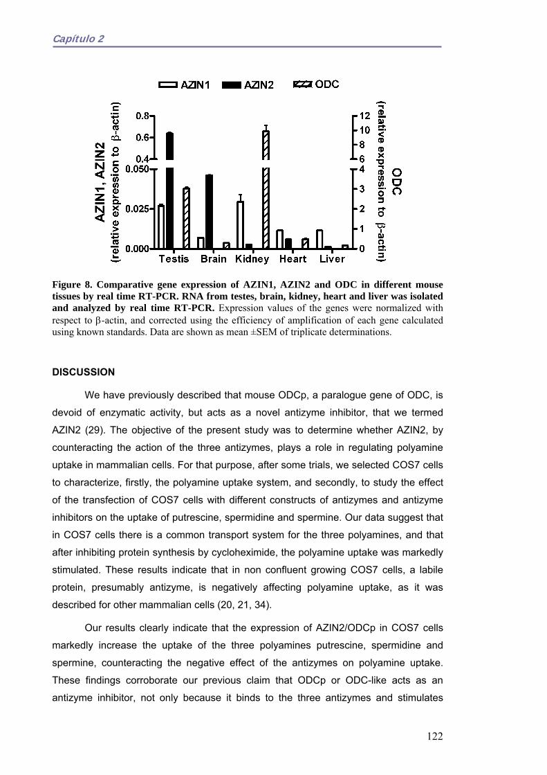
Capítulo 2
Figure 8. Comparative gene expression of AZIN1, AZIN2 and ODC in different mouse tissues by real time RT-PCR. RNA from testes, brain, kidney, heart and liver was isolated and analyzed by real time RT-PCR. Expression values of the genes were normalized with respect to β-actin, and corrected using the efficiency of amplification of each gene calculated using known standards. Data are shown as mean ±SEM of triplicate determinations.
DISCUSSION
We have previously described that mouse ODCp, a paralogue gene of ODC, is
devoid of enzymatic activity, but acts as a novel antizyme inhibitor, that we termed
AZIN2 (29). The objective of the present study was to determine whether AZIN2, by
counteracting the action of the three antizymes, plays a role in regulating polyamine
uptake in mammalian cells. For that purpose, after some trials, we selected COS7 cells
to characterize, firstly, the polyamine uptake system, and secondly, to study the effect
of the transfection of COS7 cells with different constructs of antizymes and antizyme
inhibitors on the uptake of putrescine, spermidine and spermine. Our data suggest that
in COS7 cells there is a common transport system for the three polyamines, and that
after inhibiting protein synthesis by cycloheximide, the polyamine uptake was markedly
stimulated. These results indicate that in non confluent growing COS7 cells, a labile
protein, presumably antizyme, is negatively affecting polyamine uptake, as it was
described for other mammalian cells (20, 21, 34).
Our results clearly indicate that the expression of AZIN2/ODCp in COS7 cells
markedly increase the uptake of the three polyamines putrescine, spermidine and
spermine, counteracting the negative effect of the antizymes on polyamine uptake.
These findings corroborate our previous claim that ODCp or ODC-like acts as an
antizyme inhibitor, not only because it binds to the three antizymes and stimulates
122

Capítulo 2
ODC activity (29), but also by negating the repressing effect of any of the three
antizymes on polyamine uptake. Moreover, the fact that the deletion in AZIN2 of the
sequence equivalent to the antizyme binding domain, existing in mouse ODC (37) and
in AZIN1 (38, 39), abolished the stimulatory effect of AZIN2 on polyamine uptake, also
supports that this domain is critical for the binding of AZIN2 to the antizyme molecule.
This result and previous findings on the binding of mouse ODC and AZIN1 to antizyme
(7, 17, 19, 26) support the contention that ODC, and their homologous proteins AZIN1
and AZIN2, share the ability to bind to antizymes, and that in this function the antizyme
binding element (AZBE) is a common and indispensable element. On the other hand,
our results also provide evidence that in AZIN2, the sequences containing the ~forty
terminal residues in both N and C ends are not essential for binding to antizymes.
After our report that mouse ODCp acted as an antizyme inhibitor devoid of both
ornithine decarboxylase and arginine decarboxylase activities (29), it was published
that the human orthologue of ODCp also inhibited AZ1-dependent degradation of ODC
as the murine protein (30), confirming our previous results. Our present findings have
also revealed that the human AZIN2 is able to stimulate polyamine uptake, apparently
as efficiently as mouse AZIN2 or AZIN1. This suggests that the sophisticated network
regulating polyamine metabolism mediated by antizymes and antizyme-binding
proteins is conserved from mice to humans. In fact, ODC and the two antizyme
inhibitors can bind to any of the three antizymes characterized so far. Among the
antizyme family members, AZ1 and AZ2 are expressed in numerous mammalian
tissues (40), whereas AZ3 expression is restricted to the testes (22). Both AZ1 and
AZ2 inhibit ODC and polyamine uptake (20, 21, 33). AZ3 also possesses ODC
inhibitory activity (22) but no data are available about its possible role as inhibitor of
polyamine uptake. Our results on polyamine uptake by COS7 cells transfected with
AZ3, clearly indicated that AZ3 may also negatively affect polyamine transport.
Since antizymes are able to decrease polyamine uptake, it was believed that
antizyme inhibitor should prevent the negative effect of antizymes on polyamine
transport. This prediction was confirmed by showing that the overexpression of AZIN1
in CHO cells produced a moderate increase in the uptake of spermine and some
polyamine analogues (41). Other experiments using NIH3T3 fibroblasts transfected
with AZIN1 have corroborated that overexpression of AZIN1 increases the uptake of
spermidine (27). In contrast to these results, no increase in spermine uptake was
observed in rat prostate carcinoma cells overexpressing AZIN1 (39). Our results on
COS7 cells also support that AZIN1 increases polyamine uptake, at least as efficiently
as AZIN2. Recently, Snapir et al. found that AZIN2, stably transfected into NIH 3T3
123

Capítulo 2
cells, exerted a modest increase on the spermidine uptake rate (less than 20%), when
compared with the effect of AZIN1 (about 100%), concluding that AZIN2 is less potent
than AZIN1 in regulating polyamine uptake (42). The higher increase in polyamine
uptake rate produced by both AZINs (about 300%) in our system in comparison to that
observed in stably transfected cells could be related with a lower AZs/AZINs ratio.
Nevertheless, in COS7 cells transiently cotransfected with AZ1 and AZIN1 or AZIN2,
both antizyme inhibitors exerted a similar effect (result not shown). In this regard, both
mouse and human AZIN1 and AZIN2 showed similar capacity in counteracting the
effects of AZs on ODC activity (29,30).
The physiological role of antizyme inhibitors is far from being understood.
Different experiments have indicated that AZIN1 may be related with the control of
cellular growth, not only by acting on antizymes but also through antizyme independent
effects (27). In fact, overexpression of AZIN1 increased growth rate and induced the
transformed phenotype in different type of cells (27, 39), while silencing its expression
using specific small interfering RNA (siRNA) decreased cell proliferation (27, 39, 43).
Moreover, AZIN1 is up regulated in several human cancers (44) and is rapidly
stimulated in cultured fibroblasts by serum and phorbol esters (45). AZIN1 is also up
regulated in capillary endothelial cells by fibroblast growth factor 2, a known angiogenic
factor (46). Coincidentally, the human AZIN1 gene is located in a region of
chromosome 8 that is amplified in some tumours (47, 48). In normal tissues, AZIN1
transcripts have been found in the liver, lung, kidney and heart of rats (26), but little is
known about its expression in mouse tissues. On the other hand, AZIN2 appears to be
expressed exclusively in the brain and testes, both in mouse and humans (29, 31).
However, there are not quantitative data comparing the expression of both genes in
mammalian tissues. Our results of real time RT-PCR revealed that in the mouse, the
expression levels of ODC and antizyme inhibitors among the different tissues assayed
varied significantly. ODC was the most expressed in all tissues analyzed. Interestingly,
AZIN2 was much more expressed than AZIN1 in brain and testes. In kidney and heart
the expression level of AZIN2 was lower than AZIN1, although the expression of the
latter in these tissues was far from the value of AZIN2 in testes. The fact that AZIN2
mRNA is mostly found in testes, where there is a specific testicular antizyme (AZ3)
expressed in the haploid germinal cells (22, 23), linked to our findings that AZ3 affect
polyamine uptake and that AZIN2 may counteract this effect, strongly suggests that
both proteins may be important to achieve the severe control of intracellular
polyamines, that appears to be required at the later stages of spermatogenesis (49).
124
Capítulo 2
Collectively, our previous results on the functional activity of AZIN2 regulating
ODC activity, and the present results showing that this protein markedly stimulates
polyamine uptake and is considerably expressed in testes and brain, suggest that it is
quite likely that AZIN2 may be an important and novel player in polyamine metabolism
in these tissues. The precise role of this protein in testicular and neuronal functions, as
well as its possible implication in malignant growth, is a subject of ongoing
investigations in our laboratory.
REFERENCES. 1. Cohen S.S. (1998) A guide to the polyamines, Oxford University Press, New York
2. Thomas, T., and Thomas, T.J. (2001) Cell.Mol.Life Sci. 58, 244-258
3. Wallace, H.M., Fraser, A.V., and Hughes, A. (2003) Biochem. J. 376, 1-14
4. Childs, A.C., Mehta, D.J., and Gerner, E.W. (2003) Cell.Mol.Life Sci. 60, 1394-1406
5. Igarashi, K., and Kashiwagi, K. (2000) Biochem.Biophys.Res.Commun. 271, 559-564
6. Johnson, T.D. (1996) Trends in Pharmacol. Sci. 17, 22-27
7. Coffino, P. (2001) Nat.Rev.Mol.Cell Biol. 2, 188-194
8. Seiler, N. (2004) Amino Acids 26, 217-233
9. Pegg, A.E. (1988) Cancer Res. 48, 759-774
10. Gerner, E.W., and Meyskens, F.L. (2004) Nat.Rev.Cancer 4, 781-792
11. Casero, R.A., and Marton, L.J. (2007) Nat. Rev.Drug Discov. 6, 373-390
12. Auvinen, M., Paasinen, A., Andersson, L.C., and Holtta, E. (1992) Nature 360, 355-358
13. Pegg, A. E. (2006) J.Biol.Chem. 281, 14529-14532
14. Seiler, N., Delcros, J.G., and Moulinoux, J.P. (1996) Int.J. Biochem. Cell Biol. 28, 843-861
15. Igarashi, K., and Kashiwagi, K. (1999) Biochem. J. 344, 633-642
16. Soulet, D., Covassin, L., Kaouass, M., Charest-Gaudreault, R., Audette, M., and Poulin, R. (2002) Biochem. J. 367, 347-357
17. Mangold, U. (2005) Iubmb Life 57, 671-676
18. Matsufuji, S., Matsufuji, T., Miyazaki, Y., Murakami, Y., Atkins, J. F., Gesteland, R.F., and Hayashi, S. (1995) Cell 80, 51-60
19. Hayashi, S., Murakami, Y., and Matsufuji, S. (1996) Trends Biochem. Sci. 21, 27-30
125

Capítulo 2
20. Mitchell, J.L.A., Judd, G.G., Bareyal-Leyser, A., and Ling, S.Y. (1994) Biochem. J. 299, 19-22
21. Suzuki, T., He, Y., Kashiwagi, K., Murakami, Y., Hayashi, S., and Igarashi, K. (1994) Proc.Natl.Acad.Sci.U.S.A. 91, 8930-8934
22. Ivanov, I.P., Rohrwasser, A., Terreros, D.A., Gesteland, R.F., and Atkins, J.F. (2000) Proc.Natl.Acad.Sci.U.S.A. 97, 4808-4813
23. Tosaka, Y., Tanaka, H., Yano, Y., Masai, K., Nozaki, M., Yomogida, K., Otani, S., Nojima, H., and Nishimune, Y. (2000) Genes to Cells 5, 265-276
24. Kitani, T., and Fujisawa, H. (1989) Biochim. Biophys. Acta 991, 44-49
25. Mangold, U. (2006) Cell.Mol.Life Sci. 63, 2095-2101
26. Murakami, Y., Ichiba, T., Matsufuji, S., and Hayashi, S. (1996) J.Biol.Chem. 271, 3340-3342
27. Keren-Paz, A., Bercovich, Z., Porat, Z., Erez, O., Brener, O., and Kahana, C. (2006) Oncogene 25, 5163-5172
28. Zhu, M.Y., Iyo, A., Piletz, J., and Regunathan, S. (2004) Biochim. Biophys. Acta 1670, 156–164
29. Lopez-Contreras, A.J., Lopez-Garcia, C., Jimenez-Cervantes, C., Cremades, A., and Penafiel, R. (2006) J.Biol.Chem. 281, 30896-30906
30. Kanerva, K., Matikie, L. T., Pelander, A., Heiskala, M., and Andersson, L.C. (2008) Biochem. J. 409, 187-192
31. Pitkanen, L.T., Heiskala, M., and Andersson, L.C. (2001) Biochem.Biophys.Res.Commun. 287, 1051-1057
32. Mangold, U., and Leberer, E. (2005) Biochem. J. 385, 21-28
33. Zhu, C., Lang, D.W., and Coffino, P. (1999) J.Biol.Chem. 274, 26425-26430
34. Sambrook, J., Fritsch, E.F., and Maniatis, T. (1989) Molecular Cloning, 2nd Ed., Cold Spring Harbor Laboratory Press, Cold Spring Harbor, NY
35. Bradford, M.M. (1976) Anal.Biochem. 72, 248-254
36. Mitchell, J.L.A., Diveley, R.R., and Bareyal-Leyser, A. (1992) Biochem.Biophys.Res.Commun. 186, 81-88
37. Li, X.Q., and Coffino, P. (1992) Mol.Cell.Biol. 12, 3556-3562
38. Bercovich, Z., and Kahana, C. (2004) J.Biol.Chem. 279, 54097-54102
39. Kim, S.W., Mangold, U., Waghorne, C., Mobascher, A., Shantz, L., Banyard, J., and Zetter, B.R. (2006) J. Cell Sci. 119, 2853-2861
40. Ivanov, I.P., Gesteland, R.F., and Atkins, J.F. (1998) Genomics 52, 119-129
126

Capítulo 2
41. Mitchell, J.L.A., Simkus, C.L., Thane, T.K., Tokarz, P., Bonar, M.M., Frydman, B., Valasinas, A.L., Reddy, V.K., and Marton, L.J. (2004) Biochem. J. 384, 271-279
42. Snapir, Z., Keren-Paz, A., Bercovich, Z., and Kahana, C. (2008) Biochem.J. 410, 613-619
43. Choi, K.S., Suh, Y.H., Kim, W.H., Lee, T.H., and Jung, M.H. (2005) Biochem.Biophys.Res.Commun. 328, 206-212
44. Jung, M.H., Kim, S.C., Jeon, G.A., Kim, S.H., Kim, Y., Choi, K.S., Park, S.I., Joe, M.K., and Kimm, K. (2000) Genomics 69, 281-286
45. Nilsson, J., Grahn, B., and Heby, O. (2000) Biochem. J. 346, 699-704
46. Takahashi, Y., Li, L. L., Kamiryo, M., Asteriou, T., Moustakas, A., Yamashita, H., and Heldin, P. (2005) J.Biol.Chem. 280, 24195-24204
47. Schaner, M.E., Davidson, B., Skrede, M., Reich, R., Florenes, V.A., Risberg, B., Berner, A., Goldberg, I., Givant-Horwitz, V., Trope, C.G., Kristensen, G.B., Nesland, J.M., and Borresen-Dale, A.L. (2005) Molecular Cancer 4, 26
48. van Duin, M., van Marion, R., Vissers, K., Watson, J.E. ., van Weerden, W.M., Schroder, F.H., Hop, W.C.J., van der Kwast, T.H., Collins, C., and van Dekken, H. (2005) Genes Chromosomes & Cancer 44, 438-449
49. Hakovirta, H., Keiski, A., Toppari, J., Halmekyto, M., Alhonen, L., Janne, J., and Parvinen, M. (1993) Mol. Endocrinol. 7, 1430-1436
127

Capítulo 2
128

Capítulo 3
CAPÍTULO 3
La expresión del inhibidor de antizimas 2 (AZIN2) en células haploides germinales masculinas sugiere su papel en la espermiogénesis.
RESUMEN.
En estudios previos se ha demostrado la expresión del inhibidor de antizimas 2
(AZIN2), un nuevo miembro de la familia de proteínas de unión a antizimas, en
testículo de ratón y humano. En el presente trabajo, hemos estudiado la expresión
espacial y temporal de AZIN2 y otros genes relacionados con el metabolismo de
poliaminas, como ornitina descarboxilasa (ODC), antizima 3 (AZ3) y el inhibidor de
antizimas 1 (AZIN1), en testículo de ratón, en un intento de conocer el papel de AZIN2
en la fisiología testicular. El patrón de expresión temporal de estos genes durante la
primera onda de espermatogénesis se estudió mediante RT-PCR a tiempo real, y la
distribución espacial se examinó mediante hibridación in situ de ARN e
inmunocitoquímica. Los resultados mostraron que AZIN2 se expresó específicamente
en células haploides germinals, de forma similar a AZ2. Sin embargo, el ARNm de
ODC se encontró fundamentalmente en la parte externa de los túbulos seminíferos,
donde se localizan las espermatogonias y los espermatocitos. Experimentos
funcionales de transfección y experimentos de co-inmunoprecipitación corroboraron
que AZIN2 revierte la acción inhibitoria de AZ3 sobre la actividad ODC y la captación
de poliaminas. La microscopía de fluorescencia mostró que la proteína AZIN2 se
localiza fundamentalmente en estructuras membranosas perinucleares. En conjunto,
estos resultados sugieren que AZIN2 puede jugar un importante papel durante la
espermiogénesis.
129

Capítulo 3
ABSTRACT.
Previous studies demonstrated the expression of ornithine decarboxylase
antizyme inhibitor 2 (AZIN2), a novel member of the family of antizyme binding proteins,
in the testes of mice and humans. In the present work, the spatial and temporal
expression of AZIN2, and other genes related with polyamine metabolism such as
ornithine decarboxylase (ODC), antizyme 3 (AZ3) and antizyme inhibitor 1 (AZIN1)
were studied in the mouse testis, in an attempt to understand the role of AZIN2 in
testicular physiology. The temporal expression of the genes during the first wave of
spermatogenesis was studied by real time RT-PCR, and the spatial distribution was
examined by RNA in situ hybridization and immunocytochemistry. The results indicated
that AZIN2 was specifically expressed in the haploid germinal cells, similarly to AZ3.
However, ODC mRNA was mainly found in the outer part of the seminiferous tubules
where spermatogonia and spermatocytes are located. Functional transfection assays
and co-immunoprecipitation experiments corroborated that AZIN2 counteracts the
negative action of AZ3 on ODC activity and polyamine uptake. Fluorescent microscopy
showed that AZIN2 protein was mainly located in perinuclear membranous structures.
All these results strongly suggest that AZIN2 may have a role in spermiogenesis.
INTRODUCTION.
Although the crystallization of spermine phosphate in human semen was
observed by Leeuwenhoek in the XVII century (ref. in (1)), the physiological role of
polyamines in the male reproductory system is still poorly understood. The polyamines
putrescine, spermidine and spermine are polycations ubiquitously distributed in
mammals that play a relevant role in processes of growth and cell differentiation (2-6).
In this regard, it is conceivable that polyamines may have a role in testicular physiology,
since various mitotic, meiotic, and differentiation cellular mechanisms are required for
the production of mature spermatozoa by the testis.
Intracellular polyamine levels are tightly controlled by the regulation of
biosynthesis, degradation, uptake and secretion (6,7). A key component in the
polyamine biosynthetic pathway is ornithine decarboxylase (ODC), enzyme that
converts ornithine into putrescine, diamine that serves as a precursor of spermidine
and spermine (8). ODC is subject to a complex regulation exerted at transcriptional,
translational and post-translational levels (8,9). ODC antizymes play an important role
in the control of intracellular levels of polyamines by inhibiting ODC and promoting its
degradation by the proteasome in an ubiquitin-independent manner and also by
130

Capítulo 3
restricting polyamine uptake (10,11). Three mammalian antizymes (AZ) named AZ1,
AZ2 and AZ3 have been characterized (12). AZ1 and AZ2 are present in numerous
mammalian tissues (13), whereas AZ3 has been found exclusively in the testis (14,15).
AZ expression requires an unique mechanism of translational frameshifting mediated
by polyamines (16). The action of antizymes is affected by the antizyme inhibitors
(AZIN), proteins that are highly homologous to ODC, but devoid of ODC activity (17).
The first characterized antizyme inhibitor (18,19), later named AZIN1, is expressed in
different tissues and binds strongly to antizymes (12,19). The second antizyme inhibitor
(AZIN2) has been found in testis and brain, both in mice and humans (20,21).
In the testis there are two compartments: seminiferous tubules (formed by germ
cells and supporting Sertoli cells) and the interstitium (containing various types of cells,
including Leydig cells that produce testosterone). In the seminiferous tubules there are
several germ cell populations in various stages of differentiation: spermatogonia,
spermatocytes, spermatids and spermatozoa. Spermatogonial stem cells are converted
into functionally competent spermatozoa during the complex process of
spermatogenesis (22,23). Spermiogenesis is the final stage of spermatogenesis in
which spermatids differentiate into spermatozoa. Early studies reported the existence
of significant amounts of polyamines in the rat testis and an uneven distribution of ODC
activity and polyamines in the different types of testicular cells (24-27). Changes in
ODC activity and ODC mRNA levels were also found during sexual development of
male rats and mice (28-31). Moreover, detailed analysis of the temporal and regional
expression profile of ODC mRNA in mouse testis suggested that polyamines may play
an important role during late meiosis and early spermiogenesis (29). In addition,
transgenic mice with overexpression of human ODC in the testis showed reduced
fertility (32). A detailed analysis of these transgenic mice showed higher DNA synthesis
in spermatogonia and a reduction in the number of meiotic and particularly post meiotic
cells (33). Based on these findings, it was proposed that putrescine has selective local
stimulatory and inhibitory actions during spermatogenesis. The importance of testicular
polyamine homeostasis in murine spermatogenesis was reinforced by the discovery of
antizyme 3, a new paralogue of mammalian antizymes expressed specifically in the
testis (14,15). The restricted expression of AZ3 in haploid germ cells (15) was
consistent with the idea that higher levels of ODC activity are required during the
spermatocytic and early spermatidal phases of spermatogenesis, but may be
detrimental to spermiogenesis at later stages.
We recently found that a paralogous gene of mouse ODC, named ODC-like or
ODCp (34), which is expressed in testis and brain, encodes a protein acting as an
131

Capítulo 3
antizyme inhibitor, termed antizyme inhibitor 2 (AZIN2) (20). In an attempt to
understand better the possible role of AZIN2 in testicular physiology, we first measured
the expression profile of AZIN2 in the mouse testis during the first wave of
spermatogenesis, as well as those of AZ3, ODC and AZIN1, by using real-time RT-
PCR. Secondly, we analyzed the localization of AZIN2 in testicular cells by in situ RNA
hybridization and immunocytochemistry. Finally, we also studied functional interactions
between AZIN2 and AZ3, and the subcellular distribution of AZIN2, by using transient
transfection assays of HEK293 cells and co-immunoprecipitation experiments. The
results suggest that AZIN2 is a novel modulator of polyamine metabolism, at least
during spermiogenesis.
EXPERIMENTAL PROCEDURES.
Materials.
MMLV reverse transcriptase, GenElute mammalian total RNA Miniprep kit, Taq
DNA polymerase, anti-FLAG M2 monoclonal antibody peroxidase, anti-HA monoclonal
antibody peroxidase conjugate, anti-HA affinity gel beads, protease inhibitor mixture
(EDTA, 4-(2-aminoethyl) benzenesulfonyl fluoride, bestatin, E-64, leupeptin and
aprotinin), Igepal CA-360, were purchased from Sigma (St. Louis, MO). Pfu DNA
polymerase was obtained from Biotools (Madrid, Spain). SYBR Green® PCR Master
Mix was from Applied Biosystems (Warrington, UK). Restriction endonucleases EcoRI,
XbaI and BamHI were from Fermentas Life Sciences (Vilnius, Lithuania). 14C-
Putrescine (specific activity 107 mCi/mmol), 14C-Spermidine (specific activity 112
mCi/mmol), 14C-Spermine (specific activity 113 mCi/mmol), ECL+ detection reagent,
developing reagents and films were from Amersham Biosciences (Little Chalfont,
Buckinghamshire, UK). L-[1-14C]Ornithine (specific activity, 4.7 mCi/mmol) was
purchased from Moravek Biochemicals Inc. (Brea, CA). Lipofectamine 2000
Transfection Reagent and Trypsin-EDTA were purchased from Invitrogen (Carlsbad,
CA). Primers were purchased from Sigma Genosys (Cambridge, UK). Scintillation
solution Ecoscint-H was obtained from National Diagnostics (Atlanta, GA).
Animals.
Swiss CD1 mice were provided by the Service of Laboratory Animals of the
University of Murcia. Animals were fed with standard chow (UAR A03; Panlab,
Barcelona, Spain) and water ad libitum, and maintained at 22ºC and 55% relative
humidity under a controlled 12:12 hours light-dark cycle. Animals were sacrificed by
132

Capítulo 3
cervical dislocation. Manipulations were carried out according to the institutional
guidelines of the University of Murcia, in compliance with national (RD 1201/2005) and
international laws and policies (European Economic Community Council Directive
86/009).
RNA extraction and RT-PCR.
Total RNA was extracted from mouse testis at different postnatal days with the
GenElute mammalian total RNA Miniprep kit, following the manufacturer’s instructions.
Total RNA was reverse-transcribed using (dT)18 as primer and MMLV reverse
transcriptase. Real time RT-PCR was carried out as described elsewhere (35) The
following primers were used for real time RT-PCR: qβ-actin (forward, 5’-
GATTACTGCTCTGGCTCCTAG CA-3’; reverse, 5’-
GCTCAGGAGGAGCAATGATCTT-3’); qAZIN1 (forward, 5’-CTTTCCAC GAACC
ATCTGCT-3’; reverse, 5’-TTCCAGCATCTTGCATCTCA-3’); qAZIN2 (forward, 5’-
GCTTAGAGGGAGCCAAAGTG-3’; reverse, 5’-CTCAGCAAGGATGTCCACAC-3’);
qODC (forward, 5’-ATGGGTTCCAGAGGCCAAA-3’; reverse, 5’-CTGC
TTCATGAGTTGCCACATT-3’); qAZ3 (forward, 5’-CCAGGTGGGTAGGAGCACT- 3’;
reverse, 5’-AAGCAGGGGGTCAGTTGATA-3’).
Semi-quantitative RT-PCR was performed as previously described (20). The
primers used were: β-actin (forward, 5’- TGCGTCTGGACCTGGCTG-3’; reverse, 5’-
CTGCTGGAAGGTGGACAG-3’); AZIN2 (forward, 5’-TGAATCGGACTT TGTGATGG -
3’; reverse, 5’- GGTAGCAATGCACAGAACC -3’).
RNA in situ hybridization.
The testes were fixed for 2-3 days in phosphate buffered 4% paraformaldehyde
at 4°C. They were dehydrated in methanol series (25-100%) and embedded in paraffin.
The paraffin blocks were cut serially into 100 µm-thick transversal sections. The
sections were rehydrated in a 100-25% ethanol series. They were treated with
proteinase K (20 μg/ml) in phosphate buffered saline for 5 min at ambient temperature,
and then were post-fixed in 4% paraformaldehyde. This was followed by an acetylating
step with acetic anhydride in triethanolamine buffer, dehydration through the ethanol
series, and drying by evaporation. Mouse AZIN2 and ODC RNA antisense probes were
designed against a less conserved region, corresponding to the downstream ORF
region and part of 3’ UTR of the genes, and mouse AZ3 probe was against the full ORF.
The desired fragments were cloned into pcDNA3 using the appropriate primers
133

Capítulo 3
basically as described elsewhere (20) and the plasmids were linearized with EcoRI.
The cDNA were transcribed in vitro (Promega kit) using SP6 polymerase and
digoxigenin-UTP. The digoxigenin labeled probes were purified by means of RNA
purification columns (Roche, Indinapolis, USA). RNA probes at 1 µg/ml concentration
were used for all the experiments. Hybridization was performed at 50°C for 14 h, with
the probes diluted in a hybridization buffer containing 57% formamide, 340 mM NaCl,
20 mM Tris HCl pH 7.4, 5 mM ethylenediaminetetraacetic acid, 11% dextran sulfate,
Denhardt ×1, 500 μg/ml yeast RNA, and 10 mM NaH2PO4 pH 8.0. Slides were then
washed three times in MABT (0.1 M maleic acid, 0.2 M NaOH, 0.2 M NaCl, 0.01%
Tween), incubated for 1 h in blocking solution (10% blocking solution and 10% sheep
serum in MABT), and subsequently incubated overnight with anti-digoxigenin antibody
(1:5000; Roche) diluted in a solution containing 1% sheep serum and 1% blocking
solution in MABT. Slides were next washed 10–15 times each for 30 min in MABT, with
a final overnight wash in the same solution. Next, the slides were washed three times
for 5 min in reaction buffer (0.1 M Tris, pH 9.5, 0.1 M NaCl, and 50 mM MgCl2) and
incubated in the dark in nitroblue tetrazolium (NBT)/5-bromo-4-chloro-3-indolyl
phosphate (BCIP) solution [3.4 µl/ml from NBT stock and 3.5 µl/ml from BCIP stock in
reaction buffer (100 mg/ml NBT stock in 70% dimethylformamide; 50 mg/ml BCIP stock
in 100% dimethylformamide; Roche)]. Slides were checked periodically and the
reaction was stopped in Tris-EDTA buffer. Finally, they were brought to 100% ethanol
and subsequently dried by evaporation. The primers used for cloning the fragment for
RNA antisense probes were the following: cAZIN2 (fragment size 337 bp) (forward: 5’-
GTCGAATTC(EcoRI)AGCCTGGGAAGCGCTTCGAGG -3’; reverse: 5’-GGC
TCTAGA(XbaI)TA TGTTGCATACTTGAACTAC-3’); cODC (fragment size 447 bp)
(forward: 5’-CTCGAATTC(EcoRI)CAGATCCAGAGCCATGGCTTC-3’; reverse: 5’-
CTTTCTAGA(XbaI)CACAATGGGTCACAACATAAGC-3’); cAZ3(fragment size 627 bp)
(forward, 5’-GCGGAATTC(EcoRI)CT CTACTG TTACAAATAC-3’; reverse 5’-
TTATCTAGA(XbaI)CTCACTGGCCAGGGTGGCC-3’).
Immunohistochemistry.
Testes from adult mice were removed and fixed in 10% formalin in PBS (0.01 M
phosphate buffered saline pH 7.4) for 10 hours. After washing in PBS, samples were
processed routinely, embedded in paraffin and cut in 5μm sections. Sections were
stained with haematoxylin-eosine and immunocytochemistry. For immunocytochemistry,
a two-step method was applied. Tissue sections from paraffin blocks were
deparaffinized in xylene and hydrated in a graded ethanol series. Endogenous
134

Capítulo 3
peroxidase activity was destroyed by treatment with 0.5% hydrogen peroxide in
methanol for 20 min. Sections were washed in three 5 min changes of PBS and then
incubated with normal swine serum (1:100) for 20 min, and then with rabbit polyclonal
antibody to AZIN2 (generated against the peptide ALQKKSADQPLI, corresponding to
residues 339-350 of AZIN2) (1:100) at 4ºC overnight. After washing in PBS, they were
incubated with biotin-conjugated swine anti-rabbit immunoglobulin antibody (DAKO
North America Inc., Carpinteria, CA) (1:250) for 20 min. After three 5 min washing
changes in PBS, avidin-biotin-peroxidase complex (ABC kit:VECTOR, Vector
Laboratories Inc., Burlingame, CA) was added and incubated for 20 min. They were
developed with 0.05% 3,3 diaminobenzidine tetrahydrochloride and 0.015% hydrogen
peroxide. After immunostaining, sections were counterstained with haematoxylin.
Control: substitution of anti-AZIN2 by PBS.
Cloning and genereration of AZIN2 constructs.
ODC, AZIN2, and AZ3 mouse genes were cloned into the expression vector
pcDNA3 (Invitrogen) as described elsewhere (20). AZ3 construct with an appropriate
deletion of one nucleotide in the frameshifting site (MAZ3), for full-length and functional
expression, was obtained by the QuickChange site-directed mutagenesis kit (20).
AZIN2 and MAZ3 with the FLAG or HA epitopes were generated as described in (20).
The green fluorescent protein sequence (GFP) was added to the C-terminal of AZIN2
by subcloning in the vector pEGFP-N2 (BD Biosciences Clontech) as described earlier
(35).
Cell culture and transient transfection.
HEK 293T cells were cultured in Dulbecco’s modified Eagle medium (DMEM)
supplemented with 10% fetal calf serum, 100 units/ml penicillin, and 100 μg/ml
streptomycin, at 37 °C under a 5% CO2 humidified atmosphere. Cells were grown to
about 80% confluence. Transient transfections were carried out with Lipofectamine
2000 transfection reagent (Invitrogen, Carlsbad, CA) as described elsewhere (20).
ODC assays. ODC activity was assayed in the soluble fraction of HEK 293T cells lysed
in a media containing the detergent Igepal. The extract was centrifuged at 20000xg for
20 min, and ODC activity was determined in the supernatant. ODC activity was
assayed basically as described elsewhere (20), by measuring 14CO2 release from L-
[1-14C] ornithine (specific activity, 4.7 mCi/mmol, Moravek Biochemicals Inc. Brea, CA).
Protein content was calculated using the method of Bradford (36).
135

Capítulo 3
Western Blotting.
Cells were solubilized in 50 mM Tris-HCl (pH 8), 1% Igepal, 1 mM EDTA, and
protease inhibitor mixture and centrifuged at 12000xg for 20 min. Reducing SDS-PAGE
was performed in 10% polyacrylamide gels. Gels were transferred to polyvinylidene
difluoride membranes, blocked with 5% non-fat dry milk in PBS, and incubated
overnight at 4 °C with the anti-FLAG antibody peroxidase-labeled (1:5000), anti-HA
antibody (1:20000) or anti-AZIN2 antibody (1/500). Immunoreactive bands were
detected by using ECL+ as detection reagent and commercial developing reagents and
films. For loading controls, Erk2 was determined by means of polyclonal anti-Erk2
antibody (Santa Cruz Biotechnology Inc., Santa Cruz, CA).
Immunoprecipitation.
HEK 293T cells were transfected with AZIN2-FLAG, alone or in combination
with HA epitope-labeled mutated AZ3. Approximately 2 x 10⁶ cells were processed and
immunoprecipitated by means of anti-HA affinity gel beads, as described elsewhere
(20).
Polyamine uptake assay.
HEK 293T cells grown to ~80% of confluence were washed with DMEM
medium (serum free) to remove all traces of serum, and then 200 μL of fresh DMEM
medium (serum free) was added to the cells. The uptake assay was started by the
addition of 2 μM 14C-putrescine. After incubation at 37 °C for different periods of time,
the cells were washed three times with cold PBS, lysed and radioactivity was counted.
136

Capítulo 3
RESULTS.
Temporal and regional expression of AZIN2 mRNA in the mouse testis.
The levels of AZIN2 mRNA were studied by RT-PCR in the testes of mice of
different ages. Semiquantitative analysis (Fig. 1A) showed that the expression of
AZIN2 in the testis of prepuberal mice was very low, but increased markedly after
puberty. To study with more detail the profile of AZIN2 expression during the first wave
of spermatogenesis (23), RNA was isolated from the testes of mice of 4, 10, 15, 19, 20,
21, 22, 23, 24, 27, 31, 34 and 60 days of age, and the expression levels of AZIN2 were
quantified by real-time RT-PCR. Fig. 1B shows that transcript levels were very low until
day 24, and that they markedly increased after this day. The onset of AZIN2 expression
in the testis corresponds with the appearance of round spermatids, and transcription
increased throughout the differentiation process from spermatids to spermatozoa. Fig.
2 compares the expression of AZIN2 with those of ODC and AZ3, genes related to
polyamine metabolism in the testis, and with AZIN1, the other antizyme inhibitor protein.
AZ3 showed an expression pattern similar to that of AZIN2, with extremely reduced
expression during the 3 first postnatal weeks, but increasing significantly during the
fourth week. On the other hand, the expression of ODC rose steadily from birth to
adulthood, while AZIN1 expression increased during the second and third weeks after
birth, reaching at the end of this period values equivalent to those of sexually mature
males. It should be noted that the relative expression values of AZ3 and ODC in the
testis of adult mice were about 5-6 fold higher than the expression of AZIN2. However,
the expression of this gene was about 25-fold higher than that of AZIN1. Transcripts
corresponding to AZ1 and AZ2 were also detected in the adult testis, with relative
expression values of 40% and 14%, compared to AZ3.
137

Capítulo 3
138
Figure 1. Antizyme inhibitor 2 expression during postnatal testis development. A) Semiquantitative RT-PCR analysis of the levels of AZIN2 mRNA at 4, 15, 22 and 60 days of age. βA: β actin; AZIN2: antizyme inhibitor 2. B) Real time RT-PCR analysis of AZIN2 transcripts during testis development. β-actin and L19 ribosomal protein were used as controls. Values are expressed with respect to the value of day 4 that was taken as 1. At least three animals for each age were used. The initial appearance of the different germ cell types during the first wave of spermatogenesis is marked in the bottom of the graph. SpG: spermatogonia; SpC: spermatocyte; PSpC: pachytene spermatocyte; RSpT: round spermatid; ESpT: elongated spermatid; SpZ: spermatozoa.
0 10 20 30 40 50 60 700
100
200
300
400
500
Days post-partum
AZI
N2
(rel
ativ
e ex
pres
sion
)
AZI
N2
AZI
N2
AZI
N2
AZI
N2
βA βA βA βA
Days post-partum
4 15 25 60
A)
B)
6 10 14 20 35
SpGSpC
PSpCSpT
SpZ
RSpT ESpT
0 10 20 30 40 50 60 700
100
200
300
400
500
Days post-partum
AZI
N2
(rel
ativ
e ex
pres
sion
)
AZI
N2
AZI
N2
AZI
N2
AZI
N2
βA βA βA βA
Days post-partum
4 15 25 60
AZI
N2
AZI
N2
AZI
N2
AZI
N2
βA βA βA βA
Days post-partum
4 15 25 60
A)
B)
6 10 14 20 35
SpGSpC
PSpCSpT
SpZ
6 10 14 20 35
SpGSpC
PSpCSpT
SpZ
RSpT ESpT

Capítulo 3
139
Figure 2. Comparative analysis of the expression of antizyme inhibitors, antizyme 3 and ornithine decarboxylase during testis development. mRNA levels were calculated by real
time RT-PCR. For each gene, specific pair of primers described in “Experimental procedures” were used, and the values were normalized with respect to the expression of β-actin. AZIN1: antizyme inhibitor 1; AZIN2; antizyme inhibitor 2, AZ3: antizyme 3; ODC: ornithine decarboxylase.
In order to determine the regional expression of AZIN2 in the mouse testis, RNA
in situ hybridization was performed. The results indicated that AZIN2 transcripts were
mainly found in the inner part of the seminiferous tubules, where spermatids at different
stages of differentiation and spermatozoa are localized (Fig. 3 A,D). The spatial
localization of AZIN2 was similar to that found for AZ3 (Fig. 3 B,E). In contrast, ODC
mRNA was largely localized in the outer part of the seminiferous tubules, consistent
with predominant expression in spertagonia and spermatocytes (Fig. 3 C,F). It must be
emphasized that the probes selected for the detection of AZIN2 and ODC matched with
the 3’UTR of their specific mRNA, since in this region the sequence homology between
the paralogues AZIN2 and ODC is lowest.
4 10 15 19 20 21 22 23 24 27 31 34 600.0
0.2
0.4
0.6
Age (days)
AZI
N2
(rel
ativ
e ex
pres
sion
)
4 10 15 19 20 21 22 23 24 27 31 34 600
1
2
3
4
Age (days)
AZ3
(rel
ativ
e ex
pres
sion
)
4 10 15 19 20 21 22 23 24 27 31 34 600
1
2
3
4
Age (days)
OD
C(r
elat
ive
expr
essi
on)
4 10 15 19 20 21 22 23 24 27 31 34 600.00
0.01
0.02
0.03
0.04
0.05
Age (days)
AZI
N1
(rel
ativ
e ex
pres
sion
)

Capítulo 3
140
Figure 3. Cellular and subcellular expression of AZIN2 in adult mouse testis and HEK 293 cells. (A,D) In situ hybridization of AZIN2. AZIN2 expression is restricted to the inner part of seminiferous tubules. (B,E) In situ hybridization of AZ3. Note an expression pattern similar to AZIN2. (C,F) In situ hybridization of ODC. ODC is mainly expressed in the outer part of the seminiferous tubules. (G,H) Immunocitochemical analysis of AZIN2 in mouse testis. AZIN2 immunoreactivity was localized mainly in the spermatids and spermatozoa in the inner part of the seminiferous ephitelium. (I) Subcellular localization of AZIN2 in HEK293 transfected with a construct of AZIN2 fused to GFP.
Expression of AZIN2 protein in the mouse testis.
To investigate the regional distribution of AZIN2 protein in the testis of adult
mice, immunocytochemical analysis of sections of adult testis was carried out, using a
rabbit anti-AZIN2 polyclonal antibody generated against the synthetic peptide
ALQKKSADQPLY, corresponding to the residues 339 to 351 of mouse AZIN2, purified
by affinity chromatography. Previously, the antibody was validated by western blotting,
using both cell extracts from AZIN2 transfected HEK293 cells and cell extracts from
adult mouse testis. Fig. 4 shows the western blot analysis of extracts of HEK 293 cells
C)A) B)
F)D) E)
H)G) I)
C)A) B)
F)D) E)
H)G) H)H)G)G) I)I)

Capítulo 3
141
transfected with AZIN2, and cytosolic (S-12) and mitochondrial-membranous (P12)
fractions of testis extracts. A specific band of ~50 kDa, corresponding with the
expected size of AZIN2, was observed in the samples of the transfected cells and in
the mitochondrial-membranous fraction of testicular extracts of adult mice. Fig 3G
shows that anti-AZIN2 immunoreactivity was mainly located in the inner part of the
seminiferous tubules. At higher magnification the staining was mainly found in
spermatids and spermatozoa (Fig. 3H).
Figure 4. Western blot analysis of AZIN2. HEK 293T cells were transfected either with the pcDNA3 empty vector or with a construct containing AZIN2 cDNA. The mitochondrial-membrane pellet (P12), obtained after centrifugation of cell homogenates at 12000xg, was assayed by western-blot, using an anti-AZIN2 antibody generated against residues 339-350 of mouse AZIN2 (left). In the right, cytosolic (S12) and mitochondrial-membrane pellet fraction (P12) from homogenates of adult testis were resolved by PAGE-SDS and tested with the same anti-AZIN2 antibody.
Subcellular localization and functional effects of AZIN2 in transient transfected HEK 293 cells.
The subcellular localization of AZIN2 was studied in HEK 293 cells transfected
with AZIN2-GFP. Fig. 3I shows a moderate cytoplasmic presence, and a predominant
fluorescent signal in specific perinuclear regions. A similar pattern was observed in
cells transfected with AZIN2-tag constructs of FLAG or hemagglutinin (results not
shown). These results are in agreement with earlier subcellular fractionation studies
that showed that AZIN2 was mainly found in the mitochondrial-membranous fraction of
HEK 293 cells (20), and with the presence of AZIN2 immunoreactivity in the P12
fraction of testicular extracts (Fig. 4).
To study the influence of AZIN2 on ODC in HEK 293 cells, ODC activity and
ODC protein were measured in single, double and triple transfected HEK 293 cells with
constructs of ODC, ODC+mutatedAZ3 (MAZ3) and ODC+MAZ3+AZIN2, respectively.
AZ3 significantly reduced ODC activity to about 50% of control values, but AZIN2
counteracted the negative effect elicited by AZ3 (Fig. 5A). The effect on ODC protein,
as determined by western blotting, was parallel to that found for ODC activity. These
Anti-AZIN2
pcDNA3 AZIN2
Transfected HEK cells
S12 P1250 KDa
Testis
Anti-AZIN2
pcDNA3 AZIN2
Transfected HEK cells
S12 P1250 KDa
Testis

Capítulo 3
142
results are similar to those found when wild type AZ3 having the early stop codon was
used (20), what suggests that polyamine levels in these transfected cells were
sufficient for inducing frameshifting.
DC protein and polyamine uptake in transient transfected HEK 293T cells. A) HEK 293T cells were single-transfected with ODC, double-transfected with ODC and MAZ3 or triple-transfected with ODC, MAZ3 and AZIN2. In these experiments 0.75 µg of DNA per well was used and the mixtures contained equimolecular amounts of each construct. Upper part: relative ODC activity in transfected cells. Specific ODC activity in control ODC was ~220 nmol of 14CO2 /h mg of protein. Lower part: ODC-FLAG protein levels analyzed by western blotting in the different transfection assays, using. anti-FLAG antibody to detect ODC-FLAG. Erk2 was used as loading control. B) Polyamine uptake by HEK 293T cells transfected with MAZ3 alone or in combination with AZIN2. Control cells were transfected with the empty vector. Polyamine uptake assays were carried out using 2µM 14C-labelled putrescine, spermidine or spermine. Data were corrected by subtracting polyamine uptake corresponding to non-transfected cells according to the transfection efficiency, which was estimated by fluorescence microscopy of GFP protein overexpressed under the same transfection conditions. Data are mean ±SE of triplicate determinations.
Figure 5. Influence of AZ3 and AZIN2 on ODC activity, O
OD
C
OD
C+M
AZ3
OD
C+M
AZ3
+AZI
N2
0
100
200O
DC
act
ivity
(%)
Put Spd Spm0
100
200
C MAZ3 MAZ3+AZIN2
Poly
amin
e up
take
(% c
ontr
ol c
ells
)
A)
B)
ODC
Erk2
1.0±0.1 0.57±0.1 2.3±0.3ODC/Erk2
OD
C
OD
C+M
AZ3
OD
C+M
AZ3
+AZI
N2
0
100
200O
DC
act
ivity
(%)
Put Spd Spm0
100
200
C MAZ3 MAZ3+AZIN2
Poly
amin
e up
take
(% c
ontr
ol c
ells
)
A)
B)
ODC
Erk2
1.0±0.1 0.57±0.1 2.3±0.3ODC/Erk2
ODC
Erk2
1.0±0.1 0.57±0.1 2.3±0.3ODC/Erk2

Capítulo 3
The influence of AZIN2 and AZ3 on polyamine transport was studied by
assaying the rate of radioactive polyamine uptake in transient transfected cells with
MAZ3 or MAZ3+AZIN2, and comparing this uptake with that of cells transfected with
the empty vector pcDNA3. Fig. 5B shows that MAZ3 almost abolished polyamine
uptake, whereas AZIN2 not only blocked the inhibitory action of AZ3 but also
stimulated polyamine uptake above control levels, probably by counteracting the action
of the endogenous antizymes expressed in HEK 293 cells. This effect was observed for
the three polyamines putrescine, spermidine and spermine. These results on the
influence of AZIN2 and AZ3 on polyamine uptake in HEK293T cells are similar to those
we have obtained in a more general study about the action of AZIN2 and AZIN1 on all
members of the AZ family in COS7 cells (López-Contreras et al.,unpublished results).
Analysis of interactions between AZIN2 and AZ3.
Co-immunoprecipitation experiments, using anti-hemagglutinin (HA) beads and
extracts of HEK 293 cells co-transfected with AZIN2-FLAG and pcDNA3 or AZIN2-
FLAG and MAZ3-HA, indicated interaction between AZIN2 and AZ3 (Fig. 6A).
Moreover, in the double transfectants of AZIN2 and AZ3, the amount of AZ3 protein
was markedly increased with respect to the amount found in the single transfectants
(Fig. 6B). Similarly, the level of AZIN2 was also increased by the presence of AZ3 (Fig.
6C). These results indicate not only that AZIN2 and AZ3 may interact with each other in
HEK 293 cells, likely by forming heterodimers, but also that this interaction may
stabilize both proteins under in vivo conditions, decreasing their degradation by the
proteasome. These results are in agreement with a recent report showing that AZ3
stabilized ODCp (AZIN2) by interfering with its ubiquitination (37).
143

Capítulo 3
144
Figure 6. Interactions between AZIN2 and AZ3. HEK 293T cells were transfected with construct encoding an AZIN2-FLAG, alone or in combination with HA epitope-labeled MAZ3 construct (AZ3-HA). Cells were solubilized in buffer containing 1% Igepal, and the supernatants obtained after centrifugation of homogenates at 12000xg were used in immunoprecipitation experiments. (A) Co-immunoprecipitation experiment: extracts were incubated with anti HA-affinity gel beads for 3h, and after extensive washing of the pellet, the eluted proteins were resolved by electrophoresis, and blotted AZIN2-FLAG fusion protein was detected using anti-FLAG antibody. (B) AZ3 levels in cells transfected with AZ3-HA alone or in combination with AZIN2. AZ3-HA protein was detected using an anti-HA antibody. (C) AZIN2 levels in cells transfected with AZIN2-FLAG alone or in combination with MAZ3. AZIN2-FLAG was detected with anti-FLAG antibody. All the experiments were repeated three times with similar results.
+MA
Z3-H
A
AZIN2-FLAG
+pcD
NA3A)
Erk2
MAZ3-HA
+AZI
N2
+pcD
NA3
B)
Erk2
AZIN2-FLAG
+MA
Z3
+pcD
NA3
C)
+MA
Z3-H
A
AZIN2-FLAG
+pcD
NA3A)
+MA
Z3-H
A
AZIN2-FLAG
+pcD
NA3
+MA
Z3-H
A
AZIN2-FLAG
+pcD
NA3A)
Erk2
MAZ3-HA
+AZI
N2
+pcD
NA3
B)
Erk2
AZIN2-FLAG
+MA
Z3
+pcD
NA3
C)

Capítulo 3
DISCUSSION.
In the present study we have analyzed the temporal and spatial expression of
AZIN2, a novel member in the family of antizyme binding proteins, in the mouse testis.
We had previously shown that the murine gene orthologous to the human ODC
paralogue, named ODCp or ODC-like (34) encodes a protein termed AZIN2, devoid of
ornithine or arginine decarboxylase activity but having antizyme inhibitory activity (20).
These results were later corroborated using human ODCp (21). In both cases, the
expression of AZIN2 was restricted to brain and testes. Our present results on the
expression pattern of AZIN2 in mouse testis using RNA in situ hybridization, clearly
indicated that in the adult testis, AZIN2 mRNA was exclusively expressed in haploid
germ cells. This conclusion was also supported by our quantification of the levels of
AZIN2 mRNA by real-time RT-PCR in the testis during postnatal development. In fact,
AZIN2 was almost not expressed in the testis in the postnatal period up to day 20, in
which the mouse testis does not contain any haploid germ cells. AZIN2 expression
sharply started in the testis with the onset of spermiogenesis, and the levels of its
mRNA increased in the more differentiated haploid spermatids. It should be noted that
the expression of AZIN1 did not change during spermiogenesis and the levels of AZIN1
mRNA were much lower than those of AZIN2 (~4%). Immunocytochemical analysis of
adult testis revealed that the AZIN2 protein was localized mainly in spermatids and
spermatozoa, showing again that this protein appears to be exclusive of haploid
germinal cells. These results strongly suggest that AZIN2 may have a role in
spermiogenesis.
Different studies have suggested that polyamines may play an important role
during spermatogenesis. ODC mRNA and protein are present at different levels in all
types of testicular cells (28,30). Moreover, ODC expression and polyamine levels
change significantly during the different phases of spermatogenesis (29,38). ODC
levels in haploid cells appear to be decreased in relation with those found in late
pachytene spermatocytes (28). These data and the interesting finding that transgenic
mice overexpressing ODC in the testis were infertile and presented altered
spermatogenesis (32,33) suggested that putrescine may stimulate DNA synthesis in
spermatogonia but that its excess may be deleterious to postmeiotic cells or may alter
the transition through meiosis (33). The discovery of a new antizyme gene, AZ3 or
OAZ-t, specifically expressed in haploid testicular cells (14,15), reinforced the idea that
the regulation of polyamine levels in haploid germ cells is critical for spermiogenesis.
Our results indicate that AZIN2 and AZ3 have a very similar spatial and temporal
pattern of expression in the mouse testis. On the other hand, while the levels of ODC
145

Capítulo 3
mRNA increased moderately during the first wave of spermatogenesis, the in situ
hybridization results indicated that ODC is largely expressed in cells other than haploid
germinal cells, consistently with previous findings in the literature (28). In addition, the
expression pattern of ODC in immature testes is closer to that of AZIN1 than to that of
AZIN2. Then, it appears to be a certain parallelism between the expression of ODC
and AZIN1 and that of AZ3 and AZIN2, respectively, in the mouse testis. Whereas we
and others found that AZIN2 counteracted some AZ functions as efficiently as AZIN1
(20,21), Snapir et al. (37), in a recent report, concluded that AZIN2 acts as an antizyme
inhibitor but less efficiently than AZIN1. However, even assuming a lower affinity of
AZIN2 for AZs, the higher levels of expression of AZIN2 and its co-expression with AZ3
in the haploid cells of the testis, suggest that it is very likely that AZIN2 and AZ3 are
functionally related.
It has been proposed that the expression of AZ3 in the haploid germ cells is
necessary for a more stringent control of intracellular polyamines at the later stages of
spermatogenesis (15) and that the physiological role of AZ3 is related to the rapid
extinction of ODC activity, after the period in which ODC is required for
spermatogenesis (14). With these premises, the presence of AZIN2 in this complex
scene appears to be paradoxical. It may be argued that AZIN2 is required for a more
rigorous control of polyamine biosynthesis during the later phases of spermiogenesis.
However, other alternatives are also possible, according to our findings. Our data
demonstrate that AZ3 may decrease not only ODC activity and protein level, but also
polyamine uptake. The fact that AZIN2 can interact with AZ3, counteracting the action
of AZ3 on polyamine metabolism, suggests that AZIN2 may participate in the regulation
of polyamine fluxes in the haploid testicular germinal cells. Indeed, changes in
polyamine pools have been reported during spermiogenesis. Significant and
progressive decreases in nuclear spermine levels occur during the transition from
spermatocytes to elongated spermatids (38). This diminution is concomitant with
processes of nuclear remodelling affecting chromatin structure, the replacement of
histones by protamines and transcriptional activity. Moreover, immunocytochemical
analysis of polyamines in the rat testis revealed the accumulation of polyamines in the
residual bodies of the seminiferous tubules, suggesting that polyamines are
sequestered into residual bodies when they are no longer needed for protein synthesis
(39). Taking into consideration that antizymes have been found in cytosolic,
mitochondrial and nuclear localizations (11,40,41) and that our analysis reveals that
AZIN2 is mainly located in membranous structures, one may speculate that AZ3 and
AZIN2 could participate in the redistribution of polyamines in haploid germinal cells.
146

Capítulo 3
Although the present experimental evidence fully supports a key role of
polyamines in murine spermatogenesis, it cannot be excluded that AZIN2 may affect
testicular physiology by regulating processes other than polyamine homeostasis. In fact,
recent experiments using the yeast two-hybrid approach revealed that AZ3 is a
potential interaction partner of gametogenin, a testicular germ-cell specific protein of
unknown function, expressed from late pachytene spermatogenesis through to round
spermatid phase (42). Therefore, it is not unlikely that AZIN2 may affect gametogenin
action through its interaction with AZ3. In addition, since several studies have shown
that antizymes may interact with several small proteins including the transcriptional
regulator Smad1 (43), Snip1 (a repressor of CBP/p300) (44), cyclin D (45) and Aurora-
A kinase (46), the possibility exists that AZIN2 may affect targets other than ODC or
gametogenin. It is also possible that AZIN2, as reported for AZIN1 (47), may interact
with other targets through antizyme-independent mechanisms.
In conclusion, the results presented here provide strong experimental evidence
supporting a role for AZIN2 in testicular physiology. Further studies with transgenic
mice with ablation of the AZIN2 gene are needed in order to understand better the role
of AZIN2 in male gametogenesis.
REFERENCES. 1 Cohen S.S. (1998) A guide to the polyamines, Oxford University Press, New York
2 Gerner, E.W., and Meyskens, F.L. (2004) Nat.Rev.Cancer 4, 781-792
3 Igarashi, K., and Kashiwagi, K. (2000) Biochem.Biophys.Res.Commun. 271, 559-564
4 Jänne, J., Alhonen, L., Keinanen, T.A., Pietila, M., Uimari, A., Pirinen, E., Hyvonen, M. T. and Jarvinen, A. (2005) J. Cell Mol. Med. 9, 865-882
5 Thomas, T. and Thomas, T. J. (2001) Cell. Mol .Life Sci. 58, 244-258
6 Wallace, H. M., Fraser, A. V., and Hughes, A. (2003) Biochem. J. 376, 1-14
7 Seiler, N. (2004) Amino Acids 26, 217-233
8 Pegg, A. E. (2006) J. Biol. Chem. 281, 14529-14532
9 Hayashi, S. and Murakami, Y. (1995) Biochem. J. 306, 1-10
10 Coffino, P. (2001) Nat. Rev. Mol. Cell Biol. 2, 188-194
11 Mangold, U. (2005) Iubmb Life 57, 671-676
12 Mangold, U. and Leberer, E. (2005) Biochem. J. 385, 21-28
13 Ivanov, I.P., Gesteland, R.F. and Atkins, J.F. (1998) Genomics 52, 119-129
147

Capítulo 3
14 Ivanov, I. P., Rohrwasser, A., Terreros, D. A., Gesteland, R. F., and Atkins, J. F. (2000) Proc. Natl. Acad. Sci. U.S.A. 97, 4808-4813
15 Tosaka, Y., Tanaka, H., Yano, Y., Masai, K., Nozaki, M., Yomogida, K., Otani, S., Nojima, H., and Nishimune, Y. (2000) Genes to Cells 5, 265-276
16 Matsufuji, S., Matsufuji, T., Miyazaki, Y., Murakami, Y., Atkins, J. F., Gesteland, R. F., and Hayashi, S. (1995) Cell 80, 51-60
17 Mangold, U. (2006) Cell.Mol.Life Sci. 63, 2095-2101
18 Fujita, K., Murakami, Y. and Hayashi, S. (1982) Biochem. J. 204, 647-652
19 Murakami, Y., Ichiba, T., Matsufuji, S., and Hayashi, S. (1996) J. Biol. Chem. 271, 3340-3342
20 Lopez-Contreras, A.J., Lopez-Garcia, C., Jimenez-Cervantes, C., Cremades, A., and Penafiel, R. (2006) J.Biol.Chem. 281, 30896-30906
21 Kanerva, K., Matikie, L. T., Pelander, A., Heiskala, M. and Andersson, L.C. (2008) Biochem J. 409, 187-192
22 Clermont, Y. (1972) Physiol. Rev. 52, 198-236
23 Oakberg, E.F. (1956) Am. J. Anat. 99, 507-516
24 MacIndoe, J.H., Turkington, R.W. (1973) Endocrinology 2, 595-605
25 Qian, Z.U., Tsai, Y.H., Steinberger, A., Lu, M., Greenfield, A.R.L. and Haddox, M. K. (1985) Biol. Reprod. 33, 1189-1195
26 Shubhada, S., Lin, S. N., Qian, Z.Y., Steinberger, A. and Tsai, Y.H. (1989) J.Androl 10, 145-151
27 Tsai, Y.N. and Lin, S.N. (1985) J. Androl. 6; 348-352
28 Alcivar, A.A., Hake, L.E., Mali, P., Kaipia, A., Parvinen, M. and Hecht, N.B. (1989) Biol. Reprod. 41, 1133-1142
29 Kaipia, A., Toppari, J., Mali, P., Kangasniemi, M., Alcivar, A.A., Hecht, N.B. and Parvinen,M. (1990) Mol. Cell. Endocrinol. 73, 45-52
30 Shubhada, S., Lin, S., Qian, Z., Steinberger, A. and Tsai,Y. (1989 J. Androl. 10, 145-151
31 Weiner, K.X.B. and Dias, J.A. (1992) Biol. Reprod. 46, 617-622
32 Halmekyto, M., Alhonen, L., Wahlfors, J., Sinervirta, R., Eloranta, T. and Jänne, J. (1991) Biochem. J. 278, 895-898
33 Hakovirta, H., Keiski, A., Toppari, J., Halmekyto, M., Alhonen, L., Janne, J. and Parvinen, M. (1993) Gene. Mol. Endocrinology, 7, 1430-1436
34 Pitkanen, L.T., Heiskala, M., and Andersson, L.C. (2001) Biochem.Biophys.Res.Commun. 287, 1051-1057
148

Capítulo 3
35 Lopez-Contreras, A.J., Ramos-Molina, B., Cremades, A. and Peñafiel, R. (2008) J. Biol. Chem. (in press)
36 Bradford, M. M. (1976) Anal. Biochem. 72, 248-254
37 Snapir Z., Keren-Paz, A., Bercovich, Z. and Kahana C. (2008) Biochem. J. 410, 613-619
38 Quemener, V., Blanchard, Y., Lescoat, D., Havouis, R. and Moulineux, J.P. (1992) Am. J. Physiol. Cell Physiol. 263, C343-C347
39 Shin, M., Larsson, L. and Fujiwara, K. (2007) Histochem. Cell Biol. 127, 649-655
40 Gandre, S., Bercovich, Z. and Kahana, C. (2003) Mitochondria 2, 245-256
41 Gritli-Linde, A., Nilsson, J., Bohlooly, Y., Heby, O. and Linde, A. (2001) Dev. Dyn. 220, 259-275
42 Zhang, J., Wang, Y., Zhou, Y., Cao, Z., Huang P. and Lu, B. (2005) FEBS Lett. 579, 559-566
43 Gruendler, C., Lin, Y., Farley, J. and Wang, T. (2001) J. Biol. Chem. 276, 46533-46543
44 Lin, Y., Martin, J., Gruendler, C., Farley, J., Meng, X., Li,B., Lechleider, R., Huff, C., Kim, H., Grasser, W., Paralkar, V. and Wang, T. (2002) BMC Cell Biol. 3, 15
45 Newman, R.M., Mobascher, A., Mangold, U., Koike, C., Diah, S., Schmidt, M., Finley, D., and Zetter, B.R. (2004) J. Biol. Chem. 279, 41504-41511
46 Lim, S.K.. and Gopalan, G. (2007) Oncogene 26, 6593-6603 47 Kim, S.W., Mangold, U., Waghorne, C., Mobascher, A., Shantz, L., Banyard, J. and Zetter,
B.R. (2006) J. Cell Sci. 119, 2853-2861
149

Capítulo 3
150

Capítulo 4
151
CAPÍTULO 4
Subcellular localization of antizyme inhibitor 2 (AZIN2) and their paralog proteins ornithine decarboxylase and AZIN1 in mammalian cells.
RESUMEN.
Ornitina descarboxilasa (ODC), una enzima clave en la ruta biosintética de
poliaminas y los inhibidores de antizimas (AZIN1 y AZIN2), proteínas reguladoras de
los niveles de poliaminas, son proteínas de unión a antizimas. Mientras que es
claramente aceptado que ODC se localiza fundamentalmente en el citosol, existe poca
información sobre la localización subcelular de AZIN1 y AZIN2. Mediante estudios de
microscopía confocal de células HEK 293T y COS7 transfectadas, hemos determinado
que estas proteínas, que poseen una gran homología en su secuencia aminoacídica,
presentaron una localización subcelular diversa. ODC, como había sido previamente
descrito, se encontró principalmente en el citosol, y AZIN1 predominantemente en el
núcleo. Por otro lado, interesantemente, AZIN2 se localizó en el compartimento
intermedio entre el RE y el Golgi (ERGIC) y el cis-Golgi. Aparentemente, esta
localización característica de AZIN2 no está relacionada con secuencias de
localización subcelular conocidas. Además, nuestros estudios sugieren que la región
N-terminal comprendida entre los residuos 39-113 puede ser responsable de su
particular localización, ya que la deleción de esta secuencia suprimió la incorporación
de la AZIN2 mutada al ERGIC. Además, la coexpresión de AZIN2 con cualquiera de
los miembros de la familia de antizimas (AZ1, AZ2 y AZ3) indujo un cambio en su
localización desde el ERGIC al citosol. Estos resultados ponen de manifiesto la
complejidad del sistema regulador de AZs/AZINs, sustentando previas evidencias que
relacionaban estas proteínas con funciones adicionales diferentes de la de regular la
homeostasis de poliaminas.

Capítulo 4
152
ABSTRACT.
Ornithine descarboxylase (ODC), a key enzyme in the biosynthesis of polyamines, and
the antizyme inhibitors (AZIN1 and AZIN2), regulatory proteins of polyamine levels, are
antizyme-binding proteins. Although it is amply recognized that ODC is mainly a
cytosolic enzyme, less is known about the subcellular distribution of AZIN1 and AZIN2.
Based on confocal microscopy of HEK293T and COS7 transfected cells, we found that
these proteins, which share a high homology in the amino acid sequence, presented an
uneven subcellular location in mammalian cells. Whereas ODC, as previously reported,
was mainly present in the cytosol and AZIN1 was found predominantly in the nucleus,
interestingly, AZIN2 was located in the ER-Golgi Intermediate compartment (ERGIC)
and in the cis-Golgi network. This particular location of AZIN2 is apparently not related
to any known cell-sorting sequences. Our results rather suggest that the N–terminal
region ranging from the residues 39-113 may be responsible for this particular location,
since its deletion abrogated the incorporation of the mutated AZIN2 to the ERGIC
complex and, on the other hand, the substitution of this sequence for the corresponding
sequence in ODC, translocated ODC from cytosol to the ERGIC compartment.
Furthermore, the coexpression of AZIN2 with any members of the antizyme family (AZ1,
AZ2 and AZ3) induced a shift of AZIN2 from the ERGIC to the cytosol. These findings
underline the complexity of the AZs/AZINs regulatory system, supporting early
evidence relating these proteins with additional functions other than regulating
polyamine homeostasis.
INTRODUCTION.
Polyamines (putrescine, spermidine and spermine) are ubiquitous aliphatic
amines that regulate different processes in the cell, including nucleic acids and protein
synthesis, and modulate ion channels and receptors, making these molecules essential
for cellular growth and differentiation (1-5). Polyamine levels are tightly regulated by
different mechanisms, including biosynthesis, degradation, uptake and secretion (6-10).
Moreover, in malignant cells polyamines homeostasis is deregulated, having increased
polyamines by activation of biosynthesis and uptake (11,12).
In the biosynthetic pathway, ornithine decarboxylase (ODC) is a key enzyme,
catalyzing the conversion of ornithine into putrescine, diamine that serves as precursor
of the other polyamines. ODC activity is highly regulated at transcriptional, translational
and posttranslational levels by different stimuli (13). An important factor regulating ODC
activity is the intracellular concentration of polyamines. High levels of polyamines

Capítulo 4
153
stimulate the synthesis of a regulatory protein named antizyme (AZ) by the induction of
translational frameshifting (14,15). AZ binds to ODC, inhibiting the enzyme and
directing it to the 26S proteasome for its degradation by an ubiquitin-independent
process (16-18). Three antizyme isoforms have been described, AZ1, AZ2 and AZ3,
and all of them are also able to inhibit the uptake of extracellular polyamines (16-20),
and in consequence, the antizymes negatively regulate both polyamine synthesis and
uptake. For more complexity, another protein called antizyme inhibitor (AZIN) can
counteract the actions of the antizymes (18,21,22).
Recent studies from our laboratory have demonstrated the existence of a new
antizyme inhibitory protein named AZIN2, which can be considered as another player
in polyamine metabolism in mammalian cells, since it affects not only polyamine
biosynthesis but also polyamine uptake (19,23). AZIN2 is mainly expressed in the
testes, where it might participate in the process of spermiogenesis (24). This protein
was previously known as ODC-like or ODCp in base to its high homology with ODC
(25). In fact, AZIN2, ODC and the first characterised antizyme inhibitor, AZIN1, are
homologous proteins highly conserved among mammals, and it is thought they
diverged from a single ancient gene. The three proteins share the capacity to bind to
antizymes, but during evolution the AZINs have lost their enzymatic capacity. However,
whereas ODC and AZIN1 are ubiquitously expressed, AZIN2 is mainly restricted to
testis and brain (23,25). Moreover, in contrast to ODC, that is mainly a cytosolic
enzyme, AZIN2 was fundamentally found in the mitochondrial-membranous fraction, in
cell fractionation experiments (23). Differences in the subcellular location of AZIN1 and
AZIN2 may have physiological relevance, not only by its effect regulating polyamine
distribution but also by its possible effects on proteasome degradation pathways.
The aim of the present work was to elucidate the precise subcellular location of
AZIN2, which may have implications on the regulation of intracellular polyamine
distribution by affecting ODC and polyamine uptake or on the function of other proteins
regulated by AZs, especially in male germinal haploid cells, where it is highly
expressed. For that purpose, different constructs of AZIN2, AZIN1 and ODC fused to
enhanced green fluorescent protein (GFP) or FLAG epitope were generated,
transiently expressed in HEK293T and COS7 cells and the subcellular distribution
analyzed by confocal microscopy.

Capítulo 4
154
EXPERIMENTAL PROCEDURES.
Materials.
Moloney murine leukemia virus reverse transcriptase, Taq DNA polymerase,
anti-FLAG M2 monoclonal antibody peroxidase conjugate, anti-FLAG M2 monoclonal
antibody, anti-HA monoclonal antibody, protease inhibitor mixture (4-(2-aminoethyl)
benzenesulfonyl fluoride, EDTA, bestatin, E-64, leupeptin, aprotinin) and Igepal CA-630
were purchased from Sigma. Pfu DNA polymerase was obtained from Biotools (Madrid,
Spain). Restriction endo-nucleases were from Fermentas Life Sciences (Vilnius,
Lithuania). Lipofectamine 2000 Transfection Reagent, Alexa 568-conjugated anti-
mouse antibody and MitoTracker® Red 580 were purchased from Invitrogen.
QuikChange site-directed mutagenesis kit was from Stratagene (La Jolla, CA). ECL+
detection reagent, developing reagents and films were from Amersham Biosciences
(Little Chalfont, Buckinghamshire, UK). Primers were purchased from Sigma Genosys
(Cambridge, UK
Cloning and genereration of truncated AZIN2 forms and other constructs.
ODC, AZIN1, AZIN2, AZ1, AZ2 and AZ3 mouse genes were cloned into the
expression vector pcDNA3 (Invitrogen) following standard procedures and using the
primers described elsewhere (23). Antizyme constructs with an appropriate deletion of
one nucleotide in the frameshifting site, for full-length and functional expression, were
obtained by the QuickChange site-directed mutagenesis kit (23).
ODC and AZIN2 with the FLAG epitope fused to its N-terminus were generated
as described in (23). Clonning of AZIN1 with the FLAG fused to its C-terminus is
described in (19). AZIN2 truncated forms cloning was also previously described in (19).
The green fluorescent protein sequence (GFP) was added to the C-terminal of
ODC, AZIN1 and AZIN2 by subcloning in the vector pEGFP-N2 (BD Biosciences
Clontech), between the restriction sites EcoRI and BamHI, using as forward primer the
original ODC, AZIN1 or AZIN2 cloning primer and as reverse primers one of the
following: ODC-GFP, 5’-ATGGGATCC(BamHI)ACACATTGATCCTAGCAGAAG-3’;
AZIN1-GFP, 5’-TGCGGATCC(BamHI)AAGCTTCAGTGGAAAAGCTGTC-3’; AZIN2-
GFP, 5’-GGCGGATCC(BamHI)ACATGATGCTTGCT GGGGTG-3’.
The quimeric fusion protein 113N AZIN2-ODC was generated by the adequate
amplification of the AZIN2 N-terminus introducing a NotI restriction site in the 3’ end,
and the amplification of the corresponding C-terminal sequence in ODC introducing a

Capítulo 4
155
NotI restriction site in the 5’ end. The N-terminal AZIN2 fragment was digested with the
restriction enzymes EcoRI and NotI, and the C-terminal ODC fragment was digested
with NotI and XbaI, and then a triple ligation with the pcDNA3 containing the FLAG
epitope was performed resulting the quimeric AZIN2-ODC construct. The primers used
were: N-AZIN2, forward (see original cloning primer in (23)), reverse, 5’-
CAGGCGGCCGC(NotI)ACAGATGATCTTACTGGCAGG; C-ODC, forward, 5’- ATCGCGGCCGC(NotI)TCCTTGTAAACAAGTCTCTC, reverse (see original cloning
primer).
KDELR-GFP, GFP-RAB1 and MC1R-TAG plasmids were kindly provided by
Prof. Jose Carlos García-Borrón.
The identity of the constructs was ascertained by DNA sequencing of the cloned
inserts by means of an ABI PRISM 310 Genetic Analyzer (Applied Biosystems) at the
“Servicio de Apoyo a las Ciencias Experimentales”, University of Murcia.
Cell culture and transient transfection.
HEK 293T cells and COS7 cells were cultured in Dulbecco’s modified Eagle
medium (DMEM) supplemented with 10% fetal calf serum, 100 units/ml penicillin, and
100 μg/ml streptomycin, at 37 °C under a 5% CO2 humidified atmosphere. Cells were
grown to about 80% confluence. Transient transfections were carried out with
Lipofectamine 2000 transfection reagent (Invitrogen, Carlsbad, CA) as described
elsewhere (23).
Western Blotting.
Cells collected and washed in PBS were homogenized in ice-cold Tris/sucrose
buffer using a Polytron homogenizer. The composition of the homogenizing buffer
(buffer A) was as follows: 10 mM Tris-HCl (pH 7.2), 0.1 mM pyridoxal phosphate, 0.2
mM EDTA, 1 mM di-thiothreitol, 0.25 M sucrose, 200 µM 4-(2-
aminoethyl)benzenesulfonyl fluoride, 13 µM bestatin, 1.4 µM E-64, 100 µM leupeptin,
30 nM aprotinin. The cell homogenate was centrifuged at 500 x g for 10 min to obtain a
post-nuclear supernatant that was centrifuged at 12,000 x g for 20 min to collect a post-
mitochondrial supernatant (S12) and a crude mitochondrial pellet (P12). Gels were
transferred to polyvinylidene difluoride membranes, blocked with 5% non-fat dry milk in
PBS, and incubated overnight at 4 °C with the anti-FLAG antibody peroxidase-labeled
(1:5000). Immunoreactive bands were detected by using ECL+ as detection reagent

Capítulo 4
156
and commercial developing reagents and films. For loading controls, Erk2 was
determined by means of polyclonal anti-Erk2 antibody (Santa Cruz Biotechnology Inc.,
Santa Cruz, CA).
Confocal microscopy.
Cells grown on coverslips were transfected with ODC, AZIN1 or AZIN fused to
the green fluorescente protein (GFP) or with ODC, AZIN1, AZIN2 or mutant AZIN2
constructs labeled with the FLAG epitope. In some experiments, cells were
cotransfected with the ERGIC/Golgi markers KDELR-EGFP, EGFP-Rab1, or with the
ER retained protein MC1R-TAG. 24 h after transfection, cells were fixed with 4%
paraformaldehyde in PBS and permeabilized with 0.5% Igepal. For detection of FLAG
labeled proteins, cells were incubated with an anti-FLAG M2 monoclonal antibody from
Sigma (1:7000), followed by an Alexa 568-conjugated secondary antibody (1:400). For
co-localization of AZIN2-GFP and MC1R-TAG, cells treated as above were incubated
with α-HA monoclonal antibody (1:7000), followed by Alexa 568-conjugated anti-mouse
antibody. For Mitochondria staining, cells were loaded with MitoTracker® Red 580 at
500 nM during 30 min which passively diffuses across the plasma membrane and
accumulates in active mitochondria. Then, cells were treated as previously described.
Finally, samples were mounted by standard procedures using amounting medium from
Dako (Carpinteria, USA) and examined with a Leica laser scanning confocal
microscope.

Capítulo 4
157
RESULTS.
Subcellular localization of AZIN2 and other AZ-binding proteins in transfected HEK 293T cells analyzed by confocal microscopy.
In order to asses the subcellular location of AZIN2 and to compare this with that
of other AZ-binding proteins, such as ODC and AZIN1, different constructs were
generated. They included ODC, AZIN1 and AZIN2, fused to the green fluorescence
protein (GFP) (ODC-GFP, AZIN1-GFP and AZIN2-GFP) or tagged with the FLAG
epitope (ODC-FLAG, AZIN1-FLAG and AZIN2-FLAG). These construct were used to
transfect HEK 293T and COS7 cells. The confocal microscopy analysis revealed that
for each AZ-binding protein, the subcellular location of the GFP or FLAG tagged
proteins was independent of the type of tag (Fig 1). As expected, ODC was
predominantly found in the cytosol, whereas AZIN1 was mainly found in the nuclei
(Fig 1). However, AZIN2 showed a different staining pattern, with a strong
accumulation in perinuclear structures and a weaker signal detected in cytosol (Fig 1).
These results are compatible with our previous work on cell fractionation, using
differential centrifugation, which showed that ODC-FLAG was fundamentally located in
the cytosolic fraction, whereas AZIN2-FLAG was mainly present in the mitochondrial-
membrane pellet (23). The subcellular pattern of any of these proteins in COS7 cells
and HEK 293T was similar.
Co-localization studies of AZIN2 with different subcellular markers using confocal microscopy.
In order to obtain a more precise information on the AZIN2 intracellular
distribution, different organelles markers were used. As mitochondrial marker, we used
the commercial dye MitoTracker® Red 580, a derivative of X-rosamine that is
accumulated by active mitochondria and well retained during cell fixation. Fig. 2A (left
column) shows that AZIN2 did not co-localize at all with this marker, indicating that
AZIN2 is not located in the mitochondria. Next, we tested whether AZIN2 co-localizes
with the human melanocortin 1 receptor (MC1R), tagged with three hemagglutinin
epitopes and a polyhistidine sequence in its C terminus (MC1R-TAG). MC1R belongs
to the G protein coupled receptor family that is located in the plasma membrane but
that when is fused to TAG epitope is totally retained in the endoplasmic reticulum (26).
The analysis of HEK 293T cells co-transfected with MC1R-TAG and AZIN2-GFP
revealed that although these proteins did not co-localize, they showed a
complementary staining pattern (Fig. 2A, column), suggesting that AZIN2 may be

Capítulo 4
158
Figure 1. Subcellular location of ODC, AZIN1 and AZIN2 in tranfected cells. A) Laser scanning confocal micrographs of HEK 293T or COS7 cells transfected with ODC, AZIN1 or AZIN2 fused to the fluorescent protein GFP. After transfection, cells were fixed and examined in a confocal microscope. Left column, ODC-GFP (shown in green) is mainly located in the cytosol. Middle column, AZIN1-GFP is shown in green and it is mainly found in the nuclei of cells Right column AZIN2-GFP is shown in green. Note the higher signal in perinuclear structures. B) Laser scanning confocal micrographs of HEK 293 transfected with ODC, AZIN1 or AZIN2 fused to the FLAG epitope. After corresponding transfections, cells were fixed, permeabilized and stained with anti-FLAG antibody and ALEXA anti-mouse, and then examined in a confocal microscope. Left column, ODC-FLAG shown in red, is mainly expressed in the cytosol. Middle column, AZIN1-FLAG is shown in red and it is mainly found in the nuclei of cells. Right column, AZIN2-FLAG is shown in red. Note the higher expression in perinuclear structures.
located in the proximity of the endoplasmic reticulum. AZIN2 did not co-localize with
calnexin, a known marker of endoplasmic reticulum (results not shown). To gain more
AZIN2-GFPODC-GFP AZIN1-GFPH
EK 2
93T
CO
S7
ODC-FLAG AZIN1-FLAG AZIN2-FLAG
HEK
293
TA
B
AZIN2-GFPODC-GFP AZIN1-GFPH
EK 2
93T
CO
S7
ODC-FLAG AZIN1-FLAG AZIN2-FLAG
HEK
293
TA
B

Capítulo 4
159
information, we tested the possible co-
localization of AZIN2 with Rab1 (a small
GTPase that regulates forward traffic
from the endoplasmic reticulum to the
Golgi) that can be used as a marker of
the endoplasmic reticulum-Golgi
intermediate compartment (ERGIC) and
of the cis side of the Golgi. For that
purpose, HEK 293T cells were co-
transfected with GFP-Rab1 and AZIN2-
FLAG and sequentially scanned in the
laser confocal microscopy. Fig. 2B (left
column) shows that AZIN2 did not co-
localize with Rab1, although the staining
pattern of both proteins was similar and
likely complementary, suggesting that
AZIN2 may be located nearby to Rab1.
Figure 2. Colocalization of AZIN2-FLAG and KDELR. HEK 293 cells transfected with AZIN2-FLAG or AZIN2-GFP were fixed, permeabilized, stained when needed and examined in a confocal microscopy. A) Left column, HEK 293 cells transfected with AZIN2-GFP (shown in green) were loaded with MitoTracker® Red 580 (shown in red) at 500 nM during 30 min. Then cells were fixed and analyzed in a confocal microscope. Merged images show that AZIN2-GFP is not present in mitochondria. Right column, cells were cotransfected with AZIN2-GFP (shown in green) and MC1R-TAG (protein retained in Endoplasmic Reticulum) (shown in red). Merged images show the absence of colocalization of the signals of both proteins. B) AZIN2-FLAG (shown in red) was cotrans- fected with Rab1-EGFP (shown in green) or KDELR-EGFP (shown in green). In merged images (lower row) colocalization is shown in yellow. Note the lack of colocalization of AZIN2-FLAG and Rab-1-EGFP in contrast to KDELR-GFP that shows a strong cosegregation with AZIN2-FLAG (details in the upper right square).
MITOTRACKER MC1R-TAG
A
AZI
N2-
GFP
MA
RK
ERM
ERG
EA
ZIN
2-FL
AG
Rab1-GFP
MA
RK
ERM
ERG
E
KDELR-GFP
B
MITOTRACKERMITOTRACKER MC1R-TAGMC1R-TAG
A
AZI
N2-
GFP
MA
RK
ERM
ERG
EA
ZIN
2-FL
AG
Rab1-GFPRab1-GFP
MA
RK
ERM
ERG
E
KDELR-GFPKDELR-GFP
B

Capítulo 4
160
Finally, as shown by Fig. 2B (right column) we found that AZIN2-FLAG strongly co-
localized with the KDEL receptor fused to GFP (KDELR-GFP) in co-transfected HEK
293T cells. Since KDEL receptor is a cis-Golgi/ERGIC located integral membrane
protein (27), the results suggest that AZIN2 may be located in some part in the cis-
Golgi-ERGIC complex. This co-localization of AZIN2-FLAG with KDELR-GFP was also
found in COS7 transfected cells (results not shown). The lack of co-localization of
AZIN2 with galactosil transferase-GFP, an enzyme localized in the trans-Golgi,
exclude the presence of AZIN2 in that compartment (results not shown).
Influence of antizymes on the subcellular localization of AZIN2 in HEK 293 cells.
Although previous studies had demonstrated that AZIN2 was able to interact
with all members of the antizyme family, affecting not only ODC activity but also
polyamine uptake (19,23), no data were available about the possible influence of this
interaction on the subcellular localization of AZIN2. Since it has been proven that AZs
bind to ODC and AZINs (16,23), it is likely that the overexpression of AZs could affect
AZIN2 subcellular localization. To test this possibility, HEK 293T cells were co-
transfected with AZIN2 and mutated forms of the three antizymes, which do not require
of the ribosomal frameshifting for the synthesis of functional proteins (23). Confocal
microscopy analysis revealed that in the double transfectants AZIN2 was mainly
located in the cytosol (Fig. 3A), giving a staining pattern similar to that ODC. This
suggests that the three AZs are able to induce a subcellular shift of AZIN2 from the
ERGIC related structures to the cytosol. This result was corroborated by western blot
analysis that showed that the AZIN2 protein markedly increased in the cytosolic
fraction isolated from co-transfected cells (Fig. 3B).
Determination of the AZIN2 region responsible for its accumulation in ERGIC structures.
To assess the importance of the different regions of AZIN2 protein on the
translocation to the ERGIC complex, several deletions of AZIN2-FLAG were generated
(Fig. 4A), and the subcellular localization of these mutated proteins in transfected HEK
293T cells was studied by confocal microscopy and quantified by western blotting. The
deletion of the 45 or 162 C-terminal amino acids of AZIN2-FLAG (Δ416-459 and Δ298-459)
did not affect the distribution pattern, since these AZIN2-truncated proteins still
remained in the ERGIC complex and co-localized with the KDELR protein in
cotransfected cells, and the ratio between cytosolic AZIN2 (S12) and membranous-like

Capítulo 4
161
Figure 3. Influence of antizymes on the subcellular location of AZIN2-FLAG. A) Laser scanning confocal micrographs of HEK 293 cells transfected with AZIN2-FLAG alone (left), or cotransfected with MAZ1, MAZ2 and MAZ3 indicated on top of each column. The images show a shift in the intracellular staining pattern from an accumulated pattern in cells transfected with AZIN2-FLAG alone to a cytosolic pattern when cotransfected with MAZs. B) Western-blot of the cytosolic fraction of HEK293 cells transfected with AZIN2-FLAG alone or in combination with any of the mutated (without the early stop codon) antizymes (MAZ1, MAZ2, MAZ3) or ODC. Blots were assayed with anti-FLAG antibody and then loading controls were performed with anti-Erk2 antibody.
protein (P12) was almost similar to that wild typeAZIN2 (Fig. 4B and 5). The mutant
Δ117-140-AZIN2-FLAG, lacking the sequence corresponding to the putative antizyme
binding domain (AZBE), identified in ODC (28) or AZIN1 (29,30), showed the same
subcellular pattern than AZIN2 (Fig. 4B and 5). Moreover, immunoprecipitation assays
indicated that this truncated form did not interact with AZs and, accordingly, Δ117-140-
AZIN2 was not shifted to the cytosol, in co-transfections with AZs (results not shown).
On the other hand, truncated AZIN2 protein lacking 39 residues at the N terminus
(Δ1-39), the relative accumulation in the pellet with respect to cytosol was lower than in
the wild type (Fig. 4B), but a strong fluorescence signal in the corresponding ERGIC
related structures was still observed (Fig. 5). Significantly, all these AZIN2-truncated
proteins co-localized with the KDELR protein in cotransfected cells (Fig. 5).
Interestingly, the removal of the 113 N-terminal amino acids of AZIN2 (Δ1-113-AZIN2)
AZIN2(S12)
Erk2
C +MAZ1 +MAZ2 +MAZ3 +ODC
B
AZI
N2-
FLA
G+ MAZ1
A+ MAZ2 + MAZ3CONTROL
AZIN2(S12)
Erk2
C +MAZ1 +MAZ2 +MAZ3 +ODC
B
AZIN2(S12)
Erk2
C +MAZ1 +MAZ2 +MAZ3 +ODC
B
AZI
N2-
FLA
G+ MAZ1
A+ MAZ2 + MAZ3CONTROL
AZI
N2-
FLA
G+ MAZ1
A+ MAZ2 + MAZ3CONTROL

Capítulo 4
162
Figure 4. Influence of deletion of different AZIN2 domains on its subcellular fractionation A) Scheme of the AZIN2 and ODC constructs. N-terminal deletions (∆1-39; ∆1-113), C-terminal deletions (∆416-459; ∆298-459), antizyme binding site deletion of AZIN2 (∆117-140) and the quimeric AZIN2-ODC protein (113N AZIN2-ODC) were obtained by PCR and subcloning into the pcDNA3 plasmid containing the FLAG epitope (see “Experimental procedures”). B) Cells were transfected with the wild type AZIN2, ODC or the different constructs generated. After 24 h cells were homogenized in an isotonic buffer and, after discard the nuclei fraction, were centrifuged at 12000 xg 20 min to obtain a cytosolic fraction (S12) and a mitochondria-membrane pellet (P12). Western-blots were carried out with anti-FLAG antibody.
caused a dramatic loss in the accumulation of this truncated protein in ERGIC related
structures, that was associated to a lack of co-localization with KDELR (Fig. 5), and a
marked decrease of the pellet/cytosol ratio (Fig. 4). These results suggested that the
113 N-terminal residues of AZIN2 are essential for the ERGIC specific subcellular
location of this protein, different from that those highly homologues ODC and AZIN1.
To corroborate this role of the N-terminal region of AZIN2, a quimeric form of ODC was
generated in which the 113 N-terminal residues of AZIN2 substituted to the equivalent
region of ODC. Fig.5 shows that this quimeric AZIN2-ODC protein was mainly found in
the ERGIC compartment, in contrast with ODC that was mainly found in the cytosol
(Fig. 1B).
1 459116 141
45940
1 297
1 414
459114
N1
C459
117-140
AZBE
A
S12
P12
B Ratio P12/S12
461
1 113
9,1 ± 2,3
3,7 ± 0,5
1,8 ± 0,1
11 ± 2,4
16,2 ± 5,3
10 ± 3,3
1,4 ± 0,1
113
4611
AZIN2
Δ117-140AZIN2
Δ1-39AZIN2
Δ416-459AZIN2
Δ298-459AZIN2
Δ1-113AZIN2
113N AZIN2-ODC
ODC 0,05 ± 0,02
1 459116 141
45940 45940
1 2971 297
1 4141 414
459114
N1
C459
117-140
AZBE
A
S12
P12
B Ratio P12/S12
461
1 113
9,1 ± 2,3
3,7 ± 0,5
1,8 ± 0,1
11 ± 2,4
16,2 ± 5,3
10 ± 3,3
1,4 ± 0,1
113
4611
AZIN2
Δ117-140AZIN2
Δ1-39AZIN2
Δ416-459AZIN2
Δ298-459AZIN2
Δ1-113AZIN2
113N AZIN2-ODC
ODC 0,05 ± 0,02

Capítulo 4
163
Figure 5. Colocalization of different AZIN2-FLAG truncated forms with KDELR-GFP. HEK 293 cells were cotransfected with AZIN2-FLAG truncated forms (∆298-459, ∆117-140, ∆1-39; ∆1-113 or 113N AZIN2-ODC) and KDELR-EGFP. After corresponding transfections, cells were fixed, permeabilized, stained with anti-FLAG and examined in a confocal microscope. Left column, AZIN2-FLAG truncated forms or the N-terminus-FLAG of AZIN2 fused to ODC are shown in red. Middle column, KDELR-GFP is shown in green. Right column, merged images (note the boxed areas in the overlays enlarged and depicted beside them). Merged images of both signals show that any of the AZIN2 forms and the N-terminus fused to ODC have a strong colocalization with the KDELR, except for the ∆113N AZIN2 that did not colocalize at all.
Δ1-
39Δ
1-11
3Δ
117-
140
113 N
AZI
N2-
OD
C
AZIN2-FLAGconstructs
KDELR-GFP MERGE
Δ29
8-45
9Δ
1-39
Δ1-
113
Δ11
7-14
011
3 N A
ZIN
2-O
DC
AZIN2-FLAGconstructs
KDELR-GFP MERGE
Δ29
8-45
9

Capítulo 4
164
DISCUSSION. In the present work we have analyzed the subcelullar localization of AZIN2, and
of its homologues ODC and AZIN1, using transient expression in mammalian cells and
confocal microscopy. Our results clearly show that although these three AZ binding
proteins (ODC, AZIN1 and AZIN2) have a high sequence homology, their subcellular
localization was totally different. It should be noted that although heterologous
expression systems were used, the results obtained were similar in HEK293T and
COS7 cells, and they were also independent from the type of tag used. Moreover, the
cytosolic localization found for ODC-GFP and ODC-FLAG is in agreement with early
cell fractionation studies from different types of mammalian tissues (31,32) or with
immunocytochemical studies (33). Similarly, although AZIN2 showed a dual localization,
the protein was mainly located in perinuclear structures that may co-sediment in the
mitochondrial-membrane fraction, according to our previous results (23). On the other
hand, AZIN1 was detected mainly in the nucleus. This is basically in accord with recent
findings that found AZIN1 in the nucleus, in Golgi-like structures and associated to AZ1
in centrosomes (34). These authors postulated that both proteins may have importance
in regulating the ubiquitin-independent proteasomal degradation of centrosome-
associated proteins.
In contrast to these experimental results, in silico analyses, using different
subcellular prediction programmes (PSORTII, http://www.psort.org; Predotar,
urgi.versailles.inra.fr/predotar/predotar.html; TargetP 1.1, http://www.cbs.dtu.dk/
services/TargetP; SOSUI 1.11, http://bp.nuap.nagoya-u.ac.jp/sosui) reported a
theoretically cytosolic location for any of these paralogue proteins (ODC, AZIN1 and
AZIN2). The reasons for these discrepancies are mostly unknown. One possibility is
that AZIN1 and AZIN2 could possess sequences, different to the orthodox cell-sorting
sequences, which can direct them to non-cytosolic locations. An alternative possibility
is that these proteins could interact with other partners that direct them to specific
subcellular compartments.
In accordance with the latter possibility are our results on AZIN2 that
demonstrated that the expression of any of the three antizymes can affect AZIN2
localization, shifting the protein from the perinuclear membranous structures to the
cytosol. Moreover, for this translocation the presence of the AZBE in the AZIN2 was
required, since AZIN2 mutants lacking this region were not translocated to the cytosol
in the presence of increased amount of antizymes. On the other hand, our results using
different mutated AZIN2 forms also revealed that sequences in the N-terminal region of
AZIN2 are more important than sequences in the C-terminal region, for the localization

Capítulo 4
165
of AZIN2 in non-cytosolic compartments. The fact that the substitution of the N-terminal
region of ODC by the corresponding region of AZIN2, shifted ODC from the cytosol to
the perinuclear location characteristic of AZIN2 suggest that the region from the 39
position to the 113 position of AZIN2 can be relevant for the subcellular protein
localization. Taking into account these considerations, it is likely that the presence of
AZIN2 in one or other compartment could be affected by the growing state of the cells
or by the levels of other components, such as polyamines, which may affect the
concentration of antizymes of other proteins able to interact with AZIN2. In this regard,
although ODC has been found mainly in the cytosol, under certain conditions it has
been found in the nucleus (33,35,36) or in the plasma membrane (36,37).
The dual location of proteins is an important and emergent question in cellular
biology since it is known that the subcellular location may affect the physiological
function of the protein. In this regard, ODC, AZIN1 and also AZs, are good examples of
proteins located in several organelles, although the biological significance of that is far
from being understood. Whereas it is clear that ODC activity in the cytosol is related
with polyamine biosynthesis, its presence in the nucleus is still a controversial matter. It
has been postulated that it could participate in the regulation of gene expression or
merely be present for being degraded by nuclear proteasomes (33).
The precise location of AZIN2 was determined by confocal microscopy
experiments. We found that the strongest signal of this protein corresponded to
structures related with the ER-Golgi Intermediate Compartment (ERGIC) and with the
cis-Golgi network, according to a high co-localization with the KDELR-GFP, a
transmembrane integral protein resident in these structures. However, AZIN2 did not
co-localize with Rab1-GFP, another marker protein resident in ERGIC structures and
cis-Golgi, but was detected nearby it. This could indicate that AZIN2 could be present
in vesicles or stacks in the ERGIC, but not to all of them.
The ERGIC is the site for the anterograde and retrograde vesicular transport of
proteins between the ER and the cis-Golgi network. Furthermore, the ERGIC serves for
concentrating secretory cargo that leaves the ER, and there are evidences that it is
also involved in protein quality control mechanisms and, probably, in protein folding
(38). To discuss about the potential role of AZIN2 in ERGIC related structures we must
have in consideration that this protein is specifically expressed in haploid cells in testis.
In spermatids and spermatozoa there is an important organelle for the physiology of
the cell, the acrosome, which is a Golgi-derived secretory granule formed during
spermiogenesis. One could speculate that the high expression of AZIN2 during
spermiogenesis (24) in haploid cells may imply an important subcellular representation
in the acrosome, according to its Golgi related nature. In this regard, Quian et al, in

Capítulo 4
166
1985 described an important ODC immunoreactivity in the acrosome using a polyclonal
anti-ODC antibody (39), that could likely cross-react with its homologous AZIN2, since
we have seen that ODC is not expressed in these cells (24). The possible role of
AZIN2 in spermatids and spermatozoa could be related to the regulation of polyamine
levels in these cells and between different compartments like the acrosome vesicle,
regulating polyamine effluxes. Actually, spermine has been implicated in the acrosome
reaction (40). On the other hand, the action of AZIN2 and/or AZ3 could also be related
to the regulation of other proteins such as cyclin D1 or gametogenetin, as previously
described. Protein processing or degradation is an important event during acrosome
reaction. In this respect it is noteworthy that proteasomes have been found in
acrosome vesicles (41), so AZIN2 /AZ3 could regulate the ubiquitin independent
degradation of some proteins in this organelle.
REFERENCES.
1. Childs, A. C., Mehta, D. J., and Gerner, E. W. (2003) Cell.Mol.Life Sci. 60, 1394-1406
2. Cohen S.S. (1998) A guide to the polyamines, Oxford University Press, New York
3. Igarashi, K. and Kashiwagi, K. (2000) Biochem.Biophys.Res.Commun. 271, 559-564
4. Tabor, C. W. and Tabor, H. (1984) Annu.Rev.Biochem. 53, 749-790
5. Wallace, H. M., Fraser, A. V., and Hughes, A. (2003) Biochem. J.l 376, 1-14
6. Heby, O. and Persson, L. (1990) Trends Biochem.Sci. 15, 153-158
7. Janne, J., Alhonen, L., Pietila, M., and Keinanen, T. A. (2004) Eur.J.Biochem. 271, 877-894
8. Pegg, A. E. (1986) Biochemical Journal 234, 249-262
9. Seiler, N., Delcros, J. G., and Moulinoux, J. P. (1996) International Journal of Biochemistry & Cell Biology 28, 843-861
10. Seiler, N. (2004) Amino Acids 26, 217-233
11. Casero, R. A. and Marton, L. J. (2007) Nature Reviews Drug Discovery 6, 373-390
12. Pegg, A. E. (1988) Cancer Res. 48, 759-774
13. Pegg, A. E. (2006) J.Biol.Chem.
14. Matsufuji, S., Matsufuji, T., Miyazaki, Y., Murakami, Y., Atkins, J. F., Gesteland, R. F., and Hayashi, S. (1995) Cell 80, 51-60

Capítulo 4
167
15. Rom, E. and Kahana, C. (1994) Proc.Natl.Acad.Sci.U.S.A. 91, 9195
16. Coffino, P. (2001) Nature Reviews Molecular Cell Biology 2, 188-194
17. Murakami, Y., Matsufuji, S., Kameji, T., Hayashi, S., Igarashi, K., Tamura, T., Tanaka, K., and Ichihara, A. (1992) Nature 360, 597-599
18. Murakami, Y., Ichiba, T., Matsufuji, S., and Hayashi, S. (1996) J.Biol.Chem. 271, 3340-3342
19. Lopez-Contreras, A. J., Ramos-Molina B, Cremades, A., and Penafiel, R. (2008) J.Biol.Chem.
20. Mangold, U. (2005) Iubmb Life 57, 671-676
21. Fujita, K., Murakami, Y., and Hayashi, S. (1982) Biochem.J. 204, 647-652
22. Mangold, U. (2006) Cell.Mol.Life Sci. 63, 2095-2101
23. Lopez-Contreras, A. J., Lopez-Garcia, C., Jimenez-Cervantes, C., Cremades, A., and Penafiel, R. (2006) J.Biol.Chem. 281, 30896-30906
24. Lopez-Contreras, A. J., Ramos-Molina B, Martínez de la Torres, M., Peñafiel-Verdú, C., Puelles, L., Cremades, A, and Penafiel, R. (2008) FEBS Letter
25. Pitkanen, L. T., Heiskala, M., and Andersson, L. C. (2001) Biochem.Biophys.Res.Commun. 287, 1051-1057
26. Sanchez-Laorden, B. L., Sanchez-Mas, J., Martinez-Alonso, E., Martinez-Menarguez, J. A., Garcia-Borron, J. C., and Jimenez-Cervantes, C. (2006) J.Invest Dermatol. 126, 172-181
27. Tang, B. L., Wong, S. H., Qi, X. L., Low, S. H., and Hong, W. (1993) J.Cell Biol. 120, 325-338
28. Li, X. Q. and Coffino, P. (1992) Mol.Cell.Biol. 12, 3556-3562
29. Bercovich, Z. and Kahana, C. (2004) J.Biol.Chem. 279, 54097-54102
30. Kim, S. W., Mangold, U., Waghorne, C., Mobascher, A., Shantz, L., Banyard, J., and Zetter, B. R. (2006) J.Cell Sci.119, 2853-2861
31. Grillo, M. A. and Fossa, T. (1983) Int.J.Biochem. 15, 139-141
32. McCormick, F. (1977) J.Cell Physiol 93, 285-292
33. Schipper, R. G., Cuijpers, V. M., De Groot, L. H., Thio, M., and Verhofstad, A. A. (2004) J.Histochem.Cytochem. 52, 1259-1266
34. Mangold, U., Hayakawa, H., Coughlin, M., Munger, K., and Zetter, B. R. (2008) Oncogene 27, 604-613
35. Snyder, S. H. and Russell, D. H. (1970) Fed.Proc. 29, 1575-1582
36. Bitonti, A. J. and Couri, D. (1981) Biochem.Biophys.Res.Commun. 99, 1040-1044

Capítulo 4
168
37. Heiskala, M., Zhang, J., Hayashi, S., Holtta, E., and Andersson, L. C. (1999) EMBO J. 18, 1214-1222
38. Appenzeller-Herzog, C. and Hauri, H. P. (2006) J.Cell Sci. 119, 2173-2183
39. Qian, Z. U., Tsai, Y. H., Steinberger, A., Lu, M., Greenfield, A. R., and Haddox, M. K. (1985) Biol.Reprod. 33, 1189-1195
40. Rubinstein, S. and Breitbart, H. (1991) Biochem.J. 278 ( Pt 1), 25-28
41. Ziemba, H., Bialy, L. P., Fracki, S., Bablok, L., and Wojcik, C. (2002) Folia Histochem.Cytobiol. 40, 169-170

Discusión general
DISCUSIÓN GENERAL
169

Discusión general
170

Discusión general
Los estudios de expresión mediante RT-PCR en diversos tejidos de ratón nos
mostraron que ODCp u ODClike posee un patrón de expresión restringido a testículo y
cerebro, de forma similar a los resultados obtenidos por Pitkanen y col., que
caracterizaron ODCp humana como una proteína homóloga a ODC expresada en
testículo y cerebro (Pitkanen et al., 2001). Esta expresión conservada y específica de
ODCp, contrastaba con la expresión más ubicua de otros genes relacionados con el
metabolismo de poliaminas, como ODC, AZ1, AZ2 y AZIN, mientras que presentaba
cierto paralelismo con AZ3, que apunta a una posible relación funcional entre ambas
proteínas en testículo y cerebro.
A partir de cDNA de testículo de ratón clonamos el gen ODCp en el vector de
expresión pcDNA3 y lo secuenciamos, comprobando que su secuencia correspondía a
la deducida a partir del genoma de ratón. La comparación de secuencias entre ODCp
y sus parálogos ODC y AZIN mostró una alta homología de secuencia, el 69%
respecto a ODC y el 66% respecto AZIN. En concreto, la región putativa AZBE de
ODCp posee una homología mayor, cercana al 86% respecto a ODC y AZIN, mientras
que los regiones N- y C-terminal están menos conservadas El análisis de la
comparación de secuencias de ODC y ODCp muestra que ésta última carece de
residuos que han sido considerados como fundamentales para la capacidad catalítica
de ODC, como por ejemplo Lys69, Asp88 y Cys360 (Poulin et al., 1992; Tsirka &
Coffino, 1992; Tobias & Kahana, 1993; Coleman et al., 1993; Osterman et al., 1995b).
Esto sugería que la ODCp murina difícilmente tendría capacidad descarboxilante de
ornitina o de arginina. Por su parte la comparación de las secuencias de ODCp murina
y humana nos reveló una gran conservación, cercana al 92%, faltando de igual forma
en la proteína humana residuos claves para la actividad catalítica.
En el trabajo original de Pitkanen y col., en el que se clonó y describió la
proteína ODClike, hallaron que su sobreexpresión en cultivos producía un pequeño
aumento de la actividad ODC, aunque no determinaron si se debía a una actividad
enzimática intrínseca de la proteína. Por otro lado, la expresión de esta proteína in
vitro (Pitkanen et al., 2001) o en E. coli (Coleman et al., 2004), no permitió actividad
ODC, aunque en estas condiciones podría carecerse del adecuado plegamiento o de
las modificaciones post-traduccionales necesarias. Nuestros resultados, basados en
experimentos de transfección transitoria en células de mamíferos HEK 293T, COS7 y
CHO, y ensayos de actividad ODC, mostraron un aumento de la actividad ODC
cuando se transfectó ODCp, aunque muy inferior al producido al transfectar ODC en
las mismas condiciones. Otros experimentos utilizando ARN de interferencia (siARN)
frente a la ODC endógena, nos permitieron dilucidar si ese pequeño aumento de
171

Discusión general
actividad ODC era consecuencia de una actividad catalítica reducida o se debía a la
inducción de la ODC endógena de las células transfectadas. Pudimos comprobar que
el siRNA bloqueaba completamente la ODC de las células HEK utilizadas, y cuando
transfectamos conjuntamente ODClike y el siRNA frente a ODC no se produjo ningún
aumento de actividad. Por lo que concluimos que ODClike no posee actividad
catalítica ODC, tal y como se podía deducir de su secuencia aminoacídica.
Por tanto, el aumento de actividad ODC que se detecta al ser sobreexpresado
en cultivos celulares es debido a la inducción de la ODC endógena. Y
presumiblemente se produce por su interacción con las AZs endógenas, de un modo
semejante a lo que hace AZIN. De hecho la homología entre AZIN1 y ODClike es muy
alta (44% de identidad de secuencia y 66% de homología) y ambas proteínas
producen un efecto similar de inducción de la ODC endógena al ser transfectadas.
De forma similar, sobreexpresando ODC en células HEK 293T y otras líneas
celulares de mamífero, determinamos que ODCp no posee la actividad ADC que le
habían atribuido Zhu y col. (Zhu et al., 2004). Sobreexpresamos el gen en diversas
líneas celulares, detectando la presencia de la proteína, pero no pudimos detectar en
ningún caso un aumento de actividad ADC, ni determinar el producto de la reacción,
agmatina. Realizamos los experimentos en múltiples ocasiones y en las mismas
condiciones descritas por Zhu y col., además de en otras, y utilizando sustratos
radiactivos rigurosamente comprobados. Además en paralelo y como control positivo
determinamos la actividad ADC de una enzima bacteriana. Nuestra conclusión fue que
ODClike no es la enzima ADC, y quizá la detección de dicha actividad descrita Zhu y
col., se debió a la medida de arginasa más ODC, o a la contaminación de ornitina en el
sustrato arginina radioactivo que usaron. Además, otros estudios de importantes
grupos de investigación en el campo de las poliaminas se han sumado a la afirmación
de que ODClike no posee actividad ADC (Coleman et al., 2004; Kanerva et al., 2008).
Por tanto, en la controversia sobre la existencia o no en mamíferos de una ruta
alternativa para la producción de poliaminas sigue sin ser caracterizada la enzima que
codificaría para ADC, cuyo producto de reacción sería agmatina. Es mucho más
plausible que la agmatina encontrada en algunos tejidos de mamífero, principalmente
en cerebro, proceda de la dieta o de la flora bacteriana intestinal.
A continuación, demostramos la interacción entre ODClike y las AZs y que
ODClike era capaz de revertir la acción inhibitoria de estas proteínas sobre ODC. Para
ello, clonamos las tres AZs murinas AZ1, AZ2 y AZ3, y mediante experimentos de co-
inmunoprecipitación determinamos que ODClike es capaz de interaccionar con
cualquiera de las tres AZs. Para realizar estos estudios generamos mediante
172

Discusión general
mutagénesis dirigida las correspondientes AZs mutadas (MAZ1, MAZ2 y MAZ3),
eliminando el codón stop prematuro, facilitando así la síntesis completa de estas
proteínas independientemente de los niveles de poliaminas. No podemos afirmar que
ODClike interaccione con especial afinidad con alguna AZ en concreto, ya que aunque
en cotransfecciones con AZ1 la señal detectada fue mayor, hay que decir que AZ1 se
expresó en mayor medida que AZ2 y AZ3, posiblemente debido a su mayor estabilidad
metabólica (resultados preliminares no publicados).
Una vez demostrada la capacidad de interacción entre ODClike y AZs, se
podría concluir que ODClike es en realidad una nueva proteína inhibidora de AZs, no
obstante corroboramos su efecto funcional sobre la actividad ODC. Para ellos
realizamos transfecciones triples con ODC, AZs y ODClike. Como era de esperar ODC
produjo un gran aumento de actividad al ser transfectada, en cambio al ser
cotransfectada con las AZs esa actividad disminuyó notablemente. Especialmente en
el caso de AZ1, que produjo una bloqueo casi completo de la actividad ODC, aunque
esta mayor inhibición puede ser consecuencia de la mayor expresión de AZ1 que
hemos comentado anteriormente. Además observamos, que tanto las AZs silvestres
como las mutadas (MAZ1, MAZ2 y MAZ3), producían un efecto inhibitorio similar al ser
cotransfectadas con ODC, posiblemente debido a que los niveles de poliaminas
alcanzados por la sobreexpresión de ODC fueron suficientes para una eficiente
inducción del frameshift. En cualquier caso, la adicción de ODClike en la transfección
fue capaz de revertir los efectos de las tres AZs sobre la actividad ODC, demostrando
definitivamente que ODClike es un inhibidor de AZs. Por lo que propusimos cambiar su
denominación a AZIN2, y nos referiremos al inhibidor de antizima clásico como AZIN1.
De hecho, estos experimentos se realizaron en paralelo para AZIN1 y AZIN2
encontrando resultados muy similares.
Era bien conocido que AZ1 además de inhibir la actividad ODC es capaz de
conducirla al proteasoma y acelerar su degradación. En cambio la capacidad de AZ2 y
AZ3 para aumentar la degradación de ODC no estaba clara (Chen et al., 2002).
Nuestros experimentos utilizando ODC marcada con FLAG y posteriores análisis
mediante western-blot demostraron que AZ2 y AZ3 también son capaces de degradar
a ODC. De hecho, prácticamente, encontramos un paralelismo en la inhibición de la
actividad ODC y la disminución de la cantidad de proteína. Asimismo tanto AZIN1
como AZIN2 fueron también capaces de revertir esta degradación de ODC mediada
por las AZs.
Por otro lado, la captación de poliaminas es un proceso importante en la
regulación de los niveles de poliaminas, y especialmente en la resistencia a terapias
173

Discusión general
antitumorales basadas en la inhibición de la biosíntesis de poliaminas. Como se
comentó, el transportador de poliaminas en células de mamíferos no está
caracterizado molecularmente, pero sí se sabe que se inhibe por AZ1 y AZ2, aunque
los mecanismos y los dominios de AZ necesarios para ello no son conocidos. Para
abordar el estudio del transporte, concretamente de la captación de poliaminas,
realizamos experimentos con poliaminas radioactivas en la línea celular COS7.
Comprobamos que estas células de mamífero captaban eficientemente poliaminas del
medio extracelular, caracterizando además los parámetros cinéticos. Como se había
descrito previamente en otras líneas celulares, la espermidina y la espermina
mostraron una afinidad mayor por el transportador, con una Km próxima a 1 µM,
mientras que la de putrescina está en torno a 5 µM. La precarga de poliaminas en el
medio de cultivo previa a los ensayos de captación produjo claramente una inhibición
del proceso de captación, lo que indicaba que en este sistema, la inducción de las AZs
endógenas puede bloquear eficientemente la captación de poliaminas. La adicción de
cicloheximida provocó el efecto inverso aumentando notablemente la captación de
poliaminas (aproximadamente 4 veces), presumiblemente porque al inhibir la síntesis
proteica los niveles de AZs endógenos disminuyen rápidamente (Mitchel et al., 1992).
El aumento en el transporte de poliaminas provocado por cicloheximida alcanza un
máximo a las 4 horas de preincubación con dicha sustancia y después desciende
paulatinamente. El estudio con preincubaciones a tiempos largos nos ha permitido
realizar por primera vez una estimación de la estabilidad metabólica del desconocido
transportador de poliaminas, que tendría una vida media de unas 16 horas.
Una vez comprobada la existencia de un sistema de captación de poliaminas
que parecía regulado por las AZs, diseñamos y llevamos a cabo diversos
experimentos de transfección de las distintas AZs y AZINs, cuyos resultados
produjeron nuevas aportaciones sobre los mecanismos de regulación de la captación
de poliaminas en células de mamífero. Hay que mencionar que todos los resultados
encontrados fueron similares para las tres poliaminas, lo que sugiere que todas son
transportadas por el mismo sistema.
En primer lugar, comprobamos que, como se había descrito previamente, AZ1
y AZ2 inhibían eficientemente la captación de poliaminas. Hasta ahora no se conocía
si AZ3 era también un regulador negativo del sistema de poliaminas, ya que posee
ciertas diferencias estructurales con AZ1 y AZ2 (Ivanov et al., 2000) y los elementos
implicados la interacción de las AZs con el transportador no son conocidos. Nuestros
resultados muestran por primera vez que AZ3 inhibe la captación de poliaminas.
Además en el sistema de expresión heterólogo que hemos utilizado, las tres AZs
174

Discusión general
produjeron un bloqueo total de la captación de poliaminas, quizá por los altos niveles
de expresión alcanzados. Hay que comentar que para estos experimentos de
transporte utilizamos las construcciones de AZs mutadas (MAZ1, MAZ2 y MAZ3).
En cuanto a AZIN2, principal objeto de nuestro estudio, determinamos que al
ser expresada en este sistema es capaz de aumentar la captación de poliaminas, tanto
en condiciones basales, presumiblemente interaccionando con las AZs endógenas,
como en cotransfecciones con cualquiera de las tres AZs. El incremento de captación
de poliaminas producido por AZIN2 fue similar al producido por AZIN1. De forma que,
según nuestros resultados, AZIN2 tiene una potencia similar a AZIN1 para
contrarrestar los efectos de las AZs tanto sobre ODC como sobre el transporte de
poliaminas. Por el contrario, en un estudio del grupo de Kahana y col., que corrobora
la capacidad de AZIN2 de inducir la captación de poliaminas, se afirma que AZIN1
posee una mayor potencia que AZIN2 tanto para estimular el transporte de poliaminas
como la actividad ODC (Snapir et al., 2008). En esta discusión sobre qué AZIN es más
potente, otro grupo de investigación aportó nuevos datos, en este a caso apoyando
una similar capacidad para ambas proteínas (Kanerva et al., 2008).
En cualquier caso todos estos estudios han venido a confirmar que la proteína
codificada por el gen ODClike es una proteína inhibidora de AZs (AZIN2). Nuestro
grupo de investigación inicialmente describió los efectos de la AZIN2 murina sobre la
actividad ODC, y posteriormente sobre la captación de poliaminas. Y como era
esperado, debido a la gran conservación de esta proteína entre ratón y humano,
también se ha confirmado que AZIN2 humana tiene la capacidad de contrarrestar el
efecto de AZs sobre ODC (Kanerva et al., 2008) y sobre el transporte de poliaminas,
como se describe en el presente trabajo. En el caso del transporte, realizamos
estudios en paralelo comprobando que AZIN2 murina y humana tienen una capacidad
muy similar para estimular la captación de poliaminas, al menos en el sistema de
transfección en células COS7.
Los trabajos citados anteriormente y otros sobre AZIN2 que se han publicado
en los últimos años, durante la realización de esta tesis, han aportado nuevos datos
sobre esta proteína. Así por ejemplo, se ha demostrado que AZIN2 es degradada en el
proteasoma 26S mediante un proceso dependiente de ubiquitinación, de forma similar
a AZIN1 (Kanerva et al., 2008; Snapir et al., 2008). Y de forma inversa a lo que ocurre
con ODC, el proceso de degradación de AZIN2 no depende de su extremo C-terminal
y es inhibido por AZs (Snapir et al., 2008). También se ha determinado que su
sobreexpresión en células NIH 3T3 produce un incremento de la proliferación celular,
de forma paralela a los efectos producidos por ODC o AZIN1 (Snapir et al., 2008), lo
175

Discusión general
cual puede apuntar a una posible implicación de AZIN2 en la aparición o desarrollo de
tumores o otras enfermedades hiperproliferativas. Por otra parte, el grupo que
describió que ODClike codificaba para la enzima ADC, ha publicado varios trabajos
describiendo algunos aspectos de su regulación y localización en diversas zonas del
cerebro (Iyo et al., 2006; Zhu et al., 2007).
En definitiva la existencia de una nueva proteína AZIN, AZIN2 parece haber
sido aceptada por la mayoría de los grupos en el campo de las poliaminas (Deignan et
al., 2007; Kidron et al., 2007; Ivanov & Atkins, 2007; Snapir et al., 2008; Kanerva et al.,
2008). De modo que nos encontramos con un complejo sistema de regulación formado
por tres AZs (AZ1, AZ2 y AZ3) y dos AZINs (AZIN1 y AZIN2). Uno se puede plantear
cual es la razón de la existencia de estas distintas isoformas con una aparente función
molecular semejante entre ellas.
A priori, la gran conservación de todas estas isoformas en mamíferos sugiere
su importancia. Aunque es cierto que se han generado ratones knock-out para AZ1, y
han resultado ser viables, aunque con ciertas alteraciones fenotípicas no muy
marcadas (Matsufuji, 2004). En este caso es posible que AZ2 sea capaz de
compensar la falta de AZ1, sobre todo si consideramos el hecho de que ambas
proteínas presentan una expresión ubicua. Sin embargo, no existen modelos
transgénicos de AZ3, que posee una expresión específica en células haploides
testiculares, ni de ninguno de los AZINs.
La necesidad de la existencia de estas distintas isoformas puede radicar en la
expresión diferencial de cada una de estas proteínas en tejidos diferentes. Así,
mientras AZ1, AZ2 y también AZIN1 poseen una expresión aparentemente ubicua,
AZ3 y AZIN2 se han encontrado ambas en testículo y en el SNC. Por tanto, parece
que fisiológicamente podría haber una relación funcional específica entre AZ3 y AZIN2
en testículo y cerebro.
Otro aspecto que puede justificar la existencia de distintas isoformas de AZs y
AZINs es la diferente localización subcelular de los distintos miembros de estas
familias. En la bibliografía existen diferentes trabajos que describen la localización de
ODC, AZs y AZIN1, pero no existían datos referentes a AZIN2. La localización
subcelular fundamental de ODC es citosólica (McCormick, 1977; Grillo & Fossa, 1983;
Schipper & Verhofstad, 2002). No obstante en algunos trabajos se describen
localizaciones alternativas, como por ejemplo su presencia en el núcleo o en la
membrana plasmática (Snyder & Russell, 1970; Bitonti & Couri, 1981; Heiskala et al.,
1999). Por otro lado, AZIN1 se ha detectado en citosol y núcleo, además se ha
176

Discusión general
sugerido su presencia en centrosomas, donde junto a AZs podría estar regulando la
degradación proteasomal independiente de ubiquitinación de ciertas proteínas durante
la duplicación de los centrosomas (Mangold et al, 2008). Las AZs también se han
encontrado en diversas localizaciones, la mayoría de veces en el citosol y la
mitocondria (Gritli-Linde et al., 2001;Gandre et al., 2003; Mangold, 2005), pero también
en el núcleo y los centrosomas (Mangold et al, 2008). La razón de estos datos tan
dispares sobre la localización subcelular de estas proteínas puede deberse a que son
estudios en tejidos y condiciones celulares distintas. En cualquier caso, en los últimos
años la localización de determinadas proteínas en diversos compartimentos está
cobrando un creciente interés y en muchos casos se ha comprobado que está
relacionado con funciones fisiológicas diversas de una misma proteína.
En el presente trabajo hemos estudiado la localización subcelular de AZIN2,
comparándola con la de sus proteínas homólogas ODC y AZIN1. Para ello, hemos
realizado estudios de microscopía confocal en células de mamífero HEK o COS7
transfectadas con construcciones de estas proteínas fusionadas al epítopo FLAG o a
la proteína GFP. Los resultados fueron similares entre las construcciones con FLAG o
GFP para cada proteína, lo que sugiere que estas modificaciones no alteran la
localización de la proteína silvestre.
En concordancia con trabajos previos, observamos una localización de ODC
marcadamente citosólica. Sin embargo, hay que mencionar que en un pequeño
porcentaje de células se encontró cierta señal de ODC en núcleo, probablemente en
relación con distintas etapas del ciclo celular, como había sido descrito previamente
(Schipper et al., 2004). AZIN1 presentó una intensa expresión en el núcleo en casi
todas las células transfectadas y también se detectó en el citosol.
En cuanto a AZIN2 encontramos un patrón de expresión subcelular muy
llamativo. Observamos importantes acúmulos en regiones perinucleares que eran
sugestivos de una localización en el aparato de Golgi. Posteriores experimentos
utilizando múltiples marcadores de localización subcelular confirmaron que AZIN2 se
localiza mayoritariamente en estructuras del compartimento intermedio entre el retículo
endoplásmico (RE) y el Golgi (ERGIC), y en la cara cis del Golgi. Por otro lado, se
descartó que AZIN2 se encuentre en el RE, en la mitocondria o en cisternas distales
del Golgi. En cambio, también detectamos AZIN2 en citosol y núcleo, aunque en
mucha menor proporción.
Se podría pensar que la acumulación de AZIN2 en estas estructuras podría ser
un artefacto por la sobreexpresión. Pero a nuestro juicio, varios hechos sugieren que
177

Discusión general
esta localización de AZIN2 puede ser fisiológica: 1) en experimentos paralelos sus
proteínas homólogas ODC y AZIN1 presentan una localización similar a la descrita en
trabajos previos, 2) con ambas construcciones AZIN2-FLAG y AZIN2-GFP se obtuvo
un patrón similar, 3) los estudios de actividad ODC y captación de poliaminas
demuestran que estas construcciones de AZIN2 son funcionalmente activas, y 4) la
coexpresión con AZs, que estabilizan AZIN2 aumentando aun más sus niveles,
produce un cambio en su localización hasta el citosol.
Resulta llamativo que tres proteínas con una homología de secuencia tan alta
(ODC, AZIN1 y AZIN2) posean una localización subcelular tan diferente, máxime
cuando no se han identificado en estas proteínas ninguna secuencia conocida de
localización en orgánulos intracelulares. En concreto AZIN2 no posee en su extremo
N-terminal la secuencia señal que la dirija a la vía biosintética-secretora (Pfeffer &
Rothman, 1987) y que justifique su presencia en ERGIC y cis-Golgi. Se pueden
considerar dos alternativas: que AZIN2 se introduzca en la vía biosintética-secretora
mediado por una secuencia diferente a la secuencia señal o que AZIN2 se sintetice en
el citosol y su localización en el ERGIC se deba a la interacción con algún componente
presente en la cara citosólica de estas estructuras, posiblemente una proteína integral
de membrana.
Resulta interesante que la coexpresión de AZIN2 con cualquiera de las AZs
produce un cambio drástico en su localización subcelular, desapareciendo por
completo del ERGIC y presentando una clara localización citosólica. Nuestros
experimentos de co-inmunoprecipitación, western-blot y microscopia confocal
demuestran que AZIN2 es capaz de interaccionar con AZs, lo que produce su
estabilización metabólica y además cambia su localización subcelular. Como hemos
comentado anteriormente, este hecho apoya la posibilidad de que AZIN2 se sintetice
en el citosol y luego se acumule en ERGIC, interaccionando quizá con alguna proteína.
En este supuesto, las AZs se unirían a AZIN2, evitando su unión con la hipotética
proteína de ERGIC.
Para determinar que región de la proteína AZIN2 le confiere su característica
localización en ERGIC generamos construcciones de AZIN2 con diferentes deleciones.
Una posibilidad era que el AZBE estuviera implicado en su localización, ya que la
interacción con AZs es capaz de modificarla, por lo que podría existir una competencia
entre AZs y la proteína desconocida de ERGIC para unirse a AZIN2. Pero el mutante
de AZIN2 con deleción del AZBE (∆117-140) presentó exactamente el mismo patrón de
expresión que la proteína silvestre, aunque como se podía esperar la coexpresión con
AZs no fue capaz de cambiar la localización de la construcción ∆117-140, ni de revertir
178

Discusión general
los efectos funcionales de las AZs. También generamos mutantes de AZIN2 con
deleciones de 39 aminoácidos en el N-terminal (∆39N) y 45 aminoácidos en el C-
terminal (∆45C), que son las regiones menos conservadas entre AZIN2 y sus
homólogos ODC y AZIN1, por lo que podrían ser responsables de su distinta
localización. Pero estos nuevos mutantes también se localizaban en el ERGIC y cis-
Golgi como la forma silvestre de AZIN2. Por último, realizamos deleciones de
secuencias de mayor longitud en ambos extremos. La forma ∆162C se localizó en el
ERGIC, confirmando que el extremo C-terminal no interviene en la característica
localización de AZIN2. Por fin, realizando el mutante ∆113N obtuvimos un cambio en el
patrón de expresión subcelular de la proteína. Este mutante presentó un patrón
totalmente citosólico, semejante al de ODC, o al de AZIN2 cuando se coexpresa con
AZs. De esta forma, hemos determinado que la región comprendida entre los residuos
39 y 113 parece ser la responsable de la localización fisiológica de AZIN2.
Hipotéticamente esta región sería la que interacciona con alguna proteína presente en
las estructuras del ERGIC y el cis-Golgi. Además, generamos una proteína quimérica
con los 113 residuos del N-terminal de AZIN2 fusionados a los 340 del C-terminal de
ODC que presentó una localización subcelular semejante a AZIN2, confirmando la
importancia de esta secuencia en la determinación de la localización de AZIN2.
Los distintos mutantes de AZIN2 generados fueron ensayados funcionalmente,
mediante transfección en células HEK 293T y COS7, y posteriores análisis de
actividad ODC y captación de poliaminas. Hemos comprobado que la eliminación de la
región putativa de unión a AZs (AZBE) de AZIN2 (∆117-140) le hace perder
completamente su capacidad de revertir los efectos inhibitorios de las AZs. Los
mutantes de AZIN2 truncados ∆39N y ∆45C conservan cierta capacidad funcional,
siendo capaces de estimular la actividad ODC y la captación de poliaminas, pero en
menor medida que la forma silvestre. Sin embargo la pérdida de secuencias más
largas tanto en el extremo amino como en el carboxilo (∆162C, ∆113N) produjo la pérdida
funcional total, a pesar de conservar el AZBE, posiblemente debido a una importante
alteración de la conformación de la proteína.
Para conocer cual es la misión específica que cumple AZIN2 en estructuras del
ERGIC y cis-Golgi serán necesarios estudios en mayor profundidad. No obstante, en
base a la bibliografía existente sobre las distintas funciones que pueden tener las AZs
y los AZINs, y aspectos relacionados con las poliaminas en el testículo podemos
plantear varias posibilidades. En este sentido conviene recordar que la expresión
fisiológica de AZIN2 es exclusiva de algunas regiones del SNC y, especialmente, de
testículo. En primer lugar, el trabajo de Quian y col. resulta de especial interés, ya que
179

Discusión general
mediante el uso de anticuerpos policlonales frente a ODC describe una intensa señal
en el acrosoma de espermátidas y espermatozoides (Quian et al., 1985). Sin embargo,
en base a nuestros resultados ODC no se expresa en estas células, mientras que
AZIN2 sí lo hace y con una gran intensidad. Así que podemos especular que el
anticuerpo utilizado por Quian y col., podría estar detectando en realidad la expresión
AZIN2 en el acrosoma. Este hecho podría tener relación con la localización subcelular
de AZIN2 detectada en nuestro sistema de expresión heterólogo, ya que el acrosoma
es una estructura derivada del aparato de Golgi. La función de AZIN2 en el acrosoma
podría estar relacionada con la redistribución de las poliaminas en este orgánulo de las
células haploides germinales, lo que cobra mayor relevancia si tenemos en cuenta que
se ha descrito que espermina es importante para la reacción acrosómica (Rubinstein &
Breitbart, 1991). Por otro lado, se han descrito procesos de degradación proteica
necesarios durante la reacción acrosómica, así como la presencia de proteasomas en
el acrosoma (Morales et al., 2007). Este hecho unido a estudios previos que afirman
que las AZs y los AZINs pueden regular la degradación de diversas proteínas por el
proteasoma, nos sugiere que AZIN2 podría estar regulando procesos de degradación
de determinadas proteínas durante la reacción acrosómica.
Otra posibilidad es que la presencia de AZIN2 en ERGIC constituya una
especie de reserva funcional, que estaría lista para interaccionar con una eventual
elevación de AZ3 durante el proceso de espermiogénesis, regulando la actividad ODC,
el transporte de poliaminas y los flujos de poliaminas en las células haploides
germinales.
En cualquier caso, resulta interesante comprobar que tanto ODC, como AZIN1
y AZIN2 poseen localizaciones subcelulares diferentes, al igual que ocurre con las AZs.
Y ya que hemos demostrado que AZIN2 puede interaccionar con cualquiera de las
AZs, al igual que AZIN1, la localización subcelular diferencial puede ser una de las
claves que justifique la necesidad fisiológica de las diversas isoformas.
Por otro lado, los estudios de expresión de estos genes mediante PCR
cuantitativa presentados en este trabajo demuestran que AZ3 y AZIN2 se expresan
principalmente y con unos niveles muy altos (más de 1000 copias de ARNm por célula)
en testículo, lo que refuerza la idea de su posible relación funcional en este tejido. Por
ello, nos propusimos un estudio más detallado de la expresión de AZIN2
temporalmente (en relación al proceso de espermatogénesis), y en los distintos tipos
de células testiculares.
180

Discusión general
Ya era conocido que AZ3 se expresa específicamente en células haploides y
que su expresión aumenta progresivamente durante el proceso de espermatogénsis
(Tosaka et al., 2000). Nosotros hemos encontrado un patrón de expresión de AZIN2
prácticamente paralelo al de AZ3. Se sabe que el proceso de espermatogénesis en
ratón comienza el primer día postnatal y dura aproximadamente 35 días. Nuestros
resultados mediante hibridación in situ de ARN e inmunocitoquímica muestran
claramente que AZIN2 se expresa exclusivamente en células haploides (espermátidas
y espermatozoides), no detectándose su presencia en espermatogonias o células
intersticiales. Además el análisis de la expresión de AZIN2 durante las primeras
semanas del desarrollo postnatal del testículo de ratón confirma lo expuesto
anteriormente, ya que reveló la existencia de unos niveles de expresión muy bajos
hasta el día 20 (momento en el que empiezan a formarse células haploides), y un
posterior aumento progresivo hasta alcanzar la madurez sexual, al cumplir el primer
ciclo de espermatogénesis. En definitiva, existe una expresión paralela y progresiva de
AZIN2 y AZ3 desde el día 20, que parece coincidir con el proceso de espermiogénesis,
sugiriendo que estas proteínas están funcionalmente relacionadas y tienen un papel
importante en la regulación de la homeostasis de poliaminas durante la
espermiogénesis. Por otro lado, experimentos en paralelo, demostraron que en
testículo la expresión de AZIN1 es mucho inferior y no varía a lo largo del desarrollo
postnatal. Por su parte ODC aumenta ligeramente durante la primera ronda de
espermatogénesis, aunque mediante hibridación in situ hemos comprobado que ODC
se expresa en las células precursoras, las espermatogonias y no en células haploides.
La importancia de las poliaminas en la fisiología testicular y en el proceso de
espermatogénesis es sustentada por múltiples trabajos. Curiosamente, ya en el siglo
XVII, Leeuwenhoek detectó la presencia de cristales de espermina en semen humano,
y de ahí el origen de su nomenclatura. Concretamente se ha demostrado que los
niveles de ODC y poliaminas varían significativamente durante el proceso de
espermatogénesis (Alcívar et al., 1989; Kaipia et al., 1990; Shubhada et al., 1989). Y
además, modelos de ratones que sobreexpresan ODC resultan ser infértiles
(Halmekyto et al., 1991), lo cual apoya el hecho de que la correcta homeostasis de las
poliaminas es fundamental para una correcta espermatogénesis.
La idea general más aceptada es que un incremento de ODC y de los niveles
de poliaminas son necesarios en la primera fase de la espermatogénesis, y
posteriormente ocurre un brusco descenso de la actividad ODC mediado por la gran
expresión de AZ3, durante la espermiogénesis (Ivanov et al., 2000). En este contexto
la aparición de AZIN2 con una expresión paralela a AZ3, pero una función molecular
181

Discusión general
opuesta, parece ser paradójica. Se puede argumentar que AZIN2 es necesaria para un
control más riguroso de la actividad ODC durante la etapa de espermiogénesis. Sin
embargo existen otras posibilidades. Así por ejemplo, según demuestran nuestros
resultados AZIN2 y AZ3 también pueden regular la captación de poliaminas, y podrían
estar implicados en los flujos de poliaminas entre distintas células o el fluido seminal.
Por ejemplo se ha descrito que las poliaminas pueden acumularse en los cuerpos
residuales de los túbulos seminíferos, cuando ya no son necesarias para la síntesis
proteica.
Además existe la posibilidad de que AZIN2 y AZ3 puedan afectar la fisiología
testicular regulando procesos distintos a la homeostasis de poliaminas. De hecho, se
ha descrito que AZ3 puede interaccionar con una proteína de función desconocida y
localización testicular, denominada gametogenina. Además se han descrito otras
posibles proteínas diana de AZs, o que al menos interaccionan con ellas, ampliando la
visión de las funciones fisiológicas que pueden tener las proteínas AZs y AZINs.
Dichas proteínas son Smad1 (Gruendler et al., 2001), Snip1 (Lin et al., 2002), ciclina
D (Newman et al., 2004) y Aurora A kinasa (Lim et al 2007). Y por último, AZIN2 podría
tener funciones independientes de su interacción con AZ3, si tenemos en cuenta que
se ha demostrado previamente que AZIN1 puede ejercer ciertas funciones sin mediar
su interacción con AZs (Kim et al., 2006). En cualquier caso será necesario un estudio
más detallado para esclarecer la función exacta y la relevancia de AZIN2 en la
gametogénesis, para lo cual resultaría muy adecuado la generación de animales
transgénicos que sobreexpresen o carezcan de AZ3 y o AZIN2. Por otro lado, un
estudio reciente en muestras de esperma de individuos controles e individuos que
presentan una fertilidad deficiente debido a malformaciones de los espermatozoides
(azoespermia), ha puesto de manifiesto que la expresión de AZIN2 en estos últimos
está muy disminuida (datos de microarray disponibles en ArrayExpress database
(www.ebi.ac.uk/arrayexpress), número de acceso E-GEOD-6872), resultado que
apoya la importancia de AZIN2 para una correcta espermiogénesis.
En resumen, en el presente trabajo se describe la caracterización de una nueva
proteína inhibidora de AZs (AZIN2), descartando además la posibilidad de que dicha
proteína tenga actividad ADC, como se había postulado previamente. Conviene
resaltar que diversos trabajos posteriores han corroborado este hallazgo (Kidron et al.,
2007; Snapir et al., 2008; Kanerva et al., 2008). Esta proteína, AZIN2, es por tanto un
nuevo componente del complejo mecanismo molecular que regula la homeostasis de
poliaminas. Hemos demostrado que es capaz de interaccionar con cualquiera de las
182

Discusión general
tres AZs, contrarrestando sus efectos inhibitorios sobre ODC y la captación de
poliaminas. Por último, destacar que hemos caracterizado detalladamente la expresión
de AZIN2, tanto a nivel tisular o celular, como a nivel subcelular. Comprobando que
AZIN2 se expresa fundamentalmente en las células haploides germinales de testículo
y posee una localización característica en el ERGIC y la cara cis del Golgi. Estas
características distinguen a AZIN2 de su proteína homóloga AZIN1, y pueden por tanto
explicar su relevancia fisiológica en mamíferos, que muy probablemente esté
relacionada con una adecuada espermatogénesis y, consecuentemente, con la
fertilidad masculina.
183

Discusión general
184

Conclusiones
CONCLUSIONES
185

Conclusiones
186

Conclusiones
CONCLUSIONES
1. El gen ODCp, un gen altamente homólogo a ODC y AZIN1 en mamíferos, cuya
expresión es específica de testículo y cerebro, codifica una proteína carente de
actividad descarboxilante de ornitina o arginina.
2. La función molecular de ODCp corresponde a la de una nueva proteína
inhibidora de antizimas, por lo que lo hemos denominado el inhibidor de antizimas 2
(AZIN2).
3. AZIN2 es capaz de interaccionar con los tres miembros de la familia de
antizimas (AZ1, AZ2 y AZ3), revertiendo la acción inhibitoria de las AZs sobre la
actividad ODC, tan eficientemente como AZIN1.
4. AZ2 y AZ3, al igual que AZ1, además de inhibir la actividad ODC, disminuyen
sus niveles de proteína, aunque con menor eficiencia que AZ1, posiblemente debido a
los menores niveles de expresión de AZ2 y AZ3 obtenidos al sobreexpresarlos en
cultivos de células mamíferas.
5. Las tres AZs son capaces de bloquear completamente la captación de
poliaminas en células de mamífero.
6. AZIN2 activa la velocidad de captación de las poliaminas (putrescina,
espermidina y espermina) en células de mamífero, mediante el bloqueo de la acción
de las AZs endógenas. Además, AZIN2 puede bloquear los efectos inhibitorios de la
sobreexpresión de cualquiera de las AZs sobre la captación de poliaminas.
7. Tanto AZIN2 murina y humana, como AZIN1 murina, mostraron una capacidad
similar para inducir la captación de poliaminas en células de mamífero.
187

Conclusiones
188
8. El patrón de expresión celular y temporal de AZIN2 en testículo es paralelo al
de AZ3. AZIN2 y AZ3 se expresan abundantemente y de forma muy específica en las
células haploides germinales testiculares durante la espermiogénesis, lo que sugiere la
relación fisiológica de estas dos proteínas para regular la homeostasis de las
poliaminas durante este proceso.
9. La localización subcelular de AZIN2 es fundamentalmete en el compartimento
intermedio entre el retículo endoplásmico y el aparato de Golgi y en las cisternas del
cis-Golgi. Este patrón de expresión difiere marcadamente del de sus proteínas
parálogas ODC y AZIN1, que se localizan en citosol y núcleo respectivamente.
10. Para la localización subcelular de AZIN2 en estas estructuras es fundamental la
secuencia aminoacídica desde la posición 40 a la 113 de la proteína.
11. La coexpresión de cualquiera de las AZs estabiliza la proteína AZIN2 y,
además, induce su translocación desde su localización en ERGIC hasta el citosol.

Bibliografía
BIBLIOGRAFÍA
189

Bibliografía
190

Bibliografía
Abrahamsen M.S., Li R.S., Dietrich-Goetz W., Morris D.R. (1992) Multiple DNA elements responsible for transcriptional regulation of the ornithine decarboxylase gene by protein kinase A. J.Biol.Chem., 267, 18866-18873.
Agostinelli E., Seiler N. (2007) Lysosomotropic compounds and spermine enzymatic oxidation products in cancer therapy (review). Int.J.Oncol., 31, 473-484.
Albeck S., Dym O., Unger T., Snapir Z., Bercovich Z., Kahana C. (2008) Crystallographic and biochemical studies revealing the structural basis for antizyme inhibitor function. Protein Sci., 17, 793-802.
Alcivar A.A., Hake L.E., Mali P., Kaipia A., Parvinen M., Hecht N.B. (1989) Developmental and differential expression of the ornithine decarboxylase gene in rodent testis. Biol.Reprod., 41, 1133-1142.
Alhonen-Hongisto L., Fagerstrom R., Laine R., Elo H., Jänne J. (1984) Different efflux rates may determine the cellular accumulation of various bis(guanylhydrazones). Biochem. J., 221, 273-276.
Alhonen L., Halmekyto M., Kosma V.M., Wahlfors J., Kauppinen R., Janne J. (1995) Life-long over-expression of ornithine decarboxylase (ODC) gene in transgenic mice does not lead to generally enhanced tumorigenesis or neuronal degeneration. Int.J.Cancer, 63, 402-404.
Alhonen L., Karppinen A., Uusi-Oukari M., Vujcic S., Korhonen V.P., Halmekyto M., Kramer D.L., Hines R., Janne J., Porter C.W. (1998) Correlation of polyamine and growth responses to N1 N11-diethylnorspermine in primary fetal fibroblasts derived from transgenic mice overexpressing spermidine/spermine N1-acetyltransferase. J.Biol.Chem., 273, 1964-1969.
Alhonen L., Parkkinen J.J., Keinanen T., Sinervirta R., Herzig K.H., Janne J. (2000) Activation of polyamine catabolism in transgenic rats induces acute pancreatitis. Proc.Natl.Acad.Sci.U.S.A, 97, 8290-8295.
Allen J.C., Smith C.J., Hussain J.I., Thomas J.M., Gaugas J.M. (1979) Inhibition of lymphocyte proliferation by polyamines requires ruminant-plasma polyamine oxidase. Eur.J.Biochem., 102, 153-158.
Alm K., Berntsson P., Oredsson S.M. (1999) Topoisomerase II is nonfunctional in polyamine-depleted cells. J. Cell Biochem., 75, 46-55.
Almrud J.J., Oliveira M.A., Kern A.D., Grishin N.V., Phillips M.A., Hackert M.L. (2000) Crystal structure of human ornithine decarboxylase at 2.1 A resolution: structural insights to antizyme binding. J. Mol. Biol., 295, 7-16.
Arscott P.G., Li A.Z., Bloomfield V.A. (1990) Condensation of DNA by trivalent cations. 1. Effects of DNA length and topology on the size and shape of condensed particles. Biopolymers, 30, 619-630.
Asher G., Bercovich Z., Tsvetkov P., Shaul Y., Kahana C. (2005) 20S proteasomal degradation of ornithine decarboxylase is regulated by NQO1. Mol. Cell, 17, 645-655.
Autelli R., Stjernborg L., Khomutov A.R., Khomutov R.M., Persson L. (1991) Regulation of S-adenosylmethionine decarboxylase in L1210 leukemia cells. Studies using an irreversible inhibitor of the enzyme. Eur.J.Biochem., 196, 551-556.
191

Bibliografía
Auvinen M., Paasinen A., Andersson L.C., Holtta E. (1992) Ornithine decarboxylase activity is critical for cell transformation. Nature, 360, 355-358.
Auvinen M., Laine A., Paasinen-Sohns A., Kangas A., Kangas L., Saksela O., Andersson L.C., Holtta E. (1997) Human ornithine decarboxylase-overproducing NIH3T3 cells induce rapidly growing, highly vascularized tumors in nude mice. Cancer Res., 57, 3016-3025.
Babbar N., Murray-Stewart T., Casero R.A., Jr. (2007) Inflammation and polyamine catabolism: the good, the bad and the ugly. Biochem.Soc.Trans., 35, 300-304.
Bachrach U., Wang Y.C., Tabib A. (2001) Polyamines: new cues in cellular signal transduction. News Physiol Sci., 16, 106-109.
Bachrach U. Wang Y.C. (2002) Cancer therapy and prevention by green tea: role of ornithine decarboxylase. Amino. Acids, 22, 1-13.
Bachrach U. (2004) Polyamines and cancer: minireview article. Amino. Acids, 26, 307-309.
Bachrach U. (2005) Naturally occurring polyamines: interaction with macromolecules. Curr. Protein Pept. Sci., 6, 559-566.
Bai G., Kasper S., Matusik R.J., Rennie P.S., Moshier J.A., Krongrad A. (1998) Androgen regulation of the human ornithine decarboxylase promoter in prostate cancer cells. J.Androl, 19, 127-135.
Bardocz S. (1993) The role of dietary polyamines. Eur. J. Clin. Nutr., 47, 683-690.
Bardocz S., Duguid T.J., Brown D.S., Grant G., Pusztai A., White A., Ralph A. (1995) The importance of dietary polyamines in cell regeneration and growth. Br. J. Nutr., 73, 819-828.
Bastida C.M., Tejada F., Cremades A., Peñafiel R. (2002) The preovulatory rise of ovarian ornithine decarboxylase is required for progesterone secretion by the corpus luteum. Biochem. Biophys. Res. Commun. , 293, 106-111.
Bastida C.M., Cremades A., Castells M.T., Lopez-Contreras A.J., Lopez-Garcia C., Tejada F., Peñafiel R. (2005) Influence of ovarian ornithine decarboxylase in folliculogenesis and luteinization. Endocrinology, 146, 666-674.
Bastida C.M., Cremades A., Castells M.T., Lopez-Contreras A.J., Lopez-Garcia C., Sanchez-Mas J., Peñafiel R. (2007) Sexual dimorphism of ornithine decarboxylase in the mouse adrenal: influence of polyamine deprivation on catecholamine and corticoid levels. Am.J.Physiol Endocrinol.Metab, 292, E1010-E1017.
Bello-Fernandez C., Packham G., Cleveland J.L. (1993) The ornithine decarboxylase gene is a transcriptional target of c-Myc. Proc. Natl. Acad. Sci. U. S A, 90, 7804-7808.
Belting M., Persson S., Fransson L.A. (1999) Proteoglycan involvement in polyamine uptake. Biochem. J., 338, 317-323.
Bence A.K., Worthen D.R., Stables J.P., Crooks P.A. (2003) An in vivo evaluation of the antiseizure activity and acute neurotoxicity of agmatine. Pharmacol.Biochem.Behav., 74, 771-775.
192

Bibliografía
Bercovich Z., Rosenberg-Hasson Y., Ciechanover A., Kahana C. (1989) Degradation of ornithine decarboxylase in reticulocyte lysate is ATP-dependent but ubiquitin-independent. J. Biol. Chem., 264, 15949-15952.
Bercovich Z., Kahana C. (2004) Degradation of antizyme inhibitor, an ornithine decarboxylase homologous protein, is ubiquitin-dependent and is inhibited by antizyme. J. Biol. Chem., 279, 54097-54102.
Berry M.D. (1999) N8-acetyl spermidine protects rat cerebellar granule cells from low K+-induced apoptosis. J. Neurosci. Res, 55, 341-351.
Bettuzzi S., Davalli P., Astancolle S., Pinna C., Roncaglia R., Boraldi F., Tiozzo R., Sharrard M., Corti A. (1999) Coordinate changes of polyamine metabolism regulatory proteins during the cell cycle of normal human dermal fibroblasts. FEBS Lett., 446, 18-22.
Binda C., Mattevi A., Edmondson D.E. (2002) Structure-function relationships in flavoenzyme-dependent amine oxidations: a comparison of polyamine oxidase and monoamine oxidase. J. Biol. Chem., 277, 23973-23976.
Bitonti A.J., Couri D. (1981) Drug-induced migration of cytoplasmic ornithine decarboxylase into rat liver nucleus in not related to increased RNA polymerase activity. Biochem.Biophys.Res.Commun., 99, 1040-1044.
Bixel M.G., Weise C., Bolognesi M.L., Rosini M., Brierly M.J., Mellor I.R., Usherwood P.N., Melchiorre C., Hucho F. (2001) Location of the polyamine binding site in the vestibule of the nicotinic acetylcholine receptor ion channel. J.Biol.Chem., 276, 6151-6160.
Black A.R., Black J.D., Azizkhan-Clifford J. (2001) Sp1 and kruppel-like factor family of transcription factors in cell growth regulation and cancer. J.Cell Physiol, 188, 143-160.
Bogle R.G., Mann G.E., Pearson J.D., Morgan D.M. (1994) Endothelial polyamine uptake: selective stimulation by L-arginine deprivation or polyamine depletion. Am. J. Physiol, 266, C776-C783.
Bolkenius F.N., Seiler N. (1981) Acetylderivatives as intermediates in polyamine catabolism. Int. J. Biochem., 13, 287-292.
Brabant M., McConlogue L., van Daalen W.T., Coffino P. (1988) Mouse ornithine decarboxylase gene: cloning, structure, and expression. Proc.Natl.Acad.Sci.U.S.A, 85, 2200-2204.
Brachet P., Debbabi H., Tome D. (1995) Transport and steady-state accumulation of putrescine in brush-border membrane vesicles of rabbit small intestine. Am.J.Physiol, 269, G754-G762.
Brunton V.G., Grant M.H., Wallace H.M. (1990) Spermine toxicity and glutathione depletion in BHK-21/C13 cells. Biochem.Pharmacol., 40, 1893-1900.
Brunton V.G., Grant M.H., Wallace H.M. (1991) Mechanisms of spermine toxicity in baby-hamster kidney (BHK) cells. The role of amine oxidases and oxidative stress. Biochem.J., 280 ( Pt 1), 193-198.
193

Bibliografía
Butcher N.J., Broadhurst G.M., Minchin R.F. (2007) Polyamine-dependent regulation of spermidine-spermine N1-acetyltransferase mRNA translation. J.Biol.Chem., 282, 28530-28539.
Byers T.L., Kameji R., Rannels D.E., Pegg A.E. (1987) Multiple pathways for uptake of paraquat, methylglyoxal bis(guanylhydrazone), and polyamines. Am. J. Physiol, 252, C663-C669.
Byers T.L., Pegg A.E. (1989) Properties and physiological function of the polyamine transport system. Am. J. Physiol, 257, C545-C553.
Byers T.L., Wechter R.S., Hu R.H., Pegg A.E. (1994) Effects of the S-adenosylmethionine decarboxylase inhibitor, 5'-([(Z)-4-amino-2-butenyl]methylamino)-5'-deoxyadenosine, on cell growth and polyamine metabolism and transport in Chinese hamster ovary cell cultures. Biochem. J., 303, 89-96.
Casero R.A., Jr., Pegg A.E. (1993) Spermidine/spermine N1-acetyltransferase--the turning point in polyamine metabolism. FASEB J., 7, 653-661.
Casero R.A., Jr., Wang Y., Stewart T.M., Devereux W., Hacker A., Wang Y., Smith R., Woster P.M. (2003) The role of polyamine catabolism in anti-tumour drug response. Biochem. Soc. Trans., 31, 361-365.
Casero R.A., Jr., Frydman B., Stewart T.M., Woster P.M. (2005) Significance of targeting polyamine metabolism as an antineoplastic strategy: unique targets for polyamine analogues. Proc. West Pharmacol. Soc., 48, 24-30.
Casero R.A., Jr., Marton L.J. (2007) Targeting polyamine metabolism and function in cancer and other hyperproliferative diseases. Nat.Rev.Drug Discov., 6, 373-390.
Cason A.L., Ikeguchi Y., Skinner C., Wood T.C., Holden K.R., Lubs H.A., Martinez F., Simensen R.J., Stevenson R.E., Pegg A.E., Schwartz C.E. (2003) X-linked spermine synthase gene (SMS) defect: the first polyamine deficiency syndrome. Eur. J. Hum. Genet., 11, 937-944.
Chayen R., Goldberg S., Burke M. (1985) Excretion of polyamines by humans following inhibition of diamine oxidase. Isr. J. Med. Sci., 21, 543-545.
Chen H., MacDonald A., Coffino P. (2002) Structural elements of antizymes 1 and 2 are required for proteasomal degradation of ornithine decarboxylase. J.Biol.Chem., 277, 45957-45961.
Childs A.C., Mehta D.J., Gerner E.W. (2003) Polyamine-dependent gene expression. Cell Mol. Life Sci., 60, 1394-1406.
Clermont Y. (1972) Kinetics of spermatogenesis in mammals: seminiferous epithelium cycle and spermatogonial renewal. Physiol Rev., 52, 198-236.
Coffino P. (2000) Polyamines in spermiogenesis: not now, darling. Proc. Natl. Acad. Sci. U. S A, 97, 4421-4423.
Coffino P. (2001a) Antizyme, a mediator of ubiquitin-independent proteasomal degradation. Biochimie, 83, 319-323.
Coffino P. (2001b) Regulation of cellular polyamines by antizyme. Nat. Rev. Mol. Cell Biol., 2, 188-194.
194

Bibliografía
Cohen S.S (1998). A guide to the polyamines. New York, Oxford University Press.
Coleman C.S., Wallace H.M. (1990) Polyamine excretion from human cancer cells. Biochem. Soc. Trans., 18, 1228-1229.
Coleman C.S., Stanley B.A., Pegg A.E. (1993) Effect of mutations at active site residues on the activity of ornithine decarboxylase and its inhibition by active site-directed irreversible inhibitors. J. Biol. Chem., 268, 24572-24579.
Coleman C.S., Stanley B.A., Viswanath R., Pegg A.E. (1994) Rapid exchange of subunits of mammalian ornithine decarboxylase. J.Biol.Chem., 269, 3155-3158.
Coleman C.S., Pegg A.E., Megosh L.C., Guo Y., Sawicki J.A., O'Brien T.G. (2002) Targeted expression of spermidine/spermine N1-acetyltransferase increases susceptibility to chemically induced skin carcinogenesis. Carcinogenesis, 23, 359-364.
Coleman C.S., Hu G.R., Pegg A.E. (2004) Putrescine biosynthesis in mammalian tissues. Biochemical Journal, 379, 849-855.
D'Agostino L., di Pietro M., Di Luccia A. (2006) Nuclear aggregates of polyamines. IUBMB. Life, 58, 75-82.
Deignan J.L., Livesay J.C., Shantz L.M., Pegg A.E., O'Brien W.E., Iyer R.K., Cederbaum S.D., Grody W.W. (2007) Polyamine homeostasis in arginase knockout mice. Am.J.Physiol Cell Physiol, 293, C1296-C1301.
Deloyer P., Peulen O., Dandrifosse G. (2001) Dietary polyamines and non-neoplastic growth and disease. Eur. J. Gastroenterol. Hepatol., 13, 1027-1032.
Deng H., Bloomfield V.A., Benevides J.M., Thomas G.J., Jr. (2000) Structural basis of polyamine-DNA recognition: spermidine and spermine interactions with genomic B-DNAs of different GC content probed by Raman spectroscopy. Nucleic Acids Res., 28, 3379-3385.
Desiderio M.A., Weibel M., Mamont P.S. (1992) Spermidine nuclear acetylation in rat hepatocytes and in logarithmically growing rat hepatoma cells: comparison with histone acetylation. Exp. Cell Res, 202, 501-506.
Desiderio M.A., Grassilli E., Bellesia E., Salomoni P., Franceschi C. (1995) Involvement of ornithine decarboxylase and polyamines in glucocorticoid-induced apoptosis of rat thymocytes. Cell Growth Differ., 6, 505-513.
Desiderio M.A., Dansi P., Tacchini L., Bernelli-Zazzera A. (1999) Influence of polyamines on DNA binding of heat shock and activator protein 1 transcription factors induced by heat shock. FEBS Lett., 455, 149-153.
Donato N.J., Rotbein J., Rosenblum M.G. (1991) Tumor necrosis factor stimulates ornithine decarboxylase activity in human fibroblasts and tumor target cells. J.Cell Biochem., 46, 69-77.
Feinstein S.C., Dana S.L., McConlogue L., Shooter E.M., Coffino P. (1985) Nerve growth factor rapidly induces ornithine decarboxylase mRNA in PC12 rat pheochromocytoma cells. Proc.Natl.Acad.Sci.U.S.A, 82, 5761-5765.
195

Bibliografía
Feith D.J., Shantz L.M., Pegg A.E. (2001) Targeted antizyme expression in the skin of transgenic mice reduces tumor promoter induction of ornithine decarboxylase and decreases sensitivity to chemical carcinogenesis. Cancer Res., 61, 6073-6081.
Feuerstein B.G., Pattabiraman N., Marton L.J. (1986) Theoretical models of spermine/DNA interactions. Ann.N.Y.Acad.Sci., 482, 251-254.
Feuerstein B.G., Pattabiraman N., Marton L.J. (1989) Molecular dynamics of spermine-DNA interactions: sequence specificity and DNA bending for a simple ligand. Nucleic Acids Res., 17, 6883-6892.
Feuerstein B.G., Pattabiraman N., Marton L.J. (1990) Molecular mechanics of the interactions of spermine with DNA: DNA bending as a result of ligand binding. Nucleic Acids Res., 18, 1271-1282.
Feuerstein B.G., Williams L.D., Basu H.S., Marton L.J. (1991) Implications and concepts of polyamine-nucleic acid interactions. J.Cell Biochem., 46, 37-47.
Ficker E., Taglialatela M., Wible B.A., Henley C.M., Brown A.M. (1994) Spermine and spermidine as gating molecules for inward rectifier K+ channels. Science, 266, 1068-1072.
Fogel-Petrovic M., Shappell N.W., Bergeron R.J., Porter C.W. (1993) Polyamine and polyamine analog regulation of spermidine/spermine N1-acetyltransferase in MALME-3M human melanoma cells. J. Biol. Chem., 268, 19118-19125.
Fong L.Y., Feith D.J., Pegg A.E. (2003) Antizyme overexpression in transgenic mice reduces cell proliferation, increases apoptosis, and reduces N-nitrosomethylbenzylamine-induced forestomach carcinogenesis. Cancer Res., 63, 3945-3954.
Fong W.F., Heller J.S., Canellakis E.S. (1976) The appearance of an ornithine decarboxylase inhibitory protein upon the addition of putrescine to cell cultures. Biochim.Biophys.Acta, 428, 456-465.
Fozard J.R., Part M.L., Prakash N.J., Grove J. (1980a) Inhibition of murine embryonic development by alpha-difluoromethylornithine, an irreversible inhibitor of ornithine decarboxylase. Eur. J. Pharmacol., 65, 379-391.
Fozard J.R., Part M.L., Prakash N.J., Grove J., Schechter P.J., Sjoerdsma A., Koch-Weser J. (1980b) L-Ornithine decarboxylase:an essential role in early mammalian embryogenesis. Science, 208, 505-508.
Fredlund J.O., Oredsson S.M. (1996a) Normal G1/S transition and prolonged S phase within one cell cycle after seeding cells in the presence of an ornithine decarboxylase inhibitor. Cell Prolif., 29, 457-466.
Fredlund J.O., Oredsson S.M. (1996b) Impairment of DNA replication within one cell cycle after seeding of cells in the presence of a polyamine-biosynthesis inhibitor. Eur.J.Biochem., 237, 539-544.
Fujita K., Murakami Y., Hayashi S. (1982) A macromolecular inhibitor of the antizyme to ornithine decarboxylase. Biochem. J., 204, 647-652.
Gahl W.A., Pitot H.C. (1979) Putrescine-oxidase activity in adult bovine serum and fetal bovine serum. In Vitro, 15, 252-257.
196

Bibliografía
Galliani G., Colombo G., Luzzani F. (1983) Contragestational effects of DL-alpha-difluoro-methylornithine, an irreversible inhibitor of ornithine decarboxylase, in the hamster. Contraception, 28, 159-170.
Gandre S., Bercovich Z., Kahana C. (2002) Ornithine decarboxylase-antizyme is rapidly degraded through a mechanism that requires functional ubiquitin-dependent proteolytic activity. Eur. J. Biochem., 269, 1316-1322.
Gerner E.W., Meyskens F.L., Jr. (2004) Polyamines and cancer: old molecules, new understanding. Nat. Rev. Cancer, 4, 781-792.
Ghoda L., van Daalen W.T., MacRae M., Ascherman D., Coffino P. (1989) Prevention of rapid intracellular degradation of ODC by a carboxyl-terminal truncation. Science, 243, 1493-1495.
Ghoda L., Phillips M.A., Bass K.E., Wang C.C., Coffino P. (1990) Trypanosome ornithine decarboxylase is stable because it lacks sequences found in the carboxyl terminus of the mouse enzyme which target the latter for intracellular degradation. J.Biol.Chem., 265, 11823-11826.
Gilad G.M., Gilad V.H., Rabey J.M. (1996) Arginine and ornithine decarboxylation in rodent brain: Coincidental changes during development and after ischemia. Neurosci.Lett., 216, 33-36.
Gingras A.C., Gygi S.P., Raught B., Polakiewicz R.D., Abraham R.T., Hoekstra M.F., Aebersold R., Sonenberg N. (1999) Regulation of 4E-BP1 phosphorylation: a novel two-step mechanism. Genes Dev., 13, 1422-1437.
Goodwin A.C., Jadallah S., Toubaji A., Lecksell K., Hicks J.L., Kowalski J., Bova G.S., De Marzo A.M., Netto G.J., Casero R.A., Jr. (2008) Increased spermine oxidase expression in human prostate cancer and prostatic intraepithelial neoplasia tissues. Prostate, 68, 766-772.
Grassilli E., Bettuzzi S., Monti D., Ingletti M.C., Franceschi C., Corti A. (1991) Studies on the relationship between cell proliferation and cell death: opposite patterns of SGP-2 and ornithine decarboxylase mRNA accumulation in PHA-stimulated human lymphocytes. Biochem.Biophys.Res.Commun., 180, 59-63.
Gray N.K., Hentze M.W. (1994) Regulation of protein synthesis by mRNA structure. Mol.Biol.Rep., 19, 195-200.
Greenberg M.E., Greene L.A., Ziff E.B. (1985) Nerve growth factor and epidermal growth factor induce rapid transient changes in proto-oncogene transcription in PC12 cells. J.Biol.Chem., 260, 14101-14110.
Grens A., Scheffler I.E. (1990) The 5'- and 3'-untranslated regions of ornithine decarboxylase mRNA affect the translational efficiency. J.Biol.Chem., 265, 11810-11816.
Grillo M.A., Fossa T. (1983) Ornithine decarboxylation in rat liver nuclei. Int.J.Biochem., 15, 139-141.
Grillo M.A., Colombatto S. (2004) Metabolism and function in animal tissues of agmatine, a biogenic amine formed from arginine. Amino. Acids, 26, 3-8.
197

Bibliografía
Grillo M.A., Colombatto S. (2008) S-adenosylmethionine and its products. Amino.Acids, 34, 187-193.
Gritli-Linde A., Nilsson J., Bohlooly Y., Heby O., Linde A. (2001) Nuclear translocation of antizyme and expression of ornithine decarboxylase and antizyme are developmentally regulated. Dev. Dyn., 220, 259-275.
Gruendler C., Lin Y., Farley J., Wang T.W. (2001) Proteasomal degradation of Smad1 induced by bone morphogenetic proteins. Journal of Biological Chemistry, 276, 46533-46543.
Gugliucci A. (2004) Polyamines as clinical laboratory tools. Clin. Chim. Acta, 344, 23-35.
Guo Y., Cleveland J.L., O'Brien T.G. (2005) Haploinsufficiency for odc modifies mouse skin tumor susceptibility. Cancer Res., 65, 1146-1149.
Ha H.C., Sirisoma N.S., Kuppusamy P., Zweier J.L., Woster P.M., Casero R.A., Jr. (1998) The natural polyamine spermine functions directly as a free radical scavenger. Proc. Natl. Acad. Sci. U. S A, 95, 11140-11145.
Haghighi A.P., Cooper E. (1998) Neuronal nicotinic acetylcholine receptors are blocked by intracellular spermine in a voltage-dependent manner. J.Neurosci., 18, 4050-4062.
Haghighi A.P., Cooper E. (2000) A molecular link between inward rectification and calcium permeability of neuronal nicotinic acetylcholine alpha3beta4 and alpha4beta2 receptors. J.Neurosci., 20, 529-541.
Halmekyto M., Alhonen L., Wahlfors J., Sinervirta R., Eloranta T., Jänne J. (1991) Characterization of a transgenic mouse line over-expressing the human ornithine decarboxylase gene. Biochem. J., 278, 895-898.
Hämäläinen, R. (1974) Über die quantitative Bestimmung des Spermins im Organismus und sein Vorkommen in meschlichen Geweben und Körperflüssigkeiten. Acta Soc Med "Duodecim" Series A., 23: 97–165.
Hampel K.J., Crosson P., Lee J.S. (1991) Polyamines favor DNA triplex formation at neutral pH. Biochemistry, 30, 4455-4459.
Harada J., Sugimoto M. (1997) Polyamines prevent apoptotic cell death in cultured cerebellar granule neurons. Brain Res., 753, 251-259.
Hasan R., Alam M.K., Ali R. (1995) Polyamine induced Z-conformation of native calf thymus DNA. FEBS Lett., 368, 27-30.
Hasne M.P., Ullman B. (2005) Identification and characterization of a polyamine permease from the protozoan parasite Leishmania major. J.Biol.Chem., 280, 15188-15194.
Hayashi S., Murakami Y. (1995) Rapid and regulated degradation of ornithine decarboxylase. Biochem. J., 306, 1-10.
198

Bibliografía
Hayashi S., Murakami Y., Matsufuji S. (1996) Ornithine decarboxylase antizyme: a novel type of regulatory protein. Trends Biochem. Sci., 21, 27-30.
He Y., Kashiwagi K., Fukuchi J., Terao K., Shirahata A., Igarashi K. (1993) Correlation between the inhibition of cell growth by accumulated polyamines and the decrease of magnesium and ATP. Eur.J.Biochem., 217, 89-96.
Heby O., Emanuelsson H. (1981) Role of the polyamines in germ cell differentiation and in early embryonic development. Med.Biol., 59, 417-422.
Heby O., Persson L. (1990) Molecular genetics of polyamine synthesis in eukaryotic cells. Trends Biochem. Sci., 15, 153-158.
Heby O., Roberts S.C., Ullman B. (2003) Polyamine biosynthetic enzymes as drug targets in parasitic protozoa. Biochem. Soc. Trans., 31, 415-419.
Heffner J.E., Ali R., Jeevanandam M. (1995) Urinary excretion of polyamines in the adult respiratory distress syndrome. Exp.Lung Res., 21, 275-286.
Heiskala M., Zhang J., Hayashi S., Holtta E., Andersson L.C. (1999) Translocation of ornithine decarboxylase to the surface membrane during cell activation and transformation. EMBO J., 18, 1214-1222.
Heller J.S., Fong W.F., Canellakis E.S. (1976) Induction of a protein inhibitor to ornithine decarboxylase by the end products of its reaction. Proc.Natl.Acad.Sci.U.S.A, 73, 1858-1862.
Hillary R.A. Pegg A.E. (2003) Decarboxylases involved in polyamine biosynthesis and their inactivation by nitric oxide. Biochim. Biophys. Acta, 1647, 161-166.
Hoet P.H., Nemery B. (2000) Polyamines in the lung: polyamine uptake and polyamine-linked pathological or toxicological conditions. Am. J. Physiol Lung Cell Mol. Physiol, 278, L417-L433.
Hoffman D.W., Carroll D., Martinez N., Hackert M.L. (2005) Solution structure of a conserved domain of antizyme: a protein regulator of polyamines. Biochemistry, 44, 11777-11785.
Holtta E. (1977) Oxidation of spermidine and spermine in rat liver: purification and properties of polyamine oxidase. Biochemistry, 16, 91-100.
Horyn O., Luhovyy B., Lazarow A., Daikhin Y., Nissim A., Yudkoff M., Nissim I. (2005) Biosynthesis of agmatine in isolated mitochondria and perfused rat liver: studies with N-15-labelled arginine. Biochemical Journal, 388, 419-425.
Hou M.H., Lin S.B., Yuann J.M., Lin W.C., Wang A.H., Kan L.L. (2001) Effects of polyamines on the thermal stability and formation kinetics of DNA duplexes with abnormal structure. Nucleic Acids Res., 29, 5121-5128.
Huang Y., Pledgie A., Casero R.A., Jr., Davidson N.E. (2005) Molecular mechanisms of polyamine analogs in cancer cells. Anticancer Drugs, 16, 229-241.
Hyltander A., Lind A.K., Yoshikawa T., Sandstrom R., Lundholm K. (1998) Increased urinary polyamine excretion in unselected cancer patients related to host factors. Acta Oncol., 37, 91-96.
199

Bibliografía
Hyvonen T., Seiler N., Persson L. (1994) Characterization of a COS cell line deficient in polyamine transport. Biochim.Biophys.Acta, 1221, 279-285.
Igarashi K., Kashiwagi K. (1999) Polyamine transport in bacteria and yeast. Biochem. J., 344 , 633-642.
Igarashi K., Kashiwagi K. (2000) Polyamines: mysterious modulators of cellular functions. Biochem. Biophys. Res. Commun., 271, 559-564.
Igarashi K., Ito K., Kashiwagi K. (2001) Polyamine uptake systems in Escherichia coli. Res. Microbiol., 152, 271-278.
Igarashi K. (2006) Physiological functions of polyamines and regulation of polyamine content in cells. Yakugaku Zasshi, 126, 455-471.
Igarashi K., Kashiwagi K. (2006) Polyamine Modulon in Escherichia coli: genes involved in the stimulation of cell growth by polyamines. J. Biochem. (Tokyo), 139, 11-16.
Ito K., Kashiwagi K., Watanabe S., Kameji T., Hayashi S., Igarashi K. (1990) Influence of the 5'-untranslated region of ornithine decarboxylase mRNA and spermidine on ornithine decarboxylase synthesis. J.Biol.Chem., 265, 13036-13041.
Ivanov I.P., Gesteland R.F., Matsufuji S., Atkins J.F. (1998) Programmed frameshifting in the synthesis of mammalian antizyme is +1 in mammals, predominantly +1 in fission yeast, but -2 in budding yeast. Rna-A Publication of the Rna Society, 4, 1230-1238.
Ivanov I.P., Rohrwasser A., Terreros D.A., Gesteland R.F., Atkins J.F. (2000) Discovery of a spermatogenesis stage-specific ornithine decarboxylase antizyme: antizyme 3. Proc. Natl. Acad. Sci. U. S A, 97, 4808-4813.
Ivanov I.P., Atkins J.F. (2007) Ribosomal frameshifting in decoding antizyme mRNAs from yeast and protists to humans: close to 300 cases reveal remarkable diversity despite underlying conservation. Nucleic Acids Res., 35, 1842-1858.
Iwata S., Sato Y., Asada M., Takagi M., Tsujimoto A., Inaba T., Yamada T., Sakamoto S., Yata J., Shimogori T., Igarashi K., Mizutani S. (1999) Anti-tumor activity of antizyme which targets the ornithine decarboxylase (ODC) required for cell growth and transformation. Oncogene, 18, 165-172.
Iyer R.K., Kim H.K., Tsoa R.W., Grody W.W., Cederbaum S.D. (2002) Cloning and characterization of human agmatinase. Mol.Genet.Metab., 75, 209-218.
Iyo A.H., Zhu M.Y., Ordway G.A., Regunathan S. (2006) Expression of arginine decarboxylase in brain regions and neuronal cells. J.Neurochem., 96, 1042-1050.
Jackson L.K., Brooks H.B., Osterman A.L., Goldsmith E.J., Phillips M.A. (2000) Altering the reaction specificity of eukaryotic ornithine decarboxylase. Biochemistry, 39, 11247-11257.
Janne J. (1967) Studies on the biosynthetic pathway of polyamines in rat liver. Acta Physiol Scand.Suppl, 300, 1-71.
Jänne J., Poso H., Raina A. (1978) Polyamines in rapid growth and cancer. Biochim. Biophys. Acta, 473, 241-293.
200

Bibliografía
Jänne J., Alhonen L., Pietila M., Keinanen T.A. (2004) Genetic approaches to the cellular functions of polyamines in mammals. Eur. J. Biochem., 271, 877-894.
Jänne J., Alhonen L., Keinanen T.A., Pietila M., Uimari A., Pirinen E., Hyvonen M.T., Jarvinen A. (2005) Animal disease models generated by genetic engineering of polyamine metabolism. J. Cell Mol. Med., 9, 865-882.
Janne J., Alhonen L., Pietila M., Keinanen T.A., Uimari A., Hyvonen M.T., Pirinen E., Jarvinen A. (2006) Genetic manipulation of polyamine catabolism in rodents. J.Biochem., 139, 155-160.
Janne O.A., Crozat A., Palvimo J., Eisenberg L.M. (1991) Androgen-regulation of ornithine decarboxylase and S-adenosylmethionine decarboxylase genes. J.Steroid Biochem.Mol.Biol., 40, 307-315.
Jenkinson C.P., Grody W.W., Cederbaum S.D. (1996) Comparative properties of arginases. Comp Biochem. Physiol B Biochem. Mol. Biol., 114, 107-132.
Jung M.H., Kim S.C., Jeon G.A., Kim S.H., Kim Y., Choi K.S., Park S.I., Joe M.K., Kimm K. (2000) Identification of differentially expressed genes in normal and tumor human gastric tissue. Genomics, 69, 281-286.
Kaasinen S.K., Oksman M., Alhonen L., Tanila H., Janne J. (2004) Spermidine/spermine N1-acetyltransferase overexpression in mice induces hypoactivity and spatial learning impairment. Pharmacol.Biochem.Behav., 78, 35-45.
Kahana C., Asher G., Shaul Y. (2005) Mechanisms of protein degradation: an odyssey with ODC. Cell Cycle, 4, 1461-1464.
Kahana C. (2007) Ubiquitin dependent and independent protein degradation in the regulation of cellular polyamines. Amino.Acids, 33, 225-230.
Kaipia A., Toppari J., Mali P., Kangasniemi M., Alcivar A.A., Hecht N.B., Parvinen M. (1990) Stage- and cell-specific expression of the ornithine decarboxylase gene during rat and mouse spermatogenesis. Mol.Cell Endocrinol. , 73, 45-52.
Kameji T., Pegg A.E. (1987a) Inhibition of translation of mRNAs for ornithine decarboxylase and S-adenosylmethionine decarboxylase by polyamines. J.Biol.Chem., 262, 2427-2430.
Kameji T., Pegg A.E. (1987b) Effect of putrescine on the synthesis of S-adenosylmethionine decarboxylase. Biochem.J., 243, 285-288.
Kanerva K., Matikie L.T., Pelander A., Heiskala M., Andersson L.C. (2008) Human ornithine decarboxylase-paralogue (ODCp) is an antizyme inhibitor but not an arginine decarboxylase. Biochemical Journal, 409, 187-192.
Kashiwagi K., Williams K., Igarashi K. (2007) Anthraquinone polyamines: novel channel blockers of N-methyl-D-aspartate receptors. Amino.Acids, 33, 299-304.
Katz A., Kahana C. (1988) Isolation and characterization of the mouse ornithine decarboxylase gene. J.Biol.Chem., 263, 7604-7609.
Kauppinen L., Myohanen S., Halmekyto M., Alhonen L., Jänne J. (1993) Transgenic mice over-expressing the human spermidine synthase gene. Biochem. J., 293, 513-516.
201

Bibliografía
Kee K., Foster B.A., Merali S., Kramer D.L., Hensen M.L., Diegelman P., Kisiel N., Vujcic S., Mazurchuk R.V., Porter C.W. (2004) Activated polyamine catabolism depletes acetyl-CoA pools and suppresses prostate tumor growth in TRAMP mice. J.Biol.Chem., 279, 40076-40083.
Keren-Paz A., Bercovich Z., Porat Z., Erez O., Brener O., Kahana C. (2006) Overexpression of antizyme-inhibitor in NIH3T3 fibroblasts provides growth advantage through neutralization of antizyme functions. Oncogene, 25, 5163-5172.
Kern A.D., Oliveira M.A., Coffino P., Hackert M.L. (1999) Structure of mammalian ornithine decarboxylase at 1.6 A resolution: stereochemical implications of PLP-dependent amino acid decarboxylases. Structure, 7, 567-581.
Khan A.U., Di Mascio P., Medeiros M.H., Wilson T. (1992) Spermine and spermidine protection of plasmid DNA against single-strand breaks induced by singlet oxygen. Proc. Natl. Acad. Sci. U. S A, 89, 11428-11430.
Kidron H., Repo S., Johnson M.S., Salminen T.A. (2007) Functional classification of amino acid decarboxylases from the alanine racemase structural family by phylogenetic studies. Mol.Biol.Evol., 24, 79-89.
Kilpelainen P.T., Saarimies J., Kontusaari S.I., Jarvinen M.J., Soler A.P., Kallioinen M.J., Hietala O.A. (2001) Abnormal ornithine decarboxylase activity in transgenic mice increases tumor formation and infertility. Int.J.Biochem.Cell Biol., 33, 507-520.
Kim S.W., Mangold U., Waghorne C., Mobascher A., Shantz L., Banyard J., Zetter B.R. (2006) Regulation of cell proliferation by the antizyme inhibitor: evidence for an antizyme-independent mechanism. Journal of Cell Science, 119, 2853-2861.
Kingsnorth A.N. Wallace H.M. (1985) Elevation of monoacetylated polyamines in human breast cancers. Eur. J. Cancer Clin. Oncol., 21, 1057-1062.
Kitani T. Fujisawa H. (1989) Purification and characterization of antizyme inhibitor of ornithine decarboxylase from rat liver. Biochim. Biophys. Acta, 991, 44-49.
Korhonen V.P., Niiranen K., Halmekyto M., Pietila M., Diegelman P., Parkkinen J.J., Eloranta T., Porter C.W., Alhonen L., Jänne J. (2001) Spermine deficiency resulting from targeted disruption of the spermine synthase gene in embryonic stem cells leads to enhanced sensitivity to antiproliferative drugs. Mol. Pharmacol., 59, 231-238.
Kramer D.L., Vujcic S., Diegelman P., Alderfer J., Miller J.T., Black J.D., Bergeron R.J., Porter C.W. (1999) Polyamine analogue induction of the p53-p21WAF1/CIP1-Rb pathway and G1 arrest in human melanoma cells. Cancer Res., 59, 1278-1286.
Kramer D.L., Diegelman P., Jell J., Vujcic S., Merali S., Porter C.W. (2008) Polyamine acetylation modulates polyamine metabolic flux, a prelude to broader metabolic consequences. J.Biol.Chem., 283, 4241-4251.
Kumar A.P., Butler A.P. (1997) Transcription factor Sp3 antagonizes activation of the ornithine decarboxylase promoter by Sp1. Nucleic Acids Res., 25, 2012-2019.
Laemmli U.K. (1970) Cleavage of Structural Proteins During the Assembly of the Head of Bacteriophage T4. Nature, 227, 680-685.
202

Bibliografía
Langen H., von Kietzell D., Byrd D., Arslan-Kirchner M., Vester U., Stuhrmann M., Dork T., Saar K., Reis A., Schmidtke J., Brodehl J. (2000) Renal polyamine excretion, tubular amino acid reabsorption and molecular genetics in cystinuria. Pediatr.Nephrol., 14, 376-384.
Law G.L., Itoh H., Law D.J., Mize G.J., Merchant J.L., Morris D.R. (1998) Transcription factor ZBP-89 regulates the activity of the ornithine decarboxylase promoter. J.Biol.Chem., 273, 19955-19964.
Lentini A., Abbruzzese A., Caraglia M., Marra M., Beninati S. (2004) Protein-polyamine conjugation by transglutaminase in cancer cell differentiation: review article. Amino. Acids, 26, 331-337.
Li G., Regunathan S., Barrow C.J., Eshraghi J., Cooper R., Reis D.J. (1994) Agmatine: an endogenous clonidine-displacing substance in the brain. Science, 263, 966-969.
Li J., Regunathan S., Reis, D. J. (1995) Agmatine is synthesized by a mitochondrial arginine decarboxylase in rat brain. Ann. New York Acad. Sci., 763, 325-329.
Li R.S., Law G.L., Seifert R.A., Romaniuk P.J., Morris D.R. (1999) Ornithine decarboxylase is a transcriptional target of tumor suppressor WT1. Exp.Cell Res., 247, 257-266.
Li X.Q., Coffino P. (1992) Regulated Degradation of Ornithine Decarboxylase Requires Interaction with the Polyamine-Inducible Protein Antizyme. Mol.Cell.Biol., 12, 3556-3562.
Lim S.K., Gopalan G. (2007) Antizyme1 mediates AURKAIP1-dependent degradation of Aurora-A. Oncogene, 26, 6593-6603.
Lin Y., Martin J., Gruendler C., Farley J., Meng X., Li B.Y., Lechleider R., Huff C., Kim R.H., Grasser W.A., Paralkar V., Wang T. (2002) A novel link between the proteasome pathway and the signal transduction pathway of the bone morphogenetic proteins (BMPs). BMC.Cell Biol., 3, 15.
Lindsay G.S., Wallace H.M. (1999) Changes in polyamine catabolism in HL-60 human promyelogenous leukaemic cells in response to etoposide-induced apoptosis. Biochem.J., 337 ( Pt 1), 83-87.
Lopatin A.N., Makhina E.N., Nichols C.G. (1994) Potassium channel block by cytoplasmic polyamines as the mechanism of intrinsic rectification. Nature, 372, 366-369.
Lopez-Garcia C., Lopez-Contreras A.J., Cremades A., Castells M.T., Marin F., Schreiber F., Peñafiel R. (2008) Molecular and morphological changes in placenta and embryo development associated to the inhibition of polyamine synthesis during midpregnancy in mice. Endocrinology.
Lorenzini E.C., Scheffler I.E. (1997) Co-operation of the 5' and 3' untranslated regions of ornithine decarboxylase mRNA and inhibitory role of its 3' untranslated region in regulating the translational efficiency of hybrid RNA species via cellular factor. Biochem.J., 326 ( Pt 2), 361-367.
203

Bibliografía
Lortie M.J., Novotny W.F., Peterson O.W., Vallon V., Malvey K., Mendonca M., Satriano J., Insel P., Thomson S.C., Blantz R.C. (1996) Agmatine, a bioactive metabolite of arginine - Production, degradation, and functional effects in the kidney of the rat. J.Clin.Invest., 97, 413-420.
Loser C. (2000) Polyamines in human and animal milk. Br. J. Nutr., 84, S55-S58.
Lovaas E. (1997) Antioxidative and metal-chelating effects of polyamines. Adv. Pharmacol., 38, 119-149.
Lovkvist E., Stjernborg L., Persson L. (1993) Feedback regulation of mammalian ornithine decarboxylase. Studies using a transient expression system. Eur. J. Biochem., 215, 753-759.
Lovkvist W.E., Takao K., Wendt A., Vargiu C., Yin H., Persson L. (2001) Importance of the 3' untranslated region of ornithine decarboxylase mRNA in the translational regulation of the enzyme. Biochem. J., 356, 627-634.
Lowkvist B., Heby O., Emanuelsson H. (1980) Essential role of the polyamines in early chick embryo development. J.Embryol.Exp.Morphol., 60, 83-92.
Lu L., Stanley B.A., Pegg A.E. (1991) Identification of residues in ornithine decarboxylase essential for enzymic activity and for rapid protein turnover. Biochem.J., 277 ( Pt 3), 671-675.
Lukkarainen J., Kauppinen R.A., Koistinaho J., Halmekyto, Alhonen L.M., Janne J. (1995) Cerebral energy metabolism and immediate early gene induction following severe incomplete ischaemia in transgenic mice overexpressing the human ornithine decarboxylase gene: evidence that putrescine is not neurotoxic in vivo. Eur.J.Neurosci., 7, 1840-1849.
Lukkarinen J., Grohn O.H., Sinervirta R., Jarvinen A., Kauppinen R.A., Janne J., Alhonen L.I. (1997) Transgenic rats as models for studying the role of ornithine decarboxylase expression in permanent middle cerebral artery occlusion. Stroke, 28, 639-645.
Lukkarinen J.A., Grohn O.H., Alhonen L.I., Janne J., Kauppinen R.A. (1999) Enhanced ornithine decarboxylase activity is associated with attenuated rate of damage evolution and reduction of infarct volume in transient middle cerebral artery occlusion in the rat. Brain Res., 826, 325-329.
MacIndoe J.H., Turkington R.W. (1973) Hormonal regulation of spermidine formation during spermatogenesis in the rat. Endocrinology, 92, 595-605.
Mamroud-Kidron E., Omer-Itsicovich M., Bercovich Z., Tobias K.E., Rom E., Kahana C. (1994) A unified pathway for the degradation of ornithine decarboxylase in reticulocyte lysate requires interaction with the polyamine-induced protein, ornithine decarboxylase antizyme. Eur.J.Biochem., 226, 547-554.
Mangold U. (2005) The antizyme family: polyamines and beyond. IUBMB. Life, 57, 671-676.
Mangold U., Leberer E. (2005) Regulation of all members of the antizyme family by antizyme inhibitor. Biochem. J., 385, 21-28.
204

Bibliografía
Mangold U. (2006) Antizyme inhibitor: mysterious modulator of cell proliferation. Cell Mol. Life Sci., 63, 2095-2101.
Mangold U., Hayakawa H., Coughlin M., Munger K., Zetter B.R. (2008) Antizyme, a mediator of ubiquitin-independent proteasomal degradation and its inhibitor localize to centrosomes and modulate centriole amplification. Oncogene, 27, 604-613.
Manzella J.M., Blackshear P.J. (1990) Regulation of rat ornithine decarboxylase mRNA translation by its 5'-untranslated region. J.Biol.Chem., 265, 11817-11822.
Marra M., Agostinelli E., Tempera G., Lombardi A., Meo G., Budillon A., Abbruzzese A., Giuberti G., Caraglia M. (2007) Anticancer drugs and hyperthermia enhance cytotoxicity induced by polyamine enzymatic oxidation products. Amino.Acids, 33, 273-281.
Martinez M.E., O'Brien T.G., Fultz K.E., Babbar N., Yerushalmi H., Qu N., Guo Y., Boorman D., Einspahr J., Alberts D.S., Gerner E.W. (2003) Pronounced reduction in adenoma recurrence associated with aspirin use and a polymorphism in the ornithine decarboxylase gene. Proc.Natl.Acad.Sci.U.S.A, 100, 7859-7864.
Marton L.J., Pegg A.E. (1995) Polyamines as targets for therapeutic intervention. Annu. Rev. Pharmacol. Toxicol., 35, 55-91.
Matsufuji S., Matsufuji T., Miyazaki Y., Murakami Y., Atkins J.F., Gesteland R.F., Hayashi S. (1995) Autoregulatory frameshifting in decoding mammalian ornithine decarboxylase antizyme. Cell, 80, 51-60.
Matsufuji S. (2004) Knockout mice of the frameshift gene, antizyme. RNA Mitingu, 6, 7.
Matthews H.R. (1993) Polyamines, chromatin structure and transcription. Bioessays, 15, 561-566.
McCann P.P., Pegg A.E. (1992) Ornithine decarboxylase as an enzyme target for therapy. Pharmacol.Ther., 54, 195-215.
McCormick F. (1977) Polyamine metabolism in enucleated mouse L-cells. J.Cell Physiol, 93, 285-292.
Medina M.A., Urdiales J.L., Rodriguez-Caso C., Ramirez F.J., Sanchez-Jimenez F. (2003) Biogenic amines and polyamines: similar biochemistry for different physiological missions and biomedical applications. Crit Rev. Biochem. Mol. Biol., 38, 23-59.
Medina M.A., Correa-Fiz F., Rodriguez-Caso C., Sanchez-Jimenez F. (2005) A comprehensive view of polyamine and histamine metabolism to the light of new technologies. J. Cell Mol. Med., 9, 854-864.
Megosh L., Gilmour S.K., Rosson D., Soler A.P., Blessing M., Sawicki J.A., Obrien T.G. (1995) Increased Frequency of Spontaneous Skin Tumors in Transgenic Mice Which Overexpress Ornithine Decarboxylase. Cancer Research, 55, 4205-4209.
Milovic V. (2001) Polyamines in the gut lumen: bioavailability and biodistribution. Eur. J. Gastroenterol. Hepatol., 13, 1021-1025.
Milovic V., Turchanowa L. (2003) Polyamines and colon cancer. Biochem. Soc. Trans., 31, 381-383.
205

Bibliografía
Mistry S.K., Burwell T.J., Chambers R.M., Rudolph-Owen L., Spaltmann F., Cook W.J., Morris S.M. (2002) Cloning of human agmatinase. An alternate path for polyamine synthesis induced in liver by hepatitis B virus. American Journal of Physiology-Gastrointestinal and Liver Physiology, 282, G375-G381.
Mitchell J.L.A., Diveley R.R., Bareyal-Leyser A. (1992) Feedback repression of polyamine uptake into mammalian cells requires active protein synthesis. Biochem.Biophys.Res.Commun., 186, 81-88.
Mitchell J.L., Judd G.G., Bareyal-Leyser A., Ling S.Y. (1994) Feedback repression of polyamine transport is mediated by antizyme in mammalian tissue-culture cells. Biochem. J., 299, 19-22.
Mitchell J.L., Leyser A., Holtorff M.S., Bates J.S., Frydman B., Valasinas A.L., Reddy V.K., Marton L.J. (2002) Antizyme induction by polyamine analogues as a factor of cell growth inhibition. Biochem.J., 366, 663-671.
Mitchell J.L., Simkus C.L., Thane T.K., Tokarz P., Bonar M.M., Frydman B., Valasinas A.L., Reddy V.K., Marton L.J. (2004) Antizyme induction mediates feedback limitation of the incorporation of specific polyamine analogues in tissue culture. Biochemical Journal, 384, 271-279.
Mitchell J.L., Thane T.K., Sequeira J.M., Thokala R. (2007) Unusual aspects of the polyamine transport system affect the design of strategies for use of polyamine analogues in chemotherapy. Biochem.Soc.Trans., 35, 318-321.
Miyazaki Y., Matsufuji S., Hayashi S. (1992) Cloning and characterization of a rat gene encoding ornithine decarboxylase antizyme. Gene, 113, 191-197.
Moffatt J., Hashimoto M., Kojima A., Kennedy D.O., Murakami A., Koshimizu K., Ohigashi H., Matsui-Yuasa I. (2000) Apoptosis induced by 1'-acetoxychavicol acetate in Ehrlich ascites tumor cells is associated with modulation of polyamine metabolism and caspase-3 activation. Carcinogenesis, 21, 2151-2157.
Moinard C., Cynober L., de Bandt J.P. (2005) Polyamines: metabolism and implications in human diseases. Clin. Nutr., 24, 184-197.
Montanez R., Sanchez-Jimenez F., Aldana-Montes J.F., Medina M.A. (2007) Polyamines: metabolism to systems biology and beyond. Amino.Acids, 33, 283-289.
Monti M.G., Ghiaroni S., Pernecco L., Barbieri D., Marverti G., Franceschi C. (1998) Polyamine depletion protects HL-60 cells from 2-deoxy-D-ribose-induced apoptosis. Life Sci., 62, 799-806.
Morales P., Diaz E.S., Kong M. (2007) Proteasome activity and its relationship with protein phosphorylation during capacitation and acrosome reaction in human spermatozoa. Soc.Reprod.Fertil.Suppl, 65, 269-273.
Morgan D.M. (1992) Uptake of polyamines by human endothelial cells. Characterization and lack of effect of agonists of endothelial function. Biochem. J., 286, 413-417.
Morgan D.M. (1999) Polyamines. An overview. Mol.Biotechnol., 11, 229-250.
206

Bibliografía
Morgan J.E., Blankenship J.W., Matthews H.R. (1987) Polyamines and acetylpolyamines increase the stability and alter the conformation of nucleosome core particles. Biochemistry, 26, 3643-3649.
Morrissey J., Mccracken R., Ishidoya S., Klahr S. (1995) Partial Cloning and Characterization of An Arginine Decarboxylase in the Kidney. Kidney International, 47, 1458-1461.
Murakami Y., Nishiyama M., Hayashi S. (1989) Involvement of antizyme in stabilization of ornithine decarboxylase caused by inhibitors of polyamine synthesis. Eur. J. Biochem., 180, 181-184.
Murakami Y., Ichiba T., Matsufuji S., Hayashi S. (1996) Cloning of antizyme inhibitor, a highly homologous protein to ornithine decarboxylase. J. Biol. Chem., 271, 3340-3342.
Murakami Y., Matsufuji S., Hayashi S., Tanahashi N., Tanaka K. (2000) Degradation of ornithine decarboxylase by the 26S proteasome. Biochem. Biophys. Res. Commun., 267, 1-6.
Murray-Stewart T., Wang Y., Goodwin A., Hacker A., Meeker A., Casero R.A., Jr. (2008) Nuclear localization of human spermine oxidase isoforms - possible implications in drug response and disease etiology. FEBS J., 275, 2795-2806.
Newman R.M., Mobascher A., Mangold U., Koike C., Diah S., Schmidt M., Finley D., Zetter B.R. (2004) Antizyme targets cyclin D1 for degradation. A novel mechanism for cell growth repression. J.Biol.Chem., 279, 41504-41511.
Nilsson J., Koskiniemi S., Persson K., Grahn B., Holm I. (1997) Polyamines regulate both transcription and translation of the gene encoding ornithine decarboxylase antizyme in mouse. Eur.J.Biochem., 250, 223-231.
Nilsson J., Grahn B., Heby O. (2000) Antizyme inhibitor is rapidly induced in growth-stimulated mouse fibroblasts and releases ornithine decarboxylase from antizyme suppression. Biochemical Journal, 346, 699-704.
Nilsson J.A., Maclean K.H., Keller U.B., Pendeville H., Baudino T.A., Cleveland J.L. (2004) Mnt loss triggers Myc transcription targets, proliferation, apoptosis, and transformation. Mol.Cell Biol., 24, 1560-1569.
Nilsson J.A., Keller U.B., Baudino T.A., Yang C., Norton S., Old J.A., Nilsson L.M., Neale G., Kramer D.L., Porter C.W., Cleveland J.L. (2005) Targeting ornithine decarboxylase in Myc-induced lymphomagenesis prevents tumor formation. Cancer Cell, 7, 433-444.
Nishimura K., Nakatsu F., Kashiwagi K., Ohno H., Saito T., Igarashi K. (2002) Essential role of S-adenosylmethionine decarboxylase in mouse embryonic development. Genes Cells, 7, 41-47.
Nishimura K, Shiina R., Kashiwagi K., Igarashi K. (2006) Decrease in polyamines with aging and their ingestion from food and drinks. J. Biochem., 139, 81-90.
Nitta T., Igarashi K., Yamashita A., Yamamoto M., Yamamoto N. (2001) Involvement of polyamines in B cell receptor-mediated apoptosis: spermine functions as a negative modulator. Exp.Cell Res., 265, 174-183.
207

Bibliografía
Nitta T., Igarashi K., Yamamoto N. (2002) Polyamine depletion induces apoptosis through mitochondria-mediated pathway. Exp.Cell Res., 276, 120-128.
Oakberg, E.F. (1956) Duration of spermatogenesis in the mouse and timing of stages of the cycle of the seminiferous epithelium. Am.J.Anat., 99, 507-516.
O'Brien T.G., Megosh L.C., Gilliard G., Soler A.P. (1997) Ornithine decarboxylase overexpression is a sufficient condition for tumor promotion in mouse skin. Cancer Res., 57, 2630-2637.
Oka T., Borellini F. (1989) The role of ornithine decarboxylase and polyamine biosynthesis in cellular development and differentiation. Hayashi, S. Ornithine Decarboxylase: Biology, Enzimology and Molecular Genetics. 7-19.
Oliver D., Baukrowitz T., Fakler B. (2000) Polyamines as gating molecules of inward-rectifier K+ channels. Eur. J. Biochem., 267, 5824-5829.
Oredsson S.M. (2003) Polyamine dependence of normal cell-cycle progression. Biochem. Soc. Trans., 31, 366-370.
Origanti S., Shantz L.M. (2007) Ras transformation of RIE-1 cells activates cap-independent translation of ornithine decarboxylase: regulation by the Raf/MEK/ERK and phosphatidylinositol 3-kinase pathways. Cancer Res., 67, 4834-4842.
Osterman A., Grishin N.V., Kinch L.N., Phillips M.A. (1994) Formation of functional cross-species heterodimers of ornithine decarboxylase. Biochemistry, 33, 13662-13667.
Osterman A.L., Lueder D.V., Quick M., Myers D., Canagarajah B.J., Phillips M.A. (1995a) Domain organization and a protease-sensitive loop in eukaryotic ornithine decarboxylase. Biochemistry, 34, 13431-13436.
Osterman A.L., Kinch L.N., Grishin N.V., Phillips M.A. (1995b) Acidic residues important for substrate binding and cofactor reactivity in eukaryotic ornithine decarboxylase identified by alanine scanning mutagenesis. J.Biol.Chem., 270, 11797-11802.
Osterman A.L., Brooks H.B., Jackson L., Abbott J.J., Phillips M.A. (1999) Lysine-69 plays a key role in catalysis by ornithine decarboxylase through acceleration of the Schiff base formation, decarboxylation, and product release steps. Biochemistry, 38, 11814-11826.
Packham G., Cleveland J.L. (1994) Ornithine decarboxylase is a mediator of c-Myc-induced apoptosis. Mol.Cell Biol., 14, 5741-5747.
Packham G., Cleveland J.L. (1997) Induction of ornithine decarboxylase by IL-3 is mediated by sequential c-Myc-independent and c-Myc-dependent pathways. Oncogene, 15, 1219-1232.
Pajunen A., Crozat A., Janne O.A., Ihalainen R., Laitinen P.H., Stanley B., Madhubala R., Pegg A.E. (1988) Structure and regulation of mammalian S-adenosylmethionine decarboxylase. J.Biol.Chem., 263, 17040-17049.
Pakala R., Kreisel M., Bachrach U. (1988) Polyamine metabolism and interconversion in NIH 3T3 and ras-transfected NIH 3T3 cells. Cancer Res, 48, 3336-3340.
208

Bibliografía
Palvimo J.J., Eisenberg L.M., Janne O.A. (1991) Protein-DNA interactions in the cAMP responsive promoter region of the murine ornithine decarboxylase gene. Nucleic Acids Res., 19, 3921-3927.
Panagiotidis C.A., Artandi S., Calame K., Silverstein S.J. (1995) Polyamines alter sequence-specific DNA-protein interactions. Nucleic Acids Res., 23, 1800-1809.
Parchment R.E., Pierce G.B. (1989) Polyamine oxidation, programmed cell death, and regulation of melanoma in the murine embryonic limb. Cancer Res., 49, 6680-6686.
Parchment R.E. (1993) The implications of a unified theory of programmed cell death, polyamines, oxyradicals and histogenesis in the embryo. Int.J.Dev.Biol., 37, 75-83.
Park M.H., Cooper H.L., Folk J.E. (1981) Identification of hypusine, an unusual amino acid, in a protein from human lymphocytes and of spermidine as its biosynthetic precursor. Proc. Natl. Acad. Sci. U. S A, 78, 2869-2873.
Park M.H., Wolff E.C., Folk J.E. (1993) Is hypusine essential for eukaryotic cell proliferation? Trends Biochem.Sci., 18, 475-479.
Pedreño E., Lopez-Contreras A.J., Cremades A., Peñafiel R. (2005) Protecting or promoting effects of spermine on DNA strand breakage induced by iron or copper ions as a function of metal concentration. J.Inorg.Biochem., 99, 2074-2080.
Pegg A.E. (1979) Investigation of the turnover of rat liver S-adenosylmethionine decarboxylase using a specific antibody. J.Biol.Chem., 254, 3249-3253.
Pegg A.E., McGill S. (1979) Decarboxylation of ornithine and lysine in rat tissues. Biochim.Biophys.Acta, 568, 416-427.
Pegg A.E. (1988) Polyamine metabolism and its importance in neoplastic growth and a target for chemotherapy. Cancer Res., 48, 759-774.
Pegg A.E., Stanley B., Pajunen A., Crozat A., Jänne O.A. (1988) Properties of human and rodent S-adenosylmethionine decarboxylase. Adv Exp. Med. Biol., 250, 101-109.
Pegg A.E. (2008) Spermidine/spermine-N(1)-acetyltransferase: a key metabolic regulator. Am.J.Physiol Endocrinol.Metab, 294, E995-1010.
Pegg A.E., Feith D.J., Fong L.Y., Coleman C.S., O'Brien T.G., Shantz L.M. (2003) Transgenic mouse models for studies of the role of polyamines in normal, hypertrophic and neoplastic growth. Biochem. Soc. Trans., 31, 356-360.
Pegg A.E. (2006) Regulation of ornithine decarboxylase. J. Biol. Chem., 281, 14529-14532.
Pellegrini-Giampietro D.E. (2003) An activity-dependent spermine-mediated mechanism that modulates glutamate transmission. Trends Neurosci., 26, 9-11.
Pendeville H., Carpino N., Marine J.C., Takahashi Y., Muller M., Martial J.A., Cleveland J.L. (2001) The ornithine decarboxylase gene is essential for cell survival during early murine development. Mol. Cell Biol., 21, 6549-6558.
209

Bibliografía
Peñafiel R., Ruzafa C., Pedreño E, Cremades A (1998) Agmatine metabolism in rodent tissues. In Metabolic effects of biologically active amines in food. Vol.II, 79-85. COST 917 European Comission (Bardoscz, ed.)
Penning L.C., Schipper R.G., Vercammen D., Verhofstad A.A., Denecker T., Beyaert R., Vandenabeele P. (1998) Sensitization of tnf-induced apoptosis with polyamine synthesis inhibitors in different human and murine tumour cell lines. Cytokine, 10, 423-431.
Persson L., Holm I., Heby O. (1988) Regulation of ornithine decarboxylase mRNA translation by polyamines. Studies using a cell-free system and a cell line with an amplified ornithine decarboxylase gene. J.Biol.Chem., 263, 3528-3533.
Pestova T.V., Kolupaeva V.G., Lomakin I.B., Pilipenko E.V., Shatsky I.N., Agol V.I., Hellen C.U. (2001) Molecular mechanisms of translation initiation in eukaryotes. Proc.Natl.Acad.Sci.U.S.A, 98, 7029-7036.
Pfeffer S.R., Rothman J.E. (1987) Biosynthetic protein transport and sorting by the endoplasmic reticulum and Golgi. Annu.Rev.Biochem., 56, 829-852.
Pfeffer L.M., Yang C.H., Pfeffer S.R., Murti A., McCormack S.A., Johnson L.R. (2000) Inhibition of ornithine decarboxylase induces STAT3 tyrosine phosphorylation and DNA binding in IEC-6 cells. Am.J.Physiol Cell Physiol, 278, C331-C335.
Pfeffer L.M., Yang C.H., Murti A., McCormack S.A., Viar M.J., Ray R.M., Johnson L.R. (2001) Polyamine depletion induces rapid NF-kappa B activation in IEC-6 cells. J.Biol.Chem., 276, 45909-45913.
Piacentini M., Fesus L., Farrace M.G., Ghibelli L., Piredda L., Melino G. (1991) The expression of "tissue" transglutaminase in two human cancer cell lines is related with the programmed cell death (apoptosis). Eur.J.Cell Biol., 54, 246-254.
Pietila M., Alhonen L., Halmekyto M., Kanter P., Jänne J., Porter C.W. (1997) Activation of polyamine catabolism profoundly alters tissue polyamine pools and affects hair growth and female fertility in transgenic mice overexpressing spermidine/spermine N1-acetyltransferase. J. Biol. Chem., 272, 18746-18751.
Pignatti C., Tantini B., Stefanelli C., Flamigni F. (2004) Signal transduction pathways linking polyamines to apoptosis. Amino. Acids, 27, 359-365.
Pitkanen L.T., Heiskala M., Andersson L.C. (2001) Expression of a novel human ornithine decarboxylase-like protein in the central nervous system and testes. Biochem. Biophys. Res Commun., 287, 1051-1057.
Porter C.W., McManis J., Casero R.A., Bergeron R.J. (1987) Relative abilities of bis(ethyl) derivatives of putrescine, spermidine, and spermine to regulate polyamine biosynthesis and inhibit L1210 leukemia cell growth. Cancer Res, 47, 2821-2825.
Poulin R., Lu L., Ackermann B., Bey P., Pegg A.E. (1992) Mechanism of the Irreversible Inactivation of Mouse Ornithine Decarboxylase by Alpha-Difluoromethylornithine - Characterization of Sequences at the Inhibitor and Coenzyme Binding-Sites. Journal of Biological Chemistry, 267, 150-158.
Poulin R., Coward J.K., Lakanen J.R., Pegg A.E. (1993) Enhancement of the spermidine uptake system and lethal effects of spermidine overaccumulation in
210

Bibliografía
ornithine decarboxylase-overproducing L1210 cells under hyposmotic stress. J.Biol.Chem., 268, 4690-4698.
Poulin R., Pelletier G., Pegg A.E. (1995) Induction of apoptosis by excessive polyamine accumulation in ornithine decarboxylase-overproducing L1210 cells. Biochem.J., 311 ( Pt 3), 723-727.
Poulin R., Zhao C., Verma S., Charest-Gaudreault R., Audette M. (1998) Dependence of mammalian putrescine and spermidine transport on plasma-membrane potential: identification of an amiloride binding site on the putrescine carrier. Biochem.J., 330 ( Pt 3), 1283-1291.
Qian Z.U., Tsai Y.H., Steinberger A., Lu M., Greenfield A.R., Haddox M.K. (1985) Localization of ornithine decarboxylase in rat testicular cells and epididymal spermatozoa. Biol.Reprod., 33, 1189-1195.
Quigley G.J., Teeter M.M., Rich A. (1978) Structural analysis of spermine and magnesium ion binding to yeast phenylalanine transfer RNA. Proc. Natl. Acad. Sci. U. S A, 75, 64-68.
Raasch W., Regunathan S., Li G., Reis D. J. (1995) Agmatine, the bacterial amine, is widely distributed in mammalian tissues. Life Sci., 56, 2319-2330.
Raasch W., Schafer U., Chun J., Dominiak P. (2001) Biological significance of agmatine, an endogenous ligand at imidazoline binding sites. Br.J.Pharmacol., 133, 755-780.
Ransom R.W., Stec N.L. (1988) Cooperative modulation of [3H]MK-801 binding to the N-methyl-D-aspartate receptor-ion channel complex by L-glutamate, glycine, and polyamines. J.Neurochem., 51, 830-836.
Rasanen T.L., Alhonen L., Sinervirta R., Keinanen T., Herzig K.H., Suppola S., Khomutov A.R., Vepsalainen J., Janne J. (2002) A polyamine analogue prevents acute pancreatitis and restores early liver regeneration in transgenic rats with activated polyamine catabolism. J.Biol.Chem., 277, 39867-39872.
Ray R.M., Zimmerman B.J., McCormack S.A., Patel T.B., Johnson L.R. (1999) Polyamine depletion arrests cell cycle and induces inhibitors p21(Waf1/Cip1), p27(Kip1), and p53 in IEC-6 cells. Am.J.Physiol, 276, C684-C691.
Ray R.M., Viar M.J., Yuan Q., Johnson L.R. (2000) Polyamine depletion delays apoptosis of rat intestinal epithelial cells. Am.J.Physiol Cell Physiol, 278, C480-C489.
Reguera R.M., Tekwani B.L., Balana-Fouce R. (2005) Polyamine transport in parasites: a potential target for new antiparasitic drug development. Comp Biochem. Physiol C. Toxicol. Pharmacol., 140, 151-164.
Regunathan S., Youngson C., Raasc, W., Wang H., Reis D. J. (1996) Imidazoline receptors and agmatine in blood vessels: a novel system inhibiting vascular smooth muscle proliferation. J. Pharmacol. Exp. Ther., 276, 1272-1282.
Regunathan S., Reis D.J. (2000) Characterization of arginine decarboxylase in rat brain and liver: distinction from ornithine decarboxylase. J. Neurochem., 74, 2201-2208.
211

Bibliografía
Reis D.J., Regunathan S. (1999) Agmatine: an endogenous ligand at imidazoline receptors is a novel neurotransmitter. Ann.N.Y.Acad.Sci., 881, 65-80.
Reis D.J., Regunathan S. (2000) Is agmatine a novel neurotransmitter in brain? Trends Pharmacol.Sci., 21, 187-193.
Rom E., Kahana C. (1994) Polyamines Regulate the Expression of Ornithine Decarboxylase Antizyme In-Vitro by Inducing Ribosomal Frame-Shifting (Vol 91, Pg 3959, 1994). Proc.Natl.Acad.Sci.U.S.A., 91, 9195.
Rosenberg-Hasson Y., Bercovich Z., Ciechanover A., Kahana C. (1989) Degradation of ornithine decarboxylase in mammalian cells is ATP dependent but ubiquitin independent. Eur. J. Biochem., 185, 469-474.
Rosenberg-Hasson Y., Strumpf D., Kahana C. (1991) Mouse ornithine decarboxylase is phosphorylated by casein kinase-II at a predominant single location (serine 303). Eur.J.Biochem., 197, 419-424.
Rouzina I., Bloomfield V.A. (1998) DNA bending by small, mobile multivalent cations. Biophys.J., 74, 3152-3164.
Rubinstein S., Breitbart H. (1991) Role of spermine in mammalian sperm capacitation and acrosome reaction. Biochem.J., 278, 25-28.
Ruiz-Herrera J., Ruiz-Medrano R., Dominguez A. (1995) Selective inhibition of cytosine-DNA methylases by polyamines. FEBS Lett., 357, 192-196.
Russell D., Snyder S.H. (1968) Amine synthesis in rapidly growing tissues: ornithine decarboxylase activity in regenerating rat liver, chick embryo, and various tumors. Proc. Natl. Acad. Sci. U. S. A, 60, 1420-1427.
Russell D.H. (1985) Ornithine decarboxylase: a key regulatory enzyme in normal and neoplastic growth. Drug Metab Rev., 16, 1-88.
Ruzafa C. (2001) Metabolismo de arginina en riñón de ratón: dimorfismo sexual. Tesis Doctoral. Universidad de Murcia.
Ruzafa C., Monserrat F., Cremades A., Peñafiel R. (2003) Influence of dietary arginine on sexual dimorphism of arginine metabolism in mice. Journal of Nutritional Biochemistry, 14, 333-341.
Sakata K., Kashiwagi K., Igarashi K. (2000) Properties of a polyamine transporter regulated by antizyme. Biochem.J., 347 Pt 1, 297-303.
Salvi M., Toninello A. (2004) Effects of polyamines on mitochondrial Ca(2+) transport. Biochim. Biophys. Acta, 1661, 113-124.
Sambrook J., Fritsch E.F., Maniatis T. (1989) Molecular Cloning: A Laboratory Manual, 2nd Ed., Cold Spring Harbor Laboratory Press, Cold Spring Harbor, NY
Sanchez-Capelo A., Peñafiel R., Tovar A., Galindo J.D., Cremades A. (1994) Postnatal development of ornithine decarboxylase and polyamines in the mouse kidney: influence of testosterone. Biol.Neonate, 66, 119-127.
212

Bibliografía
Sanchez-Capelo A., Tejada F., Ruzafa C., Cremades A., Peñafiel R. (1999) Postnatal exposure to androgens alters renal ornithine decarboxylase ontogeny and abolishes renal sexual dimorphism in mice. Biol.Neonate, 76, 72-83.
Sanchez M.J., Martijnez-Esparza M., Bastida C.M., Solano F., Peñafiel R., Garcija-Borron J.C. (2002) Regulation of ornithine decarboxylase in B16 mouse melanoma cells: synergistic activation of melanogenesis by alphaMSH and ornithine decarboxylase inhibition. Biochim.Biophys.Acta, 1542, 57-65.
Sastre M., Regunathan S., Reis D.J. (1998) Agmatinase activity in rat brain: a metabolic pathway for the degradation of agmatine J. Neurochem., 67, 1761-1765.
Satriano J., Matsufuji S., Murakami Y., Lortie M.J., Schwartz D., Kelly C.J., Hayashi S., Blantz R.C. (1998) Agmatine suppresses proliferation by frameshift induction of antizyme and attenuation of cellular polyamine levels. J. Biol. Chem., 273, 15313-15316.
Satriano J., Isome M., Casero R.A., Jr., Thomson S.C., Blantz R.C. (2001) Polyamine transport system mediates agmatine transport in mammalian cells. Am. J. Physiol Cell Physiol, 281, C329-C334.
Satriano J. (2003) Agmatine: at the crossroads of the arginine pathways. Ann. N. Y. Acad. Sci., 1009, 34-43.
Schipper R.G., Penning L.C., Verhofstad A.A. (2000) Involvement of polyamines in apoptosis. Facts and controversies: effectors or protectors? Semin. Cancer Biol., 10, 55-68.
Schipper R.G., Verhofstad A.A. (2002) Distribution patterns of ornithine decarboxylase in cells and tissues: facts, problems, and postulates. J.Histochem.Cytochem., 50, 1143-1160.
Schipper R.G., Romijn J.C., Cuijpers V.M., Verhofstad A.A. (2003) Polyamines and prostatic cancer. Biochem. Soc. Trans., 31, 375-380.
Schipper R.G., Cuijpers V.M., De Groot L.H., Thio M., Verhofstad A.A. (2004) Intracellular localization of ornithine decarboxylase and its regulatory protein, antizyme-1. J.Histochem.Cytochem., 52, 1259-1266.
Schnier J., Schwelberger H.G., Smit-McBride Z., Kang H.A., Hershey J.W. (1991) Translation initiation factor 5A and its hypusine modification are essential for cell viability in the yeast Saccharomyces cerevisiae. Mol. Cell Biol., 11, 3105-3114.
Seely J.E., Poso H., Pegg A.E. (1982a) Effect of androgens on turnover of ornithine decarboxylase in mouse kidney. Studies using labeling of the enzyme by reaction with [14C] alpha-difluoromethylornithine. J.Biol.Chem., 257, 7549-7553.
Seely J.E., Poso H., Pegg A.E. (1982b) Measurement of the number of ornithine decarboxylase molecules in rat and mouse tissues under various physiological conditions by binding of radiolabelled alpha-difluoromethylornithine. Biochem.J., 206, 311-318.
Seely J.E., Pegg A.E. (1983) Changes in mouse kidney ornithine decarboxylase activity are brought about by changes in the amount of enzyme protein as measured by radioimmunoassay. J.Biol.Chem., 258, 2496-2500.
213

Bibliografía
Seiler N., Knödgen B, Bink G, Sarhan S, Bolkenuis F. (1983). Diamine oxidase and and polyamine catabolism. Adv Polyamine Res, 135-154.
Seiler N. (1983) Liquid chromatographic methods for assaying polyamines using prechromatographic derivatization. Methods Enzymol., 94, 10-25.
Seiler N. (1987) Functions of polyamine acetylation. Can. J. Physiol Pharmacol., 65, 2024-2035.
Seiler N., Dezeure F. (1990) Polyamine transport in mammalian cells. Int. J. Biochem., 22, 211-218.
Seiler N., Delcros J.G., Moulinoux J.P. (1996) Polyamine transport in mammalian cells. An update. Int. J. Biochem. Cell Biol. , 28, 843-861.
Seiler N. (2000) Oxidation of polyamines and brain injury. Neurochem. Res., 25, 471-490.
Seiler N. (2004a) How important is the oxidative degradation of spermine?: minireview article. Amino. Acids, 26, 317-319.
Seiler N. (2004b) Catabolism of polyamines. Amino. Acids, 26, 217-233.
Seiler N., Raul F. (2005) Polyamines and apoptosis. J. Cell Mol. Med., 9, 623-642.
Seiler N., Raul F. (2007) Polyamines and the intestinal tract. Crit Rev.Clin.Lab Sci., 44, 365-411.
Shantz L.M., Pegg A.E. (1994) Overproduction of ornithine decarboxylase caused by relief of translational repression is associated with neoplastic transformation. Cancer Res, 54, 2313-2316.
Shantz L.M., Hu R.H., Pegg A.E. (1996) Regulation of ornithine decarboxylase in a transformed cell line that overexpresses translation initiation factor eIF-4E. Cancer Res., 56, 3265-3269.
Shantz L.M., Pegg A.E. (1999) Translational regulation of ornithine decarboxylase and other enzymes of the polyamine pathway. Int.J.Biochem.Cell Biol., 31, 107-122.
Shantz L.M., Feith D.J., Pegg A.E. (2001) Targeted overexpression of ornithine decarboxylase enhances beta-adrenergic agonist-induced cardiac hypertrophy. Biochem.J., 358, 25-32.
Shantz L.M. (2004) Transcriptional and translational control of ornithine decarboxylase during Ras transformation. Biochem.J., 377, 257-264.
Shantz L.M., Levin V.A. (2007) Regulation of ornithine decarboxylase during oncogenic transformation: mechanisms and therapeutic potential. Amino.Acids, 33, 213-223.
Shirahata A., Pegg A.E. (1985) Regulation of S-adenosylmethionine decarboxylase activity in rat liver and prostate. J.Biol.Chem., 260, 9583-9588.
Shirahata A., Pegg A.E. (1986) Increased content of mRNA for a precursor of S-adenosylmethionine decarboxylase in rat prostate after treatment with 2-difluoromethylornithine. J.Biol.Chem., 261, 13833-13837.
214

Bibliografía
Shubhada S., Lin S.N., Qian Z.Y., Steinberger A., Tsai Y.H. (1989) Polyamine profiles in rat testis, germ cells and Sertoli cells during testicular maturation. J.Androl, 10, 145-151.
Smith M.K., Trempus C.S., Gilmour S.K. (1998) Co-operation between follicular ornithine decarboxylase and v-Ha-ras induces spontaneous papillomas and malignant conversion in transgenic skin. Carcinogenesis, 19, 1409-1415.
Snapir Z., Keren-Paz A., Bercovich Z., Kahana C. (2008) ODCp, a brain- and testis-specific ornithine decarboxylase paralogue, functions as an antizyme inhibitor, although less efficiently than AzI1. Biochem.J., 410, 613-619.
Snyder S.H., Russell D.H. (1970) Polyamine synthesis in rapidly growing tissues. Fed.Proc., 29, 1575-1582.
Soininen T., Liisanantti M.K., Pajunen A.E. (1996) S-adenosylmethionine decarboxylase gene expression in rat hepatoma cells: regulation by insulin and by inhibition of protein synthesis. Biochem.J., 316 ( Pt 1), 273-277.
Solano F., Peñafiel R., Solano M.E., Lozano J.A. (1985) Equilibrium between active and inactive forms of rat liver ornithine decarboxylase mediated by L-ornithine and salts. FEBS Lett., 190, 324-328.
Soulet D., Covassin L., Kaouass M., Charest-Gaudreault R., Audette M., Poulin R. (2002) Role of endocytosis in the internalization of spermidine-C(2)-BODIPY, a highly fluorescent probe of polyamine transport. Biochem. J., 367, 347-357.
Soulet D., Gagnon B., Rivest S., Audette M., Poulin R. (2004) A fluorescent probe of polyamine transport accumulates into intracellular acidic vesicles via a two-step mechanism. J.Biol.Chem., 279, 49355-49366.
Spotheim-Maurizot M., Ruiz S., Sabattier R., Charlier M. (1995) Radioprotection of DNA by polyamines. Int.J.Radiat.Biol., 68, 571-577.Stanley B.A., Pegg A.E., Holm I. (1989) Site of pyruvate formation and processing of mammalian S-adenosylmethionine decarboxylase proenzyme. J. Biol. Chem., 264, 21073-21079.
Stjernborg L., Heby O., Mamont P., Persson L. (1993) Polyamine-mediated regulation of S-adenosylmethionine decarboxylase expression in mammalian cells. Studies using 5'-([(Z)-4-amino-2-butenyl]methylamino)-5'-deoxyadenosine, a suicide inhibitor of the enzyme. Eur.J.Biochem., 214, 671-676.
Stuber E., Alves F., Hocker M., Folsch U.R. (1993) Putrescine uptake in rat pancreatic acini: effect of secretagogues and growth factors. Pancreas, 8, 433-439.
Suzuki T., He Y., Kashiwagi K., Murakami Y., Hayashi S., Igarashi K. (1994) Antizyme protects against abnormal accumulation and toxicity of polyamines in ornithine decarboxylase-overproducing cells. Proc. Natl. Acad. Sci. U. S A, 91, 8930-8934.
Tabor C.W., Tabor H. (1984) Polyamines. Annu. Rev. Biochem., 53, 749-790.
Takeuchi J., Chen H., Hoyt M.A., Coffino P. (2008) Structural elements of the ubiquitin-independent proteasome degron of ornithine decarboxylase. Biochem.J., 410, 401-407.
215
Bibliografía
Thomas T.J., Bloomfield V.A. (1984) Ionic and structural effects on the thermal helix-coil transition of DNA complexed with natural and synthetic polyamines. Biopolymers, 23, 1295-1306.
Thomas T., Thomas T.J. (1993) Structural specificity of polyamines in modulating the binding of estrogen receptor to potential Z-DNA forming sequences. J.Recept.Res., 13, 1115-1133.
Thomas T., Balabhadrapathruni S., Gallo M.A., Thomas T.J. (2002) Development of polyamine analogs as cancer therapeutic agents. Oncol. Res., 13, 123-135.
Tobias K.E., Kahana C. (1993) Intersubunit location of the active site of mammalian ornithine decarboxylase as determined by hybridization of site-directed mutants. Biochemistry, 32, 5842-5847.
Tobias K.E., Mamroud-Kidron E., Kahana C. (1993) Gly387 of murine ornithine decarboxylase is essential for the formation of stable homodimers. Eur.J.Biochem., 218, 245-250.
Tome M.E., Fiser S.M., Gerner E.W. (1994) Consequences of aberrant ornithine decarboxylase regulation in rat hepatoma cells. J.Cell Physiol, 158, 237-244.
Tome M.E., Fiser S.M., Payne C.M., Gerner E.W. (1997) Excess putrescine accumulation inhibits the formation of modified eukaryotic initiation factor 5A (eIF-5A) and induces apoptosis. Biochem.J., 328 ( Pt 3), 847-854.
Tomitori H., Kashiwagi K., Sakata K., Kakinuma Y., Igarashi K. (1999) Identification of a gene for a polyamine transport protein in yeast. J. Biol. Chem., 274, 3265-3267.
Tosaka Y., Tanaka H., Yano Y., Masai K., Nozaki M., Yomogida K., Otani S., Nojima H., Nishimune Y. (2000) Identification and characterization of testis specific ornithine decarboxylase antizyme (OAZ-t) gene: expression in haploid germ cells and polyamine-induced frameshifting. Genes Cells, 5, 265-276.
Tovar A., Penuela A., Sanchez-Capelo A., Monserrat F., Cremades A., Peñafiel R. (1995) Neuronal regulation of ornithine decarboxylase induced by androgens in the mouse kidney. Gen.Pharmacol., 26, 997-1001.
Tsirka S., Coffino P. (1992) Dominant negative mutants of ornithine decarboxylase. J.Biol.Chem., 267, 23057-23062.
Tucker J.M., Murphy J.T., Kisiel N., Diegelman P., Barbour K.W., Davis C., Medda M., Alhonen L., Janne J., Kramer D.L., Porter C.W., Berger F.G. (2005) Potent modulation of intestinal tumorigenesis in Apcmin/+ mice by the polyamine catabolic enzyme spermidine/spermine N1-acetyltransferase. Cancer Res., 65, 5390-5398.
Uemura T., Kashiwagi K., Igarashi K. (2007) Polyamine uptake by DUR3 and SAM3 in Saccharomyces cerevisiae. J.Biol.Chem., 282, 7733-7741.
Urdiales J.L., Medina M.A., Sanchez-Jimenez F. (2001) Polyamine metabolism revisited. Eur. J. Gastroenterol. Hepatol., 13, 1015-1019.
Valcárcel H.M. (2007). Determinación de poliaminas en melanomas y otros tejidos de crecimiento rápido mediante cromatografía líquida de alta eficacia. Tesina de Licenciatura. Universidad de Murcia.
216

Bibliografía
Vujcic S., Diegelman P., Bacchi C.J., Kramer D.L., Porter C.W. (2002) Identification and characterization of a novel flavin-containing spermine oxidase of mammalian cell origin. Biochem. J., 367, 665-675.
Vujcic S., Liang P., Diegelman P., Kramer D.L., Porter C.W. (2003) Genomic identification and biochemical characterization of the mammalian polyamine oxidase involved in polyamine back-conversion. Biochem. J., 370, 19-28.
Wallace H.M., Fraser A.V., Hughes A. (2003) A perspective of polyamine metabolism. Biochem. J., 376, 1-14.
Wallace H.M. (2003) Polyamines and their role in human disease--an introduction. Biochem. Soc. Trans., 31, 354-355.
Wallace H.M., Fraser A.V. (2004) Inhibitors of polyamine metabolism: review article. Amino. Acids, 26, 353-365.
Wallace H.M. (2007) Targeting polyamine metabolism: a viable therapeutic/preventative solution for cancer? Expert.Opin.Pharmacother., 8, 2109-2116.
Wallace H.M., Niiranen K. (2007) Polyamine analogues - an update. Amino.Acids, 33, 261-265.
Wallstrom E.L., Persson L. (1999) No role of the 5' untranslated region of ornithine decarboxylase mRNA in the feedback control of the enzyme. Mol. Cell Biochem., 197, 71-78.
Wang Y., Xiao L., Thiagalingam A., Nelkin B.D., Casero R.A., Jr. (1998) The identification of a cis-element and a trans-acting factor involved in the response to polyamines and polyamine analogues in the regulation of the human spermidine/spermine N1-acetyltransferase gene transcription. J. Biol. Chem., 273, 34623-34630.
Wang Y., Devereux W., Stewart T.M., Casero R.A., Jr. (1999) Cloning and characterization of human polyamine-modulated factor-1, a transcriptional cofactor that regulates the transcription of the spermidine/spermine N(1)-acetyltransferase gene. J. Biol. Chem., 274, 22095-22101.
Wang Y., Casero R.A., Jr. (2006) Mammalian polyamine catabolism: a therapeutic target, a pathological problem, or both? J. Biochem. (Tokyo), 139, 17-25.
Wang X., Feith D.J., Welsh P., Coleman C.S., Lopez C., Woster P.M., O'Brien T.G., Pegg A.E. (2007) Studies of the mechanism by which increased spermidine/spermine N1-acetyltransferase activity increases susceptibility to skin carcinogenesis. Carcinogenesis, 28, 2404-2411.
Watanabe S., Kusama-Eguchi K., Kobayashi H., Igarashi K. (1991) Estimation of polyamine binding to macromolecules and ATP in bovine lymphocytes and rat liver. J. Biol. Chem., 266, 20803-20809.
Weiner K.X., Dias J.A. (1992) Developmental regulation of ornithine decarboxylase (ODC) in rat testis: comparison of changes in ODC activity with changes in ODC mRNA levels during testicular maturation. Biol.Reprod., 46, 617-622.
217

Bibliografía
218
Wen L., Huang J.K., Blackshear P.J. (1989) Rat ornithine decarboxylase gene. Nucleotide sequence, potential regulatory elements, and comparison to the mouse gene. J.Biol.Chem., 264, 9016-9021.
Williams K., Romano C., Molinoff P.B. (1989) Effects of polyamines on the binding of [3H]MK-801 to the N-methyl-D-aspartate receptor: pharmacological evidence for the existence of a polyamine recognition site. Mol.Pharmacol. , 36, 575-581.
Williams K., Romano C., Molinoff P.B. (1989) Effects of polyamines on the binding of [3H]MK-801 to the N-methyl-D-aspartate receptor: pharmacological evidence for the existence of a polyamine recognition site. Mol.Pharmacol. , 36, 575-581.
Williams K., Dawson V.L., Romano C., Dichter M.A., Molinoff P.B. (1990) Characterization of polyamines having agonist, antagonist, and inverse agonist effects at the polyamine recognition site of the NMDA receptor. Neuron, 5, 199-208.
Wolff E.C., Kang K.R., Kim Y.S., Park M.H. (2007) Posttranslational synthesis of hypusine: evolutionary progression and specificity of the hypusine modification. Amino.Acids, 33, 341-350.
Xie X., Gillies R.J., Gerner E.W. (1997) Characterization of a diamine exporter in Chinese hamster ovary cells and identification of specific polyamine substrates. J. Biol. Chem., 272, 20484-20489.
Zhang M., Pickart C.M., Coffino P. (2003) Determinants of proteasome recognition of ornithine decarboxylase, a ubiquitin-independent substrate. EMBO J., 22, 1488-1496.
Zhang J., Wang Y., Zhou Y., Cao Z., Huang P., Lu B. (2005) Yeast two-hybrid screens imply that GGNBP1, GGNBP2 and OAZ3 are potential interaction partners of testicular germ cell-specific protein GGN1. FEBS Lett., 579, 559-566.
Zhao Y.C., Chi Y.J., Yu Y.S., Liu J.L., Su R.W., Ma X.H., Shan C.H., Yang Z.M. (2008) Polyamines are essential in embryo implantation: expression and function of polyamine-related genes in mouse uterus during peri-implantation period. Endocrinology, 149, 2325-2332.
Zhu C., Lang D.W., Coffino P. (1999) Antizyme2 is a negative regulator of ornithine decarboxylase and polyamine transport. J. Biol. Chem., 274, 26425-26430.
Zhu M.Y., Iyo A., Piletz J.E., Regunathan S. (2004) Expression of human arginine decarboxylase, the biosynthetic enzyme for agmatine. Biochim. Biophys. Acta, 1670, 156-164.
Zhu M.Y., Wang W.P., Huang J., Regunathan S. (2007) Chronic treatment with glucocorticoids alters rat hippocampal and prefrontal cortical morphology in parallel with endogenous agmatine and arginine decarboxylase levels. J.Neurochem., 103, 1811-1820.

English Summary
ENGLISH SUMMARY
219

English Summary
220

English Summary
PhD THESIS SUMMARY
CHARACTERIZATION OF ODCp AS A NOVEL ANTIZYME INHIBITORY PROTEIN (AZIN2). STRUCTURAL AND
FUNCTIONAL ASPECTS. Author: Andrés Joaquín López Contreras
Address: Department of Biochemistry and Molecular Biology
Faculty of Medicine, University of Murcia
Campus de Espinardo
30100, Murcia, SPAIN
Advisors: Prof. Rafael Peñafiel García
Prof. Asunción Cremades Campos
Advisors during external stays: Dr. Dave Vetrie (The Wellcome Trust Sanger Institute,
Cambridge, UK)
Dr. Anthony E. Pegg (Deparment of Cellular and
Molecular Biology, PennState University, USA)
221

English Summary
INTRODUCTION The polyamines putrescine, spermidine and spermine are ubiquitous
polycations that participate in the regulation of growth, differentiation and death of
eukaryotic cells (Igarashi & Kashiwagi, 2000; Thomas & Thomas, 2001; Wallace et al.,
2003). Some functions of polyamines, related with the regulation of protein synthesis
and transcription, have been attributed to its interactions with nucleic acids, whereas
other effects are explained by their interaction with different proteins, including
enzymes of cell signalling pathways, membrane receptors and ionic channels (Williams,
1997). The content of the intracellular levels of polyamines is tightly regulated in
mammals by processes of biosynthesis, catabolism and transport (Pegg, 1986;
Wallace et al., 2003; Pegg, 2006).
Ornithine decarboxylase (ODC), enzyme that transforms L-ornithine into
putrescine, is the key enzyme in the biosynthetic pathway. ODC activity is generally
low in non proliferating cells but considerably increases in tumour cells and in normal
cells with high proliferative rate, and the forced overexpression of ODC can transform
mouse fibroblast cells (Pegg, 1988; Gerner & Meyskens, 2004; Auvinen et al., 1992;
Casero & Marton, 2007). This enzyme is subject to a complex regulation by
transcriptional, translational and post-translational mechanisms (Bello-Fernandez et al.,
1993; Pegg, 2006). At the post-translational level ODC is finely regulated by a family of
inhibitory proteins called antizymes (AZs) (Hayashi & Canellakis, 1989; Coffino, 2001a;
Mangold, 2005). AZs proteins are synthesized, when cellular polyamine levels increase,
by stimulation of an unusual translation frameshifting mechanism on the AZ messenger
RNA (Matsufuji et al., 1995). AZs bind to ODC monomers preventing the formation of
active ODC homodimers and promoting ODC degradation through the 26 S
proteasome without ubiquitination (Murakami et al., 1992; Coffino, 2001b). In addition,
AZ1 and AZ2 not only decrease polyamine biosynthesis but also prevent the
accumulation of excess polyamines by inhibiting polyamine transport (Zhu et al., 1999).
AZ1 and AZ2 are widely expressed, whereas AZ3 expression is restricted to the testes
(Ivanov et al., 2000, Tosaka et al., 2000). In this complex scene there is another protein
called antizyme inhibitor (AZIN), with a sequence highly similar to that of ODC, but
lacking decarboxilating activity that can activate both ODC and polyamine uptake by
competing for AZs, because AZIN can bind efficiently to all members of the antizyme
family negating their action (Mangold, 2006). Recent reports have revealed that AZs
and AZIN may also have functions other than those related to the regulation of
polyamine homeostasis, increasing the interest of these ODC-related proteins. In this
regard, it has been reported that AZs may interact and regulate proteins such as the
222

English Summary
signalling protein Smad-1, cyclin D1, gametogenin or Aurora kinase A (Gruendler et al.,
2001; Newman et al., 2004; Zhang et al., 2005; Lim et al 2007).
On the other hand, the presence of agmatine in mammalian cells (Li et al., 1994;
Raasch et al., 1995; Lortie et al., 1996) and the identification of the agmatinase gene in
the human genome (Iyer et al., 2002; Mistry et al., 2002), coding for the enzyme that
transform agmatine into putrescine, raised the possibility that this alternative pathway
to putrescine synthesis could be relevant in mammalian polyamine metabolism.
Although there is sound evidence supporting that agmatine is present in mammalian
tissues, the existence of authentic arginine decarboxylase (ADC), enzyme catalyzing
the conversion of L-arginine into agmatine, in mammalian tissues is subject to some
controversy. Although it is clear that different rat tissues are able to produce 14CO2 from 14C-labeled L-arginine (Li et al., 1994; Lortie et al., 1996; Sastre et al., 1998;
Regunathan & Reis, 2000), and that in some cases agmatine formation has been
reported (Lortie et al, 1996., 1996; Horyn et al 2005), in other studies agmatine
production from arginine was not detected either in mouse (Penafiel et al., 1998;
Ruzafa et al., 2003) or in rat tissues (Coleman et al., 2004). Moreover, although it was
suggested that arginine decarboxylation reaction in the rodent brain may be catalyzed
by ODC (Gilad et al., 1996), later studies reported that rat ADC was able to
decarboxylate both arginine and ornithine, this enzyme being distinct from ODC
(Regunathan & Reis, 2000). Recently, Regunathan and co-workers (Zhu et al., 2004)
identified a human cDNA clone that exhibited ADC activity when expressed in COS-7
cells. The deduced amino acid sequence of this protein was not related to bacterial or
plant ADC, but was identical to a previously identified human ODC-like protein, a ODC
paralogue named ODCp, expressed in brain and testes (Pitkanen et al., 2001).
However, the facts that in vitro translated human ODCp did not decarboxylate L-
ornithine (Pitkanen et al., 2001) and that extracts from Escherichia coli expressing
recombinant human ODCp lacked ODC or ADC activity (Coleman et al., 2004) raised
doubts on its possible ADC activity, but leave unsettled the authentic function of human
ODCp.
In previous studies from our lab, in spite that we did not detect agmatine
synthesis in mouse extracts using HPLC and electrophoretic techniques, we could not
rule out the possibility that a minor part of the 14CO2 released from mouse tissue
extracts incubated with 14C-labeled arginine could be the result of the existence of ADC
in mammalian cells (Peñafiel et al., 1998; Ruzafa et al., 2003). In this work, we wanted
to elucidate whether the ODCp gene codes for an authentic ADC enzyme or, on the
contrary, ODCp could function as an antizyme inhibitor. For that purposes we firstly
cloned the ODCp gene and studied its cellular function by means of transient
223

English Summary
transfection experiments of mammalian cells and, secondly, we analyzed its
expression pattern in the brain and testes of mice, looking for the plausible
physiological role in these tissues.
AIMS 1. To determine whether ODCp codes for an authentic mammalian ADC or has
intrinsic ODC activity.
2. To characterize the molecular functions of ODCp in relation to the AZ
regulatory system on ODC activity.
3 To study the influence of ODCp on the polyamine uptake system, determining
its interaction with the different forms of AZs and the regions of ODCp implicated.
4. To study the tissue and cell distribution of ODCp and other ODC-related genes,
in mice, especially in the testis.
5. To characterize the subcellular distribution and stability of ODCp in comparison
to its homologues ODC and AZIN, studying the influence of different domains of the
protein on these properties.
RESULTS AND DISCUSSION 1- Murine ODCp lacks intrinsic decarboxylating activity both of arginine and ornithine.
To test the possible ODC or ADC activity of mouse ODC-like protein, ODCp
was cloned in the expression vector pcDNA3 and used in functional assays with
transiently transfected HEK 293T cells. The capacity to decarboxylate ornithine or
arginine was measured in the cell homogenates, and cell fractions obtained from cells
transfected with the empty vector or with mouse ODCp or ODC cDNAs. As expected,
ODC activity in ODC-transfected cells was remarkably high when compared with
control cells, whereas in ODCp-transfected cells there was a moderate increase in the
decarboxylation of ornithine with respect to control cells, but the activity was lower than
2% that of ODC-transfected cells. In both cases, ODC activity was mainly found in the
post-mitochondrial fraction. This activity was inhibited more than 95% by 1 mM DFMO,
a specific inhibitor of mammalian ODC. To analyze whether the large differences
observed in the decarboxylating activities of the extracts of ODC and ODCp could be
due to gross variations in protein expression, HEK 293T cells were transfected with
224

English Summary
different constructs encoding ODC-FLAG, ODCp-FLAG, or AZIN-FLAG fusion proteins,
and expressed proteins were detected by an anti-FLAG antibody, showing similar
expression levels of fusion proteins. The decarboxylating activity of the fusion proteins
appeared not to be significantly altered.
To test whether the ornithine decarboxylating activity measured in ODCp-
transfected cells is the result of authentic ODC activity of mouse ODCp protein or the
consequence of increased endogenous ODC activity of HEK 293T cells, as result of a
possible interaction of mouse ODCp protein with AZ, we examined ODC activity in
ODCp-transfected cells co-transfected with human ODC-siRNA, specific for interfering
human ODC mRNA but not for human or mouse ODCp mRNA. ODC activity was
markedly decreased in ODC-siRNA transfectants compared with control cells or
control-siRNA transfectants. The rise in ODC activity detected in the ODCp
transfectants was abrogated by 30 nM human ODC siRNA. These results suggested
that ODCp protein may function as an AZ inhibitory protein.
The arginine decarboxylating activity of ODCp- and ODC-transfected cell
extracts was negligible in most experiments when recently purchased 14C-arginine was
used. However, in some cases, cell extracts from ODCp- and ODC-transfected cells
were able to release 14CO2 from 14C-labeled L-arginine. Even in these cases, the
release of 14CO2 from arginine was remarkably lower than that obtained from ornithine,
when the same homogenate was assayed with these amino acids. Moreover, this
activity was much lower in ODCp- than in ODC-transfected cells, and in both cases it
was lower in the mitochondrial fraction than in the cytosolic fraction. In all these
homogenates, the decarboxylation of arginine was not significantly affected by 1 mM
difluoromethylarginine, an effective inhibitor of bacterial and plant ADC, but, however, it
was markedly reduced (more than 95%) by 1 mM DFMO, whereas NG-hydroxyarginine,
a potent inhibitor of arginase, also decreased the release of 14CO2 from L-arginine,
about 50% inhibition at 1 mM. Although these results suggested that the 14CO2 release
from L-arginine by extracts from the transfected cells incubated with 14C-arginine may
be the result of the concerted action of arginase and ODC activities that may be
present in these extracts, or from trace amounts of labeled ornithine formed by
chemical decomposition of arginine, we also tested the formation of agmatine and
putrescine, the direct products of ADC and ODC, respectively, to check this possibility.
We analyzed by HPLC the radioactive polyamines that could be generated by
incubation of cell extracts with [U-14C]-arginine. Although in cells transfected with ODC
a peak with a retention time identical to that of putrescine was seen, evidence for
agmatine formation could not be found. In extracts from ODCp-transfected cells, we
225

English Summary
could not detect the formation of agmatine. In contrast, a peak with the same retention
time as agmatine was clearly seen when incubates of bacterial ADC with labeled
arginine were analyzed. When the products were separated according to their positive
charge by means of paper electrophoresis at pH 6.1 and detected by PhosphorImager,
the results were similar to those found by HPLC analysis. Similar results were obtained
when Chinese hamster ovary and COS-7 cells were transfected with ODCp.
2. ODCp is a novel antizyme inhibitor protein (AZIN2), according to its capacity to interact with AZs abrogating their effects on ODC.
To investigate the possibility that ODCp protein may function as an antizyme
inhibitor we also cloned cDNA corresponding to mouse ODC, AZ1, AZ2, AZ3, and
AZIN in the expression vector pcDNA3, and HEK 293T cells were transiently co-
transfected with several combinations of the different recombinant plasmids. ODC
activity was measured in the cytosolic fraction of the co-transfected cells and compared
with the values of ODC-transfected cells. The three AZ tested down-regulated ODC
activity, with AZ1 having an apparently stronger effect than AZ2 or AZ3. We also
determined that both AZIN and ODCp were able to rescue ODC from the inhibitory
effect produced by AZ. These results suggest that the levels in polyamine
concentration reached by the ODC-transfected cells seem to be sufficient to induce
ribosomal frameshifting of AZ mRNA in the co-transfected cells, and they indicate that
the product of the ODCp gene behaves similarly to that of AZIN. Moreover, to assess
the role of ODCp protein on ODC degradation in HEK cells, we studied the effect of AZ,
AZIN, and ODCp on the levels of ODC-tagged protein with the FLAG epitope, using a
specific antibody directed to this sequence and Western blot analysis. There was a
parallelism between the changes in ODC activity and ODC protein, with AZ decreasing
ODC activity and promoting the degradation of ODC, and ODCp and AZIN preventing
this effect. It must be noted that AZ2 and AZ3 also appear to promote ODC
degradation as reported previously for AZ1. However, the apparently higher effect
observed for AZ1 could be related with the higher expression of AZ1 protein in the
transfected cells, presumably as a consequence of a higher stability of AZ1 protein.
Moreover, the fact that in the double transfectants (ODC+ODCp) ODCp increased the
amount of ODC-tagged protein confirms the view that the rise in ODC activity induced
by ODCp is related to its antizyme inhibitory action rather than to an intrinsic putative
ODC activity. To assess a direct interaction of ODCp with AZs, immunoprecipitation
experiments were carried out with AZ-HA fusion proteins and ODCp tagged with FLAG.
Interactions between AZs and ODCp were observed in the homogenates of the double
226

English Summary
transfectants. Again, the higher intensity of ODCp found in AZ1 co-transfectant could
be related to a higher level of expression of AZ1. This interaction was also detectable
when AZ1 and ODCp extracts were incubated in vitro. Overall, these results clearly
indicate that ODCp counteracts AZ as effectively as AZIN, and they consequently
suggest that ODCp should be considered as a novel antizyme inhibitor (AZIN2).
3. AZIN2 stimulates polyamine uptake in mammalian cells by reverting the inhibitory actions of the AZs.
To study the influence of the antizymes and antizyme inhibitors on polyamine
uptake, COS7 cells were transiently transfected with plasmids containing the cDNA
corresponding to mouse AZ1, AZ2, AZ3, AZIN1 and AZIN2, and human AZIN2, under
the CMV promoter. In the case of the antizymes, mutated versions lacking the stop
codon of the ORF1 that synthesize antizymes without frameshift were used. These
constructs were named as MAZ1, MAZ2 and MAZ3. We determined that the uptake of
putrescine, spermidine and spermine was markedly reduced in the cells transfected
with any of the three mutated antizymes. On the contrary, in the cells transfected with
the murine AZIN2, polyamine uptake was significantly enhanced (~2-3 folds). Similar
effect was observed in transfections with human AZIN2 or mouse AZIN1. If one takes
into consideration that the transfection efficiency of COS7 cells was around 50%, as
estimated by measuring by laser confocal microscopy, the percentage of positively
transfected cells expressing the green fluorescent protein (GFP) under the control of
the CMV promoter, we could conclude that the overexpression of any of the three
antizymes in COS7 cells almost abolished polyamine uptake. On the other hand, the
corrected values in the case of the antizyme inhibitors clearly indicate that these
proteins markedly enhanced polyamine uptake, reaching levels similar to those
obtained in non-transfected cells treated with cycloheximide (drug that blocks protein
synthesis producing a rapid decrease of the short life proteins AZs). This result
suggests that antizyme inhibitors overexpression totally block the endogenous levels of
antizymes existing in COS7 cells. Similar results to those described above were
obtained when other mammalian cell lines such as HEK293T or NIH 3T3 were used.
To confirm that AZIN2 is a positive regulator of the uptake of putrescine,
spermidine and spermine by negating the inhibitory effect of antizymes on polyamine
transport, COS7 cells were co-transfected with equimolar mixtures of plasmids coding
for AZIN2 and each one of the antizymes, and polyamine uptake was compared with
cells transfected with an equivalent amount of antizymes. As expected, AZIN2
227

English Summary
prevented the negative effect of antizymes on polyamine uptake. However, in this case,
the stimulation of polyamine uptake with respect to control cells was not as high as in
the case of the single transfectants of AZIN2. Moreover, the capacity to counteract the
action of the three antizymes appeared to be different, with lower effect on AZ1. The
apparent lower effect of AZIN2 on polyamine uptake in the double transfectants, in
comparison with the single transfectants, can be explained, taking into consideration
than in the double transfectants the amount of each antizyme should be much higher
than in the single transfectants, where there is no forced expression of the antizymes.
4. Expression of AZIN2 and other ODC-related genes in different mouse tissues by semiquantitative and quantitative RT-PCR.
The expression of AZIN2 in different mouse tissues was studied by RT-PCR.
We found that among the different tissues studied, mouse AZIN2 expression was
restricted to brain and testes, in contrast to ODC, AZ1, and AZ2 that were expressed in
all tissues tested (kidney, liver, heart, lung, adrenal glands, ovary, placenta, testis and
brain). AZIN2 expression appears to be conserved because previous studies revealed
that human AZIN2 (or ODCp) expression was found in the central nervous system and
testes (Pitkanen et al., 2001). In agreement with previous findings, AZ3 was mainly
expressed in testes (Ivanov et al., 2000; Tosaka et al., 2000), although we also found
expression of this antizyme isoform in the brain.
In order to compare the expression levels of the two antizyme inhibitors and
ODC in different mouse tissues, total RNA was extracted from these tissues and
mRNA levels were quantified by using real time RT-PCR. We found that in all tissues
studied the levels of ODC transcripts were higher than those of antizyme inhibitors,
especially in the kidney of males. AZIN2 was most expressed in the testes, followed by
the brain. In these two tissues the expression of AZIN2 was about 23-fold higher than
AZIN1 in the testes and 6-fold in the brain. However, in kidney, heart and liver AZIN1
was more expressed than AZIN2.
Since AZIN1 and AZIN2 show similar function at the molecular level, interacting
with any of the AZs, the specific tissue distribution can explain the physiological
relevance of the existence of these different isoforms.
228

English Summary
5. AZIN2 has a cellular and temporal parallel expression pattern to AZ3, in the haploid germ cells, suggesting a role in spermiogenesis.
The levels of AZIN2 mRNA were studied by RT-PCR in the testes of mice of
different ages. Semiquantitative analysis showed that the expression of AZIN2 in the
testis of prepuberal mice was very low, but increased markedly after puberty. To study
with more detail the profile of AZIN2 expression during the first wave of
spermatogenesis, RNA was isolated from the testes of mice of 4, 10, 15, 19, 20, 21, 22,
23, 24, 27, 31, 34 and 60 days of age, and the expression levels of AZIN2 were
quantified by real-time RT-PCR. We found that transcript levels were very low until day
24, and that they markedly increased after this day. The onset of AZIN2 expression in
the testis corresponds with the appearance of round spermatids, and transcription
increased throughout the differentiation process from spermatids to spermatozoa. We
studied in parallel the expression of AZIN2 with those of ODC and AZ3, genes related
to polyamine metabolism in the testis, and with AZIN1, the other antizyme inhibitor
protein. AZ3 showed an expression pattern similar to that of AZIN2, with extremely
reduced expression during the 3 first postnatal weeks, but increasing significantly
during the fourth week. On the other hand, the expression of ODC rose steadily from
birth to adulthood, while AZIN1 expression increased during the second and third
weeks after birth, reaching at the end of this period values equivalent to those of
sexually mature males. It should be noted that the relative expression values of AZ3
and ODC in the testis of adult mice were about 5-6 fold higher than the expression of
AZIN2. However, the expression of this gene was about 25-fold higher than that of
AZIN1. Transcripts corresponding to AZ1 and AZ2 were also detected in the adult
testis, with relative expression values of 40% and 14%, compared to AZ3.
In order to determine the regional expression of AZIN2 in the mouse testis, RNA
in situ hybridization was performed. The results indicated that AZIN2 transcripts were
mainly found in the inner part of the seminiferous tubules, where spermatids at different
stages of differentiation and spermatozoa are localized. The spatial localization of
AZIN2 was similar to that found for AZ3. In contrast, ODC mRNA was largely localized
in the outer part of the seminiferous tubules, consistent with predominant expression in
spertagonia and spermatocytes. It must be emphasized that the probes selected for the
detection of AZIN2 and ODC matched with the 3’UTR of their specific mRNA, since in
this region the sequence homology between the paralogues AZIN2 and ODC is lowest.
We also corroborated the regional distribution of AZIN2, detecting AZIN2
protein in the testes of adult mice. Immunocytochemical analysis of sections of adult
testis was carried out, using a rabbit anti-AZIN2 polyclonal antibody generated against
229

English Summary
the synthetic peptide ALQKKSADQPLY, corresponding to the residues 339 to 351 of
mouse AZIN2, purified by affinity chromatography. Previously, the antibody was
validated by western blotting, using both cell extracts from AZIN2 transfected HEK293
cells and cell extracts from adult mouse testis. Anti-AZIN2 immunoreactivity was mainly
located in the inner part of the seminiferous tubules. At higher magnification the
staining was mainly found in spermatids and spermatozoa.
These results suggest that AZIN2 may play an important role in regulating
polyamine homeostasis during spermiogenesis.
6. AZIN2 is mainly located in the Endoplasmic Reticulum-Golgi Intermediate Compartment (ERGIC) and cis-Golgi matrix.
In order to asses the different subcellular distribution of AZ binding proteins,
constructs of ODC, AZIN1 and AZIN2 fused to the green fluorescence protein (GFP) or
fused to FLAG were transfected in HEK 293 or COS7 cells and analyzed by confocal
microscopy. GFP constructions and FLAG constructions of these proteins showed
similar results. As expected, ODC was predominantly found in the cytosol, whereas
AZIN1 was mainly localized in the nucleus. On the other hand, AZIN2 showed a
staining pattern with a strong accumulation in perinuclear structures, and a weaker
signal detected in cytosol and nucleus. We found similar subcellular patterns for any of
these proteins in HEK 293 and COS7 cells.
In order to obtain a more precise information on the intracellular distribution of
AZIN2, different organelles markers were used. We found that KDEL receptor fused to
GFP (KDELR-GFP), a cis-Golgi/ERGIC located integral membrane protein, co-localized
strongly with AZIN2-FLAG in cotransfected HEK cells, indicating that the intense
staining pattern detected for AZIN2 should correspond to its presence in some
structures in the ERGIC and cis-Golgi matrix.
Moreover, we observed that the co-transfection of AZIN2 with any of the AZs
clearly switched its distribution from the ERGIC-related structures to the cytosol, giving
a similar pattern to ODC. We also determined that AZIN2 can interact with AZs, and
that this interaction mutually stabilizes both proteins, apart from inducing a shift in the
subcellular location of AZIN2. These findings raise more complexity to the AZs/AZINs
regulatory system that could be related to functions other than regulating polyamine
homeostasis.
230

English Summary
We finally studied the importance of different domains of AZIN2 protein on its
subcellular location by generating several deletions of AZIN2-FLAG.The deletion of 45
or 162 residues in the C terminus of AZIN2-FLAG protein did not affect the subcellular
location. The mutant AZIN2, lacking the sequence corresponding to the putative
antizyme binding element (AZBE) of ODC, which lies between residues 117-140,
showed the same subcellular pattern as the wild type AZIN2. As expected, this AZIN2
form, lacking the AZBE, did not interact with AZs (as determined by
immunoprecipitation and functional assays) and accordingly, still remained in its
membranous location when co-transfected with AZs. Truncated AZIN2 protein, lacking
the 39 residues at the N terminus, also presented a strong fluorescence signal in the
ERGIC related structures. Moreover, all these AZIN2-truncated proteins co-localized
with the KDELR protein in co-transfection experiments. On the contrary, the removal of
113 residues from the N terminus of AZIN2 caused the loss of accumulation of the
protein in ERGIC related structures. This mutated form of AZIN2 was mainly located in
the cytosol and, did not co-localize with KDELR. These results indicate that these 113
N-terminal residues of AZIN2 are essential or responsible of its specific subcellular
location, different from that of the homologous proteins ODC and AZIN1.
These results show that highly homologous proteins may have different
subcellular location, due to differences in sequences not related to the well-known cell-
sorting sequences. The different location of AZIN1 and AZIN2 may be one of the
reasons for the conservation of these two isoforms in mammals. Moreover, it should be
noted that we found that AZIN2 is specifically expressed in spermatids and
spermatozoa. One could speculate that the physiological subcellular location of AZIN2
could be the acrosome, since this organelle is derived from the Golgi system.
In summary, we have demonstrated the existence in mammalian cells of a
novel antizyme inhibitory protein that has been named AZIN2, that efficiently interacts
with all the members of the antizyme family, negating their effects on ODC activity and
polyamine uptake. Moreover, we have determined that AZIN2 is expressed in the
haploid germinal cells, suggesting a physiological role during the spermiogenesis.
Finally we have found that in contrast to its homologous proteins ODC and AZIN1,
AZIN2 is mainly located in the ERGIC and cis-Golgi matrix. It must be noted that our
finding that ODCp is a novel AZIN2 devoid of decarboxilating activity has been recently
confirmed by several relevant groups in the polyamine field (Kidron et al., 2007;
Kanerva et al., 2008 ;Snapir et al., 2008).
231

English Summary
CONCLUSIONS
1. ODCp gene encodes for a protein devoid of decarboxilating activity of ornithine
or arginine.
2. The molecular function of ODCp corresponds to a novel antizyme inhibitor
protein (AZIN2). This protein is able to interact with any of the members of the
antizyme family in different types of mammalian cells, counteracting the inhibitory
actions of the antizymes on ODC activity, as efficiently as AZIN1.
3- The three antizymes (AZ1, AZ2 and AZ3) are able to increase the degradation
of ODC protein and inhibit the polyamine uptake in mammalian cells. AZ1 showed a
higher efficiency for inhibiting ODC activity and inducing its degradation in HEK 293
transfected cells, probably due to a higher expression of this isoform compared to AZ2
and AZ3.
4. AZIN2 up-regulates the uptake rate of the three polyamines (putrescine,
spermidine and spermine) in mammalian cells by blocking the actions of the
endogenous antizymes. Moreover, AZIN2 is also able to counteract the inhibitory
action of any of the antizymes on the polyamine uptake, even when they are
overexpressed. Both murine and human AZIN2, as well as murine AZIN1 up-regulate
polyamine uptake with similar efficiency.
5. In the testis, the cellular and temporal expression pattern of AZIN2 is parallel to
that of AZ3. Whereas ODC, AZIN1, AZ1 and AZ2 are ubiquitously expressed among
mammalian tissues, AZIN2 and AZ3 are highly and specifically expressed in haploid
germ cells (spermatids and spermatozoa), indicating that they are physiologically
related and suggesting a role for these proteins in the spermiogenesis.
6. AZIN2 is mainly located in the Endoplasmic Reticulum-Golgi Intermediate
Compartment (ERGIC) and cis-Golgi matrix, at least in transfected mammalian cells
(HEK 293 and COS7 cells). This expression pattern markedly differs from their
paralogues ODC and AZIN1, that are mainly located in the cytosol and the nucleus,
respectively. We have determined that residues lying in the region spanning positions
40-113 of AZIN2 are related of its subcellular location.
232

English Summary
7. The co-expression of AZs decreases the degradation rate of AZIN2 and induces a
shift from its membranous location to the cytosol.
PUBLICATIONS
López-Contreras, A.J., López-García,C., Jiménez-Cervantes,C., Cremades,A., and
Penafiel,R. (2006). Mouse ornithine decarboxylase-like gene encodes an antizyme
inhibitor devoid of ornithine and arginine decarboxylating activity. J. Biol. Chem. 281,
30896-30906.
López-Contreras, A.J., Ramos-Molina,B., Cremades,A., and Penafiel,R. (2008).
Antizyme inhibitor 2 (AZIN2/ODCP) stimulates polyamine uptake in mammalian cells. J.
Biol. Chem. (in press). DOI:http://www.jbc.org/cgi/doi/10.1074/jbc.M801024200
López-Contreras, A.J., Ramos-Molina,B., Martínez de la Torre, M. , Peñafiel-Verdú,
C., Puelles, L., Cremades,A., and Penafiel,R. (2008). Expression of antizyme inhibitor
2 (AZIN2) in male haploid germinal cells suggests a role in spermiogenesis. FEBS
Letter (submitted).
López-Contreras, A.J., Sánchez-Laorden, B.L., Ramos-Molina,B., de la Morena, E.,
Cremades,A., and Penafiel,R. (2008). Subcellular localization of antizyme inhibitor 2
(AZIN2) and their paralog proteins ornithine decarboxylase and AZIN1 in mammalian
cells. J. Cell Sci. (submitted).
233

English Summary
REFERENCES
Auvinen M., Paasinen A., Andersson L.C., Holtta E. (1992) Ornithine Decarboxylase Activity Is Critical for Cell-Transformation. Nature, 360, 355-358.
Bello-Fernandez C., Packham G., Cleveland J.L. (1993) The Ornithine Decarboxylase Gene is a Transcriptional Target of c-Myc. PNAS, 90, 7804-7808.
Casero R.A., Marton L.J. (2007) Targeting polyamine metabolism and function in cancer and other hyperproliferative diseases. Nature Reviews Drug Discovery, 6, 373-390.
Coffino P. (2001a) Regulation of cellular polyamines by antizyme. Nature Reviews Molecular Cell Biology, 2, 188-194.
Coffino P. (2001b) Antizyme, a mediator of ubiquitin-independent proteasomal degradation. Biochimie, 83, 319-323.
Coleman C.S., Hu G.R., Pegg A.E. (2004) Putrescine biosynthesis in mammalian tissues. Biochemical Journal, 379, 849-855.
Gerner E.W., Meyskens F.L. (2004) Polyamines and cancer: Old molecules, new understanding. Nature Reviews Cancer, 4, 781-792.
Gilad G.M., Gilad V.H., Rabey J.M. (1996) Arginine and ornithine decarboxylation in rodent brain: Coincidental changes during development and after ischemia. Neurosci.Lett., 216, 33-36.
Gruendler C., Lin Y., Farley J., Wang T.W. (2001) Proteasomal degradation of Smad1 induced by bone morphogenetic proteins. J.Biol.Chem., 276, 46533-46543.
Hayashi S. and Canellakis E.S. (1989) Ornithine decarboxylase antizymes. In Ornithine decarboxylase: biology, enzymology and molecular genetics pp. 47-58. Hayashi S Ed. Pergamon Press, New York.
Horyn O., Luhovyy B., Lazarow A., Daikhin Y., Nissim A., Yudkoff M., Nissim I. (2005) Biosynthesis of agmatine in isolated mitochondria and perfused rat liver: studies with N-15-labelled arginine. Biochemical Journal, 388, 419-425.
Igarashi K., Kashiwagi K. (2000) Polyamines: Mysterious modulators of cellular functions. Biochem.Biophys.Res.Commun., 271, 559-564.
Ivanov I.P., Rohrwasser A., Terreros D.A., Gesteland R.F., Atkins J.F. (2000) Discovery of a spermatogenesis stage-specific ornithine decarboxylase antizyme: Antizyme 3. Proc.Natl.Acad.Sci.U.S.A., 97, 4808-4813.
Iyer R.K., Kim H.K., Tsoa R.W., Grody W.W., Cederbaum S.D. (2002) Cloning and characterization of human agmatinase. Mol.Genet.Metab., 75, 209-218.
Kanerva K., Matikie L.T., Pelander A., Heiskala M., Andersson L.C. (2008) Human ornithine decarboxylase-paralogue (ODCp) is an antizyme inhibitor but not an arginine decarboxylase. Biochemical Journal, 409, 187-192.
234

English Summary
Kidron H., Repo S., Johnson M.S., Salminen T.A. (2007) Functional classification of amino acid decarboxylases from the alanine racemase structural family by phylogenetic studies. Mol.Biol.Evol., 24, 79-89.
Li G., Regunathan S., Barrow C.J., Eshraghi J., Cooper R., Reis D.J. (1994) Agmatine - An Endogenous Clonidine-Displacing Substance in the Brain. Science, 263, 966-969.
Lim S.K., Gopalan G. (2007) Antizyme1 mediates AURKAIP1-dependent degradation of Aurora-A. Oncogene, 26, 6593-6603.
Lortie M.J., Novotny W.F., Peterson O.W., Vallon V., Malvey K., Mendonca M., Satriano J., Insel P., Thomson S.C., Blantz R.C. (1996) Agmatine, a bioactive metabolite of arginine - Production, degradation, and functional effects in the kidney of the rat. J.Clin.Invest., 97, 413-420.
Mangold U. (2005) The antizyme family: Polyamines and beyond. Iubmb Life, 57, 671-676.
Mangold U. (2006) Antizyme inhibitor: mysterious modulator of cell proliferation. Cell.Mol.Life Sci., 63, 2095-2101.
Matsufuji S., Matsufuji T., Miyazaki Y., Murakami Y., Atkins J.F., Gesteland R.F., Hayashi S. (1995) Autoregulatory Frameshifting in Decoding Mammalian Ornithine Decarboxylase Antizyme. Cell, 80, 51-60.
Mistry S.K., Burwell T.J., Chambers R.M., Rudolph-Owen L., Spaltmann F., Cook W.J., Morris S.M. (2002) Cloning of human agmatinase. An alternate path for polyamine synthesis induced in liver by hepatitis B virus. American Journal of Physiology-Gastrointestinal and Liver Physiology, 282, G375-G381.
Murakami Y., Matsufuji S., Kameji T., Hayashi S., Igarashi K., Tamura T., Tanaka K., Ichihara A. (1992) Ornithine Decarboxylase Is Degraded by the 26S-Proteasome Without Ubiquitination. Nature, 360, 597-599.
Newman R.M., Mobascher A., Mangold U., Koike C., Diah S., Schmidt M., Finley D., Zetter B.R. (2004) Antizyme targets cyclin D1 for degradation. A novel mechanism for cell growth repression. J.Biol.Chem., 279, 41504-41511.
Pegg A.E. (1986) Recent Advances in the Biochemistry of Polyamines in Eukaryotes. Biochemical Journal, 234, 249-262.
Pegg A.E. (1988) Polyamine Metabolism and Its Importance in Neoplastic Growth and As A Target for Chemotherapy. Cancer Res., 48, 759-774.
Pegg A.E. (2006) Regulation of ornithine decarboxylase. J.Biol.Chem..
Penafiel R., Ruzafa C., Pedreño E. and Cremades A. (1998) Agmatine metabolism in rodent tissues. In In Metabolic effects of biologically active amines in food pp. 79-85. COST 917. European Comission, Luxembourg.
Pitkanen L.T., Heiskala M., Andersson L.C. (2001) Expression of a novel human ornithine decarboxylase-like protein in the central nervous system and testes. Biochem.Biophys.Res.Commun., 287, 1051-1057.
235

English Summary
236
Raasch W., Regunathan S., Li G., Reis D.J. (1995) Agmatine, the Bacterial Amine, Is Widely Distributed in Mammalian-Tissues. Life Sci., 56, 2319-2330.
Regunathan S., Reis D.J. (2000) Characterization of arginine decarboxylase in rat brain and liver: Distinction from ornithine decarboxylase. J.Neurochem., 74, 2201-2208.
Ruzafa C., Monserrat F., Cremades A., Penafiel R. (2003) Influence of dietary arginine on sexual dimorphism of arginine metabolism in mice. Journal of Nutritional Biochemistry, 14, 333-341.
Sastre M., Galea E., Feinstein D., Reis D.J., Regunathan S. (1998) Metabolism of agmatine in macrophages: modulation by lipopolysaccharide and inhibitory cytokines. Biochemical Journal, 330, 1405-1409.
Snapir Z., Keren-Paz A., Bercovich Z., Kahana C. (2008) ODCp, a brain- and testis-specific ornithine decarboxylase paralogue, functions as an antizyme inhibitor, although less efficiently than AzI1. Biochem.J., 410, 613-619.
Thomas T., Thomas T.J. (2001) Polyamines in cell growth and cell death: molecular mechanisms and therapeutic applications. Cell.Mol.Life Sci. , 58, 244-258.
Tosaka Y., Tanaka H., Yano Y., Masai K., Nozaki M., Yomogida K., Otani S., Nojima H., Nishimune Y. (2000) Identification and characterization of testis specific ornithine decarboxylase antizyme (OAZ-t) gene: expression in haploid germ cells and polyamine-induced frameshifting. Genes to Cells, 5, 265-276.
Wallace H.M., Fraser A.V., Hughes A. (2003) A perspective of polyamine metabolism. Biochemical Journal, 376, 1-14.
Williams K. (1997) Interactions of polyamines with ion channels. Biochem.J., 325 ( Pt 2), 289-297.
Zhang J., Wang Y., Zhou Y., Cao Z., Huang P., Lu B. (2005) Yeast two-hybrid screens imply that GGNBP1, GGNBP2 and OAZ3 are potential interaction partners of testicular germ cell-specific protein GGN1. FEBS Lett., 579, 559-566.
Zhu C., Lang D.W., Coffino P. (1999) Antizyme2 is a negative regulator of ornithine decarboxylase and polyamine transport. J.Biol.Chem., 274, 26425-26430.
Zhu M.Y., Iyo A., Piletz J.E., Regunathan S. (2004) Expression of human arginine decarboxylase, the biosynthetic enzyme for agmatine. Biochim.Biophys.Acta, 1670, 156-164.

Apéndice
APÉNDICE
237

Apéndice
238

Apéndice
1. Tampones.
a) Generales.
PBS 1X (pH 7.2):
NaCl 137 mM
KCl 2.7 mM
Na2HPO4 10.0 mM
KH2PO4 1.0 mM
Tampón de solubilización celular:
Tris (pH 8.0) 50 mM
Igepal-CA630 1%
EDTA 1 mM
Tampón de lavado para inmunoprecipitación (TBS)
Tris HCl 10 mM (pH 7.5)
KCl 500 mM
NaCl 300 mM
Triton X-100 0.05%
b) ADN.
Tampón de carga de muestras de ADN (5x):
Ficoll 400 20%
Azul de bromofenol 0.05%
Azul xileno 0.05%
EDTA 50 mM
239

Apéndice
Tampón de recorrido TAE:
Tris 40 mM
EDTA 1.0 mM
Ácido acético 30 mM (0.175% v/v)
c) Medidas de actividad enzimática..
Tampón A (medida de ODC)
Tris 10 mM (pH 7,2)
Sacarosa 0.25 M
Ditiotreitol 1 mM
Fosfato de piridoxal 0.1 mM
EDTA 0.2 mM
Tampón B (medida de ADC)
Tris 10 mM (pH 8,2)
Sacarosa 0.06 M
Ditiotreitol 0.2 mM
Fosfato de piridoxal 0.1 mM
EDTA 0.2 mM
MgSO4 1 mM
2. Medios de cultivo de células eucariotas.
Abajo se especifica la composición del medio de cultivo DMEM (Dulbecco´s Eagle
Medium)empleado en mg/l, tal y como aparece en el catálogo de medios de cultivo de
GIBCO (Invitrogen).
Salvo cuando se indique lo contrario, el medio se suplementó con un 10% de suero
bovino fetal, 2 mM de glutamina, 100 U/ml de penicilina y 100 μg/ml de estreptomicina.
Para tripsinizar las células se empleó tripsina (0.1 mg/ml) y EDTA (0.2 mg/ml) en PBS.
240

Apéndice
146.0L-Lys
105.0L-Leu
105.0L-Ile
42.0L-His
30.0Gly
59.0L-Glu
48.0L-cistina
53.0L-Asp
60.0L-Asn
84.0L-Arg
35.6L-Ala
Aminoácidos
140.0NaH2PO4 2H2O
3700.0NaHCO3
6400.0NaCl
200.0MgSO4 7H2O
400.0KCl
0.1FeNO3 ⋅9H2O
264.0CaCl2 2H2O
Sales inorgánicas
DMEMComponentes
146.0L-Lys
105.0L-Leu
105.0L-Ile
42.0L-His
30.0Gly
59.0L-Glu
48.0L-cistina
53.0L-Asp
60.0L-Asn
84.0L-Arg
35.6L-Ala
Aminoácidos
140.0NaH2PO4 2H2O
3700.0NaHCO3
6400.0NaCl
200.0MgSO4 7H2O
400.0KCl
0.1FeNO3 ⋅9H2O
264.0CaCl2 2H2O
Sales inorgánicas
DMEMComponentes
17.0Rojo fenol
4500.0D-glucosa
Otros componentes
4.0Tiamina
0.4Riboflavina
4.0Piridoxina
4.0Nicotinamida
7.2i-Inositol
4.0Ác. Fólico
4.0Cloruro de colina
4.0Ác. pantoténico
Vitaminas
94.0L-Val
72.0L-Tyr
16.0L-Trp
95.0L-Thr
42.0L-Ser
46.0L-Pro
66.0L-Phe
30.0L-Met
DMEMComponentes
17.0Rojo fenol
4500.0D-glucosa
Otros componentes
4.0Tiamina
0.4Riboflavina
4.0Piridoxina
4.0Nicotinamida
7.2i-Inositol
4.0Ác. Fólico
4.0Cloruro de colina
4.0Ác. pantoténico
Vitaminas
94.0L-Val
72.0L-Tyr
16.0L-Trp
95.0L-Thr
42.0L-Ser
46.0L-Pro
66.0L-Phe
30.0L-Met
DMEMComponentes
3. Reactivos SDS-PAGE y transferencia Western.
a) SDS-PAGE.
GELES de poliacrilamida: la mezcla acrilamida/bisacrilamida (AA/bAA) se utilizó en la
proporción 30:0.8. A continuación se detalla la composición de los geles utilizados
habitualmente en SDS-PAGE.
Gel separador (10% acrilamida)
H2O 2.01 ml
AA/bAA 1.67 ml
Tris-HCl 1.5 M pH 8.8 1.25 ml (375mM)
SDS 10% 50 μl (0.1%)
Persulf. amónico 10% 20 μl
TEMED 2 μl
241

Apéndice
Gel hacinador (4% poliacrilamida)
H2O 1.20 ml
AA/bAA 280 μl
Tris-HCl 0.5 M pH 6.8 0.5 ml (125 mM)
SDS 10% 20 μl (0.1%)
Persulf. amónico 10% 20 μl
TEMED 2 μl
Tampón de muestra (3x):
Tris HCl 0.18 M (pH 6.8)
Glicerol 15%
SDS 9%
Azul de bromofenol 0.075%
β-mercaptoetanol 7.5%
Tampón de recorrido:
Tris (base) 25 mM
Glicina 190 mM
SDS 0.1%
(pH aproximado 8.3)
b) Transferencia Western.
Tampón de transferencia:
Tris (base) 48 mM (pH 9.2)
Glicina 39 mM
SDS 0.04%
Metanol 20%
242

Apéndice
TBS 10x:
Tris 250 mM
Glicina 1.9 M
SDS 1%
TTBS:
TBS 1x
Tween 20 0.05%
4. Protocolo Hibridación in situ de ARN.
Día 1 (hibridación).
Consiste en la permeabilización del tejido para la entrada de la sonda. Los primeros
pasos se llevan a cabo a temperatura ambiente y en agitación.
-Tres lavados en PBS 0,1 M con 0.1% Tween-20 (PBST).
-Tratamiento con proteinasa K (10 μg/μl) en PBST (5 min). Sin agitación para evitar la
degradación excesiva.
-Dos lavados rápidos con PBST.
-Postfijación con glutaraldehído 0,25% en PFA 4% (20 min).
-Tres lavados PBST (10 min).
-Lavado con tampón de prehibridación (Formamida 50%, NaCl 0,75 M, PIPES 10 mM,
EDTA 0,1 mM, Heparina 0,5 mg/ml, CHAPS 0,5%, Tween-20 0,1%) a 58 ºC.
-Lavado con tampón de hibridación (Formamida 50%, NaCl 0,75 M, PIPES 10 mM,
ARNt 100 μg/ml, EDTA 0,1 mM, Heparina 0,5 mg/ml, CHAPS 0,5%, Tween-20 0,1%)
al cual se le añaden 2 μl/ml de la sonda específica (concentración final entre 0,2-2
μg/ml) y se deja hibridar a 58 ºC toda la noche.
Día 2 (detección de la sonda).
-Lavado con tampón 1 (NaCl 300 mM, PIPES 10 mM, EDTA 0,1 mM, Tween-20 0,1%)
dos veces 30 min a 58 ºC.
-Lavado con tampón 1,5 (NaCl 50 mM, PIPES 10 mM, EDTA 0,1 mM, Tween-20 0,1%)
dos veces 30 min a 50 ºC.
243

Apéndice
-Lavado con tampón RNAsa (PIPES 10 mM, NaCl 500 mM, Tween-20 0,1%, pH 7,2)
con RNAsa A (100 μg/ml) 1 hora a 37 ºC.
-Lavado con tampón 2 (Formamida 50%, NaCl 300 mM, PIPES 10 mM, EDTA 0,1 mM,
Tween-20 0,1%) 30 min a 50 ºC.
-Lavado con tampón 3 (Formamida 50%, NaCl 150 mM, PIPES 10 mM, EDTA 0,1 mM,
Tween-20 0,1%) 30 min a 50 ºC.
-Lavado con tampón 4 (NaCl 500 mM, PIPES 10 mM, EDTA 0,1 mM, Tween-20 0,1%)
dos veces 20 min a 65 ºC.
-Lavado con MABT (Ácido maléico 0,1 M, NaCl 0,75 M, Tween-10 0,1%, pH 7,5) 10
min. Dos veces a temperatura ambiente y en agitación.
-Lavado con tampón de bloqueo (Suero de oveja inactivado 10%, Blocking Buffer
Reagent 2% en MABT) 1 hora a temperatura ambiente y en agitación (bloquea las
uniones inespecíficas del anticuerpo).
-Lavado con tampón de bloqueo más el anticuerpo anti-digoxigenina acoplado a la
fosfatasa alcalina (1:3500) a 4 ºC durante toda la noche y en agitación. El anticuerpo
se unirá a la digoxigenina presente en los uracilos de nuestra sonda.
Día 3 (revelado).
-Lavado con MABT por lo menos cinco veces durante 30 min cada lavado.
-Lavado con NTMT (Tris pH 9,5 100 mM, NaCl 100 mM, MgCl2 50 mM, Tween-20
0,1%) con Levamisole 2 mM para bloquear las fosfatasas alcalinas endógenas
(añadido en el momento del uso).
-Se añade el revelador, NTMT, más el sustrato de la fosfatasa alcalina (BCIP, 3,5 μl/ml)
y un cromógeno (NBT, 0,45 μl/ml).
-Revelamos en oscuridad hasta que el marcaje azul oscuro se visualiza distintamente
del tejido negativo.
-Paramos la reacción de revelado con PBT + EDTA 1 mM, 15 min.
-Se postfija con PFA 4% + glutaraldehído 0,1% en PBS (toda la noche a 4 ºC).
-El tejido se lava en PBST y se monta en porta, se deshidrata en baños sucesivos de
etanol 60%, 76%, 90%, 100% y xileno. Se cubren los portas con el medio de montaje
Eukitt. Las preparaciones están listas para observaciones al microscopio.
244

Apéndice
245
5. Mapas y sitios de restricción de vectores empleados.
a) pcDNA3.1. http://www.invitrogen.com/content/sfs/vectors/pcdna3.1+.pdf
b) pEGFP N2 (Clontech)


