Download - Pablo Roberto Egas Vivero

UNIVERSIDAD SAN FRANCISCO DE QUITO USFQ
Colegio de Posgrados
Microorganisms playing key roles in bioelectrogenesis
Pablo Roberto Egas Vivero
Sonia Zapata, Ph.D. Director de Trabajo de Titulación
Trabajo de titulación de posgrado presentado como requisito
para la obtención del título de Máster en Microbiología
Quito, 29 de mayo de 2020

UNIVERSIDAD SAN FRANCISCO DE QUITO USFQ COLEGIO DE POSGRADOS
HOJA DE APROBACIÓN DE TRABAJO DE TITULACIÓN
Microorganisms playing key roles in bioelectrogénesis
Pablo Roberto Egas Vivero
Firmas
Sonia Zapata Mena, Ph.D. Director del Trabajo de Titulación
Lotfi Boubekeur, Ph.D. Miembro del Comité de Tesis
Carlos Peña-Garay, Ph.D. Miembro del Comité de Tesis
Gabriel Trueba, Ph.D. Director del Programa de Microbiología
Hugo Burgos, Ph.D
Decano del Colegio de Posgrados
Quito, mayo de 2020
2
© Derechos de Autor
Por medio del presente documento certifico que he leído todas las Políticas y Manuales
de la Universidad San Francisco de Quito USFQ, incluyendo la Política de Propiedad Intelectual
USFQ, y estoy de acuerdo con su contenido, por lo que los derechos de propiedad
intelectual del presente trabajo quedan sujetos a lo dispuesto en esas Políticas.
Asimismo, autorizo a la USFQ para que realice la digitalización y publicación de este
trabajo en el repositorio virtual, de conformidad a lo dispuesto en el Art. 144 de la Ley
Orgánica de Educación Superior.
Nombre del estudiante: Pablo Roberto Egas Vivero
Código de estudiante: 00141013
C. I.: 100401909-5
Lugar, Fecha: Quito, 29 de mayo de 2020

3
ACLARACIÓN PARA PUBLICACIÓN
Nota: El presente trabajo, en su totalidad o cualquiera de sus partes, no debe ser considerado
como una publicación, incluso a pesar de estar disponible sin restricciones a través de un
repositorio institucional. Esta declaración se alinea con las prácticas y recomendaciones
presentadas por el Committee on Publication Ethics COPE descritas por Barbour et al. (2017)
Discussion document on best practice for issues around theses publishing, disponible in
http://bit.ly/COPETheses.
UNPUBLISHED DOCUMENT
Note: The following graduation project is available through Universidad San Francisco de
Quito USFQ institutional repository. Nonetheless, this project – in whole or in part – should
not be considered a publication. This statement follows the recommendations presented by
the Committee on Publication Ethics COPE described by Barbour et al. (2017) Discussion
document on best practice for issues around theses publishing available on
http://bit.ly/COPETheses.

4
DEDICATORIA
A mi familia y a esos seres queridos que ahora forman parte de otro universo.

5
AGRADECIMIENTOS A mis padres, Pablo y Elisa, por el amor y apoyo incondicional en cada decisión que he
tomado en mi vida.
A mis amigos, quienes han sido la familia que elegí y un soporte que nunca ha fallado
incluso cuando la adversidad parecía inconmutable.
A mi directora de tesis, Sonia Zapata, quién con su conocimiento me guío y motivó en
este arduo camino de inicio de mi vida como investigador.
Al profesor Lotfi Boubekeur, Director del proyecto “Implementación de Bioceldas
Microbianas para biorremediación de Aguas Residuales” por su apoyo y conocimiento para el
desarrollo de la presente investigación.
Al profesor Carlos Peña-Garay, Director del Laboratorio Subterráneo de Canfranc por
sus importantes comentarios y apoyo a este estudio.
Al Instituto de Microbiología de la USFQ, especialmente a Dario Ramirez y Juan
Mosquera por su apoyo en el análisis bioinformático.

6
RESUMEN
Hoy en día la búsqueda de microorganismos productores de electricidad o exoelectrogénicos se ha intensificado debido al gran potencial que poseen para el desarrollo de dispositivos conocidos como Celdas de combustible Microbianas (MFC por sus siglas en inglés). Estos microorganismos exoelectrogénicos se pueden encontrar en nichos que van desde el fondo marino hasta cráteres de volcanes activos. El análisis metagenómico de las comunidades microbianas se ha convertido en una de las herramientas más poderosas y útiles para identificar consorcios microbianos productores de electricidad. En el presente estudio se caracterizó las comunidades microbianas de MFCs ensambladas con sedimentos de lagunas salinas de la isla de San Cristóbal y otras con consorcios microbianos cultivados en medios mínimos, mediante el análisis bioinformático de la región V4-V5 del gen ARNr 16S, en los software QIIME2 y Phyloseq. Los resultados denotan la presencia de microbiota halófila, quimiorganótrofa y fotoautótrofa con capacidad exoelectrogénica. Familias bacterianas como Cyanobacteriaceae, Desulfobulbaceae, Desulfobacteraceae, a las cuáles pertenecen los géneros Halothece, Electrothrix y Desulfobacter respectivamente, han sido previamente descritas en bioceldas altamente electrogénicas, lo cual sugiere que podrían ser candidatos para el desarrollo de plantas de biorremediación autosustentables en el archipiélago de Galápagos.
Palabras clave: exoelectrogénico, extremófilos, análisis metagenómico, comunidades microbianas, bioceldas microbianas, región V4-V5 del gen ARNr 16S.

7
ABSTRACT
Nowadays, exoelectrogenic microorganisms from diverse habitats have been subjected to extensive research, due to their potential to generate electrical current on devices known as Microbial Fuel Cells (MFCs). These exoelectrogenic microorganisms are found in niches ranging from the ocean floor to the craters of active volcanoes. Metagenomic analyses of microbial communities have become one the most powerful and useful tools in detecting electrogenic populations in a determined niche. Therefore, this study aimed to explore the microbial communities from MFCs assembled with microorganisms from athalassic lagoons of the San Cristóbal Island, Galapagos. The MFCs contained either sediment from the lagoons or a microbial consortium cultivated from the sediments on minimal growth media. We conducted a metagenomic analysis of the region V4-V5 from 16S rRNA from the MFCs’ bacterial communities using QIIME2 and Phyloseq software. We found that most microorganism were halophiles with a photoautotrophic and chemoautotrophic metabolism; bacterial families such as Cyanobacteriaceae, Desulfobulbaceae, Desulfobacteraceae, with genera Halothece, Electrothrix and Desulfobacter, respectively, have been previously described on microbial fuel cells with high energy input, representing excellent candidates for the development of auto sustainable biorremediation plants on the Galapagos archipelago.
Keywords: exoelectrogenic, extremophiles, metagenomic analysis, microbial communities, microbial fuel cells, rRNA 16S V4-V5.
8
CONTENTS INTRODUCTION ............................................................................................................................................ 11
Microbial fuel cells (MFCs) .................................................................................................................... 13 Single chambered MFCs ........................................................................................................................ 13 Double chambered MFCs ...................................................................................................................... 14 Parameters affecting electricity production on MFCs .......................................................................... 14 Electroactive biofilms ............................................................................................................................ 14 EET mechanisms .................................................................................................................................... 15 Short Range Electron Transfer .............................................................................................................. 16 Long Range Electron Transfer ............................................................................................................... 16 Electroactive Gram positive-like microorganisms ................................................................................. 17 Advantages of MFCs’ usage ................................................................................................................... 18
MATERIAL AND METHODS ......................................................................................................................... 20 Sample collection .................................................................................................................................. 20 Microbial fuel cells (MFC) assembly ...................................................................................................... 20 DNA extraction ...................................................................................................................................... 21 Sequencing data processing .................................................................................................................. 21 Sequencing data analyses ..................................................................................................................... 22 Microbiome Diversity Analysis .............................................................................................................. 23
DATA ANALYSIS ............................................................................................................................................ 24 Sample parameters ............................................................................................................................... 24 MFC’s energy production ...................................................................................................................... 24 Microbiome Diversity Analysis .............................................................................................................. 25 Microbial communities evolving into the MFCs .................................................................................... 26
DISCUSSION .................................................................................................................................................. 28 CONCLUSIONS .............................................................................................................................................. 31 REFERENCES .................................................................................................................................................. 32 TABLES AND FIGURES .................................................................................................................................. 39

9
Tables’ index Table 1. - Physic-chemical parameters of the sediments. ..................................................................... 46

10
Figures’ index
Figure 1. - Different pathways and mediators for EET. ......................................................................... 39 Figure 2. – Sampling sites on San Cristobal Island. ............................................................................... 40 Figure 3. – Single chamber Microbial Fuel Cells working scheme. ....................................................... 40 Figure 4. –Dual-chamber MFC’s scheme. .............................................................................................. 41 Figure 5. - Voltage data of sequenced Single Chamber MFCs. ............................................................. 42 Figure 6. – Voltage from sequenced Dual Chamber MFC.. ................................................................... 42 Figure 7. - Relative frequency or abundance of taxa on each MFC sample. ......................................... 43 Figure 8. - Diversity Indexes.. ................................................................................................................ 44 Figure 9. - Differential abundance heatmap. ........................................................................................ 45

11
INTRODUCTION
Microbial fuel cells (MFCs) are devices that can generate electricity based on the
metabolism of exoelectrogenic microbes (Santoro et al., 2017). These devices are a promising
tool for fighting against climate change, not only for clean energy production but also for CO2
consumption, wastewater bioremediation and other applications. The increasing interest in
MFCs’ microbes has raised due to environmental pollution by fossil fuel consumption
(Azevedo-Santos et al., 2016).
The consequences of climate change such as food insecurity, extreme weather events
and sea level rising (Hoegh-Guldberg et al., 2018) could also harm the development and
stability of entire ecosystems, such as the Galápagos Archipelago. Since the current rate of
greenhouse emissions, mainly caused by fossil fuels and other human activities, is expected
to increase the average temperature by 0.2°C per decade (IPCC, 2018), it is imperative to
research more about improving new sources of sustainable energy, such as bio-
electrochemical energy in the case of MFCs.
Nowadays, the seeking for clean energy has encouraged scientists to look down
intosoils of extreme environments for microorganisms, who can supply electrical current
feeding on organic substrates (Logan et al., 2019). Some studies have found exoelectrogenic
microbes in niches ranging from volcanic lagoons to anaerobic sludge (Saratale et al., 2017).
The Galapagos Archipelago is of great interest because it has been considered a
paradise to study the evolution of species since the Darwin era. On these days, this paradise
is seriously harmed by human activity, mainly the pollution caused by oil spillovers into deep
12
waters (Alava, Palomera, Bendell & Rose, 2014). These oil leaking could be prevented if the
Islands’ population would rely its energy supply on sustainable sources like bio-
electrochemical systems.
The MFCs implementation at large scale is still a challenge, basically for the weak
MFCs’ current production compared to electro-chemical systems, and lack of knowledge of
the interactions between exo-electrogenic microbes. Most studies are based on the culture
of electroactive microbes such as Geobacter sulfurreducens (Wei, Liang, Cao, & Huang, 2010),
and Shewanella oneidensis (Li et al., 2017) into the anode of MFCs. Few or almost none
researches have focused on the microbial interactions as a whole ecosystem, so this could be
the bottleneck of MFCs’ development. Besides, most of the MFCs researches followed-up the
development of MFCs in a short scale of time, meanwhile this research tries to study the
microbial communities playing key roles after a year of cultivation of sediments from
athalassic lagoons into Microbial Fuel Cells (MFCs).
The main purpose of this study is to understand the biological system carrying out
biochemical reactions that generate electrical current into Microbial Fuel Cells assembled with
sediments from athalassic lagoons from San Cristobal Island,Galápagos Archipelago.

13
LITERATURE REVIEW
Microbial fuel cells (MFCs)
Microbial fuel cells (MFCs) are defined as systems that can transform chemical energy
into electricity by microbial metabolism (Santoro et al., 2017). The microbial communities of
MFCs may be forming biofilms to retain or expel electrons from the soil populations (Saratale
et al., 2017). Extracellular Electron Transfer (EET) biofilms have been studied by some authors
(Gimkiewicz & Harnisch, 2013; Jain et al., 2011; Khan et al., 2013). Among MFCs biofilm
populations, some EET microbes have been identified and isolated, such as Aeromonas
hydrophila; Citrobacter sp., Clostridium butyricum; Enterococcus gallinarum; Geobacter spp.,
Pseudomonas aeruginosa; Rhodobacter sphaeroides; Rhodoferax ferrireducens; Shewanella
spp. Some studies have found that populations with deletion in biofilm promoting genes, such
as PilA, had decreased energy production inside the MFC’s (Richter et al., 2009). Hence the
importance of the EET biofilm formation in the functioning of microbial fuel cells. On the other
hand, there are many types of microbial fuel cells, according to their design and assembly, on
this research we assembled two types of MFC, single-chamber (scMFCs) and double-chamber
(dcMFCs) (Javed et al., 2018).
Single chambered MFCs
The single-chamber MFC was designed to have an aerobic cathode (carbon cloth) in
the upper place of the MFC compartment. Down the cathode, a layer of sediment is placed,
then the anode (carbon cloth) and another layer of sediment -are put at the bottom of the

14
MFC. This design has the purpose of decrease diffusion of oxygen to the anode (anaerobic)
and has a low cost and high energy input compared to other designs (Javed et al., 2018).
Double chambered MFCs
These MFCs are made of two chambers linked to each other by a tube containing an
ion exchange membrane made of nafion. Glass bottles were designed to have a tube on one
of their sides, so linking to each other could be easily made with the help of a clip or harness.
One chamber contains the anolyte (carbon cloth) with inoculated sediment and its growth
media, and the other chamber contains the catholyte (carbon cloth) with saturated salt
solution (Javed et al., 2018).
Parameters affecting electricity production on MFCs
The complex system that is conferring electrogenic abilities to the MFCs’
microorganisms is under constant research. Thanks to these studies, parameters affecting
electricity production into the MFCs have been documented (Saratale et al., 2017). Among
these parameters are included: Electrogenic communities’ diversity, biofilm production on
anode, system design, operating and environmental conditions. Since we are focus on the
biological system of the MFCs, we are going to briefly describe some of the parameters that
affect the microbiological development into the MFC chambers the most.
Electroactive biofilms
One of the main parameters affecting MFCs’ energy input is biofilm formation and
availability, diversity and abundance of electroactive populations (Logan et al., 2019). The
capacity to form biofilms is given by the presence of certain genes, such as those that codify
Quorum sensing signals, for example, LasI, RhlI genes and PqsABCDH operon from

15
Pseudomonas aeruginosa (Wolska et al., 2016); another important parameter is the microbial
ability to synthesize redox mediators or the now-known as nanowires or electron shuttles
(figure 1.a). Those mediators might be acting as electron bridges or wires to transfer electrons
from the intracellular to the extracellular matrix (Liu et al., 2018).
EET mechanisms
Inside the MFCs’ biofilms, it is known that extracellular electron transfer (EET) is one
the main process performed by electrogenic microbes (Reguera, 2018). EET mechanisms
(Figure 1) have been mainly described from bacteria of the genus Geobacter (Poddar &
Khurana, 2011), although in recent years other microorganisms have been studied because of
their potential role in electroactive systems, such as Desulfovibrio desulfuricans (Kang et al.,
2014), Desulfovibrio alaskensis (Keller et al., 2014), Thermincola ferriacetica (Parameswaran
et al., 2013).
One mechanism is directly mediated by membrane-bounded cytochromes, also known
as short-range electron transfer (Kumar et al., 2015). This mechanism helps microorganisms
to perform Extracellular Electron Transfer (EET) using redox-active proteins like Geobacter’ c-
type cytochromes containing heme groups in their motifs (Logan et al. 2019); another direct
mechanism involves the presence of conductive pili, known as long-range electron transfer,
although these conductive pili have been only found on Geobacteraceae and Shewanellaceae
bacterial families (Kumar et al., 2015). Finally, the other is an indirect mechanism or mediated
by secondary metabolites, such as magnetite from Fe (III) reduction carried out by Geobacter
metallireducens (Reguera, 2018), pyocianin from Pseudomonas aeruginosa (Sheng et al.,

16
2014), riboflavin secreted by Geothrix fermentans which promotes the reduction of Fe (III)
oxides (Mehta-Kolte & Bond, 2012), among others.
Short Range Electron Transfer
The direct electron transfer via cytochromes has been profoundly studied in Geobacter
sulfurreducens (Liu et al., 2015). The cytochromes act in an orchestrated way with other
proteins of the electron transport chain (ETC) like b-type cytochromes, quinones and iron-
sulfur proteins (Aklujkar et al., 2013). Among the major proteins involved in EET are the Outer
Membrane c-type Cytrocromes (Omc), especially OmcZ, OmcB and OmcE, which were found
to be the most abundant in current harvesting cells (Inoue et al., 2011). The importance of
OmcZ inside the MFCs could be due to its intervention on the electron transfer through the
biofilm, meanwhile, OmcB helps transfer electrons across the biofilm to the electrode
interface or resistance (Richter et al., 2009). Little is known about the function of OmcE but it
is suggested to play a secondary role in the electron transfer through the biofilm (Voordeckers
et al., 2010).
Long Range Electron Transfer
Long range electron transfer is one of the main topics of interest inside the EET
mechanism researches. This fact could be explained because of the high current production
associated with this EET mechanism (Liu & Bond, 2012). This form of EET is mediated by a vast
network of conductive pili, produced mainly by bacterial families Geobacteraceae and
Shewanellaceae (Kumar et al., 2015). Nowadays, it is known that conductive pili, especially G.
sulfurreducens’ pili, are Type IV pili composed by monomers of PilA proteins (Richter et al.,
2009). The conductive features of the pili are given by a conserved sequence of aromatic
amino acids (Trp, Phe, Tyr, His and Met) located at the C terminus region of PilA proteins

17
(Vargas et al., 2013). Although long range electron transfer is associated to conductive pili,
another mechanism has been shown, the pilus-associated c-type cytochrome OmcS seemed
to work together with pili to transfer electrons in an interspecies manner, thus it is called DIET
(Direct Interspecies Electron Transfer) (Shrestha et al., 2013).
The so-called electron shuttles could facilitate the electron transfer independently
from pili or outer membrane cytochromes. These soluble electron shuttles are released by
some microorganisms, they help by promoting redox reactions and electron transfer to the
anode (Brutinel & Gralnick, 2012). Some examples of these shuttles are pyocianin and
phenazine-l-carboxamide produced by Pseudomonas aeruginosa (Wang, Kern & Newman,
2010), Riboflavin produced by Geothrix fermentans (Mehta-Kolte & Bond, 2012), 2-amino-3-
dicarboxy-1,4-naphthoquinone from Lactococcus lactis (Freguia, Masuda, Tsujimura & Kano,
2019), and 2,6-di-tert-butyl-p-benzoquinon from Klebsiella pneumonia L17 (Deng et al., 2010).
Thanks to these mechanisms, microbes are capable of transferring electrons to the
extracellular matrix, forming some kind of network between other microorganisms, resulting
in “electrogenic symbiotic relationships” among communities in a determined habitat, such
as soil (Wolińska et al., 2014).
Electroactive Gram positive-like microorganisms
Even though EET mechanisms have been well documented, most of those studies have
been carried out on Gram negative mesophilic bacteria, such as Geobacter and Shewanella
spp. Recent studies have tried to explore electroactive features of thermophilic
microorganisms with Gram positive-like cell wall because of their potential use into MFCs
coupled with bioremediation systems (Lusk, 2019). Due to the intrinsic characteristics of the

18
cell wall, it is expected that EET mechanisms vary between Gram positive and Gram negative
microorganisms.
In this scenario, we placed some of the features that made Gram positive-like
thermophiles EET mechanisms differ from those of the Gram negative mesophilic bacteria.
Thermophiles have smaller genomes than the organisms that thrive below 45°C; hence Gram
positive thermophiles have reduced protein length and family size, a lower rate of non-
synonymous substitutions in protein-coding regions (Wang, Cen & Zhao, 2015) thus increasing
stabilizing selection, which implies that thermophilic protein structure and function are under
a strong selective pressure (Berezovsky & Shakhnovich, 2005). This low non-synonymous
mutation rates result in a functional stability better than mesophilic Gram negative bacteria,
so thermophiles are the best candidates to build bioreactors for long-term bioremediation
with high reproducibility (Lusk, 2019).
Advantages of MFCs’ usage
Electric current production is just one of the main benefits of using Microbial fuel cells
as an alternative fuel to fight against the use of fossil fuels and greenhouse gases pollution.
One of the most interesting applications of MFCs is their potential in the bioremediation of
wastewaters (Li, Yu, & He, 2014), application as a biosensor (Yang et al., 2015), plant-based
power generation (Deng, Chen & Zhao, 2012), water desalination plants (Luo et al., 2012),
electrolysis for H2 recovery (Wang et al., 2011), among the major ones.
Thanks to these noticeable features, MFCs have been drawing attention from scientific
community in the past few years. The race for clean energy development to fight climate
change has speeded up exponentially. In this scenario, scientist have tried to find improved

19
electricity production of bioelectrochemical systems but approaches to biological systems
involved in electricity production are less known (Javed et al., 2018).

20
MATERIAL AND METHODS
Sample collection
The samples used for this study were collected from two athalassic lagoons at San
Cristobal Island in the Galapagos archipelago; one of brackish features, Cerro Brujo, and a
man-made hypersaline pond, Punta Pitt (Figure 2). Approximately 10 pounds of each sediment
were collected using sterile instruments. Two sites from each lagoon were sampled, hence we
call the samples from Cerro Brujo: CB 1 and CB 2; and the samples from Punta Pitt: PP 1 and
PP 2. Once the samples were taken, they were kept at 4°C until they arrived at the laboratory.
In situ parameters (GPS coordinates, salinity, pH and temperature) were taken using a
portable YSY model water quality sonde. Furthermore, conductivity, pH and Dissolved Oxygen
(DO) were measured in vitro using the Thermo Scientific™ Orion™ Versa Star Pro™ pH/
Conductivity/Dissolved Oxygen Multiparameter Benchtop Meter (Table 1).
Microbial fuel cells (MFC) assembly
Approximately 40 grams from each sediment were placed into the single chamber MFC
(scMFC), 20 grams under the anode and 20 grams below the cathode (Figure 3); from each
sediment, two MFCs were assembled, and thus we called the MFCs: CB 1.1, CB 1.2, CB 2.1, CB
2.2, PP 1.1, PP 1.2, PP 2.1 and PP 2.2. Energy production was measured once a week using a
standard voltmeter. Sterile water was added to the MFC at least once per month or when low
energy production and dry appearance of the sediment were detected.
Furthermore, based on the previous results of the microbial diversity from the
sediments (data not shown) we tried to isolate certain microbial communities because of their
relative abundance into the sediments. For this purpose, we inoculate approximately 10

21
grams of each sediment into 90 ml of the following growth media: m9 (minimal), BG11
(cyanobacteria), Chlorobium (Green sulfur bacteria), and Chromatium (Purple sulfur bacteria).
After one month of culture, we proceed to place 10 ml of the primary culture into fresh growth
media to renew the cultures and propagate them. After 6 months from the first isolation, we
selected 8 samples for dual-chamber MFC (dcMFC) assembling (Figure 4); those samples and
their growth medium were added into the anolyte or anode as follow: PP 1 - Chlorobium, PP
2 – Chlorobium, PP 2 – BG11, PP 2 – Chromatium, PP 2 – M9, CB 1 – M9, CB 1 – Chromatium,
and CB 1 – Chlorobium; on the other chamber or catholyte, we poured 200 ml of a saturated
salts solution (M9 salts 5x). Once the MFCs were assembled, we followed-up the current
production during the first month, later we measured energy production once per week and
then once per two weeks until six months were completed.
DNA extraction
One year after single-chamber MFC assembly, we selected the MFCs that produced
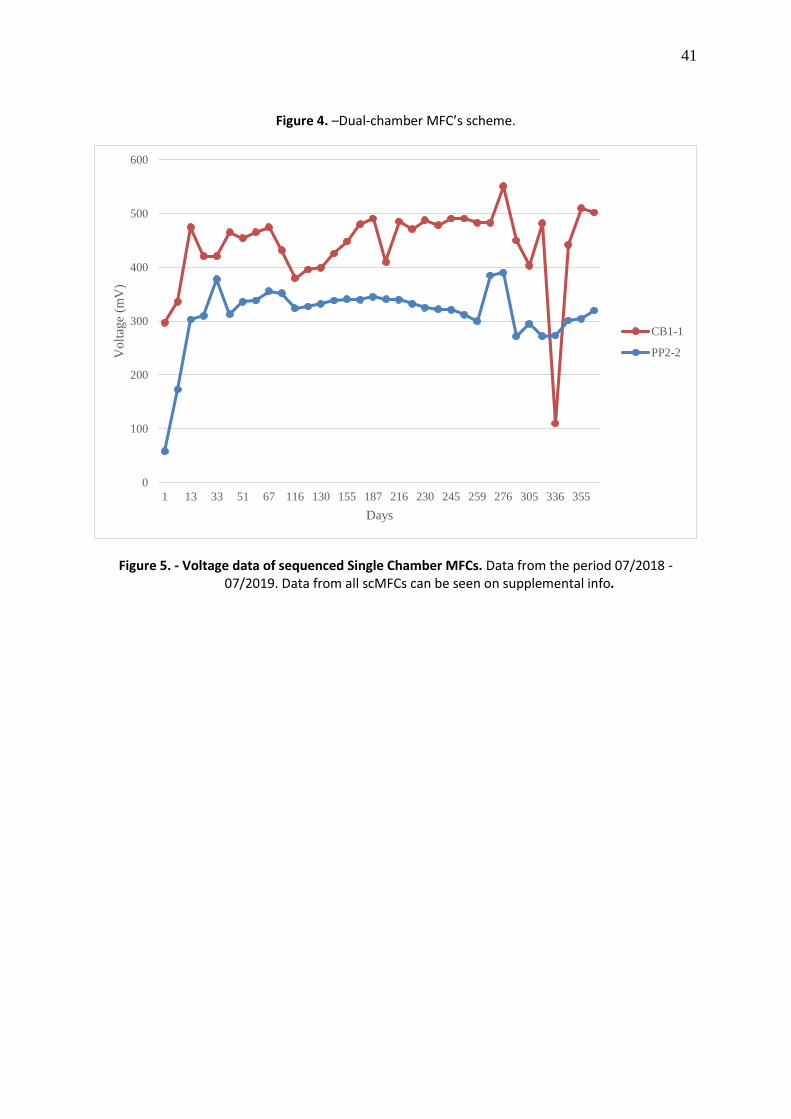
the highest and constant amount of energy, which was CB1.1 and PP2.2 (Figure 5). Four
samples of the cathode were collected and mixed into a pool. DNA extraction was carried out
using the QIAGEN™ DNeasy PowerSoil kit® following manufacturer’s instructions. Once the
DNA was extracted, quality parameters were measured using fluorometry. DNA was stored at
-20°C for later use. On the other hand, six months after dual-chamber MFC assembly, we
chose PP2.BG11 dcMFC for DNA extraction due to its higher input compared to the other
dcMFCs (Figure 6)
Sequencing data processing
Library preparation and sequencing runs were performed at Macrogen in South Korea.
Paired-end runs were sequenced by Illumina technology.

22
Sequencing data analyses
Sequencing results came as fasta.gz files, one per forward and reverse read of each
sample. Raw data was imported to QIIME2 (Quantitative Insights into Microbial Ecology)
(Bolyen et al., 2018) program using the function qiime tools import. The sequenced raw data
contained quality information that could be depicted by the function qiime demux summarize,
figures 8 and 9 shows forward and reverse reads quality information at base 240, just before
quality score started to fall. Primers were removed with qiime cutadapt trim-paired, to prevent
the formation of chimeras.
Quality control and denoising, which are removing sequences with low quality,
chimeras, and redundant, were performed using the dada2 pipeline. Function qiime dada2
denoise-paired was set to admit 2 errors from each read and truncate the sequence at base 240.
Three archives were obtained from this analysis, a feature table with the number of reads
from each sample, the representative sequences and the denoising stats, all these data can
be found on supplemental information. Representative sequences were aligned using MAFFT
(Katoh & Standley, 2013), with the following command line: qiime alignment mafft.
Once the sequences were aligned, we proceed to assign taxonomy to each sequence.
rRNA 16S Green Genes database (Balvočiūtė & Hudson., 2017) was used as the reference
database. The sequence classifier was generated using the Naive-Bayes method; function
qiime feature-classifier fit-classifier-naive-bayes was set to train the classifier on the V4-V5 region
of the rRNA 16S gene, which was the targeted gene region of the primers we used on this
research (Hughert et al., 2015). Representative sequences classification was done thanks to
the program Scikit-learn (Pedregosa et al., 2011), this was called by the function qiime feature-
classifier classify-sklearn.

23
Microbiome Diversity Analysis
The classified sequences and feature table were used to generate a relative abundance
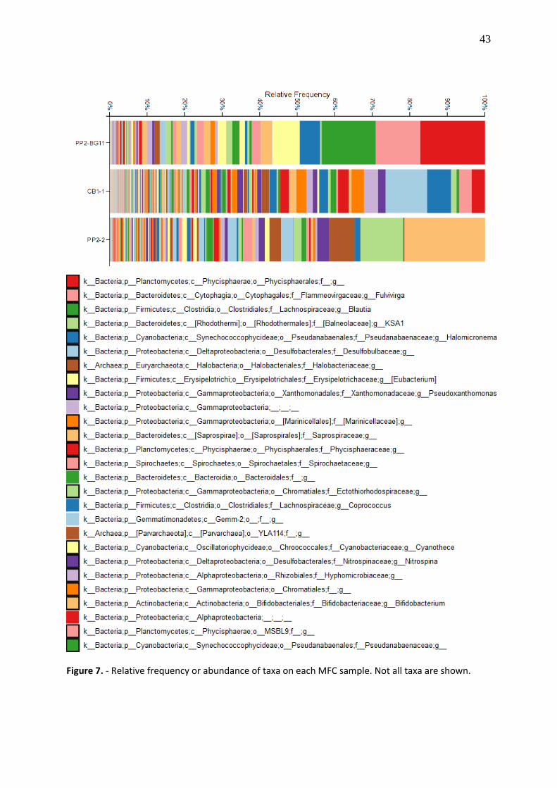
taxa barplot. Figure 7 depicts the relative abundance of taxa in each sample. QIIME2 command
line for bar plotting was qiime taxa barplot.
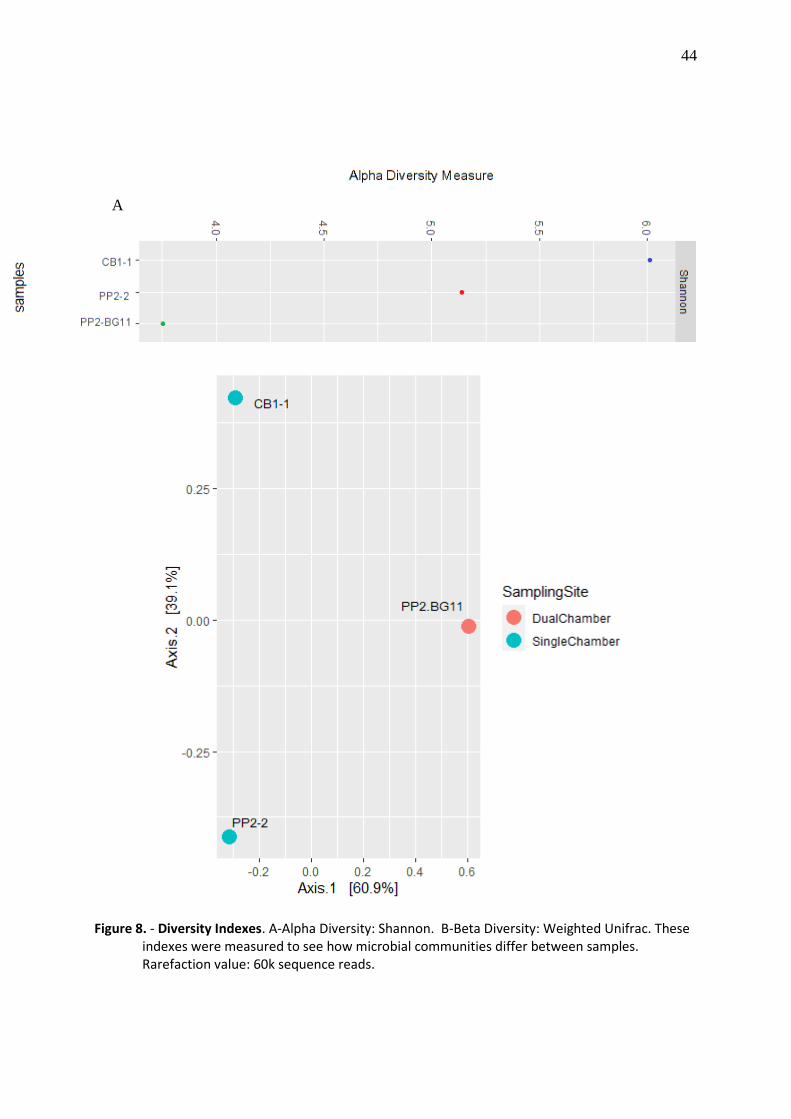
Alpha diversity indexes were calculated to see the species richness and evenness inside
of each sample. Shannon index was calculated using qiime diversity alpha-rarefaction.
Beta diversity was calculated as weighted UniFrac index, line command: qiime diversity
beta-rarefaction. Weighted UniFrac includes phylogenic relationships as a parameter, so we
created a phylogenetic tree using the program MAFFT-FAST TREE; line command: qiime
phylogeny align-to-tree-mafft-fasttree.
Moreover, to obtain high quality plots we exported QIIME2 files to R, to analyze the
data in Phyloseq pipeline (McMurdie & Holmes, 2013). In Phyloseq, we created a heatmap of
the 20th most abundant families and re-run the diversity index.
24
DATA ANALYSIS
Sample parameters
Physic-chemical parameters were measured at the time of sampling (In situ) and after
one month of arriving at the lab (In vitro) (Table 1). The main differences between In situ and
In vitro measures could be due to several factors, such as the variation of the measuring
instruments because in situ measures were taken by a portable sonde meanwhile In vitro
measures were taken by a specific benchtop. Changes in temperature and altitude also could
be explained by the fact that MFCs were assembled in Quito, which is 2890 meters above sea
level (m.a.s.l.) on the highlands of Ecuador, while the samples of sediment were taken on
Galapagos islands whose altitude is around 350 m.a.s.l. Finally, reduced dissolved oxygen (DO)
availability on In vitro measures might be due to anoxic conditions of the MFCs (Saratale et
al., 2017) as a consequence of microbial metabolism.
MFC’s energy production
Bioelectrochemical activity of the MFCs was measured as voltage generation;
measures were taken using a standard voltmeter. After one year of assembly, we chose the
scMFC with the higher and constant energy input for DNA sequencing, these MFCs were
CB1.1and PP2.2, whose average input was 441 and 314 mV, respectively (Figure 5). Same as
single-chamber MFCs, we chose the dual-chamber PP2.BG11 that produces the highest and
most constant energy input, (Figure 6). One interesting fact is that the energy production of
the MFCs was reestablished after the addition of sterile water (Supplement info) or when the
MFCs were opened for sampling purposes; this could indicate oxygenic metabolism and a
burst of Extracellular Electron Transfer (EET).
25
Microbiome Diversity Analysis
Relative frequency or abundance of taxa showed us the microbial composition of each
microbial fuel cell (Figure 7). The single-chamber MFCs are more diverse (CB1.1, PP2.2) than
dual-chamber MFC (PP2.BG11). This was corroborated by the number of sequences reads on
each sample, being CB1.1 the sample with the higher count of reads: 88k, followed by PP2.2:
85k reads and PP2.BG11; 76k reads. Besides, these data were confirmed by alpha and beta
diversity analysis. Alpha diversity is a measure of diversity from each sample. Figure 8. A
depicts alpha diversity index, Shannon index calculated the distribution of microbial
communities into each sample (Kim et al., 2017). Single chamber MFCs had higher Shannon
index as sequencing depth was increasing, CB1.1 had the highest Shannon score: 6, while
PP2.2 and PP2.BG11 had Shannon scores of 5.5 and 3.5, respectively, meaning that CB1.1was
the most diverse of all MFCs and also had more evenly distributed communities.
Beta diversity index displayed dissimilarity between samples, which meant that
samples did not share representative amounts of microbial abundance or phylogenetic
relationships between them (Figure 8.B). Weighted UniFrac based its index on phylogenetic
distances and relative abundance (Schroeder & Jenkins, 2018). The differences between
samples were mainly explained by the type of MFC (60.9%) because of PP2.BG11 dcMFC was
assembled with enriched sediment and scMFCs (CB1.1 and PP2.2) were assembled with raw
sediment; the other 39.1% of dissimilarity between samples was explained by the origin of the
sediments, Cerro Brujo for CB and Punta Pitt for PP.

26
Microbial communities evolving into the MFCs
Since we had the information of microbial communities abundance and taxonomy
assignment from the sediments or starting point (data not shown), we proceeded to compare
the communities that were at the starting point and a year after they were inoculated into the
MFC’s. These results suggest the microbial consortia could be playing a major role in this
bioelectrochemical system. The relative abundance of the communities that were found at
the two points of analysis can be seen on supplemental info.
Differences in the relative abundance at different points of analysis might help us to
infer microbial activities happening inside the MFCs. Some microbial families had decreased
in relative abundance after one year of scMFCs culturing, such as the following ones:
Chromatiaceae, Flavobacteriaceae, Geobacteraceae and Rhodospirillaceae in both scMFCs,
CB1.1 and PP2.2; while almost the rest of the families had increased their abundance over the
year (Supplemental info), especially the bacterial families Anaerolineaceae, Caldilineaceae,
Cyanobacteriaceae, Chlorobiaceae, Planctomycetae, Spirochaetaceae and arqueal
Halobacteriaceae on CB1.1 scMFC and bacterial families Cyanobacteriaceae, Chlorobiaceae,
Desulfobacteraceae, Desulfohalobiaceae, Ectothiorhodospiraceae and arqueal
Halobacteriaceae on PP2.2 scMFC.
The most abundant microbial families on the MFCs were Alteromonadaceae,
Balneollaceae, Cyanobacteriaceae, Hyphomicrobiaceae, Marinicellaceae, Phycisphaeraceae,
Pirellulaceae, Pseudanabaenaceae, Rhodobacteraceae and arqueal Halobacteriaceae that
were present in all MFCs (CB1.1, PP2.2, and PP2.BG11), bacterial families Flammeovirgaceae
and Xanthomonadaceae were only present in Punta Pitt MFCs (PP2.2 and PP2.BG11),
Coriobacteriaceae, Erysipelothricaceae, Lachnospiraceae were only found on PP2.2; on the
27
other hand, Spirochaetaceae and Priscirickettsiaceae were shared by PP2.2 and CB1.1. Finally,
Chromatiaceae, Desulfobulbaceae and Nitrospiraceae were only present in Cerro Brujo MFC
(CB1.1). (Figure 9).
An interesting finding was the presence of Desulfobulbaceae, and Nitrospiraceae only
in CB1.1, which was the most electrogenic MFC (441mV). Delsufobulbaceae members such as
Candidatus Electrothrix and Electronema had been proposed as new genera of EET bacteria
(Trojan et al., 2016). On the other hand, members of Nitrospiraceae family, like genera
Leptospirilum and Thermodesulfovibrio, are known to be potentially useful for wastewater
treatment, acid mine drainage and extracellular polymeric substance production (Daims,
2014) this could lead us to test MFC as a sustainable way of bioremediation, biomass and
energy production.
In contrast, family Lachnospiraceae was found only in PP2.2, this family had been
characterized in soils with high energy production on MFCs (Jiang, Zhong, Han & Deng, 2016).
Cultivation on BG11 medium might inhibit the growth of this and other families, resulting in
the decreased energy input of PP2.BG11 MFC.
Also, we notice that some microbial families were found most abundantly on PP2.BG11
dcMFC (Figure 9), we compared PP2.BG11 relative abundance with PP2.2 because both
samples were inoculated with the same sediment. An interesting finding was the increased
abundance of the family Alteromonadaceae, which includes the genus Marinobacter
hydrocarbonoclasticus, an old extremophile bacteria which can degrade hydrocarbons (Vance
et al., 2019).

28
DISCUSSION
In this study, we were trying to understand the microbial ecosystem inside the new
devices called microbial fuel cells (MFC). For this purpose, we compare the most prevalent
microbial community’s relative abundance from sediments of athalassic lagoons (data not
shown) versus the same communities found a year after cultivation on single-chamber MFC
and six months on dual-chamber MFC. Besides characterizing those communities, we
manually search for the kind of metabolism and carbon source to have an idea of what it’s
happening inside the MFC’s in terms of biochemistry (supplemental info).
Microbial families found in this study might be replicating the natural cycles of
chemical elements. In this case, oxygenic photosynthesis carried out by Cyanobacteria such
as Haloteche, Nostoc, Cyanothece might be participating in the cycling of hydrogen and
carbon, this could be exploited by other microorganisms inside the MFC systems, (Pisciotta,
Zou & Baskakov, 2010). The sulfur cycle could be carried out by members of the family
Desulfuromonadales, they might be reducing elemental sulfur from the sediments to H2S;
Thiotrichaceae family could oxidize H2S to sulfate (SO4), Desulfobacteraceae and
Desulfobulbaceae, might be reducing sulfate to H2S (Kuever, 2014), and then, that H2S
molecule is recycled, preventing its lethal action on the MFCs microhabitat. Finally, Nitrogen
cycle might be executed in the first place by nitrogen fixers like Clostridiaceae,
Cyanobacteriaceae, Rhodospirillaceae, they fix nitrogen from the atmosphere and make it
available to Nitrogen reducers such as Pirellulaceae. Nitrogen reducers form NH3 that might
be nitrificated by Chromatiaceae and Nitrospiraceae families. Nitrate formed by nitrogen
reducers could be de-nitrificated by Hyphomicrobiaceae and Rhodobacteracea families,
preventing NO3 accumulation and hence eutrophication of the microhabitat.

29
Besides replicating natural cycles of chemical elements, families that had increased
their relative abundance over the time were most likely to be halophiles with
photolithoautotropic metabolism, which means they can use light as their main source of
energy, carbon dioxide as carbon source and also use an inorganic electron donor (Stambler
& Dubinsky, 2007). These types of metabolism are likely to be found on MFCs coupled with
bioremediation systems. For instance, family Chlorobiaceae are predominant on benzene and
ammonium-contaminated groundwater MFCs (Wei et al., 2015). Likewise, some genus of the
family Anaerolineaceae such as Anaerolinea thermophila has been found on activated sludge
and oil spillover treatment plants (Sekiguchi et al.,2003), Another interesting discovery was
made on PP2.BG11 dcMFC, a bacterial genus increased its abundance once was cultivated on
BG11 medium, Marinobacter hydrocarbonoclasticus, an old bacteria that can degrade
hydrocarbons (Vance et al., 2019). This could lead us to test BG11 growth medium on
bioreactors to propagate M. hydrocarbonoclasticus and analyze the bioremediation potential
of this bacteria. All these findings can suggest MFCs as a possible source of auto-sustainable
bioremediation plants.
The fact that CB1.1 produced higher electrical current input than the other
MFCs could not be elucidated with this analysis, but we could have an initial approach looking
at the communities that have survived all this time after MFC’s culturing. In this scenario,
Desulfobulbaceae and Nitrospiraceae might be playing a key role in bioelectrogenesis, this is
because they have increased on relative abundance compared to the Starting Point and were
only found on CB1.1 (Supplemental info). Moreover, Alpha diversity showed us that single-
chamber MFCs are more diverse than dual-chamber MFCS, which could be explained since
PP2.BG11 dcMFC was under selective pressure of MFC conditions and the presence of a

30
specific growth media (BG11), meanwhile, scMFCs allowed the growth of more taxa due to
the absence of a specific growth media. On the other side, Beta diversity depicted the
difference between all of the MFCs, meaning that every MFC has a low proportion of shared
taxa and phylogenetic relationships. A fact that could explain the difference in diversity
between sites is that Cerro Brujo is a pristine habitat where humans are not allowed to enter
without special permission, and Punta Pitt is an old saltern where humans used to extract salt
for consumption.
The change in relative abundance and biochemical characteristics of the most
prevalent microbes could give us an idea of the type of reactions that are happening on the
MFCs, but we need to seek deeper into this microhabitat to clarify its functioning. Lower
abundant taxa that could not be detected in this study might be fine-tuning
bioelectrochemical reactions. Transcriptomic and metabolomic tools could show us what
reactions are predominating between microorganisms, not only the predominant taxa,
leading to discoveries about how to improve MFC electrical current in a short period (Logan
et al., 2019).

31
CONCLUSIONS
In this study, we found that microbial communities might have evolved into the MFCs
in a syntrophic way. Most microbial communities play key roles in biogeochemical cycles,
hence maintaining the functioning of microbial fuel cells over time. The most prevalent class
of metabolism among microbial families are anaerobic photolithotrophy. This type of
metabolism allows microbial communities to obtain energy from the sun while using an
inorganic electron donor and could be the MFC’s main source of energy. Alongside
photolithotrophs, heterotrophs might be reusing microbial debris and consuming oxygen
from the environment, thus preventing its lethal action over the anaerobic populations;
chemolithoautotrophs might be oxidizing and reducing chemical compounds present in the
sediments and making them available to the other communities and Cyanobacteria could be
providing protons to the habitat while fixing CO2 from the atmosphere. Also, almost all the
families found in the MFCs were halotolerant or halophile, with high bioremediation potential,
which is not surprising due to the conditions of the athalassic lagoons we sampled; these
findings might drive us to test MFC as a bioremediation process of wastewaters, reducing CO2
environmental levels, draining acid mines, and production of non-oil derivate polymers. All
these phenomena make us think about what it’s happening inside the microbial fuel cells, and
that not only Geobacteraceae and Shewanellaceae are the rare microorganisms that can
produce significant amounts of energy. Although we could only infer these phenomena until
we establish a “core microbiome” of the MFCs and their transcriptome and metabolome
involved in energy production.
32
REFERENCES
Alava, J. J., Palomera, C., Bendell, L., & Ross, P. S. (2014). Pollution as an emerging threat for the conservation of the Galapagos Marine Reserve: environmental impacts and management perspectives. In The Galapagos Marine Reserve (pp. 247-283). Springer, Cham.
Aklujkar, M., Coppi, M. V., Leang, C., Kim, B. C., Chavan, M. A., Perpetua, L. A., ... & Holmes, D. E. (2013). Proteins involved in electron transfer to Fe (III) and Mn (IV) oxides by Geobacter sulfurreducens and Geobacter uraniireducens. Microbiology, 159(3), 515-535.
Azevedo-Santos, V. M., Garcia-Ayala, J. R., Fearnside, P. M., Esteves, F. A., Pelicice, F. M., Laurance, W. F., & Benine, R. C. (2016). Amazon aquatic biodiversity imperiled by oil spills. Biodiversity and conservation, 25(13), 2831-2834.
Balvočiūtė, M., Huson, H. (2017). SILVA, RDP, Greengenes, NCBI and OTT — how do these taxonomies compare?. BMC Genomics. 18, 114. DOI: 10.1186/s12864-017-3501-4
Berezovsky, I. N., & Shakhnovich, E. I. (2005). Physics and evolution of thermophilic adaptation. Proceedings of the National Academy of Sciences, 102(36), 12742- 12747.
Bolyen, E., Rideout, J., Dillon, M., Bokulich, N., Abnet, C. … Caporaso, J. (2018). QIIME 2: Reproducible, interactive, scalable, and extensible microbiome data science. PeerJ Preprints. 6:e27295v2. doi: doi.org/10.7287/peerj.preprints.27295v2
Breheny, M., Bowman, K., Farahmand, N., Gomaa, O., Keshavarz, T., & Kyazze, G. (2019). Biocatalytic electrode improvement strategies in microbial fuel cell systems. Journal of Chemical Technology & Biotechnology, 94(7), 2081-2091.
Brutinel, E. D., & Gralnick, J. A. (2013). On the role of endogenous electron shuttles in extracellular electron transfer. In Microbial Metal Respiration (pp. 83-105). Springer, Berlin, Heidelberg.
Daims, H. (2014) The Family Nitrospiraceae. In: Rosenberg E., DeLong E.F., Lory S., Stackebrandt E., Thompson F. (eds) The Prokaryotes. Springer, Berlin, Heidelberg
Deamer, D., & Weber, A. L. (2010). Bioenergetics and life's origins. Cold Spring Harbor perspectives in biology, 2(2), a004929.

33
Deng, H., Chen, Z., & Zhao, F. (2012). Energy from plants and microorganisms: progress in plant–microbial fuel cells. ChemSusChem, 5(6), 1006-1011.
Deng, L., Li, F., Zhou, S., Huang, D., & Ni, J. (2010). A study of electron-shuttle mechanism in Klebsiella pneumoniae based-microbial fuel cells. Chinese Science Bulletin, 55(1), 99-104.
Freguia, S., Masuda, M., Tsujimura, S., & Kano, K. (2009). Lactococcus lactis catalyses electricity generation at microbial fuel cell anodes via excretion of a soluble quinone. Bioelectrochemistry, 76(1-2), 14-18.
Gimkiewicz, C., & Harnisch, F. (2013). Waste water derived electroactive microbial biofilms: growth, maintenance, and basic characterization. JoVE (Journal of Visualized Experiments), (82), e50800.
Hoegh-Guldberg, O., Jacob, D., Taylor, M., Bindi, M., Brown, S., Camilloni, I., ... & Guiot, J. (2018). Impacts of 1.5 C global warming on natural and human systems. In Global warming of 1.5° C.: An IPCC Special Report (pp. 175-311). IPCC Secretariat. Hugerth, L. W., Wefer, H. A., Lundin, S., Jakobsson, H. E., Lindberg, M., Rodin, S., ... & Andersson, A. F. (2014). DegePrime, a program for degenerate primer design for broad-taxonomic-range PCR in microbial ecology studies. Applied Environmental Microbiology, 80(16), 5116-5123. DOI: 10.1128/AEM.01403-14
Inoue, K., Leang, C., Franks, A. E., Woodard, T. L., Nevin, K. P., & Lovley, D. R. (2011). Specific localization of the c‐type cytochrome OmcZ at the anode surface in current‐producing biofilms of Geobacter sulfurreducens. Environmental Microbiology Reports, 3(2), 211-217.
Intergovernmental Panel on Climate Change – IPCC. (2018). First Joint Session of Working Groups I, Ii And Iii. Incheon, Republic of Korea, 1 - 5 October 2018. (30.IX.2018)
Jain, A., Gazzola, G., Panzera, A., Zanoni, M., & Marsili, E. (2011). Visible spectroelectrochemical characterization of Geobacter sulfurreducens biofilms on optically transparent indium tin oxide electrode. Electrochimica Acta, 56(28), 10776- 10785.
Javed, M. M., Nisar, M. A., Ahmad, M. U., Yasmeen, N., & Zahoor, S. (2018). Microbial fuel cells as an alternative energy source: current status. Biotechnology and Genetic Engineering Reviews, 34(2), 216-242.

34
Jiang, Y. B., Zhong, W. H., Han, C., & Deng, H. (2016). Characterization of electricity generated by soil in microbial fuel cells and the isolation of soil source exoelectrogenic bacteria. Frontiers in microbiology, 7, 1776.
Kang, C. S., Eaktasang, N., Kwon, D. Y., & Kim, H. S. (2014). Enhanced current production by Desulfovibrio desulfuricans biofilm in a mediator-less microbial fuel cell. Bioresource technology, 165, 27-30.
Katoh, K., & Standley, D. M. (2013). MAFFT multiple sequence alignment software version 7: improvements in performance and usability. Molecular biology and evolution, 30(4), 772–780. DOI:10.1093/molbev/mst010
Keller, K. L., Rapp-Giles, B. J., Semkiw, E. S., Porat, I., Brown, S. D., & Wall, J. D. (2014). New model for electron flow for sulfate reduction in Desulfovibrio alaskensis G20. Applied Environmental Microbiology, 80(3), 855-868.
Khan, M. M., Ansari, S. A., Lee, J. H., Lee, J., & Cho, M. H. (2013). Mixed culture electrochemically active biofilms and their microscopic and spectroelectrochemical studies. ACS Sustainable Chemistry & Engineering, 2(3), 423-432.
Kim, B. R., Shin, J., Guevarra, R., Lee, J. H., Kim, D. W., Seol, K. H., ... & Isaacson, R. E. (2017). Deciphering diversity indices for a better understanding of microbial communities. J. Microbiol. Biotechnol, 27(12), 2089-2093.
Kuever J. (2014) The Family Desulfobacteraceae. In: Rosenberg E., DeLong E.F., Lory S., Stackebrandt E., Thompson F. (eds) The Prokaryotes. Springer, Berlin, Heidelberg
Kumar, R., Singh, L., Wahid, Z. A., & Din, M. F. M. (2015). Exoelectrogens in microbial fuel cells toward bioelectricity generation: a review. International Journal of Energy Research, 39(8), 1048-1067.
Logan, B. E., Rossi, R., Ragab, A., & Saikaly, P. E. (2019). Electroactive microorganisms in bioelectrochemical systems. Nature Reviews Microbiology. DOI:10.1038/s41579-019- 0173-x
Logan, B. E. (2009). Exoelectrogenic bacteria that power microbial fuel cells. Nature Reviews Microbiology, 7(5), 375.
Li, F., Li, Y., Sun, L., Li, X., Yin, C., An, X. ... & Song, H. (2017). Engineering Shewanella oneidensis enables xylose-fed microbial fuel cell. Biotechnology for biofuels, 10(1), 196.

35
Li, W. W., Yu, H. Q., & He, Z. (2014). Towards sustainable wastewater treatment by using microbial fuel cells-centered technologies. Energy & Environmental Science, 7(3), 911-924.
Liu, X., Shi, L., & Gu, J. D. (2018). Microbial electrocatalysis: redox mediators responsible for extracellular electron transfer. Biotechnology advances, 36, 1815–1827.
Liu, Y., Kim, H., Franklin, R. R., & Bond, D. R. (2011). Linking spectral and electrochemical analysis to monitor c‐type cytochrome redox status in living Geobacter sulfurreducens biofilms. ChemPhysChem, 12(12), 2235-2241.
Liu, Y., & Bond, D. R. (2012). Long‐distance electron transfer by G. sulfurreducens biofilms results in accumulation of reduced c‐type cytochromes. ChemSusChem, 5(6), 1047-1053.
Luo, H., Xu, P., Roane, T. M., Jenkins, P. E., & Ren, Z. (2012). Microbial desalination cells for improved performance in wastewater treatment, electricity production, and desalination. Bioresource Technology, 105, 60-66.
Lusk, B. G. (2019). Thermophiles; or, the Modern Prometheus: The Importance of Extreme Microorganisms for Understanding and Applying Extracellular Electron Transfer. Frontiers in microbiology, 10, 818.
McMurdie and Holmes (2013) phyloseq: An R Package for Reproducible Interactive Analysis and Graphics of Microbiome Census Data. PLoS ONE, 8(4):e61217.
Mehta-Kolte, M. G., & Bond, D. R. (2012). Geothrix fermentans secretes two different redox-active compounds to utilize electron acceptors across a wide range of redox potentials. Applied Environmental Microbiology, 78(19), 6987-6995.
Parameswaran, P., Bry, T., Popat, S. C., Lusk, B. G., Rittmann, B. E., & Torres, C. I. (2013). Kinetic, electrochemical, and microscopic characterization of the thermophilic, anode-respiring bacterium Thermincola ferriacetica. Environmental science & technology, 47(9), 4934-4940.
Pedregosa, F., Varoquaux, G., Gramfort, A., Michel, V., Thirion, B., Grisel, O., ... & Vanderplas, J. (2011). Scikit-learn: Machine learning in Python. The Journal of machine Learning research, 12, 2825-2830.

36
Pisciotta, J. M., Zou, Y., & Baskakov, I. V. (2010). Light-dependent electrogenic activity of cyanobacteria. PloS one, 5(5).
Poddar, S., & Khurana, S. (2011). Geobacter: the electric microbe! Efficient microbial fuel cells to generate clean, cheap electricity. Indian journal of microbiology, 51(2), 240.
Reguera, G. (2018). Microbial nanowires and electroactive biofilms. FEMS Microbiology Ecology, 94(7). DOI:10.1093/femsec/fiy086.
Richter, H., Nevin, K. P., Jia, H., Lowy, D. A., Lovley, D. R., & Tender, L. M. (2009). Cyclic voltammetry of biofilms of wild type and mutant Geobacter sulfurreducens on fuel cell anodes indicates possible roles of OmcB, OmcZ, type IV pili, and protons in extracellular electron transfer. Energy & Environmental Science, 2(5), 506-516.
Santoro, C., Arbizzani, C., Erable, B., & Ieropoulos, I. (2017). Microbial fuel cells: from fundamentals to applications. A review. Journal of power sources, 356, 225-244.
Saratale, G. D., Saratale, R. G., Shahid, M. K., Zhen, G., Kumar, G., Shin, H. S. ... & Kim, S. H. (2017). A comprehensive overview on electro-active biofilms, role of exo- electrogens and their microbial niches in microbial fuel cells (MFCs). Chemosphere, 178, 534-547.
Schroeder, P. J., & Jenkins, D. G. (2018). How robust are popular beta diversity indices to sampling error?. Ecosphere, 9(2), e02100.
Sekiguchi, Y., Yamada, T., Hanada, S., Ohashi, A., Harada, H., & Kamagata, Y. (2003). Anaerolinea thermophila gen. nov., sp. nov. and Caldilinea aerophila gen. nov., sp. nov., novel filamentous thermophiles that represent a previously uncultured lineage of the domain Bacteria at the subphylum level. International journal of systematic and evolutionary microbiology, 53(6), 1843-1851.
Shen, H. B., Yong, X. Y., Chen, Y. L., Liao, Z. H., Si, R. W., Zhou, J., ... & Zheng, T. (2014). Enhanced bioelectricity generation by improving pyocyanin production and membrane permeability through sophorolipid addition in Pseudomonas aeruginosa- inoculated microbial fuel cells. Bioresource technology, 167, 490-494.
Shrestha, P. M., Rotaru, A. E., Summers, Z. M., Shrestha, M., Liu, F., & Lovley, D. R. (2013). Transcriptomic and genetic analysis of direct interspecies electron transfer. Applied Environmental Microbiology, 79(7), 2397-2404.
Stambler, N., & Dubinsky, Z. (2007). Marine phototrophs in the twilight zone. In Algae and cyanobacteria in extreme environments (pp. 79-97). Springer, Dordrecht.

37
Tigre, M. A. (2017). Threats to the Amazon Rainforest: Deforestation and Climate Change. In Regional Cooperation in Amazonia (pp. 48-75). Brill Nijhoff. Trojan, D., Schreiber, L., Bjerg, J. T., Bøggild, A., Yang, T., Kjeldsen, K. U., & Schramm, A. (2016). A taxonomic framework for cable bacteria and proposal of the candidate genera Electrothrix and Electronema. Systematic and applied microbiology, 39(5), 297-306.
Vance, T. D., Guo, S., Assaie-Ardakany, S., Conroy, B., & Davies, P. L. (2019). Structure and functional analysis of a bacterial adhesin sugar-binding domain. PloS one, 14(7).
Vargas, M., Malvankar, N. S., Tremblay, P. L., Leang, C., Smith, J. A., Patel, P., ... & Lovley, D. R. (2013). Aromatic amino acids required for pili conductivity and long- range extracellular electron transport in Geobacter sulfurreducens. MBio, 4(2), e00105-13.
Voordeckers, J. W., Kim, B. C., Izallalen, M., & Lovley, D. R. (2010). Role of Geobacter sulfurreducens outer surface c-type cytochromes in reduction of soil humic acid and anthraquinone-2, 6-disulfonate. Applied Environmental Microbiology, 76(7), 2371- 2375.
Vos, P., Garrity, G., Jones, D., Krieg, N. R., Ludwig, W., Rainey, F. A., ... & Whitman, W. B. (Eds.). (2011). Bergey's manual of systematic bacteriology: Volume 3: The Firmicutes (Vol. 3). Springer Science & Business Media.
Wang, Y., Kern, S. E., & Newman, D. K. (2010). Endogenous phenazine antibiotics promote anaerobic survival of Pseudomonas aeruginosa via extracellular electron transfer. Journal of bacteriology, 192(1), 365-369.
Wang, A., Sun, D., Cao, G., Wang, H., Ren, N., Wu, W. M., & Logan, B. E. (2011). Integrated hydrogen production process from cellulose by combining dark fermentation, microbial fuel cells, and a microbial electrolysis cell. Bioresource Technology, 102(5), 4137-4143.
Wang, Q., Cen, Z., & Zhao, J. (2015). The survival mechanisms of thermophiles at high temperatures: an angle of omics. Physiology, 30(2), 97-106.
Wei, J., Liang, P., Cao, X., & Huang, X. (2010). A new insight into potential regulation on growth and power generation of Geobacter sulfurreducens in microbial fuel cells based on energy viewpoint. Environmental science & technology, 44(8), 3187-3191.

38
Wei, M., Harnisch, F., Vogt, C., Ahlheim, J., Neu, T. R., & Richnow, H. H. (2015). Harvesting electricity from benzene and ammonium-contaminated groundwater using a microbial fuel cell with an aerated cathode. RSC Advances, 5(7), 5321-5330.
Wolińska, A., Stępniewska, Z., Bielecka, A., & Ciepielski, J. (2014). Bioelectricity production from soil using microbial fuel cells. Applied biochemistry and biotechnology, 173(8), 2287-2296.
Wolska, K. I., Grudniak, A. M., Rudnicka, Z., & Markowska, K. (2016). Genetic control of bacterial biofilms. Journal of applied genetics, 57(2), 225-238.
Yang, H., Zhou, M., Liu, M., Yang, W., & Gu, T. (2015). Microbial fuel cells for biosensor applications. Biotechnology letters, 37(12), 2357-2364.
Zhuang, L., Zheng, Y., Zhou, S., Yuan, Y., Yuan, H., & Chen, Y. (2012). Scalable microbial fuel cell (MFC) stack for continuous real wastewater treatment. Bioresource technology, 106, 82-88.

39
TABLES AND FIGURES
Figure 1. - Different pathways and mediators for EET. a) Electron shuttles; b) pili-like nanowires; c) outer-membrane redox proteins. (Logan et al, 2019)

40
Figure 2. – Sampling sites on San Cristobal Island. Two athalassic lagoons, Cerro Brujo and Punta Pitt (Shown with blue mark).
Figure 3. – Single chamber Microbial Fuel Cells working scheme.
41
Figure 4. –Dual-chamber MFC’s scheme.
Figure 5. - Voltage data of sequenced Single Chamber MFCs. Data from the period 07/2018 -07/2019. Data from all scMFCs can be seen on supplemental info.
0
100
200
300
400
500
600
1 13 33 51 67 116 130 155 187 216 230 245 259 276 305 336 355
Vo
ltag
e (m
V)
Days
CB1-1
PP2-2

42
Figure 6. – Voltage from sequenced Dual Chamber MFC. Data of the period 02/2019 - 08/2019. Information from all dcMFCs can be seen on supplemental info.
0
50
100
150
200
250
300
350
400
1 3 5 7 9
11
13
15
17
19
21
23
25
27
29
35
45
55
65
75
85
95
11
0
13
0
15
0
17
0
VO
LT
AG
E (
MV
)PP2-BG11
PP2-BG11
43
Figure 7. - Relative frequency or abundance of taxa on each MFC sample. Not all taxa are shown.
44
Figure 8. - Diversity Indexes. A-Alpha Diversity: Shannon. B-Beta Diversity: Weighted Unifrac. These indexes were measured to see how microbial communities differ between samples. Rarefaction value: 60k sequence reads.
B
A

45
Figure 9. - Differential abundance heatmap. Blue marks show more counts while green shows fewer. Blank gaps show no counts for the sample. H: Heterotrophy; A: Autotrophy.

46
Table 1. - Physic-chemical parameters of the sediments. Measures were taken “In situ” on the sampling area, and “In vitro” in the laboratory
In situ measures
Sample Salinity (ppt) DO (mg/ml) pH Temperature (°C)
Cerro Brujo 1 19,70 ± 7,29 7,97± 0,38 7,78 ± 0,85 31,10 ± 2,07
Cerro Brujo 2 19,70 ± 7,30 7,97± 0,39 7,78 ± 0,86 31,10 ± 2,08
Punta Pitt 1 72,09 ± 11,74 8,09 ± 0,56 5,92 ± 0,76 33,10 ± 2,07
Punta Pitt 2 72,09 ± 11,75 8,09 ± 0,57 5,92 ± 0,77 33,10 ± 2,08
In vitro measures
Sample Conductivity (ms/cm) DO (mg/ml) pH Temperature (°C)
Cerro Brujo 1 40,105 0,27 7.25 22.2
Cerro Brujo 2 34,82 0,29 7.72 22.2°C
Punta Pitt 1 77,9575 1,35 7.28 22.2°C
Punta Pitt 2 71,3775 0,17 7.57 23°C


