contribuciÓn al establecimiento de las bases...
TRANSCRIPT

FACULTAT DE FARMÀCIA
DEPARTAMENT NUTRICIÓ I BROMATOLOGIA
CONTRIBUCIÓN AL ESTABLECIMIENTO DE LAS BASES
CIENTÍFICAS PARA EL USO DE FRACCIONES
POLIFENÓLICAS Y FIBRA DIETÉTICA ANTIOXIDANTE
EN LA PREVENCIÓN DEL CÁNCER
SONIA TOURIÑO EIRIN 2009

INSTITUT DE QUÍMICA AVANÇADA
DE CATALUNYA (IQAC-CSIC)
DEPARTAMENT QUÍMICA BIOLÒGICA I MODELITZACIÓ MOLECULAR (QBM)
NUTRACÈUTICS I RADICALS LLIURES
UNIVERSITAT DE BARCELONA
FACULTAT DE FARMÀCIA
DEPARTAMENT NUTRICIÓ I BROMATOLOGIA
Programa de doctorat
NUTRICIÓ I METABOLISME
Bienni 2005-2007
CONTRIBUCIÓN AL ESTABLECIMIENTO DE LAS BASES
CIENTÍFICAS PARA EL USO DE FRACCIONES
POLIFENÓLICAS Y FIBRA DIETÉTICA ANTIOXIDANTE
EN LA PREVENCIÓN DEL CÁNCER
Memòria presentada per Sonia Touriño Eirín per optar al títol de doctor per
la Universitat de Barcelona
Director de la tesi: Tutor de la tesi:
Josep Lluís Torres Simón Rosa Mª Lamuela Raventós
SONIA TOURIÑO EIRIN 2009


Este trabajo ha sido financiado por:
Ministerio de Ciencia e Innovación
Beca de Formación de Personal Investigador
(FPI): 2005-2009
AGL2004-07579-C04-02/ALI


Science is organized knowledge. Wisdom is organized life. Immanuel Kant

Agradecimientos Quisiera expresar mi más profundo agradecimiento a todas y cada una de las personas
que han contribuido en este trabajo y que han hecho posible la consecución de un reto
profesional y personal
En primer lugar, me gustaría dar las gracias el Dr. Josep Lluís Torres por haberme
brindado la gran oportunidad de realizar esta tesis doctoral bajo su dirección y depositar
en mí su confianza desde el primer día que aterricé en Barcelona. Por guiarme a lo largo
de todo este período no sólo académicamente sino que también personalmente. Por
hacerme participe de todos sus proyectos y por despertar en mí el entusiasmo por la
ciencia.
Mi agradecimiento al Dr. Pere Clapés por tratarme de igual a igual, por conseguir que
controle mi carácter pero sobretodo por su ayuda desinteresada en todo momento. Al Dr.
Jesús Joglar por estar siempre dispuesto a ayudar y transmitir su buen humor. Al Dr.
Lluís Julià por que una parte de esta tesis no podría ser realizada sin su colaboración.
A la Dra. Pilar Vinardell y Dra. Montse Mitjans, del departamento de Fisiologia de la
Universitat de Barcelona, por su ayuda en los estudios de experimentación animal.
Al grupo de Bioquímica Integrativa i Teràpia del Càncer de Universitat de Barcelona.
Especialmente, a la Dra. Marta Cascante por que sin su colaboración está tesis no podría
llevarse a cabo. A la Dra. Daneida Lizárraga por que parte de esta tesis es suya. A los
demás integrantes del grupo (Susana, S; Marisa, F; Silvia, M; Cecilia, M….) por acogerme
como una más del equipo.
Al personal dels Serveis Científico-Tècnics de la Universitat de Barcelona, personalmente
a la Dra. Olga Jáuregui, por cogerme de la mano en los primeros pasos y enseñarme a
caminar en el mundo de la espectrometría de masas, ojalá pueda contar con tu ayuda en
el largo camino que todavía me queda por recorrer.
Al personal del Servei d’espectrometria de masses del IADEA-CSIC, a la Dra. Roser
Chaler y Maria por ayudarme con el uso del Q-TOF.
Mi más profundo agradecimiento para el Profesor Jason Morrow por ofréceme la
oportunidad de realizar una estancia en su laboratorio y dar un millón de gracias al Dr.
Brian Cox por su paciencia y buen humor en todo momento. Por supuesto, quisiera
agradecer al Dr. Álvarez su inestimable ayuda para poder establecer los vínculos con el
Profesor Morrow.

A todos mis compañeros y amigos del laboratorio, a los que están y a los que se han ido.
Ellos han sido cómplices del esfuerzo, de las alegrías y también de los lloros que
conllevan esta tesis. Compartir y convivir con vosotros gran parte del día a día ha sido
fantástico. Mi más profundo agradecimiento a Ariadna Selga y el Dr. Carles Lozano por su
amistad y por ser mis maestros durante los primeros años en el laboratorio. E incluir al
Dr. Jordi Calveras y el Dr. José A. Castillo por ayudarme con mis dudas y problemas
químicos. Gracias a todos por hacerme sentir como en mi propia casa, apoyándome en
los momentos más difíciles.
A la Dra. Elisabet Fuguet por su ayuda incondicional y por su contribución fundamental
durante estos dos últimos años. Por ser, además de consejera, amiga y por ensañarme a
ver de manera analítica cada uno de mis experimentos.
A Sònia Castellanos por iluminar el laboratorio más oscuro del CSIC (la cueva) con sus
radicales y sus placas solares, con su buen humor. Te echaremos de menos estos meses.
A Debi Castiella y a Sonia Lorenzo por aquellos buenos momentos que pasamos juntas
en el laboratorio. Al Guillem Rocasalbas por no olvidar nunca su sonrisa, por ser
realmente un amigo.
Gracias a Cristina Alonso por estar disponible cada vez que necesitaba envasar al vacío.
Ya sabes que eres una más de nuestro grupo.
Gracias Anna, Xavi, Bruno y Aris por una convivencia ejemplar en el laboratorio
A Mariana por compartir su alegría con nosotros y….. su comida, su café. Por traernos los
mejores alfajores del mundo. Mis mejores deseos para esta nueva etapa de la vida que
comienzas.
A Livia, G. y Alda Lisa, C. por ser mis compinches dentro y fuera del laboratorio. Todavía
nos quedan muchos rosarios por disfrutar.
Al Jordi P. por la paciencia de estos últimos meses y por confiar en mis indicaciones a
ciegas. Estoy segura de que éste es el comienzo de una carrera científica fructificante.
Al equipo Bioglane; Susana, Sara, Dani, Ester por que el ambiente del Laboratorio no
sería el mismo sin vosotros. Susana, no olvidaré nunca nuestras aventuras en la gran
ciudad. Ojalá tengamos la posibilidad de poder compartir alguna más. Yo creo que
somos un buen equipo.
A Marisa y a Juan por su alegría sureña, y por echarnos una mano con parte nuestro
trabajo.

Gracias a los chicos del RUBAM, a los del Lab. 309 y como no, a la Patricia y el Gerard del
Lab. 305 por mostrar su alegría en el día a día.
Grazas ós que me deron tanto apoio dende tan lonxe.
A todas aquellas personas y amigos que no he citado y que de alguna forma han
contribuido a que esta tesis llegue a buen termino.
A todos, muchas gracias.

Índice 1. INTERÉS Y JUSTIFICACIÓN…………………………………………………………………………………………… 1
2. INTRODUCCIÓN…………………………………………………………………………………………………………….. 5
2.1 Radicales libres y especies reactivas de oxígeno…………………………………………………. 7
2.2 Principales fuentes de ROS en la célula……………………………………………………………….. 9
2.3 Funciones biológicas de ROS………………………………………………………………………………… 10
2.4 Citotoxicidad de ROS……………………………………………………………………………………………. 11
2.5 Sistemas de defensa antioxidante………………………………………………………………………... 14
2.5.1 Definición de antioxidante............................................................................. 14
2.5.2 Clasificación de los antioxidantes.................................................................. 14
2.6 Estrés oxidativo............................................................................................................... 16
2.6.1 Factores externos implicados en el incremento de ROS............................ 17
2.6.2 Patologías relacionadas con el estrés oxidativo.......................................... 21
2.6.3 Cáncer y estrés oxidativo............................................................................... 22
2.6.3.1 Concepto de cáncer......................................................................... 22
2.6.3.2 Cáncer y Especies reactivas de oxígeno....................................... 22
2.7 Los flavonoides como ingredientes de alimentos funcionales................................... 25
2.7.1 La dieta en la prevención de enfermedades................................................ 25
2.7.2 Alimentos funcionales. Concepto.................................................................. 25
2.7.3 Los flavonoides................................................................................................ 26
2.7.4 Los subproductos agroforestales. Fuentes de obtención
de flavonoides y otros compuestos fenólicos.............................................. 29
2.7.5 Actividad biológica de los flavonoides........................................................... 31
2.7.6 Biodisponibilidad de los flavonoides.............................................................. 34
2.7.6.1 Absorción y transporte celular....................................................... 35
2.7.6.2 Distribución....................................................................................... 36
2.7.6.3 Metabolismo o biotransformación.................................................. 36
2.7.6.3.1 Transformaciones enzimáticas....................................... 37
2.7.6.3.2 Transformaciones colónicas............................................ 39
2.8 Metodología de identificación y análisis de compuestos fenólicos............................ 41

2.8.1 Extracción de compuestos fenólicos............................................................ 41
2.8.2 Métodos cromatográficos de separación..................................................... 41
2.8.2.1 Cromatografía de gases (GC)....................................................... 41
2.8.2.2 Cromatografía de exclusión por tamaño (SEC)......................... 42
2.8.2.3 Cromatografía líquida de alta resolución (HPLC)........................ 42
2.8.3 Técnicas de detección .................................................................................... 43
2.8.3.1 Espectroscopía UV-Vis.................................................................... 43
2.8.3.2 Espectroscopía de fluorescencia.................................................... 44
2.8.3.3 Espectrometría de masas............................................................... 44
2.8.4 Evaluación de la actividad antioxidante....................................................... 47
2.8.4.1 Métodos químicos en solución....................................................... 47
2.8.4.1.1 Métodos de evaluación de
los mecanismos HAT y SET............................................ 48
2.8.4.1.2 Métodos de evaluación del mecanismo HAT................. 49
2.8.4.1.3 Métodos de evaluación del mecanismo SET................. 50
2.8.4.2 Ensayos en medios lipídicos........................................................... 51
2.8.4.3 Ensayos con modelos celulares...................................................... 53
2.8.4.4 Biomarcadores de estrés oxidativo............................................... 53
2.8.4.4.1 Indicadores de daño oxidativo en el citosol................. 55
2.8.4.4.2 Indicadores de daño oxidativo en el ADN..................... 55
2.8.4.4.3 Indicadores de daño oxidativo en lípidos..................... 56
2.8.4.4.4 Indicadores de daño oxidativo en proteínas................ 57
3. OBJETIVOS.............................................................................................................................. 59
4. RESULTADOS........................................................................................................................... 63
4.1 Estudio de la relación entre el grado de polimerización y porcentaje
de galoización de fracciones fenólicas con sus efectos químicos y biológicos......... 65
4.1.1 Evaluación de la actividad antioxidante y biológica de fracciones
fenólicas exentas de galoización................................................................... 69
Publicación 1: Procyanidin Fractions from pine (Pinus pinaster)
bark: Radical scavenging power in solution, antioxidant activity in
emulsion, and antiproliferative effect in melanoma cells.............................. 73
4.1.2 Evaluación de la actividad antioxidante y biológica
de fracciones de fenólicas de elevada galoización...................................... 81
Publicación 2: Highly galloylated tannin fractions from Witch
Hazel (Hamamelis virginiana) bark: electron transfer capacity, in vitro
antioxidant activity, and effects on skin-related cells......................................... 83
4.2 Estudio de la absorción/metabolización de proantocianidinas.................................. 93
4.2.1 Identificación de los compuestos fenólicos de la fracción
extraíble de fibra antioxidante de uva (GADF) .......................................... 95
Publicación 3: High-resolution liquid chromatography/electrospray
ionization time-of-flight mass spectrometry combined with liquid
chromatography/electrospray ionization tandem mass spectrometry
to identify polyphenols from grape antioxidant dietary fiber......................... 97
4.2.2 Identificación de metabolitos de GADF en orina de ratas......................... 109
Publicación 4: Phenolic metabolites of grape antioxidant
dietary fiber in rat urine.......................................................................... 111
5. RESULTADOS GLOBALES/ DISCUSIÓN GENERAL............................................................. 123
6. CONCLUSIONES...................................................................................................................... 137
7. BIBLIOGRAFÍA......................................................................................................................... 141
8. ANEXOS.................................................................................................................................... 167
8.1 Publicaciones complementarias..................................................................................... 171
I. Electron-transfer capacity of catechin derivatives and influence
on the cell cycle and apoptosis in HT29 cells ...................................................... 173
II. Functional fatty fish supplemented with grape procyanidins.
Antioxidant and proapoptotic properties on colon cell lines................................. 187
III. Procyanidins from pine bark: relationships between structure,
composition and antiradical activity...................................................................... 195
IV. The importance of polymerization and galloylation for the
antiproliferative properties of procyanidin-rich natural extracts……..………… 207
V. Comparative antioxidant and cytotoxic effect of procyanidin
fractions from grape and pine………………………………………………………………………..………… 219
VI. Witch Hazel (Hamamelis virginiana) fractions and the
importance of gallate moieties electron transfer capacities
in their antitumoral properties………………………………………………………………………..………… 227
VII. The maize ZmMYB42 represses the phenylpropanoid pathway
and affects the cell wall structure, composition and degradability
in Arabidopsis thaliana..................................................................................... 287
8.2 Material suplementario........................................................................................................ 253
8.2.1 Material suplementario publicación 1....................................................................... 255
8.2.2 Material suplementario publicación 2....................................................................... 273
8.2.3 Material suplementario publicación 4....................................................................... 287
Índice de figuras de la introducción
TÍTULO FIGURA PÁGINA
Figura 2.1: Reducción tetravalente del oxígeno molecular.
Estado de los spins direccionales de las moléculas implicadas........... ...................7
Figura 2.2: Especies reactivas generadas por leucocitos activados...................... 9
Figura 2.3: Degradación de las purinas vía xantina oxidasa (XO) y
Formación de ROS........................................................................................ 10
Figura 2.4: Esquema de citotoxicidad en la célula............................................ 11
Figura 2.5: Proceso esquemático de la peroxidación lipídica.............................. 12
Figura 2.6: Estrés oxidativo.......................................................................... 16
Figura 2.7: Metabolismo del etanol en los hepatocitos...................................... 18
Figura 2.8: Generación de ROS en la piel y defensas antioxidantes..................... 19
Figura 2.9: Estimación del porcentaje de muertes correspondientes
a los cánceres más comunes en la UE en el año 2006........................................ 22
Figura 2.10: Etapas de formación del cáncer en función del
estrés oxidativo............................................................................................ 24
Figura 2.11: Estructura básica de los flavonoides............................................. 26
Figura 2.12: Estructura química de las subclases de flavonoides
más relevantes............................................................................................. 27
Figura 2.13: Ejemplo de taninos condensados y taninos hidrolizables.................. 28
Figura 2.14: Mecanismo de oxidación de la (―)-epicatequina ............................. 31
Figura 2.15: Características estructurales de los flavonoides relacionadas
con la capacidad antioxidante.......................................................................... 32
Figura 2.16: Proceso esquemático del mecanismo de quelación de
metales de los flavonoides.............................................................................. 33
Figura 2.17: Representación de biodisponibilidad de un compuesto..................... 34
Figura 2.18: Esquema básico de las posibles rutas de los nutrientes
y xenobióticos............................................................................................... 35
Figura 2.19: Esquema de absorción, transformación y excreción de
xenobióticos................................................................................................. 37
Figura 2.20: Posibles biotransformaciones colónicas de las (epi)catequinas.......... 40
Figura 2.21: Esquema de ionización química a presión atmosférica (API)............. 45
Figura 2.22: Representación esquemática de un triple cuadrupolo (QqQ)............. 46
TÍTULO FIGURA PÁGINA
Figura 2.23: Transferencia electrónica entre la (―)-epicatequina y
el radical estable HNTTM................................................................................ 51
Figura 2.24: Ejemplo de un ensayo de inhibición de autooxidación lipídica
medida por calorimetría diferencial de barrido (DSC).......................................... 52
Figura 2.25: Algunos de los productos generados tras la oxidación
de biomoléculas............................................................................................ 54
Figura 2.26: Resultados del ensayo cometa..................................................... 55
Índice de tablas de la introducción TÍTULO TABLA PÁGINA
Tabla 2.1: Especies reactivas de oxígeno y nitrógeno.......................................... 8
Tabla 2.2: Principales sistemas antioxidantes del organismo................................ 15
Tabla 2.3: Enfermedades asociadas a estrés oxidativo........................................ 21
Tabla 2.4: Principales familias de los compuestos fenólicos.................................. 26
Tabla 2.5: Ejemplos de fuentes dietéticas de flavonoides..................................... 29
Tabla 2.6: Ejemplos de subproductos utilizados como fuente de
compuestos fenólicos....................................................................................... 30
Tabla 2.7: Enzimas implicadas en las biotransformaciones de Fase I..................... 38
Tabla 2.8: Bandas de absorción UV-Vis de algunos compuestos fenólicos............... 43
Tabla 2.9: Modelos celulares para evaluar los efectos protectores de
un compuesto antioxidante en el desarrollo del cáncer......................................... 53

I
Abreviaturas
A• Radical libre no tóxico
AAPH 2,2’-azo-bis (2-amidinopropano) dihidrocloruro
AAS R-aminoadípico semialdeído
AAT Actividad Antioxidante Total
ABAP 2,2’-azo-bis (2-amidinopropano)
ABTS 2,2-azino-bis (3-etilbezotiazolin-6-sulfonato)
ABTS•+ Radical catión ABTS
ACE Enzima convertidora de angiotensina I
ACN Acetonitrilo
ADH Alcohol deshidrogenasa
ADN Ácido Desoxirribonucleico
AH Antioxidante
ALDH Aldehído deshidrogenasa
APCI Ionización química a presión atmosférica
API Ionización a presión atmosférica
ARP Poder antirradicalario
ATP Adenosín trifosfato
AUC Área bajo la curva (del inglés, Area under curve)
BHA Butilhidroxianisol
BHT Butilhidroxitolueno
B-PE B-Ficoeritrina
CAT Catalasa
Cat Catequina
CE Electroforesis capilar
CID Disociación inducida por colisión
Cmax Concentración máxima
COMT Catecol metilo transferasa
COMTs Catecol metilo transferasas
COX Enzima ciclooxigenasa
Cya Cisteamina
Cya-Cat 4β-(2-aminoetiltio)catequina
Cya-Ec 4β-(2-aminoetiltio)epicatequina
Cya-EcG 4β-(2-aminoetiltio)epicatequin-3-O-galato
CYP Enzima del complejo citocromo P450
II
DAD Detector de diodos en línea (del inglés, Diode array detector)
DHA Ácido docohexanoico
DH-IsoP Dihomo-Isoprostanos
DMPD radical catión N,N-dimetil-p-fenilendiamina
DMPO 5,5’-dimetil-1-pirrolín-N-oxido
DMSO Dimetilsulfóxido
DNA Ácido desoxirribonucleico
DPPH Radical 2,2-difenil-1-picrilhidracilo
DSC Calorimetría Diferencial de Barrido
DT di-tirosina
Ec (―)-epicatequina
EcG (―)-epicatequina 3-O-galato
EGC (―)-epigalocatequina
EGCG (―)-epigalocatequín 3-O-galato
ELISA Inmunoensayo (del inglés, Enzyme linked inmunosorbent Assay)
EPR Resonancia Paramagnética Electrónica
ESI Ionización por electrospray
EUFIC Consejo Europeo de información sobre la alimentación ( del ingles, The
European Food Information Council)
FL 3,6-Dihidroxispiro[isobenzofuran-1(3H),9-[9H] xanten]-3-ona
FlO· Radical fenoxil
FlOH Flavonoide
FRAP Reducción del Hierro/Poder antioxidante (del inglés, Ferring
Reducing/Antioxidant Power)
G porcentaje de galoización
GADF Fibra antioxidante dietética (del inglés, Grape antioxidant dietary fiber)
GC Cromatografía de gases (del inglés, Gas chromatography)
GC-MS GC acoplada a MS
GGS δ-glutámico semialdeído
GHz Frecuencia de banda-X
GPx Glutatión peroxidasa
GRed Glutatión reductasa
GSH Glutatión reducido
GSH Glutatión reducido
GSSG Glutatión oxidado
GST Glutatión-s-transferasa
H2O2 Peróxido de hidrógeno
III
HAT Transferencia de hidrógenos
HNE 4-Hidroxinonenal
HNTTM Radical tris (2,4,6-tricloro-3,5-dinitrofenil)metil
HPLC Cromatografía líquida de alta eficacia
HPLC-CE HPLC acoplado a CE
HPLC-MS HPLC acoplado a MS
HPLC-MS/MS HPLC acoplado a MS/MS
HPLC-UV HPLC acoplado a UV
HRP Peroxidasa (del inglés, horseradish peroxidase)
Hx Hidroperóxidos
IT Trampa de iones (del inglés, Ion Trap)
LC Cromatografía líquida
LDL Lipoproteínas de baja densidad
LPH Lactasa phlorodzin hidrolasa
MALDI Ionización por desorción de una matriz mediante láser
MALDI-TOF Técnica de MALDI acoplada a la técnica de TOF
MDA Malonaldehído
mDP Grado de polimerización medio
MeOH Metanol
mMW Masa molecular media
MPO Mieloperoxidasa
MRM Monitorización de reacciones múltiples (del inglés, Monitoring multple
reaction)
MS Espectrometría de masas
MS/MS ó MSn Espectrometría en tándem
NAC N-acetil cisteina
NAD Nicotín Adenín nucleótido
NADH Nicotín Adenín nucleótido reducido
NADPH Nicotinamida-Adenina Dinucleotido fosfato
NL Barrido de perdidas neutras (del inglés, Neutral loss scan)
NOS Nítrico oxido sintasa
NP Fase normal ( del inglés, Normal phase)
NP-HPLC Cromatografía líquida de alta resolución de fase normal
O/W Emulsión aceite en agua
OMS Organización Mundial de la Salud
ORAC Capacidad de absorción de radicales libres (del inglés, Oxygen radical
absorbance capacity)
IV
o-Tyr orto-Tirosina
OWP Extracto crudo de corteza de pino soluble en disolventes orgánicos y
agua
OWH Extracto crudo de Hamamelis virginiana soluble en disolventes
orgánicos y agua
PAHs Hidrocarburos policíclicos aromáticos
PAPs fosfoadenosil-5'-fosfosulfato
PBS Tampón fosfato salino
pH Logaritmo negativo de base 10 de la actividad de los iones hidrógeno
PI Barrido de ión precursor (del inglés, precursor ión)
PIDS Dispersión diferencial por Intensidad de polarización (del inglés,
Polarization Intensity Diferencial Scatter)
Q Detector de cuadrupolo de barras
Q• Quinonas
QqQ Triple cuadrupolo
Q-TOF Cuadrupolo acoplado a un detector de tiempo de vuelo
R• Radical alquilo
RL Radical libre
RNA Ácido ribonucleico.
RNOS Especies reactivas de oxígeno y nitrógeno
RO• Radical alcoxilo
ROCl Especies reactivas de oxígeno y cloro
ROO• Radical peroxilo
ROONO Alquil peroxinitrito
ROS ó ERO Especies reactivas de oxígeno (del inglés, Reactive oxygen species)
RP Fase reversa (del inglés, Reverse phase)
RP-HPLC Cromatografía líquida de alta resolución en fase reversa.
SAM S-adenosil metionina
SEC Cromatografía de exclusión por tamaño
SET Transferencia de electrones
SGLT Proteínas con transporte sodio-glucosa
SOD Superóxido dismutasa
ST Sulfotransferasa
STs Sulfotransferasas
TBARS Sustancias reactivas al ácido tiobarbitúrico (del inglés,Thiobarbituric
acid reactive-substances)

V
TEAC Capacidad Antioxidante equivalente al Trolox (del inglés,Trolox
Equivalent Antioxidant Capacity)
TFA Ácido trifluoroacético
Tmax Tiempo máximo
TNPTM Radical tris (2,4,6-tricloro-3,5-dinitrofenil)-metilo
TOF Detector de tiempo de vuelo (del inglés, Time of fly)
TOSC Capacidad total de captación de radicales libres ( del inglés,Total
oxidant scavenging capacity)
TPTZ Complejo 2,4,6-tripiridil-s-triazina
Trolox Ácido 6-hidroxi-2,5,7,8-tetrametilcroman-2-carboxílico
UDP Uridina difosfato
UGTs Uridina glucuroniltransferasas
UPLC Cromatografía líquida de ultra resolución
UV Ultravioleta (200-400nm)
UVA Radiación ultravioleta de onda larga (320-400 nm)
UVB Radiación ultravioleta de onda media (290-320 nm)
UV-vis Ultravioleta-visible (200-750nm)
Vit Vitamina
X Xantina
XO Xantina oxidasa
8-OH-dG 8-hidroxi-2’-deoxiguanosina
8-OHG 8-hidroxiguanina


1. INTERÉS Y JUSTIFICACIÓN

1. Interés y justificación
3
Los organismos aerobios necesitan oxígeno para vivir. La oxidación de los nutrientes para
la obtención de energía fisiológica en forma de ATP implica una reducción del oxígeno
escalonada en cuatro etapas monoeléctricas. Esta reducción del oxígeno a agua permite
la liberación energética de manera gradual. Entre un 2 y un 5% de la reducción del
oxígeno ocurre de forma incompleta en la mitocondria generando otras especies
derivadas del oxígeno que se caracterizan por ser altamente oxidantes y reactivas. Las
características que presentan estas especies por un lado les permite actuar como
mensajeros secundarios modulando diversas funciones fisiológicas y por otro lado, sin
embargo, pueden provocar daño oxidativo en elementos celulares debido a su
inespecificidad y elevada capacidad oxidante. Como consecuencia, los organismos
aerobios han desarrollado un sistema antioxidante capaz de mitigar el daño producido.
Factores ambientales relacionados con la dieta y la exposición a agentes pro-oxidantes
(radiaciones, metales, pesticidas, contaminación, etc.) aumentan considerablemente las
especies reactivas en la célula. Cuando el incremento de las especies reactivas excede la
capacidad de actuación del sistema antioxidante endógeno, provoca un estado de estrés
oxidativo. Daños sobre las membranas celulares y sobre el ADN son consecuencia directa
de una desregulación en el estado redox de la célula. Existen evidencias que sugieren la
existencia de una relación entre el estrés oxidativo y los estados patológicos de
numerosas enfermedades. Es por ello que la modulación del equilibrio antioxidante
fisiológico parece ser esencial para prevenir o reducir el daño al ADN y otras
biomoléculas y por tanto, para disminuir la posibilidad de desarrollo de cáncer u otras
enfermedades.
Estudios epidemiológicos han comprobado que una dieta rica en frutas y verduras parece
ser importante para la prevención de enfermedades. Parece bastante plausible que
alguno de los compuestos o la interacción de varios compuestos que constituyen frutas y
verduras son responsables del efecto protector que presentan. Los flavonoides son los
principales candidatos debido su abundancia en el reino vegetal y la actividad
antioxidante que presentan en su estado natural.
De la ingesta total de compuestos fenólicos, se ha estimado que los ácidos fenólicos
podrían representar aproximadamente 1/3 y los flavonoides los 2/3 restantes. La
cantidad y la proporción de los diferentes compuestos fenólicos ingeridos varían
ampliamente según el tipo de alimentos consumidos en función de los hábitos dietéticos
y las preferencias. El estilo de vida al que se tiende hoy en día, ha aumentado la
demanda de productos saludables que contienen compuestos biológicamente activos,
conocidos por el nombre de alimentos funcionales, que además del aporte nutritivo
pueden ofrecer beneficios para la salud disminuyendo el riesgo de padecer
enfermedades.
En la actualidad, las investigaciones referentes a los compuestos fenólicos y a sus
actividades biológicas están en auge, aunque el número de antioxidantes incluidos en las
1. Interés y justificación
4
listas positivas de ingredientes es de momento bajo. Compuestos fenólicos han sido
extraídos, purificados y evaluada su actividad biológica. Los resultados prometedores en
cuanto a las propiedades beneficiosas y la aceptación de los consumidores para utilizar
antioxidantes naturales como complementos nutricionales han disparado el mercado de
estos productos. La necesidad de disponer de grandes fuentes de compuestos fenólicos
ha puesto en el punto de mira de numerosas investigaciones la obtención compuestos
fenólicos a partir de subproductos de la industria agrícola y forestal. De hecho, algunos
extractos obtenidos a partir de subproductos agroforestales han sido utilizados como
ingredientes o suplementos nutricionales, aplicaciones dérmicas, y/o antioxidantes
alimentarios. Las diferentes aplicaciones de los extractos fenólicos dependerán de
factores físico-químicos (solubilidad, coeficiente de partición, estructura) y de la actividad
biológica que presenten.
Una elevada proporción de los 2/3 ingeridos de flavonoides se encuentra en forma
polimérica, generalmente en forma de proantocianidinas. Sin embargo, la mayoría
estudios in vitro y ex vivo que parecen corroborar la capacidad de prevención de
determinadas enfermedades son generalmente realizados con productos purificados que
no son reflejo de una ingesta de frutas y/o verduras. Además, una gran parte de ellos
utilizan enzimas que hidrolizan las conjugaciones sufridas en el organismo. Es cierto que,
estos estudios facilitan el análisis y aporta información sobre la biodisponibilidad, no
obstante, el hecho de no conocer las estructuras reales de los flavonoides en el
organismo impide evaluar y descifrar la actividad de estos compuestos en la prevención
de enfermedades.
Las proantocianidinas parecen tener una absorción limitada en el intestino, por lo que
una parte de las proantocianidinas ingeridas van directamente al colon. Las bacterias
que habitan el colon de los mamíferos fermentan los productos no digeridos en el
estomago para obtener sustrato. Los compuestos fenólicos que no han sido absorbidos y
los que se excretan vía bilis pueden ser degradados por la acción de las bacterias en
compuestos más pequeños. La actividad de los metabolitos de los compuestos fenólicos
y en concreto de las proantocianidinas todavía no es clara.
Un mayor conocimiento de los aspectos metabólicos relacionados con la ingesta de
polifenoles ha de permitir explicar los potenciales efectos beneficiosos de los flavonoides
de la dieta. Como resultado de todos los estudios con polifenoles se podría llegar a
establecer recomendaciones dietéticas optimizadas en antioxidantes para distintos
grupos de la población (ancianos, enfermos, deportistas, etc.) tendentes a mejorar la
calidad de vida.

2. INTRODUCCIÓN


2. Introducción
7
2.1 Radicales libres y especies reactivas de oxígeno Un radical libre (RL) es una especie química (átomo, ión, o molécula) capaz de existir
de forma independiente (de ahí, el termino de libre) y que presenta uno o más electrones
desapareados en su estructura. Precisamente, debido a la existencia de electrones
desapareados, son extremadamente reactivos hasta que consiguen transformarse en una
molécula estable a través de una reacción de oxidación-reducción, en la cual el radical
gana electrones o hidrogeniones (átomos de hidrógeno) de otra molécula. En general la
mayor parte de las reacciones de oxidación son de deshidrogenación, es decir, que
entrañan la perdida de átomos de hidrogeno.
Cualquier molécula es susceptible de convertirse en un radical libre tras la ganancia o
pérdida de un electrón o tras la ruptura de un enlace homolítico. Un radical puede
generar una cascada de reacciones en cadena clasificadas como: de iniciación
(generación de un radical o especie reactiva), propagación (el número de radicales
libres se mantiene constante aumentado el número de productos de la reacción y
disminuyendo el de reactivos) y terminación (desaparición de los radicales libres por
aniquilación o combinación de sus dos electrones desapareados).
Figura 2.1: Reducción tetravalente del oxígeno molecular. Estado de los spins
direccionales de las moléculas implicadas.
El oxígeno molecular (O2), que necesitan todos los organismos aerobios presenta en su
estructura dos electrones desapareados lo que lo convierte en un bi-radical. La
disposición de estos dos electrones, en diferentes orbitales libres y ambos en el mismo
sentido, permite al O2 tener una cierta estabilidad y propiedades paramagnéticas, aunque
por otro lado, impide que el oxígeno capte dos electrones simultáneamente en su
reducción. En la mitocondria la reducción del oxígeno implicada en la producción de ATP
tiene lugar mediante cuatro reacciones univalentes para dar lugar a dos moléculas de

2.1 Radicales libres y especies reactivas de oxígeno
8
agua (Figura 2.1). Durante este proceso se generan especies intermediarias altamente
reactivas y oxidantes de naturaleza radical (O2•⎯ y OH•) y no radical (H2O2). Ambas son
incluidas en la definición de especies reactivas de oxígeno (ROS; del inglés reactive
oxygen species).
Entre los más reactivos se encuentra el radical hidroxilo (OH•) que presenta un elevado
potencial de reducción y una vida media de tan sólo 10-9 s (Liochev et al., 1994). La
formación del radical OH• tiene lugar en presencia de iones metálicos y H2O2 por
reacciones de Haber-Weiss y Fenton. Aunque es cierto que en los sistemas biológicos el
Fe se encuentra generalmente en su forma férrica y en pequeñas cantidades (Halliwell et
al., 1986), éste puede ser reducido por el ascorbato y por el radical superóxido, con lo
que se genera un ciclo de producción continua de radicales hidroxilo (Frei, 1994)
El peróxido de hidrógeno (H2O2), en ausencia de iones metálicos, es la especie menos
reactiva y la más estable en condiciones de pH y temperatura fisiológicas. Se origina por
dismutación del anión superóxido vía superóxido dismutasa o partir de la actividad de
enzimas como la aminoácido oxidasa y la xantina oxidasa (Lee et al., 2004). Es
considerado un agente oxidante débil. Sin embargo tal y como se ha explicado
anteriormente, el peróxido de hidrógeno puede generar radicales hidroxilo en presencia
de iones metálicos y además puede producir in vivo oxígeno singlete al reaccionar con el
anión superóxido o con cloraminas (Stief, 2000). Existen otras especies reactivas que
pueden generarse en los organismos aerobios. Las cuales son dependientes de otros
elementos tales como nitrógeno (RNOS); cloro (ROCl), etc. Las ROS y las RNOS son las
principales especies reactivas implicadas en la biología redox (Tabla 2.1) (Halliwell,
1999).
Tabla 2.1: Especies reactivas de oxígeno y nitrógeno
Radicales No radicalarias
Anión superóxido (O2•⎯) Peróxido de hidrógeno (H2O2)
Hidroxilo (OH•) Ácido hipocloroso (HClO)
Alcoxilo (RO•) Ozono (O3 ) ROS
Peroxilo (ROO•) Oxígeno singlete (1∆O2)
Óxido nítrico (NO•) Anión nitrosilo ( NO-)
Dióxido de nitrógeno (NO2•) Catión nitrosilo (NO+)
Ácido nitroso (HNO2) RNOS
Alquil peroxinitrito (ROONO)
2. Introducción
9
2.2 Principales fuentes de ROS en la célula
La mitocondria constituye la principal fuente de ROS. La obtención de energía a partir
de la reducción del O2 tiene lugar a nivel de cadena de electrones por la acción del
complejo citocromo oxidasa. En general, más de un 95% del O2 consumido es reducido
en cuatro etapas monoeléctricas, adquiriendo 4 electrones y generando 2 moléculas de
agua (Figura 2.1). Sin embargo, el 5% restante la reducción del oxígeno no es
completa formándose compuestos intermediarios: anión superóxido (O2•⎯), peróxido de
hidrógeno (H2O2) y/o hidroxilo (OH•)
Los peroxisomas son orgánulos que tienen un papel fundamental en el metabolismo
lipídico y están encargados de la degradación de ácidos grasos de cadena larga para su
completa oxidación en la mitocondria. Además, participan en la degradación de bases
púricas de los aminoácidos (adenina y guanina) y juegan un papel importante en la
detoxificación de moléculas tóxicas como el etanol (Masters, 1998). Los procesos de
degradación que ocurren en los peroxisomas son realizados por numerosas enzimas de
tipo oxidasa que utilizan oxígeno molecular para conseguir átomos de hidrógeno de
sustratos específicos generando como consecuencia grandes cantidades de H2O2 (Del Río
et al., 1992). El peróxido de hidrógeno formado en condiciones normales es eliminado
por la enzima catalasa pero bajo ciertas condiciones de estrés oxidativo la actividad de la
enzima catalasa disminuye liberándose grandes concentraciones de H2O2 en la célula
(Schrader et al., 2006).
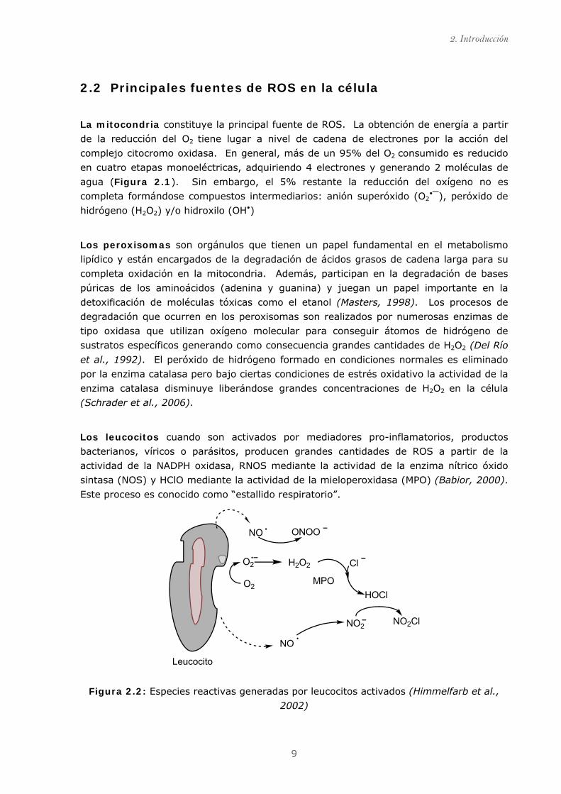
Los leucocitos cuando son activados por mediadores pro-inflamatorios, productos
bacterianos, víricos o parásitos, producen grandes cantidades de ROS a partir de la
actividad de la NADPH oxidasa, RNOS mediante la actividad de la enzima nítrico óxido
sintasa (NOS) y HClO mediante la actividad de la mieloperoxidasa (MPO) (Babior, 2000).
Este proceso es conocido como “estallido respiratorio”.
NO ONOO
O2
O2 H2O2
MPO
Cl
HOCl
NO2ClNO2
NO
Leucocito
Figura 2.2: Especies reactivas generadas por leucocitos activados (Himmelfarb et al.,
2002)

2.3 Funciones biológicas de ROS
10
Pequeñas moléculas solubles tales como tioles, hidroquinonas, catecolaminas,
flavinas y tetrahidropterinas pueden autooxidarse de forma natural generando radical
superóxido que la mayor parte de las veces acaba derivando en peróxido de hidrógeno
(Martínez-Cayuela, 1995).
Otras enzimas, principalmente enzimas citosólicas solubles como la aldehído oxidasa
involucrada en el metabolismo del etanol y la óxido nítrico sintasa responsable de
transformar la L-arginina en óxido nítrico y enzimas unidas a la membrana plasmática
como la lipooxigenasa y la ciclooxigenasa (COX) que participan en el metabolismo del
ácido araquidónico generando radicales libres durante el ciclo de catálisis (Balazy et al.,
2008;Caro et al., 2006). Algunas enzimas implicadas en la hidroxilación de purinas
también son responsables de contribuir a un aumento de especies reactivas en la célula.
Por ejemplo, la enzima xantina oxidasa (XO) que es encargada de catalizar la reacción de
hipoxantina a xantina y de xantina a ácido úrico (Figura 2.3). Ambas reacciones
generan O2•⎯(Vorbach et al., 2003).
Figura 2.3: Degradación de las purinas vía xantina oxidasa (XO) y formación de ROS
2.3 Funciones biológicas de ROS Pequeñas concentraciones de ROS parecen ser necesarias para el correcto
funcionamiento de los organismo vivos (Droge, 2002). Entre las funciones más
destacadas de las especies reactivas de oxígeno se encuentran:
(i) Regulación de óxido nítrico generado por la enzima óxido nítrico sintasa (NOS) (Chen
et al., 2009);
2. Introducción
11
(ii) Activación de la enzima NAD(P)H en leucocitos para la producción masiva de ROS en
la defensa de microorganismos externos y en células no fagocíticas, como
fibroblastos o células endoteliales vasculares (Thannickal et al., 1995), que tras la
producción de ROS pueden activar rutas intracelulares necesarias en la regulación
de las células cardiacas y factores de crecimiento en fibroblastos (Griendling et al.,
2000;Jacobi et al., 2005).
(iii) Regulación del tono vascular del músculo liso (Griendling et al., 1994);
(iv) Inhibición de la agregación plaquetaria (Begonja et al., 2006);
(v) Detección de cambios en la concentración del oxígeno intracelular(Droge,
2002;MacFarlane et al., 2008);
(vi) Programación de apoptosis (Herdener et al., 2000); Pequeñas cantidades de
especies reactivas juegan un papel crucial como segundos mensajeros en la
transducción de la señal para la activación, diferenciación y proliferación celular. La
inducción o la inhibición de la proliferación parece ser dependiente del balance
ROS/antioxidantes en la célula. (Schreck R et al., 1992).
(vii) Modulación de la respuesta inmunitaria de linfocitos-T y macrófagos (Victor et al.,
2004).
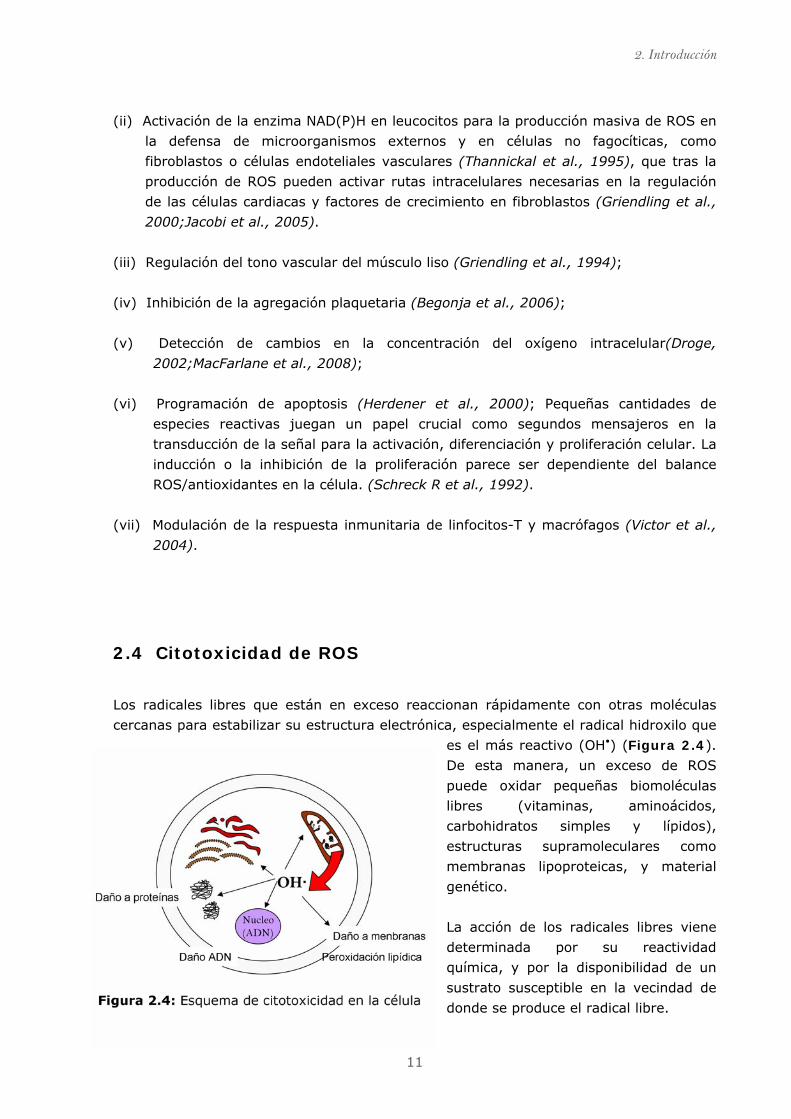
2.4 Citotoxicidad de ROS
Los radicales libres que están en exceso reaccionan rápidamente con otras moléculas
cercanas para estabilizar su estructura electrónica, especialmente el radical hidroxilo que
es el más reactivo (OH•) (Figura 2.4).
De esta manera, un exceso de ROS
puede oxidar pequeñas biomoléculas
libres (vitaminas, aminoácidos,
carbohidratos simples y lípidos),
estructuras supramoleculares como
membranas lipoproteicas, y material
genético.
La acción de los radicales libres viene
determinada por su reactividad
química, y por la disponibilidad de un
sustrato susceptible en la vecindad de
donde se produce el radical libre.
2.5 Sistema de defensa antioxidante
12
Daño oxidativo a lípidos: La acción de las ROS sobre los lípidos tiene lugar
fundamentalmente sobre los ácidos grasos poliinsaturados generando peróxidos, los
cuales son degradados hasta formar especies reactivas. La oxidación de los lípidos que
forman parte de la membrana celular afecta tanto a las propiedades físicas (fluidez y
permeabilidad) como a la funcionalidad de las proteínas de membrana (Droge, 2002).
Los radicales libres que pueden iniciar esta reacción son: el radical hidroxilo (OH•), el
peroxilo (ROO•), el alcoxilo (RO•) y el radical alquilo (R•).
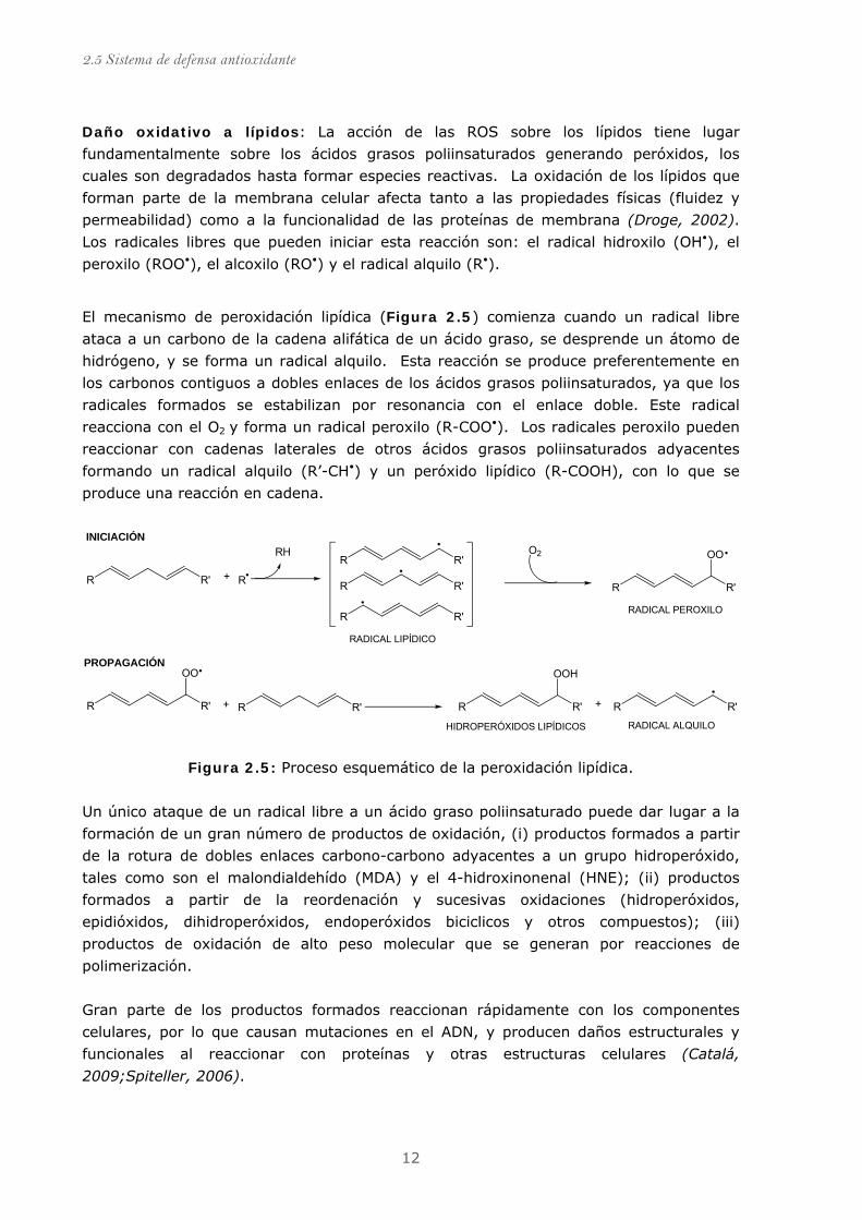
El mecanismo de peroxidación lipídica (Figura 2.5) comienza cuando un radical libre
ataca a un carbono de la cadena alifática de un ácido graso, se desprende un átomo de
hidrógeno, y se forma un radical alquilo. Esta reacción se produce preferentemente en
los carbonos contiguos a dobles enlaces de los ácidos grasos poliinsaturados, ya que los
radicales formados se estabilizan por resonancia con el enlace doble. Este radical
reacciona con el O2 y forma un radical peroxilo (R-COO•). Los radicales peroxilo pueden
reaccionar con cadenas laterales de otros ácidos grasos poliinsaturados adyacentes
formando un radical alquilo (R’-CH•) y un peróxido lipídico (R-COOH), con lo que se
produce una reacción en cadena.
R R'
R R'
R R'
R R'
R R'
OO
R R'
OO
R R' R R'
OOH
R R'
R
RH O2
+
+ +
INICIACIÓN
RADICAL LIPÍDICO
RADICAL PEROXILO
PROPAGACIÓN
HIDROPERÓXIDOS LIPÍDICOS RADICAL ALQUILO
Figura 2.5: Proceso esquemático de la peroxidación lipídica.
Un único ataque de un radical libre a un ácido graso poliinsaturado puede dar lugar a la
formación de un gran número de productos de oxidación, (i) productos formados a partir
de la rotura de dobles enlaces carbono-carbono adyacentes a un grupo hidroperóxido,
tales como son el malondialdehído (MDA) y el 4-hidroxinonenal (HNE); (ii) productos
formados a partir de la reordenación y sucesivas oxidaciones (hidroperóxidos,
epidióxidos, dihidroperóxidos, endoperóxidos biciclicos y otros compuestos); (iii)
productos de oxidación de alto peso molecular que se generan por reacciones de
polimerización.
Gran parte de los productos formados reaccionan rápidamente con los componentes
celulares, por lo que causan mutaciones en el ADN, y producen daños estructurales y
funcionales al reaccionar con proteínas y otras estructuras celulares (Catalá,
2009;Spiteller, 2006).
2. Introducción
13
La peroxidación del ácido araquidónico, componente estructural de las membranas
celulares origina además de hidroperóxidos, unos peróxidos cíclicos conocidos como
isoprostanoides. Estos compuestos son una familia de eicosanoides de origen no
enzimático que se forman a partir de la peroxidación mediada por radicales libres
(Morrow et al., 1997;Roberts Ii et al., 1997)
Daño oxidativo al ADN: El ADN del núcleo y de la mitocondria, son susceptibles de
sufrir daños oxidativos debido, principalmente, a la cercanía donde las especies reactivas
son generadas. Existen diferentes tipos de daño que sufre el ADN tales como: ruptura
del esqueleto azúcar fosfato de una o de las 2 hebras; modificación de las bases
nitrogenadas (saturación y fragmentación del anillo de timina) y la formación de uniones
cruzadas (cross-links) ADN‑ADN o ADN‑proteína. Por ejemplo, la acción del .OH• da lugar
a más de 20 modificaciones y entre ellas la más frecuente es la 8-hidroxi-2'-
desoxiguanosina (8-OH-dG) que tiene un potencial altamente mutagénico (Jaruga et al.,
1996). El daño producido en el ADN induce error frecuentemente de forma irreversible en
las señales de trascripción y traducción. Los errores en la replicación y la inestabilidad
genómica están asociados a carcinogénesis (Aruoma et al., 1995;Dizdaroglu et al.,
2002).
Daño oxidativo a proteínas: Todos los aminoácidos presentes en las proteínas tienen
residuos susceptibles (sobre todo prolina, histidina, arginina y metionina) de ser atacados
por los radicales libres, principalmente por el radical hidroxilo. El inicio de la oxidación
proteica se ve favorecido por la presencia de iones metálicos formando complejos en el
interior del la proteína. Dichos iones son capaces de catalizar la descomposición del H2O2
generando, un sitio especifico en la proteína para el anclaje del radical y una posterior
ruptura de la proteína (Davies, 1986). El radical peroxinitrito (ONOO⎯) oxida
esencialmente grupos –SH, y da lugar a productos estables que originan un cambio
conformacional en la proteína (Virág et al., 2003). La estructura proteica también puede
ser atacada por productos secundarios formados en la peroxidación lipídica, tales como el
MDA y HNE que generan productos de enlaces cruzados con aminoácidos específicos
(Lecomte et al., 1993). En cualquiera de los casos, la oxidación da lugar a un cambio
conformacional en proteína que puede resultar en una pérdida o modificación de su
función biológica (Valko et al., 2007).
Daño oxidativo a glúcidos: Los mono y disacáridos resisten la acción de los radicales
libres de oxígeno. De hecho, la glucosa es captadora de radical superóxido y la manosa
y el manitol eliminan radicales hidroxilo, por lo que son considerados como agentes
antioxidantes en la célula (Albertini et al., 1996) (De Lederkremer et al., 2003).
El daño oxidativo a los glúcidos reviste importancia cuando se trata de polisacáridos de
función estructural, ya que los polisacáridos son despolimerizados por los radicales libres
dando lugar a procesos degenerativos. Un caso especial es el del ácido hialurónico cuya

2.5 Sistema de defensa antioxidante
14
función estructural reside en mantener la viscosidad del fluido sinovial. La exposición a
agentes oxidantes (sobre todo radical superóxido) provoca su fragmentación, lo que
conduce a la desestabilización del tejido conectivo y a la pérdida de viscosidad del fluido
sinovial, como ocurre en el caso de la artritis reumatoide (Stern et al., 2007) (Halliwell et
al., 1995b).
2.5 Sistemas de defensa antioxidante
Para minimizar el daño que las ROS puede producir a las biomoléculas, los organismos
aeróbicos han desarrollado un sistema de defensa antioxidante ante las especies
reactivas.
2.5.1 Definición de antioxidante
Un antioxidante es una molécula capaz de retardar o prevenir la oxidación de otras
moléculas (Halliwell et al., 1995a). Los antioxidantes (AH) actúan generalmente
cediendo un electrón o hidrogenión a los radicales libres (RL) transformándose a su vez
en un radical libre de naturaleza no tóxica (A•) y que en algunos casos puede ser
regenerado por la acción de otros antioxidantes. De está manera, los antioxidantes
puede detener reacciones de propagación e inhibir la oxidación de moléculas evitando la
alteración en el funcionamiento normal de la célula.
AH + R· A· RH+AH + R·AH + R·R· A· RH+A· RH+
Un compuesto es considerado antioxidante cuando cumple al menos una de las
siguientes propiedades: (i) eliminar ROS y/o otras especies reactivas; (ii) disminuir la
disponibilidad de especies pro-oxidantes; (iii) proteger moléculas de la oxidación.
2.5.2 Clasificación de los antioxidantes
Existen numerosas formas de clasificar los antioxidantes. Las más utilizadas son la
clasificación según su naturaleza enzimática o no enzimática (Tabla 2.2) y según el
mecanismo de acción por el cual actúan: (i) de manera preventiva (impiden la formación
de RL y secuestran metales del medio); (ii) disminuyendo RL del medio y rompiendo las
reacciones en cadena; (iii) reparando y reconstituyendo daños que pueden provocar los
RL. En condiciones fisiológicas normales la actuación y los niveles de ambos
antioxidantes está en equilibrio. Este balance es esencial para la supervivencia de los
seres aerobios (Valko et al., 2007)
2. Introducción
15
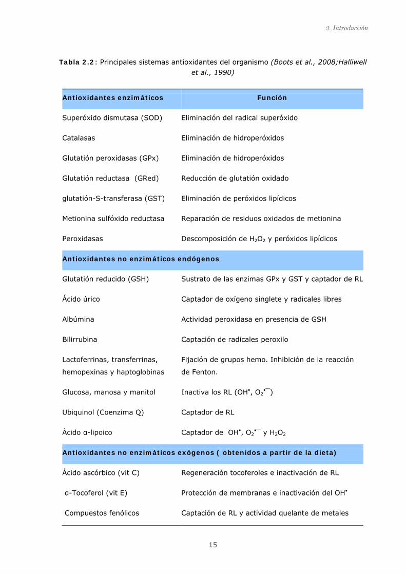
Tabla 2.2: Principales sistemas antioxidantes del organismo (Boots et al., 2008;Halliwell
et al., 1990)
Antioxidantes enzimáticos Función
Superóxido dismutasa (SOD) Eliminación del radical superóxido
Catalasas Eliminación de hidroperóxidos
Glutatión peroxidasas (GPx) Eliminación de hidroperóxidos
Glutatión reductasa (GRed) Reducción de glutatión oxidado
glutatión-S-transferasa (GST) Eliminación de peróxidos lipídicos
Metionina sulfóxido reductasa Reparación de residuos oxidados de metionina
Peroxidasas Descomposición de H2O2 y peróxidos lipídicos
Antioxidantes no enzimáticos endógenos
Glutatión reducido (GSH) Sustrato de las enzimas GPx y GST y captador de RL
Ácido úrico Captador de oxígeno singlete y radicales libres
Albúmina Actividad peroxidasa en presencia de GSH
Bilirrubina Captación de radicales peroxilo
Lactoferrinas, transferrinas,
hemopexinas y haptoglobinas
Fijación de grupos hemo. Inhibición de la reacción
de Fenton.
Glucosa, manosa y manitol Inactiva los RL (OH•, O2•⎯)
Ubiquinol (Coenzima Q) Captador de RL
Ácido α-lipoico Captador de OH•, O2•⎯ y H2O2
Antioxidantes no enzimáticos exógenos ( obtenidos a partir de la dieta)
Ácido ascórbico (vit C) Regeneración tocoferoles e inactivación de RL
α-Tocoferol (vit E) Protección de membranas e inactivación del OH•
Compuestos fenólicos Captación de RL y actividad quelante de metales

2.6 Estrés oxidativo
16
2.6 Estrés oxidativo
En situaciones fisiológicas, cada célula presenta un estado redox característico. En
condiciones normales la cantidad de radicales libres es controlado por el sistema
antioxidante endógeno. Sin embargo, existen diferentes situaciones fisiológicas en las
que puede alterarse el equilibrio en favor de las especies oxidantes dando lugar a lo que
se conoce como estrés oxidativo y éste es originado por dos motivos fundamentales
(Halliwell et al., 2004):
1. Disminución de los niveles de antioxidantes debido a, mutaciones que afectan a
los sistemas antioxidantes o toxinas que causan depleción de las defensas
antioxidantes.
2. Incremento en la producción de especies reactivas a causa de la exposición a
diferentes compuestos que generan especies radicales.
Estás dos causas son mayoritariamente generadas por factores externos que
desequilibran la homeostasis de los organismos aerobios.
Figura 2.6: Factores exógenos que generan
ROS (Limón-Pacheco et al., 2009).
Contaminación ambiental, radiaciones
ionizantes, metales pesados, pesticidas e
hidrocarburos policíclicos aromáticos (PAHs)
entre otros, inducen a la formación de ROS y
RNOS en células y tejidos provocando estrés
oxidativo. En este estado el sistema
antioxidante endógeno es sobrepasado por
las elevadas proporciones de ROS, las cuales
son capaces de dañar proteínas, lípidos y
ADN.
2. Introducción
17
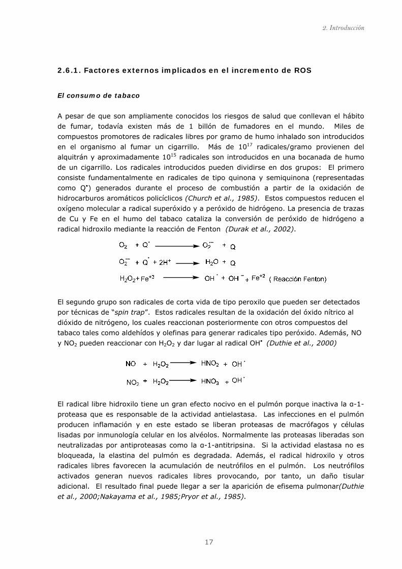
2.6.1. Factores externos implicados en el incremento de ROS
El consumo de tabaco
A pesar de que son ampliamente conocidos los riesgos de salud que conllevan el hábito
de fumar, todavía existen más de 1 billón de fumadores en el mundo. Miles de
compuestos promotores de radicales libres por gramo de humo inhalado son introducidos
en el organismo al fumar un cigarrillo. Más de 1017 radicales/gramo provienen del
alquitrán y aproximadamente 1015 radicales son introducidos en una bocanada de humo
de un cigarrillo. Los radicales introducidos pueden dividirse en dos grupos: El primero
consiste fundamentalmente en radicales de tipo quinona y semiquinona (representadas
como Q•) generados durante el proceso de combustión a partir de la oxidación de
hidrocarburos aromáticos policíclicos (Church et al., 1985). Estos compuestos reducen el
oxígeno molecular a radical superóxido y a peróxido de hidrógeno. La presencia de trazas
de Cu y Fe en el humo del tabaco cataliza la conversión de peróxido de hidrógeno a
radical hidroxilo mediante la reacción de Fenton (Durak et al., 2002).
El segundo grupo son radicales de corta vida de tipo peroxilo que pueden ser detectados
por técnicas de “spin trap”. Estos radicales resultan de la oxidación del óxido nítrico al
dióxido de nitrógeno, los cuales reaccionan posteriormente con otros compuestos del
tabaco tales como aldehídos y olefinas para generar radicales tipo peróxido. Además, NO
y NO2 pueden reaccionar con H2O2 y dar lugar al radical OH• (Duthie et al., 2000)
El radical libre hidroxilo tiene un gran efecto nocivo en el pulmón porque inactiva la α-1-
proteasa que es responsable de la actividad antielastasa. Las infecciones en el pulmón
producen inflamación y en este estado se liberan proteasas de macrófagos y células
lisadas por inmunología celular en los alvéolos. Normalmente las proteasas liberadas son
neutralizadas por antiproteasas como la α-1-antitripsina. Si la actividad elastasa no es
bloqueada, la elastina del pulmón es degradada. Además, el radical hidroxilo y otros
radicales libres favorecen la acumulación de neutrófilos en el pulmón. Los neutrófilos
activados generan nuevos radicales libres provocando, por tanto, un daño tisular
adicional. El resultado final puede llegar a ser la aparición de efisema pulmonar(Duthie
et al., 2000;Nakayama et al., 1985;Pryor et al., 1985).

2.6 Estrés oxidativo
18
Los efectos inflamatorios del humo de tabaco se relacionan con condiciones que
potencian los procesos de carcinogénesis tales como la oxidación del glutatión (GSH) que
incrementa los niveles de disulfuro de glutatión en el tejido pulmonar; el incremento de
los niveles de 8-OH-dG; la disminución de niveles de antioxidantes en el corriente
sanguíneo y el incremento de marcadores de peroxidación lipídica.
El consumo de alcohol
Un consumo excesivo y habitual de alcohol conlleva a un aumento en la producción de
ROS. La hipótesis más plausible sobre el consumo de alcohol y el incremento de ROS
tiene su explicación en el metabolismo del etanol a acetaldehído vía alcohol
deshidrogenada (Figura 2.7).
Las reacciones metabólicas del etanol están localizas en diferentes compartimentos
celulares: (i) en el citosol, vía alcohol deshidrogenasa (ADH); (ii) en los microsomas
mediado por enzimas de complejo citocromo P450.; (iii) en los peroxisomas, vía catalasa
(Lieber et al., 2000).
Figura 2.7: Metabolismo del etanol en los hepatocitos. ADH, alcohol deshidrogenasa;
CYP2E1, citocromo P450 2E1; ALDH2, aldehído deshidrogenasa 2. (Zakhari, 2006)
El acetaldehído (1), producto formado en la reacción mediada por la enzima alcohol
deshidrogenasa, es rápidamente metabolizado por la enzima aldehído deshidrogenasa a
acetato. El Acetaldehído es capaz de formar aductos con proteínas lo que resulta en la
formación de anticuerpos e inactivación de enzimas (Dicker et al., 1988) además de una
disminución en la capacidad de reparación de errores en la replicación (Lieber et al.,
2. Introducción
19
1989). La actividad de la isoenzima CYP2E1 (2), del complejo citocromo P-450
(localizada en los microsomas), asume un papel importante en la metabolización del
etanol a acetaldehído especialmente cuando la concentración de etanol es elevada
(Km=8-10mM, comparada con 0.2- 2.0mM de la ADH hepática), pero a su vez, este
proceso genera especies reactivas como hidroxietil, anión superóxido y radicales
hidroxilo. El proceso de oxidación mediado por la alcohol deshidrogenasa (ADH) implica
la transferencia de dos electrones del dinucleótido de nicotinamida adenina (NAD+) el
cual es reducido a NADH (3). NADH inhibe la enzima xantina deshidrogenasa
promoviendo la oxidación de purinas vía xantina oxidasa y provoca un estado reductor en
la célula que hace más vulnerable la defensa de las biomoléculas al ataque de ROS y a
los productos formados en el metabolismo del etanol.
El consumo habitual de alcohol ha sido relacionado con un incremento de H2O2 en la
célula debido al daño, directo o indirecto, del acetaldehído sobre la actividad de enzimas
antioxidantes como la glutatión peroxidasa (Lieber et al., 2000;Misra et al., 1992).
Además el alcohol disminuye los niveles de GSH e induce la actividad de ciertas enzimas
del complejo citrocromo P-450 (CYP2E1) que impulsan la activación de pro-carcinógenos
en carcinógenos
Radiaciones ultravioletas (UVA) y ultravioletas-visible (UVB)
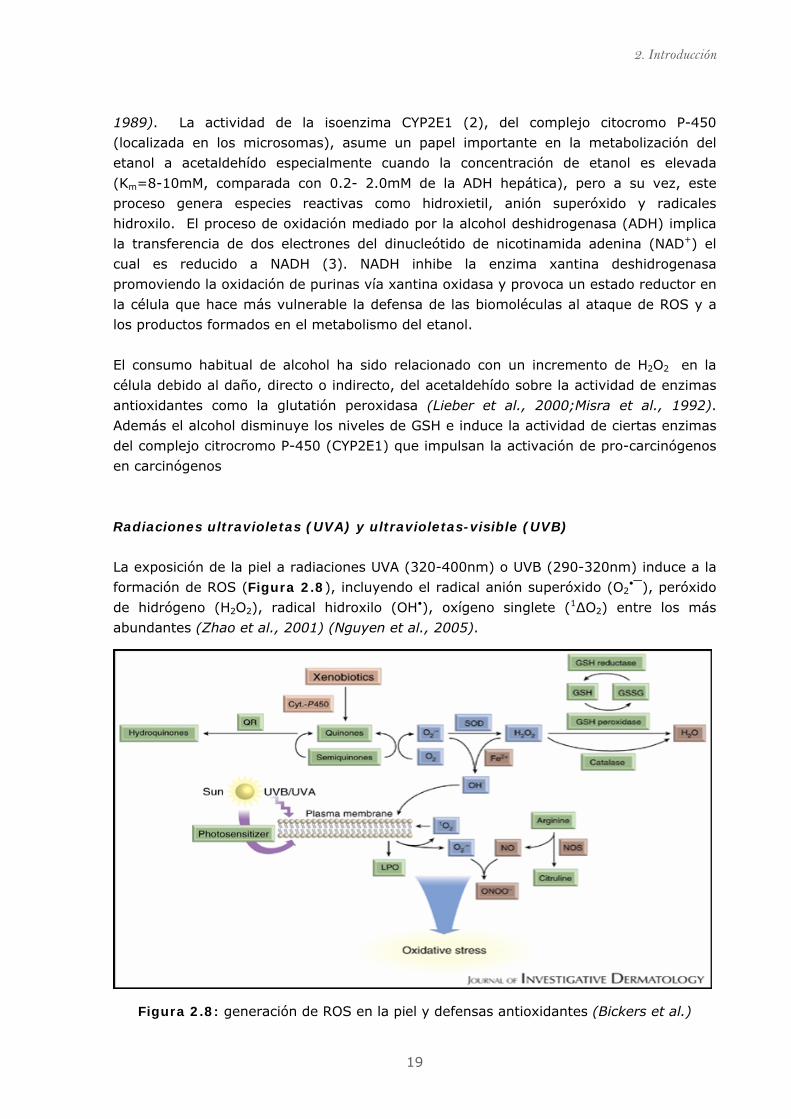
La exposición de la piel a radiaciones UVA (320-400nm) o UVB (290-320nm) induce a la
formación de ROS (Figura 2.8), incluyendo el radical anión superóxido (O2•⎯), peróxido
de hidrógeno (H2O2), radical hidroxilo (OH•), oxígeno singlete (1∆O2) entre los más
abundantes (Zhao et al., 2001) (Nguyen et al., 2005).
Figura 2.8: generación de ROS en la piel y defensas antioxidantes (Bickers et al.)
2.6 Estrés oxidativo
20
Las radiaciones solares que corresponden en más de un 90% a radiaciones UVA,
penetran en la dermis causando degeneración de colágeno e inflamación por formación
de ROS. La exposición continua de UVA deriva en un envejecimiento prematuro de la
piel.
Las radiaciones UVB (causantes de las quemaduras solares) incrementan los niveles de
ROS en células de la epidermis. Activan rutas involucradas en el crecimiento celular,
diferenciación y proliferación, facilitando la expansión de tumores celulares (Bickers et
al.). La exposición continuada a UVB induce a la oxidación de lípidos y proteínas de las
membranas epiteliales ocasionando perdida de fluidez, inactivación de enzimas y
alteración de la permeabilidad de iones, que finalmente derivan en choque osmótico y
ruptura celular (Bommareddy et al., 2007).
Biocidas y pesticidas
Trazas de herbicidas y pesticidas son detectados en la mayoría de alimentos vegetales y
aguas no comerciales que ingieren los humanos. La naturaleza de estos compuestos es
variada y en ello radica su grado de toxicidad. Los pesticidas pueden inducir al estrés
oxidativo por diferentes vías (Bagchi et al., 1995;Banerjee et al., 2001). La alteración de
enzimas del sistema antioxidante endógeno (catalasa y superóxido dismutasa) es una de
las vías más frecuentes (Braconi et al., 2008).
Metales
El desarrollo tecnológico, el consumo masivo e indiscriminado y la producción de
desechos principalmente urbanos (pilas, pinturas, baterías de automóviles), han
provocado la presencia de numerosos metales en cantidades importantes en el ambiente.
Estos pueden incorporarse con los alimentos o como partículas que se respiran y se van
acumulando en el organismo. La formación e incremento de ROS ha sido observada en
diferentes órganos después de estar expuestos a arsénico (As) (Bardullas et al.), cadmio
(Cd)(Nemmiche et al., 2007), cromo (Cr)(Bagchi et al., 2002) o mercurio (Hg)(Göksel
Şener et al., 2003).
Otros factores
Situaciones de estrés psicológico (Chalmers et al., 2003), ejercicio físico intenso
(Bloomer et al., 2008;Ji, 2000), una dieta desequilibrada, rica en grasas (Brewster et al.,
2006), o incluso escribir una tesis (Touriño, 2009) pueden dar lugar a la formación y/o
incremento de ROS en el organismo y derivar en un estado de estrés oxidativo.
2. Introducción
21
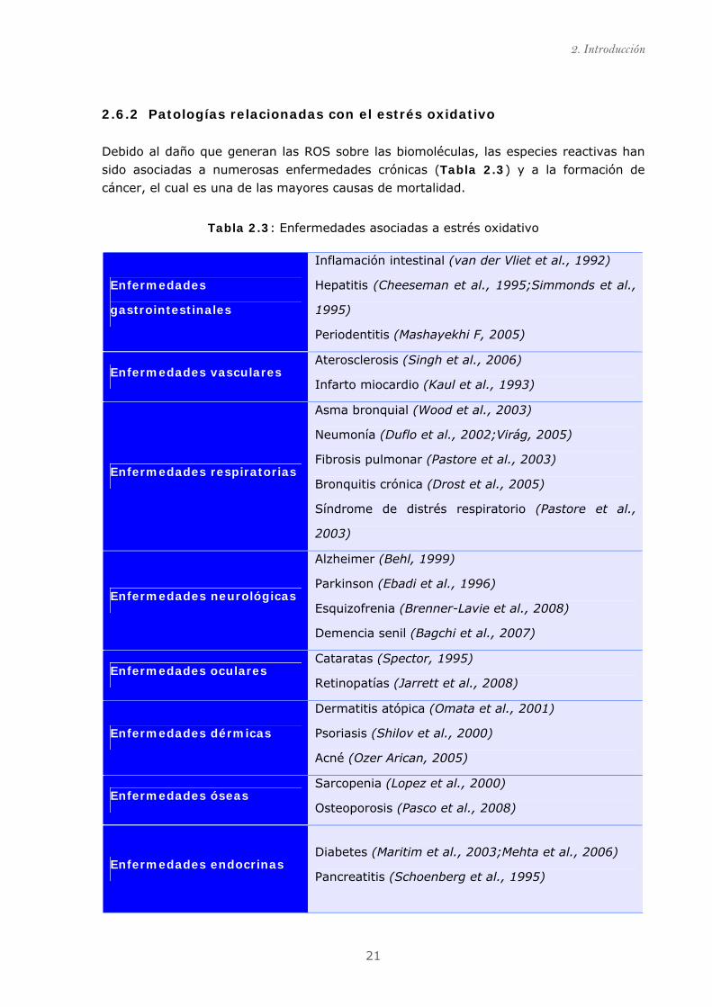
2.6.2 Patologías relacionadas con el estrés oxidativo
Debido al daño que generan las ROS sobre las biomoléculas, las especies reactivas han
sido asociadas a numerosas enfermedades crónicas (Tabla 2.3) y a la formación de
cáncer, el cual es una de las mayores causas de mortalidad.
Tabla 2.3: Enfermedades asociadas a estrés oxidativo
Enfermedades
gastrointestinales
Inflamación intestinal (van der Vliet et al., 1992)
Hepatitis (Cheeseman et al., 1995;Simmonds et al.,
1995)
Periodentitis (Mashayekhi F, 2005)
Enfermedades vasculares Aterosclerosis (Singh et al., 2006)
Infarto miocardio (Kaul et al., 1993)
Enfermedades respiratorias
Asma bronquial (Wood et al., 2003)
Neumonía (Duflo et al., 2002;Virág, 2005)
Fibrosis pulmonar (Pastore et al., 2003)
Bronquitis crónica (Drost et al., 2005)
Síndrome de distrés respiratorio (Pastore et al.,
2003)
Enfermedades neurológicas
Alzheimer (Behl, 1999)
Parkinson (Ebadi et al., 1996)
Esquizofrenia (Brenner-Lavie et al., 2008)
Demencia senil (Bagchi et al., 2007)
Enfermedades oculares Cataratas (Spector, 1995)
Retinopatías (Jarrett et al., 2008)
Enfermedades dérmicas
Dermatitis atópica (Omata et al., 2001)
Psoriasis (Shilov et al., 2000)
Acné (Ozer Arican, 2005)
Enfermedades óseas Sarcopenia (Lopez et al., 2000)
Osteoporosis (Pasco et al., 2008)
Enfermedades endocrinas Diabetes (Maritim et al., 2003;Mehta et al., 2006)
Pancreatitis (Schoenberg et al., 1995)
2.6 Estrés oxidativo
22
2.6.3 Cáncer y estrés oxidativo
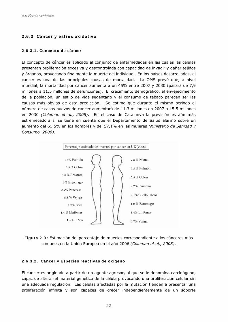
2.6.3.1. Concepto de cáncer
El concepto de cáncer es aplicado al conjunto de enfermedades en las cuales las células
presentan proliferación excesiva y descontrolada con capacidad de invadir y dañar tejidos
y órganos, provocando finalmente la muerte del individuo. En los países desarrollados, el
cáncer es una de las principales causas de mortalidad. La OMS prevé que, a nivel
mundial, la mortalidad por cáncer aumentará un 45% entre 2007 y 2030 (pasará de 7,9
millones a 11,5 millones de defunciones). El crecimiento demográfico, el envejecimiento
de la población, un estilo de vida sedentario y el consumo de tabaco parecen ser las
causas más obvias de esta predicción. Se estima que durante el mismo periodo el
número de casos nuevos de cáncer aumentará de 11,3 millones en 2007 a 15,5 millones
en 2030 (Coleman et al., 2008). En el caso de Catalunya la previsión es aún más
estremecedora si se tiene en cuenta que el Departamento de Salud alarmó sobre un
aumento del 61,5% en los hombres y del 57,1% en las mujeres (Ministerio de Sanidad y
Consumo, 2006).
Figura 2.9: Estimación del porcentaje de muertes correspondiente a los cánceres más
comunes en la Unión Europea en el año 2006 (Coleman et al., 2008).
2.6.3.2. Cáncer y Especies reactivas de oxígeno
El cáncer es originado a partir de un agente agresor, al que se le denomina carcinógeno,
capaz de alterar el material genético de la célula provocando una proliferación celular sin
una adecuada regulación. Las células afectadas por la mutación tienden a presentar una
proliferación infinita y son capaces de crecer independientemente de un soporte
2. Introducción
23
estructural. Cuando un grupo de células se divide de forma anormal es llamada tumor.
Si el tumor es capaz de separarse de su origen y llegar a otros órganos por el corriente
sanguíneo o linfa invadiendo otros tejidos, es denominado metástasis.
Los mecanismos propuestos por numerosos autores para explicar la aparición de cáncer
son: (i) incremento de síntesis y mitosis de ADN originado a partir de un carcinógeno
capaz de inducir mutaciones en los genes encargados de regular la proliferación y el
crecimiento celular (conocidos como pro-oncogenes) y que dan lugar a la formación de
oncogenes (pro-oncogenes mutados); (ii) desequilibrio entre la proliferación de las
células y la muerte celular programada (apoptosis). Estudios epidemiológicos y
biológicos han demostrado que el proceso de oncogénesis ocurre en varias etapas, donde
las especies radicales juegan un papel decisivo para el desarrollo del cáncer (Frenkel,
1992;Valko et al., 2004).
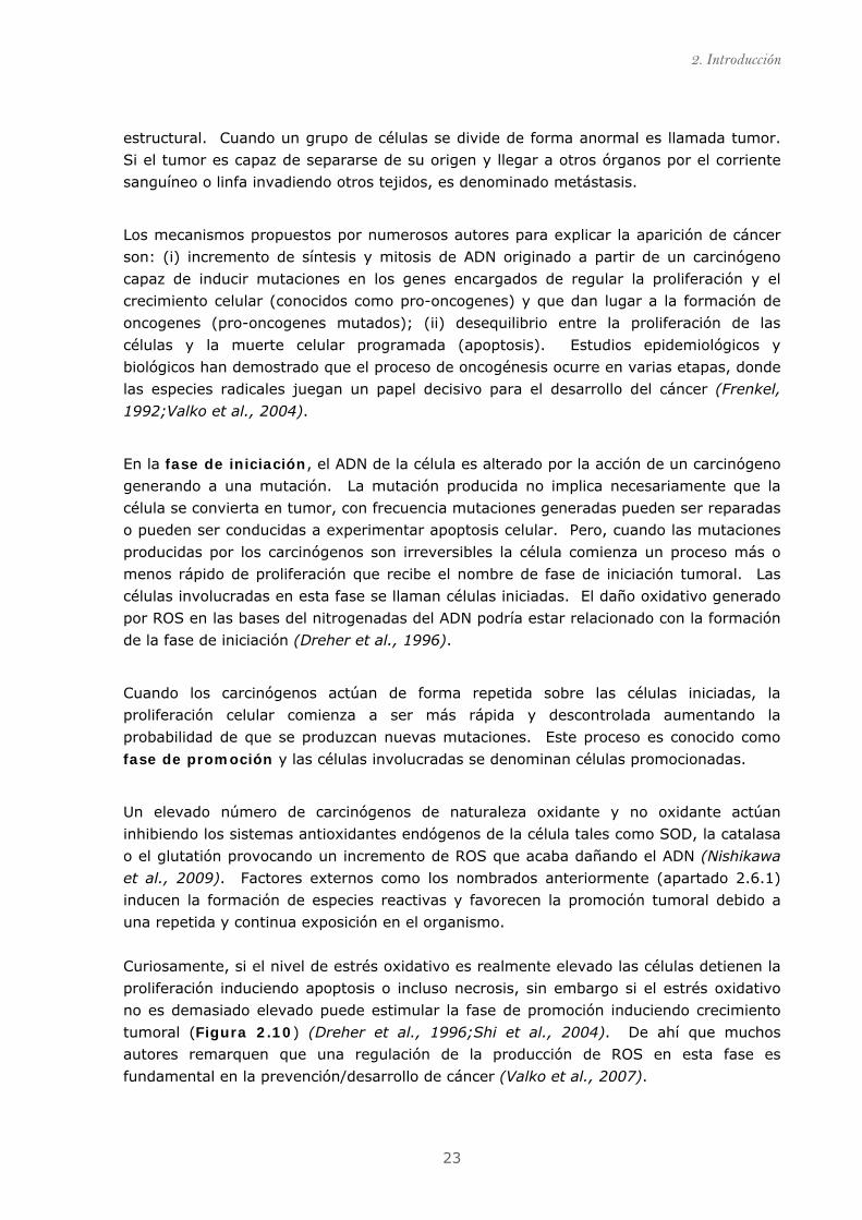
En la fase de iniciación, el ADN de la célula es alterado por la acción de un carcinógeno
generando a una mutación. La mutación producida no implica necesariamente que la
célula se convierta en tumor, con frecuencia mutaciones generadas pueden ser reparadas
o pueden ser conducidas a experimentar apoptosis celular. Pero, cuando las mutaciones
producidas por los carcinógenos son irreversibles la célula comienza un proceso más o
menos rápido de proliferación que recibe el nombre de fase de iniciación tumoral. Las
células involucradas en esta fase se llaman células iniciadas. El daño oxidativo generado
por ROS en las bases del nitrogenadas del ADN podría estar relacionado con la formación
de la fase de iniciación (Dreher et al., 1996).
Cuando los carcinógenos actúan de forma repetida sobre las células iniciadas, la
proliferación celular comienza a ser más rápida y descontrolada aumentando la
probabilidad de que se produzcan nuevas mutaciones. Este proceso es conocido como
fase de promoción y las células involucradas se denominan células promocionadas.
Un elevado número de carcinógenos de naturaleza oxidante y no oxidante actúan
inhibiendo los sistemas antioxidantes endógenos de la célula tales como SOD, la catalasa
o el glutatión provocando un incremento de ROS que acaba dañando el ADN (Nishikawa
et al., 2009). Factores externos como los nombrados anteriormente (apartado 2.6.1)
inducen la formación de especies reactivas y favorecen la promoción tumoral debido a
una repetida y continua exposición en el organismo.
Curiosamente, si el nivel de estrés oxidativo es realmente elevado las células detienen la
proliferación induciendo apoptosis o incluso necrosis, sin embargo si el estrés oxidativo
no es demasiado elevado puede estimular la fase de promoción induciendo crecimiento
tumoral (Figura 2.10) (Dreher et al., 1996;Shi et al., 2004). De ahí que muchos
autores remarquen que una regulación de la producción de ROS en esta fase es
fundamental en la prevención/desarrollo de cáncer (Valko et al., 2007).
2.6 Estrés oxidativo
24
Las células promocionadas siguen sufriendo mutaciones debido a la desregulación del
ciclo celular tornándose más anómalas en su crecimiento y comportamiento hasta
originar un tumor. La suma de estos procesos es conocida como fase de progresión.
Los tumores, en los que la presencia de ROS parece ser evidente (Hsu et al., 1991),
pueden adquirir la capacidad de invasión tanto a nivel local, infiltrando los tejidos de
alrededor, como a distancia, originando metástasis. Niveles elevados de stress oxidativo
han sido observados en esta fase (Gupte et al., 2009;Nishikawa, 2008).
Figura 2.10: Etapas de formación del cáncer en función del estrés oxidativo (Valko et
al., 2006)
Enfermedades crónicas y cáncer presentan en común un elevado estrés oxidativo.
Todavía queda por aclarar sí el stress oxidativo es generado por los mecanismos
patológicos de cada enfermedad, o si las enfermedades son causa directa o indirecta del
daño celular que causan los radicales libres. Sin embargo, es clara la idea de que la
presencia de estrés oxidativo en la célula convierte el sistema antioxidante endógeno
insuficiente para la homeostasis. La ingesta de antioxidantes podría ser necesaria para
compensar un déficit del sistema antioxidante endógeno. El ácido ascórbico (vit C) o la
vitamina E son necesarias para el correcto funcionamiento celular auque no son
sintetizadas en los humanos y requieren ser incorporadas por la dieta. Es por ello, que
una dieta rica en antioxidantes podría prevenir enfermedades.
2. Introducción
25
2.7 Los flavonoides como ingredientes de alimentos
funcionales 2.7.1 La dieta en la prevención de enfermedades
Estudios epidemiológicos han demostrado, que la Dieta Mediterránea contribuye a
disminuir el riesgo de padecer patologías crónicas como son las enfermedades
cardiovasculares (Fung et al., 2009), la obesidad (Mendez et al., 2006), la diabetes
(Martínez-González et al., 2008) y cáncer (Benetou et al.). La conocida paradoja
francesa esta relacionada con la prevención de enfermedades cardiovasculares (Cheng,
2001;Vidavalur et al., 2006). Ambas tienen en común una elevada ingesta de frutas y
verduras ricas en productos antioxidantes. Muchos de los beneficios asignados a estás
dietas puede ser debido a la actividad sinérgica entre los compuestos antioxidantes
ingeridos (Rahman, 2009). Es por ello, que una dieta equilibrada suplementada con
antioxidantes podría prevenir o/y curar enfermedades relacionadas con el estrés
oxidativo.
En la actualidad, se han realizado estudios clínicos suplementando la dieta con (i)
compuestos antioxidantes esenciales de la dieta tales como vitamina E y C, (ii)
compuestos precursores de antioxidantes, N-acetil cisteina (NAC) y un mayor número de
estudios con, (iii) antioxidantes exógenos, los cuales no son esenciales para el organismo
pero que juegan un papel importante en el buen funcionamiento del organismo, tales
como ácido α-lipoico y los polifenoles. Algunos de éstos estudios dieron como resultado,
efectos beneficiosos en los enfermos (Autier et al., 2007) (Christen et al., 2009;Hercberg
et al., 2004). Sin embargo, algunos no presentaron diferencias significativas en los
resultados obtenidos (Beazley et al., 2005;Cook et al., 2007;Farouque et al., 2006;Keith
et al., 2001;Kucuk et al., 2001), e incluso, algún estudio presentó resultados negativos
(Bairati et al., 2005).
La utilización de antioxidantes exógenos para la prevención y/o tratamiento de
enfermedades asociadas al stress oxidativo es un tema donde todavía quedan muchos
puntos que aclarar. El trabajo realizado durante la presente tesis tiene como objetivo
general contribuir en un mayor conocimiento sobre el uso de antioxidantes exógenos
como suplementos nutricionales e ingredientes de alimentos funcionales.
2.7.2 Alimentos funcionales. Concepto.
Los alimentos funcionales no han sido definidos hasta el momento por la legislación
europea. Generalmente, se considera que son aquellos alimentos, que se consumen
como parte de una dieta normal y contienen componentes biológicamente activos, que
ofrecen beneficios para la salud y reducen el riesgo de sufrir enfermedades. Entre
algunos ejemplos de alimentos funcionales, destacan: los alimentos que contienen
determinados minerales, vitaminas, ácidos grasos o fibra dietética; los alimentos a los
2.7 Los flavonoides como ingredientes de alimentos funcionales
26
que se han añadido sustancias biológicamente activas (fitoquímicos u otros
antioxidantes); y los probióticos (alimentos con cultivos vivos beneficiosos ) (EUFIC,
2009).
2.7.3 Los flavonoides
Los flavonoides están incluidos en el grupo de los compuestos fenólicos, que se
encuentran ampliamente distribuidos en la naturaleza con más de 8000 estructuras
conocidas (Harborne, 1993). Los compuestos fenólicos son metabolitos secundarios de
las plantas y ejercen diversas funciones; desde la coloración de flores hasta la
impregnación de lignina de las paredes pecto-celulósicas y son encargados de los
mecanismos de defensa frente agresiones externas (radiación UVA, predadores, ataques
fúngicos y víricos)(Dixon et al., 1996). Los animales no son capaces de sintetizar este
tipo de compuestos es por ello que tienen que incorporarlos por la dieta.
El esqueleto de los compuestos fenólicos consta de un anillo bencénico que contiene uno
o diversos grupos hidroxilo. Las distintas familias de compuestos fenólicos son
clasificadas principalmente por el número de átomos de carbono que presentan en su
estructura (Tabla 2.4)
Tabla 2.4: Principales familias de los compuestos fenólicos.
Estructura Familia
C6 Fenoles simples C6-C1 Ácidos fenólicos C6-C2 Ácidos fenólicos y acetofenonas C6-C3 Cumarinas y ácidos hidroxicinámicos C6-C1-C6 Xantonas y benzofenonas C6-C2-C6 Estilbenos C6-C3-C6 Chalconas C6-C3-C6 Flavonoides (C6-C3-C6)n Taninos condensados (C6-C1)n Taninos hidrolizables
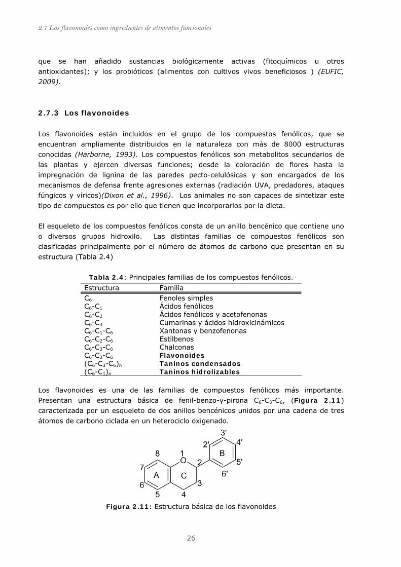
Los flavonoides es una de las familias de compuestos fenólicos más importante.
Presentan una estructura básica de fenil-benzo-γ-pirona C6-C3-C6, (Figura 2.11)
caracterizada por un esqueleto de dos anillos bencénicos unidos por una cadena de tres
átomos de carbono ciclada en un heterociclo oxigenado.
Figura 2.11: Estructura básica de los flavonoides
2. Introducción
27
Figura 2. 12: Estructura química de las subclases de flavonoides más relevantes.
2.7 Los flavonoides como ingredientes de alimentos funcionales
28
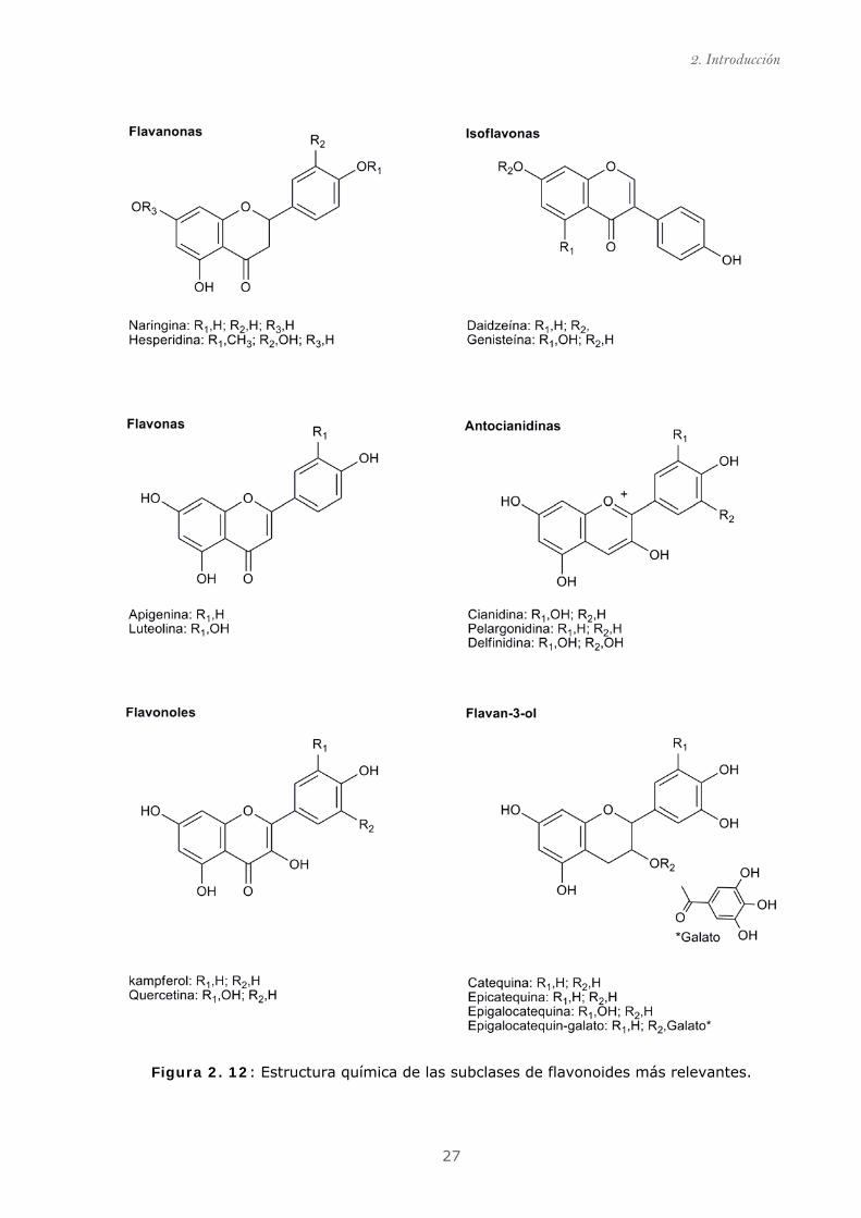
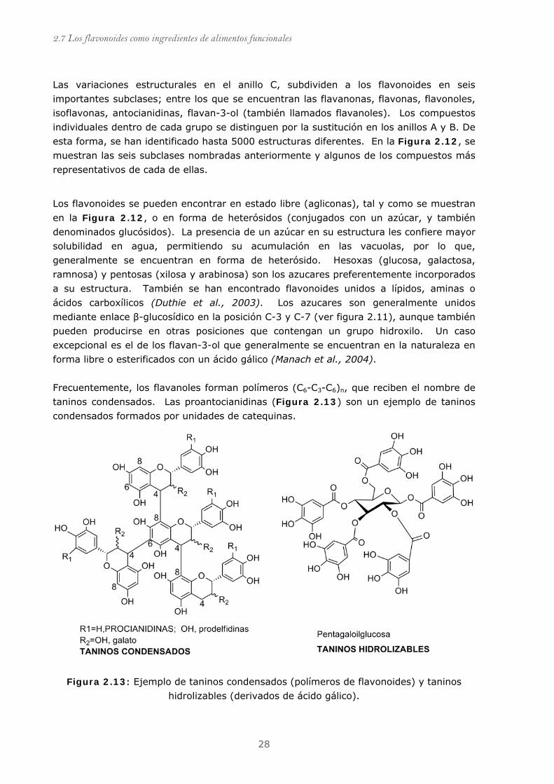
Las variaciones estructurales en el anillo C, subdividen a los flavonoides en seis
importantes subclases; entre los que se encuentran las flavanonas, flavonas, flavonoles,
isoflavonas, antocianidinas, flavan-3-ol (también llamados flavanoles). Los compuestos
individuales dentro de cada grupo se distinguen por la sustitución en los anillos A y B. De
esta forma, se han identificado hasta 5000 estructuras diferentes. En la Figura 2.12, se
muestran las seis subclases nombradas anteriormente y algunos de los compuestos más
representativos de cada de ellas.
Los flavonoides se pueden encontrar en estado libre (agliconas), tal y como se muestran
en la Figura 2.12, o en forma de heterósidos (conjugados con un azúcar, y también
denominados glucósidos). La presencia de un azúcar en su estructura les confiere mayor
solubilidad en agua, permitiendo su acumulación en las vacuolas, por lo que,
generalmente se encuentran en forma de heterósido. Hesoxas (glucosa, galactosa,
ramnosa) y pentosas (xilosa y arabinosa) son los azucares preferentemente incorporados
a su estructura. También se han encontrado flavonoides unidos a lípidos, aminas o
ácidos carboxílicos (Duthie et al., 2003). Los azucares son generalmente unidos
mediante enlace β-glucosídico en la posición C-3 y C-7 (ver figura 2.11), aunque también
pueden producirse en otras posiciones que contengan un grupo hidroxilo. Un caso
excepcional es el de los flavan-3-ol que generalmente se encuentran en la naturaleza en
forma libre o esterificados con un ácido gálico (Manach et al., 2004).
Frecuentemente, los flavanoles forman polímeros (C6-C3-C6)n, que reciben el nombre de
taninos condensados. Las proantocianidinas (Figura 2.13) son un ejemplo de taninos
condensados formados por unidades de catequinas.
Figura 2.13: Ejemplo de taninos condensados (polímeros de flavonoides) y taninos
hidrolizables (derivados de ácido gálico).
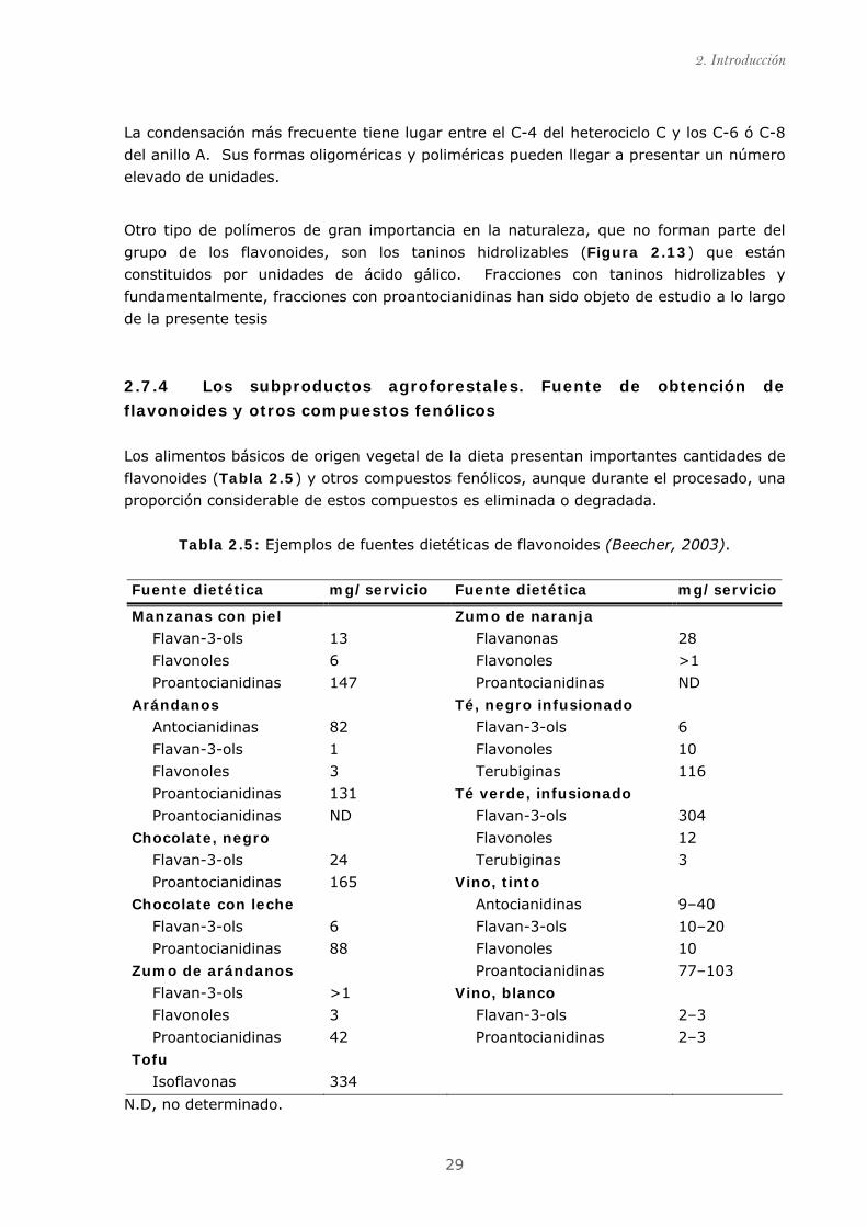
2. Introducción
29
La condensación más frecuente tiene lugar entre el C-4 del heterociclo C y los C-6 ó C-8
del anillo A. Sus formas oligoméricas y poliméricas pueden llegar a presentar un número
elevado de unidades.
Otro tipo de polímeros de gran importancia en la naturaleza, que no forman parte del
grupo de los flavonoides, son los taninos hidrolizables (Figura 2.13) que están
constituidos por unidades de ácido gálico. Fracciones con taninos hidrolizables y
fundamentalmente, fracciones con proantocianidinas han sido objeto de estudio a lo largo
de la presente tesis
2.7.4 Los subproductos agroforestales. Fuente de obtención de
flavonoides y otros compuestos fenólicos
Los alimentos básicos de origen vegetal de la dieta presentan importantes cantidades de
flavonoides (Tabla 2.5) y otros compuestos fenólicos, aunque durante el procesado, una
proporción considerable de estos compuestos es eliminada o degradada.
Tabla 2.5: Ejemplos de fuentes dietéticas de flavonoides (Beecher, 2003).
Fuente dietética mg/servicio Fuente dietética mg/servicio
Manzanas con piel Zumo de naranja
Flavan-3-ols 13 Flavanonas 28
Flavonoles 6 Flavonoles >1
Proantocianidinas 147 Proantocianidinas ND
Arándanos Té, negro infusionado
Antocianidinas 82 Flavan-3-ols 6
Flavan-3-ols 1 Flavonoles 10
Flavonoles 3 Terubiginas 116
Proantocianidinas 131 Té verde, infusionado
Proantocianidinas ND Flavan-3-ols 304
Chocolate, negro Flavonoles 12
Flavan-3-ols 24 Terubiginas 3
Proantocianidinas 165 Vino, tinto
Chocolate con leche Antocianidinas 9–40
Flavan-3-ols 6 Flavan-3-ols 10–20
Proantocianidinas 88 Flavonoles 10
Zumo de arándanos Proantocianidinas 77–103
Flavan-3-ols >1 Vino, blanco
Flavonoles 3 Flavan-3-ols 2–3
Proantocianidinas 42 Proantocianidinas 2–3
Tofu
Isoflavonas 334
N.D, no determinado.
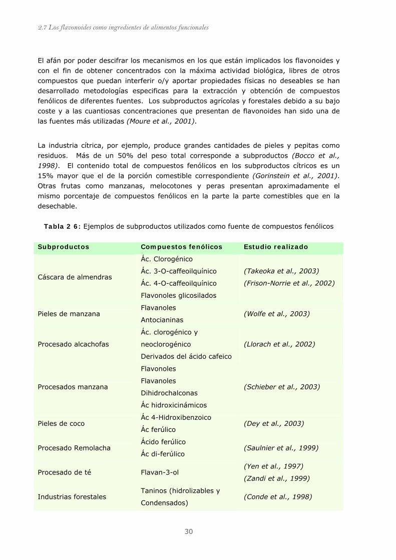
2.7 Los flavonoides como ingredientes de alimentos funcionales
30
El afán por poder descifrar los mecanismos en los que están implicados los flavonoides y
con el fin de obtener concentrados con la máxima actividad biológica, libres de otros
compuestos que puedan interferir o/y aportar propiedades físicas no deseables se han
desarrollado metodologías especificas para la extracción y obtención de compuestos
fenólicos de diferentes fuentes. Los subproductos agrícolas y forestales debido a su bajo
coste y a las cuantiosas concentraciones que presentan de flavonoides han sido una de
las fuentes más utilizadas (Moure et al., 2001).
La industria cítrica, por ejemplo, produce grandes cantidades de pieles y pepitas como
residuos. Más de un 50% del peso total corresponde a subproductos (Bocco et al.,
1998). El contenido total de compuestos fenólicos en los subproductos cítricos es un
15% mayor que el de la porción comestible correspondiente (Gorinstein et al., 2001).
Otras frutas como manzanas, melocotones y peras presentan aproximadamente el
mismo porcentaje de compuestos fenólicos en la parte la parte comestibles que en la
desechable.
Tabla 2 6: Ejemplos de subproductos utilizados como fuente de compuestos fenólicos
Subproductos Compuestos fenólicos Estudio realizado
Ác. Clorogénico
Ác. 3-O-caffeoilquínico Cáscara de almendras
Ác. 4-O-caffeoilquínico
Flavonoles glicosilados
(Takeoka et al., 2003)
(Frison-Norrie et al., 2002)
Pieles de manzana Flavanoles
Antocianinas (Wolfe et al., 2003)
Ác. clorogénico y
neoclorogénico Procesado alcachofas
Derivados del ácido cafeico
(Llorach et al., 2002)
Procesados manzana
Flavonoles
Flavanoles
Dihidrochalconas
Ác hidroxicinámicos
(Schieber et al., 2003)
Pieles de coco Ác 4-Hidroxibenzoico
Ác ferúlico (Dey et al., 2003)
Procesado Remolacha Ácido ferúlico
Ác di-ferúlico (Saulnier et al., 1999)
Procesado de té Flavan-3-ol (Yen et al., 1997)
(Zandi et al., 1999)
Industrias forestales Taninos (hidrolizables y
Condensados) (Conde et al., 1998)

2. Introducción
31
Otro ejemplo es la industria vitivinícola que procesa anualmente más de 60 millones de
toneladas de uva. Más de un 13% corresponden a subproductos del prensado que
básicamente consisten en hollejo, y semillas ricas proantocianidinas y otros compuestos
fenólicos (Schieber et al., 2001;Torres et al., 2001)
El grupo del Dr. Saura-Calixto y colaboradores ha desarrollado un nuevo concepto de
fibra dietética a partir del prensando de hollejo de uva que proviene de subproductos de
la industria vitivinícola (Saura-Calixto et al., 1999). La fibra dietética de uva (GADF,
grape antioxidant dietary fiber) combina los efectos beneficiosos de la fibra con los de los
compuestos polifenólicos antioxidantes (Martín-Carron et al., 1997). Recientemente, se
han obtenido fibras dietéticas de otros subproductos generados del procesado de: los
espárragos (Fuentes-Alventosa et al., 2009); la coliflor (Stojceska et al., 2008); el trigo
(Esposito et al., 2005); los cítricos (Lario et al., 2004;Marín et al., 2007) o subproductos
derivados del procesado de la soja (Redondo-Cuenca et al., 2008).
2.7.5 Actividad biológica de los flavonoides
Los compuestos fenólicos, entre ellos los flavonoides, son considerados como
antioxidantes gracias a la capacidad de captar radicales libres. Los grupos hidroxilo que
forman parte de su estructura donan electrones o hidrogeniones inactivando a los
radicales. El radical fenoxilo generado es muy poco reactivo, debido a que se estabiliza por resonancia con los electrones π del anillo aromático (Figura 2.14).
OHO
OH
OH
OHOHO
OH
OHA A
Rad RadH
OH OH
otras especies oxidadasoligómeros, polímeros
Rad
RadH
( )-epicatequina (EC)semiquinona
OHO
OH
O
O
B
A
OHorto-quinona
O
B
Figura 2.14: mecanismo de oxidación de la (—)-epicatequina.
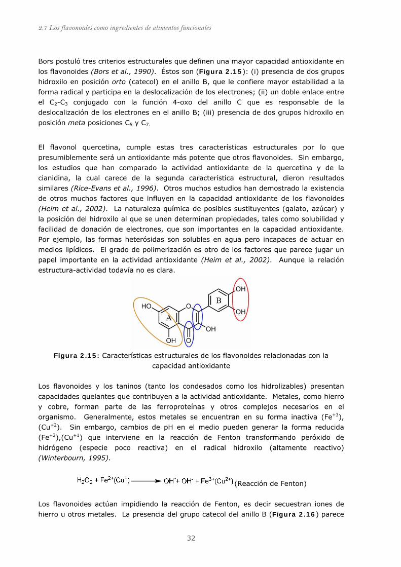
2.7 Los flavonoides como ingredientes de alimentos funcionales
32
Bors postuló tres criterios estructurales que definen una mayor capacidad antioxidante en
los flavonoides (Bors et al., 1990). Éstos son (Figura 2.15): (i) presencia de dos grupos
hidroxilo en posición orto (catecol) en el anillo B, que le confiere mayor estabilidad a la
forma radical y participa en la deslocalización de los electrones; (ii) un doble enlace entre
el C2-C3 conjugado con la función 4-oxo del anillo C que es responsable de la
deslocalización de los electrones en el anillo B; (iii) presencia de dos grupos hidroxilo en
posición meta posiciones C5 y C7.
El flavonol quercetina, cumple estas tres características estructurales por lo que
presumiblemente será un antioxidante más potente que otros flavonoides. Sin embargo,
los estudios que han comparado la actividad antioxidante de la quercetina y de la
cianidina, la cual carece de la segunda característica estructural, dieron resultados
similares (Rice-Evans et al., 1996). Otros muchos estudios han demostrado la existencia
de otros muchos factores que influyen en la capacidad antioxidante de los flavonoides
(Heim et al., 2002). La naturaleza química de posibles sustituyentes (galato, azúcar) y
la posición del hidroxilo al que se unen determinan propiedades, tales como solubilidad y
facilidad de donación de electrones, que son importantes en la capacidad antioxidante.
Por ejemplo, las formas heterósidas son solubles en agua pero incapaces de actuar en
medios lipídicos. El grado de polimerización es otro de los factores que parece jugar un
papel importante en la actividad antioxidante (Heim et al., 2002). Aunque la relación
estructura-actividad todavía no es clara.
Figura 2.15: Características estructurales de los flavonoides relacionadas con la
capacidad antioxidante
Los flavonoides y los taninos (tanto los condesados como los hidrolizables) presentan
capacidades quelantes que contribuyen a la actividad antioxidante. Metales, como hierro
y cobre, forman parte de las ferroproteínas y otros complejos necesarios en el
organismo. Generalmente, estos metales se encuentran en su forma inactiva (Fe+3),
(Cu+2). Sin embargo, cambios de pH en el medio pueden generar la forma reducida
(Fe+2),(Cu+1) que interviene en la reacción de Fenton transformando peróxido de
hidrógeno (especie poco reactiva) en el radical hidroxilo (altamente reactivo)
(Winterbourn, 1995).
(Reacción de Fenton)
Los flavonoides actúan impidiendo la reacción de Fenton, es decir secuestran iones de
hierro u otros metales. La presencia del grupo catecol del anillo B (Figura 2.16) parece
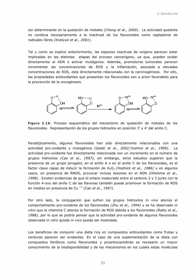
2. Introducción
33
ser determinante en la quelación de metales (Cheng et al., 2000). La actividad quelante
no conlleva necesariamente a la inactivad de los flavonoides como captadores de
radicales libres (Kostyuk et al., 2001).
Tal y como se explicó anteriormente, las especies reactivas de oxígeno parecen estar
implicadas en las distintas etapas del proceso cancerígeno, ya que, pueden oxidar
directamente al ADN o activar mutágenos. Además, promotores tumorales parecen
incrementar las concentraciones de ROS y la inflamación, asociada a elevadas
concentraciones de ROS, esta directamente relacionada con la carcinogénesis. Por ello,
las propiedades antioxidantes que presentan los flavonoides son a priori favorables para
la prevención de la oncogénesis.
Figura 2.16: Proceso esquemático del mecanismo de quelación de metales de los
flavonoides. Representación de los grupos hidroxilos en posición 3’ y 4’ del anillo C.
Paradójicamente, algunos flavonoides han sido directamente relacionados con una
actividad pro-oxidante y mutagénica (Galati et al., 2002;Yoshino et al., 1999). La
actividad pro-oxidante fue directamente relacionada con un incremento en el número de
grupos hidroxilos (Cao et al., 1997), sin embargo, otros estudios sugieren que la
presencia de un grupo pirogalol, en el anillo A o en el anillo C de los flavonoides, es el
factor clave capaz de inducir la formación de H2O2 (Hodnick et al., 1986) y en algunos
casos, en presencia de RNOS, provocar incluso lesiones en el ADN (Ohshima et al.,
1998). Existen evidencias de que el enlace insaturado entre el carbono 2 y 3 junto con la
función 4-oxo del anillo C de las flavonas también puede promover la formación de ROS
en medios en presencia de Cu +2 (Cao et al., 1997).
Por otro lado, la conjugación que sufren los grupos hidroxilos in vivo atenúa el
comportamiento pro-oxidante de los flavonoides (Zhu et al., 1994) y se ha observado in
vitro que la vitamina C atenúa la formación de ROS debida a los flavonoides (Ratty et al.,
1988), por lo que se podría pensar que la actividad pro-oxidante de algunos flavonoides
observada in vitro quizás in vivo pueda ser modulada.
Los beneficios de consumir una dieta rica en compuestos antioxidantes como frutas y
verduras parecen ser evidentes. En el caso de una suplementación de la dieta con
compuestos fenólicos como flavonoides y proantocianidinas es necesario un mayor
conocimiento de la biodisponibilidad y de los mecanismos en los cuales estas moléculas
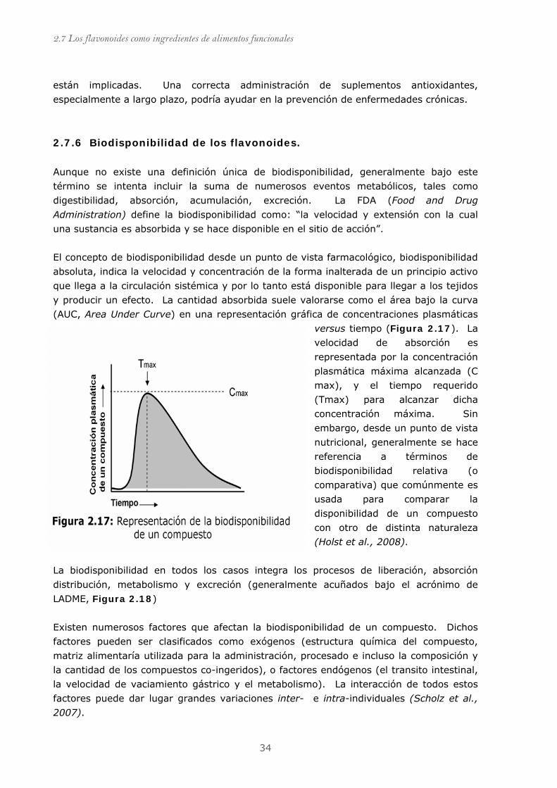
2.7 Los flavonoides como ingredientes de alimentos funcionales
34
están implicadas. Una correcta administración de suplementos antioxidantes,
especialmente a largo plazo, podría ayudar en la prevención de enfermedades crónicas.
2.7.6 Biodisponibilidad de los flavonoides.
Aunque no existe una definición única de biodisponibilidad, generalmente bajo este
término se intenta incluir la suma de numerosos eventos metabólicos, tales como
digestibilidad, absorción, acumulación, excreción. La FDA (Food and Drug
Administration) define la biodisponibilidad como: “la velocidad y extensión con la cual
una sustancia es absorbida y se hace disponible en el sitio de acción”.
El concepto de biodisponibilidad desde un punto de vista farmacológico, biodisponibilidad
absoluta, indica la velocidad y concentración de la forma inalterada de un principio activo
que llega a la circulación sistémica y por lo tanto está disponible para llegar a los tejidos
y producir un efecto. La cantidad absorbida suele valorarse como el área bajo la curva
(AUC, Area Under Curve) en una representación gráfica de concentraciones plasmáticas
versus tiempo (Figura 2.17). La
velocidad de absorción es
representada por la concentración
plasmática máxima alcanzada (C
max), y el tiempo requerido
(Tmax) para alcanzar dicha
concentración máxima. Sin
embargo, desde un punto de vista
nutricional, generalmente se hace
referencia a términos de
biodisponibilidad relativa (o
comparativa) que comúnmente es
usada para comparar la
disponibilidad de un compuesto
con otro de distinta naturaleza
(Holst et al., 2008).
La biodisponibilidad en todos los casos integra los procesos de liberación, absorción
distribución, metabolismo y excreción (generalmente acuñados bajo el acrónimo de
LADME, Figura 2.18)
Existen numerosos factores que afectan la biodisponibilidad de un compuesto. Dichos
factores pueden ser clasificados como exógenos (estructura química del compuesto,
matriz alimentaría utilizada para la administración, procesado e incluso la composición y
la cantidad de los compuestos co-ingeridos), o factores endógenos (el transito intestinal,
la velocidad de vaciamiento gástrico y el metabolismo). La interacción de todos estos
factores puede dar lugar grandes variaciones inter- e intra-individuales (Scholz et al.,
2007).

2. Introducción
35
Figura 2.18: Esquema básico de las posibles rutas de los nutrientes y xenobióticos
La biodisponibilidad de los flavonoides es considerada baja en comparación con algunos
macronutrientes (ácidos grasos, amino ácidos o azucares) ingeridos por vía oral. Una de
las razones fundamentales que explican su baja disponibilidad es que son reconocidos
como xenobióticos (término utilizado generalmente para designar una sustancia exógena
al organismo) por lo que tienden a ser excretados por diferentes vías en el menor tiempo
posible.
2.7.6.1 Absorción y transporte celular.
La absorción se define como el paso de un compuesto desde el sitio de administración,
generalmente el tracto gastrointestinal, hasta el corriente sanguíneo. Muchas veces éste
termino es utilizado de forma equivocada como sinónimo de disponibilidad.
De nuevo, una multitud de factores tanto exógenos del compuesto (solubilidad,
ionización, tamaño, concentración), como endógenos (interacciones con proteínas o
competencia por los transportadores de membrana) están involucrados en los procesos
de absorción.
Los flavonoides glicosilados necesitan ser hidrolizados para su absorción. Así, la
quercetina 4'-glucosa y genistina-7-glucosa (u otros flavonoides conjugados con una
glucosa) son rápidamente hidrolizados en la saliva y absorbidos por las células epiteliales
bucales. Este mecanismo de hidrólisis es gracias a la acción de las β-glucosidasa,
2.7 Los flavonoides como ingredientes de alimentos funcionales
36
aunque también se ha comprobado que las bacterias de la cavidad bucal juegan un papel
importante (Walle et al., 2005). Otros grupo de flavonoides como las antocianinas, son
hidrolizados en el intestino a la forma de aglicona mediante la enzima lactasa phlorodzin
hidrolasa (LPH) y posteriormente absorbidos (Sesink et al., 2003). Aunque algunos
autores (Walgren et al., 2000), tras haber realizado experimentos con cultivos celulares,
sugieren que los glucósidos son absorbidos intestinalmente vía transporte sodio
dependiente (SGLT-1)
Los flavan-3-ol, grupo de flavonoides que se encuentra de forma libre monomérica, son
fácilmente absorbibles por procesos de transporte de difusión pasiva. En el caso de los
esteres de galato, existen evidencias de que el ácido gálico es liberado mediante enzimas
esterasas (Yang et al., 1999). La absorción de oligómeros (procianidinas) y polímeros
(proantocianidas) es ampliamente discutida. Algunos autores sugieren que tanto los
oligómeros como los polímeros no pueden ser absorbidos por los enterocitos (células que
forman la pared intestinal) de manera que llegan intactos al colon donde posteriormente
son degradados por las bacterias colónicas (Gonthier et al., 2003). Sin embargo, otros
autores se decantan por postular la absorción de oligómeros en el intestino tras una
previa hidrólisis a sus unidades monoméricas (Spencer et al., 2001). Por otro lado,
recientes estudios han demostrado la existencia de oligómeros, en concreto dímeros, en
el corriente sanguíneo indicando que los oligómeros, pese a su gran tamaño, también
pueden ser absorbidos (Shoji et al., 2006).
2.7.6.2 Distribución
Pocos estudios se han realizado sobre la distribución de flavonoides, debido a la
complejidad de los mismos. Experimentos con isótopos radiactivos han permitido una
aproximación de la distribución de los flavonoides y sus metabolitos acumulados en los
distintos tejidos. Estudios recientemente realizados de experimentación animal, en los
cuales se administraba quercetina glucosa marcada radiactivamente con 14C o 3-
[3H]epicatequina respectivamente, mostraba que estos dos flavonoides son ampliamente
distribuidos y de forma similar en solo media hora por gran parte del aparato digestivo y
otros órganos tales como cerebro, testículos, tejido muscular y sangre (Abrahamse et
al., 2005;Graf et al., 2005).
2.7.6.3 Metabolismo o biotransformación.
El término de metabolismo es de origen griego, significa cambio, y se refiere al conjunto
de biotransformaciones químicas, físicas y biológicas que realizan los seres vivos, tanto
en moléculas propias como en xenobióticos, con el fin de poder ser utilizadas para el
desarrollo de las funciones vitales o para ser eliminadas evitando posibles daños en el
organismo.
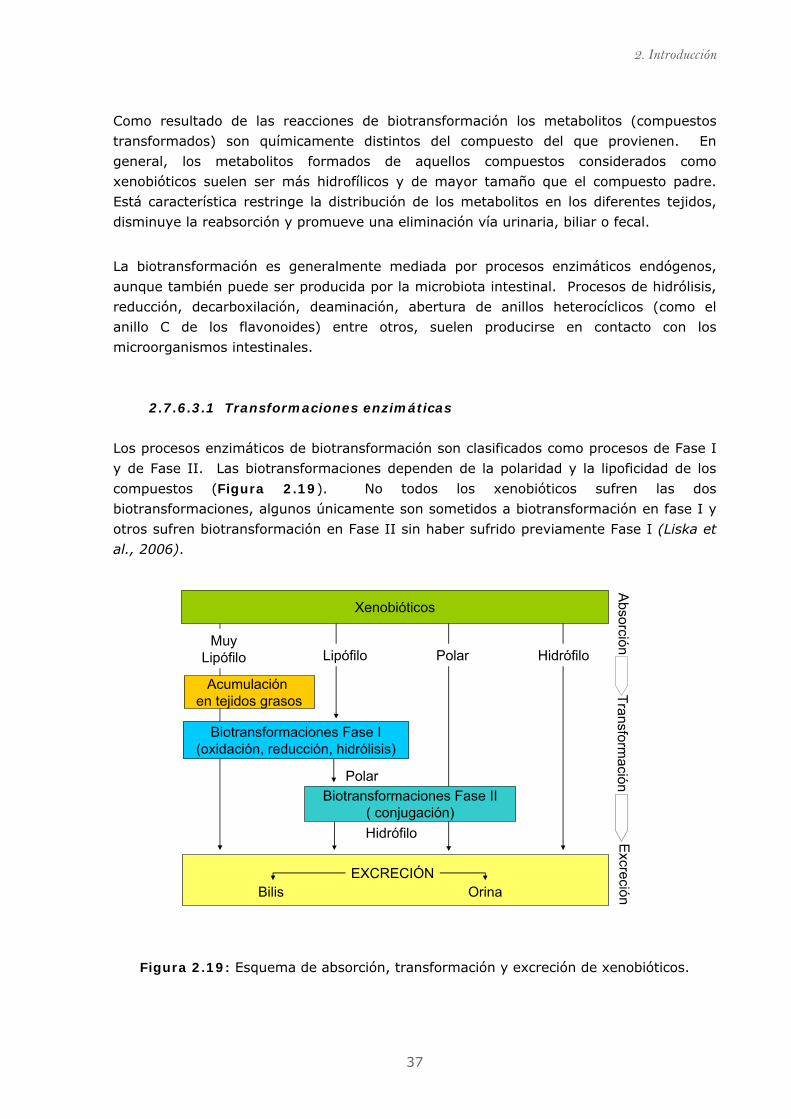
2. Introducción
37
Como resultado de las reacciones de biotransformación los metabolitos (compuestos
transformados) son químicamente distintos del compuesto del que provienen. En
general, los metabolitos formados de aquellos compuestos considerados como
xenobióticos suelen ser más hidrofílicos y de mayor tamaño que el compuesto padre.
Está característica restringe la distribución de los metabolitos en los diferentes tejidos,
disminuye la reabsorción y promueve una eliminación vía urinaria, biliar o fecal.
La biotransformación es generalmente mediada por procesos enzimáticos endógenos,
aunque también puede ser producida por la microbiota intestinal. Procesos de hidrólisis,
reducción, decarboxilación, deaminación, abertura de anillos heterocíclicos (como el
anillo C de los flavonoides) entre otros, suelen producirse en contacto con los
microorganismos intestinales.
2.7.6.3.1 Transformaciones enzimáticas
Los procesos enzimáticos de biotransformación son clasificados como procesos de Fase I
y de Fase II. Las biotransformaciones dependen de la polaridad y la lipoficidad de los
compuestos (Figura 2.19). No todos los xenobióticos sufren las dos
biotransformaciones, algunos únicamente son sometidos a biotransformación en fase I y
otros sufren biotransformación en Fase II sin haber sufrido previamente Fase I (Liska et
al., 2006).
Xenobióticos
Acumulaciónen tejidos grasos
Biotransformaciones Fase I(oxidación, reducción, hidrólisis)
ExcrecciónBilis Orina
EXCRECIÓN
PolarMuy
Lipófilo
Polar
Hidrófilo
Biotransformaciones Fase II( conjugación)
HidrófiloLipófilo
Absorción
Transformación
Excreción
Figura 2.19: Esquema de absorción, transformación y excreción de xenobióticos.
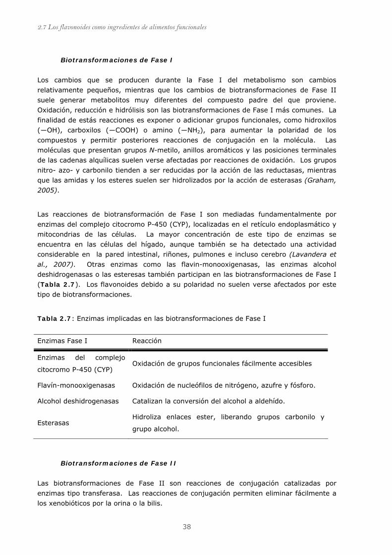
2.7 Los flavonoides como ingredientes de alimentos funcionales
38
Biotransformaciones de Fase I
Los cambios que se producen durante la Fase I del metabolismo son cambios
relativamente pequeños, mientras que los cambios de biotransformaciones de Fase II
suele generar metabolitos muy diferentes del compuesto padre del que proviene.
Oxidación, reducción e hidrólisis son las biotransformaciones de Fase I más comunes. La
finalidad de estás reacciones es exponer o adicionar grupos funcionales, como hidroxilos
(―OH), carboxilos (―COOH) o amino (―NH2), para aumentar la polaridad de los
compuestos y permitir posteriores reacciones de conjugación en la molécula. Las
moléculas que presentan grupos N-metilo, anillos aromáticos y las posiciones terminales
de las cadenas alquílicas suelen verse afectadas por reacciones de oxidación. Los grupos
nitro- azo- y carbonilo tienden a ser reducidas por la acción de las reductasas, mientras
que las amidas y los esteres suelen ser hidrolizados por la acción de esterasas (Graham,
2005).
Las reacciones de biotransformación de Fase I son mediadas fundamentalmente por
enzimas del complejo citocromo P-450 (CYP), localizadas en el retículo endoplasmático y
mitocondrias de las células. La mayor concentración de este tipo de enzimas se
encuentra en las células del hígado, aunque también se ha detectado una actividad
considerable en la pared intestinal, riñones, pulmones e incluso cerebro (Lavandera et
al., 2007). Otras enzimas como las flavin-monooxigenasas, las enzimas alcohol
deshidrogenasas o las esteresas también participan en las biotransformaciones de Fase I
(Tabla 2.7). Los flavonoides debido a su polaridad no suelen verse afectados por este
tipo de biotransformaciones.
Tabla 2.7: Enzimas implicadas en las biotransformaciones de Fase I
Enzimas Fase I Reacción
Enzimas del complejo
citocromo P-450 (CYP) Oxidación de grupos funcionales fácilmente accesibles
Flavín-monooxigenasas Oxidación de nucleófilos de nitrógeno, azufre y fósforo.
Alcohol deshidrogenasas Catalizan la conversión del alcohol a aldehído.
Esterasas Hidroliza enlaces ester, liberando grupos carbonilo y
grupo alcohol.
Biotransformaciones de Fase II
Las biotransformaciones de Fase II son reacciones de conjugación catalizadas por
enzimas tipo transferasa. Las reacciones de conjugación permiten eliminar fácilmente a
los xenobióticos por la orina o la bilis.
2. Introducción
39
La glucuronidación es la reacción de conjugación más frecuente. La enzimas encargadas
de está reacción son las uridina glucuroniltransferasas (UGTs) que utiliza el UDP-
glucuronido como cofactor. En concreto, los flavonoides sufren transformaciones de tipo
O-glucuronido, y son considerados como buenos sustratos para estas enzimas.(Zhang et
al., 2007) Las UGTs están localizadas en el retículo endoplasmático, principalmente, de
las células hepáticas, aunque también están presentes en otros tejidos como, intestino,
bazo, riñón, piel y testículos (Nakamura et al., 2008).
La reacción de sulfatación consiste en la transferencia de un grupo sulfato desde el
cofactor 3´-fosfoadenosil-5´-fosfosulfato (PAPs) a un grupo hidroxilo de los flavonoides.
La reacción es catalizada por sulfotransferasas (STs), enzimas solubles que se
encuentran en el citosol de células que forman parte de hígado, riñón, intestino y
pulmones. Las enzimas pueden conjugar más de un grupo hidroxilo en una misma
molécula, siempre y cuando dispongan de PAPs. El sulfato inorgánico libre necesario
para la síntesis de PAPs puede ser un factor limitante de esta reacción y genera
variaciones inter/intra-individuales (Pai et al., 2001). Cuando un flavonoide se conjuga
con un sulfato se convierte en un compuesto ionizado, soluble en agua que se excreta
por la orina.
La enzima catecol metilo transferasa (COMT) es la encargada de transferir un grupo
metilo a uno de los hidroxilos que forman parte del grupo catecol en presencia de Mn+2
utilizando compuesto donador de grupos metilo la S-adenosil metionina (SAM) (Guldberg
et al., 1975). Las COMTs se encuentran en el citosol de casi todas las células, y al igual
que las otras enzimas implicadas en los procesos de biotransformación de Fase II, el
hígado es el tejido donde se concentra un mayor número (Mannisto et al., 1999).
La glutationización, que consiste en la adición de glutatión (GSH), a través de su grupo
sulfidrilo, y la aminoacidación, que consiste en la formación de una unión peptídica entre
el grupo amino de la glicina, y un carboxilo en el xenobiótico, son otras reacciones de
biotransformación de Fase II. Los flavonoides debido a su estructura no son buenos
sustratos para estas reacciones.
2.7.6.3.2 Transformaciones producidas por la microbiota intestinal.
El intestino de los humanos está colonizado por un elevado número de microorganismos
(en el colón, más de 1012 microorganismos/cm3), que viven en simbiosis con los
organismos a los que habitan. Las bacterias del tracto gastrointestinal contribuyen a
aumentar el rendimiento de la digestión, fermentando proteínas y carbohidratos que no
fueron digeridos en el intestino delgado, y produciendo ácidos grasos de cadena corta
(butirato, acetato, propionato) que son utilizados como fuente de energía para el
organismo. A diferencia de las transformaciones enzimáticas, que producen compuestos
polares de mayor tamaño molecular, las transformaciones de la microbiota intestinal
consisten fundamentalmente en reacciones de reducción, hidrólisis y rotura de anillos
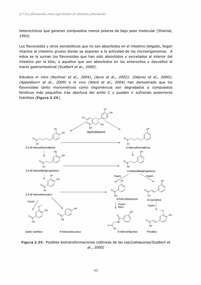
2.7 Los flavonoides como ingredientes de alimentos funcionales
40
heterocíclicos que generan compuestos menos polares de bajo peso molecular (Shamat,
1993).
Los flavonoides y otros xenobióticos que no son absorbidos en el intestino delgado, llegan
intactos al intestino grueso donde se exponen a la actividad de los microorganismos. A
estos se le suman los flavonoides que han sido absorbidos y excretados al interior del
intestino por la bilis; o aquellos que son absorbidos en los enterocitos y devueltos al
tracto gastrointestinal (Scalbert et al., 2000).
Estudios in vitro (Rechner et al., 2004), (Aura et al., 2002); (Déprez et al., 2000);
(Appeldoorn et al., 2009) e in vivo (Ward et al., 2004) han demostrado que los
flavonoides tanto monoméricos como oligoméricos son degradados a compuestos
fenólicos más pequeños tras abertura del anillo C y pueden ir sufriendo posteriores
hidrólisis (Figura 2.20).
Figura 2.20: Posibles biotransformaciones colónicas de las (epi)catequinas(Scalbert et
al., 2000)
2. Introducción
41
2.8 Metodología de identificación y análisis de compuestos
fenólicos. Los extractos vegetales contienen mezclas complejas de compuestos fenólicos que varían
en composición desde ácidos fenólicos simples hasta taninos condensados o hidrolizables.
Para establecer una relación estructura-actividad es necesario definir los compuestos
mayoritarios que componen la mezcla y, así posteriormente, poder explicar su actividad
biológica.
2.8.1 Extracción de los compuestos fenólicos
La extracción de los compuestos fenólicos de una muestra vegetal se realiza mediante el
uso de disolventes de diferente polaridad. La elección de los disolventes está en función
de los compuestos que se quieran separar. Los compuestos menos polares son extraídos
con disolventes orgánicos, mientras que los compuestos más polares (glucósidos) son
extraídos con alcoholes o mezclas de alcohol-agua. El rendimiento de los métodos de
extracción no sólo está en función del disolvente, la temperatura y el tiempo de
extracción son otros de los factores importantes (Spigno et al., 2007). Los taninos tanto
hidrolizables como condensados pueden ser difíciles de extraer de la matriz vegetal que
los envuelve. La cantidad de compuestos fenólicos no extraíbles puede llegar a un 90%
en algunos casos y está en función de la matriz vegetal y del grado de polimerización de
los taninos (Matthews et al., 1997).
2.8.2 Métodos cromatográficos de separación
El extracto crudo obtenido de la fuente vegetal, como se comentó anteriormente, es una
mezcla compleja que puede variar desde fenoles simples hasta compuestos fenólicos de
naturaleza polimérica. Los métodos cromatográficos permiten separar los diferentes
compuestos fenólicos según su tamaño, volatilidad o su grado de hidrofobicidad.
2.8.2.1 Cromatografía de gases. (GC)
Para la separación de mezclas de compuestos polifenólicos, la cromatografía de gases es
generalmente descartable, debido a la baja volatilidad de los compuestos (especialmente
polímeros) y al tedioso proceso de derivatización de los compuestos para aumentar su
volatilidad. Sin embargo, el acoplamiento de detectores de espectrómetros de masas a
la cromatografía de gases ha empujado a algunos investigadores a optimizar y aplicar
métodos de separación y determinación de mezclas de compuestos fenólicos utilizando
estas técnicas. (Deng et al., 2003;Fiamegos et al., 2004;Proestos et al., 2006)

2.8 Metodología de identificación y análisis de compuestos fenólicos
42
2.8.2.2 Cromatografía de exclusión por tamaño (SEC)
Está técnica se usa para separar de manera preparativa los compuestos de un extracto
polifenólico en fracciones según el tamaño molecular, aunque también se ha utilizado
para determinar el peso molecular de proantocianidinas mediante el uso de patrones
(Williams et al., 1983). La separación de las procianidinas según el grado de
polimerización se basa en el tamaño y la polaridad de los oligómeros. A medida que
aumenta el grado de polimerización los polímeros son más polares.
Sephadex LH-20 y Toyopearl HW-40 son las resinas frecuentemente utilizados para
separar proantocianidinas según el grado de polimerización (Sun et al., 1998;Yanagida et
al., 1999) (Jerez et al., 2007;Torres et al., 2002).
2.8.2.3 Cromatografía líquida de alta resolución (HPLC)
La cromatografía líquida de alta resolución (HPLC) es la técnica más utilizada en la
separación y análisis de los compuestos fenólicos.
El uso de columnas de fase normal (NP, normal phase) ha sido preferentemente escogido
para el análisis de proantocianidinas (Lazarus et al., 1999;Rigaud et al., 1993). NP-HPLC
permite separar y cuantificar proantocianidinas con grados de polimerización entre cinco
y doce (Adamson et al., 1999;Sudjaroen et al., 2005). Sin embargo, esta técnica parece
presentar varias limitaciones que complican el uso rutinario tales como: la falta de
resolución, el uso de disolventes clorados para la elución o una incompleta elución de las
proantocianidinas de elevado grado de polimerización (Wolfgang Hümmer, 2008).
Recientemente, Kelm y colaboradores (Kelm et al., 2006) han desarrollado un método,
tanto a escala preparativa como analítica, aparentemente mucho más eficaz utilizando
una fase estacionaria de tipo diol.
La cromatografía líquida de alta resolución en fase reversa (RP-HPLC) es la técnica
generalmente usada para separar compuestos fenólicos tanto a nivel de escala analítica
como preparativa. Existen numerosos artículos de revisión de los diferentes métodos
utilizados para la separación de los compuestos fenólicos ("Bibliography of liquid column
chromatography 1971-1973 and survey of applications," 1976;Daigle et al.,
1983;Robards et al., 1997) (Merken et al., 2000). La separación se basa en el uso de
sistemas de gradiente binarios, que consisten en una solución acuosa con un modificador
de pH y un solvente orgánico soluble en agua como metanol (MeOH) o Acetonitrilo
(ACN). Los modificadores de pH dependen de la técnica de detección que se utilice.
Para UV-DAD el ácido trifluroroacético (TFA) es frecuentemente utilizado, sin embargo,
esté ácido presenta numerosos problemas en la detección por espectrometría de masas
(Shou et al., 2005). El ácido fórmico y el ácido acético son otros modificadores de pH
ampliamente utilizados.
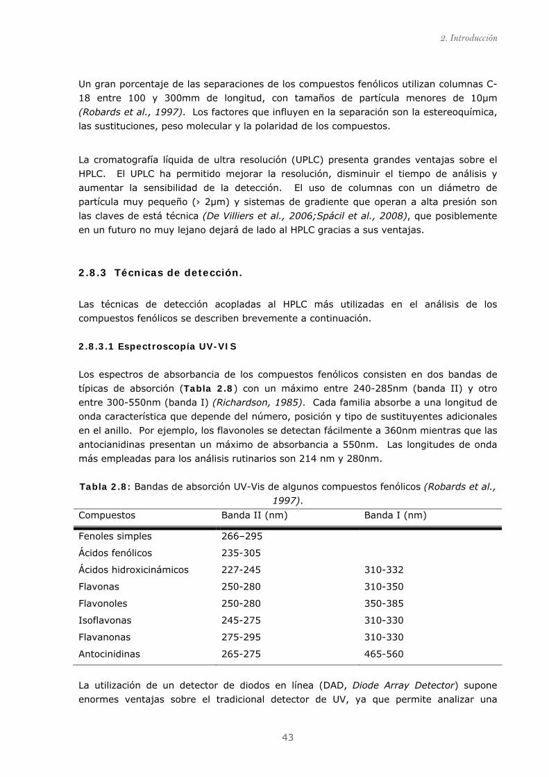
2. Introducción
43
Un gran porcentaje de las separaciones de los compuestos fenólicos utilizan columnas C-
18 entre 100 y 300mm de longitud, con tamaños de partícula menores de 10µm
(Robards et al., 1997). Los factores que influyen en la separación son la estereoquímica,
las sustituciones, peso molecular y la polaridad de los compuestos.
La cromatografía líquida de ultra resolución (UPLC) presenta grandes ventajas sobre el
HPLC. El UPLC ha permitido mejorar la resolución, disminuir el tiempo de análisis y
aumentar la sensibilidad de la detección. El uso de columnas con un diámetro de
partícula muy pequeño (› 2µm) y sistemas de gradiente que operan a alta presión son
las claves de está técnica (De Villiers et al., 2006;Spácil et al., 2008), que posiblemente
en un futuro no muy lejano dejará de lado al HPLC gracias a sus ventajas.
2.8.3 Técnicas de detección.
Las técnicas de detección acopladas al HPLC más utilizadas en el análisis de los
compuestos fenólicos se describen brevemente a continuación.
2.8.3.1 Espectroscopía UV-VIS
Los espectros de absorbancia de los compuestos fenólicos consisten en dos bandas de
típicas de absorción (Tabla 2.8) con un máximo entre 240-285nm (banda II) y otro
entre 300-550nm (banda I) (Richardson, 1985). Cada familia absorbe a una longitud de
onda característica que depende del número, posición y tipo de sustituyentes adicionales
en el anillo. Por ejemplo, los flavonoles se detectan fácilmente a 360nm mientras que las
antocianidinas presentan un máximo de absorbancia a 550nm. Las longitudes de onda
más empleadas para los análisis rutinarios son 214 nm y 280nm.
Tabla 2.8: Bandas de absorción UV-Vis de algunos compuestos fenólicos (Robards et al.,
1997).
Compuestos Banda II (nm) Banda I (nm)
Fenoles simples 266–295
Ácidos fenólicos 235-305
Ácidos hidroxicinámicos 227-245 310-332
Flavonas 250-280 310-350
Flavonoles 250-280 350-385
Isoflavonas 245-275 310-330
Flavanonas 275-295 310-330
Antocinidinas 265-275 465-560
La utilización de un detector de diodos en línea (DAD, Diode Array Detector) supone
enormes ventajas sobre el tradicional detector de UV, ya que permite analizar una
2.8 Metodología de identificación y análisis de compuestos fenólicos
44
muestra a diferentes longitudes de onda, ofreciendo un espectro en tres dimensiones y
mejorando los métodos de cuantificación. La técnica DAD permite en una sola inyección
analizar diferentes familias de compuestos fenólicos (Merken et al., 2000).
2.8.3.2 Espectroscopía de fluorescencia
La técnica de detección por fluorescencia se usa relativamente poco para los compuestos
fenólicos debido a que el número de flavonoides que exhiben de manera natural
fluorescencia es limitado (Bader et al., 2004). La ventaja que ofrece esta técnica es que
los compuestos que pueden ser analizados son fácilmente detectados en matrices o
mezclas complejas (De Rijke et al., 2001). Las isoflavonas (De Rijke et al., 2002) y los
flavonoides con un grupo hidroxilo libre en el C-3, como es el caso de las catequinas
pueden ser detectadas por esta técnica (Sengupta et al., 1979).
2.8.3.3 Espectrometría de masas (MS)
La espectrometría de masas es una técnica de detección basada en la ionización de los
compuestos, la separación y registro de los iones producidos, según su relación
masa/carga (m/z). Esta técnica es idónea para compuestos no volátiles, polares y
termolábiles. La técnica de HPLC-MS proporciona una alta sensibilidad, ofreciendo muy
buenos limites de detección.
El acoplamiento de la técnica de espectrometría de masas a la cromatografía de líquidos
de alta resolución supuso bastantes problemas debido a que la MS opera a presión
atmosférica ionizando los compuestos mientras que HPLC trabaja en fase líquida. Los
caudales relativamente elevados que se manejan en HPLC obligan al empleo de una
interfase cuya misión es compatibilizar la cromatografía líquida con el espectrómetro de
masas. Las principales interfases generalmente usadas para ionizar a presión atmosférica
(API) son la ionización química a presión atmosférica (APCI) y la ionización por
electrospray (ESI) (Vékey, 2001). En la Figura 2.21 se muestra un esquema simple de
cada una de ellas.
ESI ioniza las muestras directamente desde soluciones acuosas, orgánicas o mezclas de
ambos, mediante la formación de un spray de gotas. Estas gotas van reduciendo su
tamaño debido a fenómenos de evaporación del solvente (por efecto del gas y de la
temperatura) y a la desintegración de las mismas gotas en otras de menor tamaño (3-10
nm). Al disminuir las gotas de tamaño, la densidad de carga eléctrica en su superficie
aumenta. La mutua repulsión entre cargas de igual signo en la superficie llega a ser tan
grande que se superan las fuerzas de cohesión superficial y los iones dejan las gotas en
la forma que se conoce como “cono de Taylor”, siendo dirigidas hacia el analizador
mediante la aplicación de vacíos crecientes, que van desde presión atmosférica en la
introducción de la muestra, hasta valores de vacío de 10-9 en la zona del analizador-
detector. La vaporización de estas gotas cargadas da lugar a la producción de iones con
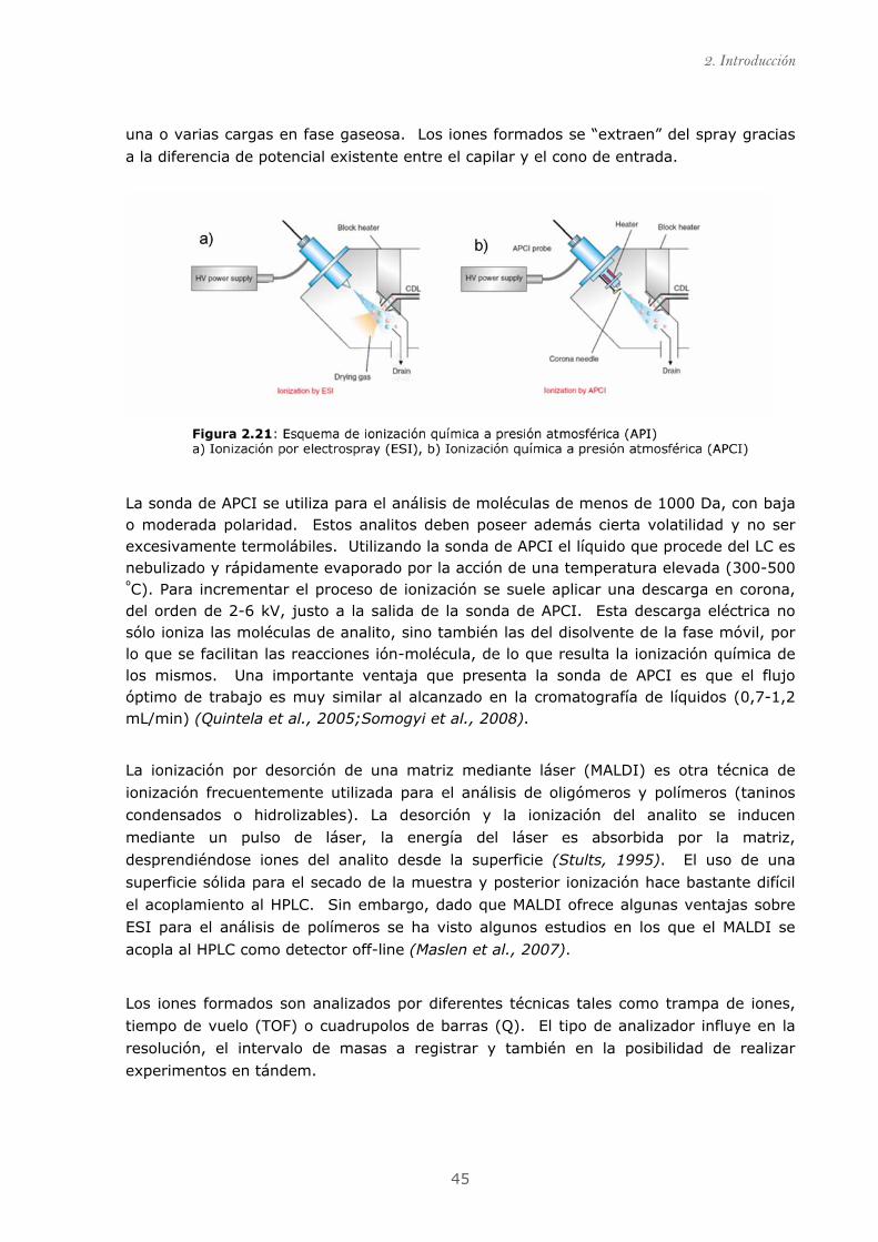
2. Introducción
45
una o varias cargas en fase gaseosa. Los iones formados se “extraen” del spray gracias
a la diferencia de potencial existente entre el capilar y el cono de entrada.
La sonda de APCI se utiliza para el análisis de moléculas de menos de 1000 Da, con baja o moderada polaridad. Estos analitos deben poseer además cierta volatilidad y no ser excesivamente termolábiles. Utilizando la sonda de APCI el líquido que procede del LC es nebulizado y rápidamente evaporado por la acción de una temperatura elevada (300-500 ºC). Para incrementar el proceso de ionización se suele aplicar una descarga en corona, del orden de 2-6 kV, justo a la salida de la sonda de APCI. Esta descarga eléctrica no sólo ioniza las moléculas de analito, sino también las del disolvente de la fase móvil, por lo que se facilitan las reacciones ión-molécula, de lo que resulta la ionización química de los mismos. Una importante ventaja que presenta la sonda de APCI es que el flujo óptimo de trabajo es muy similar al alcanzado en la cromatografía de líquidos (0,7-1,2 mL/min) (Quintela et al., 2005;Somogyi et al., 2008).
La ionización por desorción de una matriz mediante láser (MALDI) es otra técnica de
ionización frecuentemente utilizada para el análisis de oligómeros y polímeros (taninos
condensados o hidrolizables). La desorción y la ionización del analito se inducen
mediante un pulso de láser, la energía del láser es absorbida por la matriz,
desprendiéndose iones del analito desde la superficie (Stults, 1995). El uso de una
superficie sólida para el secado de la muestra y posterior ionización hace bastante difícil
el acoplamiento al HPLC. Sin embargo, dado que MALDI ofrece algunas ventajas sobre
ESI para el análisis de polímeros se ha visto algunos estudios en los que el MALDI se
acopla al HPLC como detector off-line (Maslen et al., 2007).
Los iones formados son analizados por diferentes técnicas tales como trampa de iones,
tiempo de vuelo (TOF) o cuadrupolos de barras (Q). El tipo de analizador influye en la
resolución, el intervalo de masas a registrar y también en la posibilidad de realizar
experimentos en tándem.

2.8 Metodología de identificación y análisis de compuestos fenólicos
46
Espectrometría de masas en tándem (MS/MS ó MSn) es una técnica que se basa en la
detección de los compuestos mediante la fragmentación de los iones. La trampa de iones
(IT), el triple cuadrupolo (QqQ) y el cuadrupolo-TOF (Q-TOF) permiten realizar
experimentos en tándem (Schermann, 2008). Todos estos diseños presentan el mismo
esquema general. Primero, los iones de la muestra son formados en la fuente de
ionización. Uno de los iones es seleccionado para ser analizado por tándem. El ión
seleccionado es excitado y fragmentado con la ayuda de un gas inerte en una celda de
colisión formando iones producto. Este proceso recibe el nombre de disociación inducida
por colisión (CID). Los iones productos son nuevamente analizados de acuerdo con su
relación m/z (Vékey, 2001).
Los cuatro modos principales de operación MS/MS se describen a continuación para el
triple cuadrupolo (QqQ) que se usa frecuentemente debido a su relativa sencillez y
versatilidad (Watson et al., 2003).
HPLCFuenteionización
Q1 Q3CID DetectorHPLC
Fuenteionización
Q1 Q3CID Detector
Figura 2.22: Representación esquemática de un triple cuadrupolo (QqQ).
(i) Barrido de producto de iones (product ion scan), este modo filtra en Q1 (Figura 2.22)
el ión seleccionado y Q3 registra todos los iones productos formados del ión
seleccionado; (ii) Barrido del ión precursor (precursor ion scan), este modo permite
fragmentar el ión seleccionado en Q3 y el barrido en Q1 muestra todos los precursores
que han sido fragmentados y que presentaban el ión precursor;.(iii) Barrido de perdidas
neutras (neutral loss scan), este modo permite escanear en Q1 y Q3 de manera
sincronizada, registrando los iones que presentan perdidas de una masa neutra
especifica; (iv) Monitorización de reacción múltiple (multiple reacction monitoring),
permite seleccionar un ión en Q1 y un ión producto en Q3.
En general, ESI y APCI han sido ampliamente utilizadas para la ionización de los
compuestos fenólicos tanto en modo positivo como en modo negativo (Cuyckens et al.,
2004). El modo positivo es más adecuado para la detección de antocinidinas, que se
encuentran en forma catiónica en medio ácido, sin embargo, cuando están derivadas con
ácidos carboxílicos pueden ser analizadas en modo negativo (Fulcrand et al., 1998).
Entre las fuentes API más utilizadas, ESI parece presentar la mayor eficiencia de
ionización sobre los compuestos fenólicos (Jussi-Pekka Rauha, 2001). MALDI es
fundamentalmente usada para el análisis de oligómeros y polímeros de compuestos
fenólicos. Taninos condensados, polímeros de antocianinas y taninos hidrolizables han
2. Introducción
47
sido analizados utilizando MALDI-TOF (Babior, 2000) (Vivas et al., 2004), (Ishida et al.,
2005), (Monagas et al.).
Las técnicas de espectrometría en tándem, gracias a su alta sensibilidad y robustez
permiten el análisis de compuestos fenólicos de matrices complejas (Liu et al., 2005a),
(Sánchez-Rabaneda et al., 2004), (Määttä-Riihinen et al., 2004), el análisis de
metabolitos generados tras la ingesta de compuestos fenólicos (Dvorakova et al.,
2008;Felgines et al., 2003;Meng et al., 2001;Sang et al., 2008;Xu et al., 2007) y
también, el análisis de biomarcadores de estrés oxidativo (Watson et al., 2003).
2.8.4 Evaluación de la actividad antioxidante
La evaluación de la actividad antioxidante de un compuesto o en particular, la actividad
antioxidante de un alimento funcional puede realizarse respondiendo a las siguientes
pautas.
I. Evaluación de la actividad antioxidante in vitro mediante pruebas químicas.
II. Evaluación de la actividad antioxidante y de la actividad frente al daño
oxidativo en cultivos celulares.
III. Evaluación de la actividad antioxidante in vivo tras la ingesta en animales y/o
humanos.
Los métodos utilizados en cada uno de estos niveles son numerosos y la gran mayoría
han sido adaptados según los requerimientos, por ejemplo, métodos químicos en
solución son utilizados, tras algunas modificaciones, en cultivos celulares. Por ello es
difícil etiquetar los métodos aplicados a los diferentes niveles. A continuación se
describen de manera general los métodos más utilizados para evaluar la actividad
antioxidante in vitro e in vivo.
2.8.4.1 Métodos químicos en solución
Evaluar la capacidad antioxidante in vitro, de compuestos puros, extractos o alimentos,
aporta de manera, simple, rápida y no invasiva, información relevante sobre las posibles
sinergias u otros procesos que puedan generarse como resultado de la interacción de los
compuestos con el medio o la matriz de la muestra analizada. Generalmente los
resultados difieren de los obtenidos in vivo, pero los resultados in vitro pueden
ayudarnos a resolver problemas iniciales. Así, la capacidad antioxidante puede variar
según el pH, la solubilidad, la temperatura del tratamiento al que pueda someterse la
muestra o el almacenamiento de los compuestos escogidos como antioxidantes. Por lo
tanto, el estudio y conocimiento de la relación estructura-actividad antioxidante de los
2.8 Metodología de identificación y análisis de compuestos fenólicos
48
compuestos seleccionados nos dará una aproximación a las propiedades que pueda
ejercer in vivo.
La transferencia de átomos de hidrógeno o/y la transferencia electrónica son los dos
mecanismos fundamentales de actuación de un antioxidante. Estas reacciones difieren
en la cinética y el potencial de ionización pero el resultado final de ambas es el mismo
(Prior et al., 2005). Ambos procesos pueden ocurrir simultáneamente y dominará uno u
otro de los mecanismos en función de la estructura del antioxidante y/o del radical, de la
solubilidad, del coeficiente de partición y del medio donde tenga lugar la reacción.
La necesidad de estandarizar los métodos antioxidantes usados en alimentos, extractos
vegetales, nutracéuticos u otros suplementos dietéticos ha derivado en una clasificación
de los métodos antioxidantes en función del mecanismo de actuación.
Los métodos de transferencia de átomos de hidrógeno (HAT) son los más comunes y
miden la habilidad de un antioxidante para neutralizar un radical por donación de un
átomo de hidrógeno. Las reacciones HAT son independientes del pH y del disolvente.
Generalmente, los métodos que miden la transferencia de átomos de hidrógeno son
rápidos debido a que las reacciones se completan en cuestión de minutos.
Por el contrario, los métodos basados en la transferencia electrónica (SET) miden la
eficacia de un antioxidante para transferir un electrón y reducir un compuesto (Wright et
al., 2001). Las reacciones SET dependen del pH y son lentas ,por ello, los resultados de
estos métodos se basan en el porcentaje de disminución del radical (Prior et al., 2005).
Por lo general, la actividad antioxidante ocurre simultáneamente por ambos mecanismos,
y tal como hemos comentado anteriormente, la manifestación del mecanismo dominante
estará en función de la estructura del antioxidante y del pH. En ocasiones, diferenciar
entre uno u otro mecanismo es bastante complicado por lo que se ha creado un grupo de
clasificación de métodos antioxidantes basado en ambos mecanismos de actuación (Prior
et al., 2005).
2.8.4.1.1 Métodos de evaluación de la actividad antioxidante basados en
ambos mecanismos de actuación: HAT y SET
Ensayo del DPPH (2,2-Difenil-1-picrilhidracilo)
Este ensayo se basa en la reducción del radical DPPH por los antioxidantes de la
muestra. La acción de los antioxidantes puede ser medida por disminución de la señal
del radical por EPR o por lo disminución de absorbancia que se observa a medida que el
radical es reducido por el antioxidante (Prior et al., 2005). Se considera un método
sencillo y fácil de aplicar, los resultados que se obtienen son altamente reproducibles y

2. Introducción
49
comparables con otros métodos que miden de la misma forma la capacidad
secuestradora de radicales libres como es el caso del ABTS (Gil et al., 2000). El DPPH
es un buen método para medir la capacidad antioxidante en fracciones o alimentos. No
obstante algunos antioxidantes que presentan un espectro de absorción similar al del
radical, como es el caso de los carotenoides, pueden causar interferencias si son
cuantificados por UV-VIS (Nomura et al., 1997).
Ensayo TEAC (Trolox Equivalent Antioxidant Capacity)
El ensayo TEAC o ensayo del radical ABTS (2,2-azinobis–(3-etilbenzotiazolin-6-
sulfonato)) se basa en medir la capacidad de captación del radical cation ABTS•+. En
este ensayo, el ABTS es oxidado in situ, generalmente por peróxido de hidrógeno,
hasta su forma de radical catión. El ABTS•+ presenta una coloración verde-azulada
muy intensa a una longitud de onda de 734nm que disminuye a medida que los
antioxidantes reducen el radical (Sánchez-Moreno, 2002). El ensayo TEAC presenta
otras variaciones de protocolo que consisten en hacer reaccionar el ABTS con dióxido
de manganeso, persulfato potásico o ABAP (2,2’-azo-bis-(2-amidinopropano), pero
estas modificaciones presentan el inconveniente de que requieren largos tiempos de
incubación o altas temperaturas (Prior et al., 2005).
A diferencia del DPPH, el radical catión ABTS presenta la ventaja de que es soluble
tanto en medios acuosos como orgánicos, lo cual convierte al ensayo TEAC en un
método apto para determinar la capacidad antioxidante en soluciones químicas y
también en fluidos biológicos (Awika et al., 2003).
2.8.4.1.2 Métodos de evaluación de actividad antioxidante basados en
mecanismos HAT
Ensayo ORAC (oxygen radical absorbance capacity)
Este método permite medir la inhibición del radicales peróxido (ROO•) generados
mediante la descomposición térmica de un compuesto azo, generalmente AAPH (2,2’-
azo-bis (2-amidinopropano) dihidrocloruro). En las primeras versiones de este método
se hacia reaccionar al radical peroxilo con un producto fluorescente (B-ficoeritrina, B-
PE) para formar uno no fluorescente. La disminución de fluorescencia indicaba el daño
producido por la reacción del radical (Cao et al., 1994). Debido a la inespecificidad del
B-PE, el método fue posteriormente mejorado. La utilización de FL (3,6-
Dihidroxispiro[isobenzofuran-1(3H),9-[9H] xanten]-3-ona) un producto fluorescente
mucho más estable y más selectivo, ha permitido medir con exactitud la capacidad
antioxidante frente a radicales peróxido (Huang et al., 2002).
2.8 Metodología de identificación y análisis de compuestos fenólicos
50
Ensayo TRAP (total peroxyl radical-trapping antioxidant parameter)
Es un ensayo muy similar al anteriormente explicado, el ORAC. Este ensayo permite
medir la eficacia con la que los compuestos antioxidantes actúan frente a los radicales
peroxilo generados por compuestos azo tales como AAPH o ABAP ([2-2’-azobis(2-
amidinopropano)dihidrocloridro]). Dicha habilidad se puede medir por fluorescencia
mediante el uso de R-ficoeritrina (DeLange et al., 1989) o por absorbancia mediante el
uso de ABTS (2,2-azino-bis (3-etilbezotiazolin-6-sulfonato)) (Bartosz et al., 1998).
El ensayo del TRAP es generalmente escogido para medir la capacidad antioxidante de
compuestos como glutatión, vitamina C y α-tocoferol en muestras de suero o plasma
(Wayner et al., 1987).
Ensayo TOSC ( total oxidant scavenging capacity)
Este método permite medir la capacidad de transferir un átomo de hidrógeno a
radicales hidroxilo, peroxilo y peroxinitrito. El método utiliza el área bajo la curva para
determinar la respuesta de los antioxidantes frente a los radicales (Regoli et al., 1999).
2.8.4.1.3 Evaluación de actividad antioxidante basados en mecanismo SET
Ensayo FRAP (Ferric reducing antioxidant power)
Este ensayo estima la actividad antioxidante como una medida de la capacidad de los
antioxidantes para reducir el complejo 2,4,6-tripiridil-s-triazina (TPTZ) a su forma
ferrosa. La forma ferrosa es un producto coloreado que permite ser cuantificado por
espectrofotometría a 595nm.
Este ensayo ha sido ampliamente utilizado para medir la capacidad antioxidante en
muestras de suero o plasma, sin embargo no es recomendable debido a que no detecta
la capacidad de los antioxidantes que actúan mediante transferencia de hidrógenos, por
lo que genera resultados erróneos (Ou et al., 2002).
Ensayo del radical HNTTM (tris (2,4,6-tricloro-3,5-dinitrofenil)-metilo)
El radical HNTTM, (tris (2,4,6-tricloro-3,5-dinitrofenil)-metilo), (Figura 2.23) fue
sintetizado en nuestro laboratorio del actual Instituto de Química Avanzada de
Catalunya (IQAC). La estructura química del radical estable HNTTM, no permite la
transferencia de átomos de hidrógeno de los antioxidantes debido al impedimento
estérico de los seis átomos de cloro en posición -orto. El elevado potencial de
reducción (0.58V) convierte al radical HNTTM en un buen captador de electrones.
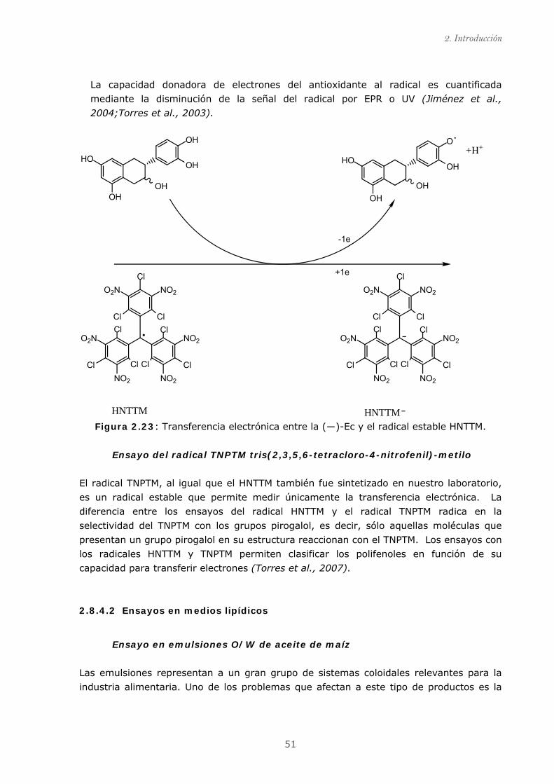
2. Introducción
51
La capacidad donadora de electrones del antioxidante al radical es cuantificada
mediante la disminución de la señal del radical por EPR o UV (Jiménez et al.,
2004;Torres et al., 2003).
OHOH
HO
OH
OH
OHOH
HO
O
OH
ClNO2O2N
NO2
ClNO2NO2
Cl
O2N
Cl Cl
Cl ClCl Cl
ClNO2O2N
NO2
ClNO2NO2
Cl
O2N
Cl Cl
Cl ClCl Cl
HNTTM HNTTM
-1e
+1e
+H+
Figura 2.23: Transferencia electrónica entre la (―)-Ec y el radical estable HNTTM.
Ensayo del radical TNPTM tris(2,3,5,6-tetracloro-4-nitrofenil)-metilo
El radical TNPTM, al igual que el HNTTM también fue sintetizado en nuestro laboratorio,
es un radical estable que permite medir únicamente la transferencia electrónica. La
diferencia entre los ensayos del radical HNTTM y el radical TNPTM radica en la
selectividad del TNPTM con los grupos pirogalol, es decir, sólo aquellas moléculas que
presentan un grupo pirogalol en su estructura reaccionan con el TNPTM. Los ensayos con
los radicales HNTTM y TNPTM permiten clasificar los polifenoles en función de su
capacidad para transferir electrones (Torres et al., 2007).
2.8.4.2 Ensayos en medios lipídicos
Ensayo en emulsiones O/W de aceite de maíz
Las emulsiones representan a un gran grupo de sistemas coloidales relevantes para la
industria alimentaria. Uno de los problemas que afectan a este tipo de productos es la
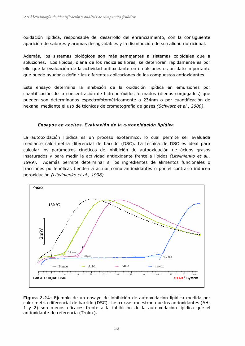
2.8 Metodología de identificación y análisis de compuestos fenólicos
52
oxidación lipídica, responsable del desarrollo del enranciamiento, con la consiguiente
aparición de sabores y aromas desagradables y la disminución de su calidad nutricional.
Además, los sistemas biológicos son más semejantes a sistemas coloidales que a
soluciones. Los lípidos, diana de los radicales libres, se deterioran rápidamente es por
ello que la evaluación de la actividad antioxidante en emulsiones es un dato importante
que puede ayudar a definir las diferentes aplicaciones de los compuestos antioxidantes.
Este ensayo determina la inhibición de la oxidación lipídica en emulsiones por
cuantificación de la concentración de hidroperóxidos formados (dienos conjugados) que
pueden son determinados espectrofotométricamente a 234nm o por cuantificación de
hexanal mediante el uso de técnicas de cromatografía de gases (Schwarz et al., 2000).
Ensayos en aceites. Evaluación de la autooxidación lipídica
La autooxidación lipídica es un proceso exotérmico, lo cual permite ser evaluada
mediante calorimetría diferencial de barrido (DSC). La técnica de DSC es ideal para
calcular los parámetros cinéticos de inhibición de autooxidación de ácidos grasos
insaturados y para medir la actividad antioxidante frente a lípidos (Litwinienko et al.,
1999). Además permite determinar si los ingredientes de alimentos funcionales o
fracciones polifenólicas tienden a actuar como antioxidantes o por el contrario inducen
peroxidación (Litwinienko et al., 1998)
Figura 2.24: Ejemplo de un ensayo de inhibición de autooxidación lipídica medida por calorimetría diferencial de barrido (DSC). Las curvas muestran que los antioxidantes (AH-1 y 2) son menos eficaces frente a la inhibición de la autooxidación lipídica que el antioxidante de referencia (Trolox).
150 ºC
9,7 min15,0 min 45,2 min
min 0 5 10 15 20 25 30 35 40 45 50 55
^exo
Lab A.T.: IIQAB.CSIC System e STAR
AH-1 AH-2 Trolox Blanco
2mW
2. Introducción
53
2.8.4.3 Ensayos con modelos celulares
Los modelos celulares son considerados una buena herramienta para evaluar la actividad
antioxidante y otras actividades biológicas relacionadas en la prevención de
enfermedades como el cáncer (Liu et al., 2005b). Las pautas ha seguir en los ensayos
celulares presentan los siguientes pasos: (i) caracterización de las dosis citotóxicas de la
muestra; (ii) ensayos en cultivos celulares con dosis no citotóxicas; (iii) inducción al
estrés oxidativo o ensayos en líneas celulares especificas (Caco-2, carcinoma de colon;
HepG2 carcinoma hepático; U937, leucemia mieloide; Jurkat-T, leucemia) y (iv)
valoración del efecto protector proporcionado (García, 2005).
En la Tabla 2.9 se incluyen algunos métodos que pueden ayudarnos entender los
mecanismos de acción de los compuestos fenólicos frente al cáncer u otras
enfermedades.
Tabla 2.9: modelos celulares para evaluar los efectos protectores de un compuesto
antioxidante en el desarrollo de cáncer (Liu et al., 2005b)
Modelos celulares Valoración del efecto protector
Evaluación de proliferación celular Inhibición de la proliferación
Estudio del ciclo celular Control de la etapa G1, evaluación del cociente G1/S
Estudio de apoptosis Inhibición /Inducción de apoptosis
Angiogénesis Inhibición de la angiogénesis
Estudio de inhibición de enzimas Inhibición de COX-2, formación prostaglandinas
Daño oxidativo al ADN 8-OH-dG, ensayo cometa
2.8.4.4 Marcadores de estrés oxidativo.
El gran numero de fuentes por las que se pueden generar especies reactivas en un
individuo y la variedad de biomoléculas con las que puede interaccionar, dificulta en gran
medida realizar una predicción del daño oxidativo o de la acción de los antioxidantes
introducidos por la dieta. En ciertos casos, un incremento de especies reactivas puede
ocurrir sin generarse una disminución de los antioxidantes; o una oxidación lipídica
puede tener lugar sin que se produzca daño oxidativo al ADN (Han et al., 2000). Una
alternativa a los problemas analíticos generados a la hora de medir el estrés oxidativo es

2.8 Metodología de identificación y análisis de compuestos fenólicos
54
la utilización de biomarcadores. Los marcadores de estrés oxidativo reflejan cambios en
los sistemas biológicos relacionados con el daño de los radicales sobre las biomoléculas.
Las productos estables originados como consecuencia del daño oxidativo a las
biomoléculas (ver apartado 2.4.) son utilizados como marcadores de estrés oxidativo en
individuos (Figura 2.25). El objetivo de usar marcadores de estrés oxidativo es la
detección de un cambio fisiológico en el organismo, el cual esta íntimamente relacionado
con un efecto adverso de salud.
Figura 2.25 : Algunos de los productos generados tras la oxidación de biomoléculas(De
Zwart et al., 1999)
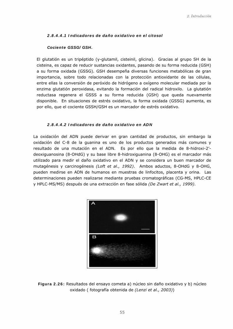
2. Introducción
55
2.8.4.4.1 Indicadores de daño oxidativo en el citosol
Cociente GSSG/GSH.
El glutatión es un tripéptido (γ-glutamil, cisteinil, glicina). Gracias al grupo SH de la
cisteina, es capaz de reducir sustancias oxidantes, pasando de su forma reducida (GSH)
a su forma oxidada (GSSG). GSH desempeña diversas funciones metabólicas de gran
importancia, sobre todo relacionadas con la protección antioxidante de las células,
entre ellas la conversión de peróxido de hidrógeno a oxígeno molecular mediada por la
enzima glutatión peroxidasa, evitando la formación del radical hidroxilo. La glutatión
reductasa regenera el GSSS a su forma reducida (GSH) que queda nuevamente
disponible. En situaciones de estrés oxidativo, la forma oxidada (GSSG) aumenta, es
por ello, que el cociente GSSH/GSH es un marcador de estrés oxidativo.
2.8.4.4.2 Indicadores de daño oxidativo en ADN
La oxidación del ADN puede derivar en gran cantidad de productos, sin embargo la
oxidación del C-8 de la guanina es uno de los productos generados más comunes y
resultado de una mutación en el ADN. Es por ello que la medida de 8-hidroxi-2’-
deoxiguanosina (8-OHdG) y su base libre 8-hidroxiguanina (8-OHG) es el marcador más
utilizado para medir el daño oxidativo en el ADN y se considera un buen marcador de
mutagénesis y carcinogénesis (Loft et al., 1992). Ambos aductos, 8-OHdG y 8-OHG,
pueden medirse en ADN de humanos en muestras de linfocitos, placenta y orina. Las
determinaciones pueden realizarse mediante pruebas cromatográficas (CG-MS, HPLC-CE
y HPLC-MS/MS) después de una extracción en fase sólida (De Zwart et al., 1999).
Figura 2.26: Resultados del ensayo cometa a) núcleo sin daño oxidativo y b) núcleo
oxidado ( fotografía obtenida de (Lenzi et al., 2003))
2.8 Metodología de identificación y análisis de compuestos fenólicos
56
La detección por electroforesis en gel de la rotura de hebras que componen la doble
hélice (ADN) es otro de los ensayos utilizado para medir el daño producido por las
especies radicales. Es conocido como ensayo cometa (comet), debido a que la rotura de
las hebras permite que los bucles de ADN superenrrollado se desplacen en el gel durante
la electroforesis dando lugar a una serie de colores que vistos al microscopio parecen un
cometa. Para este ensayo se utilizan cultivos celulares obtenidos de frotis bucales, de
útero o fecales (Ferguson et al., 2006). El ensayo cometa permite evaluar el daño del
ADN de forma relativamente simple, barata, y rápida y por lo tanto, es considerado un
buen ensayo para la detección de estrés oxidativo y para determinar el efecto de los
compuestos fenólicos con respecto al daño oxidativo (Wasson et al., 2008).
La quercetina ha sido ampliamente evaluada con el ensayo cometa y parece proteger del
daño oxidativo en distintos tipos de células, linfocitos (Duthie et al., 1997a;Johnson et
al., 2000) colon, hepatocitos y células epiteliales (Duthie et al., 1997b), melanoma
(Horváthová et al., 2005) y macrófagos. La luteolina parece proteger frente al daño del
hidrógeno de peróxido en melanoma (Horváthová et al., 2005) y las catequinas reducen
el daño del ter-butilhidroperóxido en macrófagos (Kanupriya et al., 2006).
2.8.4.4.3 Indicadores de daño oxidativo en lípidos
Medir la oxidación lipídica es realmente difícil debido a la variedad de productos que se
generan. Éstos dependen de la composición lipídica de la cual son generada y de la fase
en la que se encuentre el proceso oxidación. Es por este motivo, que existe un gran
rango de biomarcadores para medir la oxidación lipídica. El método más rápido y sencillo
es la determinación colorimétrica (532nm) de las sustancias reactivas con el ácido
tiobarbitúrico (TBARS). Mediante está técnica se mide un cromógeno formado por el
ácido tiobarbitúrico y malondialdehído (MDA) (Han et al., 2000). Sin embargo, este
método ha sido ampliamente criticado debido al gran número de artefactos existentes en
muestras biológicas que absorben a la misma longitud de onda.
Una alternativa ha sido el ensayo de determinación de dienos conjugados, que son
productos primarios de oxidación lipídica. Los dienos conjugados son detectados y
cuantificados espectrofotométricamente a longitudes de onda de 230-235nm. Pero al
igual que sucede en el método TBARS, gran cantidad de sustancias biológicas absorben
en este rango de UV. Con el fin de mejorar el método, se cambiaron las técnicas de
detección de las mediciones, de HPLC-UV a CG, aunque sin buenos resultados. Es por
ello que el mejor indicador de peroxidación lipídica parece ser la determinación de
isoprostanos (Kadiiska et al., 2005).
Los isoprostanos son un grupo de compuestos formados vía no enzimática debido a la
ataque de los radicales libres sobre el ácido grasos poliinsaturados. Los isoprostanos son
generados in situ en los fosfolípidos de los cuales forman parte y posteriormente,
liberados al medio por la acción de las enzimas fosfolipasa tipo A. Dependiendo del ácido
2. Introducción
57
graso a partir del cual es generado se conocen los F2-isoprostanos, originados del ácido
araquidónico (Morrow et al., 1992); F4-neuroprostanos, originados del ácido
docohexanoico (DHA) (Roberts Ii et al., 1998) y los dihomo-isoprostanos (DH-IsoP),
generados tras la oxidación del ácido adrénico (abundante en membranas de neuronas)
(VanRollins et al., 2008). La determinación de isoprostanos se realiza generalmente
mediante CG-MS o HPLC-MS. Aunque, se han desarrollado métodos inmunológicos para
realizar esta determinación, se ha comprobado que no son demasiado sensibles ni
precisos (Kadiiska et al., 2005)
2.8.4.4.4 Indicadores de daño oxidativo en proteínas
El daño oxidativo derivado de la acción de los radicales libres y/o del ataque de los
productos secundarios formados tras la peroxidación lipídica, genera grupos carbonilo y
otros grupos funcionales como hidroxilos que son resultado de oxidaciones producidas en
los enlaces entre aminoácidos. La carbonilación de proteínas es una oxidación más
severa que la generación de metionín sulfóxidos y derivados de cisteína por lo que
indican un estrés oxidativo más severo y representan un marcador de oxidación proteica
estable.
La cuantificación de los grupos carbonilos es el método mas usado aunque es poco
selectivo, éste se realiza mediante un ensayo espectrofotométrico (generalmente a
370nm) tras el marcaje con dinitrofenilhidrazina. Otra alternativa es la inmunodetección,
que hace uso de anticuerpos contra el grupo dinitrofenilo y puede llevarse a cabo tanto
por la técnica de Western Blot, separando las proteínas en una o dos dimensiones como
por Enzyme Linked Immunosorbent Assay (ELISA). Aunque estos métodos proporcionan
valores absolutos diferentes, parecen presentar una buena correlación cuando se
emplean en determinaciones plasmáticas.
Recientemente se han desarrollado métodos que permiten determinar los grupos
carbonilo más abundantes tras la oxidación de proteínas, el (R-aminoadípico semialdeído
(AAS)) y el δ-glutámico semialdeído (GGS), permiten conocer el mecanismo de
oxidación y el grado de distribución in vivo después de una reacción de reducción con
borohidruro de sodio (NaBH4), que es medida por fluorescencia. (Akagawa et al., 2006)
Medir los productos de oxidación de los aminoácidos parece dar información más
detallada sobre el daño de las especies radicales sobre las proteínas. Algunos ejemplos,
son la medida de orto-tirosina (o-Tyr) que se genera tras la oxidación de la fenilalanina,
y la formación di-tirosina (DT) , que se forma tras la oxidación de L-tirosina (De Zwart et
al., 1999). La formación de los productos de oxidación es generalmente medida por
HPLC o CG en muestras de sangre y orina (De Zwart et al., 1999).


3. OBJETIVOS


3. Objetivos
61
El objetivo general de la presente tesis es contribuir al conocimiento de la relación
estructura/efecto(s) de los compuestos fenólicos naturales, en especial de las
proantocianidinas, con relación a las diferentes aplicaciones a las que pueden ser
destinados, por ejemplo como antioxidantes alimentarios, suplementos nutricionales o
ingredientes funcionales. Para ello, y teniendo en cuenta que la presente tesis está
enmarcada en el desarrollo de dos proyectos nacionales diferentes: PPQ2003-06602-
C04-01 (Obtención de fracciones polifenólicas a partir de subproductos agroalimentarios
y forestales) y AGL2004-07579-C04-02/ALI (Establecimiento de las bases científicas para
el uso de la fibra dietética antioxidante de uva), los objetivos concretos planteados
fueron los siguientes:
1. Relacionar el grado de polimerización y porcentaje de galoización de
proantocianidinas procedentes de fracciones fenólicas con sus efectos químicos y
biológicos.
2. Evaluar la biodisponibilidad (absorción/metabolización) de compuestos fenólicos,
en particular proantocianidinas, procedentes de una matriz compleja parecida a
un alimento (fibra antioxidante de uva).
3. Proponer explicaciones a la actividad antioxidante de polifenoles, en particular
proantocianidinas, en modelos de oxidación y en el organismo vivo.


4. RESULTADOS


4. Resultados
65
Los resultados de esta tesis se concretan en tres artículos publicados en revistas del
Science Citation Index y un cuarto artículo en proceso de revisión.
Previo a cada una de las publicaciones, se acompaña un breve resumen y el material
suplementario es incluido en los Anexos (apartado 8.2, ver pág. 253).
Los resultados se presentan en dos grandes bloques atendiendo a los objetivos marcados
al inicio de la presente tesis:
4.1. Estudio de la relación entre el grado de polimerización y porcentaje
de galoización de fracciones fenólicas con sus efectos químicos y
biológicos.
• Publicación 1: Evaluación de la actividad antioxidante y biológica de
fracciones fenólicas exentas de galoización ( ver pág. 73)
• Publicación 2: Evaluación de la actividad antioxidante y biológica de
fracciones fenólicas de elevada galoización ( ver pág. 83)
4.2 Estudio de la absorción/metabolización de proantocianidinas
• Publicación 3: Identificación de los compuestos fenólicos de la fracción
extraíble de fibra antioxidante de uva ( ver pág. 97)
• Publicación 4: Identificación de metabolitos de fibra antioxidante de uva en
orina de ratas ( ver pág. 111)


4.1. Estudio de la relación entre el grado de
polimerización y porcentaje de galoización de
fracciones fenólicas con sus efectos químicos
y biológicos.


4. Resultados
69
4.1.1. PUBLICACIÓN 1: Evaluación de la actividad antioxidante y
biológica de fracciones fenólicas exentas de galoización.
Título original: Procyanidin fractions from pine (Pinus pinaster) bark: Radical
scavenging power in solution, antioxidant activity in emulsion, and
antiproliferative effect in melanoma cells.
Autores: Sonia Touriño, Ariadna Selga, Aurora Jiménez, Lluís Juliá, Carles
Lozano, Daneida Lizárraga, Marta Cascante and Josep Lluís Torres. Journal
of Agricultural and Food Chemistry, 2005, 53, 4728-4735
Índice de impacto: 2,507
Clasificación y categoría: 7/ 103 (Ciencia y Tecnología de los alimentos); 8/62
(Química aplicada); 1/35 (Agricultura, Multidisciplinar)
RESUMEN:
La capacidad antioxidante de los compuestos fenólicos es responsable de que extractos
de naturaleza fenólica se utilicen en diversas aplicaciones. Los subproductos de las
industrias agroforestales son una importante fuente de polifenoles, es por ello que en los
últimos años el número de extractos comerciales obtenidos a partir de subproductos de
la industria agroforestal ha experimentado un considerable aumento. Por ejemplo, la
corteza de pino es uno de los subproductos más utilizados desde que Masquelier en 1987
puso de manifiesto la elevada capacidad antioxidante de las procianidinas que forman
parte de su composición (Masquelier, 1987).
Un estudio previo realizado en nuestro laboratorio (Torres et al., 2002), consistió en
elaborar un abanico de fracciones, con diferente grado de polimerización y galoización, a
partir de un extracto obtenido de bagazo de uva soluble tanto en agua como acetato de
etilo. Los extractos de bagazo de uva se componen fundamentalmente de monómeros
de flavan-3-ol (catequina, epicatequina, epigalocatequíngalato), proantocianidinas y
diferentes clases de flavonoides glicosilados.
El porcentaje de gailozación de estos extractos es la proporción de ésteres de galato en
posición 3 de la estructura de los flavanoles. El tamaño y composición medios de las
diferentes fracciones fue estimado mediante una despolimerización ácida en presencia de
cisteamina (Torres et al., 2001). Se evaluó la capacidad antioxidante de las fracciones
utilizando el ensayo del DPPH, ensayos de inhibición de la oxidación lipídica en
emulsiones aceite/agua y se realizaron ensayos de antiproliferación de cada una de estas
fracciones.

4. Resultados
70
Los resultados de este estudio (Torres et al., 2002) mostraron una relación entre el
grado de polimerización y un aumento de la capacidad antioxidante. Además, las
fracciones más galoizadas presentaron un aumento de actividad antioxidante
comparadas con las fracciones menos galoizadas pero de igual grado de polimerización.
El aumento en el número de grupos hidroxilo es la explicación a esta observación.
Por otro lado, éste y otros estudios demostraron que la presencia de compuestos
galoizados podía presentar efecto pro-oxidante (Kondo et al., 1999;Long et al., 2000).
A partir de este trabajo y de otros, realizados por diferentes autores, se partió de la
hipótesis de que el grado de galoización y polimerización son un punto clave para definir
el tipo de posibles aplicaciones de los extractos.
Con el fin de evaluar la influencia de la galoización en la actividad de los extractos
fenólicos, el primer trabajo de la presente tesis fue obtener un abanico de fracciones de
grado de polimerización análogo al obtenido a partir del bagazo de uva y con grado de
galoización cero.
Las procianidinas de la corteza de pino (Pinus pinaster) fueron escogidas para realizar
este trabajo, ya que carecen de esteres de galato en su composición (Packer et al.,
1999). Una vez obtenido el extracto crudo, se realizó el mismo esquema de
fraccionamiento que el utilizado en el estudio de la actividad biológica de fracciones de
bagazo de uva, con el fin de obtener fracciones comparables en grado de polimerización
y diferentes en cuanto al porcentaje de galoización.
El tamaño y composición de las diferentes fracciones fue estimado, al igual que en las
fracciones obtenidas a partir de bagazo de uva, mediante una despolimerización ácida en
presencia de cisteamina. Se realizaron ensayos para evaluar la capacidad antioxidante
(DPPH; TEAC), antirradicalaria (HNTTM), inhibición de la oxidación lipídica en emulsiones
aceite/agua y ensayos en la línea celular de melanoma (SK-MEL-28) para evaluar la
capacidad antiproliferativa.
Los resultados mostraron que las fracciones de bagazo de uva son mejores antioxidantes
que las de corteza de pino, posiblemente debido a un mayor número de grupos hidroxilo
en la molécula. Las fracciones de corteza de pino resultaron ser buenos agentes
antioxidantes y de baja toxicidad debido a la ausencia de esteres de galato. Los ensayos
de inhibición lipídica en emulsiones aceite/agua demostraron que la presencia de galatos
no incrementa la actividad antioxidante. Así, fracciones de bagazo de uva y corteza de
pino presentaron una actividad antioxidante similar, e incluso algunas fracciones de
corteza de pino (XP, XIP) fueron ligeramente mejores protectores que sus análogas de
bagazo de uva.
Teniendo en cuenta que la presencia de esteres de galato parece jugar un papel
importante en ciclo celular, las fracciones de corteza de pino podrían ser una buena
4. Resultados
71
opción para aplicaciones donde se requieran agentes antioxidantes eficientes que no
alteren las funciones normales de la célula.
A continuación se muestra la publicación derivada de este estudio cuyo material
suplementario está incluido en los Anexos (apartado 8.2.1, ver pág. 255).

Procyanidin Fractions from Pine ( Pinus pinaster ) Bark: RadicalScavenging Power in Solution, Antioxidant Activity in
Emulsion, and Antiproliferative Effect in Melanoma Cells
SONIA TOURINO,† ARIADNA SELGA,† AURORA JIMEÄ NEZ,† LLUIÄS JULIAÄ ,†
CARLES LOZANO,†,‡ DANEIDA LIZAÄ RRAGA,‡ MARTA CASCANTE,‡ AND
JOSEPLLUIÄS TORRES* ,†
Institute for Chemical and Environmental Research (IIQAB-CSIC), Jordi Girona 18-26,08034 Barcelona, Spain, and Department of Biochemistry and Molecular Biology,
Associated Unit to CSIC, University of Barcelona, Martı i Franques 1-11, 08028 Barcelona, Spain
Pine (Pinus pinaster) bark is a rich source of procyanidin oligomers. From a total polyphenolic extract,we have generated fractions of different procyanidin composition. The mixtures, devoid of gallateesters, were active as free radical scavengers against ABTS•+, DPPH, and HNTTM. Pine bark fractionswere tested for antioxidant activity in solution (hydrogen donation and electron transfer) and emulsion(inhibition of lipid peroxidation) and compared with their galloylated counterparts from grape origin.While galloylation clearly influenced the free radical scavenging efficiency in solution, it did not seemto play a determinant role in protection against lipid peroxidation in emulsion. The fractions werevery mild inhibitors of cell proliferation. Because gallate esters appear to interfere with crucial cellfunctions, gallate free pine procyanidins may be the innocuous chemopreventative agents of choicefor many applications in food and skin protection.
KEYWORDS: Pinus pinaster ; polyphenols; procyanidins; catechins; antioxidants; free radical scavenging
activity; emulsions; cell proliferation
INTRODUCTION
Polyphenols from plant origin, particularly flavonoids, arewidely appreciated for their putative health promoting properties(1, 2). The antioxidant activity, taken in a broad sense, isbelieved to be responsible for the preventative properties offlavonoids. The main mechanisms behind this antioxidantactivity are direct free radical scavenging (3, 4), transition metalchelation (5,6), and maintenance of endogenous antioxidantssuch as the glutathione and superoxide dismutase systems (7-9). Moreover, the efficiency of any antioxidant very muchdepends on its distribution within the different biological andphysicochemical environments where oxidation of key mol-ecules (e.g., lipids, proteins, DNA) takes place (10,11). Becauseso many factors influence the oxidative degradation of biomol-ecules, the antioxidant activity should be evaluated by morethan one experimental setup including methods in solution andemulsion (12). This is particularly true for the protection ofcolloidal systems such as food products and skin care formula-tions.
Apart from their direct antioxidant properties, flavonoids exertother activities that may or may not be related to their radicalscavenging capacity. These activities, mostly mediated by
receptor-ligand interactions, include antiproliferation, cell cycleregulation, and induction of apoptosis (13-17). It is becomingevident that the activity of plant flavonoids as preventive agentsmust be evaluated from different angles to cover not only thescavenging power but also the influence of the physicochemicalenvironment on the antioxidant effectiveness and the occurrenceof other biological activities.
Proanthocyanidins, which include both procyanidins andprodelphinidins (Figure 1B), are a particularly interesting typeof flavonoids consisting of oligomers of flavan-3-ols (catechins).They are potent free radical scavengers, efficient antioxidants,and antiproliferative and antimflammatory agents (18, 19). Thesize and composition of oligomeric proanthocyanidins appearto be related to their antioxidant activity, through differencesin both the intrinsic scavenging capacity and the physicochem-ical properties governing their partition behavior within biologi-cal environments (20-22).
One of the main interests in our laboratories is to assess thesuitability of proanthocyanidins (Figure 1B) obtained from plantbyproducts as food antioxidants and preventative agents againstskin aging and cancer. Oligomeric proanthocyanidins of theprodelphinidin type are present in grape skin and stems (23-26), whereas grape seeds contain only procyanidins (27, 28).The three components (skin, seeds, and stems) of the grapepomace are galloylated (presence of gallate esters in C-3,Figure1B) to some extent (23-28). Pine bark appears to contain onlyprocyanidins with no measurable galloylation (29-31). The fact
* Author to whom correspondence should be addressed [telephone 3493 400 61 12; fax 34 93 204 59 04; e-mail [email protected]].
† Institute for Chemical and Environmental Research-CSIC.‡ University of Barcelona.
4728 J. Agric. Food Chem. 2005, 53, 4728−4735
10.1021/jf050262q CCC: $30.25 © 2005 American Chemical SocietyPublished on Web 04/30/2005
73
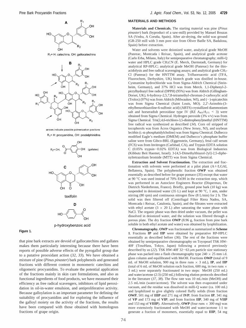
that pine bark extracts are devoid of gallocatechins and gallatesmakes them particularly interesting because there have beenreports of possible adverse effects of the pyrogallol group dueto a putative prooxidant action (32, 33). We have obtained amixture of pine (Pinus pinaster) bark polyphenols and generatedfractions with different content in monomeric catechins andoligomeric procyanidins. To evaluate the potential applicationof the fractions mainly in skin care formulations, and also asfunctional ingredients of food products, we have measured theirefficiency as free radical scavengers, inhibitors of lipid peroxi-dation in oil-in-water emulsion, and antiproliferative activity.Because galloylation is an important parameter for defining thesuitability of procyanidins and for exploring the influence ofthe galloyl moiety on the activity of the fractions, the resultshave been compared with those obtained with homologousfractions of grape origin.
MATERIALS AND METHODS
Materials and Chemicals.The starting material was pine (Pinuspinaster) bark (byproduct of a saw-mill) provided by Manuel BouzasSA (Vedra, A Coruna, Spain). After air-drying, the solid was ground(GR-250 mill with 3 mm pore size from Oliver Batlle SA, Badalona,Spain) before extraction.
Water and solvents were deionized water, analytical grade MeOH(Panreac, Montcada i Reixac, Spain), and analytical grade acetone(Carlo Erba, Milano, Italy) for semipreparative chromatography; milli-Qwater and HPLC grade CH3CN (E. Merck, Darmstadt, Germany) foranalytical RP-HPLC; analytical grade MeOH (Panreac) for the thio-acidolysis and free radical scavenging assays; and analytical grade CH3-Cl (Panreac) for the HNTTM assay. Trifluoroacetic acid (TFA,Fluorochem, Derbyshire, UK) biotech grade was distilled in-house.Cysteamine hydrochloride was from Sigma-Aldrich Chemical (Stein-heim, Germany), and 37% HCl was from Merck. 1,1-Diphenyl-2-picrylhydrazyl free radical (DPPH) (95%) was from Aldrich (Gillingham-Dorset, UK), 6-hydroxy-2,5,7,8-tetramethyl-chroman-2-carboxylic acid(Trolox) (97%) was from Aldrich (Milwaukee, WI), and (-)-epicatechinwas from Sigma Chemical (Saint Louis, MO). 2,2′-Azinobis-(3-ethylbenzothiazoline-6-sulfonic acid) (ABTS) crystallized diammoniumsalt and horseradish peroxidase type IV (RZA403/A275 < 3) wereobtained from Sigma Chemical. Hydrogen peroxide (3% v/v) was fromSigma Chemical. Tris(2,4,6-trichloro-3,5-dinitrophenyl)methyl (HNTTM)free radical was synthesized as described (34). Corn oil stripped oftocopherols was from A¨ cros Organics (New Jersey, NJ), and soybeanlecithin (L-R-phosphatidylcholine) was from Sigma Chemical. Dulbeccomodified Eagle’s medium (DMEM) and Dulbecco’s phosphate buffersaline were from Gibco-BRL (Eggenstein, Germany), fetal calf serum(FCS) was from Invitrogen (Carlsbad, CA), and Trypsin EDTA solutionC (0.05% trypsin-0.02% EDTA) was from Biological Industries(Kibbutz Beit Haemet, Israel). 3-[4,5-Dimethylthiazol-2yl]-2,5-diphe-nyltetrazolium bromide (MTT) was from Sigma Chemical.
Extraction and Solvent Fractionation. The extraction and frac-tionation with solvents were performed at a pilot plant (A+/LGAI,Bellaterra, Spain). The polyphenolic fractionOWP was obtainedessentially as described before for grape pomace (35) except that waterat 90°C was used instead of 70% EtOH in the extraction step, whichwas performed in an Autoclave Engineers Reactor (Dispermax, DeDietrich Niederbronn, France). Briefly, ground pine bark (10 kg) wassuspended in deionized water (35 L) and kept at 90°C, 1 atm, understirring (80 rpm) and continuous nitrogen flow (8 L/min) for 2 h. Thesolid was then filtered off (Centrifugal Filter Riera Nadeu, SA,Montcada i Reixac, Catalonia, Spain), and the filtrates were extractedwith ethyl acetate (3× 20 L) after saturating the water phase withNaCl. The organic phase was then dried under vacuum, the pellet wasdissolved in deionized water, and the solution was filtered through aporous plate. The dry fractionOWP (8.96 g, fraction from pine barksoluble in both ethyl acetate and water) was obtained by lyophilization.
Chromatography. OWP was fractionated as summarized inScheme1. FractionsIP and IIP were obtained by preparative RP-HPLCessentially as described before (36). The rest of the fractions wereobtained by semipreparative chromatography on Toyopearl TSK HW-40F (TosoHass, Tokyo, Japan) following a protocol previouslydescribed by us (22). TSK HW-40F 32-63 µm particle size stationaryphase was packed into a flash chromatography type 240× 25 mm i.d.glass column and equilibrated with MeOH. FractionsOWP (total of 9mL of MeOH solution, 900 mg in three runs× 3 mL), IP, and IIP(total of 6 mL of MeOH solution each fraction, 600 mg, in two runs×3 mL) were separately fractionated in two steps: MeOH (250 mL)and water/acetone (2:3) (250 mL) following elution protocols describedin the literature (37,38). The flow rate was 10 mL/min (MeOH) and2.5 mL/min (water/acetone). The solvent was then evaporated undervacuum, and the residue was dissolved in milli-Q water (ca. 100 mL)and lyophilized to give slightly colored fluffy solids (from fractionOWP, 501 mg ofIIIP and 271 mg ofIVP; from fraction IP, 341 mgof VP and 173 mg ofVIP; and from fractionIIP, 340 mg of VIIPand 153 mg ofVIIIP). Alternatively, OWP (four runs× 300 mg) wasmore extensively fractionated with MeOH and water/acetone 1:1 togenerate a fraction of monomers, essentially equal toIIIP , 54 mg
Figure 1.
Pine Bark Procyanidin Fractions J. Agric. Food Chem., Vol. 53, No. 12, 2005 4729
74
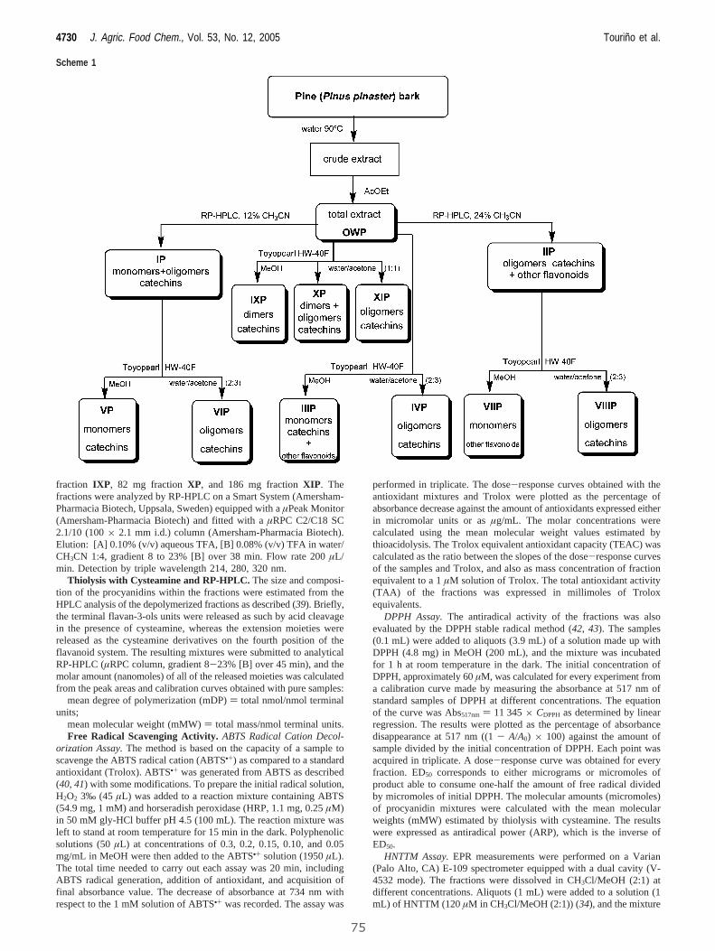
fraction IXP, 82 mg fraction XP, and 186 mg fractionXIP. Thefractions were analyzed by RP-HPLC on a Smart System (Amersham-Pharmacia Biotech, Uppsala, Sweden) equipped with aµPeak Monitor(Amersham-Pharmacia Biotech) and fitted with aµRPC C2/C18 SC2.1/10 (100× 2.1 mm i.d.) column (Amersham-Pharmacia Biotech).Elution: [A] 0.10% (v/v) aqueous TFA, [B] 0.08% (v/v) TFA in water/CH3CN 1:4, gradient 8 to 23% [B] over 38 min. Flow rate 200µL/min. Detection by triple wavelength 214, 280, 320 nm.
Thiolysis with Cysteamine and RP-HPLC.The size and composi-tion of the procyanidins within the fractions were estimated from theHPLC analysis of the depolymerized fractions as described (39). Briefly,the terminal flavan-3-ols units were released as such by acid cleavagein the presence of cysteamine, whereas the extension moieties werereleased as the cysteamine derivatives on the fourth position of theflavanoid system. The resulting mixtures were submitted to analyticalRP-HPLC (µRPC column, gradient 8-23% [B] over 45 min), and themolar amount (nanomoles) of all of the released moieties was calculatedfrom the peak areas and calibration curves obtained with pure samples:
mean degree of polymerization (mDP)) total nmol/nmol terminalunits;
mean molecular weight (mMW)) total mass/nmol terminal units.Free Radical Scavenging Activity.ABTS Radical Cation Decol-
orization Assay.The method is based on the capacity of a sample toscavenge the ABTS radical cation (ABTS•+) as compared to a standardantioxidant (Trolox). ABTS•+ was generated from ABTS as described(40,41) with some modifications. To prepare the initial radical solution,H2O2 3‰ (45 µL) was added to a reaction mixture containing ABTS(54.9 mg, 1 mM) and horseradish peroxidase (HRP, 1.1 mg, 0.25µM)in 50 mM gly-HCl buffer pH 4.5 (100 mL). The reaction mixture wasleft to stand at room temperature for 15 min in the dark. Polyphenolicsolutions (50µL) at concentrations of 0.3, 0.2, 0.15, 0.10, and 0.05mg/mL in MeOH were then added to the ABTS•+ solution (1950µL).The total time needed to carry out each assay was 20 min, includingABTS radical generation, addition of antioxidant, and acquisition offinal absorbance value. The decrease of absorbance at 734 nm withrespect to the 1 mM solution of ABTS•+ was recorded. The assay was
performed in triplicate. The dose-response curves obtained with theantioxidant mixtures and Trolox were plotted as the percentage ofabsorbance decrease against the amount of antioxidants expressed eitherin micromolar units or asµg/mL. The molar concentrations werecalculated using the mean molecular weight values estimated bythioacidolysis. The Trolox equivalent antioxidant capacity (TEAC) wascalculated as the ratio between the slopes of the dose-response curvesof the samples and Trolox, and also as mass concentration of fractionequivalent to a 1µM solution of Trolox. The total antioxidant activity(TAA) of the fractions was expressed in millimoles of Troloxequivalents.
DPPH Assay.The antiradical activity of the fractions was alsoevaluated by the DPPH stable radical method (42,43). The samples(0.1 mL) were added to aliquots (3.9 mL) of a solution made up withDPPH (4.8 mg) in MeOH (200 mL), and the mixture was incubatedfor 1 h at room temperature in the dark. The initial concentration ofDPPH, approximately 60µM, was calculated for every experiment froma calibration curve made by measuring the absorbance at 517 nm ofstandard samples of DPPH at different concentrations. The equationof the curve was Abs517nm ) 11 345× CDPPH as determined by linearregression. The results were plotted as the percentage of absorbancedisappearance at 517 nm ((1- A/A0) × 100) against the amount ofsample divided by the initial concentration of DPPH. Each point wasacquired in triplicate. A dose-response curve was obtained for everyfraction. ED50 corresponds to either micrograms or micromoles ofproduct able to consume one-half the amount of free radical dividedby micromoles of initial DPPH. The molecular amounts (micromoles)of procyanidin mixtures were calculated with the mean molecularweights (mMW) estimated by thiolysis with cysteamine. The resultswere expressed as antiradical power (ARP), which is the inverse ofED50.
HNTTM Assay.EPR measurements were performed on a Varian(Palo Alto, CA) E-109 spectrometer equipped with a dual cavity (V-4532 mode). The fractions were dissolved in CH3Cl/MeOH (2:1) atdifferent concentrations. Aliquots (1 mL) were added to a solution (1mL) of HNTTM (120µM in CH3Cl/MeOH (2:1)) (34), and the mixture
Scheme 1
4730 J. Agric. Food Chem., Vol. 53, No. 12, 2005 Tourino et al.
75
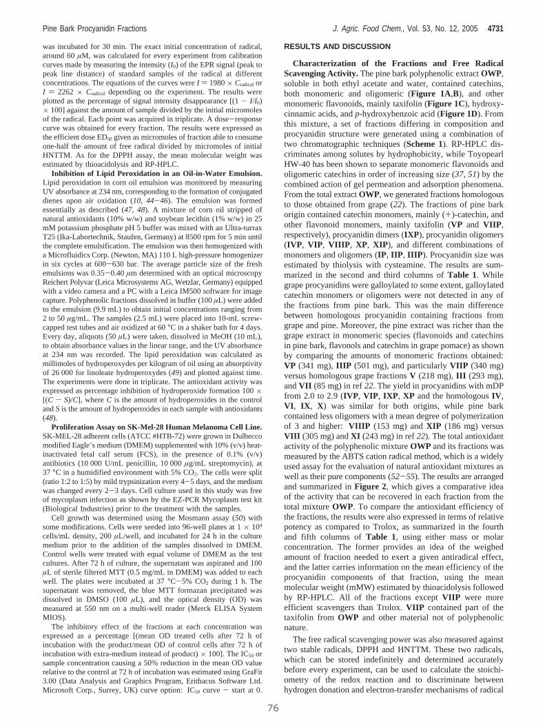
was incubated for 30 min. The exact initial concentration of radical,around 60µM, was calculated for every experiment from calibrationcurves made by measuring the intensity (I0) of the EPR signal (peak topeak line distance) of standard samples of the radical at differentconcentrations. The equations of the curves wereI ) 1980× CradicalorI ) 2262 × Cradical depending on the experiment. The results wereplotted as the percentage of signal intensity disappearance [(1- I/I 0)× 100] against the amount of sample divided by the initial micromolesof the radical. Each point was acquired in triplicate. A dose-responsecurve was obtained for every fraction. The results were expressed asthe efficient dose ED50 given as micromoles of fraction able to consumeone-half the amount of free radical divided by micromoles of initialHNTTM. As for the DPPH assay, the mean molecular weight wasestimated by thioacidolysis and RP-HPLC.
Inhibition of Lipid Peroxidation in an Oil-in-Water Emulsion.Lipid peroxidation in corn oil emulsion was monitored by measuringUV absorbance at 234 nm, corresponding to the formation of conjugateddienes upon air oxidation (10,44-46). The emulsion was formedessentially as described (47, 48). A mixture of corn oil stripped ofnatural antioxidants (10% w/w) and soybean lecithin (1% w/w) in 25mM potassium phosphate pH 5 buffer was mixed with an Ultra-turraxT25 (Ika-Labortechnik, Staufen, Germany) at 8500 rpm for 5 min untilthe complete emulsification. The emulsion was then homogenized witha Microfluidics Corp. (Newton, MA) 110 L high-pressure homogenizerin six cycles at 600-630 bar. The average particle size of the freshemulsions was 0.35-0.40µm determined with an optical microscopyReichert Polyvar (Leica Microsystems AG, Wetzlar, Germany) equippedwith a video camera and a PC with a Leica IM500 software for imagecapture. Polyphenolic fractions dissolved in buffer (100µL) were addedto the emulsion (9.9 mL) to obtain initial concentrations ranging from2 to 50 µg/mL. The samples (2.5 mL) were placed into 10-mL screw-capped test tubes and air oxidized at 60°C in a shaker bath for 4 days.Every day, aliquots (50µL) were taken, dissolved in MeOH (10 mL),to obtain absorbance values in the linear range, and the UV absorbanceat 234 nm was recorded. The lipid peroxidation was calculated asmillimoles of hydroperoxydes per kilogram of oil using an absorptivityof 26 000 for linoleate hydroperoxydes (49) and plotted against time.The experiments were done in triplicate. The antioxidant activity wasexpressed as percentage inhibition of hydroperoxide formation 100×[(C - S)/C], whereC is the amount of hydroperoxides in the controlandS is the amount of hydroperoxides in each sample with antioxidants(48).
Proliferation Assay on SK-Mel-28 Human Melanoma Cell Line.SK-MEL-28 adherent cells (ATCC #HTB-72) were grown in Dulbeccomodified Eagle’s medium (DMEM) supplemented with 10% (v/v) heat-inactivated fetal calf serum (FCS), in the presence of 0.1% (v/v)antibiotics (10 000 U/mL penicillin, 10 000µg/mL streptomycin), at37 °C in a humidified environment with 5% CO2. The cells were split(ratio 1:2 to 1:5) by mild trypsinization every 4-5 days, and the mediumwas changed every 2-3 days. Cell culture used in this study was freeof mycoplasm infection as shown by the EZ-PCR Mycoplasm test kit(Biological Industries) prior to the treatment with the samples.
Cell growth was determined using the Mosmann assay (50) withsome modifications. Cells were seeded into 96-well plates at 1× 10⁴
cells/mL density, 200µL/well, and incubated for 24 h in the culturemedium prior to the addition of the samples dissolved in DMEM.Control wells were treated with equal volume of DMEM as the testcultures. After 72 h of culture, the supernatant was aspirated and 100µL of sterile filtered MTT (0.5 mg/mL in DMEM) was added to eachwell. The plates were incubated at 37°C-5% CO2 during 1 h. Thesupernatant was removed, the blue MTT formazan precipitated wasdissolved in DMSO (100µL), and the optical density (OD) wasmeasured at 550 nm on a multi-well reader (Merck ELISA SystemMIOS).
The inhibitory effect of the fractions at each concentration wasexpressed as a percentage [(mean OD treated cells after 72 h ofincubation with the product/mean OD of control cells after 72 h ofincubation with extra-medium instead of product)× 100]. The IC50 orsample concentration causing a 50% reduction in the mean OD valuerelative to the control at 72 h of incubation was estimated using GraFit3.00 (Data Analysis and Graphics Program, Erithacus Software Ltd.Microsoft Corp., Surrey, UK) curve option: IC50 curve - start at 0.
RESULTS AND DISCUSSION
Characterization of the Fractions and Free RadicalScavenging Activity.The pine bark polyphenolic extractOWP,soluble in both ethyl acetate and water, contained catechins,both monomeric and oligomeric (Figure 1A,B), and othermonomeric flavonoids, mainly taxifolin (Figure 1C), hydroxy-cinnamic acids, andp-hydroxybenzoic acid (Figure 1D). Fromthis mixture, a set of fractions differing in composition andprocyanidin structure were generated using a combination oftwo chromatographic techniques (Scheme 1). RP-HPLC dis-criminates among solutes by hydrophobicity, while ToyopearlHW-40 has been shown to separate monomeric flavonoids andoligomeric catechins in order of increasing size (37,51) by thecombined action of gel permeation and adsorption phenomena.From the total extractOWP, we generated fractions homologousto those obtained from grape (22). The fractions of pine barkorigin contained catechin monomers, mainly (+)-catechin, andother flavonoid monomers, mainly taxifolin (VPand VIIP,respectively), procyanidin dimers (IXP ), procyanidin oligomers(IVP, VIP, VIIIP, XP, XIP), and different combinations ofmonomers and oligomers (IP, IIP, IIIP). Procyanidin size wasestimated by thiolysis with cysteamine. The results are sum-marized in the second and third columns ofTable 1. Whilegrape procyanidins were galloylated to some extent, galloylatedcatechin monomers or oligomers were not detected in any ofthe fractions from pine bark. This was the main differencebetween homologous procyanidin containing fractions fromgrape and pine. Moreover, the pine extract was richer than thegrape extract in monomeric species (flavonoids and catechinsin pine bark, flavonols and catechins in grape pomace) as shownby comparing the amounts of monomeric fractions obtained:VP (341 mg),IIIP (501 mg), and particularlyVIIP (340 mg)versus homologous grape fractionsV (218 mg),III (293 mg),andVII (85 mg) in ref22. The yield in procyanidins with mDPfrom 2.0 to 2.9 (IVP, VIP, IXP, XP and the homologousIV,VI, IX, X) was similar for both origins, while pine barkcontained less oligomers with a mean degree of polymerizationof 3 and higher:VIIIP (153 mg) andXIP (186 mg) versusVIII (305 mg) andXI (243 mg) in ref22). The total antioxidantactivity of the polyphenolic mixtureOWP and its fractions wasmeasured by the ABTS cation radical method, which is a widelyused assay for the evaluation of natural antioxidant mixtures aswell as their pure components (52-55). The results are arrangedand summarized inFigure 2, which gives a comparative ideaof the activity that can be recovered in each fraction from thetotal mixtureOWP. To compare the antioxidant efficiency ofthe fractions, the results were also expressed in terms of relativepotency as compared to Trolox, as summarized in the fourthand fifth columns ofTable 1, using either mass or molarconcentration. The former provides an idea of the weighedamount of fraction needed to exert a given antiradical effect,and the latter carries information on the mean efficiency of theprocyanidin components of that fraction, using the meanmolecular weight (mMW) estimated by thioacidolysis followedby RP-HPLC. All of the fractions exceptVIIP were moreefficient scavengers than Trolox.VIIP contained part of thetaxifolin from OWP and other material not of polyphenolicnature.
The free radical scavenging power was also measured againsttwo stable radicals, DPPH and HNTTM. These two radicals,which can be stored indefinitely and determined accuratelybefore every experiment, can be used to calculate the stoichi-ometry of the redox reaction and to discriminate betweenhydrogen donation and electron-transfer mechanisms of radical
Pine Bark Procyanidin Fractions J. Agric. Food Chem., Vol. 53, No. 12, 2005 4731
76
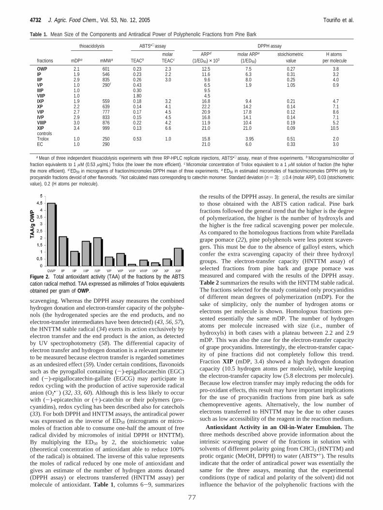
scavenging. Whereas the DPPH assay measures the combinedhydrogen donation and electron-transfer capacity of the polyphe-nols (the hydrogenated species are the end products, and noelectron-transfer intermediates have been detected) (43,56,57),the HNTTM stable radical (34) exerts its action exclusively byelectron transfer and the end product is the anion, as detectedby UV spectrophotometry (58). The differential capacity ofelectron transfer and hydrogen donation is a relevant parameterto be measured because electron transfer is regarded sometimesas an undesired effect (59). Under certain conditions, flavonoidssuch as the pyrogallol containing (-)-epigallocatechin (EGC)and (-)-epigallocatechin-gallate (EGCG) may participate inredox cycling with the production of active superoxide radicalanion (O2
•-) (32, 33, 60). Although this is less likely to occurwith (-)-epicatechin or (+)-catechin or their polymers (pro-cyanidins), redox cycling has been described also for catechols(33). For both DPPH and HNTTM assays, the antiradical powerwas expressed as the inverse of ED50 (micrograms or micro-moles of fraction able to consume one-half the amount of freeradical divided by micromoles of initial DPPH or HNTTM).By multiplying the ED50 by 2, the stoichiometric value(theoretical concentration of antioxidant able to reduce 100%of the radical) is obtained. The inverse of this value representsthe moles of radical reduced by one mole of antioxidant andgives an estimate of the number of hydrogen atoms donated(DPPH assay) or electrons transferred (HNTTM assay) permolecule of antioxidant.Table 1, columns 6-9, summarizes
the results of the DPPH assay. In general, the results are similarto those obtained with the ABTS cation radical. Pine barkfractions followed the general trend that the higher is the degreeof polymerization, the higher is the number of hydroxyls andthe higher is the free radical scavenging power per molecule.As compared to the homologous fractions from white Parelladagrape pomace (22), pine polyphenols were less potent scaven-gers. This must be due to the absence of galloyl esters, whichconfer the extra scavenging capacity of their three hydroxylgroups. The electron-transfer capacity (HNTTM assay) ofselected fractions from pine bark and grape pomace wasmeasured and compared with the results of the DPPH assay.Table 2summarizes the results with the HNTTM stable radical.The fractions selected for the study contained only procyanidinsof different mean degrees of polymerization (mDP). For thesake of simplicity, only the number of hydrogen atoms orelectrons per molecule is shown. Homologous fractions pre-sented essentially the same mDP. The number of hydrogenatoms per molecule increased with size (i.e., number ofhydroxyls) in both cases with a plateau between 2.2 and 2.9mDP. This was also the case for the electron-transfer capacityof grape procyanidins. Interestingly, the electron-transfer capac-ity of pine fractions did not completely follow this trend.FractionXIP (mDP, 3.4) showed a high hydrogen donationcapacity (10.5 hydrogen atoms per molecule), while keepingthe electron-transfer capacity low (5.8 electrons per molecule).Because low electron transfer may imply reducing the odds forpro-oxidant effects, this result may have important implicationsfor the use of procyanidin fractions from pine bark as safechemopreventive agents. Alternatively, the low number ofelectrons transferred to HNTTM may be due to other causessuch as low accessibility of the reagent in the reaction medium.
Antioxidant Activity in an Oil-in-Water Emulsion. Thethree methods described above provide information about theintrinsic scavenging power of the fractions in solution withsolvents of different polarity going from CHCl3 (HNTTM) andprotic organic (MeOH, DPPH) to water (ABTS•+). The resultsindicate that the order of antiradical power was essentially thesame for the three assays, meaning that the experimentalconditions (type of radical and polarity of the solvent) did notinfluence the behavior of the polyphenolic fractions with the
Table 1. Mean Size of the Components and Antiradical Power of Polyphenolic Fractions from Pine Bark
thioacidolysis ABTS•+ assay DPPH assay
fractions mDPa mMWa TEACbmolarTEACc
ARPd
(1/ED50) × 103molar ARPe
(1/ED50)stoichiometric
valueH atoms
per molecule
OWP 2.1 601 0.23 2.3 12.5 7.5 0.27 3.8IP 1.9 546 0.23 2.2 11.6 6.3 0.31 3.2IIP 2.9 835 0.26 3.0 9.6 8.0 0.25 4.0VP 1.0 290f 0.43 6.5 1.9 1.05 0.9IIIP 1.0 0.30 9.5VIIP 1.0 1.80 4.5IXP 1.9 559 0.18 3.2 16.8 9.4 0.21 4.7XP 2.2 639 0.14 4.1 22.2 14.2 0.14 7.1VIP 2.7 777 0.17 4.5 20.9 17.8 0.12 8.6IVP 2.9 833 0.15 4.5 16.8 14.1 0.14 7.1VIIIP 3.0 876 0.22 4.2 11.9 10.4 0.19 5.2XIP 3.4 999 0.13 6.6 21.0 21.0 0.09 10.5controlsTrolox 1.0 250 0.53 1.0 15.8 3.95 0.51 2.0EC 1.0 290 21.0 6.0 0.33 3.0
a Mean of three independent thioacidolysis experiments with three RP-HPLC replicate injections, ABTS•+ assay, mean of three experiments. b Micrograms/microliter offraction equivalents to 1 µM (0.53 µg/mL) Trolox (the lower the more efficient). c Micromolar concentration of Trolox equivalent to a 1 µM solution of fraction (the higherthe more efficient). d ED50 in micrograms of fraction/micromoles DPPH mean of three experiments. e ED50 in estimated micromoles of fraction/micromoles DPPH only forprocyanidin fractions devoid of other flavonoids. f Not calculated mass corresponding to catechin monomer. Standard deviation (n ) 3): e0.4 (molar ARP), 0.03 (stoichiometricvalue), 0.2 (H atoms per molecule).
Figure 2. Total antioxidant activity (TAA) of the fractions by the ABTScation radical method. TAA expressed as millimoles of Trolox equivalentsobtained per gram of OWP.
4732 J. Agric. Food Chem., Vol. 53, No. 12, 2005 Tourino et al.
77
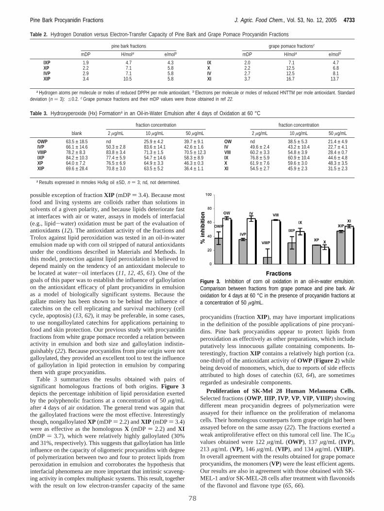
possible exception of fractionXIP (mDP) 3.4). Because mostfood and living systems are colloids rather than solutions insolvents of a given polarity, and because lipids deteriorate fastat interfaces with air or water, assays in models of interfacial(e.g., lipid-water) oxidation must be part of the evaluation ofantioxidants (12). The antioxidant activity of the fractions andTrolox against lipid peroxidation was tested in an oil-in-wateremulsion made up with corn oil stripped of natural antioxidantsunder the conditions described in Materials and Methods. Inthis model, protection against lipid peroxidation is believed todepend mainly on the tendency of an antioxidant molecule tobe located at water-oil interfaces (11,12, 45, 61). One of thegoals of this paper was to establish the influence of galloylationon the antioxidant efficacy of plant procyanidins in emulsionas a model of biologically significant systems. Because thegallate moiety has been shown to be behind the influence ofcatechins on the cell replicating and survival machinery (cellcycle, apoptosis) (13, 62), it may be preferable, in some cases,to use nongalloylated catechins for applications pertaining tofood and skin protection. Our previous study with procyanidinfractions from white grape pomace recorded a relation betweenactivity in emulsion and both size and galloylation indistin-guishably (22). Because procyanidins from pine origin were notgalloylated, they provided an excellent tool to test the influenceof galloylation in lipid protection in emulsion by comparingthem with grape procyanidins.
Table 3 summarizes the results obtained with pairs ofsignificant homologous fractions of both origins.Figure 3depicts the percentage inhibition of lipid peroxidation exertedby the polyphenolic fractions at a concentration of 50µg/mLafter 4 days of air oxidation. The general trend was again thatthe galloylated fractions were the most effective. Interestinglythough, nongalloylatedXP (mDP) 2.2) andXIP (mDP) 3.4)were as effective as the homologousX (mDP ) 2.2) andXI(mDP ) 3.7), which were relatively highly galloylated (30%and 31%, respectively). This suggests that galloylation has littleinfluence on the capacity of oligomeric procyanidins with degreeof polymerization between two and four to protect lipids fromperoxidation in emulsion and corroborates the hypothesis thatinterfacial phenomena are more important that intrinsic scaveng-ing activity in complex multiphasic systems. This result, togetherwith the result on low electron-transfer capacity of the same
procyanidins (fractionXIP), may have important implicationsin the definition of the possible applications of pine procyani-dins. Pine bark procyanidins appear to protect lipids fromperoxidation as effectively as other preparations, which includeputatively less innocuous gallate containing components. In-terestingly, fractionXIP contains a relatively high portion (ca.one-third) of the antioxidant activity ofOWP (Figure 2) whilebeing devoid of monomers, which, due to reports of side effectsattributed to high doses of catechin (63, 64), are sometimesregarded as undesirable components.
Proliferation of SK-Mel 28 Human Melanoma Cells.Selected fractions (OWP, IIIP, IVP, VP, VIP, VIIIP) showingdifferent mean procyanidin degrees of polymerization wereassayed for their influence on the proliferation of melanomacells. Their homologous counterparts form grape origin had beenassayed before on the same assay (22). The fractions exerted aweak antiproliferative effect on this tumoral cell line. The IC50
values obtained were 122µg/mL (OWP), 137µg/mL (IVP),213 µg/mL (VP), 146µg/mL (VIP), and 134µg/mL (VIIIP).In overall agreement with the results obtained for grape pomaceprocyanidins, the monomers (VP) were the least efficient agents.Our results are also in agreement with those obtained with SK-MEL-1 and/or SK-MEL-28 cells after treatment with flavonoidsof the flavonol and flavone type (65, 66).
Table 2. Hydrogen Donation versus Electron-Transfer Capacity of Pine Bark and Grape Pomace Procyanidin Fractions
pine bark fractions grape pomace fractionsc
mDP H/mola e/molb mDP H/mola e/molb
IXP 1.9 4.7 4.3 IX 2.0 7.1 4.7XP 2.2 7.1 5.8 X 2.2 12.5 6.8IVP 2.9 7.1 5.8 IV 2.7 12.5 8.1XIP 3.4 10.5 5.8 XI 3.7 16.7 13.7
a Hydrogen atoms per molecule or moles of reduced DPPH per mole antioxidant. b Electrons per molecule or moles of reduced HNTTM per mole antioxidant. Standarddeviation (n ) 3): e0.2. c Grape pomace fractions and their mDP values were those obtained in ref 22.
Table 3. Hydroxyperoxide (Hx) Formationa in an Oil-in-Water Emulsion after 4 days of Oxidation at 60 °C
fraction concentration fraction concentration
blank 2 µg/mL 10 µg/mL 50 µg/mL 2 µg/mL 10 µg/mL 50 µg/mL
OWP 63.5 ± 18.5 nd 25.9 ± 4.2 39.7 ± 9.1 OW nd 38.5 ± 5.3 21.4 ± 4.9IVP 66.1 ± 14.6 50.3 ± 2.8 83.6 ± 14.1 42.6 ± 1.6 IV 49.6 ± 2.4 43.2 ± 10.4 22.7 ± 4.1VIIIP 78.2 ± 8.3 83.8 ± 3.4 71.3 ± 1.5 70.5 ± 12.3 VIII 60.2 ± 3.3 54.8 ± 3.9 28.4 ± 0.7IXP 84.2 ± 10.3 77.4 ± 5.9 54.7 ± 14.6 58.3 ± 8.9 IX 76.8 ± 5.9 60.9 ± 10.4 44.6 ± 4.8XP 64.0 ± 7.2 76.5 ± 6.9 64.9 ± 3.3 46.3 ± 0.3 X 61.9 ± 7.6 59.6 ± 3.0 48.3 ± 3.5XIP 69.6 ± 28.4 70.8 ± 3.0 63.5 ± 5.2 36.4 ± 1.1 XI 54.5 ± 2.7 45.9 ± 2.3 31.5 ± 2.3
a Results expressed in mmoles Hx/kg oil ±SD, n ) 3; nd, not determined.
Figure 3. Inhibition of corn oil oxidation in an oil-in-water emulsion.Comparison between fractions from grape pomace and pine bark. Airoxidation for 4 days at 60 °C in the presence of procyanidin fractions ata concentration of 50 µg/mL.
Pine Bark Procyanidin Fractions J. Agric. Food Chem., Vol. 53, No. 12, 2005 4733
78
In conclusion, oligomeric procyanidin fractions from pinebark appear to be efficient and safe antioxidant agents. Becausethey do not include gallate esters, they are less potent free radicalscavengers in solution than the corresponding galloylatedfractions of grape origin. Interestingly, the galloyl moiety isnot necessary for lipid protection in emulsion. This has importantimplications for the application of mixtures of this kind in foodmanufacturing and the formulation of skin care products.Because the galloyl moiety influences crucial biochemicalpathways in cell replication and survival, efficient gallate freemixtures such as our fractionXIP may be the best option forproducts designed to offer antioxidant protection by exclusivelyscavenging an excess of radicals without altering the normalcell functions.
ABBREVIATIONS USED
ABTS, 2,2′-azinobis(3-ethylbenzothiazoline-6-sulphonic acid);EC, (-)-epicatechin; DMEM, Dulbecco modified Eagle’smedium; DPPH, 1,1-diphenyl-2-picrylhydrazyl free radical;FCS, fetal calf serum; HNTTM, tris(2,3,6-trichloro-3,5-dinitro-phenyl)methyl; MTT, 3-[4,5-dimethylthiazol-2yl]-2,5-diphe-nyltetrazolium bromide; RP-HPLC, reversed-phase high-performance liquid chromatography; TAA, total antioxidantactivity; TEAC, Trolox equivalent antioxidant capacity; TFA,trifluoroacetic acid; Trolox, 2,5,7,8-tetramethyl-chroman-2-carboxylic acid.
ACKNOWLEDGMENT
We are thankful to Drs. C. Solans and J. Esquena for their adviceand help in the preparation of the emulsion.
Supporting Information Available: RP-HPLC chromato-grams obtained for all of the fractions before and afterthioacidolysis, plots of time course oil oxidation in emulsion,and dose-response curves from the proliferation assay. Thismaterial is available free of charge via the Internet at http://pubs.acs.org.
LITERATURE CITED
(1) Rusznyak, S. P.; Szent-Gyorgyi, A. Vitamin P: flavonols asvitamins.Nature1936,138, 27.
(2) Ross, J. A.; Kasum, C. M. Dietary flavonoids: Bioavailability,metabolic effects, and safety.Annu. ReV. Nutr.2002,22, 19-34.
(3) Rice-Evans, C. A.; Miller, N. J.; Paganga, G. Structure-antioxidant activity relationships of flavonoids and phenolicacids.Free Radical Biol. Med.1996,20, 933-956.
(4) Yokozawa, T.; Chen, C. P.; Dong, E.; Tanaka, T.; Nonaka, G.-I.; Nishioka, I. Study on the inhibitory effect of tannins andflavonoids against the 1,1-diphenyl-2-picrylhydrazyl radical.Biochem. Pharmacol.1998,56, 213-222.
(5) Brown, J. E.; Khodr, H.; Hider, R. C.; Rice-Evans, C. A.Structural dependence of flavonoid interactions with Cu2+ ions:implications for their antioxidant properties.Biochem. J.1998,330, 1173-1178.
(6) Hider, R. C.; Liu, Z. D.; Khodr, H. H. Metal chelation ofpolyphenols. InFlaVonoids and Other Polyphenols; Packer, L.,Ed.; Academic Press Inc.: San Diego, CA, 2001; pp 190-203.
(7) Katiyar, S. K.; Afaq, F.; Perez, A.; Mukhtar, H. Green teapolyphenol (-)-epigallocatechin-3-gallate treatment of humanskin inhibits ultraviolet radiation-induced oxidative stress.Car-cinogenesis2001,22, 287-294.
(8) Ishige, K.; Schubert, D.; Sagara, Y. Flavonoids protect neuronalcells from oxidative stress by three distinct mechanisms.FreeRadical Biol. Med.2001,30, 433-446.
(9) Inal, M. E.; Kahraman, A. The protective effect of flavonolquercetin against ultraviolet a induced oxidative stress in rats.Toxicology2000,154, 21-29.
(10) Frankel, E. N.; Huang, S.-W.; Kanner, J.; German, J. B.Interfacial phenomena in the evaluation of antioxidants: Bulkoils vs emulsions.J. Agric. Food Chem.1994,42, 1054-1059.
(11) Frankel, E. N. Interfacial lipid oxidation and antioxidation.J.Oleo Sci.2001,50, 387-391.
(12) Frankel, E. N.; Meyer, A. S. The problems of using one-dimensional methods to evaluate multifunctional food andbiological antioxidants.J. Sci. Food Agric.2000, 80, 1925-1941.
(13) Liang, Y. C.; Lin-shiau, S. Y.; Chen, C. F.; Lin, J. K. Suppressionof extracellular signals and cell proliferation through EGFreceptor binding by (-)-epigallocatechin gallate in human A431epidermoid carcinoma cells.J. Cell Biochem.1997,67, 55-65.
(14) Ahmad, N.; Gali, H.; Javed, S.; Agarwal, R. Skin cancerchemopreventive effects of a flavonoid antioxidant silymarin aremediated via impairment of receptor tyrosine kinase signalingand perturbation in cell cycle progression.Biochem. Biophys.Res. Commun.1998,247, 294-301.
(15) Ahmad, N.; Cheng, P. Y.; Mukhtar, H. Cell cycle dysregulationby green tea polyphenol epigallocatechin-3-gallate.Biochem.Biophys. Res. Commun.2000,275, 328-334.
(16) Hayakawa, S.; Kimura, T.; Saeki, K.; Koyama, Y.; Aoyagi, Y.;Noro, T.; Nakamura, Y.; Isemura, M. Apoptosis-inducing activityof high molecular weight fractions of tea extracts.Biosci.Biotechnol. Biochem.2001,65, 459-462.
(17) Williams, R. J.; Spencer, J. P. E.; Rice-Evans, C. Flavonoids:Antioxidants or signaling molecules?Free Radical Biol. Med.2004,36, 838-849.
(18) Santos-Buelga, C.; Scalbert, A. Proanthocyanidins and tannin-like compounds- nature, occurrence, dietary intake and effectson nutrition and health.J. Sci. Food Agric.2000, 80, 1094-1117.
(19) Cos, P.; De Bruyne, T.; Hermans, N.; Apers, S.; Berghe, D. V.;Vlietinck, A. J. Proanthocyanidins in health care: Current andnew trends.Curr. Med. Chem.2004,11, 1345-1359.
(20) Plumb, G. W.; De Pascual-Teresa, S.; Santos-Buelga, C.;Cheynier, V.; Williamson, G. Antioxidant properties of catechinsand proanthocyanidins: effect of polymerisation, galloylationand glycosylation.Free Radical Res.1998,29, 351-358.
(21) Saint-Cricq de Gaulejac, N.; Provost, C.; Vivas, N. Comparativestudy of polyphenol scavenging activities assessed by differentmethods.J. Agric. Food Chem.1999,47, 425-431.
(22) Torres, J. L.; Varela, B.; Garcıa, M. T.; Carilla, J.; Matito, C.;Centelles, J. J.; Cascante, C.; Sort, X.; Bobet, R. Valorizationof grape (VitisVinifera) byproducts. Antioxidant and biologicalproperties of polyphenolic fractions differing in procyanidincomposition and flavonol content.J. Agric. Food Chem.2002,50, 7548-7555.
(23) Escribano Bailon, M. T.; Guerra, M. T.; Rivas Gonzalo, J. C.;Santos Buelga, C. Proanthocyanidin composition in skin and seedfrom grapes.Polyphenols1994, 225-226.
(24) Souquet, J.-M.; Cheynier, V.; Brossaud, F.; Moutounet, M.Polymeric proanthocyanidins from grape skins.Phytochemistry1996,43, 509-512.
(25) Kennedy, J. A.; Hayasaka, Y.; Vidal, S.; Waters, E. J.; Jones,G. P. Composition of grape skin proanthocyanidins at differentstages of berry development.J. Agric. Food Chem.2001,49,5348-5355.
(26) Souquet, J. M.; Labarbe, B.; LeGuerneve, C.; Cheynier, V.;Moutounet, M. Phenolic composition of grape stems.J. Agric.Food Chem.2000,48, 1076-1080.
(27) Prieur, C.; Rigaud, J.; Cheynier, V.; Moutounet, M. Oligomericand polymeric procyanidins from grape seeds.Phytochemistry1994,36, 781-784.
(28) Fuleki, T.; Ricardo da Silva, J. M. Catechin and procyanidincomposition of seeds from grape cultivars grown in Ontario.J.Agric. Food Chem.1997,45, 1156-1160.
(29) Packer, L.; Rimbach, G.; Virgili, F. Antioxidant activity andbiologic properties of a procyanidin-rich extract from pine (Pinusmaritima) bark, pycnogenol.Free Radical Biol. Med.1999,27,704-724.
4734 J. Agric. Food Chem., Vol. 53, No. 12, 2005 Tourino et al.
79
(30) Rohdewald, P. A review of the French maritime pine bark extract(Pycnogenol (R)), a herbal medication with a diverse clinicalpharmacology.Int. J. Clin. Pharmacol. Ther.2002,40, 158-168.
(31) Selga, A.; Sort, X.; Bobet, R.; Torres, J. L. Efficient one potextraction and depolymerization of grape (Vitis Vinifera) pomaceprocyanidins for the preparation of antioxidant thio-conjugates.J. Agric. Food Chem.2004,52, 467-473.
(32) Kondo, K.; Kurihara, M.; Miyata, N.; Suzuki, T.; Toyoda, M.Mechanistic studies of catechins as antioxidants against radicaloxidation.Arch. Biochem. Biophys.1999,362, 79-86.
(33) Long, L. H.; Clement, M. V.; Halliwell, B. Artifacts in cellculture: Rapid generation of hydrogen peroxide on addition of(-)-epigallocatechin, (-)-epigallocatechin gallate, (+)-catechin,and quercetin to commonly used cell culture media.Biochem.Biophys. Res. Commun.2000,273, 50-53.
(34) Torres, J. L.; Varela, B.; Brillas, E.; Julia, L. Tris(2,4,6-Trichloro-3,5-dinitrophenyl)methyl radical: a new stable coloured magneticspecies as a chemosensor for natural polyphenols.Chem.Commun.2003, 74-75.
(35) Torres, J. L.; Bobet, R. New flavanol-derivatives from grape(Vitis Vinifera) byproducts. Antioxidant aminoethylthio-flavan-3-ol conjugates from a polymeric waste fraction used as a sourceof flavanols.J. Agric. Food Chem.2001,49, 4627-4634.
(36) Matito, C.; Mastorakou, F.; Centelles, J. J.; Torres, J. L.;Cascante, M. Antiproliferative effect of antioxidant polyphenolsfrom grape in murine Hepa-1c1c7.Eur. J. Nutr.2003,42, 43-49.
(37) Sun, B. S.; Belchior, G. P.; Ricardo da Silva, J. M.; Spranger,M. I. Isolation and purification of dimeric and trimeric procya-nidins from grape seeds.J. Chromatogr., A1999,841, 115-121.
(38) Labarbe, B.; Cheynier, V.; Brossaud, F.; Souquet, J. M.;Moutounet, M. Quantitative fractionation of grape proanthocya-nidins according to their degree of polymerization.J. Agric. FoodChem.1999,47, 2719-2723.
(39) Torres, J. L.; Selga, A. Procyanidin size and composition bythiolysis with cysteamine hydrochloride and chromatography.Chromatographia2003,57, 441-445.
(40) Cano, A.; Hernandez-Ruiz, J.; Garcıa-Canovas, F.; Acosta, M.;Arnao, M. An end-point method for estimation of the totalantioxidant capacity in plant material.Phytochem. Anal.1998,9, 196-202.
(41) Alcolea, J. F.; Cano, A.; Acosta, M.; Arnao, M. B. Hydrophilicand lipofilic antioxidant activities of grapes.Nahrung2002,46,353-356.
(42) Blois, M. S. Antioxidant determinations by the use of stable freeradical.Nature1958,181, 1199-1200.
(43) Brand-Williams, W.; Cuvelier, M. E.; Berset, C. Use of a freeradical method to evaluate antioxidant activity.Lebensm.-Wiss.Technol.1995,28, 25-30.
(44) Buege, J. A.; Aust, S. D. Microsomal lipid peroxidation.MethodsEnzymol.1978,52, 302-310.
(45) Huang, S.-W.; Hopia, A.; Schwarz, K.; Frankel, E. N.; German,J. B. Antioxidant activity ofR-tocopherol and trolox in differentlipid substrates: Bulk oils vs oil-in-water emulsions.J. Agric.Food Chem.1996,44, 444-452.
(46) Sanchez-Moreno, C.; Satue-Gracia, M. T.; Frankel, E. N.Antioxidant activity of selected spanish wines in corn oilemulsions.J. Agric. Food Chem.2000,48, 5581-5587.
(47) Huang, S.-W.; Satue-Gracia, M. T.; Frankel, E. N.; German, J.B. Effect of lactoferrin on oxidative stability of corn oil emulsionsand liposomes.J. Agric. Food Chem.1999,47, 1356-1361.
(48) Schwarz, K.; Huang, S. W.; German, J. B.; Tiersch, B.;Hartmann, J. Activities of antioxidants are affected by colloidalproperties of oil-in-water and water-in-oil emulsions and bulkoils. J. Agric. Food Chem.2000,48, 4874-4882.
(49) Chan, H. W.-S.; Lewet, G. Autoxidation of methyl linoleate.Separation and analysis of isomeric mixtures of methyl linoleatehydroperoxides and methyl hydroxylinoleates.Lipids 1977,12,99-104.
(50) Mosmann, T. Rapid colorimetric assay for cellular growth andsurvival: Application to proliferation and cytotoxicity assays.J. Immunol. Methods1983, 55-63.
(51) Derdelinckx, G.; Jerumanis, J. Separation of malt and hopproanthocyanidins on Fractogel TSK HW-40S.J. Chromatogr.1984,285, 231-234.
(52) Miller, N. J.; Rice Evans, C. The relative contributions of ascorbicacid and phenolic antioxidants to the total antioxidant activityof orange and apple fruit juices and blackcurrant drink.FoodChem.1997,60, 331-337.
(53) Pietta, P.; Simonetti, P.; Mauri, P. Antioxidant activity of selectedmedicinal plants.J. Agric. Food Chem.1998,46, 4487-4490.
(54) Re, R.; Pellegrini, N.; Proteggente, A.; Pannala, A.; Yang, M.;Rice-Evans, C. Antioxidant activity applying an improved ABTSradical cation decolorization assay.Free Radical Biol. Med.1999,26, 1231-1237.
(55) Luximon-Ramma, A.; Bahorun, T.; Soobrattee, M. A.; Aruoma,O. I. Antioxidant activities of phenolic, proanthocyanidin, andflavonoid components in extracts of Cassia fistula.J. Agric. FoodChem.2002,50, 5042-5047.
(56) Dangles, O.; Fargeix, G.; Dufour, C. Antioxidant properties ofanthocyanins and tannins: a mechanistic investigation withcatechin and the 3′,4′,7-trihydroxyflavylium ion.J. Chem. Soc.,Perkin Trans. 22000, 1653-1666.
(57) Goupy, P.; Dufour, C.; Loonis, M.; Dangles, O. Quantitativekinetic analysis of hydrogen transfer reactions from dietarypolyphenols to the DPPH radical.J. Agric. Food Chem.2003,51, 615-622.
(58) Jimenez, A.; Selga, A.; Torres, J. L.; Julia, L. Reducing activityof polyphenols with stable radicals of the TTM series. Electrontransfer versus H-abstraction reactions in flavan-3-ols.Org. Lett.2004,6, 4583-4586.
(59) Kovacic, P.; Sacman, A.; Wu-Weis, M. Nephrotoxins: wide-spread role of oxidative stress and electron transfer.Curr. Med.Chem.2002,9, 823-847.
(60) Kondo, K.; Kurihara, M.; Miyata, N.; Suzuki, T.; Toyoda, M.Scavenging mechanisms of (-)-epigallocatechin gallate and (-)-epicatechin gallate on peroxyl radicals and formation of super-oxide during the inhibitory action.Free Radical Biol. Med.1999,27, 855-863.
(61) Huang, S.-W.; Frankel, E. N.; Aeschbach, R.; German, J. B.Partition of selected antioxidants in corn-water model systems.J. Agric. Food Chem.1997,45, 1991-1994.
(62) Liberto, M.; Cobrinik, D. Growth factor-dependent induction ofp21(CIP1) by the green tea by polyphenol, epigallocatechingallate.Cancer Lett.2000,154, 151-161.
(63) Jaeger, A.; Walti, M.; Neftel, K. Side effects of flavonoids inmedical practice. InPlant FlaVonoids in Biology and MedicineII: Biochemical, Cellular, and Medicinal Properties; Cody, V.,Ed.; Alan R. Liss Inc.: New York, 1988; pp 379-394.
(64) Stuhlinger, W.; Berek, K.; Grosswang, F.; Schauer, N. Severalyears observation of a Catergen (cyanidanol-3) induced immu-nohemolysis.Schweiz. Med. Wochenschr.1990,120, 345-348.
(65) Rodrıguez, J.; Ya´nez, J.; Vicente, V.; Alcaraz, M.; Benavente-Garcıa, O.; Castillo, J.; Lorente, J.; Lozano, J. A. Effects ofseveral flavonoids on the growth of B16F10 and SK-MEL-1melanoma cell lines: relationship between structure and activity.Melanoma Res.2002,12, 99-107.
(66) Lama, G.; Angelucci, C.; Bruzzese, N.; Iacopino, F.; Nori, S.L.; D’Atri, S.; Turriziani, M.; Bonmassar, E.; Sica, G. Sensitivityof human melanoma cells to oestrogens, tamoxifen and quer-cetin: is there any relationship with type I and II oestrogenbinding site expression.Melanoma Res.1998,8, 313-322.
Received for review February 4, 2005. Accepted March 22, 2005.Financial support from the Spanish Ministry of Education and Science(research grants PPQ2003-06602-C04-01 and -04, and doctoral fellow-ships to C.L. and D.L.) is acknowledged. A private donation byEsperanca and Rosa Soler Mo´dena is also gratefully acknowledged.
JF050262Q
Pine Bark Procyanidin Fractions J. Agric. Food Chem., Vol. 53, No. 12, 2005 4735
80
4. Resultados
81
4.1.2. PUBLICACIÓN 2: Evaluación de la actividad antioxidante y
biológica de fracciones fenólicas de elevada galoización.
Título original: Highly galloylated tannin fractions from witch hazel (Hamamelis
virginiana) bark: Electron transfer capacity, in vitro antioxidant activity, and effects on skin-related cells.
Autores: Sonia Touriño, Daneida Lizárraga, Anna Carreras, Sonia Lorenzo,
Vanessa Ugartondo, Montserrat Mitjans, Mª Pilar Vinardell, Luis Juliá, Marta Cascante and Josep Lluís Torres. Chemical Research in Toxicology, 2008, 21, 696-704.
Índice de impacto: 3,508 Clasificación y categoría: 10/73 (Toxicología); 6/41 (Química médica); 19/128
(Química, multidisciplinar)
RESUMEN:
Con el fin de poder completar el primer objetivo y continuando en la línea de los trabajos
anteriormente realizados, se buscó una fuente de proantocianidinas de galoización
elevada. La corteza de hamamelis (Hamamelis virginiana) permitió obtener un extracto
rico en grupos galato gracias a la abundancia de taninos tanto condensados como
hidrolizables en su composición. Los taninos condensados son polímeros formados por
unidades de flavan-3-ol (en el caso de hamamelis, (epi)catequinas y
(epi)galocatequinas), mientras que los taninos hidrolizables son unidades de ácido gálico
unidos a una D-glucosa (galotaninos) que pueden derivar a elagitaninos cuando dos
moléculas de ácido gálico sufren un acoplamiento oxidativo (Haslam, 2007).
El procedimiento para generar las fracciones fue el mismo que el utilizado para los
estudios de bagazo de uva y corteza de pino. Para poder comparar la actividad de las
fracciones de hamamelis con las anteriormente obtenidas de corteza de pino y bagazo de
uva se realizaron ensayos capacidad antioxidante/antirradicalaria, citotoxicidad
(queratinocitos y fibroblastos) y actividad antiproliferativa en la línea celular de
melanoma (SK-Mel-28).
Además, en este estudio se incluyó la evaluación de las fracciones de proantocianidinas
frente al radical estable TNPTM (Torres et al., 2007). Este radical, similar al HNTTM,
únicamente capta electrones de especies altamente reactivas. Las fracciones de
Hamamelis mostraron actividad frente al TNPTM, mientras que las fracciones de bagazo
de uva y corteza de pino no reaccionaron con el TNPTM.
4. Resultados
82
Los resultados mostraron que las fracciones de hamamelis de elevada galoización en su
composición presentan mayor capacidad antioxidante (medida por los ensayos DPPH,
TEAC, HNTTM) que las fracciones de bagazo de uva y corteza de pino. A la vez, el
incremento del poder antioxidante resultó en un incrementó de la capacidad
antiproliferativa y de la citotoxicidad de las fracciones de hamamelis.
La presencia de un elevado porcentaje de grupos pirogalol parece ser la clave de la
elevada reactividad que estas fracciones presentaron. Dicha reactividad podría estar
asociada a un efecto pro-oxidante. Por lo tanto, el ensayo con el radical TNPTM podría
ayudar a predecir los efectos pro-oxidantes que algunos compuestos fenólicos pueden
presentan.
A continuación se muestra el artículo original. El material suplementario de esta
publicación se incluye en los Anexos (apartado 8.2.2, ver pág. 273)
Highly Galloylated Tannin Fractions from Witch Hazel (HamamelisWirginiana) Bark: Electron Transfer Capacity, In Vitro Antioxidant
Activity, and Effects on Skin-Related Cells
Sonia Touriño,† Daneida Lizárraga,‡ Anna Carreras,† Sonia Lorenzo,† Vanessa Ugartondo,§
Montserrat Mitjans,§ Marıa Pilar Vinardell,§ Luis Juliá,† Marta Cascante,‡ andJosep Lluís Torres*,†
Institute for Chemical and EnVironmental Research (IIQAB-CSIC), Jordi Girona 18-26, 08034 Barcelona,Department of Biochemistry and Molecular Biology, Associated Unit to CSIC, UniVersitat de Barcelona,
AVinguda Diagonal 645, 08028 Barcelona, and Departament de Fisiologia, Associated Unit to CSIC, Facultatde Farmàcia, UniVersitat de Barcelona, AV. Joan XXIII s/n, 08028 Barcelona, Spain
ReceiVed December 5, 2007
Witch hazel (Hammamelis Virginiana) bark is a rich source of both condensed and hydrolizableoligomeric tannins. From a polyphenolic extract soluble in both ethyl acetate and water, we have generatedfractions rich in pyrogallol-containing polyphenols (proanthocyanidins, gallotannins, and gallates). Themixtures were highly active as free radical scavengers against ABTS, DPPH (hydrogen donation andelectron transfer), and HNTTM (electron transfer). They were also able to reduce the newly introducedTNPTM radical, meaning that they included some highly reactive components. Witch hazel phenolicsprotected red blood cells from free radical-induced hemolysis and were mildly cytotoxic to 3T3 fibroblastsand HaCat keratinocytes. They also inhibited the proliferation of tumoral SK-Mel 28 melanoma cells atlower concentrations than grape and pine procyanidins. The high content in pyrogallol moieties may bebehind the effect of witch hazel phenolics on skin cells. Because the most cytotoxic and antiproliferativemixtures were also the most efficient as electron transfer agents, we hypothesize that the final putativeantioxidant effect of polyphenols may be in part attributed to the stimulation of defense systems by mildprooxidant challenges provided by reactive oxygen species generated through redox cycling.
Introduction
Phenolics from plants are appreciated for their putative health-promoting properties (1, 2). The antioxidant activity, taken ina broad sense, is believed to be responsible for the preventativeproperties of flavonoids. The main mechanisms behind thisantioxidant activity are direct free radical scavenging (3, 4),transition metal chelation (5, 6), and maintenance of endogenousantioxidants such as the glutathione and superoxide dismutasesystems (7). Interestingly, polyphenols may be antioxidant andprooxidant at the same time (8, 9). While all phenolics arescavengers of reactive oxygen species (ROS), strongly reducingspecies such as pyrogallol (three adjacent phenol groups)containing (-)-epigallocatechin (EGC)1 and (-)-epigallocat-echingallate (EGCG) are able to form the superoxide radical
from molecular oxygen (10, 11) (Figure 1). Moreover, the ortho-quinones formed by the loss of two electrons from pyrogalloland catechol moieties may participate in enzymatic redoxcycling with the formation of superoxide and other ROS(12, 13). Apart from their participation in redox-related events,tannins may modify cell functions by substrate-receptorinteractions (e.g., kinase inhibition), which may or may notinvolve redox reactions (14). This ensemble of activitiesinfluence cell proliferation, cell cycle regulation, and apoptosis,and the pyrogallol moieties, both on ring B and as gallate esterat C-3, appear to play a pivotal role (15, 16).
Whether all of these effects detected in vitro have anysignificance in vivo is controversial. Because polyphenols areextensively metabolized into less reactive species (17) and thecell redox system is too carefully regulated to be influenced bylow concentrations of scavengers, it has been argued thatpolyphenols may not exert any significant effect on the cellredox status of complex organisms (18, 19). However, it canalso be argued that polyphenols, particularly the less metabolizedoligomeric species, may still have a significant influence onorgans such as the skin and the intestinal tract (20, 21). In anycase, flavonoid-containing nutritional supplements and over thecounter drugs have become so popular and available that peoplerisk overdosing. This is why it is important to examine the actionof these plant actives from different angles and to evaluate theirputative benefits and risks. Most of the information availablein the literature about the antioxidant/prooxidant activities andsubstrate-receptor interactions of phenolics relates to mono-meric EGCG and quercetin (13, 14, 22). Because the redox andbinding properties of phenolics are affected by polymerization
* To whom correspondence should be addressed. Tel: 34 93 400 61 12.Fax: 34 93 204 59 04. E-mail: [email protected].
† CSIC.‡ Department of Biochemistry and Molecular Biology, Universitat de
Barcelona.§ Departament de Fisiologia, Universitat de Barcelona.1 Abbreviations: AAPH, 2,2′-azobis(amidinopropane)dihydrochloride;
ARC, antiradical capacity; C, catechin; Cya, cysteamine; EC, epicatechin;ECG, epicatechin-gallate; EGC, epigallocatechin; EGCG, epigallocatechin-gallate; GC, gallocatechin; DMEM, Dulbecco’s modified Eagle’s medium;DPPH, 1,1-diphenyl-2-picrylhydrazyl free radical; FCS, fetal calf serum;HNTTM, Tris(2,3,6-trichloro-3,5-dinitrophenyl)methyl; HPLC-DAD, high-performance liquid chromatography with diode array detection; MTT, 3-[4,5-dimethylthiazol-2yl]-2,5-diphenyltetrazolium bromide; NRU, neutral reduptake; RP-HPLC, reversed-phase high-performance liquid chromatography;RCBs, red blood cells; TFA, trifluoroacetic acid; Trolox, 2,5,7,8-tetramethyl-chroman-2-carboxylic acid; TNPTM, tris(2,3,5,6-tetrachloro-4-nitrophenyl)-methyl.
Chem. Res. Toxicol. 2008, 21, 696–704696
10.1021/tx700425n CCC: $40.75 2008 American Chemical SocietyPublished on Web 03/01/2008
83
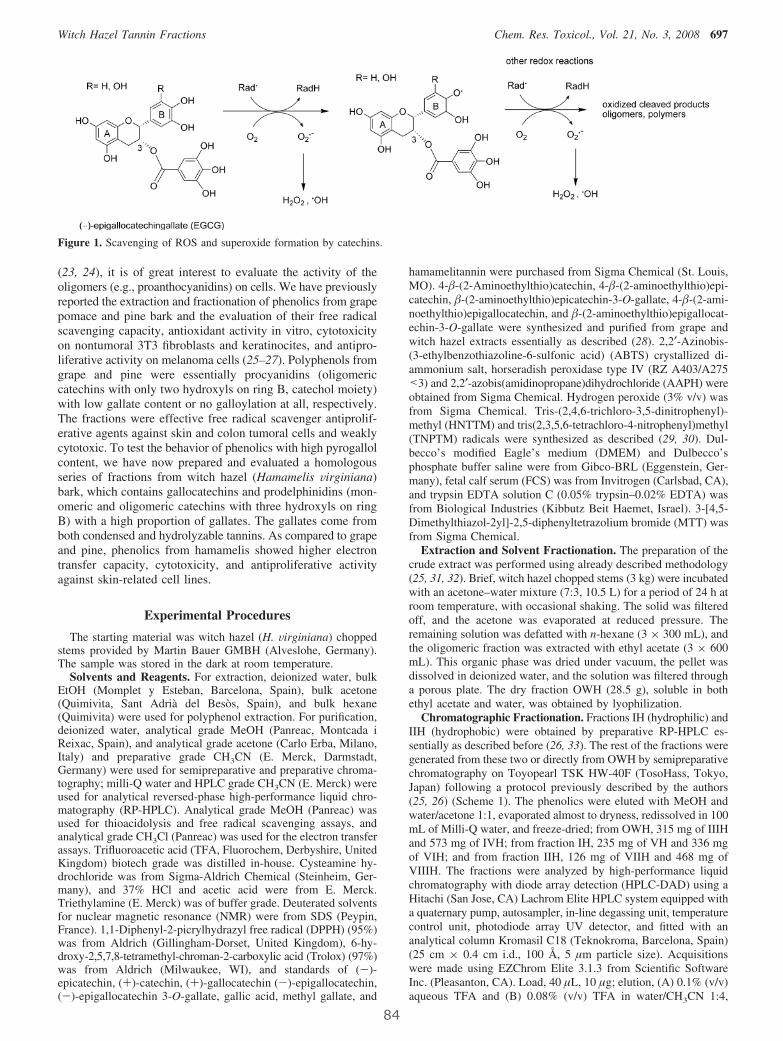
(23, 24), it is of great interest to evaluate the activity of theoligomers (e.g., proanthocyanidins) on cells. We have previouslyreported the extraction and fractionation of phenolics from grapepomace and pine bark and the evaluation of their free radicalscavenging capacity, antioxidant activity in vitro, cytotoxicityon nontumoral 3T3 fibroblasts and keratinocites, and antipro-liferative activity on melanoma cells (25–27). Polyphenols fromgrape and pine were essentially procyanidins (oligomericcatechins with only two hydroxyls on ring B, catechol moiety)with low gallate content or no galloylation at all, respectively.The fractions were effective free radical scavenger antiprolif-erative agents against skin and colon tumoral cells and weaklycytotoxic. To test the behavior of phenolics with high pyrogallolcontent, we have now prepared and evaluated a homologousseries of fractions from witch hazel (Hamamelis Virginiana)bark, which contains gallocatechins and prodelphinidins (mon-omeric and oligomeric catechins with three hydroxyls on ringB) with a high proportion of gallates. The gallates come fromboth condensed and hydrolyzable tannins. As compared to grapeand pine, phenolics from hamamelis showed higher electrontransfer capacity, cytotoxicity, and antiproliferative activityagainst skin-related cell lines.
Experimental Procedures
The starting material was witch hazel (H. Virginiana) choppedstems provided by Martin Bauer GMBH (Alveslohe, Germany).The sample was stored in the dark at room temperature.
Solvents and Reagents. For extraction, deionized water, bulkEtOH (Momplet y Esteban, Barcelona, Spain), bulk acetone(Quimivita, Sant Adrià del Besòs, Spain), and bulk hexane(Quimivita) were used for polyphenol extraction. For purification,deionized water, analytical grade MeOH (Panreac, Montcada iReixac, Spain), and analytical grade acetone (Carlo Erba, Milano,Italy) and preparative grade CH3CN (E. Merck, Darmstadt,Germany) were used for semipreparative and preparative chroma-tography; milli-Q water and HPLC grade CH3CN (E. Merck) wereused for analytical reversed-phase high-performance liquid chro-matography (RP-HPLC). Analytical grade MeOH (Panreac) wasused for thioacidolysis and free radical scavenging assays, andanalytical grade CH3Cl (Panreac) was used for the electron transferassays. Trifluoroacetic acid (TFA, Fluorochem, Derbyshire, UnitedKingdom) biotech grade was distilled in-house. Cysteamine hy-drochloride was from Sigma-Aldrich Chemical (Steinheim, Ger-many), and 37% HCl and acetic acid were from E. Merck.Triethylamine (E. Merck) was of buffer grade. Deuterated solventsfor nuclear magnetic resonance (NMR) were from SDS (Peypin,France). 1,1-Diphenyl-2-picrylhydrazyl free radical (DPPH) (95%)was from Aldrich (Gillingham-Dorset, United Kingdom), 6-hy-droxy-2,5,7,8-tetramethyl-chroman-2-carboxylic acid (Trolox) (97%)was from Aldrich (Milwaukee, WI), and standards of (-)-epicatechin, (+)-catechin, (+)-gallocatechin (-)-epigallocatechin,(-)-epigallocatechin 3-O-gallate, gallic acid, methyl gallate, and
hamamelitannin were purchased from Sigma Chemical (St. Louis,MO). 4-�-(2-Aminoethylthio)catechin, 4-�-(2-aminoethylthio)epi-catechin, �-(2-aminoethylthio)epicatechin-3-O-gallate, 4-�-(2-ami-noethylthio)epigallocatechin, and �-(2-aminoethylthio)epigallocat-echin-3-O-gallate were synthesized and purified from grape andwitch hazel extracts essentially as described (28). 2,2′-Azinobis-(3-ethylbenzothiazoline-6-sulfonic acid) (ABTS) crystallized di-ammonium salt, horseradish peroxidase type IV (RZ A403/A275<3) and 2,2′-azobis(amidinopropane)dihydrochloride (AAPH) wereobtained from Sigma Chemical. Hydrogen peroxide (3% v/v) wasfrom Sigma Chemical. Tris-(2,4,6-trichloro-3,5-dinitrophenyl)-methyl (HNTTM) and tris(2,3,5,6-tetrachloro-4-nitrophenyl)methyl(TNPTM) radicals were synthesized as described (29, 30). Dul-becco’s modified Eagle’s medium (DMEM) and Dulbecco’sphosphate buffer saline were from Gibco-BRL (Eggenstein, Ger-many), fetal calf serum (FCS) was from Invitrogen (Carlsbad, CA),and trypsin EDTA solution C (0.05% trypsin–0.02% EDTA) wasfrom Biological Industries (Kibbutz Beit Haemet, Israel). 3-[4,5-Dimethylthiazol-2yl]-2,5-diphenyltetrazolium bromide (MTT) wasfrom Sigma Chemical.
Extraction and Solvent Fractionation. The preparation of thecrude extract was performed using already described methodology(25, 31, 32). Brief, witch hazel chopped stems (3 kg) were incubatedwith an acetone–water mixture (7:3, 10.5 L) for a period of 24 h atroom temperature, with occasional shaking. The solid was filteredoff, and the acetone was evaporated at reduced pressure. Theremaining solution was defatted with n-hexane (3 × 300 mL), andthe oligomeric fraction was extracted with ethyl acetate (3 × 600mL). This organic phase was dried under vacuum, the pellet wasdissolved in deionized water, and the solution was filtered througha porous plate. The dry fraction OWH (28.5 g), soluble in bothethyl acetate and water, was obtained by lyophilization.
Chromatographic Fractionation. Fractions IH (hydrophilic) andIIH (hydrophobic) were obtained by preparative RP-HPLC es-sentially as described before (26, 33). The rest of the fractions weregenerated from these two or directly from OWH by semipreparativechromatography on Toyopearl TSK HW-40F (TosoHass, Tokyo,Japan) following a protocol previously described by the authors(25, 26) (Scheme 1). The phenolics were eluted with MeOH andwater/acetone 1:1, evaporated almost to dryness, redissolved in 100mL of Milli-Q water, and freeze-dried; from OWH, 315 mg of IIIHand 573 mg of IVH; from fraction IH, 235 mg of VH and 336 mgof VIH; and from fraction IIH, 126 mg of VIIH and 468 mg ofVIIIH. The fractions were analyzed by high-performance liquidchromatography with diode array detection (HPLC-DAD) using aHitachi (San Jose, CA) Lachrom Elite HPLC system equipped witha quaternary pump, autosampler, in-line degassing unit, temperaturecontrol unit, photodiode array UV detector, and fitted with ananalytical column Kromasil C18 (Teknokroma, Barcelona, Spain)(25 cm × 0.4 cm i.d., 100 Å, 5 µm particle size). Acquisitionswere made using EZChrom Elite 3.1.3 from Scientific SoftwareInc. (Pleasanton, CA). Load, 40 µL, 10 µg; elution, (A) 0.1% (v/v)aqueous TFA and (B) 0.08% (v/v) TFA in water/CH3CN 1:4,
Figure 1. Scavenging of ROS and superoxide formation by catechins.
Witch Hazel Tannin Fractions Chem. Res. Toxicol., Vol. 21, No. 3, 2008 697
84

gradient 12-30% B over 30 min at a flow rate of 1 mL/min. DADdetection was performed from 210 to 380 nm. Data were acquiredin triplicate.
Characterization by Thiolysis with Cysteamine and RP-HPLC. The size and composition of the proanthocyanidins withinthe fractions were estimated from the HPLC analysis of acid-catalyzed degradation of proanthocyanidins in the presence ofcysteamine, followed by RP-HPLC as described (34). Briefly, theterminal flavan-3-ols units were released as such by acid cleavagein the presence of cysteamine whereas the extension moietieswere released as the cysteamine derivatives on the fourth positionof the flavanoid system. The resulting mixtures were submitted toanalytical RP-HPLC using the same conditions described abovefor the intact samples, and the molar amount (nanomoles) of all ofthe released moieties was calculated from the peak areas andcalibration curves obtained with pure samples. Terminal units: (+)-gallocatechin (GC), (-)-EGC, (+)-C, (-)-epicatechin (EC), (-)-EGCG, and (-)-ECG; extension units: cysteamine (Cya)-C, Cya-EC, Cya-EGC, Cya-EGCG, and Cya-ECG. Mean degree ofpolymerization (mDP) ) total nmol/nmol terminal units.
Characterization by Chromatography Coupled to MassSpectrometry. Liquid chromatography–mass spectrometry (LC-MS-MS) was used for the identification of gallotannins and gallateswithin the fractions. The analyses were carried out on an Agilent1100 (Waldrom, Germany) coupled to an API 3000 triple quad-rupole mass spectrometer (Perkin-Elmer Sciex, Concord, ON,Canada) at the Parc Cientific de la Universitat de Barcelona. Massscan (MS) and daughter (MS/MS) spectra were measured from m/z100 to 1500. Mass spectrometry data were acquired in the negativeionization mode.
ABTS Radical Cation Decolorization Assay. The method isbased on the capacity of a sample to scavenge the ABTS radicalcation (ABTS•+) as compared to a standard antioxidant (Trolox).ABTS•+was generated from ABTS as described (35) with somemodifications (26). To prepare the initial ABTS•+ solution, 3‰H2O2 (45µL) was added to a reaction mixture containing ABTS(54.9 mg, 1 mM) and horseradish peroxidase (HRP, 1.1 mg, 0.25µM) in 50 mM gly HCl buffer, pH 4.5 (100 mL). The reactionmixture was left to stand at room temperature for 15 min in thedark. Then, the polyphenolic solutions (50 µL) at concentrationsof 0.3, 0.2, 0.15, 0.10, and 0.05 mg/mL in MeOH were added tothe ABTS•+ solution (1950 µL). The total time needed to carryout each assay was 20 min, including ABTS radical generation,addition of antioxidant, and acquisition of final absorbance value.The decrease of absorbance at 734 nm with respect to the 1 mMsolution of ABTS•+ was recorded on a UV spectrophotometer Cary300-Bio (Varian, Palo Alto, CA). The assay was performed in
triplicate. The dose–response curves obtained with the antioxidantmixtures and Trolox were plotted as the percentage of absorbancedecrease against the amount of antioxidants expressed as µg/mL.The total antioxidant activity (TAA) of the fractions was expressedin mmol Trolox equiv/g of OWH.
DPPH Assay. The antiradical efficiency of the fractions wasevaluated by the DPPH stable radical method (36, 37). The samples(0.1 mL) were added to aliquots (3.9 mL) of a solution made upwith DPPH (4.8 mg) in MeOH (200 mL), and the mixture wasincubated for 1 h at room temperature in the dark. The initialconcentration of DPPH, approximately 60 µM, was calculated forevery experiment from a calibration curve made by measuring theabsorbance at 517 nm of standard samples of DPPH at differentconcentrations. The equation of the curve was Abs517nm ) 11345× CDPPH as determined by linear regression. The results wereplotted as the percentage of absorbance disappearance at 517 nm[(1 - A/A0) × 100] against the amount of sample divided by theinitial concentration of DPPH. Each point was acquired in triplicate.A dose–response curve was obtained for every fraction. ED50
corresponds to micrograms of fraction able to consume half theamount of free radical divided by micromoles of initial DPPH. Theresults were expressed as antiradical capacity (ARC), which isthe inverse of ED50. UV measurements were made on a UVspectrophotometer Cary 300-Bio (Varian).
Electron Transfer Capacity against HNTTM and TNPTM.The fractions were dissolved in CH3Cl/MeOH (2:1) at differentconcentrations. Aliquots (1 mL) were added to a solution (1 mL)of HNTTM [120 µM in CH3Cl/MeOH (2:1)] (29), and the mixturewas incubated for 7 h. The exact initial concentration of radical,around 60 µM, was calculated for every experiment from calibrationcurves made by measuring the absorbance (A0) at 385 nm ofstandard samples of the radical at different concentrations. Theequations of the curve was A0 ) 21170 × Cradical. The results wereplotted as the percentage of absorbance disappearance [(1 - A/A0)× 100] against the amount of sample divided by the initialmicromoles of the radical as described for DPPH. Each point wasacquired in triplicate. A dose–response curve was obtained forevery fraction. The results were expressed as the efficient doseED50 given as micromoles of fraction able to consume half theamount of free radical divided by micromoles of initial HNTTM.
The working conditions with TNPTM were essentially thosedescribed for HNTTM with some differences. The incubationtime was 48 h, and the absorbance was measured at 378 nm. Theequation for the calibration curve was A0 ) 17153 × Cradical. Theresults were plotted as described for HNTTM. UV measurementswere made on a UV spectrophotometer Cary 300-Bio (Varian). Asolution of pyrogallol [60 µM in CH3Cl/MeOH (2:1)] was stablefor 48 h as ascertained by RP-HPLC under the elution conditionsdescribed before for the analysis of the fractions.
Antioxidant Activity on Red Blood Cells by the AAPHAssay. Blood samples were obtained from healthy donors byvenipuncture (Blood Bank of Hospital Vall d’Ebron, Barcelona,Spain) following the ethical guidelines of the hospital and collectedin citrated tubes. Red blood cells (RBCs) were separated fromplasma and buffy coat by centrifugation at 1000g for 10 min. Theerythrocyte layer was washed three times in phosphate bufferisotonic saline (PBS) containing 22.2 mM Na2HPO4, 5.6 mMKH2PO4, 123.3 mM NaCl, and 10.0 mM glucose in distilled water(pH 7.4). The cells were then suspended in isotonic saline solutionat a density of 8 × 10⁹ cells/mL. We measured the hemolysis ofRBCs mediated by AAPH using a modification of the methoddescribed previously (38). The addition of AAPH (a peroxyl radicalinitiator) to the suspension of RBCs induces the oxidation of cellmembrane lipids and proteins, thereby resulting in hemolysis. Theerythrocyte suspension (250 µL) was incubated in the presence ofAAPH at a final concentration of 100 mM for 150 min at 37 °C toachieve 100% hemolysis. Hemolysis was assessed by measuringthe absorbance of the supernatant fraction, that is, the hemoglobinrelease, at 540 nm in a Shimadzu spectrophotometer (Shimadzu,Japan). The antihemolytic activity of the fractions was studied byadding the compounds at several concentrations (10-150 µg/mL)
Scheme 1
698 Chem. Res. Toxicol., Vol. 21, No. 3, 2008 Touriño et al.
85
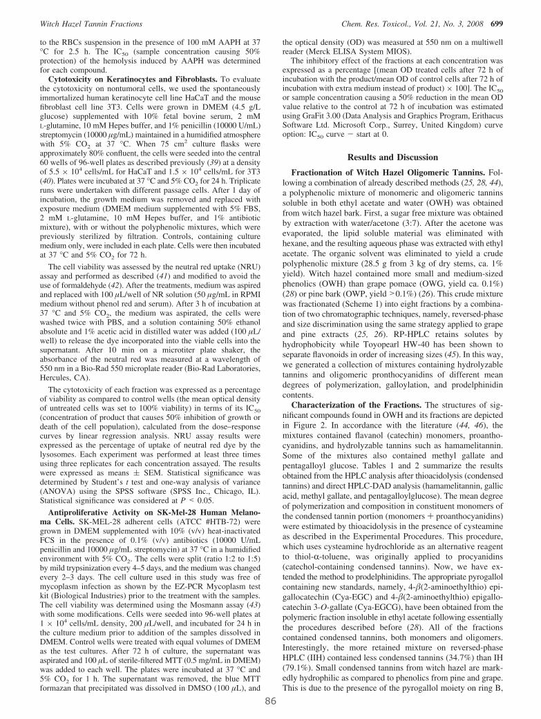
to the RBCs suspension in the presence of 100 mM AAPH at 37°C for 2.5 h. The IC50 (sample concentration causing 50%protection) of the hemolysis induced by AAPH was determinedfor each compound.
Cytotoxicity on Keratinocytes and Fibroblasts. To evaluatethe cytotoxicity on nontumoral cells, we used the spontaneouslyimmortalized human keratinocyte cell line HaCaT and the mousefibroblast cell line 3T3. Cells were grown in DMEM (4.5 g/Lglucose) supplemented with 10% fetal bovine serum, 2 mML-glutamine, 10 mM Hepes buffer, and 1% penicillin (10000 U/mL)streptomycin (10000 µg/mL) maintained in a humidified atmospherewith 5% CO2 at 37 °C. When 75 cm2 culture flasks wereapproximately 80% confluent, the cells were seeded into the central60 wells of 96-well plates as described previously (39) at a densityof 5.5 × 10⁴ cells/mL for HaCaT and 1.5 × 10⁴ cells/mL for 3T3(40). Plates were incubated at 37 °C and 5% CO2 for 24 h. Triplicateruns were undertaken with different passage cells. After 1 day ofincubation, the growth medium was removed and replaced withexposure medium (DMEM medium supplemented with 5% FBS,2 mM L-glutamine, 10 mM Hepes buffer, and 1% antibioticmixture), with or without the polyphenolic mixtures, which werepreviously sterilized by filtration. Controls, containing culturemedium only, were included in each plate. Cells were then incubatedat 37 °C and 5% CO2 for 72 h.
The cell viability was assessed by the neutral red uptake (NRU)assay and performed as described (41) and modified to avoid theuse of formaldehyde (42). After the treatments, medium was aspiredand replaced with 100 µL/well of NR solution (50 µg/mL in RPMImedium without phenol red and serum). After 3 h of incubation at37 °C and 5% CO2, the medium was aspirated, the cells werewashed twice with PBS, and a solution containing 50% ethanolabsolute and 1% acetic acid in distilled water was added (100 µL/well) to release the dye incorporated into the viable cells into thesupernatant. After 10 min on a microtiter plate shaker, theabsorbance of the neutral red was measured at a wavelength of550 nm in a Bio-Rad 550 microplate reader (Bio-Rad Laboratories,Hercules, CA).
The cytotoxicity of each fraction was expressed as a percentageof viability as compared to control wells (the mean optical densityof untreated cells was set to 100% viability) in terms of its IC50
(concentration of product that causes 50% inhibition of growth ordeath of the cell population), calculated from the dose–responsecurves by linear regression analysis. NRU assay results wereexpressed as the percentage of uptake of neutral red dye by thelysosomes. Each experiment was performed at least three timesusing three replicates for each concentration assayed. The resultswere expressed as means ( SEM. Statistical significance wasdetermined by Student’s t test and one-way analysis of variance(ANOVA) using the SPSS software (SPSS Inc., Chicago, IL).Statistical significance was considered at P < 0.05.
Antiproliferative Activity on SK-Mel-28 Human Melano-ma Cells. SK-MEL-28 adherent cells (ATCC #HTB-72) weregrown in DMEM supplemented with 10% (v/v) heat-inactivatedFCS in the presence of 0.1% (v/v) antibiotics (10000 U/mLpenicillin and 10000 µg/mL streptomycin) at 37 °C in a humidifiedenvironment with 5% CO2. The cells were split (ratio 1:2 to 1:5)by mild trypsinization every 4–5 days, and the medium was changedevery 2–3 days. The cell culture used in this study was free ofmycoplasm infection as shown by the EZ-PCR Mycoplasm testkit (Biological Industries) prior to the treatment with the samples.The cell viability was determined using the Mosmann assay (43)with some modifications. Cells were seeded into 96-well plates at1 × 104 cells/mL density, 200 µL/well, and incubated for 24 h inthe culture medium prior to addition of the samples dissolved inDMEM. Control wells were treated with equal volumes of DMEMas the test cultures. After 72 h of culture, the supernatant wasaspirated and 100 µL of sterile-filtered MTT (0.5 mg/mL in DMEM)was added to each well. The plates were incubated at 37 °C and5% CO2 for 1 h. The supernatant was removed, the blue MTTformazan that precipitated was dissolved in DMSO (100 µL), and
the optical density (OD) was measured at 550 nm on a multiwellreader (Merck ELISA System MIOS).
The inhibitory effect of the fractions at each concentration wasexpressed as a percentage [(mean OD treated cells after 72 h ofincubation with the product/mean OD of control cells after 72 h ofincubation with extra medium instead of product) × 100]. The IC50
or sample concentration causing a 50% reduction in the mean ODvalue relative to the control at 72 h of incubation was estimatedusing GraFit 3.00 (Data Analysis and Graphics Program, ErithacusSoftware Ltd. Microsoft Corp., Surrey, United Kingdom) curveoption: IC50 curve - start at 0.
Results and Discussion
Fractionation of Witch Hazel Oligomeric Tannins. Fol-lowing a combination of already described methods (25, 28, 44),a polyphenolic mixture of monomeric and oligomeric tanninssoluble in both ethyl acetate and water (OWH) was obtainedfrom witch hazel bark. First, a sugar free mixture was obtainedby extraction with water/acetone (3:7). After the acetone wasevaporated, the lipid soluble material was eliminated withhexane, and the resulting aqueous phase was extracted with ethylacetate. The organic solvent was eliminated to yield a crudepolyphenolic mixture (28.5 g from 3 kg of dry stems, ca. 1%yield). Witch hazel contained more small and medium-sizedphenolics (OWH) than grape pomace (OWG, yield ca. 0.1%)(28) or pine bark (OWP, yield >0.1%) (26). This crude mixturewas fractionated (Scheme 1) into eight fractions by a combina-tion of two chromatographic techniques, namely, reversed-phaseand size discrimination using the same strategy applied to grapeand pine extracts (25, 26). RP-HPLC retains solutes byhydrophobicity while Toyopearl HW-40 has been shown toseparate flavonoids in order of increasing sizes (45). In this way,we generated a collection of mixtures containing hydrolyzabletannins and oligomeric pronthocyanidins of different meandegrees of polymerization, galloylation, and prodelphinidincontents.
Characterization of the Fractions. The structures of sig-nificant compounds found in OWH and its fractions are depictedin Figure 2. In accordance with the literature (44, 46), themixtures contained flavanol (catechin) monomers, proantho-cyanidins, and hydrolyzable tannins such as hamamelitannin.Some of the mixtures also contained methyl gallate andpentagalloyl glucose. Tables 1 and 2 summarize the resultsobtained from the HPLC analysis after thioacidolysis (condensedtannins) and direct HPLC-DAD analysis (hamamelitannin, gallicacid, methyl gallate, and pentagalloylglucose). The mean degreeof polymerization and composition in constituent monomers ofthe condensed tannin portion (monomers + proanthocyanidins)were estimated by thioacidolysis in the presence of cysteamineas described in the Experimental Procedures. This procedure,which uses cysteamine hydrochloride as an alternative reagentto thiol-R-toluene, was originally applied to procyanidins(catechol-containing condensed tannins). Now, we have ex-tended the method to prodelphinidins. The appropriate pyrogallolcontaining new standards, namely, 4-�(2-aminoethylthio) epi-gallocatechin (Cya-EGC) and 4-�(2-aminoethylthio) epigallo-catechin 3-O-gallate (Cya-EGCG), have been obtained from thepolymeric fraction insoluble in ethyl acetate following essentiallythe procedures described before (28). All of the fractionscontained condensed tannins, both monomers and oligomers.Interestingly, the more retained mixture on reversed-phaseHPLC (IIH) contained less condensed tannins (34.7%) than IH(79.1%). Small condensed tannins from witch hazel are mark-edly hydrophilic as compared to phenolics from pine and grape.This is due to the presence of the pyrogallol moiety on ring B,
Witch Hazel Tannin Fractions Chem. Res. Toxicol., Vol. 21, No. 3, 2008 699
86
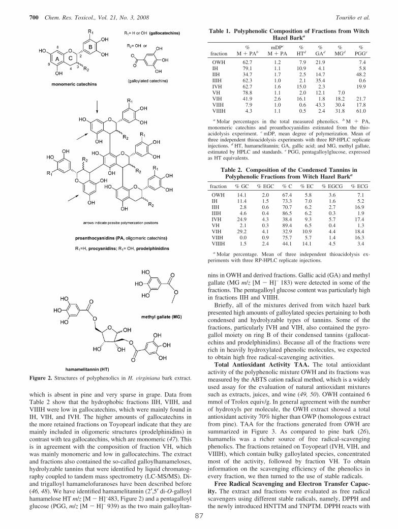
which is absent in pine and very sparse in grape. Data fromTable 2 show that the hydrophobic fractions IIH, VIIH, andVIIIH were low in gallocatechins, which were mainly found inIH, VIH, and IVH. The higher amounts of gallocatechins inthe more retained fractions on Toyopearl indicate that they aremainly included in oligomeric structures (prodelphinidins) incontrast with tea gallocatechins, which are monomeric (47). Thisis in agreement with the composition of fraction VH, whichwas mainly monomeric and low in gallocatechins. The extractand fractions also contained the so-called galloylhamameloses,hydrolyzable tannins that were identified by liquid chromatog-raphy coupled to tandem mass spectrometry (LC-MS/MS). Di-and trigalloyl hamamelofuranoses have been described before(46, 48). We have identified hamamelitannin (2′,5′ di-O-galloylhamamelose HT m/z [M - H]–483, Figure 2) and a pentagalloylglucose (PGG, m/z [M - H]– 939) as the two main galloyltan-
nins in OWH and derived fractions. Gallic acid (GA) and methylgallate (MG m/z [M - H]– 183) were detected in some of thefractions. The pentagalloyl glucose content was particularly highin fractions IIH and VIIIH.
Briefly, all of the mixtures derived from witch hazel barkpresented high amounts of galloylated species pertaining to bothcondensed and hydrolyzable types of tannins. Some of thefractions, particularly IVH and VIH, also contained the pyro-gallol moiety on ring B of their condensed tannins (gallocat-echins and prodelphinidins). Because all of the fractions wererich in heavily hydroxylated phenolic molecules, we expectedto obtain high free radical-scavenging activities.
Total Antioxidant Activity TAA. The total antioxidantactivity of the polyphenolic mixture OWH and its fractions wasmeasured by the ABTS cation radical method, which is a widelyused assay for the evaluation of natural antioxidant mixturessuch as extracts, juices, and wine (49, 50). OWH contained 6mmol of Trolox equiv/g. In general agreement with the numberof hydroxyls per molecule, the OWH extract showed a totalantioxidant activity 70% higher than OWP (homologous extractfrom pine). TAA for the fractions generated from OWH aresummarized in Figure 3. As compared to pine bark (26),hamamelis was a richer source of free radical-scavengingphenolics. The fractions retained on Toyopearl (IVH, VIH, andVIIIH), which contain bulky galloylated species, concentratedmost of the activity, followed by fraction VH. To obtaininformation on the scavenging efficiency of the phenolics inevery fraction, we then turned to the use of stable radicals.
Free Radical Scavenging and Electron Transfer Capac-ity. The extract and fractions were evaluated as free radicalscavengers using different stable radicals, namely, DPPH andthe newly introduced HNTTM and TNPTM. DPPH reacts with
Figure 2. Structures of polyphenolics in H. Virginiana bark extract.
Table 1. Polyphenolic Composition of Fractions from WitchHazel Barka
fraction%
M + PAbmDPc
M + PA%
HTd%
GAd%
MGd%
PGGe
OWH 62.7 1.2 7.9 21.9 7.4IH 79.1 1.1 10.9 4.1 5.8IIH 34.7 1.7 2.5 14.7 48.2IIIH 62.3 1.0 2.1 35.4 0.6IVH 62.7 1.6 15.0 2.3 19.9VH 78.8 1.1 2.0 12.1 7.0VIH 41.9 2.6 16.1 1.8 18.2 21.7VIIH 7.9 1.0 0.6 43.3 30.4 17.8VIIIH 4.3 1.1 0.5 2.4 31.8 61.0
a Molar percentages in the total measured phenolics. b M + PA,monomeric catechins and proanthocyanidins estimated from the thio-acidolysis experiment. c mDP, mean degree of polymerization. Mean ofthree independent thioacidolysis experiments with three RP-HPLC replicateinjections. d HT, hamamelitannin; GA, gallic acid; and MG, methyl gallate,estimated by HPLC and standards. e PGG, pentagalloylglucose, expressedas HT equivalents.
Table 2. Composition of the Condensed Tannins inPolyphenolic Fractions from Witch Hazel Barka
fraction % GC % EGC % C % EC % EGCG % ECG
OWH 14.1 2.0 67.4 5.8 3.6 7.1IH 11.4 1.5 73.3 7.0 1.6 5.2IIH 2.8 0.6 70.7 6.2 2.7 16.9IIIH 4.6 0.4 86.5 6.2 0.3 1.9IVH 24.9 4.3 38.4 9.3 5.7 17.4VH 2.1 0.3 89.4 6.5 0.4 1.3VIH 29.2 4.1 32.9 10.9 4.4 18.4VIIH 0.0 0.9 75.7 5.7 1.4 16.3VIIIH 1.5 2.4 44.1 14.1 4.5 3.4
a Molar percentage. Mean of three independent thioacidolysis ex-periments with three RP-HPLC replicate injections.
700 Chem. Res. Toxicol., Vol. 21, No. 3, 2008 Touriño et al.
87
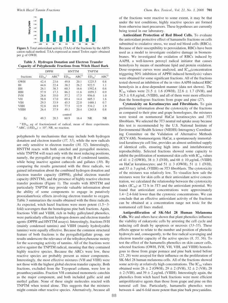
polyphenols by mechanisms that may include both hydrogendonation and electron transfer (37, 51), while the new radicalsare only sensitive to electron transfer (30, 52). Interestingly,HNTTM reacts with both catechol and pyrogallol moieties,while TNPTM will react only with the most reducing positions,namely, the pyrogallol group on ring B of condensed tannins,while being inactive against cathecols and gallates (30). Bycomparing the results generated with the three radicals, wegained information about the combined hydrogen donation andelectron transfer capacity (DPPH), global electron transfercapacity (HNTTM), and the presence of highly reactive electrontransfer positions (Figure 1). The results with HNTTM andparticularly TNPTM may provide valuable information aboutthe ability of some components to engage in putativelyprooxidant/toxic effects involving electron transfer to oxygen.Table 3 summarizes the results obtained with the three radicals.As expected, witch hazel fractions were more potent (1.5-3-fold) scavengers than the homologous pine bark fractions. Again,fractions VIH and VIIIH, rich in bulky galloylated phenolics,were particularly efficient hydrogen donors and electron transferagents (DPPH and HNTTM assays). Interestingly, fractions IVH(mainly condensed tannins) and VIIIH (mainly hydrolyzabletannins) were equally effective. Because the common structuralfeature of both fractions is the pyrogallol/gallate group, ourresults underscore the relevance of the trihydroxybenzene moietyfor the scavenging activity of tannins. All of the fractions wereactive against the TNPTM radical, meaning that they containedhighly reactive species. Because the ARCs were low, thesereactive species are probably present as minor components.Interestingly, the most effective mixtures (VH and VIIH) werenot those with the highest global electron transfer capacity. Bothfractions, excluded from the Toyopearl column, were low inproanthocyanidins. Fraction VH contained monomeric catechinas the major component, and both included gallic acid andmethyl gallate. None of these single molecules reacted withTNPTM when tested alone. This suggests that the mixturesmight contain other reactive species. Alternatively, because all
of the fractions were reactive to some extent, it may be thatunder the test conditions, highly reactive species are formedfrom otherwise inert precursors. These hypotheses are currentlybeing tested in our laboratory.
Antioxidant Protection of Red Blood Cells. To evaluatethe antioxidant protective effect of hamamelis fractions on cellssubmitted to oxidative stress, we used red blood cells (RBCs).Because of their susceptibility to peroxidation, RBCs have beenused as a model to investigate oxidative damage in biomem-branes. We investigated the oxidation of RBCs induced byAAPH, a well-known peroxyl radical initiator that causeshemolysis by means of membrane lipid and protein oxidation.Dose–response curves were analyzed, and IC50(concentrationtriggering 50% inhibition of APPH induced hemolysis) valueswere obtained for some significant fractions. All of the fractionstested showed an inhibition of the in vitro AAPH-induced RBChemolysis in a dose-dependent manner (data not shown). TheIC50 values were 21.5 ( 1.6 (OWH), 22.6 ( 1.7 (IVH), and24.5 ( 0.8 µg/mL (VIIIH), and all of them were more effectivethan the homologous fractions from grape and pine (27).
Cytotoxicity on Keratinocytes and Fibroblasts. To gainpreliminary information about the cytotoxicity of the fractionsas compared to their pine and grape homologues, the mixtureswere tested on nontumoral HaCat keratinocytes and 3T3fibroblasts. We selected the 3T3 neutral red uptake assay becausethis test is recommended by the U.S. National Institute ofEnvironmental Health Science (NIEHS) Interagency Coordinat-ing Committee on the Validation of Alternative Methods(ICCVAM). Nontumorigenic HaCat, a spontaneously immortal-ized keratinocyte cell line, provides an almost unlimited supplyof identical cells, ensuring high intra- and interlaboratoryreproducibility. Selected fractions showed some capacity toinhibit the proliferation of nontumoral skin cells with IC50 valuesof 41 ( 2 (OWH), 38 ( 3 (IVH), and 68 ( 10 µg/mL (VIIIH)on HaCat keratinocytes; and 51 ( 3 (OWH), 51 ( 1 (IVH),and 33 ( 3 µg/mL (VIIIH) on 3T3 fibroblasts. The cytotoxicityof the mixtures was relatively low. To visualize how safe themixtures were for skin cells at their antioxidant active concen-tration, we calculated the relationship between the cytotoxicityindex (IC50) at 72 h in 3T3 and the antioxidant potential. Wefound that antioxidant concentrations were approximately1.4-2.4-fold lower than the cytotoxic concentrations. We canconclude that an effective antioxidant activity of the fractionscan be obtained at a concentration range not toxic for thenontumoral cell lines studied.
Antiproliferation of SK-Mel 28 Human MelanomaCells. We and others have shown that plant phenolics influencethe viability of eukaryotic cells by arresting the cell cycle andinducing cell death by apoptosis or necrosis (53–55). Theseeffects appear to relate to the number and position of phenolichydroxyls and, consequently, to the free radical scavenging andelectron transfer capacity of the active species (8, 55, 56). Totest the effect of the hamamelis phenolics on skin cancer cells,selected fractions (OWH, IVH, VH, VIH, and VIIIH) homolo-gous to those from grape pomace and pine bark tested before(25, 26) were assayed for their influence on the proliferation ofSK-Mel 28 human melanoma cells. All of the fractions showedsome activity at relatively high concentrations. The IC50 valuesobtained were 26 ( 2 (OWH), 29 ( 2 (IVH), 32 ( 2 (VH), 28( 2 (VIH), and 39 ( 2 µg/mL (VIIIH). Interestingly again, thephenolics from witch hazel bark fractions were more efficientantiproliferative agents than those from grape and pine on thistumoral cell line. Particularly, hamamelis phenolics werebetween 4- and 6-fold more potent than pine bark procyanidins.
Figure 3. Total antioxidant activity (TAA) of the fractions by the ABTScation radical method. TAA expressed as mmol Trolox equiv obtainedper g of OWH.
Table 3. Hydrogen Donation and Electron TransferCapacity of Polyphenolic Fractions from Witch Hazel Bark
DPPH HNTTM TNPTM
fractions ED50a ARCb ED50 ARCb ED50
a ARCb
OWH 42.4 23.6 49.8 20.1 1225.5 0.8IH 44.6 22.4 38.2 26.2 922.5 1.1IIH 26.1 38.3 60.3 16.6 1592.4 0.6IIIH 57.9 17.3 86.2 11.6 1059.3 0.9IVH 28.6 35.0 57.2 17.5 956.0 1.0VH 58.8 17.0 69.4 14.4 605.3 1.6VIH 29.5 33.9 45.5 22.0 1488.1 0.7VIIH 52.8 18.9 77.5 12.9 534.2 1.9VIIIH 26.4 37.9 49.1 20.4 1265.8 0.8
controlEc 49.3 20.3 60.9 16.4 NR NR
a ED50 µg of fraction/µmol of radical, mean of three experiments.b ARC, (1/ED50) × 103; NR, no reaction.
Witch Hazel Tannin Fractions Chem. Res. Toxicol., Vol. 21, No. 3, 2008 701
88
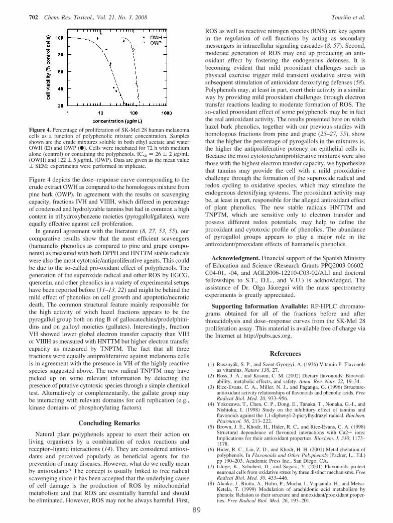
Figure 4 depicts the dose–response curve corresponding to thecrude extract OWH as compared to the homologous mixture frompine bark (OWP). In agreement with the results on scavengingcapacity, fractions IVH and VIIIH, which differed in percentageof condensed and hydrolyzable tannins but had in common a highcontent in trihydroxybenzene moieties (pyrogallol/gallates), wereequally effective against cell proliferation.
In general agreement with the literature (8, 27, 53, 55), ourcomparative results show that the most efficient scavengers(hamamelis phenolics as compared to pine and grape compo-nents) as measured with both DPPH and HNTTM stable radicalswere also the most cytotoxic/antiproliferative agents. This couldbe due to the so-called pro-oxidant effect of polyphenols. Thegeneration of the superoxide radical and other ROS by EGCG,quercetin, and other phenolics in a variety of experimental setupshave been reported before (11–13, 22) and might be behind themild effect of phenolics on cell growth and apoptotic/necroticdeath. The common structural feature mainly responsible forthe high activity of witch hazel fractions appears to be thepyrogallol group both on ring B of gallocatechins/prodelphini-dins and on galloyl moieties (gallates). Interestingly, fractionVH showed lower global electron transfer capacity than VIHor VIIIH as measured with HNTTM but higher electron transfercapacity as measured by TNPTM. The fact that all threefractions were equally antiproliferative against melanoma cellsis in agreement with the presence in VH of the highly reactivespecies suggested above. The new radical TNPTM may havepicked up on some relevant information by detecting thepresence of putative cytotoxic species through a simple chemicaltest. Alternatively or complementarily, the gallate group maybe interacting with relevant domains for cell replication (e.g.,kinase domains of phosphorylating factors).
Concluding Remarks
Natural plant polyphenols appear to exert their action onliving organisms by a combination of redox reactions andreceptor–ligand interactions (14). They are considered antioxi-dants and perceived popularly as beneficial agents for theprevention of many diseases. However, what do we really meanby antioxidants? The concept is usually linked to free radicalscavenging since it has been accepted that the underlying causeof cell damage is the production of ROS by mitochondrialmetabolism and that ROS are essentially harmful and shouldbe eliminated. However, ROS may not be always harmful. First,
ROS as well as reactive nitrogen species (RNS) are key agentsin the regulation of cell functions by acting as secondarymessengers in intracellular signaling cascades (8, 57). Second,moderate generation of ROS may end up producing an anti-oxidant effect by fostering the endogenous defenses. It isbecoming evident that mild prooxidant challenges such asphysical exercise trigger mild transient oxidative stress withsubsequent stimulation of antioxidant detoxifying defenses (58).Polyphenols may, at least in part, exert their activity in a similarway by providing mild prooxidant challenges through electrontransfer reactions leading to moderate formation of ROS. Theso-called prooxidant effect of some polyphenols may be in factthe real antioxidant activity. The results presented here on witchhazel bark phenolics, together with our previous studies withhomologous fractions from pine and grape (25–27, 55), showthat the higher the percentage of pyrogallols in the mixtures is,the higher the antiproliferative potency on epithelial cells is.Because the most cytotoxic/antiproliferative mixtures were alsothose with the highest electron transfer capacity, we hypothesizethat tannins may provide the cell with a mild prooxidativechallenge through the formation of the superoxide radical andredox cycling to oxidative species, which may stimulate theendogenous detoxifying systems. The prooxidant activity maybe, at least in part, responsible for the alleged antioxidant effectof plant phenolics. The new stable radicals HNTTM andTNPTM, which are sensitive only to electron transfer andpossess different redox potentials, may help to define theprooxidant and cytotoxic profile of phenolics. The abundanceof pyrogallol groups appears to play a major role in theantioxidant/prooxidant effects of hamamelis phenolics.
Acknowledgment. Financial support of the Spanish Ministryof Education and Science (Research Grants PPQ2003-06602-C04-01, -04, and AGL2006-12210-C03-02/ALI and doctoralfellowships to S.T., D.L., and V.U.) is acknowledged. Theassistance of Dr. Olga Jáuregui with the mass spectrometryexperiments is greatly appreciated.
Supporting Information Available: RP-HPLC chromato-grams obtained for all of the fractions before and afterthioacidolysis and dose–response curves from the SK-Mel 28proliferation assay. This material is available free of charge viathe Internet at http://pubs.acs.org.
References
(1) Rusznyák, S. P., and Szent-Györgyi, A. (1936) Vitamin P: Flavonolsas vitamins. Nature 138, 27.
(2) Ross, J. A., and Kasum, C. M. (2002) Dietary flavonoids: Bioavail-ability, metabolic effects, and safety. Annu. ReV. Nutr. 22, 19–34.
(3) Rice-Evans, C. A., Miller, N. J., and Paganga, G. (1996) Structure-antioxidant activity relationships of flavonoids and phenolic acids. FreeRadical Biol. Med. 20, 933–956.
(4) Yokozawa, T., Chen, C. P., Dong, E., Tanaka, T., Nonaka, G.-I., andNishioka, I. (1998) Study on the inhibitory effect of tannins andflavonoids against the 1,1-diphenyl-2-picrylhydrazyl radical. Biochem.Pharmacol. 56, 213–222.
(5) Brown, J. E., Khodr, H., Hider, R. C., and Rice-Evans, C. A. (1998)Structural dependence of flavonoid interactions with Cu2+ ions:Implications for their antioxidant properties. Biochem. J. 330, 1173–1178.
(6) Hider, R. C., Liu, Z. D., and Khodr, H. H. (2001) Metal chelation ofpolyphenols. In FlaVonoids and Other Polyphenols (Packer, L., Ed.)pp 190–203, Academic Press Inc., San Diego, CA.
(7) Ishige, K., Schubert, D., and Sagara, Y. (2001) Flavonoids protectneuronal cells from oxidative stress by three distinct mechanisms. FreeRadical Biol. Med. 30, 433–446.
(8) Alanko, J., Riutta, A., Holm, P., Mucha, I., Vapaatalo, H., and Metsa-Ketela, T. (1999) Modulation of arachidonic acid metabolism byphenols: Relation to their structure and antioxidant/prooxidant proper-ties. Free Radical Biol. Med. 26, 193–201.
Figure 4. Percentage of proliferation of SK-Mel 28 human melanomacells as a function of polyphenolic mixture concentration. Samplesshown are the crude mixtures soluble in both ethyl acetate and waterOWH (0) and OWP (b). Cells were incubated for 72 h with mediumalone (control) or containing the polyphenols. IC50 ) 26 ( 2 µg/mL(OWH) and 122 ( 5 µg/mL (OWP). Data are given as the mean value( SEM; experiments were performed in triplicate.
702 Chem. Res. Toxicol., Vol. 21, No. 3, 2008 Touriño et al.
89

(9) Lambert, J. D., Sang, S., and Yang, C. S. (2007) Possible controversyover dietary polyphenols: Benefits vs risks. Chem. Res. Toxicol. 20,583–585.
(10) Kondo, K., Kurihara, M., Miyata, N., Suzuki, T., and Toyoda, M.(1999) Mechanistic studies of catechins as antioxidants against radicaloxidation. ArchiV. Biochem. Biophys. 362, 79–86.
(11) Kondo, K., Kurihara, M., Miyata, N., Suzuki, T., and Toyoda, M.(1999) Scavenging mechanisms of (-)-epigallocatechin gallate and(-)-epicatechin gallate on peroxyl radicals and formation of superoxideduring the inhibitory action. Free Radical Biol. Med. 27, 855–863.
(12) Nutter, L. M., Wu, Y. Y., Ngo, E. O., Sierra, E. E., Gutierrez, P. L.,and Abul-Hajj, Y. J. (1994) An o-quinone form of estrogen producesfree radicals in human breast cancer cells: Correlation with DNAdamage. Chem. Res. Toxicol. 7, 23–28.
(13) Boots, A. W., Li, H., Schins, R. P. F., Duffin, R., Heemskerk, J. W. M.,Bast, A., and Haenen, G. (2007) The quercetin paradox. Toxicol. Appl.Pharmacol. 222, 89–96.
(14) Sang, S., Hou, Z., Lambert, J. D., and Yang, C. S. (2005) Redoxproperties of tea polyphenols and related biological activities. Antioxid.Redox Signaling 7, 1704–1714.
(15) Liang, Y. C., Lin-shiau, S. Y., Chen, C. F., and Lin, J. K. (1997)Suppression of extracellular signals and cell proliferation through EGFreceptor binding by (-)-epigallocatechin gallate in human A431epidermoid carcinoma cells. J. Cell Biochem. 67, 55–65.
(16) Ahmad, N., Cheng, P. Y., and Mukhtar, H. (2000) Cell cycledysregulation by green tea polyphenol epigallocatechin-3-gallate.Biochem. Biophys. Res. Commun. 275, 328–334.
(17) Scalbert, A., Morand, C., Manach, C., and Remesy, C. (2002)Absorption and metabolism of polyphenols in the gut and impact onhealth. Biomed. Pharmacother. 56, 276–282.
(18) Halliwell, B. (2006) Reactive species and antioxidants. Redox biologyis a fundamental theme of aerobic life. Plant Physiol. 141, 312–322.
(19) Linnane, A. W., Kios, M., and Vitetta, L. (2007) Healthy aging:regulation of the metabolome by cellular redox modulation andprooxidant signaling systems: The essential roles of superoxide anionand hydrogen peroxide. Biogerontology 8, 445–467.
(20) Halliwell, B., Zhao, K., and Whiteman, M. (2000) The gastrointestinaltract: A major site of antioxidant action? Free Radical Res. 33, 819–830.
(21) Cross, C. E., van der Vliet, A., Louie, S., Thiele, J. J., and Halliwell,B. (1998) Oxidative stress and antioxidants at biosurfaces: Plants, skin,and respiratory tract surfaces. EnViron. Health Perspect. 106 (Suppl.5), 1241–1251.
(22) Azam, S., Hadi, N., Khan, N. U., and Hadi, S. M. (2004) Prooxidantproperty of green tea polyphenols epicatechin and epigallocatechin-3-gallate: Implications for anticancer properties. Toxicol. in Vitro 18,555–561.
(23) Plumb, G. W., De Pascual-Teresa, S., Santos-Buelga, C., Cheynier,V., and Williamson, G. (1998) Antioxidant properties of catechinsand proanthocyanidins: Effect of polymerization, galloylation andglycosylation. Free Radical Res. 29, 351–358.
(24) Saint-Cricq de Gaulejac, N., Vivas, N., Freitas, V., and Bourgeois,G. (1999) The influence of various phenolic compounds on scavengingactivity assessed by an enzymatic method. J. Sci. Food Agric. 79,1081–1090.
(25) Torres, J. L., Varela, B., García, M. T., Carilla, J., Matito, C., Centelles,J. J., Cascante, C., Sort, X., and Bobet, R. (2002) Valorization of grape(Vitis Vinifera) byproducts. Antioxidant and biological properties ofpolyphenolic fractions differing in procyanidin composition andflavonol content. J. Agric. Food Chem. 50, 7548–7555.
(26) Touriño, S., Selga, A., Jiménez, A., Juliá, L., Lozano, C., Lizárraga,D., Cascante, C., and Torres, J. L. (2005) Procyanidin fractions frompine (Pinus pinaster) bark: Radical scavenging power in solution,antioxidant activity in emulsion and antiproliferative effect in mela-noma cells. J. Agric. Food Chem. 53, 4728–4735.
(27) Ugartondo, V., Mitjans, M., Touriño, S., Torres, J. L., and Vinardell,M. P. (2007) Comparative antioxidant and cytotoxic effect ofprocyanidin fractions from grape and pine. Chem. Res. Toxicol. 20,1543–1548.
(28) Torres, J. L., and Bobet, R. (2001) New flavanol-derivatives from grape(Vitis Vinifera) byproducts. Antioxidant aminoethylthio-flavan-3olconjugates from a polymeric waste fraction used as a source offlavanols. J. Agric. Food Chem. 49, 4627–4634.
(29) Torres, J. L., Varela, B., Brillas, E., and Juliá, L. (2003) Tris(2,4,6Tri-chloro-3,5-dinitrophenyl)methyl radical: A new stable coloured mag-netic species as a chemosensor for natural polyphenols. Chem.Commun. 74–75.
(30) Torres, J. L., Carreras, A., Jiménez, A., Brillas, E., Torrelles, X., Rius,J., and Juliá, L. (2007) Reducing power of simple polyphenols byelectron-transfer reactions using a new stable radical of the PTM series,tris(2,3,5,6-tetrachloro-4-nitrophenyl)methyl radical. J. Org. Chem. 72,3750–3756.
(31) Dauer, A., Metzner, P., and Schimmer, O. (1998) Proanthocyanidinsfrom the bark of Hamamelis Virginiana exibit antimutagenic propertiesagainst nitroaromatic compounds. Planta Med. 64, 324–327.
(32) Deters, A., Dauer, A., Schnetz, E., Fartasch, M., and Hensel, A. (2001)High molecular compounds (polysaccharides and proanthocyanidins)from Hamamelis Virginiana bark: Influence on human skin kerati-nocyte proliferation and differentiation and influence on irritated skin.Phytochemistry 58, 949–958.
(33) Matito, C., Mastorakou, F., Centelles, J. J., Torres, J. L., and Cascante,M. (2003) Antiproliferative effect of antioxidant polyphenols fromgrape in murine Hepa-1c1c7. Eur. J. Nutr. 42, 43–49.
(34) Torres, J. L., and Selga, A. (2003) Procyanidin size and compositionby thiolysis with cysteamine hydrochloride and chromatography.Chromatographia 57, 441–445.
(35) Alcolea, J. F., Cano, A., Acosta, M., and Arnao, M. B. (2002)Hydrophilic and lipofilic antioxidant activities of grapes. Nahrung 46,353–356.
(36) Blois, M. S. (1958) Antioxidant determinations by the use of stablefree radical. Nature 181, 1199–1200.
(37) Brand-Williams, W., Cuvelier, M. E., and Berset, C. (1995) Use of afree radical method to evaluate antioxidant activity. Lebensm.-Wiss.Technol. 28, 25–30.
(38) Miki, M., Tamai, H., Mino, M., Yamamoto, Y., and Niké, E. (1987)Free radical chain oxidation of rat red blood cells by molecular oxygenand its inhibition by R-tocopherol. Arch. Biochem. Biophys. 258, 373–380.
(39) Ugartondo, V., Mitjans, M., Lozano, C., Torres, J. L., and Vinardell,M. P. (2006) Comparative study of the cytotoxicity induced byantioxidant epicatechin conjugates obtained from grape. J. Agric. FoodChem. 54, 6945–6950.
(40) Babich, H., Krupka, M. E., Nissim, H. A., and Zuckerbraun, H. L.(2005) Differential in vitro cytotoxicity of (-)-epicatechin gallate(ECG) to cancer and normal cells from the human oral cavity. Toxicol.in Vitro 19, 231–242.
(41) Borenfreund, E. P., and Rapad, J. (1983) A colorimetric assay tocellular growth and survival: Application to proliferation and cyto-toxicity assay. Toxicol. Lett. 24, 119–124.
(42) Riddell, R. J., Clothier, R. H., and Ball, M. (1986) An evaluation ofthree in vitro cytotoxicity assays. Food Chem. Toxicol. 24, 469–471.
(43) Mosmann, T. (1983) Rapid colorimetric assay for cellular growth andsurvival: Application to proliferation and cytotoxicity assays. J. Im-munol. Methods 55–63.
(44) Dauer, A., Rimpler, H., and Hensel, A. (2003) Polymeric proanthocya-nidins from the bark of Hamamelis Virginiana. Planta Med. 69, 89–91.
(45) Sun, B. S., Belchior, G. P., Ricardo da Silva, J. M., and Spranger,M. I. (1999) Isolation and purification of dimeric and trimericprocyanidins from grape seeds. J. Chromatogr. A 841, 115–121.
(46) Hartisch, C., and Kolodziej, H. (1996) Galloylhamameloses andproanthocyanidins from Hamamelis Virginiana. Phytochemistry 42,191–198.
(47) Harbowy, M. E., and Balentine, D. A. (1997) Tea chemistry. Crit.ReV. Plant Sci. 16, 415–480.
(48) Haberland, C., and Kolodziej, H. (1994) Novel galloylhamamelosesfrom Hamamelis Virginiana. Planta Med. 60, 464–466.
(49) Miller, N. J., and Rice Evans, C. (1997) The relative contributions ofascorbic acid and phenolic antioxidants to the total antioxidant activityof orange and apple fruit juices and blackcurrant drink. Food Chem.60, 331–337.
(50) Pietta, P., Simonetti, P., and Mauri, P. (1998) Antioxidant activity ofselected medicinal plants. J. Agric. Food Chem. 46, 4487–4490.
(51) Dangles, O., Fargeix, G., and Dufour, C. (2000) Antioxidant propertiesof anthocyanins and tannins: A mechanistic investigation with catechinand the 3′,4′,7-trihydroxyflavylium ion. J. Chem. Soc., Perkin Trans.2 1653–1666.
(52) Jiménez, A., Selga, A., Torres, J. L., and Juliá, L. (2004) Reducingactivity of polyphenols with stable radicals of the TTM series. Electrontransfer versus H-abstraction reactions in flavan-3-ols. Org. Lett. 6,4583–4586.
(53) Tan, X. H., Hu, D. R., Li, S. R., Han, Y., Zhang, Y. L., and Zhou,D. Y. (2000) Differences of four catechins in cell cycle arrest andinduction of apoptosis in LoVo cells. Cancer Lett. 158, 1–6.
(54) Kozikowski, A. P., Tuckmantel, W., Bottcher, G., and Romanczyk,L. J., Jr. (2003) Studies in polyphenol chemistry and bioactivity. 4.(1) Synthesis of trimeric, tetrameric, pentameric, and higher oligomericepicatechin-derived procyanidins having all-4�,8-interflavan con-nectivity and their inhibition of cancer cell growth through cell cyclearrest. J. Org. Chem. 68, 1641–1658.
(55) Lizárraga, D., Lozano, C., Briede, J. J., van Delft, J. H., Touriño, S.,Centelles, J. J., Torres, J. L., and Cascante, M. (2007) The importanceof polymerization and galloylation for the antiproliferative propertiesof procyanidin-rich natural extracts. FEBS J. 274, 4802–4811.
Witch Hazel Tannin Fractions Chem. Res. Toxicol., Vol. 21, No. 3, 2008 703
90
(56) Lozano, C., Torres, J. L., Juliá, L., Jiménez, A., Centelles, J. J., andCascante, M. (2005) Effect of new antioxidant cysteinyl-flavanolconjugates on skin cancer cells. FEBS Lett. 579, 4219–4225.
(57) Valko, M., Leibfritz, D., Moncol, J., Cronin, M. T. D., Mazur, M.,and Telser, J. (2007) Free radicals and antioxidants in normalphysiological functions and human disease. Int. J. Biochem. Cell Biol.39, 44–84.
(58) Ascensao, A., Magalhaes, J. F., Soares, J. M., Ferreira, R. M.,Neuparth, M. J., Appell, H. J., and Duarte, J. A. (2005) Cardiacmitochondrial respiratory function and oxidative stress: The role ofexercise. Int. J. Sports Med. 26, 258–267.
TX700425N
704 Chem. Res. Toxicol., Vol. 21, No. 3, 2008 Touriño et al.
91

4.2 Estudio de la absorción/metabolización de
proantocianidinas

4. Resultados
95
4.2.1 PUBLICACIÓN 3: Identificación de los compuestos fenólicos de la
fracción extraíble de fibra antioxidante de uva.
Título original: High-resolution liquid chromatography/electrospray ionization
time-of-flight mass spectrometry combined with liquid chromatography/electrospray ionization tandem mass spectrometry to identify polyphenols from grape antioxidant dietary fiber.
Autores: Sonia Touriño, Elisabet Fuguet, Olga Jáuregui, Fulgencio Saura-
Calixto, Marta Cascante and Josep Lluís Torres. Rapid Communications in Mass Spectrometry, 2008, 22, 3489-3500.
Índice de impacto: 2,971 Clasificación y categoría: 13/ 70 (Química analítica); 9/39 (Espectrometría)
RESUMEN
La industria vitivinícola genera grandes cantidades de subproductos que son una
importante fuente de compuestos de naturaleza fenólica, fibra y otras moléculas
bioactivas. El término de fibra dietética es referido al conjunto de polisacáridos y lignina
de los alimentos vegetales que resisten a la acción de los enzimas digestivos. Las fibras
dietéticas promueven efectos beneficiosos fisiológicos como laxantes y pueden atenuar
los niveles de colesterol y/o glucosa en sangre (Guillon et al., 2000). Debido a que no
son absorbibles en el intestino delgado, cuando estas fibras llegan al colon son
susceptibles de ser degradadas por la microbiota colónica y los productos formados
promueven el crecimiento y la actividad de las bacterias que habitan en el colón, es por
ello que las fibras dietéticas se engloban dentro del término de alimentos prebióticos.
La fibra antioxidante dietética (GADF) patentada por el grupo del Dr. Saura-Calixto
(Saura-Calixto et al., 1999), es un ingrediente que además de combinar los efectos
beneficiosos de una fibra dietética incluye en su composición grandes cantidades de
polifenoles (20%), fundamentalmente proantocianidinas. Algunos de estos de
compuestos fenólicos son absorbidos en el intestino delgado y otros, debido a que están
embebidos en la matriz de lignina, son progresivamente liberados en el colon como
consecuencia de la fermentación bacteriana. GADF presenta buenas condiciones de
fermentabilidad y digestibilidad (Goñi et al., 2005). Particularmente interesante, son los
resultados obtenidos de un estudio realizado recientemente por Pérez-Jiménez y
colaboradores (Pérez-Jiménez et al., 2008) donde se observó que GADF podía disminuir
la presión arterial y el colesterol. La presencia de los compuestos fenólicos como
principios activos en fibras dietéticas parece aumentar los beneficios que éstas ejercen.

4. Resultados
96
La identificación de los compuestos polifenólicos mayoritarios de la fracción extraíble de
la fibra fue el objetivo de este trabajo como primer paso para comprender su
metabolización y la actividad que puedan ejercer en el organismo.
La fracción fenólica extraíble de GADF es una mezcla compleja con una gran cantidad de
compuestos de distintas clases. Para mejorar la identificación de los compuestos
fenólicos se realizó un fraccionamiento mediante técnicas cromatográficas que permitió
separar monómeros, oligómeros de proantocianidinas, antocianinas y polímeros en
cuatro fracciones. Para la identificación de los compuestos de cada una de las fracciones
se utilizaron dos técnicas complementarias. El uso LC/ESI-TOF-MS permitió obtener un
primer barrido general de cada una de las fracciones y determinar las masas exactas de
cada uno de los compuestos. Los experimentos en tándem (barrido del ión precursor,
barrido de perdidas neutras y barrido de iones producto) fueron realizados en un triple
cuadrupolo acoplado a un HPLC (LC/ESI-MS/MS) y posibilitaron la determinación de las
estructuras en base a los fragmentos característicos de cada molécula. La combinación
de ambas técnicas permitió identificar un elevado número de compuestos fenólicos de
diferentes clases.
A continuación se muestra la publicación original.

RAPID COMMUNICATIONS IN MASS SPECTROMETRY
Rapid Commun. Mass Spectrom. 2008; 22: 3489–3500
) DOI: 10.1002/rcm.3756
Published online in Wiley InterScience (www.interscience.wiley.comHigh-resolution liquid chromatography/electrospray
ionization time-of-flight mass spectrometry combined
with liquid chromatography/electrospray ionization
tandem mass spectrometry to identify polyphenols from
grape antioxidant dietary fiber
Sonia Tourino1, Elisabet Fuguet1, Olga Jauregui2, Fulgencio Saura-Calixto3,
Marta Cascante4 and Josep Lluıs Torres1*1Institute for Chemical and Environmental Research (IIQAB-CSIC), Jordi Girona 18–26, E-08034 Barcelona, Spain2Scientific and Technical Services, University of Barcelona, Josep Samitier 1–5, E-08028 Barcelona, Spain3Instituto del Frıo, CSIC. Jose Antonio Novais 10, E-28040 Madrid, Spain4Department of Biochemistry and Molecular Biology, Institute of Biomedicine of the University of Barcelona (IBUB) and Centre for Research in
Theoretical Chemistry, Barcelona Science Park (CeRQT-PCB), Unit Associated with CSIC, Diagonal 645, E-08028 Barcelona, Spain
Received 13 June 2008; Revised 10 September 2008; Accepted 10 September 2008
*CorrespoEnvironmE-08034 BE-mail: jlContract/Science;-03; AGL
Grape antioxidant dietary fiber (GADF) is a dietary supplement that combines the benefits of both
fiber and antioxidants that help prevent cancer and cardiovascular diseases. The antioxidant poly-
phenolic components in GADF probably help prevent cancer in the digestive tract, where they are
bioavailable.Mass spectrometry coupled to liquid chromatography is a powerful tool for the analysis
of complex plant derivatives such as GADF. We use a combination of MS techniques, namely liquid
chromatography/electrospray ionization time-of-flight mass spectrometry (LC/ESI-TOF-MS) and
liquid chromatography/electrospray ionization tandem mass spectrometry (LC/ESI-MS/MS) on a
triple quadrupole, for the identification of the polyphenolic constituents of the soluble fraction of
GADF. First, we separated themixture into four fractions whichwere tested for phenolic constituents
using the TOF system in the full scan mode. The high sensitivity and resolution of the TOF detector
over the triple quadrupole facilitate the preliminary characterization of the fractions. Then we used
LC/ESI-MS/MS to identify the individual phenols through MS/MS experiments (product ion scan,
neutral loss scan, precursor ion scan). Finally, most of the identities were unequivocally confirmed by
accurate mass measurements on the TOF spectrometer. LC/ESI-TOF-MS combined with MS/MS
correctly identifies the bioactive polyphenolic components from the soluble fraction of GADF. High-
resolution TOF-MS is particularly useful for identifying the structure of compounds with the same
LC/ESI-MS/MS fragmentation patterns. Copyright # 2008 John Wiley & Sons, Ltd.
In recent years, there has been increasing interest in the use of
plant byproducts as natural sources of compounds with
useful nutritional and pharmaceutical properties.1 Grapes
(Vitis vinifera) are the world’s largest fruit crop. Approxi-
mately 60 million tons are produced annually, most of which
is used for making wine. This industrial activity generates
huge amounts of byproducts that are rich in fiber,
polyphenols and other bioactive molecules. Dietary fiber
(non-digestible polymers such as non-starch polysacharides
and lignin) is of particular interest owing to its putative
benefits for human health. Fibers have laxative properties
and some of them have been reported to reduce blood
ndence to: J. L. Torres, Institute for Chemical andental Research (IIQAB-CSIC), Jordi Girona 18–26,arcelona, Spain.
[email protected] sponsor: Spanish Ministry of Education and
contract/grant number: AGL2004-07579-C04-01, -02,2006-12210-C03-02/ALI.
97
cholesterol and glucose.2 Moreover, when dietary fiber
reaches the colon, the polysaccharides and other non-
digestible dietary compounds (e.g. resistant starches and
proteins, and high molecular weight polyphenols) are
fermented to some extent by the colonic microbiota. Since
these compounds help to stabilize the number of colonic
bacteria, they are called prebiotics and are believed to
contribute to the health benefits of fiber. The polyphenolic
components of some fibers may add their antioxidant
potential to the putative benefits of the whole preparation.
Polyphenols are powerful free-radical scavengers with
antioxidant activity and antitumor effects.3 The consumption
of polyphenol-rich food and beverages has been associated
with the prevention of diseases, especially different types of
cancer and coronary heart conditions.4
Grape pomace is a particularly interesting source of
dietary fiber because, apart from lignin and polysaccharides,
it contains large amounts of polyphenols. These are
Copyright # 2008 John Wiley & Sons, Ltd.
3490 S. Tourino et al.
associated with the fiber matrix and produce considerable
antioxidant activity. Some of these phenols are absorbed
in the small intestine and the rest reach the colon where,
to some extent, they are hydrolyzed, metabolized and
absorbed.5,6 As a result, an array of bioactive compounds
may become bioavailable at the colonic epithelial tissue and
released into the bloodstream. Thus, the new concept of
grape antioxidant dietary fiber (GADF) has come into being.7
GADF incorporates the advantages of both fiber and phenols
in a single dietary product.
To advance our understanding of the benefits of GADF8 it
is important to thoroughly characterize its putatively active
phenolic components. Mass spectrometry (MS) alone or
coupled to liquid chromatography (LC/MS) has been
increasingly used in the structural characterization of
complex mixtures such as bioactive plant extracts.9–13 LC/
MS with electrospray ionization (LC/ESI-MS) has been
successfully used to identify anthocyanins,14,15 flavonols,12,16
proanthocyanidins (PAs),17,18 pyranoanthocyanins,16,19 and
phenolic acids in wine-related products.19–22 The identifi-
cation of polymeric PAs with a high degree of polymeri-
zation is particularly complicated. The different polymers
cannot be separated through conventional high-performance
liquid chromatography (HPLC) since they elute together as
an unresolved broad peak. ESI-MS and matrix-assisted laser
desorption/ionization time-of-flight mass spectrometry
(MALDI-TOF-MS) are complementary alternatives for char-
acterizing polymeric PAs.23,24 These soft ionization tech-
niques provide mass spectra with little fragmentation,
making it possible to define the distribution of the polymers
around the mean value.18,25–27
The objective of this work was to identify the polyphenols
in the soluble fraction of GADF with the hope of eventually
establishing structure/activity relationships between the
components of GADF on the one hand, and its cancer and
cardiovascular chemopreventive effects on the other.
Because the soluble fraction contains a huge number of
polyphenolic compounds, and to facilitate their identifi-
cation by LC/MS, the mixture was separated into four
fractions by chromatographic methods. To identify the
phenolic compounds, each fraction was analyzed using
two complementary instruments: a high-resolution time-of-
flight mass spectrometer (LC/ESI-TOF-MS), for the first full-
scan acquisitions and to determine accurate masses; and a
triple quadrupole (QqQ) mass spectrometer (LC/ESI-MS/
MS) using several modalities (product ion scan, neutral
loss scan and precursor ion scan) to determine structures
based on fragmentation patterns. Triple quadrupole instru-
ments have well-described MS/MS capabilities that are
extensively used in the identification of phenolic compounds
in natural product samples.28,29 TOF analyzers provide
greatly improved mass resolution compared to the unit
resolution of traditional quadrupole instruments and, in
addition, they offer significantly higher sensitivity and
accuracy when acquiring full-fragment spectra. These
characteristics make them suitable for both initial charac-
terization of complex mixtures and final confirmation of
component identities through the resolution of ambiguities.
Thus we combined TOF and QqQ systems to qualitatively
characterize the phenols in the soluble fraction of GADF.
Copyright # 2008 John Wiley & Sons, Ltd.
9
EXPERIMENTAL
Reagents and materialsGrape antioxidant dietary fiber was obtained from red
grapes (the Cencibel variety), of the vintage year 2005, from
La Mancha region in Spain, as described in published
patents.30,31 For the extraction and fractionation of poly-
phenols, the following (analytical grade) reagents were used:
methanol, ethyl acetate, and hydrochloric acid 37% from
Panreac (Montcada i Reixac, Spain); acetone and diethyl
ether from Carlo Erba (Milano, Italy). Acetonitrile (HPLC
grade) and formic acid (analytical grade) were from Merck
(Darmstadt, Germany). Water was purified by a Milli-Q plus
system (Millipore, Bedford, MA, USA) to a resistivity of
18.2 MVcm.
Standards of (�)-epicatechin (�97%), (þ)-catechin (�98%),
(�)-epicatechin gallate (�98%), 4-hydroxybenzoic acid
(�97%), vanillic acid (�97%), syringic acid (�95%), dehy-
drocaffeic acid (>98%), protocatechuic acid (�97%), p-
coumaric acid (�98%), o-coumaric acid (�97%), and gallic
acid (>99%) were from Sigma Chemical (St. Louis, MO,
USA).
Apparatus and experimental conditionsThe ESI-TOF instrument was a LCT Premier (Waters,
Milford, MA, USA), equipped with a 4 GHz time-to-digital
converter (TDC) combined with an Acquity ultra-perform-
ance liquid chromatography (UPLC) system. The separations
were conducted using a Luna C18(2) 3mm particle size
column (50� 2.1 mm i.d.; Phenomenex, Torrance, CA, USA)
equipped with a Phenomenex Securityguard C18 column
(4� 3 mm i.d.). The mass spectrometer was equipped with
a dual ESI source (LockSpray). The second sprayer provided
the lock mass calibrant leucine enkephalin (m/z 556.2771). It
was operated in the W-optics mode, thus providing a
mass resolution of at least 10 000 full-width at half maximum
(FWHM). The acquisition time per spectrum was set to 0.2 s,
and the mass range was from 100 to 3000 Da. Data were
acquired using a cone voltage of 50 V, capillary voltage of
2800 V, desolvation temperature of 3508C, and source
temperature of 1008C. The desolvation gas flow was set at
400 L/h and the cone gas flow was set at 30 L/h. MassLynx
4.1 software from Waters was used for data acquisition and
processing.
An Agilent series 1100 HPLC instrument (Agilent,
Waldbronn, Germany) equipped with a quaternary
pump, an ultraviolet (UV) detector, an autosampler and a
column oven was used for the HPLC-UV/ESI-MS/MS
experiments. The separations were conducted using the
column described above. The injection volume was 5mL,
the flow rate was 400mL/min, and the temperature 258C. An
API 3000 triple quadrupole mass spectrometer (PE Sciex,
Concord, Ontario, Canada) equipped with a TurboIon spray
source was used to obtain MS/MS data. The settings were:
capillary voltage �3500 V (negative mode) or 5000 V
(positive mode), nebulizer gas (N2) 10 arbitrary units
(a.u.), curtain gas (N2) 12 a.u., collision gas (N2) 10 a.u.,
declustering potential (DP) between�30 and�60 V, focusing
potential �200 V, entrance potential 10 V, and collision
energy (CE) �30 V. The drying gas (N2) was heated to
Rapid Commun. Mass Spectrom. 2008; 22: 3489–3500
DOI: 10.1002/rcm
8
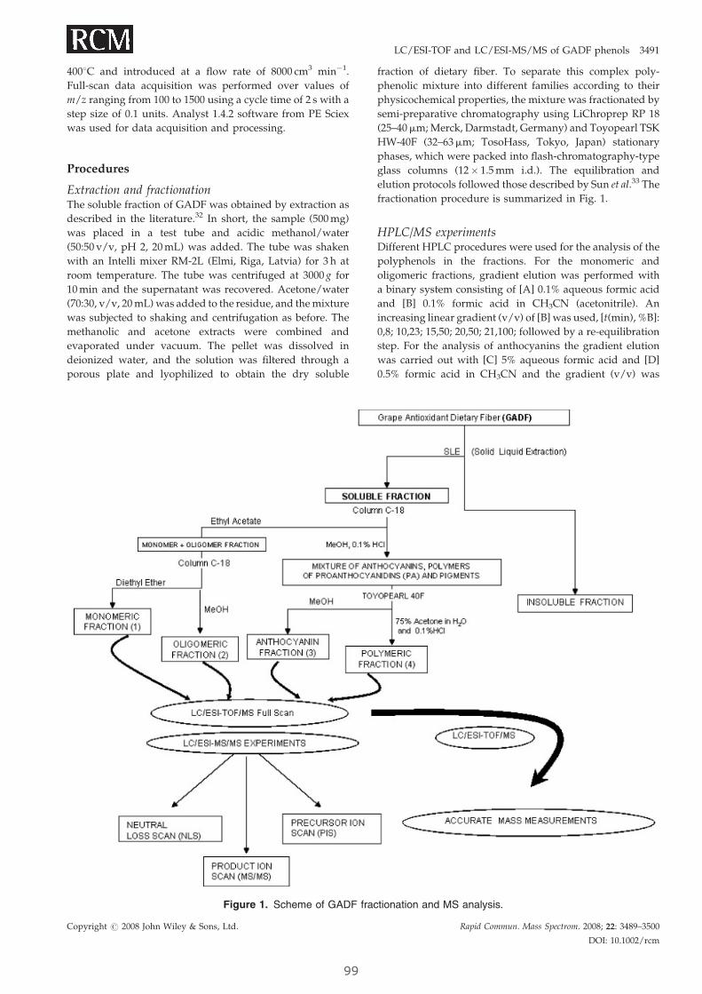
LC/ESI-TOF and LC/ESI-MS/MS of GADF phenols 3491
4008C and introduced at a flow rate of 8000 cm3 min�1.
Full-scan data acquisition was performed over values of
m/z ranging from 100 to 1500 using a cycle time of 2 s with a
step size of 0.1 units. Analyst 1.4.2 software from PE Sciex
was used for data acquisition and processing.
Procedures
Extraction and fractionationThe soluble fraction of GADF was obtained by extraction as
described in the literature.32 In short, the sample (500 mg)
was placed in a test tube and acidic methanol/water
(50:50 v/v, pH 2, 20 mL) was added. The tube was shaken
with an Intelli mixer RM-2L (Elmi, Riga, Latvia) for 3 h at
room temperature. The tube was centrifuged at 3000 g for
10 min and the supernatant was recovered. Acetone/water
(70:30, v/v, 20 mL) was added to the residue, and the mixture
was subjected to shaking and centrifugation as before. The
methanolic and acetone extracts were combined and
evaporated under vacuum. The pellet was dissolved in
deionized water, and the solution was filtered through a
porous plate and lyophilized to obtain the dry soluble
Figure 1. Scheme of GADF frac
Copyright # 2008 John Wiley & Sons, Ltd.
99
fraction of dietary fiber. To separate this complex poly-
phenolic mixture into different families according to their
physicochemical properties, the mixture was fractionated by
semi-preparative chromatography using LiChroprep RP 18
(25–40mm; Merck, Darmstadt, Germany) and Toyopearl TSK
HW-40F (32–63mm; TosoHass, Tokyo, Japan) stationary
phases, which were packed into flash-chromatography-type
glass columns (12� 1.5 mm i.d.). The equilibration and
elution protocols followed those described by Sun et al.33 The
fractionation procedure is summarized in Fig. 1.
HPLC/MS experimentsDifferent HPLC procedures were used for the analysis of the
polyphenols in the fractions. For the monomeric and
oligomeric fractions, gradient elution was performed with
a binary system consisting of [A] 0.1% aqueous formic acid
and [B] 0.1% formic acid in CH3CN (acetonitrile). An
increasing linear gradient (v/v) of [B] was used, [t(min), %B]:
0,8; 10,23; 15,50; 20,50; 21,100; followed by a re-equilibration
step. For the analysis of anthocyanins the gradient elution
was carried out with [C] 5% aqueous formic acid and [D]
0.5% formic acid in CH3CN and the gradient (v/v) was
tionation and MS analysis.
Rapid Commun. Mass Spectrom. 2008; 22: 3489–3500
DOI: 10.1002/rcm
3492 S. Tourino et al.
[t(min), %D]: 0,10; 20,40; 21,100. Detection was carried out at
280 nm (monomeric and oligomeric fractions) and 530 nm
(anthocyanins). MS/MS experiments were performed in the
negative mode for monomeric and oligomeric fractions (1, 2)
and in the positive mode for the anthocyanin fraction (3).
The polymeric fraction (4) was analyzed by direct infusion
into the ESI-TOF-MS system in the negative mode. The
sample was dissolved in formic acid/water/acetonitrile
(5:15:80 v/v/v) at a concentration of 1 mg/mL, filtered
through a 0.45mm syringe filter (Millex-LH; Millipore,
Bedford, MA, USA), and infused at a speed of 5mL/min
into the ESI source with a syringe pump (Cole-Parmer,
Vernon, IL, USA).
RESULTS AND DISCUSSION
The methanol/acetone/water extract of GADF was very rich
in polyphenolic species. Initial HPLC/MS analysis using the
TOF system in both positive and negative modes revealed
that the mixture was too complex to be sorted without
further sample pre-treatment. To minimize the number of co-
eluting compounds and simplify identification, the extracts
were subjected to chromatographic fractionation33 and four
fractions were obtained. The monomeric fraction (1) was rich
in phenolic acids and their derivatives, catechins (mono-
meric units of proanthocyanidins) and flavonols; the
oligomeric fraction (2) was mainly composed of oligomeric
PAs (up to tetramers) and flavonol derivatives; the
anthocyanin fraction (3) included conjugates of anthocyani-
dins with sugar moieties and hydroxy acids; and finally
fraction 4 contained the polymeric PAs. Figure 1 summarizes
the fractionation and analysis process.
Fractions 1, 2 and 3 were analyzed using the LC/ESI-TOF
system in the full-scan mode. Then the ions identified were
submitted to precursor ion scan, neutral loss scan, and
product ion scan experiments using the LC-UV/ESI-MS/MS
system. Finally, to achieve the maximum number of
identification points, the accurate masses of the target
compounds were measured by high-resolution LC/ESI-
TOF-MS. When available, standards were used to confirm
identities. Tables 1 and 2 summarize the information
generated through the MS/MS and TOF experiments for
each compound: molecular ion ([M–H]� or Mþ), product
ions (MS/MS ions), neutral losses (NL), precursor ions (PI),
accurate mass, error associated with the accurate mass (ppm)
and the molecular formula. Because polymeric PAs are
poorly separated in conventional reversed-phase (RP)-LC,
the polymeric fraction (4) was analyzed by direct infusion
into the ESI-TOF-MS system and the mass spectra revealed
the degree of polymerization and galloylation of the
polymeric PAs, as shown in Table 4.
Monomeric fraction 1Small phenols are the first to be absorbed in the small
intestine, and the most immediate effects of consuming
GADF are probably related to them. Hydroxybenzoic acids,
together with some of their derivatives, and monomeric
flavonoids (catechins and flavonol aglycones) were found in
this fraction (Table 1). The UV profile at 280 nm showed eight
main components and some other minor peaks. Protoca-
Copyright # 2008 John Wiley & Sons, Ltd.
10
techuic acid (23), p-hydroxybenzoic acid (1), vanillic acid
(24), syringic acid (25), 3,4-dihydroxyphenylacetic acid (40),
and sinapic acid (71) were all present in the fraction. The
typical loss of 44 Da corresponding to the carboxylic acid
moiety was observed for all of these compounds. Fertaric
acid (13) was also identified, since the MS/MS experiments
yielded the [M–H]� ion at 193 Da, corresponding to the loss
of the tartaric acid moiety (neutral loss of 132 Da).34 cis-
Coutaric acid and caftaric acid were not detected in the
mixture, maybe because they are eliminated during the
grape fermentation process. Other phenolic derivatives
identified were p-hydroxybenzaldehyde (9), coniferyl alde-
hyde (7), syringic aldehyde (56), and coumaric acid gluco-
sides (8, 11, 15). The latter gave neutral losses of 162 Da
(glucose unit), and showed typical fragments of coumaric
acid in the product ion scan (m/z 163, 145, 119).35
Monomeric flavan-3-ols were also present in this fraction.
(þ)-Catechin (4) and (�)-epicatechin (19) gave the same
deprotonated ion [M–H]�, and originated several fragments
at m/z 179, 205, 245 and 271. The fragment at m/z 205
corresponded to the loss of the flavonoid A-ring (Fig. 2(a)),22
whereas the one at 179 Da was due to the loss of the B-ring.
The two stereoisomers were identified by the use of
standards. For epicatechin-3-O-gallate (42), also identified
in this fraction, the fragment ions were the result of the
cleavage of the ester bond between the epicatechin unit
(fragment at m/z 289) and the gallic acid moiety (m/z 169).
Kaempferol (70) and quercetin (65) (flavonol aglycones)
were also found in the monomeric fraction. Both molecular
ions yielded fragments in agreement with the fragmentation
pattern of flavonols, mainly consisting of the breakage of the
C-ring through a retro-Diels-Alder (RDA) reaction (Fig. 2(b)),
to give the A- and B-ring residues. For kaempferol, the loss of
the A-ring residue gave fragments at m/z 151 and 107 Da,
whereas for quercetin the same fragmentation pattern
generated ions at m/z 179 and 151 Da.
Oligomeric fraction 2Oligomeric PAs are phenolic components of GADF which
may be partially absorbed in the small intestine and partially
metabolized in the colon.36,37 Galloylated PAs are particu-
larly interesting as they are more resistant to metabolization
than non-galloylated PAs and therefore the former are more
bioavailable.38 This is important for the structure/activity
relationships of GADF, since, as we have described,39 there is
a relationship between galloylation and the activity of PAs
against colon cancer cells. Most of the compounds identified
in this fraction were flavonoids (oligomeric PAs, flavonols,
flavones, and flavanones) and all of them are listed in Table 1.
Figure 3 shows the full-scan chromatogram for this fraction
obtained by LC/TOF-MS. As mentioned above, the higher
sensitivity of the TOF system in full-scan mode as compared
to the triple quadrupole system provided quite a sharp
profile from which the main components of the fraction can
be easily identified. Low intensity signals corresponding to
monomeric flavanols and phenolic acids (p-hydroxybenzoic
acid (1), catechin (4), epicatechin (19), vanillic acid (24),
syringic acid (25), quercetin (65) and sinapic acid (71)) were
also recorded in this analysis. Coumaric acid glucosides
Rapid Commun. Mass Spectrom. 2008; 22: 3489–3500
DOI: 10.1002/rcm
0
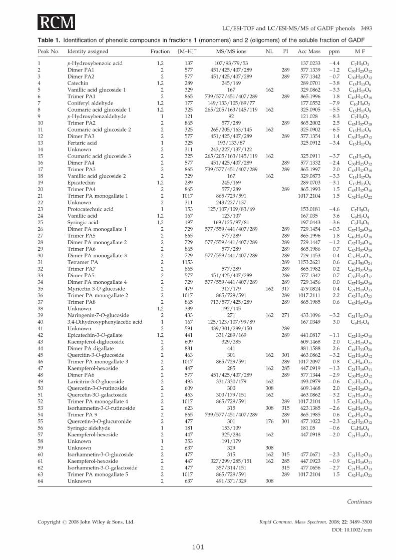
Table 1. Identification of phenolic compounds in fractions 1 (monomers) and 2 (oligomers) of the soluble fraction of GADF
Peak No. Identity assigned Fraction [M–H]S MS/MS ions NL PI Acc Mass ppm M F
1 p-Hydroxybenzoic acid 1,2 137 107/93/79/53 137.0233 �4.4 C7H5O3
2 Dimer PA1 2 577 451/425/407/289 289 577.1339 �1.2 C30H25O12
3 Dimer PA2 2 577 451/425/407/289 289 577.1342 �0.7 C30H25O12
4 Catechin 1,2 289 245/169 289.0701 �3.8 C15H13O6
5 Vanillic acid glucoside 1 2 329 167 162 329.0862 �3.3 C14H17O9
6 Trimer PA1 2 865 739/577/451/407/289 289 865.1996 1.8 C45H37O18
7 Coniferyl aldehyde 1,2 177 149/133/105/89/77 177.0552 �7.9 C10H9O3
8 Coumaric acid glucoside 1 1,2 325 265/205/163/145/119 162 325.0905 �5.5 C15H17O8
9 p-Hydroxybenzaldehyde 1 121 92 121.028 �8.3 C7H5O2
10 Trimer PA2 2 865 577/289 289 865.2002 2.5 C45H37O18
11 Coumaric acid glucoside 2 2 325 265/205/163/145 162 325.0902 �6.5 C15H17O8
12 Dimer PA3 2 577 451/425/407/289 289 577.1354 1.4 C30H25O12
13 Fertaric acid 1 325 193/133/87 325.0912 �3.4 C15H17O8
14 Unknown 2 311 243/227/137/12215 Coumaric acid glucoside 3 2 325 265/205/163/145/119 162 325.0911 �3.7 C15H17O8
16 Dimer PA4 2 577 451/425/407/289 289 577.1332 �2.4 C30H25O12
17 Trimer PA3 2 865 739/577/451/407/289 289 865.1997 2.0 C45H37O18
18 Vanillic acid glucoside 2 2 329 167 162 329.0873 �3.3 C14H17O9
19 Epicatechin 1,2 289 245/169 289.0703 �3.1 C15H13O6
20 Trimer PA4 2 865 577/289 289 865.1993 1.5 C45H37O18
21 Trimer PA monogallate 1 2 1017 865/729/591 1017.2104 1.5 C52H41O22
22 Unknown 2 311 243/227/13723 Protocatechuic acid 1 153 125/107/109/83/69 153.0181 �4.6 C7H5O4
24 Vanillic acid 1,2 167 123/107 167.035 3.6 C8H7O4
25 Syringic acid 1,2 197 169/125/97/81 197.0443 �3.6 C9H9O5
26 Dimer PA monogallate 1 2 729 577/559/441/407/289 289 729.1454 �0.3 C37H29O16
27 Trimer PA5 2 865 577/289 289 865.1996 1.8 C45H37O18
28 Dimer PA monogallate 2 2 729 577/559/441/407/289 289 729.1447 �1.2 C37H29O16
29 Trimer PA6 2 865 577/289 289 865.1986 0.7 C45H37O18
30 Dimer PA monogallate 3 2 729 577/559/441/407/289 289 729.1453 �0.4 C37H29O16
31 Tetramer PA 2 1153 289 1153.2621 0.6 C60H50O24
32 Trimer PA7 2 865 577/289 289 865.1982 0.2 C45H37O18
33 Dimer PA5 2 577 451/425/407/289 289 577.1342 �0.7 C30H25O12
34 Dimer PA monogallate 4 2 729 577/559/441/407/289 289 729.1456 0.0 C37H29O16
35 Myricetin-3-O-glucoside 2 479 317/179 162 317 479.0824 0.4 C21H19O13
36 Trimer PA monogallate 2 2 1017 865/729/591 289 1017.2111 2.2 C52H41O22
37 Trimer PA8 2 865 713/577/425/289 289 865.1985 0.6 C45H37O19
38 Unknown 1,2 339 192/14539 Naringenin-7-O-glucoside 2 433 271 162 271 433.1096 �3.2 C21H21O10
40 3,4-Dihydroxyphenylacetic acid 1 167 125/123/107/99/89 167.0349 3.0 C8H7O4
41 Unknown 2 591 439/301/289/150 28942 Epicatechin-3-O-gallate 1,2 441 331/289/169 289 441.0817 �1.1 C22H17O10
43 Kaempferol-diglucoside 2 609 329/285 609.1468 2.0 C27H29O16
44 Dimer PA digallate 2 881 441 881.1588 2.6 C44H33O20
45 Quercitin-3-O-glucoside 2 463 301 162 301 463.0862 �3.2 C21H19O12
46 Trimer PA monogallate 3 2 1017 865/729/591 289 1017.2097 0.8 C52H41O22
47 Kaempferol-hexoside 2 447 285 162 285 447.0919 �1.3 C21H19O11
48 Dimer PA6 2 577 451/425/407/289 289 577.1344 �2.9 C30H25O12
49 Laricitrin-3-O-glucoside 2 493 331/330/179 162 493.0979 �0.6 C22H21O13
50 Quercetin-3-O-rutinoside 2 609 300 308 609.1468 2.0 C27H29O16
51 Quercetin-3O-galactoside 2 463 300/179/151 162 463.0862 �3.2 C21H19O12
52 Trimer PA monogallate 4 2 1017 865/729/591 289 1017.2104 1.5 C52H41O22
53 Isorhamnetin-3-O-rutinoside 2 623 315 308 315 623.1385 �2.6 C28H31O16
54 Trimer PA 9 2 865 739/577/451/407/289 289 865.1985 0.6 C45H37O18
55 Quercetin-3-O-glucuronide 2 477 301 176 301 477.1022 �2.3 C22H21O12
56 Syringic aldehyde 1 181 153/109 181.05 �0.6 C9H9O4
57 Kaempferol-hexoside 2 447 325/284 162 447.0918 �2.0 C21H19O11
58 Unknown 1 353 191/17959 Unknown 2 637 329 30860 Isorhamnetin-3-O-glucoside 2 477 315 162 315 477.0671 �2.3 C21H17O13
61 Kaempferol-hexoside 2 447 327/299/285/151 162 285 447.0923 �0.9 C21H19O11
62 Isorhamnetin-3-O-galactoside 2 477 357/314/151 315 477.0656 �2.7 C21H17O13
63 Trimer PA monogallate 5 2 1017 865/729/591 289 1017.2104 1.5 C52H41O22
64 Unknown 2 637 491/371/329 308
Continues
Copyright # 2008 John Wiley & Sons, Ltd. Rapid Commun. Mass Spectrom. 2008; 22: 3489–3500
DOI: 10.1002/rcm
LC/ESI-TOF and LC/ESI-MS/MS of GADF phenols 3493
101
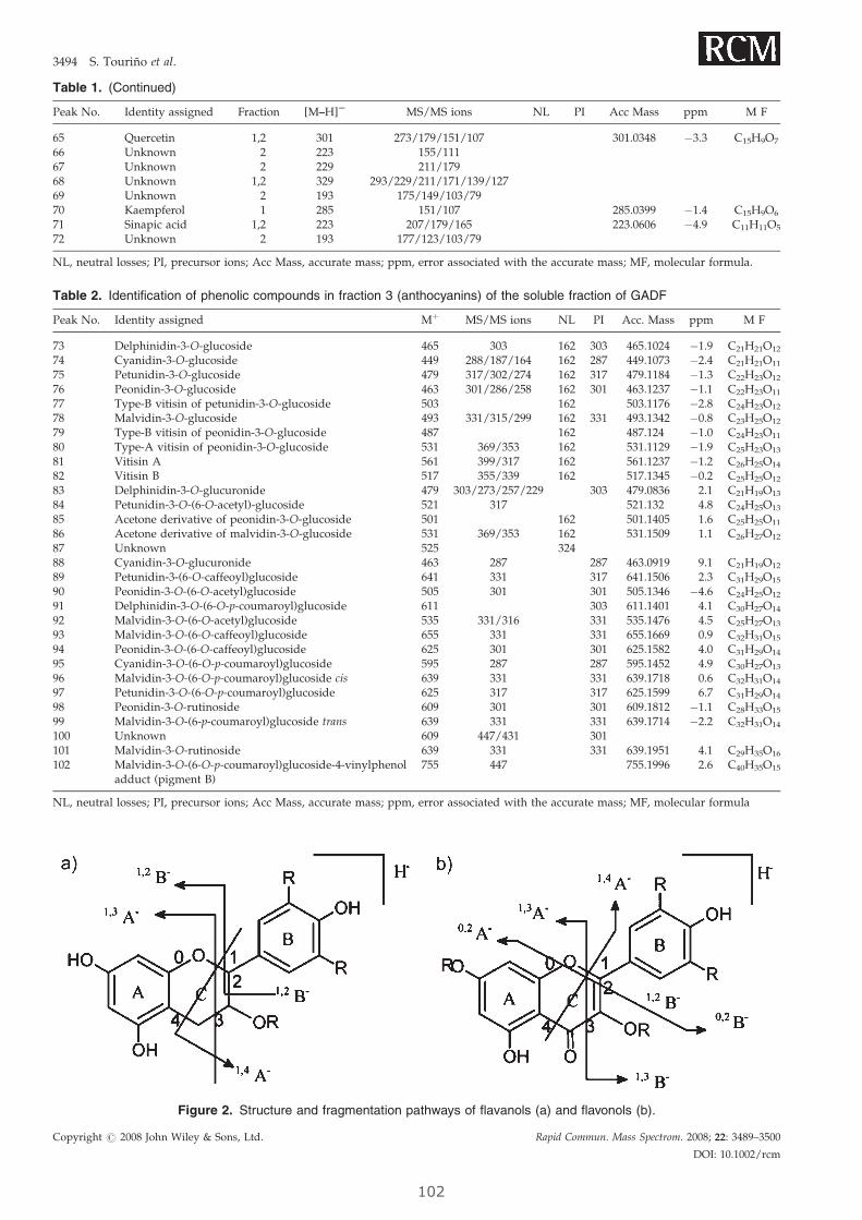
Table 2. Identification of phenolic compounds in fraction 3 (anthocyanins) of the soluble fraction of GADF
Peak No. Identity assigned Mþ MS/MS ions NL PI Acc. Mass ppm M F
73 Delphinidin-3-O-glucoside 465 303 162 303 465.1024 �1.9 C21H21O12
74 Cyanidin-3-O-glucoside 449 288/187/164 162 287 449.1073 �2.4 C21H21O11
75 Petunidin-3-O-glucoside 479 317/302/274 162 317 479.1184 �1.3 C22H23O12
76 Peonidin-3-O-glucoside 463 301/286/258 162 301 463.1237 �1.1 C22H23O11
77 Type-B vitisin of petunidin-3-O-glucoside 503 162 503.1176 �2.8 C24H23O12
78 Malvidin-3-O-glucoside 493 331/315/299 162 331 493.1342 �0.8 C23H25O12
79 Type-B vitisin of peonidin-3-O-glucoside 487 162 487.124 �1.0 C24H23O11
80 Type-A vitisin of peonidin-3-O-glucoside 531 369/353 162 531.1129 �1.9 C25H23O13
81 Vitisin A 561 399/317 162 561.1237 �1.2 C26H25O14
82 Vitisin B 517 355/339 162 517.1345 �0.2 C25H25O12
83 Delphinidin-3-O-glucuronide 479 303/273/257/229 303 479.0836 2.1 C21H19O13
84 Petunidin-3-O-(6-O-acetyl)-glucoside 521 317 521.132 4.8 C24H25O13
85 Acetone derivative of peonidin-3-O-glucoside 501 162 501.1405 1.6 C25H25O11
86 Acetone derivative of malvidin-3-O-glucoside 531 369/353 162 531.1509 1.1 C26H27O12
87 Unknown 525 32488 Cyanidin-3-O-glucuronide 463 287 287 463.0919 9.1 C21H19O12
89 Petunidin-3-(6-O-caffeoyl)glucoside 641 331 317 641.1506 2.3 C31H29O15
90 Peonidin-3-O-(6-O-acetyl)glucoside 505 301 301 505.1346 �4.6 C24H25O12
91 Delphinidin-3-O-(6-O-p-coumaroyl)glucoside 611 303 611.1401 4.1 C30H27O14
92 Malvidin-3-O-(6-O-acetyl)glucoside 535 331/316 331 535.1476 4.5 C25H27O13
93 Malvidin-3-O-(6-O-caffeoyl)glucoside 655 331 331 655.1669 0.9 C32H31O15
94 Peonidin-3-O-(6-O-caffeoyl)glucoside 625 301 301 625.1582 4.0 C31H29O14
95 Cyanidin-3-O-(6-O-p-coumaroyl)glucoside 595 287 287 595.1452 4.9 C30H27O13
96 Malvidin-3-O-(6-O-p-coumaroyl)glucoside cis 639 331 331 639.1718 0.6 C32H31O14
97 Petunidin-3-O-(6-O-p-coumaroyl)glucoside 625 317 317 625.1599 6.7 C31H29O14
98 Peonidin-3-O-rutinoside 609 301 301 609.1812 �1.1 C28H33O15
99 Malvidin-3-O-(6-p-coumaroyl)glucoside trans 639 331 331 639.1714 �2.2 C32H31O14
100 Unknown 609 447/431 301101 Malvidin-3-O-rutinoside 639 331 331 639.1951 4.1 C29H35O16
102 Malvidin-3-O-(6-O-p-coumaroyl)glucoside-4-vinylphenoladduct (pigment B)
755 447 755.1996 2.6 C40H35O15
NL, neutral losses; PI, precursor ions; Acc Mass, accurate mass; ppm, error associated with the accurate mass; MF, molecular formula
Figure 2. Structure and fragmentation pathways of flavanols (a) and flavonols (b).
Table 1. (Continued)
Peak No. Identity assigned Fraction [M–H]S MS/MS ions NL PI Acc Mass ppm M F
65 Quercetin 1,2 301 273/179/151/107 301.0348 �3.3 C15H9O7
66 Unknown 2 223 155/11167 Unknown 2 229 211/17968 Unknown 1,2 329 293/229/211/171/139/12769 Unknown 2 193 175/149/103/7970 Kaempferol 1 285 151/107 285.0399 �1.4 C15H9O6
71 Sinapic acid 1,2 223 207/179/165 223.0606 �4.9 C11H11O5
72 Unknown 2 193 177/123/103/79
NL, neutral losses; PI, precursor ions; Acc Mass, accurate mass; ppm, error associated with the accurate mass; MF, molecular formula.
Copyright # 2008 John Wiley & Sons, Ltd. Rapid Commun. Mass Spectrom. 2008; 22: 3489–3500
DOI: 10.1002/rcm
3494 S. Tourino et al.
102
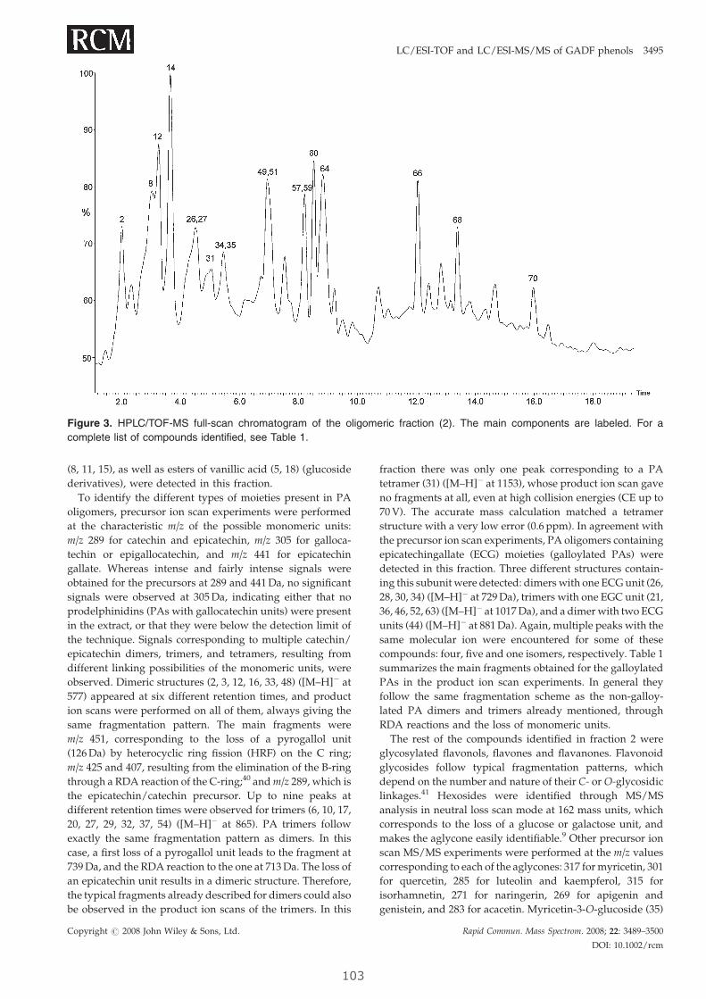
Figure 3. HPLC/TOF-MS full-scan chromatogram of the oligomeric fraction (2). The main components are labeled. For a
complete list of compounds identified, see Table 1.
LC/ESI-TOF and LC/ESI-MS/MS of GADF phenols 3495
(8, 11, 15), as well as esters of vanillic acid (5, 18) (glucoside
derivatives), were detected in this fraction.
To identify the different types of moieties present in PA
oligomers, precursor ion scan experiments were performed
at the characteristic m/z of the possible monomeric units:
m/z 289 for catechin and epicatechin, m/z 305 for galloca-
techin or epigallocatechin, and m/z 441 for epicatechin
gallate. Whereas intense and fairly intense signals were
obtained for the precursors at 289 and 441 Da, no significant
signals were observed at 305 Da, indicating either that no
prodelphinidins (PAs with gallocatechin units) were present
in the extract, or that they were below the detection limit of
the technique. Signals corresponding to multiple catechin/
epicatechin dimers, trimers, and tetramers, resulting from
different linking possibilities of the monomeric units, were
observed. Dimeric structures (2, 3, 12, 16, 33, 48) ([M–H]� at
577) appeared at six different retention times, and product
ion scans were performed on all of them, always giving the
same fragmentation pattern. The main fragments were
m/z 451, corresponding to the loss of a pyrogallol unit
(126 Da) by heterocyclic ring fission (HRF) on the C ring;
m/z 425 and 407, resulting from the elimination of the B-ring
through a RDA reaction of the C-ring;40 and m/z 289, which is
the epicatechin/catechin precursor. Up to nine peaks at
different retention times were observed for trimers (6, 10, 17,
20, 27, 29, 32, 37, 54) ([M–H]� at 865). PA trimers follow
exactly the same fragmentation pattern as dimers. In this
case, a first loss of a pyrogallol unit leads to the fragment at
739 Da, and the RDA reaction to the one at 713 Da. The loss of
an epicatechin unit results in a dimeric structure. Therefore,
the typical fragments already described for dimers could also
be observed in the product ion scans of the trimers. In this
Copyright # 2008 John Wiley & Sons, Ltd.
103
fraction there was only one peak corresponding to a PA
tetramer (31) ([M–H]� at 1153), whose product ion scan gave
no fragments at all, even at high collision energies (CE up to
70 V). The accurate mass calculation matched a tetramer
structure with a very low error (0.6 ppm). In agreement with
the precursor ion scan experiments, PA oligomers containing
epicatechingallate (ECG) moieties (galloylated PAs) were
detected in this fraction. Three different structures contain-
ing this subunit were detected: dimers with one ECG unit (26,
28, 30, 34) ([M–H]� at 729 Da), trimers with one EGC unit (21,
36, 46, 52, 63) ([M–H]� at 1017 Da), and a dimer with two ECG
units (44) ([M–H]� at 881 Da). Again, multiple peaks with the
same molecular ion were encountered for some of these
compounds: four, five and one isomers, respectively. Table 1
summarizes the main fragments obtained for the galloylated
PAs in the product ion scan experiments. In general they
follow the same fragmentation scheme as the non-galloy-
lated PA dimers and trimers already mentioned, through
RDA reactions and the loss of monomeric units.
The rest of the compounds identified in fraction 2 were
glycosylated flavonols, flavones and flavanones. Flavonoid
glycosides follow typical fragmentation patterns, which
depend on the number and nature of their C- or O-glycosidic
linkages.41 Hexosides were identified through MS/MS
analysis in neutral loss scan mode at 162 mass units, which
corresponds to the loss of a glucose or galactose unit, and
makes the aglycone easily identifiable.9 Other precursor ion
scan MS/MS experiments were performed at the m/z values
corresponding to each of the aglycones: 317 for myricetin, 301
for quercetin, 285 for luteolin and kaempferol, 315 for
isorhamnetin, 271 for naringerin, 269 for apigenin and
genistein, and 283 for acacetin. Myricetin-3-O-glucoside (35)
Rapid Commun. Mass Spectrom. 2008; 22: 3489–3500
DOI: 10.1002/rcm
3496 S. Tourino et al.
and naringerin-7-O-glucoside (39) were directly identified
through these experiments. Two quercetin hexosides were
detected, most probably quercetin-3-glucoside (45) and
quercetin-3-galactoside (51), as described for grapes.20,21
The glucoside and galactoside were distinguished by their
retention times, since it has been described42 that, under the
analytical conditions of this work, glucosides elute earlier
than galactosides. Both spectra presented the aglycone
fragment at 301 Da, and the galactoside also yielded the
radical anion of the aglycone at m/z 300. The presence of
radical aglycones in the product ion scan of flavonol-3-O-
glycosides has been reported in the literature.43 The relative
abundance of the radical ion varies according to the nature
and position of the sugar moiety, increasing with the number
of hydroxyl substituents in the B-ring (myrice-
tin>quercetin> kaempferol-glucoside). A precursor ion
scan at m/z 285 revealed the presence of three compounds at
m/z 447, all of them kaempferol or luteolin -C- or -O-
hexosides. The radical anion of the aglycone at m/z 284 was
also detected. Although product ion scan spectra were
recorded for all of them, it proved hard to assign a structure
on the basis of the fragment ions obtained alone. As the
molecular formulas of these compounds (combinations of a
kaempferol or luteolin unit with a hexose) are identical,
measuring the accurate masses is of no help. Although
luteolin derivatives have been found in several natural
products, they have not been reported in grape products. The
presence of kaempferol derivatives has, however, been
widely reported.21,44,45 The three peaks at m/z 447 (47, 57, 61)
therefore probably belong to kaempferol -C- or -O-hexosides.
Precursor ion scan MS/MS experiments at m/z 315 led to the
identification of isorhamnetin-3-O-glucoside (60) and iso-
rhamnetin-3-O-galactoside (62). As for quercetin, the two
hexoses were differentiated by their retention times.
Laricitin-3-O-glucoside (49) ([M–H]� 493), a compound
already found in red grapes,46 was also present in this
fraction. The product ion spectra showed an intense aglycone
peak (m/z 331) as well as one for its radical anion (m/z 330).
Neutral loss scan MS/MS experiments at 176 mass
units (glucuronide) revealed the presence of quercetin-3-
O-glucuronide (55). Some rutinosides such as quercetin-3-
O-rutinoside (50) and isorhamnetin-3-O-rutinoside (53)
were identified through neutral losses of 308 mass units.
The assignment of the glycoside substitutions to position 3
of the flavonoid ring was based on the literature cited.
The accurate masses measured by the high-resolution
system matched the structures proposed with very low
errors.
Anthocyanin fraction 3Anthocyanins constitute another biologically interesting
polyphenolic fraction of GADF. They are absorbed in the
digestive tract and have been found intact in a variety of
tissues.47 They even appear to be able to cross the blood brain
barrier and enter the brain, where they might perform some
neuroprotective activity.47,48 LC/ESI-TOF enabled us to
identify a number of putatively active small anthocyanins.
Because anthocyanins are already positively charged in their
natural form, this fraction was analyzed in the positive mode
and Mþ molecular ions were detected. The results are
Copyright # 2008 John Wiley & Sons, Ltd.
10
summarized in Table 2. We identified six different groups
of compounds by precursor ion scan experiments, corre-
sponding to derivatives of six aglycones (anthocyanidins):
delphinidin (m/z 303), cyanidin (m/z 287), petunidin
(m/z 317), peonidin (m/z 301), pelargonidin (m/z 271), and
malvidin (m/z 331). Neutral loss scan experiments helped us
to identify the sugar moieties. Anthocyanins produced less
fragmentation than flavonols and PAs did. On some
occasions the aglycone fragment was the only one present
in the product ion scan spectra, and this made the
measurement of the accurate mass crucial to be able to
assign a structure. Table 3 summarizes the chemical
structures of the anthocyanins identified in this fraction.
The compounds eluting first (73, 74, 75, 76 and 78) on LC
were the monoglucoside derivatives of delphinidin, cyani-
din, petunidin, peonidin, and malvidin, respectively, as
revealed by the characteristic fragment corresponding to the
loss of glucose and confirmed by the exact mass measure-
ments. Acetylglucoside derivatives of petunidin (84), peo-
nidin (90) and malvidin (92) were also identified through the
loss of 204 mass units (162 Da from the glucose moiety plus
42 Da from the acetyl moiety). In the same way, glucuronides
of delphinidin (83) and cyanidin (88) were detected (loss of
the glucuronide moiety, 176 Da). Another group of com-
pounds found in this fraction showed a loss of 324 Da in
the product ion scan spectra. This mass is compatible
with the loss of two glucose units (diglucosides) or the loss
of a caffeoylglucose unit. Two criteria allowed us to
identify the compounds as caffeoylglucose derivatives: the
absence of signals corresponding to loses of glucose in
the neutral loss experiments (as we would expect if
diglucosides were present) and the accurate mass measure-
ments (which matched structures with caffeoylglucose
moieties). Thus, the caffeoylglucoside derivatives of petuni-
din (89), malvidin (93) and peonidin (94) were unequivocally
identified. A number of ions yielded a loss of 308 Da.
This loss may be attributed to either rutinoside or
coumaroylglucoside moieties. Again, the fragmentation
was too low to decide between the two and LC/ESI-TOF
was crucial to identify the compounds. Rutinosides and
coumaroylglucosides have different molecular formulas, so
the rutinoside derivatives of peonidin (98) and malvidin
(101) were easily identified by measuring the accurate
masses. In the same way compounds 91, 95, 96, 97, and
99 were identified as coumaroylglucoside derivatives of
delphinidin, cyanidin, malvidin, petunidin, and malvidin,
respectively. The two malvidin derivatives corresponded to
the cis- (96) and trans- (99) isomers, which were distinguished
by their retention times.49 Both isomers have previously been
detected in grapes50 and wine.49,51 Some of the structures
(91–97) were identified as coumaroylglucosides. Although
accurate mass measurements could not differentiate between
coumaroylglucoside and glucoside-vinylguaiacol deriva-
tives (which have identical molecular formula) the loss of
a 308 Da fragment is quite unlikely to be from vinylguaiacol
derivatives because this would imply the fragmentation of
the main structure of the aglycone. Furthermore, according
to the literature,52,53 glucoside-vinylguaiacol derivatives lose
the glucose moiety easily, and this was not observed in
compounds 91–97.
Rapid Commun. Mass Spectrom. 2008; 22: 3489–3500
DOI: 10.1002/rcm
4
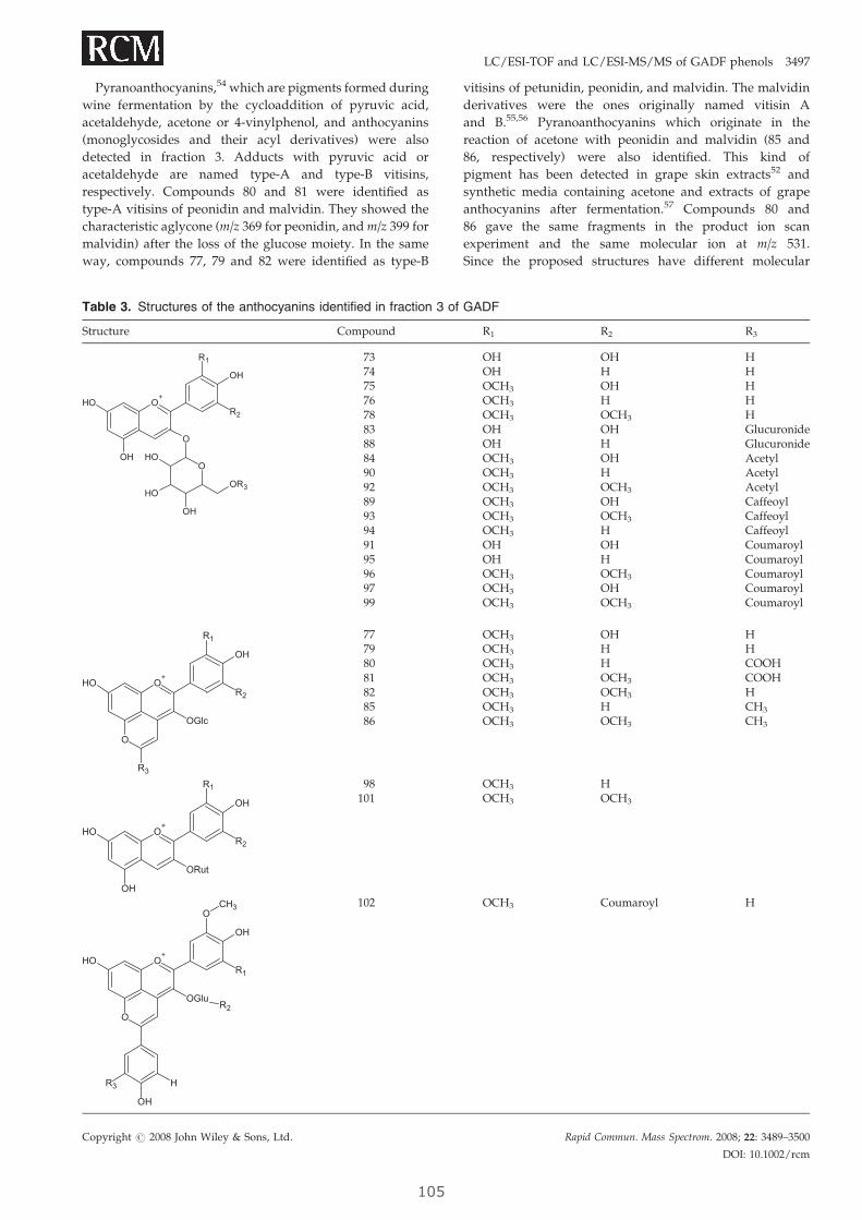
LC/ESI-TOF and LC/ESI-MS/MS of GADF phenols 3497
Pyranoanthocyanins,54 which are pigments formed during
wine fermentation by the cycloaddition of pyruvic acid,
acetaldehyde, acetone or 4-vinylphenol, and anthocyanins
(monoglycosides and their acyl derivatives) were also
detected in fraction 3. Adducts with pyruvic acid or
acetaldehyde are named type-A and type-B vitisins,
respectively. Compounds 80 and 81 were identified as
type-A vitisins of peonidin and malvidin. They showed the
characteristic aglycone (m/z 369 for peonidin, and m/z 399 for
malvidin) after the loss of the glucose moiety. In the same
way, compounds 77, 79 and 82 were identified as type-B
Table 3. Structures of the anthocyanins identified in fraction 3 of
Structure Compound
O+
O
O
OH
OH
R1
OH
R2
OH
OH
OR3
OH
737475767883888490928993949195969799
O+
OGlc
OH
O
R1
OH
R2
R3
77798081828586
O+
ORut
OH
OH
R1
OH
R2
98101
R2
O+
OGlu
OH
O
O
OH
R1
OH
HR3
CH3 102
Copyright # 2008 John Wiley & Sons, Ltd.
105
vitisins of petunidin, peonidin, and malvidin. The malvidin
derivatives were the ones originally named vitisin A
and B.55,56 Pyranoanthocyanins which originate in the
reaction of acetone with peonidin and malvidin (85 and
86, respectively) were also identified. This kind of
pigment has been detected in grape skin extracts52 and
synthetic media containing acetone and extracts of grape
anthocyanins after fermentation.57 Compounds 80 and
86 gave the same fragments in the product ion scan
experiment and the same molecular ion at m/z 531.
Since the proposed structures have different molecular
GADF
R1 R2 R3
OH OH HOH H HOCH3 OH HOCH3 H HOCH3 OCH3 HOH OH GlucuronideOH H GlucuronideOCH3 OH AcetylOCH3 H AcetylOCH3 OCH3 AcetylOCH3 OH CaffeoylOCH3 OCH3 CaffeoylOCH3 H CaffeoylOH OH CoumaroylOH H CoumaroylOCH3 OCH3 CoumaroylOCH3 OH CoumaroylOCH3 OCH3 Coumaroyl
OCH3 OH HOCH3 H HOCH3 H COOHOCH3 OCH3 COOHOCH3 OCH3 HOCH3 H CH3
OCH3 OCH3 CH3
OCH3 HOCH3 OCH3
OCH3 Coumaroyl H
Rapid Commun. Mass Spectrom. 2008; 22: 3489–3500
DOI: 10.1002/rcm

Table 4. Identification of phenolic compounds in fraction 4 (PA polymers) of the soluble fraction of GADF
Type-B PAs Type-A PAs
n
Monogallates Digallates Trigallates Monogallates
[M–H]� [M–H]2� [M–H]� [M–H]2� [M–H]� [M–H]2� [M–H]� [M–H]2� [M–H]� [M–H]2� [M–H]� [M–H]2�
1 289.1 441.12 577.1 729.1 881.2 575.13 865.2 1017.2 508.6 1169.2 1321.3 863.2 1015.24 1153.3 1305.3 652.6 1457.3 1609.3 1151.2 575.6 1303.35 1441.3 720.6 1593.3 796.7 1745.4 1439.3 719.6 1591.3 795.76 1729.4 864.7 1016.7 1092.7 1727.4 863.77 1008.7 1007.78 1152.7 1151.79 1295.710 1440.811 1584.812 1728.9
n, number of flavanol (catechin or epicatechin) units.
Figure 4. ESI-TOF-MS spectra in negative ion mode of the polymeric fraction (4): (a) fromm/z 300 to 2000 and (b) from m/z 700 to
910.
3498 S. Tourino et al.
formulas, they were easily differentiated by measuring
the accurate mass with the LC/ESI-TOF system. Com-
pounds 100 and 102 displayed the same fragment ion at
m/z 447 in their respective product ion scans. Compound
100 gave a signal at m/z 301 in the precursor ion scan
experiment, which identified the compound as a peonidin
Copyright # 2008 John Wiley & Sons, Ltd.
10
derivative; probably a glucose derivative as the 447 Da
fragment implies the loss of glucose. The accurate mass
measurement was compatible with the peonidin-3-O-
glucoside-4-vinylguaiacol adduct. However, because the
loss of 308 Da is not favored for vinylguaiacols, we do not
suggest any structure. Compound 102 was identified as
Rapid Commun. Mass Spectrom. 2008; 22: 3489–3500
DOI: 10.1002/rcm
6
LC/ESI-TOF and LC/ESI-MS/MS of GADF phenols 3499
the malvidin-3-O-(600-p-coumaroyl)glucoside-4-vinylphenol
adduct, also named pigment B.58 In this case, m/z 447
corresponded to the aglycone (see Table 3) after the loss of
the coumaroylglucoside moiety.
Polymeric fraction 4Polymeric PAs are a particularly important fraction of
GADF. Studies in vitro and in vivo have suggested that only
the smaller oligomers (up to trimers) are adsorbed to some
extent through the gut barrier, whereas larger PAs reach
the colon intact.36,37 There, these larger PAs are degraded by
the colonic microbiota into smaller units, which in turn are
degraded to phenolic acids, which are then absorbed.6,59 All
these molecules are bioavailable and bioactive in the colon
where they may greatly contribute to the preventative effects
of GADF. To characterize the polymeric fraction, we used
ESI-TOF-MS in negative mode. As RP-HPLC was not able to
separate the higher molecular mass polymers, the fraction
was injected directly into the MS system. In a first attempt to
identify the masses corresponding to PAs with high degree
of polymerization, the fraction was scanned over a wide
range of mass units (300–3000). Above 2000 mass units
there was no significant signal from any possible ions of
interest. To increase sensitivity, the fraction was analyzed
over ranges of only 210 mass units (300–510, 500–710, and so
on). Table 4 summarizes the PA polymers found in the
fraction, indicating the degree of polymerization and
galloylation. Figure 4(a) shows the whole spectrum of the
fraction, and Fig. 4(b) includes expansions of the range
from m/z 700 to 910. The most intense peaks corresponded
to polymeric type-B PAs, with and without galloyl units
attached. Two regular series of peaks were clearly observed
with a peak occurring every 288 mass units, starting at 577
(epicatechin dimer) and 441 (monogallate monomer) mass
units. A more accurate inspection of the spectrum revealed
peaks corresponding to digallate and trigallate PAs, which
generated series of signals at intervals of 152 (galloyl moiety)
mass units. Doubly charged ions (see Fig. 4(b)) allowed us to
identify PAs with a higher degree of polymerization.
Polymers of up to 12 catechin units were observed for
type-B PAs. The highest degree of polymerization for gallates
was 5 (monogallates) and 6 (digallates and trigallates).
Signals from triply charged ions were not observed.
Type-A PAs were also detected. Compared to type-B PAs,
type-A PAs contain an extra interflavanic linkage (C2–C7)
between catechin units. Therefore, type-A PA signals are 2
mass units lower than the corresponding type-B PA signals.
Table 4 includes the type-A PAs. The highest degree of
polymerization was 8 for catechin/epicatechin polymers and
4 for monogallates. MS techniques, particularly ESI-TOF,
provide a wealth of information about the distribution of PA
sizes and their galloylation that may eventually be related to
the activity of GADF in the colon.
CONCLUSIONS
To advance the study of the structure/activity relationships
of GADF, thorough identification of its bioactive polyphe-
nols is of the utmost importance. We applied a combination
of LC/MS techniques, namely high-resolution LC/ESI-TOF-
Copyright # 2008 John Wiley & Sons, Ltd.
107
MS and LC/ESI-MS/MS, to accurately identify many
important polyphenolic compounds from the soluble frac-
tion of GADF. The phenols include hydroxycinnamic acids,
anthocyanins, flavonols, flavones, flavanones, catechins and
PAs. Compared to LC/ESI-MS/MS on a triple quadrupole,
LC/ESI-TOF-MS gave more sensitive and better-resolved
full-fragment mass spectra, which facilitated the initial
identification of the components from each LC run. Then
LC/ESI-MS/MS fragmentation experiments suggested
structures which were confirmed by the accurate masses
as measured by LC/ESI-TOF-MS. High-resolution LC/ESI-
TOF-MS has proved to be a particularly useful technique for
discriminating between possible structures that are equally
compatible with the fragmentation pattern generated by
MS/MS.
AcknowledgementsFinancial support from the Spanish Ministry of Education
and Science (research grants AGL2004-07579-C04-01, -02, -03;
AGL2006-12210-C03-02/ALI, doctoral fellowship to S.T. and
post-doctoral contract to E.F.) is acknowledged.
REFERENCES
1. Schieber A, Stintzing FC, Carle R. Trends Food Sci. Technol.2001; 12: 401.
2. Guillon F, Champ M. Food Res. Int. 2000; 33: 233.3. Rice-Evans C, Miller NJ. Biochem. Soc. Trans. 1996; 24: 790.4. Scalbert A, Manach C, Morand C, Remesy C, Jimenez L. Crit.
Rev. Food Sci. Nutr. 2005; 45: 287.5. Manach C, Williamson G, Morand C, Scalbert A, Remesy C.
Am. J. Clin. Nutr. 2005; 81: 230S.6. Gonthier M-P, Donovan JL, Texier O, Felgines C, Remesy C,
Scalbert A. Free Radical Biol. Med. 2003; 35: 837.7. Saura-Calixto F. J. Agric. Food Chem. 1998; 46: 4303.8. Jimenez JP, Serrano J, Tabernero M, Arranz S, Dıaz-Rubio
ME, Garcıa-Diz L, Goni I, Saura-Calixto F. Nutrition 2008; 24:646.
9. Cuyckens F, Claeys M. J. Mass Spectrom. 2004; 39: 1.10. Kammerer DR, Saleh ZS, Carle R, Stanley RA. Eur. Food Res.
Technol. 2007; 224: 605.11. Liu R, Sun J, Bi K, Guo D-a. J. Chromatogr. B 2005; 829: 35.12. Flamini R. Mass Spectrom. Rev. 2003; 22: 218.13. Sanoner P, Guyot S, Marnet N, Molle D, Drilleau JF. J. Agric.
Food Chem. 1999; 47: 4847.14. Mozetic B, Trebse P. Acta Chim. Slov. 2004; 51: 151.15. Vidal S, Hayasaka Y, Meudec E, Cheynier V, Skouroumou-
nis G. J. Agric. Food Chem. 2004; 52: 713.16. Monagas M, Bartolome B, Gomez-Cordoves C. Crit. Rev.
Food Sci. Nutr. 2005; 45: 85.17. Peng Z, Hayasaka Y, Iland PG, Sefton M, Høj P, Waters EJ.
J. Agric. Food Chem. 2001; 49: 26.18. Hayasaka Y, Waters EJ, Cheynier V, Herderich MJ, Vidal S.
Rapid Commun. Mass Spectrom. 2003; 17: 9.19. Pozo-Bayon MA, Monagas M, Polo MC, Gomez-Cordoves C.
J. Agric. Food Chem. 2004; 52: 1300.20. Kammerer D, Claus A, Carle R, Schieber A. J. Agric. Food
Chem. 2004; 52: 4360.21. Monagas M, Garrido I, Bartolome B, Gomez-Cordoves C.
Anal. Chim. Acta 2006; 563: 401.22. Bravo MN, Silva S, Coelho AV, Boas LV, Bronze MR. Anal.
Chim. Acta 2006; 563: 84.23. Reed JD, Krueger CG, Vestling MM. Phytochemistry 2005; 66:
2248.24. Ohnishi-Kameyama M, Yanagida A, Kanda T, Nagata T.
Rapid Commun. Mass Spectrom. 1997; 11: 31.25. Vivas N, Nonier MF, Vivas De Gaulejac N, Absalon C,
Bertrand A, Mirabel M. Anal. Chim. Acta 2004; 513: 247.26. Guyot S, Doco T, Souquet JM, Moutounet M, Drilleau JF.
Phytochemistry 1997; 44: 351.27. Ishida Y, Kitagawa K, Goto K, Ohtani H. Rapid Commun.
Mass Spectrom. 2005; 19: 706.
Rapid Commun. Mass Spectrom. 2008; 22: 3489–3500
DOI: 10.1002/rcm

3500 S. Tourino et al.
28. Marchese S, Gentili A, Perret D, D’Ascenzo G, Pastori F.Rapid Commun. Mass Spectrom 2003; 17: 879.
29. Fletcher AT, Mader JC. J. Plant Growth Regul. 2007; 26: 351.30. Saura-Calixto F, Goni-Cambrodon I. In CSIC-UCM, ES
2259258 A1.31. Saura-Calixto F, Larrauri-Garcıa JA. In CSIC, ES 2130092 A1.32. Perez-Jimenez J, Saura-Calixto F. Int. J. Food Sci. Technol.
2008; 43: 185.33. Sun B, Leandro MC, de Freitas V, Spranger MI.
J. Chromatgr. A 2006; 1128: 27.34. Maier T, Sanzenbacher S, Kammerer DR, Berardini N, Con-
rad J, Beifuss U, Carle R, Schieber A. J. Chromatgr. A 2006;1128: 61.
35. Maatta-Riihinen KR, Kahkonen MP, Torronen AR, Heino-nen IM. J. Agric. Food Chem. 2005; 53: 8485.
36. Jimenez-Ramsey LM, Rogler JC, Housley TL, Butler LG,Elkin RG. J. Agric. Food Chem. 1994; 42: 963.
37. Terrill TH, Waghorn GC, Woolley DJ, McNabb WC, BarryTN. Br. J. Nutr. 1994; 72: 467.
38. Meselhy R, Nakamura N, Hattori M. Chem. Pharm. Bull. 1997;45: 888.
39. Lizarraga D, Lozano C, Briede JJ, van Delft JH, Tourino S,Centelles JJ, Torres JL, Cascante M. FEBS J. 2007; 274: 4802.
40. Gu L, Kelm MA, Hammerstone JF, Zhang Z, Beecher G,Holden J, Haytowitz D, Prior RL. J. Mass Spectrom. 2003; 38:1272.
41. Ferreres F, Llorach R, Gil-Izquierdo A. J. Mass Spectrom. 2004;39: 312.
42. Sanchez-Rabaneda F, Jauregui O, Lamuela-Raventos RM,Viladomat F, Bastida J, Codina C. Rapid Commun. MassSpectrom. 2004; 18: 553.
43. Hvattum E, Ekeberg D. J. Mass Spectrom. 2003; 38: 43.
Copyright # 2008 John Wiley & Sons, Ltd.
10
44. Kammerer D, Carle R, Schieber A. Rapid Commun. MassSpectrom. 2003; 17: 2407.
45. Flamini R, Panighel A. Mass Spectrom. Rev. 2006; 25: 741.46. Ruberto G, Renda A, Daquino C, Amico V, Spatafora C,
Tringali C, Tommasi ND. Food Chem. 2007; 100: 203.47. Kalt W, Blumberg JB, McDonald JE, Vinqvist-Tymchuk MR,
Fillmore SAE, Graf BA, O’Leary JM, Milbury PE. J. Agric.Food Chem. 2008; 56: 705.
48. Andres-Lacueva C, Shukitt-Hale B, Galli RL, Jauregui O,Lamuela-Raventos RM, Joseph JA. Nutr. Neurosci. 2005; 8:111.
49. Alcalde-Eon C, Escribano-Bailon MT, Santos-Buelga C,Rivas-Gonzalo JC. J. Mass Spectrom. 2007; 42: 735.
50. Nunez V, Monagas M, Lopez-Cordoves MC, Bartolome B.Postharvest Biol. Technol. 2004; 31: 69.
51. Monagas M, Nunez V, Bartolome B, Gomez-Cordoves C.Am. J. Enol. Vitic. 2003; 54: 163.
52. Hayasaka Y, Asenstorfer RE. J. Agric. Food Chem. 2002; 50:756.
53. de Villiers A, Vanhoenacker G, Majek P, Sandra P.J. Chromatgr. A 2004; 1054: 195.
54. Rentzsch W, Schwarz M, Winterhalter P. Trends Food Sci.Technol. 2007; 18: 526.
55. Bakker J, Timberlake CF. J. Agric. Food Chem. 1997; 45: 35.56. Fulcrand H, Benabdeljalil C, Rigaud J, Cheynier V, Moutou-
net M. Phytochemistry 1998; 47: 1401.57. Benabdeljalil C, Cheynier V, Fulcrand H, Hakiki A, Mosad-
dak M, Moutounet M. Sci. Aliment. 2000; 20: 203.58. Fulcrand H, dos Santos PJC, Sarni Manchado P, Cheynier V,
Favre Bonvin J. J. Chem. Soc. Perkin Trans. 1 1996; 735.59. Deprez S, Brezillon C, Rabot S, Philippe C, Mila I, Lapierre C,
Scalbert A. J. Nutr. 2000; 130: 2733.
Rapid Commun. Mass Spectrom. 2008; 22: 3489–3500
DOI: 10.1002/rcm
8

4. Resultados
109
4.2.2 PUBLICACIÓN 4: Identificación de metabolitos de fibra
antioxidante de uva en orina de ratas.
Título original: Phenolic metabolites of grape antioxidant dietary fiber in rat urine.
Autores: Sonia Touriño, Elisabet Fuguet, Mª Pilar Vinardell, Marta Cascante,
Josep Lluís Torres. Journal of Agricultural and Food Chemistry (En proceso de revisión)
Índice de impacto: 2,507 Clasificación y categoría: 7/ 103 (Ciencia y Tecnología de los alimentos); 8/62
(Química aplicada); 1/35 (Agricultura, multidisciplinar)
RESUMEN: La fibra dietética de uva es particularmente interesante como suplemento nutricional
gracias a sus efectos beneficiosos sobre la salud cardiovascular (Pérez-Jiménez et al.,
2008). Esta fibra dietética se caracteriza por presenta una elevada cantidad de
flavonoides y proantocianidinas superando en porcentaje a otras fibras dietéticas
similares (Goñi et al., 2009). La actividad biológica de los flavonoides y proantocianidinas
podría ser directamente responsable de los beneficios a la salud cardiovascular que
aporta GADF. Los flavonoides son metabolizados de manera extensiva cuando son
ingeridos por lo que las actividades biológicas podrían estar relacionadas con los
metabolitos más que con los compuestos en su forma original en la fuente vegetal.
El objetivo de este trabajo fue identificar los metabolitos mayoritarios tras la ingesta de
GADF. Para alcanzar este objetivo se realizó un estudio con animales de
experimentación, ratas Sprague-Dawley (n=12), divididos en tres grupos (n=4) que
correspondían a un grupo control, un grupo al que se le administró Epicatequina pura (1g
/kg) y un grupo al que se le administró GADF (1,6g/kg). Posterior a la administración, se
recogió muestra de orina a diferentes horas (2, 4, 6, 8, 10 y 24h). Las muestras fueron
purificadas por extracción en fase sólida (SPE) previamente al análisis por LC-MS y LC-
MS/MS.
Experimentos en tándem (MRM, NL, PI y barrido de iones producto) permitieron
identificar 18 metabolitos de epicatequina (mono-, di-, tri- conjugada con metilo, sulfato
y glucurónido) que podrían proceder de oligómeros de proantocianidinas
4. Resultados
110
despolimerizados a lo largo del intestino y de ácidos fenólicos libres procedentes de las
transformaciones colónicas. También fueron identificados ácidos fenólicos conjugados con
sulfato y glucuronido. Éstos podrían ser productos de las fermentaciones de la microbiota
colónica absorbidos y metabolizados probablemente en el hígado.
Los experimentos en tándem realizados, también aportaron información estructural de
las posiciones de conjugación en los compuestos fenólicos. Esta información es
particularmente interesante si se tiene en cuenta que algunos de los metabolitos
identificados en este estudio presentaron el grupo catecol libre, lo cual indica que cierta
capacidad antioxidante in vivo persiste durante los procesos de biotransformación.
El estudio de la identificación de los metabolitos de GADF aportó un mayor conocimiento
sobre la biodisponibilidad de los polifenoles en mezclas complejas. Teniendo en cuenta
que frutas y verduras no procesadas contienen grandes cantidades de proantocianidinas,
los resultados de este estudio son una contribución al conocimiento de los posibles
efectos beneficiosos de los polifenoles dietéticos en la salud.
A continuación se muestra la publicación original. El material suplementario de este
artículo se incluye en los Anexos (apartado 8.2.3, ver pág. 287)
Phenolic metabolites of grape antioxidant dietary fiber in rat urine
SONIA TOURIÑO†, ELISABET FUGUET†+, MARÍA PILAR VINARDELL‡, MARTA CASCANTE§ and
JOSEP LLUÍS† TORRES*†
† Institute for Advanced Chemistry of Catalonia, CSIC (ICAQ-CSIC). Jordi Girona 18-26, 08034-Barcelona, Spain. +Current address: Departament de Química Analítica, Universitat de Barcelona, Martí i Franquès 1-11, 08028-Barcelona, Spain
‡ Department of Physiology, Faculty of Pharmacy, University of Barcelona, Av. Joan XXIII s/n, 08028-Barcelona, Spain § Department of Biochemistry and Molecular Biology, Institute of Biomedicine of the University of Barcelona (IBUB) and Centre for
Research in Theoretical Chemistry, Barcelona Science Park (CeRQT-PCB), Unit Associated with CSIC, Diagonal 645, 08028-Barcelona, Spain
Grape antioxidant dietary fiber (GADF) combines the putative health benefits of fiber and polyphenols. Polyphenolic metabolites may play a key role in the overall biological effects of this supplement. We identified phenolic GADF metabolites in rat urine at different times after oral administration, using HPLC-ESI-MS/MS techniques. The phenolic metabolic outcome of GADF is essentially an array of mono and polyconjugated epicatechins and free or conjugated smaller phenolic acids, some of them never reported before. We have detected 18 mono-, di-, and tri-conjugates of EC with glucuronide, methyl and sulfate moieties and small phenolic acids both free and conjugated. Procyanidin oligomers are depolymerized in the small intestine and the polymers are degraded by the colonic microbiota. For several hours after ingestion of GADF, a great variety of phenolic species including some with an intact catechol group, are in contact with the digestive tract tissues before, during and after metabolization, and many of them are systemically bioavailable before being excreted.
KEYWORDS: grape antioxidant dietary fiber, polyphenols, procyanidins, epicatechin, metabolites, bioavailability, mass
spectrometry, urine.
INTRODUCTION Dietary fiber is believed to play an important role in maintaining the functional integrity of the gastrointestinal tract. A high fiber intake is associated with body weight control (1) and a reduced risk of diseases such as colon cancer (2) and atherosclerosis (3). However, not all fibers are equally good and the benefits of a fiber-rich diet greatly depend on the solubility and fermentability of the fiber eaten as well as on other functional constituents, particularly antioxidant polyphenols (4, 5). Polyphenols are scavengers of reactive oxygen species (ROS) and appear to interact with biologically significant proteins. Grape antioxidant dietary fiber (GADF) is a particularly interesting nutritional supplement obtained from grape pomace (6). GADF is superior to other fibers in terms of digestibility and intestinal fermentation (7). Most promisingly, GADF may contribute to cardiovascular health, mainly by lowering blood pressure and levels of cholesterol (8). GADF is 58% insoluble fiber, 16% soluble fiber and it contains a large proportion (20%) of polyphenolic compounds, including phenolic acids and flavonoids, mainly polymeric procyanidins (8, 9). Oligomeric procyanidins, rather than other polyphenols, are reported to be responsible for lowering blood pressure in humans (10). ∗
∗Autor to whom correspondence should be addressed, [Phone, +34 93 400 61 12; Fax, +34 93 204 59 04; e-mail, [email protected]] † Institute for Advanced Chemistry of Catalonia-CSIC (IQAC-CSIC) ‡, § University of Barcelona
Therefore, the cardiovascular benefits of GADF may very well be due to the procyanidins it contains, or perhaps more precisely to their metabolites. Procyanidins are extensively metabolized in the intestinal tract and liver, as are other polyphenols. It therefore follows that the putatively beneficial effects of polyphenols at the systemic level should be attributed to the metabolites rather than to the parent compounds. Flavonoid metabolites are formed by Phase I (cleavage/degradation) and Phase II (derivatisation) metabolism to give and array of structures. Some of them are shown in Figure 1. The number of possible metabolites is enormous and the amount of each individual metabolite may be small. That is why most studies in the literature use enzymes to strip off the derivatizing moieties and detect only the hydroxylic form of the metabolite (11, 12). However, in this way valuable information on what particular active compounds may actually be bioavailable is lost. The literature on metabolization of procyanidins is still incomplete. The most accurate accounts in terms of epicatechin (EC) derivatization (glucuronates, sulfates, methyl ethers) lack crucial information on phenolic acids (13), whereas the reports on colonic metabolites (phenolic acids) miss the variability of derivatized species (11, 14). The aim of this study is to identify the main phenolic metabolites present in rat urine after GADF ingestion, using a combination of mass spectrometry (MS)
111
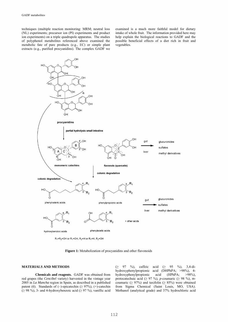
GADF metabolites
techniques (multiple reaction monitoring: MRM; neutral loss (NL) experiments; precursor ion (PI) experiments and product ion experiments) on a triple quadrupole apparatus. The studies of polyphenol metabolites referenced above examined the metabolic fate of pure products (e.g., EC) or simple plant extracts (e.g., purified procyanidins). The complex GADF we
examined is a much more faithful model for dietary intake of whole fruit. The information provided here may help explain the biological reactions to GADF and the possible beneficial effects of a diet rich in fruit and vegetables.
Figure 1: Metabolization of procyanidins and other flavonoids
MATERIALS AND METHODS Chemicals and reagents. GADF was obtained from red grapes (the Cencibel variety) harvested in the vintage year 2005 in La Mancha region in Spain, as described in a published patent (6). Standards of (–)-epicatechin (≥ 97%), (+)-catechin (≥ 98 %), 3- and 4-hydroxybenzoic acid (≥ 97 %), vanillic acid
(≥ 97 %), caffeic acid (≥ 95 %), 3,4-di-hydroxyphenylpropionic acid (DHPhPA; >98%), 4-hydroxyphenylpropionic acid (HPhPA; >98%), protocatechuic acid (≥ 97 %), p-coumaric (≥ 98 %), m-coumaric (≥ 97%) and taxifolin (≥ 85%) were obtained from Sigma Chemical (Saint Louis, MO, USA). Methanol (analytical grade) and 37% hydrochloric acid
112
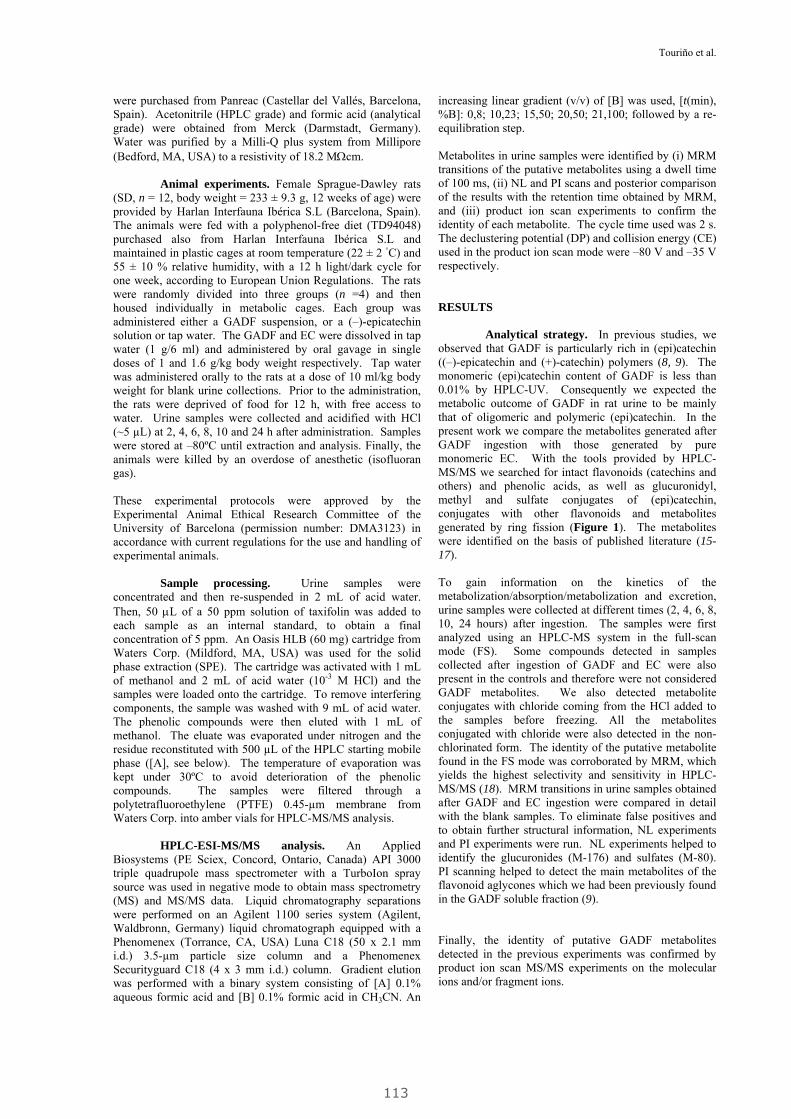
Touriño et al.
were purchased from Panreac (Castellar del Vallés, Barcelona, Spain). Acetonitrile (HPLC grade) and formic acid (analytical grade) were obtained from Merck (Darmstadt, Germany). Water was purified by a Milli-Q plus system from Millipore (Bedford, MA, USA) to a resistivity of 18.2 MΩcm. Animal experiments. Female Sprague-Dawley rats (SD, n = 12, body weight = 233 ± 9.3 g, 12 weeks of age) were provided by Harlan Interfauna Ibérica S.L (Barcelona, Spain). The animals were fed with a polyphenol-free diet (TD94048) purchased also from Harlan Interfauna Ibérica S.L and maintained in plastic cages at room temperature (22 ± 2 ◦C) and 55 ± 10 % relative humidity, with a 12 h light/dark cycle for one week, according to European Union Regulations. The rats were randomly divided into three groups (n =4) and then housed individually in metabolic cages. Each group was administered either a GADF suspension, or a (–)-epicatechin solution or tap water. The GADF and EC were dissolved in tap water (1 g/6 ml) and administered by oral gavage in single doses of 1 and 1.6 g/kg body weight respectively. Tap water was administered orally to the rats at a dose of 10 ml/kg body weight for blank urine collections. Prior to the administration, the rats were deprived of food for 12 h, with free access to water. Urine samples were collected and acidified with HCl (~5 µL) at 2, 4, 6, 8, 10 and 24 h after administration. Samples were stored at –80ºC until extraction and analysis. Finally, the animals were killed by an overdose of anesthetic (isofluoran gas). These experimental protocols were approved by the Experimental Animal Ethical Research Committee of the University of Barcelona (permission number: DMA3123) in accordance with current regulations for the use and handling of experimental animals. Sample processing. Urine samples were concentrated and then re-suspended in 2 mL of acid water. Then, 50 μL of a 50 ppm solution of taxifolin was added to each sample as an internal standard, to obtain a final concentration of 5 ppm. An Oasis HLB (60 mg) cartridge from Waters Corp. (Mildford, MA, USA) was used for the solid phase extraction (SPE). The cartridge was activated with 1 mL of methanol and 2 mL of acid water (10-3 M HCl) and the samples were loaded onto the cartridge. To remove interfering components, the sample was washed with 9 mL of acid water. The phenolic compounds were then eluted with 1 mL of methanol. The eluate was evaporated under nitrogen and the residue reconstituted with 500 µL of the HPLC starting mobile phase ([A], see below). The temperature of evaporation was kept under 30ºC to avoid deterioration of the phenolic compounds. The samples were filtered through a polytetrafluoroethylene (PTFE) 0.45-µm membrane from Waters Corp. into amber vials for HPLC-MS/MS analysis. HPLC-ESI-MS/MS analysis. An Applied Biosystems (PE Sciex, Concord, Ontario, Canada) API 3000 triple quadrupole mass spectrometer with a TurboIon spray source was used in negative mode to obtain mass spectrometry (MS) and MS/MS data. Liquid chromatography separations were performed on an Agilent 1100 series system (Agilent, Waldbronn, Germany) liquid chromatograph equipped with a Phenomenex (Torrance, CA, USA) Luna C18 (50 x 2.1 mm i.d.) 3.5-µm particle size column and a Phenomenex Securityguard C18 (4 x 3 mm i.d.) column. Gradient elution was performed with a binary system consisting of [A] 0.1% aqueous formic acid and [B] 0.1% formic acid in CH3CN. An
increasing linear gradient (v/v) of [B] was used, [t(min), %B]: 0,8; 10,23; 15,50; 20,50; 21,100; followed by a re-equilibration step. Metabolites in urine samples were identified by (i) MRM transitions of the putative metabolites using a dwell time of 100 ms, (ii) NL and PI scans and posterior comparison of the results with the retention time obtained by MRM, and (iii) product ion scan experiments to confirm the identity of each metabolite. The cycle time used was 2 s. The declustering potential (DP) and collision energy (CE) used in the product ion scan mode were –80 V and –35 V respectively. RESULTS Analytical strategy. In previous studies, we observed that GADF is particularly rich in (epi)catechin ((–)-epicatechin and (+)-catechin) polymers (8, 9). The monomeric (epi)catechin content of GADF is less than 0.01% by HPLC-UV. Consequently we expected the metabolic outcome of GADF in rat urine to be mainly that of oligomeric and polymeric (epi)catechin. In the present work we compare the metabolites generated after GADF ingestion with those generated by pure monomeric EC. With the tools provided by HPLC-MS/MS we searched for intact flavonoids (catechins and others) and phenolic acids, as well as glucuronidyl, methyl and sulfate conjugates of (epi)catechin, conjugates with other flavonoids and metabolites generated by ring fission (Figure 1). The metabolites were identified on the basis of published literature (15-17). To gain information on the kinetics of the metabolization/absorption/metabolization and excretion, urine samples were collected at different times (2, 4, 6, 8, 10, 24 hours) after ingestion. The samples were first analyzed using an HPLC-MS system in the full-scan mode (FS). Some compounds detected in samples collected after ingestion of GADF and EC were also present in the controls and therefore were not considered GADF metabolites. We also detected metabolite conjugates with chloride coming from the HCl added to the samples before freezing. All the metabolites conjugated with chloride were also detected in the non-chlorinated form. The identity of the putative metabolite found in the FS mode was corroborated by MRM, which yields the highest selectivity and sensitivity in HPLC-MS/MS (18). MRM transitions in urine samples obtained after GADF and EC ingestion were compared in detail with the blank samples. To eliminate false positives and to obtain further structural information, NL experiments and PI experiments were run. NL experiments helped to identify the glucuronides (M-176) and sulfates (M-80). PI scanning helped to detect the main metabolites of the flavonoid aglycones which we had been previously found in the GADF soluble fraction (9). Finally, the identity of putative GADF metabolites detected in the previous experiments was confirmed by product ion scan MS/MS experiments on the molecular ions and/or fragment ions.
113
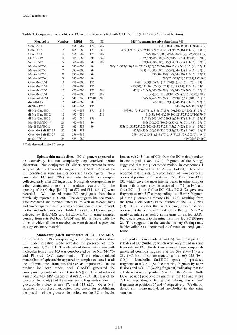
GADF metabolites
Metabolite Number MRM NL PI MS2 fragments (relative abundance %) Gluc-EC-1 1 465→289 176 289 465(1);289(100);245(25);175(6)113(7)Gluc-EC-2 2 465→289 176 289 465 (12)327(9);289(100);245(31);203(12);175(16);151(12);113(10)Gluc-EC-3 3 465→289 176 289 465(1);289(100);245(25);203(9);179(20);137(9)Sulf-EC-1* 4 369→289 80 289 369(37);289(100);245(80);217(33);203(44);137(62)Sulf-EC-2* 5 369→289 80 289 369(16);289(100);245(45);231(23);151(15);137(30)Me-Sulf-EC-1 6 383→303 80 383(13);303(100);259( 22);245(36);220(24);204(15);165(18);151(6);137(11)Me-Sulf-EC-2 7 383→303 80 383(15); 303(100);285(20);244(13);217(16);137(50)Me-Sulf-EC-3 8 383→303 80 383(39);303(100);244(20);217(17);137(33)Me-Sulf-EC-4 9 383→303 80 383(25);303(79);217(25);137(100)Gluc-Me-EC-1 10 479→303 176 479(5);303(100);285(13);244(10);165(6);137(7);113(15)Gluc-Me-EC-2 11 479→303 176 479(10);303(100);285(9);259(15);175(10); 137(10);113(30)Gluc-Me-EC-3 12 479→303 176 289 479(1);313(5);303(20);289(100);245(35);203(11);137(10)Gluc-Me-EC-4 13 479→303 176 289 313(7);303(1);289(100);245(28);203(10);179(8)Gluc-Sulf-EC-1 14 545→369 176;80 289 545(5);465(22);369(10);289(20);271(100);151(15)di-Sulf-EC-1 15 449→369 80 369(100);289(15);245(15);231(19);217(13)di-Gluc-EC-1 16 641→465 176 641(90);465(50);289(20)di-Me-Gluc-EC-1 17 493→289 176 289 493(6);475(8);317(11); 313(10)289(100);245(25);205(11)137(16)di-Me-Gluc-EC-2 18 493→289 289 313(3); 303(6);289(100);245(25);205(10)179(6)di-Me-Gluc-EC-3 19 493→289 176 317(6); 303(100);259(11);244(17);151(10);137(23)Me-di-Sulf-EC-1* 20 463→383 80 383(100);303(40);245(35);217(17);165(9);137(10)Me-di-Sulf-EC-2 21 463→383 80 383(80);303(23);273(100);245(35);231(45);217(25):189(16);137(20)Gluc-Me-Sulf-EC-1* 22 559→383 625(2);335(100);289(4);193(12);175(52);159(91);113(35)Gluc-Me-Sulf-EC-2* 23 559→383 559 (100);313(11);289 (70);245 (9);231(29);203(6);149 (6)tri-Sulf-EC-1* 24 529→289 449(25);369(100)
Table 1: Conjugated metabolites of EC in urine from rats fed with GADF or EC (HPLC-MS/MS identification).
* Only detected in the EC group Epicatechin metabolites. EC oligomers appeared to be extensively but not completely depolymerized before absorption. Non-conjugated EC dimers were present in urine samples taken 2 hours after ingestion of GADF. Most of the EC identified in urine samples occurred as conjugates. Non-conjugated EC (m/z 289) was only detected in samples collected early after EC ingestion. No signals corresponding to either conjugated dimers or to products resulting from the opening of the C-ring ([M–H] – at 579 and 581) (14, 19) were recorded. We detected more EC conjugates than those previously reported (13, 20). The conjugates include mono-glucuronidated and mono-sulfated EC as well as di-conjugates and tri-conjugates resulting from combinations of glucuronidyl, methyl and sulfate moieties. Table 1 lists all the EC conjugates detected by HPLC-MS and HPLC-MS/MS in urine samples coming from rats fed both GADF and EC. A Table with the times at which all these metabolites were detected is provided as supplementary material. Mono-conjugated metabolites of EC. The MRM transition 465→289 corresponding to EC glucuronides (Gluc-EC) under negative mode revealed the presence of three compounds: 1, 2 and 3. The identity of these metabolites with molecular ions at m/z 465 was corroborated by the NL (M-176) and PI (m/z 289) experiments. These glucuronidated metabolites of epicatechin appeared in samples collected at all the different times from rats fed GADF or pure EC. In the product ion scan mode, each Gluc-EC generated the corresponding molecular ion at m/z 465 ([M–H]–) that released a main MS/MS (MS2) fragment at m/z 289 (EC after loss of the glucuronide moiety) and the characteristic fragments of the glucuronide moiety at m/z 175 and 113 (21). Other MS2 fragments from these metabolites were useful for establishing the position of the glucuronide moiety on the EC molecule.
Ions at m/z 245 (loss of CO2 from the EC moiety) and an intense signal at m/z 137 (a fragment of the A-ring) suggested that the glucuronide moiety in compounds 1 and 3 was attached to the A-ring. Indeed, it has been reported that in rats, glucuronidation of (–)-epicatechin occurs at position 7 of the A-ring (22). Thus, Gluc-EC-3 (3), which gave the most intense peaks in urine samples from both groups, may be assigned to 7-Gluc-EC, and Gluc-EC-1 (1) to 5-Gluc-EC. Gluc-EC-2 (2) gave one fragment at m/z 327 corresponding to a B-ring fragment plus the glucuronide moiety (151+176), resulting from the retro Diels-Alder (RDA) fission of the EC C-ring (23). This indicates that in this case, glucuronidation occurred at the positions 3’ or 4’ of the B-ring. Peak 2 is nearly as intense as peak 3 in the urine of rats fed GADF fed rats, in contrast to the urine from rats fed EC (Figure 2). This suggests that the catechol moiety (B-ring) may be bioavailable as a combination of intact and conjugated forms. Two peaks (compounds 4 and 5) were assigned to sulfates of EC (Sulf-EC) which were only found in urine from rats fed EC. Product ion scans of these compounds generated common fragments at m/z 369 ([M–H]–) and 289 (EC, loss of sulfate moiety) and at m/z 245 (EC–CO2). Metabolite Sulf-EC-1 (peak 4) produced fragments at m/z 217 (Sulfate + A-ring fragment by RDA fission) and m/z 137 (A-ring fragment) indicating that the sulfate occurred at position 5 or 7 of the A-ring. Sulf-EC-2 (peak 5) produced fragments at m/z 151 and at m/z 231 corresponding to B-ring and “B-ring plus sulfate” fragments at positions 3’ and 4’ respectively. We did not detect any mono-methylated metabolite in the urine samples.
114
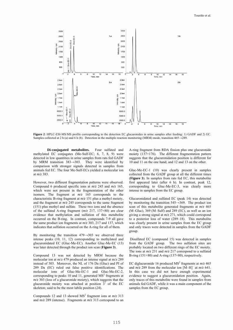
Touriño et al.
Figure 2: HPLC-ESI-MS/MS profile corresponding to the detection EC glucuronides in urine samples after feeding: 1) GADF and 2) EC. Samples collected at 2 h (a) and 6 h (b). Detection in the multiple reaction monitoring (MRM) mode, transition 465→289. Di-conjugated metabolites. Four sulfated and methylated EC conjugates (Me-Sulf-EC; 6, 7, 8, 9) were detected in low quantities in urine samples from rats fed GADF by MRM transition 383→303. They were identified by comparison with stronger signals detected in samples from animals fed EC. The four Me-Sulf-ECs yielded a molecular ion at m/z 383. However, two different fragmentation patterns were observed. Compound 6 produced specific ions at m/z 245 and m/z 165, which were not present in the fragmentation of the other isomers. The fragment at m/z 165 corresponds to the characteristic B-ring fragment at m/z 151 plus a methyl moiety, and the fragment at m/z 245 corresponds to the same fragment (151) plus methyl and sulfate. These two ions and the absence of the sulfated A-ring fragment (m/z 217, 137+80) are clear evidence that methylation and sulfation of this metabolite occurred on the B-ring. In contrast, compounds 7-9 all gave the same product ion fragments at m/z 303, 217 and 137, which indicates that sulfation occurred on the A-ring for all of them. By monitoring the transition 479→303 we observed three intense peaks (10, 11, 12) corresponding to methylated and glucuronidated EC (Gluc-Me-EC). Another Gluc-Me-EC (13) was later detected through the product ion scan (Figure 3). Compound 13 was not detected by MRM because the molecular ion at m/z 479 produced an intense signal at m/z 289 instead of 303. Moreover, the NL of 176 Da (Gluc) and PI of 289 Da (EC) ruled out false positive identifications. The molecular ions of Gluc-Me-EC-1 and Gluc-Me-EC-2, corresponding to peaks 10 and 11, generated MS2 fragments at m/z 303 (loss of a glucuronide moiety), which suggests that the glucuronide moiety was attached at position 3’ of the EC skeleton; said to be the most labile position (24). Compounds 12 and 13 showed MS2 fragment ions at m/z 313 and m/z 289 (intense). Fragments at m/z 313 correspond to an
A-ring fragment from RDA fission plus one glucuronide moiety (137+176). The different fragmentation pattern suggests that the glucuronidation position is different for 10 and 11 on the one hand, and 12 and 13 on the other. Gluc-Me-EC-1 (10) was clearly present in samples collected from the GADF group at all the different times (Figure 3). In samples from rats fed EC, this metabolite first appeared later (after 6 h). In contrast, peak 12, corresponding to Gluc-Me-EC-3, was clearly more intense in samples from the EC group. Glucuronidated and sulfated EC (peak 14) was detected by monitoring the transition 545→369. The product ion scan of this metabolite generated fragments at m/z 465 (M–Gluc), 369 (M–Sulf) and 289 (EC), as well as an ion giving a strong signal at m/z 271, which could correspond to a posterior loss of water (289–18). This metabolite was clearly present in urine samples from the EC group and only traces were detected in samples from the GADF group. Disulfated EC (compound 15) was detected in samples from the GADF group. The two sulfation sites are probably located on two different rings of the EC moiety. The ions at m/z 231 and m/z 217 correspond to a sulfated B-ring (151+80) and A-ring (137+80), respectively. EC diglucuronide 16 produced MS2 fragments at m/z 465 and m/z 289 from the molecular ion [M–H]– at m/z 641. In this case we did not have enough experimental evidence to suggest a glucuronidation position. Again, only traces of this metabolite were found in samples from animals fed GADF, while it was a main component of the samples from the EC group.
115
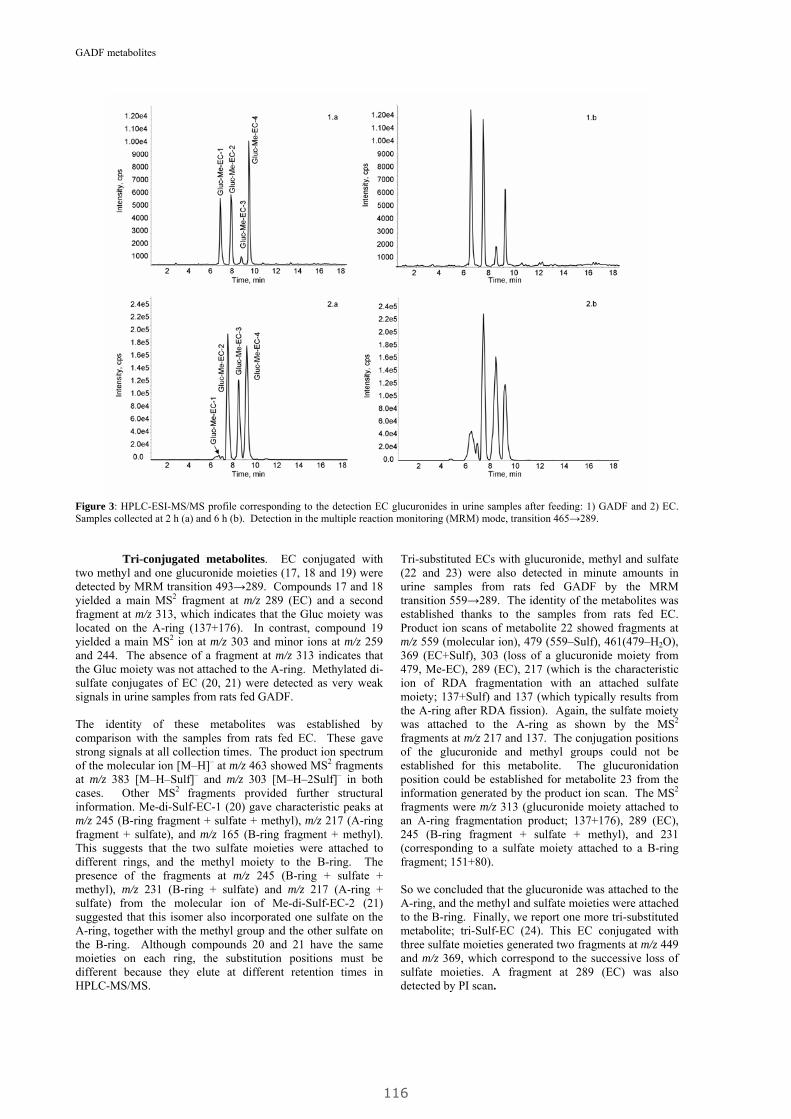
GADF metabolites
Figure 3: HPLC-ESI-MS/MS profile corresponding to the detection EC glucuronides in urine samples after feeding: 1) GADF and 2) EC. Samples collected at 2 h (a) and 6 h (b). Detection in the multiple reaction monitoring (MRM) mode, transition 465→289.
Tri-conjugated metabolites. EC conjugated with two methyl and one glucuronide moieties (17, 18 and 19) were detected by MRM transition 493→289. Compounds 17 and 18 yielded a main MS2 fragment at m/z 289 (EC) and a second fragment at m/z 313, which indicates that the Gluc moiety was located on the A-ring (137+176). In contrast, compound 19 yielded a main MS2 ion at m/z 303 and minor ions at m/z 259 and 244. The absence of a fragment at m/z 313 indicates that the Gluc moiety was not attached to the A-ring. Methylated di-sulfate conjugates of EC (20, 21) were detected as very weak signals in urine samples from rats fed GADF. The identity of these metabolites was established by comparison with the samples from rats fed EC. These gave strong signals at all collection times. The product ion spectrum of the molecular ion [M–H]– at m/z 463 showed MS2 fragments at m/z 383 [M–H–Sulf]– and m/z 303 [M–H–2Sulf]– in both cases. Other MS2 fragments provided further structural information. Me-di-Sulf-EC-1 (20) gave characteristic peaks at m/z 245 (B-ring fragment + sulfate + methyl), m/z 217 (A-ring fragment + sulfate), and m/z 165 (B-ring fragment + methyl). This suggests that the two sulfate moieties were attached to different rings, and the methyl moiety to the B-ring. The presence of the fragments at m/z 245 (B-ring + sulfate + methyl), m/z 231 (B-ring + sulfate) and m/z 217 (A-ring + sulfate) from the molecular ion of Me-di-Sulf-EC-2 (21) suggested that this isomer also incorporated one sulfate on the A-ring, together with the methyl group and the other sulfate on the B-ring. Although compounds 20 and 21 have the same moieties on each ring, the substitution positions must be different because they elute at different retention times in HPLC-MS/MS.
Tri-substituted ECs with glucuronide, methyl and sulfate (22 and 23) were also detected in minute amounts in urine samples from rats fed GADF by the MRM transition 559→289. The identity of the metabolites was established thanks to the samples from rats fed EC. Product ion scans of metabolite 22 showed fragments at m/z 559 (molecular ion), 479 (559–Sulf), 461(479–H2O), 369 (EC+Sulf), 303 (loss of a glucuronide moiety from 479, Me-EC), 289 (EC), 217 (which is the characteristic ion of RDA fragmentation with an attached sulfate moiety; 137+Sulf) and 137 (which typically results from the A-ring after RDA fission). Again, the sulfate moiety was attached to the A-ring as shown by the MS2
fragments at m/z 217 and 137. The conjugation positions of the glucuronide and methyl groups could not be established for this metabolite. The glucuronidation position could be established for metabolite 23 from the information generated by the product ion scan. The MS2
fragments were m/z 313 (glucuronide moiety attached to an A-ring fragmentation product; 137+176), 289 (EC), 245 (B-ring fragment + sulfate + methyl), and 231 (corresponding to a sulfate moiety attached to a B-ring fragment; 151+80). So we concluded that the glucuronide was attached to the A-ring, and the methyl and sulfate moieties were attached to the B-ring. Finally, we report one more tri-substituted metabolite; tri-Sulf-EC (24). This EC conjugated with three sulfate moieties generated two fragments at m/z 449 and m/z 369, which correspond to the successive loss of sulfate moieties. A fragment at 289 (EC) was also detected by PI scan.
116

Touriño et al.
Table 2: Identification and occurrence of phenolic acids in urine from rats fed with GADF or EC.
Metabolite MRM
Parent MRM
Daugther
MS2 fragments (% relative abundance)
3-hydroxyphenylvaleric acid (3-HPhVA) 193→175 — —
3,4-di-hydroxyphenylpropionic acid (DHPhPA) 181→137 — 181(5);137(35);135(47);121(73);109(3)
4-hydroxyphenylpropionic acid (4-HPhPA) 165→121 — 165(30);121(56);93(100);77(100)
caffeic acid 179→135 — 135(100);107(9);89(8)
p-coumaric acid 163→119 — 163(1);119(100);93(4);91(2)
m-coumaric acid 163→119 — 163(3);119(100);93(5);91(10)
3,4-di-hydroxyphenylacetic acid (DHPhAcA) 167→123 — 123(100);105(7);95(17)
3-hydroxyphenylacetic acid (3-HPAcA) 151→107 — 107(100);91(23)
4-hydroxybenzoic acid (4-HBA) 137→ 93 — 137(57);119(8);108(100);93(64)
Sulf-di-hydroxyphenylpropionic acid (Sulf-DHPhPA) 261→181 181→137 261(10);199(37);181(85);137(100)
Gluc-di-hydroxyphenylpropionic acid (Gluc-DHPhPA) 357→181 181→137 181(58);166(100);121(9)
Sulf-hydroxyphenylpropionic acid (Sulf-HPhPA) 245→165 165→121 245(5);165(35);121(100);119(40);106(18)
Sulf-coumaric acid 243→163 163→119 243(5);163(15);119(100)
Phenolic acids. Both non-conjugated and conjugated phenolic acids were detected and identified in urine samples from rats fed GADF. Only a limited number of non-conjugated acids were detected in samples from the EC group. The results are summarized in Table 2. The species listed were confirmed by the use of standards and gave MS signals that were clearly stronger than those from the control animals. Some of phenolic acids described in the literature as metabolites of EC (e.g., 3-HBA) are not included in the list because the signals they yielded were no stronger than those from the control samples obtained from non-supplemented animals. A Table with the times at which all these metabolites were detected is provided as supplementary material. Phenolic acids derived from GADF diet. Hydroxyphenylvaleric acid (HPhVA) detected at 24 h was one of the main metabolites in rats fed GADF. p-coumaric and m-coumaric were detected and identified in GADF samples collected all times. p-coumaric signals were more intense than m-coumaric signals in all the samples, and were clearly stronger at 8 h and 10 h. 3,4-di-hydroxyphenylacetic acid (DHPhAcA) was detected at 24 h by MRM transition 167→123. Hydroxyphenylacetic acid (HPhAcA) and 4-hydroxybenzoic acid (4-HBA) were also detected from 6 to 24h. Conjugated phenolic acids were detected in urine samples from the GADF group by MRM and product ion scan on parent/daughter pairs of ions. 3,4-DHPhPA conjugated with either a sulfate (Figure 4a) or a glucuronide moiety gave transitions: 261→181 plus 181→137, and 357→181 plus 181→137, respectively, and were confirmed by the product ion scan. We also detected sulfated coumaric acid (243→163 plus 163→119) as early as 2 h after ingestion of GADF and sulfated HPhPA (245→165) some time later (from 8 to 24 h) (Figure 4b). Phenolic acids derived from EC diet. The colonic metabolization of pure EC gave different phenolic acids with the three-carbon atom scaffold. Caffeic acid (179→135) gave signals of increasing intensity from 8 to 24 h. 3,4-DHPhPA, also called dihydrocaffeic acid (181→137) was only detected after 24 h. 4-HPhPA was detected from 8 to 24 h. p-coumaric
and m-coumaric acids gave signals of increasing intensity from 8 to 24 h. We also detected shorter phenolic acids after EC ingestion. 3-HPhAcA and 4-HBA appeared at 10 and 24 h. We also observed MRM signals corresponding to sulfated caffeic acid, which were confirmed by a second transition of the free acids at the same retention time. We do not include this metabolite in Table 2 because the less sensitive product ion scan did not yield any fragment. DISCUSSION Food polyphenols such as monomeric (epi)catechins and phenolic acids are rapidly glucuronidated and methylated in the small intestine (25), absorbed into the bloodstream and further conjugated (glucuronidated, sulfated) in the liver of both rats and humans (26-29). Those species that are not absorbed in the small intestine (e.g., polymeric (epi)catechins) reach the colon where they are fermented by the colonic microbiota and absorbed in the form of smaller phenolic acids (30). These smaller species may in turn be conjugated in the liver. The metabolites found in urine are the result of all these processes and may have exerted their biological effect during the period while they crossed membranes and circulated in the bloodstream. GADF contains a complex mixture the polyphenols including monomers, oligomers and polymers of EC, anthocyanins, flavonols and hydroxycinnamic acids associated with a fiber matrix of both soluble and insoluble polymers such as polycaccharides and lignins (8, 9) which may influence the absorption of the putatively bioactive GADF components. We report here that the phenolic metabolites in rat urine after the ingestion of GADF are mainly conjugates of (–)-epicatechin and phenolic acids. An important question is where the different polyphenolic components of GADF are absorbed and metabolized, as the putative protective effects of the polyphenols will be exerted where they are bioavailable..
117
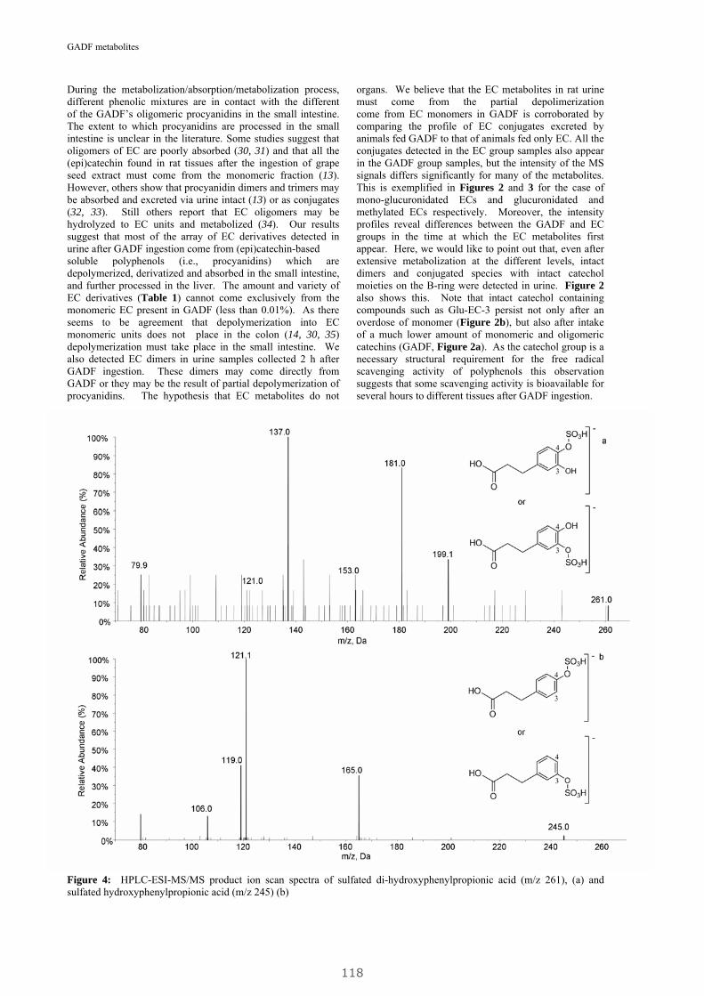
GADF metabolites
During the metabolization/absorption/metabolization process, different phenolic mixtures are in contact with the different
organs. We believe that the EC metabolites in rat urine must come from the partial depolimerization
of the GADF’s oligomeric procyanidins in the small intestine. The extent to which procyanidins are processed in the small intestine is unclear in the literature. Some studies suggest that oligomers of EC are poorly absorbed (30, 31) and that all the (epi)catechin found in rat tissues after the ingestion of grape seed extract must come from the monomeric fraction (13). However, others show that procyanidin dimers and trimers may be absorbed and excreted via urine intact (13) or as conjugates (32, 33). Still others report that EC oligomers may be hydrolyzed to EC units and metabolized (34). Our results suggest that most of the array of EC derivatives detected in urine after GADF ingestion come from (epi)catechin-based soluble polyphenols (i.e., procyanidins) which are depolymerized, derivatized and absorbed in the small intestine, and further processed in the liver. The amount and variety of EC derivatives (Table 1) cannot come exclusively from the monomeric EC present in GADF (less than 0.01%). As there seems to be agreement that depolymerization into EC monomeric units does not place in the colon (14, 30, 35) depolymerization must take place in the small intestine. We also detected EC dimers in urine samples collected 2 h after GADF ingestion. These dimers may come directly from GADF or they may be the result of partial depolymerization of procyanidins. The hypothesis that EC metabolites do not
come from EC monomers in GADF is corroborated by comparing the profile of EC conjugates excreted by animals fed GADF to that of animals fed only EC. All the conjugates detected in the EC group samples also appear in the GADF group samples, but the intensity of the MS signals differs significantly for many of the metabolites. This is exemplified in Figures 2 and 3 for the case of mono-glucuronidated ECs and glucuronidated and methylated ECs respectively. Moreover, the intensity profiles reveal differences between the GADF and EC groups in the time at which the EC metabolites first appear. Here, we would like to point out that, even after extensive metabolization at the different levels, intact dimers and conjugated species with intact catechol moieties on the B-ring were detected in urine. Figure 2 also shows this. Note that intact catechol containing compounds such as Glu-EC-3 persist not only after an overdose of monomer (Figure 2b), but also after intake of a much lower amount of monomeric and oligomeric catechins (GADF, Figure 2a). As the catechol group is a necessary structural requirement for the free radical scavenging activity of polyphenols this observation suggests that some scavenging activity is bioavailable for several hours to different tissues after GADF ingestion.
Figure 4: HPLC-ESI-MS/MS product ion scan spectra of sulfated di-hydroxyphenylpropionic acid (m/z 261), (a) and sulfated hydroxyphenylpropionic acid (m/z 245) (b)
118
Touriño et al.
The EC metabolites probably came from oligomers with low degree of polymerization (dimers, trimers). Bulkier oligomers and polymers are associated with proteins (36). The protein-associated material, as well as both soluble and insoluble procyanidins more or less associated with the fiber matrix (polysaccharides and lignin) and maybe the remaining monomers, eventually reach the colon, where the colonic microbiota break them into smaller compounds (phenolic acids) as described for both rats (30) and humans (11, 37). It is believed that procyanidins are directly degraded to phenolic acids, without previous depolymerization into (epi)catechin units (14). In agreement with the observations from other authors working with purified oligomers (14, 30, 35), we found that the urine samples from animals fed GADF contained more phenolic acids than the samples from animals given only monomeric EC. The GADF colonic metabolites (Table 2) most probably come from the colonic fermentation of polymeric EC. The main MS signals corresponded to 3-HPhVA, 4-HPhPA and 3-HPhAcA. All these metabolites may be generated by ring cleavage of the EC moieties within the procyanidin polymers (14, 35). m- and p-coumaric acids were detected in samples from both the GADF and EC groups. A small proportion of the coumaric acids may come directly from the fiber supplement, as they are minor components of GADF (9). Since coumaric acids have been shown to be absorbed rapidly in the small intestine (38), they may account for some of the phenolics detected soon after ingestion (2 h). The rest must be generated by degradation of the EC skeleton. We detected sulfated and glucuronidated conjugates of hydroxyphenyl acids only in the GADF group samples. These metabolites (e.g. Sulf-DHPhPA and Sulf-HPhPA, Figure 4) had not been detected from procyanidins before and may be produced in the liver from the colonic degradation products. Again, as for the EC dimers and EC metabolites with intact catechol moieties, some of the phenolic acids existed in their non-conjugated form and may provide scavenging potential for several hours after GADF ingestion. However, it is evident that GADF polyphenols are extensively conjugated and the levels of free catechols may be too low to have any significant effect. In fact, many authors believe that the scavenging capacity of plant polyphenols is negligible “in vivo”. While we have to agree in part, we certainly do not think that this means that plant polyphenols, in particular procyanidins, exert no significant antioxidant effect. Apart from the observation that the circulating free catechol may not be negligible, the extent of metabolization shows how polyphenols are treated by the living organism as harmful. In fact, it is known that polyphenols may be pro-oxidant depending on their nature and concentration. Thus, they may stimulate the endogenous antioxidant systems by acting as mild pro-oxidants. Similarly, calorie restriction and moderate exercise are weak prooxidant insults that may result in antioxidant responses “in vivo” (39, 40). The antioxidant/pro-oxidant balance provided by polyphenols and their metabolites may result in an overall antioxidant effect of GADF, and of fruit and vegetables in general. For several hours after GADF ingestion, a great variety of phenolic species are in contact with the gut tissues before, during and after metabolization, and many of them (mainly mono and polyconjugated epicatechin and free or conjugated smaller phenolic acids) are systemically bioavailable before being excreted. Some of these metabolites include the free catechol moiety. The metabolic outcome of GADF essentially corresponds to the degradation products of epicatechin oligomers and polymers
ACKNOWLEDGEMENT GADF was a generous gift from Prof. Fulgencio Saura-Calixto, ICTAN-CSIC. The expert assistance of Dr. Olga Jáuregui from the Servies Científico-Tècnics de la Universitat de Barcelona is gratefully appreciated. Language revision by Christopher Evans is also appreciated. Supporting Information Available: Tables summarizing the occurrence of the metabolites at the different times. This material is available free of charge via the Internet at http://pubs.acs.org LITERATURE CITED (1) Slavin, J. L., Dietary fiber and body weight. Nutrition 2005, 21, (3), 411-418. (2) Bingham, S. A.; Day, N. E.; Luben, R.; Ferrari, P.; Slimani, N.; Norat, T.; Clavel-Chapelon, F.; Kesse, E.; Nieters, A.; Boeing, H.; Tjnneland, A.; Overvad, K.; Martinez, C.; Dorronsoro, M.; Gonzalez, C. A.; Key, T. J.; Trichopoulou, A.; Naska, A.; Vineis, P.; Tumino, R.; Krogh, V.; Bueno-de-Mesquita, H. B.; Peeters, P. H. M.; Berglund, G.; Hallmans, G.; Lund, E.; Skeie, G.; Kaaks, R.; Riboli, E., Dietary fibre in food and protection against colorectal cancer in the European Prospective Investigation into Cancer and Nutrition (EPIC): an observational study. Lancet 2003, 361, (9368), 1496-1501. (3) Dana, E. K., Dietary fiber, inflammation, and cardiovascular disease. Mol. Nutr. Food Res. 2005, 49, (6), 594-600. (4) Klenovv, S.; Glei, M.; Haber, B.; Owen, R.; Pool-Zobel, B. L., Carob fibre compounds modulate parameters of cell growth differently in human HT29 colon adenocarcinoma cells than in LT97 colon adenoma cells. Food Chem. Toxicol. 2008, 46, (4), 1389-1397. (5) Fardet, A.; Rock, E.; Rémésy, C., Is the in vitro antioxidant potential of whole-grain cereals and cereal products well reflected in vivo? J. Cereal Sci. 2008, 48, (2), 258-276. (6) Saura-Calixto, F.; Larrauri García, J. A. Concentrate of natural antioxidant dietetic fiber from grape, and preparation process, In CSIC: ES2130092 A1, 1999. (7) Goñi, I.; Martín, N.; Saura-Calixto, F., In vitro digestibility and intestinal fermentation of grape seed and peel. Food Chem. 2005, 90, (1-2), 281-286. (8) Pérez-Jiménez, J.; Serrano, J.; Tabernero, M.; Arranz, S.; Díaz-Rubio, M. E.; García-Diz, L.; Goñi, I.; Saura-Calixto, F., Effects of grape antioxidant dietary fiber in cardiovascular disease risk factors. Nutrition 2008, 24, (7-8), 646-653. (9) Touriño, S.; Fuguet, E.; Jáuregui, O.; Saura-Calixto, F.; Cascante, M.; Torres, J. L., High-resolution liquid chromatography/electrospray ionization time-of-flight mass spectrometry combined with liquid chromatography/electrospray ionization tandem mass spectrometry to identify polyphenols from grape antioxidant dietary fiber. Rapid Commun. Mass Spectrom. 2008, 22, (22), 3489-3500. (10) Taubert, D.; Roesen, R.; Schomig, E., Effect of cocoa and tea intake on blood pressure: a meta-analysis. Arch. Intern. Med. 2007, 167, (7), 626-634. (11) Ward, N. C.; Croft, K. D.; Puddey, I. B.; Hodgson, J. M., Supplementation with grape seed polyphenols
119

GADF metabolites
results in increased urinary excretion of 3-hydroxyphenylpropionic acid, an important metabolite of proanthocyanidins in humans. J. Agric. Food Chem. 2004, 52, (17), 5545-5549. (12) Baba, S.; Osakabe, N.; Natsume, M.; Muto, Y.; Takizawa, T.; Terao, J., In vivo comparison of the bioavailability of (+)-Catechin, (–)-Epicatechin and their mixture in orally administered rats. J. Nutr. 2001, 131, (11), 2885-2891. (13) Tsang, C.; Auger, C.; Mullen, W.; Bornet, A.; Rouanet, J. M.; Crozier, A.; Teissedre, P. L., The absorption, metabolism and excretion of flavan-3-ols and procyanidins following the ingestion of a grape seed extract by rats. Br. J. Nutr. 2005, 94, (2), 170-181. (14) Appeldoorn, M. M.; Vincken, J.-P.; Aura, A.-M.; Hollman, P. C. H.; Gruppen, H., Procyanidin dimers are metabolized by human microbiota with 2-(3,4-Dihydroxyphenyl)acetic acid and 5-(3,4-Dihydroxyphenyl)-g-valerolactone as the major metabolites. J. Agric. Food Chem. 2009, 57, (3), 1084-1092. (15) Spencer, J. P. E., Metabolism of tea flavonoids in the gastrointestinal tract. J. Nutr. 2003, 133, (10), 3255S-3261S. (16) Walle, T., Absorption and metabolism of flavonoids. Free Radical Biol. Med. 2004, 36, (7), 829-837. (17) Rechner, A. R.; Kuhnle, G.; Bremner, P.; Hubbard, G. P.; Moore, K. P.; RiceEvans, C. A., The metabolic fate of dietary polyphenols in humans. Free Radical Biol. Med. 2002, 33, (2), 220-235. (18) Liu, D. Q.; Hop, C. E. C. A., Strategies for characterization of drug metabolites using liquid chromatography-tandem mass spectrometry in conjunction with chemical derivatization and on-line H/D exchange approaches. J. Pharm. Biomed. Anal. 2005, 37, (1), 1-18. (19) Groenewoud, G.; Hundt, H. K. L., The microbial metabolism of condensed (+)-catechins by the rat-cecal microflora. Xenobiotica 1986, 16, (2), 99-107. (20) Sang, S.; Lee, M. J.; Yang, I.; Buckley, B.; Yang, C. S., Human urinary metabolite profile of tea polyphenols analyzed by liquid chromatography/electrospray ionization tandem mass spectrometry with data-dependent acquisition. Rapid Commun. Mass Spectrom. 2008, 22, (10), 1567-1578. (21) Levsen, K.; Schiebel, H.-M.; Behnke, B.; Dötzer, R.; Dreher, W.; Elend, M.; Thiele, H., Structure elucidation of phase II metabolites by tandem mass spectrometry: an overview. J. Chromatogr. A 2005, 1067, (1-2), 55-72. (22) Natsume, M.; Osakabe, N.; Oyama, M.; Sasaki, M.; Baba, S.; Nakamura, Y.; Osawa, T.; Terao, J., Structures of (–)-epicatechin glucuronide identified from plasma and urine after oral ingestion of (–)-epicatechin: differences between human and rat. Free Radical Biol. Med. 2003, 34, (7), 840-849. (23) Gu, L.; Kelm, M. A.; Hammerstone, J. F.; Beecher, G.; Holden, J.; Haytowitz, D.; Prior, R. L., Screening of foods containing proanthocyanidins and their structural characterization using LC-MS/MS and thiolytic degradation. J. Agric. Food Chem. 2003, 51, (25), 7513-7521. (24) Cren-Olivé, C.; Déprez, S.; Lebrun, S.; Coddeville, B.; Rolando, C., Characterization of methylation site of monomethylflavan-3-ols by liquid chromatography/electrospray ionization tandem mass spectrometry. Rapid Commun. Mass Spectrom. 2000, 14, (23), 2312-2319. (25) Kuhnle, G.; Spencer, J. P. E.; Schroeter, H.; Shenoy, B.; Debnam, E. S.; Srai, S. K. S.; Rice-Evans, C.; Hahn, U., Epicatechin and catechin are O-methylated and glucuronidated in the small intestine. Biochem. Biophys. Res. Commun. 2000, 277, (2), 507-512. (26) Okushio, K.; Suzuki, M.; Matsumoto, N.; Nanjo, F.; Hara, Y., Identification of (–)-Epicatechin metabolites and their
metabolic fate in the rat. Drug Metab. Dispos. 1999, 27, (2), 309-316. (27) Vaidyanathan, J. B.; Walle, T., Glucuronidation and sulfation of the tea flavonoid (–)-Epicatechin by the human and rat enzymes. Drug Metab. Dispos. 2002, 30, (8), 897-903. (28) Poquet, L.; Clifford, M. N.; Williamson, G., Investigation of the metabolic fate of dihydrocaffeic acid. Biochem. Pharmacol. 2008, 75, (5), 1218-1229. (29) Matsui, Y.; Nakamura, S.; Kondou, N.; Takasu, Y.; Ochiai, R.; Masukawa, Y., Liquid chromatography-electrospray ionization-tandem mass spectrometry for simultaneous analysis of chlorogenic acids and their metabolites in human plasma. J. Chromatogr. B 2007, 858, (1-2), 96-105. (30) Gonthier, M. P.; Donovan, J. L.; Texier, O.; Felgines, C.; Rémésy, C.; Scalbert, A., Metabolism of dietary procyanidins in rats. Free Radical Biol. Med. 2003, 35, (8), 837-844. (31) Scalbert, A.; Williamson, G., Dietary intake and bioavailability of polyphenols. J. Nutr. 2000, 130, (8), 2073S-2085S. (32) Prasain, J. K.; Barnes, S., Metabolism and bioavailability of flavonoids in chemoprevention: current analytical strategies and future prospectus. Mol. Pharm. 2007, 4, (6), 846-864. (33) Shoji, T.; Masumoto, S.; Moriichi, N.; Akiyama, H.; Kanda, T.; Ohtake, Y.; Goda, Y., Apple procyanidin oligomers absorption in rats after oral administration: analysis of procyanidins in plasma using the porter method and high-performance liquid chromatography/tandem mass spectrometry. J. Agric. Food Chem. 2006, 54, (3), 884-892. (34) Spencer, J. P. E.; Chaudry, F.; Pannala, A. S.; Srai, S. K.; Debnam, E.; Rice-Evans, C., Decomposition of cocoa procyanidins in the gastric milieu. Biochem. Biophys. Res. Commun. 2000, 272, (1), 236-241. (35) Déprez, S.; Brezillon, C.; Rabot, S.; Philippe, C.; Mila, I.; Lapierre, C.; Scalbert, A., Polymeric proanthocyanidins are catabolized by human colonic microflora into low-molecular-weight phenolic acids. J. Nutr. 2000, 130, (11), 2733-2738. (36) Sarni-Manchado, P.; Cheynier, V.; Moutounet, M., Interactions of grape seed tannins with salivary proteins. J. Agric. Food Chem. 1999, 47, (1), 42-47. (37) Rios, L. Y.; Gonthier, M. P.; Rémésy, C.; Mila, I.; Lapierre, C.; Lazarus, S. A.; Williamson, G.; Scalbert, A., Chocolate intake increases urinary excretion of polyphenol-derived phenolic acids in healthy human subjects. Am. J. Clin. Nutr. 2003, 77, (4), 912-918. (38) Konishi, Y.; Hitomi, Y.; Yoshioka, E., Intestinal absorption of p-coumaric and gallic acids in rats after oral administration. J. Agric. Food Chem. 2004, 52, (9), 2527-2532. (39) Schulz, T. J.; Zarse, K.; Voigt, A.; Urban, N.; Birringer, M.; Ristow, M., Glucose restriction extends Caenorhabditis elegans life span by inducing mitochondrial respiration and increasing oxidative stress. Cell Metab. 2007, 6, (4), 280-293. (40) Ascensao, A.; Magalhaes, J. F.; Soares, J. M.; Ferreira, R. M.; Neuparth, M. J.; Appell, H. J.; Duarte, J. A., Cardiac mitochondrial respiratory function and oxidative stress: The role of exercise. Int. J. Sports Med. 2005, 26, (4), 258-267.
This work was supported by the Spanish Ministry of Education and Science (research grants AGL2004-
120
Touriño et al.
07579-C04-02, -03; AGL2006-12210-C03-02/ALI; SAF2008-00164); doctoral fellowship to S.T. and post-doctoral contract to E.F.); the European Commission (Etherpaths project KBBE-grant agreement nº222639); ISCIII-RTICC (RD06/0020/0046); and Generalitat de Catalunya (2005SGR00204).
121


5. DISCUSIÓN GENERAL

5. Discusión general
125
5.1. Relación estructura/efecto de proantocianidinas
La discusión que se expone a continuación integra las publicaciones troncales generas de
los estudios con las fracciones polifenólicas de corteza de pino y hamamelis (publicación
1; apartado 4.1.1 y publicación 2; apartado 4.1.2) y otras realizadas en colaboración
(Anexos; Apartado 8.1) que han permitido llegar a una idea global de la influencia de
la estructura de las proantocianidinas, (grado de polimerización y porcentaje de
galoización) sobre sus efectos químicos y biológicos.
La actividad antioxidante de las (epi)catequinas y proantocianidinas ha sido atribuida en
parte a la presencia del grupo catecol o pirogalol en el anillo B, capaz de reducir con
elevada eficacia radicales libres del medio (Rice-Evans et al., 1996) mediante donación
de hidrógenos y/o transferencia electrónica. Por otro lado la capacidad de transferencia
electrónica puede resultar en una actividad pro-oxidante, especialmente en compuestos
más reductores como (epi)galocatequinas (Kondo et al., 1999), cuando son capaces de
transferir un electrón al oxígeno y formar radical anión superóxido que a su vez puede
generar el radical hidroxilo. Los mecanismos por los cuales actúan las proantocianidinas
para ejercer posibles efectos beneficiosos sobre la salud todavía no son claros. Cada vez
son más los estudios que descartan la idea clásica de que los polifenoles actúan
exclusivamente como agentes captadores de radicales libres. Primero los polifenoles
pueden ser tanto antioxidantes como pro-oxidantes. Segundo, los compuestos fenólicos
son ampliamente metabolizados in vivo alterando sus potenciales redox.
La presente tesis pretende ser una aportación a la discusión sobre los efectos de los
polifenoles, principalmente de las proantocianidinas. El grado de polimerización y el
porcentaje de galoización son factores que influyen en los efectos de las
proantocianidinas, es por ello que en el primer bloque de la tesis y con el fin de evaluar
la influencia de estos dos factores sobre sus efectos químicos y biológicos se generaron
fracciones de proantocianidinas con diferente composición en cuanto a grado de
polimerización y porcentaje de galoización. Fracciones de corteza de pino (Pinus pinaster)
constituidas por procianidinas, (epi)catequinas exentas en su estructura de ésteres de
galato (Touriño et al., 2005), fracciones de bagazo de uva (Vitis vinifera) en las que se
incluían mayoritariamente procianidinas con un porcentaje en ésteres de galato
intermedio (Torres et al., 2002) y fracciones de hamamelis (Hamamelis virginiana) con
un elevado porcentaje de galoización gracias a que su composición incluye una mezcla
rica de (epi)galocatequinas (prodelfinidinas), (epi)catequinas (procianidinas) y
galotaninos (unidades de ácido gálico unidos a una glucosa)(Touriño et al., 2008a)
(Lizárraga et al., 2008).
5.1.1 Evaluación de la influencia del grado de polimerización
La influencia del grado de polimerización sobre la actividad antioxidante parece evidente,
según los resultados de nuestros estudios (Touriño et al., 2005) y en los de muchos otros

5.1 Relación estructura/efecto de procianidinas
126
autores (Burda et al., 2001;Cao et al., 1997;Heim et al., 2002). Un aumento en el
número de grupos catecol del anillo B es de esperar que resulte en un incremento de la
capacidad antioxidante de las fracciones. Sin embargo, los resultados de las fracciones
de bagazo de uva (Torres et al., 2002) en los distintos ensayos antioxidantes mostraron
que fracciones de similar grado de galoización con un grado de polimerización en torno a
2 (Fracción X) y 4 (Fracción XI) mostraron similar eficacia antioxidante. Es decir, existe
un punto a partir del cual la capacidad antioxidante de las proantocianidinas deja de
aumentar aún cuando el grado de polimerización vaya en incremento. Este resultado
coincidió con el obtenido por Gaulejac y colaboradores (De Gaulejac et al., 1999) en un
estudio realizado igualmente con proantocianidinas de uva. Los autores sugirieron que la
proporción directa entre grado de polimerización y actividad antioxidante deja de existir a
partir de un grado de polimerización entre 2 y 3 posiblemente porque se produce un
plegamiento de los oligómeros que esconde los grupos hidroxilos activos.
En el caso de las fracciones de corteza de pino, exentas de galato, se observó el mismo
efecto para un grado de polimerización en torno a 4 (publicación 1; apartado 4.1.1)
(Touriño et al., 2005). En contraste con los resultados con procianidinas de uva, se
comprobó, gracias a un estudio realizado en colaboración con el grupo de la Escuela
Técnica Superior de Ingeniería de la Universidad de Santiago (Anexo 8.1 III) (Jerez et
al., 2007), que la capacidad antioxidante fracciones obtenidas de corteza de pino (Pinus
pinaster y Pinus radiata) seguía luego aumentando hasta un grado de polimerización en
torno a 7. La causa más plausible en la diferencia de los resultados, entre las fracciones
de bagazo de uva y pino, es la presencia/ausencia de ésteres de galato en la estructura
de las proantocianidinas que podrían contribuir a un plegamiento más compacto de la
molécula.
Los métodos más utilizados (principalmente DPPH) para medir la capacidad reductora de
radicales libres miden la transferencia de átomos de hidrógeno aunque se cree que el
mecanismo redox principal es la transferencia electrónica. Además, radicales libres
estables como el DPPH son parcialmente reactivos frente a solventes como el metanol.
El radical HNTTM sintetizado en nuestro laboratorio (Torres et al., 2003) es
exclusivamente sensible a la transferencia de electrones debido a la imposibilidad de
captar hidrógenos por impedimento estérico del radical por lo que permite asegurar que
un polifenol actúe por transferencia electrónica minimizando reacciones con el solvente.
Al poner en contacto las fracciones de bagazo de uva y hamamelis con el radical HNTTM
se observó que las fracciones que mostraban mayor actividad antioxidante medida por
DPPH o TEAC, mostraron una mayor transferencia de electrones. Los resultados de las
fracciones de pino (exentas de galato) mostraron un incremento de la capacidad de
donación de hidrógenos ligada al incremento en el grado de polimerización y mayor que
el incremento en la capacidad de transferencia electrónica. Este resultado podría ser
particularmente importante si se tiene en cuenta que una elevada capacidad de
transferencia electrónica, como se ha comentado anteriormente, podría estar relacionada
5. Discusión general
127
con efectos pro-oxidantes. Las fracciones oligoméricas de pino mostraron una buena
eficacia antioxidante (método DPPH) manteniendo una baja transferencia de electrones
(método HNTTM) (Touriño et al., 2005).
Uno de los parámetros más importantes para la conservación de gran parte de los
alimentos procesados que se encuentran en forma de emulsión, desde zumos (<1%)
hasta mayonesas (>80%), es la inhibición del enranciamiento lipídico. Además, los
sistemas biológicos se asemejan más a emulsiones que a soluciones concentradas. Por
ello, se evaluó la capacidad de inhibición de la peroxidación lipídica en emulsiones
aceite/agua (Torres et al., 2002;Touriño et al., 2005). Los resultados mostraron que los
oligómeros son más eficientes que los monómeros en emulsiones. Dicho resultado
concordó con la observación de que los compuestos hidrófobos tienden a ser acumulados
en la interfase aceite/agua, mientras que los compuestos hidrófilos son menos activos
debido a que se disuelven en la fase acuosa desde la cual es difícil de acceder a los
radicales lipídicos (Frankel, 2005). El carácter hidrófobo de las proantocianidinas es
fácilmente deducible por su comportamiento en RP-HPLC, es decir, cuanto mayor es el
tiempo de retención más hidrofobicidad presentan. La naturaleza hidrofóbica de las
proantocianidinas en combinación con los grupos hidroxilos hidrófilos permite la
exposición de las proantocianidinas en ambas fases de la emulsión. Entre las fracciones
oligoméricas de diferentes grados de polimerización no se observaron diferencias
significativas en los resultados, aunque sí se observó una tendencia a un incremento de
la inhibición de la oxidación lipídica con respecto a un aumento en el grado de
polimerización (Pazos et al., 2005;Torres et al., 2002;Touriño et al., 2005). La similar
eficacia observada entre las fracciones podría ser debida a la posibilidad que tienen las
proantocianidinas de establecer interacciones hidrófobas y/o hidrofílicas en función del
medio, lo cual corroboraría la hipótesis de que la distribución en las diferentes fases
puede llegar a ser más importante que la actividad antioxidante en sí (Plumb et al.,
1998). En los estudios de inhibición de oxidación lipídica de membranas celulares
realizados con estás fracciones se obtuvieron resultados muy similares a los obtenidos en
emulsiones, las fracciones monoméricas fueron muy poco activas mientras que entre las
fracciones oligoméricas se observo un ligero incremento de la actividad captadora del
radical peroxilo con relación a un aumento en el grado de polimerización (Anexo 8.1 V)
(Ugartondo et al., 2007).
En los ensayos de evaluación de inhibición de la proliferación realizados con cultivos de
células tumorales de melanoma humano (SK-MEL-28) se observó que las fracciones
monoméricas tanto de pino como de uva mostraron menor actividad antiproliferativa que
las fracciones oligoméricas. Entre las fracciones oligoméricas de ambos extractos (bagazo
de uva y corteza de pino) no se observó un incremento significativo en función del grado
de polimerización. En cambio, el grado de polimerización mostró ser más importante en
los ensayos realizados sobre inhibición del ciclo celular, donde las fracciones tanto de
corteza de pino como de bagazo de uva aumentaron a medida que aumentaba su grado
de polimerización (Anexo 8.1 IV)(Lizárraga et al., 2007).

5.1 Relación estructura/efecto de procianidinas
128
5.1.2 Evaluación de la influencia del porcentaje de galoización (grupos
galato) y de galo(epi)catequinas (pirogalol en el anillo B)
El efecto de los grupos galato en la estructura de las proantocianidinas se evidenció en el
estudio con fracciones de hamamelis (publicación 2; apartado 4.1.2) (Touriño et al.,
2008a). El incremento en el porcentaje de galoización que supone un mayor número de
grupos hidroxilo resultó en un aumento de la capacidad de donación de átomos de
hidrógeno. Las fracciones de hamamelis presentaron un elevado porcentaje de
galoización procedente de procianidinas (epicatequíngalato), prodelfinidinas
(epigalocatequíngalato) y de los taninos hidrolizables (ésteres de ácido gálico). Las
fracciones de hamamelis mostraron una capacidad antioxidante 1,5 y 3 veces mayor que
las fracciones homologas de bagazo de uva (porcentaje de galoización medio) y corteza
de pino (exentas de galatos) respectivamente. Un resultado a remarcar es la similar
capacidad de transferencia de electrones que presentaron una de las fracciones
compuestas fundamentalmente de taninos condensados (IVH) y otra de las fracciones
compuesta mayoritariamente de taninos hidrolizables (VIIIH). La presencia de
estructuras trihidroxibencénicas (ácido gálico / grupo pirogalol en el anillo B), común en
ambas fracciones, parece ser responsable de la elevada transferencia electrónica.
Sabiendo que catequinas con presencia de un grupo pirogalol en el anillo B, tales como
(epi)galocatequinas, son más reactivas que aquellas que presentan un grupo catecol
((epi)catequinas) o incluyen un galato en su estructura (Kondo et al., 1999) y, teniendo
en cuenta que la transferencia electrónica puede derivar en un efecto pro-oxidante, se
sintetizó en nuestro laboratorio el radical TNPTM, similar al HNTTM, capaz de ser
reducido únicamente por un grupo pirogalol (1,2,3 trihidroxibenzeno)(Torres et al.,
2007). El uso de este radical permitió clasificar a los polifenoles o mezclas de polifenoles
en función de su reactividad como agentes capaces de transferir electrones. Las
fracciones IVH (proantocianidinas) y VIIIH (taninos hidrolizables), de similar eficacia en
la transferencia electrónica frente al radical HNTTM, mostraron un efecto similar frente el
radical TNPTM, probablemente debido a las galocatequinas o a compuestos minoritarios
reactivos(Touriño et al., 2008a). Fracciones de bagazo de uva y corteza de pino no
reaccionaron con el radical TNPTM con lo que se comprueba que el grupo galato no es
tan reactivo como el pirogalol del anillo B.
Los estudios realizados de bloqueo de ciclo celular, inducción de apoptosis, inducción de
necrosis y captación de radicales hidroxilo y superóxido en células, mostraron una clara
relación entre una elevada galoización y una alta eficacia en los ensayos (Anexo 8.1 VI)
(Lizárraga et al., 2008). La fracción VIIIH fue un potente agente desregulador del ciclo
celular en células tumorales de colon (HT29), induciendo cambios significativos en todas
las fases del ciclo celular (G1, S y G2), especialmente bloqueando la fase S de las células
tumorales y posteriormente inhibiendo la síntesis de DNA. Curiosamente esta misma
fracción fue capaz de inducir eficazmente la apoptosis y necrosis en las células
tumorales. La apoptosis parece estar relacionada con el porcentaje de grupos galato y la
necrosis podría estar relacionada con un efecto pro-oxidante. Los resultados obtenidos
5. Discusión general
129
para las fracciones de bagazo de uva (galoización media) y fracciones de corteza de pino
(exentas de galoización) en los mismos ensayos mostraron una eficacia entre 10 y 15
veces menor respectivamente (Lizárraga et al., 2007;Lizárraga et al., 2008). En el
estudio de inhibición de proliferación de células de melanoma (SK-MEL-28) las fracciones
mostraron un efecto similar al observado en células tumorales HT29. Existió una
considerable diferencia en los resultados entre las fracciones de hamamelis y las
fracciones de bagazo de uva y corteza de pino (Touriño et al., 2008a). La gran diferencia
entre el efecto sobre el ciclo celular de las fracciones de hamamelis con respecto a las de
bagazo de uva y corteza de pino y la poca diferencia entre las de bagazo de uva y pino
concordaría con otro de los resultados obtenidos en nuestro laboratorio tras realizar un
estudio de la influencia de la transferencia electrónica de derivados de catequinas sobre
el ciclo celular y la apoptosis (Anexo 8.1 I) (Lozano et al., 2006). Los resultados de
dicho estudio sugirieron que la apoptosis está ligada a la capacidad de transferencia
electrónica y a la presencia de ésteres de galato. De entre todas las fracciones
estudiadas, aquellas que presentan mayor capacidad de reducir al radical TNPTM parecen
ser las que provocan más apoptosis tardía y/o necrosis, seguramente por un efecto pro-
oxidante directo.
Los estudios de citotoxicidad in vitro realizados mediante ensayos de viabilidad celular en
queratinocitos humanos (HaCaT) y fibroblastos de ratón de origen embrionario (3T3)
(Ugartondo et al., 2007) mostraron que las fracciones más antioxidantes y con mayor
capacidad de transferencia electrónica son las más citotóxicas. En general, las fracciones
ensayadas de bagazo de uva, corteza de pino y hamamelis presentaban una eficacia
antioxidante entre 1,3-2,5 veces mayor que la eficacia citotóxica, es decir que en las
diferentes líneas celulares ensayadas las fracciones presentaron una actividad
antioxidante efectiva en un rango no citotóxico. Las fracciones de pino presentaron la
mejor relación actividad antioxidante–citotoxicidad en los cultivos de queratinocitos y
fibroblastos.
En general y en concordancia con otros estudios publicados (Alanko et al., 1999;Lee et
al., 2006) el orden de influencia de los factores estudiados es de menor a mayor: Grado
de polimerización, porcentaje de esteres de galato, porcentaje de grupos pirogalol. Los
resultados expuestos muestran una relación directa entre la capacidad antioxidante/
antirradicalaria, la capacidad antiproliferativa, la citotoxicidad y el porcentaje de grupos
galato más pirogalol en el anillo B. Cuanto más antioxidantes fueron las fracciones, más
capacidad antiproliferativa presentaron y más citotóxicas se mostraron. Esta relación
podría ser explicada debido al efecto dual antioxidante/pro-oxidante de los polifenoles, o
dicho de otra forma la capacidad de generar radicales o frustrar la actividad de los
mismos dependiendo del medio en el que se encuentren, la concentración y/o la
naturaleza del extracto polifenólico.
5.1 Relación estructura/efecto de procianidinas
130
5.1.3 Posibles aplicaciones de las fracciones de proantocianidinas
estudiadas.
Se ha demostrado con los resultados expuestos en esta tesis que las fracciones fenólicas
con un grado de polimerización entre 2 y 4 son eficaces antioxidantes. Fracciones de pino
con baja transferencia electrónica podrían ser utilizadas para aplicaciones cosméticas,
especialmente la fracción XIP, que en nuestra consideración, sería la mejor opción para
aplicaciones donde se requiera de un buen captador de radicales del medio sin afectar a
las funciones celulares. Las fracciones de bagazo, de uva debido a su buen coeficiente
actividad antioxidante-baja citotoxicidad, podrían utilizarse como antioxidantes
alimentarios, en concreto la Fracción IVG ha sido considerada como un buen antioxidante
para emulsiones y filetes de caballa congelados (Pazos et al., 2005) e incluso la misma
fracción la hemos propuesto como ingrediente alimentario en pescado graso combinando
los efectos beneficiosos de los ácidos poliinsaturados con las propiedades antioxidantes y
ligeramente proapoptóticas de las procianidinas de extractos de uva (Anexo 8.1 II)
(Medina et al., 2006). Las fracciones de hamamelis debido a su elevada transferencia
electrónica y a una elevada capacidad apoptótica/necrótica podrían ser agentes
quimiopreventivos más potentes.
5. Discusión general
131
5.2 Absorción/metabolismo de proantocianidinas.
El segundo bloque de la tesis (publicaciones 3 y 4; apartados: 4.2.1 y 4.2.2) tuvo
como objetivo el estudio de la biodisponibilidad de la fracción polifenólica de fibra
dietética antioxidante de uva (GADF, grape antioxidant dietary fiber), o más
concretamente de las proantocianidinas que forman parte de su composición. Frutas y
verduras son una fuente de proantocianidinas y también de fibra dietética. El término de
fibra dietética incluye a los polisacáridos, lignina y otras substancias asociadas a paredes
celulares de los alimentos de origen vegetal resistentes a la hidrólisis de las enzimas
digestivas humanas, que pueden ser sustrato de la fermentación de las bacterias
colónicas. Los efectos benecifiosos de una dieta rica en fibra son numerosos. En la boca
fibra desencadena un aumento de la salivación porque necesita más tiempo de
masticación y causa posteriormente un retraso en el vaciado gástrico. En el intestino
delgado disminuye o retrasa la absorción de materias orgánicas e inorgánicas y
finalmente cuando llega al intestino grueso, la fibra acelera el tránsito intestinal
aumentando el volumen de la masa fecal y una vez en el colón es fermentada por las
bacterias, generando ácidos grasos volátiles de cadena corta (AGCC) como el acético, el
propiónico y el butírico, los cuales son absorbidos a nivel del colon (85%) y son
reutilizados por el organismo para proporcionar energía en el Ciclo de Krebs, aportando
aproximadamente el 3% del toda de la energía obtenida de la dieta.
La fibra dietética antioxidante (Saura-Calixto et al., 1999), es obtenida a partir de un
concentrado de subproductos de la vinificación que combina los efectos de la fibra
dietética con el de los polifenoles presentes en un elevado porcentaje (20%). Estos
polifenoles presentan una alta capacidad antioxidante in vitro (Saura-Calixto, 1998) y
recientemente se ha observado que GADF presenta efectos beneficiosos sobre la presión
sanguínea y el colesterol en humanos (Pérez-Jiménez et al., 2008). Otros estudios
clínicos (Taubert et al., 2007) han sugerido que la ingesta de proantocianidinas podría
derivar en una disminución de la presión sanguínea. Por ello, los efectos sobre la salud
cardiovascular de GADF podrían ser debidos en parte a los efectos de los polifenoles en la
fibra dietética de uva o más concretamente a sus metabolitos.
Aproximadamente entre un 5-10 % de los polifenoles incluidos en frutas y verduras
están asociados a los polisacáridos celulósicos y hemicelulósicos que forman parte de las
paredes vegetales (Pinelo et al., 2006;Saura-Calixto, 1998). Estos polifenoles son
difíciles de separar de la matriz que los envuelve (Ishizu et al., 1999;Le Bourvellec et al.,
2004) y por eso, los polifenoles de la dieta se dividen en extraíbles y no extraíbles.
GADF al ser un concentrado de subproducto de la vinificación (básicamente pieles y
pepitas) presentó polifenoles en ambas fracciones (extraíble y no extraíble). Los
compuestos fenólicos de la fracción extraíble son fundamentalmente monómeros de
flavonoides y oligómeros de proantocianidinas (Pérez-Jiménez et al., 2008).
5.1 Absorción y metabolismo de proantocianidinas
132
No obstante y, teniendo en cuenta que uno de los objetivos de la tesis es estudiar la
biotransformación o metabolismo de los compuestos de la fracción polifenólica de GADF,
se realizó un estudió minucioso de identificación de los compuestos fenólicos de la
fracción extraíble de GADF (publicación 3; apartado 4.2.1) (Touriño et al., 2008b). La
identificación de estos compuestos facilitó posteriormente la búsqueda de los posibles
metabolitos de la ingesta de GADF.
Un primer análisis por HPLC-DAD de los compuestos fenólicos de la fracción extraíble de
GADF, mostró una mezcla compleja de diferentes familias de compuestos fenólicos. Con
el fin de mejorar la identificación de los compuestos se utilizó una combinación de
técnicas cromatográficas (Sun et al., 2006) que permitió separar los compuestos según
su naturaleza en cuatro fracciones: (i) La fracción monomérica rica en ácidos fenólicos y
derivados, (epi)catequinas y flavonoles; (ii) la fracción oligomérica compuesta
fundamentalmente por oligómeros de proantocianidinas y derivados de flavonoles; (iii) la
fracción antocianidínica que incluyó en su composición antocianinas conjugadas con
azúcar u otros ácidos; (iv) la fracción polimérica formada por proantocianidinas. La
obtención de estas cuatro fracciones minimizó el número de compuestos fenólicos que
previamente co-eluían por RP-HPLC y simplificó el análisis e identificación.
La combinación de técnicas de espectrometría de masas ESI-TOF y ESI-MS/MS y ESI-
TOF-MS acopladas a un RP-HPLC permitieron identificar de manera precisa los
compuestos mayoritarios de la fracción soluble de GADF. Experimentos en tándem
(barrido de neutros (NL), barrido de ión precursor (PI), barrido de ión producto)
realizados por LC-MS/MS se utilizaron para determinar las estructuras en función de su
fragmentación, mientras que ESI-TOF-MS posibilitó confirmar con masas exactas más de
100 compuestos fenólicos presentes en la fracción extraíble de GADF. La fracción
polimérica, debido a que es difícil de separar por RP-HPLC, fue determinada por inyección
directa al espectrómetro de masas (ESI-TOF-MS) con un barrido entre 300-3000
unidades de masa, encontrando proantocianidinas hasta un grado de polimerización en
torno a 12, entre las cuales se observaron proantocianidinas con esteres de galato en su
composición (mono-, di- y tri- galoizadas).
Pocos trabajos de identificación de compuestos fenólicos en matrices naturales han sido
tan minuciosos como el aquí expuesto. Los resultados que se obtenidos en la
publicación 3 (apartado 4.2.1) (Touriño et al., 2008b) demuestran que la combinación
de técnicas de espectrometría de masas permite resolver con mayor facilidad y fiabilidad,
que cada uno de ellos por separado, la identidad de los compuestos, además se observó
que la técnica de ESI-TOF-MS es particularmente útil debido que permite discriminar
entre estructuras que presentan la misma fragmentación en ESI-MS/MS.
Los polifenoles de GADF son en términos generales una mezcla de ácidos
hidroxicinámicos, antocianinas, flavonoides y fundamentalmente proantocianidinas y una
fracción no extraíble compuesta fundamentalmente por proantocianidinas poliméricas (Le
5. Discusión general
133
Bourvellec et al., 2004). Aunque sin demasiado éxito, técnicas de extracción
consideradas como eficaces para la liberación de compuestos embebidos en matrices
celulósicas como la despolimerización en medio ácido (Matthews et al., 1997) y la “steam
explosion” (Fernández-Bolaños et al., 2001) fueron probadas en nuestro laboratorio con
el fin de determinar la composición fenólica de la fracción no extraíble pero los resultados
obtenidos mostraron rendimientos bajos y de elevada variabilidad (información no
adjunta).
Es de notable importancia conocer los metabolitos de los compuestos fenólicos de GADF
para avanzar en la comprensión de los mecanismos de acción de la fibra antioxidante.
Numerosos estudios han demostrado que los flavonoides monoméricos tipo catequinas
(Tsang et al., 2005) y ácidos fenólicos (Poquet et al., 2008) son metabolizados en los
enterocitos del intestino delgado alcanzando el corriente sanguíneo para llegar al hígado
donde son nuevamente metabolizados aumentando su polaridad (metilados, sulfatados y
glucuronidados) con el fin de ser eliminados vía orina. Las metabolizaciones que sufren
los flavonoides disminuyen la citotoxicidad y parecen también disminuir la actividad
biológica tal y como sugieren algunos estudios (Cren-Olivé et al., 2003). Sin embargo,
recientes investigaciones han demostrado que algunos metabolitos de epicatequina y
otros flavonoides en condiciones fisiológicas, son mejores antioxidantes que el α-tocoferol
(Dueñas et al.) y algunos sugieren que los metabolitos formados de flavonoides siguen
ejerciendo la misma actividad biológica que los compuestos de los que provienen, tal es
el caso del glucuronido de quercetina que aunque no ejerce efectos protectores en
condiciones de equilibrio celular, cuando se forma un estado de estrés oxidativo protege,
al menos parcialmente, de los radicales que desrregulan la función endotelial (Lodi et al.,
2009).
Los estudios de biodisponibilidad de flavonoides monoméricos aportan importante
información de la reacción del organismo al ingerir dichos compuestos. No obstante, no
son un modelo fiel de lo que realmente sucede cuando ingerimos frutas y verduras, o
suplementos de la dieta como extractos polifenólicos o fibras dietéticas puesto que la
mayoría de los polifenoles dietéticos están presentes en forma polimérica (Santos-Buelga
et al., 2000). Por ello, se realizó el estudio de la metabolización de los compuestos
fenólicos de GADF en ratas (publicación 4; apartado 4.2.2) (Touriño et al., 2009)
En los pocos estudios realizados de biodisponibilidad de proantocianidinas no parece
haber un consenso sobre los procesos de absorción y metabolización. Algunos estudios
sugieren que las proantocianidinas debido a que presentan una absorción muy limitada
en el intestino delgado llegan intactas al colon donde son degradadas por la fermentación
bacteriana (Gonthier et al., 2003;Scalbert et al., 2000). Sin embargo, otros han
observado que dímeros y trímeros pueden ser absorbidos y excretados vía orina (Tsang
et al., 2005) e incluso conjugados como tales (Shoji et al., 2006). Otros sugieren que los
oligómeros son previamente hidrolizados en sus unidades monoméricas que son
posteriormente metabolizadas (Spencer et al., 2000). Nuestros resultados (publicación

5.1 Absorción y metabolismo de proantocianidinas
134
4; apartado 4.2.2 )(Touriño et al., 2009) sugieren que por un lado, oligómeros de
pequeño tamaño pueden ser absorbidos y seguramente metabolizados y por otro lado, el
gran número de metabolitos de epicatequina monomérica identificados en orina de rata,
nos hace pensar que los oligómeros con grado de polimerización baja han sido
previamente hidrolizados, mientras que los polímeros de mayor grado de polimerización
alcanzan el colon asociados a proteínas (Sarni-Manchado et al., 1999) y con la matriz de
lignina (Matthews et al., 1997) donde son degradados por la microbiota. Nuestra
hipótesis se basa en la gran cantidad de metabolitos de epicatequina identificados y la
baja concentración de monómeros libres de epicatequina en GADF (<0. 01%). Más de 18
metabolitos de epicatequina (metilos, sulfatos y glucurónidos) fueron identificados en las
muestras de orina después de la ingesta de GADF. La identificación de los compuestos
realizada por técnicas de MS/MS permitió determinar la posición de conjugación de gran
parte de los metabolitos. Curiosamente algunos presentaron el grupo catecol del anillo B
libre, este resultado es particularmente interesante si se tiene en cuenta que la presencia
del grupo catecol es una condición necesaria para presentar capacidad antioxidante
(captadora de radicales libres) (Rice-Evans et al., 1999). Por otro lado, la identificación
de un mayor número de metabolitos colónicos en las muestras de orina después de
haber ingerido GADF que en las muestras del grupo de EC indican que las
proantocianidinas que no son absorbidas en el intestino delgado y que llegan al colón son
directamente degradadas a ácidos hidroxicinámicos y fenólicos, concordando con los
resultados obtenidos por Appeldoorn y colaboradores (Appeldoorn et al., 2009). De
nuevo, algunos de los metabolitos identificados en las muestras de orina de rata tras la
ingesta de GADF mostraron el grupo catecol libre (ácido 3,4–di-hidroxifenilacético y ácido
3,4-dihidroxifenilpropiónico), aunque algunos fueron identificados con uno o los dos
grupos hidroxilos conjugados por sulfatos o glucurónidos. La extensa metabolización
sugiere que los polifenoles, en concreto las proantocianidinas, son considerados por el
organismo como agentes tóxicos. Ésto podría estar relacionado con la actividad pro-
oxidante que pueden ejercer algunos flavonoides dependiendo de su naturaleza y
concentración. De forma aparentemente paradójica tal actividad pro-oxidante puede
resultar en un efecto antioxidante final. De hecho, se ha comprobado que estímulos pro-
oxidantes, como restricción calórica (Schulz et al., 2007) y ejercicio físico (Ascensão et
al., 2005), resultan en una activación de los sistemas endógenos de defensa y
consecuentemente un efecto antioxidante, siempre y cuando los sistemas antioxidantes
no se vean desbordados por un elevada concentración de ROS.
Existe un balance homeostático en el organismo sobre el cual podrían ejercer alguna
influencia los polifenoles, o concretamente las proantocianidinas. La metabolización
disminuye tanto la citotoxicidad como las actividades biológicas de los flavonoides, pero
tal y como nos indican nuestros resultados, grupos catecol responsables de la capacidad
antioxidante siguen biodisponibles en el organismo. Los resultados obtenidos en la
presente tesis tanto con fracciones polifenólicas de uva, pino y hamamelis como con fibra
antioxidante de uva muestran que el efecto de los polifenoles, particularmente de las
proantocianidinas, seguramente depende del balance antioxidante/pro-oxidante y de
cómo este carácter dual incide en el equilibrio redox del organismo. La consecuencia

5. Discusión general
135
puede ser reforzar: (i) un estado reducido del organismo, relacionado con un estado
general de salud; (ii) un estado levemente oxidado relacionado con procesos como la
apoptosis o; (iii) un estado oxidado relacionado con procesos de necrosis y pérdida de
salud en general.
Posiblemente, los polifenoles incluidos en frutas y verduras faciliten el estado inicial
reducido mediante una combinación de efecto captador de radicales libres y estimulador
de los sistemas endógenos de defensa. Éste sería también el efecto de extractos menos
agresivos como los de pino o uva. Otros extractos más reactivos como los de hamamelis
o té, ejercerían este mismo efecto o un efecto pro-oxidante neto en función de la
cantidad ingerida.


6. CONCLUSIONES


6. Conclusiones
139
Las conclusiones parciales de cada trabajo se pueden encontrar al final de las
correspondientes discusiones. A continuación se exponen las conclusiones globales más
significativas:
Los efectos químicos y biológicos en los que están implicados las
proantocianidinas están directamente relacionadas con el grado de polimerización,
porcentaje de galatos y porcentaje de grupos pirogalol en el anillo B.
En las fracciones de proantocianidinas se observa una clara relación directa entre
capacidad antioxidante, efecto antiproliferativo, efecto apoptótico y citotoxicidad.
Las fracciones más eficaces como captadoras de radicales libres (antioxidantes)
son también las más potencialmente pro-oxidantes.
Ensayos con los radicales HNTTM y TNPTM permiten evaluar la reactivad de los
polifenoles y predecir sus efectos antioxidantes/pro-oxidantes.
La combinación de técnicas de espectrometría de masas (LC-ESI-MS/MS y LC-
ESI-TOF) permite identificar con mayor fiabilidad los compuestos fenólicos en
mezclas naturales complejas.
Los polifenoles de GADF son biodisponibles como productos de despolimerización
en el intestino delgado y de degradación en el colón, conjugados con grupos
glucuronato, metilo y sulfato.
La extensa metabolización de GADF sugiere que el organismo considera las
proantocianidinas como compuestos tóxicos, tal toxicidad podría estar relacionada
con la actividad pro-oxidante que pueden ejercer en algunas situaciones las
proantocianidinas.
Una parte de los metabolitos de GADF presentan el grupo catecol libre, indicando
que la metabolización no siempre resulta en una supresión de la actividad
reductora (antioxidante) de radicales libres de las proantocianidinas.
Tanto las fracciones polifenólicas purificadas como la fibra antioxidante de uva
presentan una dualidad antioxidante/pro-oxidante. De forma aparentemente
paradójica la actividad pro-oxidante puede resultar en un efecto antioxidante
final.


7. BIBLIOGRAFÍA


7. Bibliografía
143
Abrahamse, S. L.; Kloots, W. J., van Amelsvoort, J. M. M.; Absorption, distribution,
and secretion of epicatechin and quercetin in the rat. Nutrition Research. 2005, 25 (3),305-317. Adamson, G. E.; Lazarus, S. A.; Mitchell, A. E.; Prior, R. L.; Cao, G.; Jacobs, P. H.; Kremers, B. G.; Hammerstone, J. F.; Rucker, R. B.; Ritter, K. A., Schmitz, H. H.; HPLC method for the quantification of procyanidins in cocoa and chocolate samples and correlation to total antioxidant capacity. Journal of Agricultural and Food Chemistry. 1999, 47 (10),4184-4188. Akagawa, M.; Sasaki, D.; Ishii, Y.; Kurota, Y.; Yotsu-Yamashita, M.; Uchida, K., Suyama, K.; New method for the quantitative determination of major protein carbonyls, a-aminoadipic and d-glutamic semialdehydes investigation of the formation mechanism and chemical nature in vitro and in vivo. Chemical Research in Toxicology. 2006, 19 (8),1059-1065. Alanko, J.; Riutta, A.; Holm, P.; Mucha, I.; Vapaatalo, H., Metsa-Ketela, T.; Modulation of arachidonic acid metabolism by phenols: Relation to their structure and antioxidant/prooxidant properties. Free Radical Biology and Medicine. 1999, 26 (1-2),193-201. Albertini, R.; Rindi, S.; Passi, A.; Bardoni, A.; Salvini, R.; Pallavicini, G., De Luca, G.; The effect of cornea proteoglycans on liposome peroxidation. Archives of Biochemistry and Biophysics. 1996, 327 (2),209-214. Appeldoorn, M. M.; Vincken, J. P.; Aura, A.M.; Hollman, P. C. H., Gruppen, H.; Procyanidin dimers are metabolized by human microbiota with 2-(3,4-Dihydroxyphenyl)acetic acid and 5-(3,4-Dihydroxyphenyl)-δ-valerolactone as the major metabolites. Journal of Agricultural and Food Chemistry. 2009, 57 (3),1084-1092. Aruoma, O. I.; Halliwell, B.; David, B., Paul, G. W., Immunopharmacology of Free Radical Species, Academic Press, London 1995, pp. 199-214. Ascensão, A.; Soares, J. M.; Ferreira, R. M.; Neuparth, M. J. H.; Appell, J., Duarte, A. J.; Cardiac mitochondrial respiratory function and oxidative stress: the role of exercise. International Journal of Sports Medicine. 2005, 26 (4),258-267. Aura, A. M.; O'Leary, K. A.; Williamson, G.; Ojala, M.; Bailey, M.; Puupponen-Pimia, R.; Nuutila, A. M.; Oksman-Caldentey, K. M., Poutanen, K.; Quercetin derivatives are deconjugated and converted to hydroxyphenylacetic acids but not methylated by human fecal flora in vitro. Journal of Agricultural and Food Chemistry. 2002, 50 (6),1725-1730. Autier, P., Gandini, S.; Vitamin D supplementation and total mortality: A meta-analysis of randomized controlled trials. Archives of Internal Medicine. 2007, 167 (16),1730-1737. Awika, J. M.; Rooney, L. W.; Wu, X.; Prior, R. L., Cisneros-Zevallos, L.; Screening methods to measure antioxidant activity of sorghum (Sorghum bicolor) and sorghum products. Journal of Agricultural and Food Chemistry. 2003, 51 (23),6657-6662.
7. Bibliografía
144
Babior, B. M.; Phagocytes and oxidative stress. The American Journal of Medicine.
2000, 109 (1),33-44. Bader, A. N.; Pivovarenko, V. G.; Demchenko, A. P.; Ariese, F., Gooijer, C.; Excited state and ground state proton transfer rates of 3-hydroxyflavone and its derivatives studied by Shpol'skii Spectroscopy: The influence of redistribution of electron density. The Journal of Physical Chemistry B. 2004, 108 (29),10589-10595. Bagchi, D.; Bagchi, M.; Hassoun, E. A., Stohs, S. J.; In vitro and in vivo generation of reactive oxygen species, DNA damage and lactate dehydrogenase leakage by selected pesticides. Toxicology. 1995, 104 (1-3),129-140. Bagchi, D.; Balmoori, J.; Bagchi, M.; Ye, X.; Williams, C. B., Stohs, S. J.; Comparative effects of TCDD, endrin, naphthalene and chromium (VI) on oxidative stress and tissue damage in the liver and brain tissues of mice. Toxicology. 2002, 175 (1-3),73-82. Bagchi, M.; Zafra-Stone, S.; Bagchi, D.; Patel, S.; Ramesh, C. G.; Dvm; Mvsc; Phd; Dabt, Fact, Veterinary Toxicology, Academic Press, Oxford 2007, pp. 313-334. Bairati, I.; Meyer, F.; Gelinas, M.; Fortin, A.; Nabid, A.; Brochet, F.; Mercier, J.-P.; Tetu, B.; Harel, F.; Masse, B.; Vigneault, E.; Vass, S.; del Vecchio, P., Roy, J.; A randomized trial of antioxidant vitamins to prevent second primary cancers in head and neck cancer patients. Journal of the National Cancer Institute. 2005, 97 (7),481-488. Balazy, M., Chemtob, S.; Trans-arachidonic acids: new mediators of nitro-oxidative stress. Pharmacology & Therapeutics. 2008, 119 (3),275-290. Banerjee, B. D.; Seth, V., Ahmed, R. S.; Pesticide-induced oxidative stress: Perspectives and trends. Reviews on Environmental Health. 2001, 16 (1),1-40. Bardullas, U.; Limón-Pacheco, J. H.; Giordano, M.; Carrizales, L.; Mendoza-Trejo, M. S., Rodríguez, V. M.; Chronic low-level arsenic exposure causes gender-specific alterations in locomotor activity, dopaminergic systems, and thioredoxin expression in mice. Toxicology and Applied Pharmacology. In Press, Corrected Proof. Bartosz, G.; Janaszewska, A.; Ertel, D., Bartosz, M.; Simple determination of peroxyl radical-trapping capacity. IUBMB Life. 1998, 46 (3),519 - 528. Beazley, D.; Ahokas, R.; Livingston, J.; Griggs, M., Sibai, B. M.; Vitamin C and E supplementation in women at high risk for preeclampsia: A double-blind, placebo-controlled trial. American Journal of Obstetrics and Gynecology. 2005, 192 (2),520-521. Beecher, G. R.; Overview of dietary flavonoids: nomenclature, occurrence and intake. The Journal of Nutrition. 2003, 133 (10),3248S-3254. Begonja, A. J.; Teichmann, L.; Geiger, J.; Gambaryan, S., Walter, U.; Platelet regulation by NO/cGMP signaling and NAD(P)H oxidase-generated ROS. Blood Cells, Molecules, and Diseases. 2006, 36 (2),166-170. Behl, C.; Alzheimer's disease and oxidative stress: implications for novel therapeutic approaches. Progress in Neurobiology. 1999, 57 (3),301-323.
7. Bibliografía
145
Benetou, V.; Trichopoulou, A.; Orfanos, P.; Naska, A.; Lagiou, P.; Boffetta, P., Trichopoulos, D.; Conformity to traditional Mediterranean diet and cancer incidence: the Greek EPIC cohort. British Journal of Cancer. 99 (1),191-195. Bickers, D. R., Athar, M.; Oxidative Stress in the Pathogenesis of Skin Disease. Journal of Investigative Dermatology. 2006, 126 (12),2565-2575. Bloomer, R. J., Gregory, S. M., Advances in Clinical Chemistry, Elsevier 2008, pp. 1-50. Bocco, A.; Cuvelier, M. E.; Richard, H., Berset, C.; Antioxidant activity and phenolic composition of citrus peel and seed extracts. Journal of Agricultural and Food Chemistry. 1998, 46 (6),2123-2129. Bommareddy, A.; Hora, J.; Cornish, B., Dwivedi, C.; Chemoprevention by a-santalol on UVB radiation-induced skin tumor development in mice. Anticancer Research. 2007, 27 (4 B),2185-2188. Boots, A. W.; Haenen, G. R. M. M., Bast, A.; Health effects of quercetin: From antioxidant to nutraceutical. European Journal of Pharmacology. 2008, 585 (2-3),325-337. Bors, W.; Heller, W.; Michel, C., Saran, M.; Flavonoids as antioxidants: determination of radical-scavenging efficiencies. Methods in Enzymology. 1990, 186 343-355. Braconi, D.; Possenti, S.; Laschi, M.; Geminiani, M.; Lusini, P.; Bernardini, G., Santucci, A.; Oxidative damage mediated by herbicides on yeast cells. Journal of Agricultural and Food Chemistry. 2008, 56 (10),3836-3845. Brenner-Lavie, H.; Klein, E.; Zuk, R.; Gazawi, H.; Ljubuncic, P., Ben-Shachar, D.; Dopamine modulates mitochondrial function in viable SH-SY5Y cells possibly via its interaction with complex I: Relevance to dopamine pathology in schizophrenia. Biochimica et Biophysica Acta (BBA) - Bioenergetics. 2008, 1777 (2),173-185. Brewster, A. M.; El-Zein, R.; Hajek, R.; Hernandez, M. S.; Gor, B., Jones, L. A.; Low fat diet and oxidative damage. AACR Meeting Abstracts. 2006, 2006 (3),B13. Burda, S., Oleszek, W.; Antioxidant and antiradical activities of flavonoids. Journal of Agricultural and Food Chemistry. 2001, 49 (6), 2774-2779.
Cao, G.; Alessio, H., Cutler, R.; Oxygen-radical absorbance capacity assay for
antioxidants. Free Radical Biology and Medicine. 1994, 16 (1), 135-137. Cao, G.; Sofic, E., Prior, R. L.; Antioxidant and prooxidant behavior of flavonoids: structure-Activity relationships. Free Radical Biology and Medicine. 1997, 22 (5), 749-760. Caro, A. A., Cederbaum, A. I.; Role of cytochrome P450 in phospholipase A2 and arachidonic acid-mediated cytotoxicity. Free Radical Biology and Medicine. 2006, 40 (3), 364-375. Catalá, A.; Lipid peroxidation of membrane phospholipids generates hydroxy-alkenals and oxidized phospholipids active in physiological and/or pathological conditions. Chemistry and Physics of Lipids. 2009, 157 (1), 1-11.
7. Bibliografía
146
Chalmers, A. H.; Blake-Mortimer, J. S., Winefield, A. H.; The prooxidant state and psychologic stress. Environmental health perspectives. 2003, 111 (1). Cheeseman, K. H.; David, B., Paul, G. W., Immunopharmacology of free radical species, Academic Press, London 1995, pp. 233-245. Chen, H. P.; Zhang, L.; Xu, B. H.; Kang, L. M.; Yang, S. L.; Zeng, H. H., Shi, X. Y.; Nitric oxide stimulates embryonic somatotroph differentiation and growth hormone mRNA and protein expression through a cyclic guanosine monophosphate-independent mechanism. Tissue and Cell. 2009, 41 (2),133-140. Cheng, I. F., Breen, K.; On the ability of four flavonoids, baicilein, luteolin, naringenin, and quercetin, to suppress the fenton reaction of the iron-ATP complex. BioMetals. 2000, 13 (1),77-83. Cheng, T. O.; The Mediterranean diet revisited. An International Journal of Medicine. 2001, 94 (3),174-175. Christen, W. G.; Glynn, R. J.; Chew, E. Y.; Albert, C. M., Manson, J. E.; Folic acid, pyridoxine, and cyanocobalamin combination treatment and age-related macular degeneration in women: The women's antioxidant and folic acid cardiovascular study. Archives of Internal Medicine. 2009, 169 (4),335-341. Church, D. F., Pryor, W. A.; Free radical chemistry of cigarettes smoke and its toxicological implications . Environmental Health Perspectives. 1985, 64 111-126. Coleman, M. P.; Alexe, D.-M.; Albreht, T., McKee, M. (Eds.), Responding to the challenge of cancer in Europe, Institute of Public Health of the Republic of Slovenia, Ljubljana 2008. Conde, E.; Cadahía, E.; García-Vallejo, M. C., Fernández de Simón, B.; Polyphenolic Composition of Quercus suber Cork from Different Spanish Provenances. Journal of Agricultural and Food Chemistry. 1998, 46 (8),3166-3171. Cook, N. R.; Albert, C. M.; Gaziano, J. M.; Zaharris, E.; MacFadyen, J.; Danielson, E.; Buring, J. E., Manson, J. E.; A randomized factorial trial of vitamins C and E and beta carotene in the secondary prevention of cardiovascular events in women: results from the women's antioxidant cardiovascular study. Archives of Internal Medicine. 2007, 167 (15),1610-1618. Cren-Olivé, C. é.; Teissier, E.; Duriez, P., Rolando, C.; Effect of catechin O-methylated metabolites and analogues on human LDL oxidation. Free Radical Biology and Medicine. 2003, 34 (7),850-855. Cuyckens, F., Claeys, M.; Mass spectrometry in the structural analysis of flavonoids. Journal of Mass Spectrometry. 2004, 39 (1),1-15.
Daigle, D. J., Conkerton, E. J.; Analysis of Flavonoids by HPLC. Journal of Liquid
Chromatography & Related Technologies. 1983, 6 (2 supp 1),105 - 118. Davies, K. J. A.; Intracellular proteolytic systems may function as secondary antioxidant defenses: An hypothesis. Journal of Free Radicals in Biology & Medicine. 1986, 2 (3),155-173.

7. Bibliografía
147
De Gaulejac, N. S. C.; Vivas, N.; de Freitas, V., Bourgeois, G.; The influence of various phenolic compounds on scavenging activity assessed by an enzymatic method. Journal of the Science of Food and Agriculture. 1999, 79 (8),1081-1090. De Lederkremer, R. M.; Marino, C., Horton, D., Advances in Carbohydrate Chemistry and Biochemistry, Academic Press 2003, pp. 199-306. De Rijke, E.; Zafra-Gómez, A.; Ariese, F.; Brinkman, U. A. T., Gooijer, C.; Determination of isoflavone glucoside malonates in Trifolium pratense L. (red clover) extracts: quantification and stability studies. Journal of Chromatography A. 2001, 932 (1-2),55-64. De Rijke, E.; Joshi, H. C.; Sanderse, H. R.; Ariese, F.; Brinkman, U. A. T., Gooijer, C.; Natively fluorescent isoflavones exhibiting anomalous Stokes' shifts. Analytica Chimica Acta. 2002, 468 (1),3-11. De Villiers, A.; Lestremau, F.; Szucs, R.; Gélébart, S.; David, F., Sandra, P.; Evaluation of ultra performance liquid chromatography: Part I. Possibilities and limitations. Journal of Chromatography A. 2006, 1127 (1-2),60-69. De Zwart, L. L.; Meerman, J. H. N.; Commandeur, J. N. M., Vermeulen, N. P. E.; Biomarkers of free radical damage : Applications in experimental animals and in humans. Free Radical Biology and Medicine. 1999, 26 (1-2),202-226. Del Río, L. A.; Sandalio, L. M.; Palma, J.; Bueno, P., Corpas, F. J.; Metabolism of oxygen radicals in peroxisomes and cellular implications. Free Radical Biology and Medicine. 1992, 13 (5),557-580. DeLange, R. J., Glazer, A. N.; Phycoerythrin fluorescence-based assay for peroxy radicals: A screen for biologically relevant protective agents. Analytical Biochemistry. 1989, 177 (2),300-306. Deng, F., Zito, S. W.; Development and validation of a gas chromatographic-mass spectrometric method for simultaneous identification and quantification of marker compounds including bilobalide, ginkgolides and flavonoids in Ginkgo biloba L. extract and pharmaceutical preparations. Journal of Chromatography A. 2003, 986 (1),121-127. Déprez, S.; Brezillon, C.; Rabot, S.; Philippe, C.; Mila, I.; Lapierre, C., Scalbert, A.; Polymeric proanthocyanidins are catabolized by human colonic microflora into low-molecular-weight phenolic acids. The Journal of Nutrition. 2000, 130 (11),2733-2738. Dey, G.; Sachan, A.; Ghosh, S., Mitra, A.; Detection of major phenolic acids from dried mesocarpic husk of mature coconut by thin layer chromatography. Industrial Crops and Products. 2003, 18 (2),171-176. Dicker, E., Cederbaum, A. I.; Increased oxygen radical-dependent inactivation of metabolic enzymes by liver microsomes after chronic ethanol consumption. The FASEB Journal: Official Publication of the Federation of American Societies for Experimental Biology. 1988, 2 (13),2901-2906. Dixon, R. A.; Lamb, C. J.; Masoud, S.; Sewalt, V. J. H., Paiva, N. L.; Metabolic engineering: prospects for crop improvement through the genetic manipulation of phenylpropanoid biosynthesis and defense responses - a review. Gene. 1996, 179 (1),61-71.
7. Bibliografía
148
Dizdaroglu, M.; Jaruga, P.; Birincioglu, M., Rodriguez, H.; Free radical-induced damage to DNA: mechanisms and measurement. Free Radical Biology and Medicine. 2002, 32 (11),1102-1115. Dreher, D., Junod, A. F.; Role of oxygen free radicals in cancer development. European Journal of Cancer. 1996, 32 (1),30-38. Droge, W.; Free Radicals in the Physiological Control of Cell Function. Physiological Reviews. 2002, 82 (1),47-95. Drost, E.; Skwarski, K.; Sauleda, J.; Soler, N.; Roca, J.; Agusti, A., MacNee, W.; Oxidative stress and airway inflammation in severe exacerbations of COPD. Thorax. 2005, 60 (4),2943-2300. Dueñas, M.; González-Manzano, S.; González-Paramás, A., Santos-Buelga, C.; Antioxidant evaluation of O-methylated metabolites of catechin, epicatechin and quercetin. Journal of Pharmaceutical and Biomedical Analysis. In Press, Corrected Proof. Duflo, F.; Debon, R.; Goudable, J.; Chassard, D., Allaouchiche, B.; Alveolar and serum oxidative stress in ventilator-associated pneumonia. British Journal of Anaesthesia. 2002, 89 (2),231-236. Durak, L.; Elgün, S. N.; Bingöl, N. K.; Cimen, M. Y. B.; Kacmaz, M.; BüyüKKocaK, S., Öztürk, S.; Effects of cigarette smoking with different tar content on erythrocyte oxidant/antioxidant status. Addiction Biology. 2002, 7 (2),255-258. Duthie, G. G.; Arthur, J. R., Duthie, S. J.; in: Sen, C. K., Packer, L., Hiinninen, O. (Eds.), Handbook of Oxidants and Antioxidants in Exercise, Elsevier Science B.V., Amsterdam 2000, pp. 977-993. Duthie, G. G.; Gardner, P. T., Kyle, J. A. M.; Plant polyphenols: are they the new magic bullet? Proceedings of the Nutrition Society. 2003, 62 (03),599-603. Duthie, S.; Collins, A.; Duthie, G., Dobson, V.; Quercetin and myricetin protect against hydrogen peroxide-induced DNA damage (strand breaks and oxidised pyrimidines) in human lymphocytes. Mutation Research. 1997a, 393 (3),223-231. Duthie, S.; Johnson, W., Dobson, V.; The effect of dietary flavonoids on DNA damage (strand breaks and oxidised pyrimdines) and growth in human cells. Mutation Research. 1997b, 390 (1-2),141-151. Dvorakova, M.; Moreira, M. M.; Dostalek, P.; Skulilova, Z.; Guido, L. F., Barros, A. A.; Characterization of monomeric and oligomeric flavan-3-ols from barley and malt by liquid chromatography-ultraviolet detection-electrospray ionization mass spectrometry. Journal of Chromatography A. 2008, 1189 (1-2),398-405.
Ebadi, M.; Srinivasan, S. K., Baxi, M. D.; Oxidative stress and antioxidant therapy in
Parkinson's disease. Progress in Neurobiology. 1996, 48 (1),1-19. Esposito, F.; Arlotti, G.; Maria Bonifati, A.; Napolitano, A.; Vitale, D., Fogliano, V.; Antioxidant activity and dietary fibre in durum wheat bran by-products. Food Research International. 2005, 38 (10),1167-1173. EUFIC, www.eufic.org 2009.
7. Bibliografía
149
Farouque, H. M. O.; Leung, M.; Hope, S. A.; Baldi, M.; Schechter, C.; Cameron, J. D.,
Meredith, I. T.; Acute and chronic effects of flavanol-rich cocoa on vascular function in subjects with coronary artery disease: A randomized double-blind placebo-controlled study. Clinical Science. 2006, 111 (1),71-80. Felgines, C.; Talavera, S.; Gonthier, M. P.; Texier, O.; Scalbert, A.; Lamaison, J. L., Rémésy, C.; Strawberry Anthocyanins are recovered in urine as glucuro- and sulfoconjugates in humans. The Journal of Nutrition. 2003, 133 (5),1296-1301. Ferguson, L. R.; Philpott, M., Karunasinghe, N.; Oxidative DNA damage and repair: significance and biomarkers. The Journal of Nutrition. 2006, 136 (10),2687S-2689. Fernández-Bolaños, J.; Felizón, B.; Heredia, A.; Rodríguez, R.; Guillén, R., Jiménez, A.; Steam-explosion of olive stones: hemicellulose solubilization and enhancement of enzymatic hydrolysis of cellulose. Bioresource Technology. 2001, 79 (1),53-61. Fiamegos, Y. C.; Nanos, C. G.; Vervoort, J., Stalikas, C. D.; Analytical procedure for the in vial derivatization extraction of phenolic acids and flavonoids in methanolic and aqueous plant extracts followed by gas chromatography with mass-selective detection. Journal of Chromatography A. 2004, 1041 (1-2),11-18. Frankel, E. N.; Lipid oxidation, The Oily Press & Lipid Library 2005. Frei, B.; Reactive oxygen species and antioxidant vitamins: mechanisms of action. . The American Journal of Medicine. 1994, 97 (3A),5s-13s. Frenkel, K.; Carcinogen-mediated oxidant formation and oxidative DNA damage. Pharmacology & Therapeutics. 1992, 53 (1),127-166. Frison-Norrie, S., Sporns, P.; Identification and quantification of flavonol glycosides in almond seedcoats using MALDI-TOF MS. Journal of Agricultural and Food Chemistry. 2002, 50 (10),2782-2787. Fuentes-Alventosa, J. M.; Rodríguez-Gutiérrez, G.; Jaramillo-Carmona, S.; Espejo-Calvo, J. A.; Rodríguez-Arcos, R.; Fernández-Bolaños, J.; Guillén-Bejarano, R., Jiménez-Araujo, A.; Effect of extraction method on chemical composition and functional characteristics of high dietary fibre powders obtained from asparagus by-products. Food Chemistry. 2009, 113 (2),665-671. Fulcrand, H.; Benabdeljalil, C.; Rigaud, J.; Cheynier, V., Moutounet, M.; A new class of wine pigments generated by reaction between pyruvic acid and grape anthocyanins. Phytochemistry. 1998, 47 (7),1401-1407. Fung, T. T.; Rexrode, K. M.; Mantzoros, C. S.; Manson, J. E.; Willett, W. C., Hu, F. B.; Mediterranean diet and incidence of and mortality from coronary heart disease and stroke in women. Circulation. 2009, 119 (8),1093-1100.
Galati, G.; Sabzevari, O.; Wilson, J. X., O'Brien, P. J.; Prooxidant activity and cellular
effects of the phenoxyl radicals of dietary flavonoids and other polyphenolics. Toxicology. 2002, 177 (1),91-104.
7. Bibliografía
150
García, F. J.,Evaluación in vitro e in vivo de la funcionalidad de un producto rico en antioxidantes.(Tesis), Nutrición y Bromatología, Universidad de Murcia, 2005 Gil, M. I.; Tomás-Barberán, F. A.; Hess-Pierce, B.; Holcroft, D. M., Kader, A. A.; Antioxidant activity of pomegranate juice and its relationship with phenolic composition and processing. Journal of Agricultural and Food Chemistry. 2000, 48 (10),4581-4589. Göksel Şener, A.; Özer, Ş., Gül, A. l.-D.; Melatonin protects against mercury (II)-induced oxidative tissue damage in rats. Pharmacology & Toxicology. 2003, 93 (6),290-296. Goñi, I.; Martín, N., Saura-Calixto, F.; In vitro digestibility and intestinal fermentation of grape seed and peel. Food Chemistry. 2005, 90 (1-2),281-286. Goñi, I.; Díaz-Rubio, M. E.; Pérez-Jiménez, J., Saura-Calixto, F.; Towards an updated methodology for measurement of dietary fiber, including associated polyphenols, in food and beverages. Food Research International. 2009, In Press, Corrected Proof. Gonthier, M.-P.; Donovan, J. L.; Texier, O.; Felgines, C.; Remesy, C., Scalbert, A.; Metabolism of dietary procyanidins in rats. Free Radical Biology and Medicine. 2003a, 35 (8),837-844. Gonthier, M. P.; Cheynier, V.; Donovan, J. L.; Manach, C.; Morand, C.; Mila, I.; Lapierre, C.; Rémésy, C., Scalbert, A.; Microbial aromatic acid metabolites formed in the gut account for a major fraction of the polyphenols excreted in urine of rats fed red wine polyphenols. The Journal of Nutrition. 2003b, 133 (2),461-467. Gorinstein, S.; Martín-Belloso, O.; Park, Y.-S.; Haruenkit, R.; Lojek, A.; Cíz, M.; Caspi, A.; Libman, I., Trakhtenberg, S.; Comparison of some biochemical characteristics of different citrus fruits. Food Chemistry. 2001, 74 (3),309-315. Graf, B. A.; Mullen, W.; Caldwell, S. T.; Hartley, R. C.; Duthie, G. G.; Lean, M. E. J.; Crozier, A., Edwards, C. A.; Disposition and metabolism of [2
14C]quercetin-4'-glucoside in rats. Drug Metabolism and Disposition: The Biological Fate of Chemicals. 2005, 33 (7),1036-1043. Graham, L. P. (Ed.), An introduction to medicinal chemistry, Oxford University Press, New York 2005. Griendling, K. K.; Minieri, C. A.; Ollerenshaw, J. D., Alexander, R. W.; Angiotensin II stimulates NADH and NADPH oxidase activity in cultured vascular smooth muscle cells. Circulation Research. 1994, 74 (6),1141-1148. Griendling, K. K.; Sorescu, D.; Lassègue, B., Ushio-Fukai, M.; Modulation of protein kinase activity and gene expression by reactive oxygen species and their role in vascular physiology and pathophysiology. Arteriosclerosis, Thrombosis, and Vascular Biology. 2000, 20 (10),2175-2183. Guillon, F., Champ, M.; Structural and physical properties of dietary fibres, and consequences of processing on human physiology. Food Research International. 2000, 33 (3-4),233-245. Guldberg, H. C., Marsden, C. A.; Catechol-O-methyl transferase: Pharmacological aspects and physiological role. Pharmacol Rev. 1975, 27 (2),135-206. Gupte, A., Mumper, R. J.; Elevated copper and oxidative stress in cancer cells as a target for cancer treatment. Cancer Treatment Reviews. 2009, 35 (1),32-46.

7. Bibliografía
151
Halliwell, B.; Gutteridge, J.; Oxygen free radicals and iron in relation to biology and
medicine: some problems and concepts. Archives of Biochemistry and Biophysics. 1986, 246 (2),501-514. Halliwell, B.; Gutteridge, J. M. C.; The antioxidants of human extracellular fluids. Archives of Biochemistry and Biophysics. 1990, 280 (1),1-8. Halliwell, B., Gutteridge, J. M. C.; The definition and measurement of antioxidants in biological systems. Free Radical Biology and Medicine. 1995a, 18 (1),125-126. Halliwell, B.; Henderson, B.; Edwards, J. C. W., Pettipher, E. R., Mechanisms and Models in Rheumatoid Arthritis, Academic Press, London 1995b, pp. 301-316. Halliwell, B.; Free Radicals in Byology and Medicine, Oxford University 1999. Halliwell, B., Whiteman, M.; Measuring reactive species and oxidative damage in vivo and in cell culture: how should you do it and what do the results mean? British Journal of Pharmacology. 2004, 142 (2),231-255. Han, D.; Loukianoff, S.; McLaughlin, L.; Chandan, K. S.; Ph.D; Facsm; Lester, P.; Ph.D; Fos; Osmo, O. P. H.; M.D, Ph.D, Handbook of Oxidants and Antioxidants in Exercise, Elsevier Science B.V., Amsterdam 2000, pp. 433-483. Harborne, J. B.; The flavonoids: advances in research since 1986, Chapman and Hall, London 1993. Haslam, E.; Vegetable tannins - Lessons of a phytochemical lifetime. Phytochemistry. 2007, 68 (22-24),2713-2721. Heim, K. E.; Tagliaferro, A. R., Bobilya, D. J.; Flavonoid antioxidants: chemistry, metabolism and structure-activity relationships. The Journal of Nutritional Biochemistry. 2002, 13 (10),572-584. Hercberg, S.; Galan, P.; Preziosi, P.; Bertrais, S.; Mennen, L.; Malvy, D.; Roussel, A.-M.; Favier, A., Briancon, S.; The SU.VI.MAX Study: A Randomized, Placebo-Controlled Trial of the Health Effects of Antioxidant Vitamins and Minerals. Archives of Internal Medicine. 2004, 164 (21),2335-2342. Herdener, M.; Heigold, S.; Saran, M., Bauer, G.; Target cell-derived superoxide anions cause efficiency and selectivity of intercellular induction of apoptosis. Free Radical Biology and Medicine. 2000, 29 (12),1260-1271. Himmelfarb, J.; Stenvinkel, P.; Ikizler, T. A., Hakim, R. M.; The elephant in uremia: Oxidant stress as a unifying concept of cardiovascular disease in uremia. Kidney International. 2002, 62 (5),1524-1538. Hodnick, W. F.; Kung, F. S.; Roettger, W. J.; Bohmont, C. W., Pardini, R. S.; Inhibition of mitochondrial respiration and production of toxic oxygen radicals by flavonoids : A structure-activity study. Biochemical Pharmacology. 1986, 35 (14),2345-2357. Holst, B., Williamson, G.; Nutrients and phytochemicals: from bioavailability to bioefficacy beyond antioxidants. Current Opinion in Biotechnology. 2008, 19 (2),73-82.
7. Bibliografía
152
Horváthová, K.; Chalupa, I.; Sebová, L.; Tóthová, D., Vachálková, A.; Protective effect of quercetin and luteolin in human melanoma HMB-2 cells. Mutation Research/Genetic Toxicology and Environmental Mutagenesis. 2005, 565 (2),105-112. Hsu, I. C.; Metcalf, R. A.; Sun, T.; Welsh, J. A.; Wang, N. J., Harris, C. C.; Mutational hot spot in the p53 gene in human hepatocellular carcinomas. Nature. 1991, 350 (6317),427-428. Huang, D.; Ou, B.; Hampsch-Woodill, M.; Flanagan, J. A., Deemer, E. K.; Development and validation of oxygen radical absorbance capacity assay for lipophilic antioxidants using randomly methylated b-cyclodextrin as the solubility enhancer. Journal of Agricultural and Food Chemistry. 2002, 50 (7),1815-1821.
Ishida, Y.; Kitagawa, K.; Goto, K., Ohtani, H.; Solid sampling technique for direct
detection of condensed tannins in bark by matrix-assisted laser desorption/ionization mass spectrometry. Rapid Communications in Mass Spectrometry. 2005, 19 (5),706-710. Ishizu, T.; Kintsu, K., Yamamoto, H.; NMR Study of the Solution Structures of the Inclusion Complexes of -b-Cyclodextrin with (+)-Catechin and (-)-Epicatechin. The Journal of Physical Chemistry B. 1999, 103 (42),8992-8997.
Jacobi, J.; Kristal, B.; Chezar, J.; Shaul, S. M., Sela, S.; Exogenous superoxide
mediates pro-oxidative, proinflammatory, and procoagulatory changes in primary endothelial cell cultures. Free Radical Biology and Medicine. 2005, 39 (9),1238-1248. Jarrett, S. G.; Lin, H.; Godley, B. F., Boulton, M. E.; Mitochondrial DNA damage and its potential role in retinal degeneration. Progress in Retinal and Eye Research. 2008, 27 (6),596-607. Jaruga, P., Dizdaroglu, M.; Repair of products of oxidative DNA base damage in human cells. Nucleic Acids Research. 1996, 24 (8),1389-1394. Jerez, M.; Touriño, S.; Sineiro, J.; Torres, J. L., Núñez, M. J.; Procyanidins from pine bark: Relationships between structure, composition and antiradical activity. Food Chemistry. 2007, 104 (2),518-527. Ji, L. L.; in: Sen, C. K., Packer, L., Hiinninen, O. (Eds.), Handbook of Oxidants and Antioxidants in Exercise, Elsevier Science B.V., Amsterdam 2000, pp. 689-712. Jiménez, A.; Selga, A.; Torres, J. L., Juliá, L.; Reducing activity of polyphenols with stable radicals of the TTM series. Electron transfer versus H-abstraction reactions in flavan-3-ols. Organic Letters. 2004, 6 (24),4583-4586. Johnson, M. K., Loo, G.; Effects of epigallocatechin gallate and quercetin on oxidative damage to cellular DNA. Mutation Research/DNA Repair. 2000, 459 (3),211-218. Jussi-Pekka Rauha, H. V. R. K.; Effect of eluent on the ionization efficiency of flavonoids by ion spray, atmospheric pressure chemical ionization, and atmospheric pressure photoionization mass spectrometry. Journal of Mass Spectrometry. 2001, 36 (12),1269-1280.

7. Bibliografía
153
Kadiiska, M. B.; Gladen, B. C.; Baird, D. D.; Graham, L. B.; Parker, C. E.; Ames, B.
N.; Basu, S.; FitzGerald, G. A.; Lawson, J. A.; Marnett, L. J.; Morrow, J. D.; Murray, D. M.; Plastaras, J.; Roberts Ii, L. J.; Rokach, J.; Shigenaga, M. K.; Sun, J.; Walter, P. B.; Tomer, K. B.; Barrett, J. C., Mason, R. P.; Biomarkers of oxidative stress study: III. Effects of the nonsteroidal anti-inflammatory agents indomethacin and meclofenamic acid on measurements of oxidative products of lipids in CCl4 poisoning. Free Radical Biology and Medicine. 2005, 38 (6),711-718. Kanupriya; Dipti, P.; Sharma, S. K.; Sairam, M.; Ilavazhagan, G.; Sawhney, R. C., Banerjee, P. K.; Flavonoids protect U-937 macrophages against tert-butylhydroperoxide induced oxidative injury. Food and Chemical Toxicology. 2006, 44 (7),1024-1030. Kaul, N.; Siveski-Iliskovic, N.; Hill, M.; Slezak, J., Singal, P. K.; Free radicals and the heart. Journal of Pharmacological and Toxicological Methods. 1993, 30 (2),55-67. Keith, M. E.; Jeejeebhoy, K. N.; Langer, A.; Kurian, R.; Barr, A.; O'Kelly, B., Sole, M. J.; A controlled clinical trial of vitamin E supplementation in patients with congestive heart failure. Am J Clin Nutr. 2001, 73 (2),219-224. Kelm, M. A.; Johnson, J. C.; Robbins, R. J.; Hammerstone, J. F., Schmitz, H. H.; High-performance liquid chromatography separation and purification of cacao (Theobroma cacao L.) procyanidins according to degree of polymerization using a diol stationary phase. Journal of Agricultural and Food Chemistry. 2006, 54 (5),1571-1576. Kondo, K.; Kurihara, M.; Miyata, N.; Suzuki, T., Toyoda, M.; Mechanistic studies of catechins as antioxidants against radical oxidation. Archiv. Biochem. Biophys. 1999a, 362 (1),79-86. Kondo, K.; Kurihara, M.; Miyata, N.; Suzuki, T., Toyoda, M.; Mechanistic studies of catechins as antioxidants against radical oxidation. Archives of Biochemistry and Biophysics. 1999b, 362 (1),79-86. Kostyuk, V. A.; Potapovich, A. I.; Vladykovskaya, E. N.; Korkina, L. G., Afanas'ev, I. B. A.; Influence of metal ions on flavonoid protection against asbestos-induced cell injury. Archives of Biochemistry and Biophysics. 2001, 385 (1),129-137. Kucuk, O.; Sarkar, F. H.; Sakr, W.; Djuric, Z.; Pollak, M. N.; Khachik, F.; Li, Y.-W.; Banerjee, M.; Grignon, D.; Bertram, J. S.; Crissman, J. D.; Pontes, E. J., Wood, D. P., Jr.; Phase II randomized clinical trial of lycopene supplementation before radical prostatectomy. Cancer Epidemiology, Biomarkers & Prevention : a Publication of the American Association for Cancer Research, cosponsored by the American Society of Preventive Oncology. 2001, 10 (8),861-868.
Lario, Y.; Sendra, E.; García-Pérez, J.; Fuentes, C.; Sayas-Barberá, E.; Fernández-
López, J., Pérez-Alvarez, J. A.; Preparation of high dietary fiber powder from lemon juice by-products. Innovative Food Science & Emerging Technologies. 2004, 5 (1),113-117. Lavandera, J.; Batlle, A., Buzaleh, A.; Metabolization of porphyrinogenic agents in brain: Involvement of the Phase I drug metabolizing system. A comparative study in liver and kidney. Cellular and Molecular Neurobiology. 2007, 27 (6),717-729.
7. Bibliografía
154
Lazarus, S. A.; Adamson, G. E.; Hammerstone, J. F., Schmitz, H. H.; High-Performance liquid chromatography/mass spectrometry analysis of proanthocyanidins in foods and beverages. Journal of Agricultural and Food Chemistry. 1999, 47 (9),3693-3701. Le Bourvellec, C.; Guyot, S., Renard, C. M. G. C.; Non-covalent interaction between procyanidins and apple cell wall material: Part I. Effect of some environmental parameters. Biochimica et Biophysica Acta (BBA) - General Subjects. 2004, 1672 (3),192-202. Lecomte, E.; Artur, Y.; Chancerelle, Y.; Herbeth, B.; Galteau, M.-M.; Jeandel, C., Siest, G.; Malondialdehyde adducts to, and fragmentation of, apolipoprotein B from human plasma. Clinica Chimica Acta. 1993, 218 (1),39-46. Lee, J.; Koo, N., Min, D. B.; Reactive oxygen species, aging, and antioxidative nutraceuticals. Comprehensive Reviews in Food Science and Food Safety. 2004, 3 (1),21-33. Lee, K. W., Lee, H. J.; Biphasic effects of dietary antioxidants on oxidative stress-mediated carcinogenesis. Mechanisms of Ageing and Development. 2006, 127 (5),424-431. Lenzi, P.; Frenzilli, G.; Gesi, M.; Ferrucci, M.; Lazzeri, G.; Fornai, F., Nigro, M.; DNA damage associated with ultrastructural alterations in rat myocardium after loud noise exposure. Environmental Health Perspectives. 2003, 111 (4),467-471. Lieber, C. S.; Baraona, E.; Hernández-Muñoz, R.; Kubota, S.; Sato, N.; Kawano, S.; Matsumura, T., Inatomi, N.; Impaired oxygen utilization. A new mechanism for the hepatotoxicity of ethanol in sub-human primates. The Journal of Clinical Investigation. 1989, 83 (5),1682-1690. Lieber, C. S.; Chandan, K. S.; Ph.D; Facsm; Lester, P.; Ph.D; Fos; Osmo, O. P. H.; M.D, Ph.D, Handbook of Oxidants and Antioxidants in Exercise, Elsevier Science B.V., Amsterdam 2000, pp. 951-975. Limón-Pacheco, J., Gonsebatt, M. E.; The role of antioxidants and antioxidant-related enzymes in protective responses to environmentally induced oxidative stress. Mutation Research/Genetic Toxicology and Environmental Mutagenesis. 2009, 674 (1-2),137-147. Liochev, S., Fridovich, I.; The role of O2
.- in the production of HO.: in vitro and in vivo. Free Radical Biology and Medicine. 1994, 16 (1),29-33. Liska, D.; Lyon, M., Jones, D. S.; Detoxification and biotransformational imbalances. Explore:The Journal of Science and Healing. 2006, 2 (2),122-140. Litwinienko, G., Kasprzycka-Guttman, T.; A DSC study on thermoxidation kinetics of mustard oil. Thermochimica Acta. 1998, 319 (1-2),185-191. Litwinienko, G.; Kasprzycka-Guttman, T., Jamanek, D.; DSC study of antioxidant properties of dihydroxyphenols. Thermochimica Acta. 1999, 331 (1),79-86. Liu, R.; Ye, M.; Guo, H.; Bi, K., Guo, D.; Liquid chromatography/electrospray ionization mass spectrometry for the characterization of twenty-three flavonoids in the extract of Dalbergia odorifera. Rapid Communications in Mass Spectrometry. 2005a, 19 (11),1557-1565.
7. Bibliografía
155
Liu, R. H., Finley, J.; Potential Cell Culture Models for Antioxidant Research. Journal of Agricultural and Food Chemistry. 2005b, 53 (10),4311-4314. Lizárraga, D.; Lozano, C.; Briedé, J. J.; Van Delft, J. H.; Touriño, S.; Centelles, J. J.; Torres, J. L., Cascante, M.; The importance of polymerization and galloylation for the antiproliferative properties of procyanidin-rich natural extracts. FEBS Journal. 2007, 274 (18),4802-4811. Lizárraga, D.; Touriño, S.; Reyes-Zurita, F. J.; De Kok, T. M.; Van Delft, J. H.; Maas, L. M.; Briedé, J. J.; Centelles, J. J.; Torres, J. L., Cascante, M.; Witch Hazel (Hamamelis virginiana) fractions and the importance of gallate moieties - Electron transfer capacities in their antitumoral properties. Journal of Agricultural and Food Chemistry. 2008, 56 (24),11675-11682. Llorach, R.; Espín, J. C.; Tomás-Barberán, F. A., Ferreres, F.; Artichoke (Cynara scolymus L.) Byproducts as a Potential Source of Health-Promoting Antioxidant Phenolics. Journal of Agricultural and Food Chemistry. 2002, 50 (12),3458-3464. Lodi, F.; Jiménez, R.; Moreno, L.; Kroon, P. A.; Needs, P. W.; Hughes, D. A.; Santos-Buelga, C.; González-Paramas, A.; Cogolludo, A.; López-Sepulveda, R.; Duarte, J., Pérez-Vizcaino, F.; Glucuronidated and sulfated metabolites of the flavonoid quercetin prevent endothelial dysfunction but lack direct vasorelaxant effects in rat aorta. Atherosclerosis. 2009, 204 (1),34-39. Loft, S.; Vistisen, K.; Ewertz, M.; Tjonneland, A.; Overvad, K., Poulsen, H. E.; Oxidative DNA damage estimated by 8-hydroxydeoxyguanosine excretion in humans: influence of smoking, gender and body mass index. Carcinogenesis. 1992, 13 (12),2241-2247. Long, L. H.; Clement, M. V., Halliwell, B.; Artifacts in cell culture: Rapid generation of hydrogen peroxide on addition of (-)-epigallocatechin, (-)- epigallocatechin gallate, (+)-catechin, and quercetin to commonly used cell culture media. Biochemical and Biophysical Research Communications. 2000, 273 (1),50-53. López, M. E.; Zainal, T. A.; Chung, S. S.; Aiken, J. M.; Weindruch, R.; Chandan, K. S.; Ph.D; Facsm; Lester, P.; Ph.D; Fos; Osmo, O. P. H.; M.D, Ph.D, Handbook of Oxidants and Antioxidants in Exercise, Elsevier Science B.V., Amsterdam 2000, pp. 831-880. Lozano, C.; Juliá, L.; Jiménez, A.; Touriño, S.; Centelles, J. J.; Cascante, M., Torres, J.; Electron-transfer capacity of catechin derivatives and influence on the cell cycle and apoptosis in HT29 cells. FEBS Journal. 2006, 273 (11),2475-2486.
Määttä-Riihinen, K. R.; Kamal-Eldin, A., Törrönen, A. R.; Identification and
quantification of phenolic compounds in berries of Fragaria and Rubus species (family rosaceae). Journal of Agricultural and Food Chemistry. 2004, 52 (20),6178-6187. MacFarlane, P. M.; Wilkerson, J. E. R.; Lovett-Barr, M. R., Mitchell, G. S.; Reactive oxygen species and respiratory plasticity following intermittent hypoxia. Respiratory Physiology & Neurobiology. 2008, 164 (1-2),263-271. Manach, C.; Scalbert, A.; Morand, C.; Rémésy, C., Jiménez, L.; Polyphenols: food sources and bioavailability. The American Journal of Clinical Nutrition. 2004, 79 (5),727-747.

7. Bibliografía
156
Mannisto, P. T., Kaakkola, S.; Catechol-O-methyltransferase (COMT): Biochemistry, Molecular Biology, Pharmacology, and Clinical Efficacy of the New Selective COMT Inhibitors. Pharmacol Rev. 1999, 51 (4),593-628. Marín, F. R.; Soler-Rivas, C.; Benavente-García, O.; Castillo, J., Pérez-Alvarez, J. A.; By-products from different citrus processes as a source of customized functional fibres. Food Chemistry. 2007, 100 (2),736-741. Maritim, A. C.; Sanders, R. A., Watkins, J. B.; Diabetes, oxidative stress, and antioxidants: A review. Journal of Biochemical and Molecular Toxicology. 2003, 17 (1),24-38. Martín-Carron, N.; García-Alonso, A.; Goñi, I., Saura-Calixto, F.; Nutritional and physiological properties of grape pomace as a potential food ingredient. American Journal of Enology and Viticulture. 1997, 48 (3),328-332. Martínez-Cayuela, M.; Oxygen free radicals and human disease. Biochimie. 1995, 77 (3),147-161. Martínez-González, M. A.; de la Fuente-Arrillaga, C.; Nunez-Cordoba, J. M.; Basterra-Gortari, F. J.; Beunza, J. J.; Vazquez, Z.; Benito, S.; Tortosa, A., Bes-Rastrollo, M.; Adherence to Mediterranean diet and risk of developing diabetes: prospective cohort study. British Medical Journal. 2008, 336 (7657),1348-1351. Mashayekhi F, A.-h. F., Rezaie A, Zamani MJ, Khorasani R, Abdollahi M.; Alteration of cyclic nucleotides levels and oxidative stress in saliva of human subjects with periodontitis. The Journal of Contemporary Dental Practice. 2005, 6 (4),46-53. Maslen, S. L.; Goubet, F.; Adam, A.; Dupree, P., Stephens, E.; Structure elucidation of arabinoxylan isomers by normal phase HPLC-MALDI-TOF/TOF-MS/MS. Carbohydrate Research. 2007, 342 (5),724-735. Masquelier, J., Société Civile d'Investigations Pharmacologiques d'Aquitanie, Horpah Overseas Ltd., International Nutrition Company, US 4698360 1987. Masters, C. J.; On the role of the peroxisome in the metabolism of drugs and xenobiotics. Biochemical Pharmacology. 1998, 56 (6),667-673. Matthews, S.; Mila, I.; Scalbert, A., Donnelly, D. M. X.; Extractable and non-extractable proanthocyanidins in barks. Phytochemistry. 1997, 45 (2),405-410. Medina, I.; Lois, S.; Lizárraga, D.; Pazos, M.; Touriño, S.; Cascante, M., Torres, J. L.; Functional fatty fish supplemented with grape procyanidins. Antioxidant and proapoptotic properties on colon cell lines. Journal of Agricultural and Food Chemistry. 2006, 54 (10),3598-3603. Mehta, J. L.; Rasouli, N.; Sinha, A. K., Molavi, B.; Oxidative stress in diabetes: A mechanistic overview of its effects on atherogenesis and myocardial dysfunction. The International Journal of Biochemistry & Cell Biology. 2006, 38 (5-6),794-803. Méndez, M. A.; Popkin, B. M.; Jakszyn, P.; Berenguer, A.; Tormo, M. J.; Sanchéz, M. J.; Quiros, J. R.; Pera, G.; Navarro, C.; Martínez, C.; Larrañaga, N.; Dorronsoro, M.; Chirlaque, M. D.; Barricarte, A.; Ardanaz, E.; Amiano, P.; Agudo, A., Gonzalez, C. A.; Adherence to a Mediterranean Diet Is Associated with Reduced 3-Year Incidence of Obesity. The Journal of Nutrition. 2006, 136 (11),2934-2938.
7. Bibliografía
157
Meng, X.; Lee, M.-J.; Li, C.; Sheng, S.; Zhu, N.; Sang, S.; Ho, C.-T., Yang, C. S.; Formation and Identification of 4'-O-Methyl-(-)-Epigallocatechin in Humans. Drug metabolism and disposition: the biological fate of chemicals. 2001, 29 (6),789-793. Merken, H. M., Beecher, G. R.; Measurement of food flavonoids by High-performance liquid chromatography: A Review. Journal of Agricultural and Food Chemistry. 2000, 48 (3),577-599. Ministerio de Sanidad y Consumo, 2006. Misra, U. K.; Bradford, B. U.; Handier, J. A., Thurman, R. G.; Chronic Ethanol Treatment Induces H2O2 Production Selectively in Pericentral Regions of the Liver Lobule. Alcoholism: Clinical and Experimental Research. 1992, 16 (5),839-842. Monagas, M.; Quintanilla-López, J. E.; Gómez-Cordovés, C.; Bartolomé, B., Lebrón-Aguilar, R.; MALDI-TOF MS analysis of plant proanthocyanidins. Journal of Pharmaceutical and Biomedical Analysis. In Press, Corrected Proof. Morrow, J. D.; Awad, J. A.; Boss, H. J.; Blair, I. A., Roberts Lj, II; Non-Cyclooxygenase-Derived Prostanoids (F2-Isoprostanes) are Formed In situ on Phospholipids. Proceedings of the National Academy of Sciences of the United States of America. 1992, 89 (22),10721-10725. Morrow, J. D., Roberts, L. J.; The isoprostanes: Unique bioactive products of lipid peroxidation. Progress in Lipid Research. 1997, 36 (1),1-21. Moure, A.; Cruz, J. M.; Franco, D.; Domínguez, J. M.; Sineiro, J.; Domínguez, H.; José Núñez, M., Parajó, J. C.; Natural antioxidants from residual sources. Food Chemistry. 2001, 72 (2),145-171.
Nakamura, A.; Nakajima, M.; Yamanaka, H.; Fujiwara, R., Yokoi, T.; Expression of
UGT1A and UGT2B mRNA in human normal tissues and various cell lines. Drug metabolism and disposition: the biological fate of chemicals. 2008, 36 (8),1461-1464. Nakayama, T.; Kaneko, M.; Kodama, M., Nagata, C.; Cigarette smoke induces DNA single-strand breaks in human cells. Nature. 1985, 314 (6010),462-464. Nemmiche, S.; Chabane-Sari, D., Guiraud, P.; Role of a-tocopherol in cadmium-induced oxidative stress in Wistar rat's blood, liver and brain. Chemico-Biological Interactions. 2007, 170 (3),221-230. Nguyen, T. D.; Maquart, F.-X., Monboisse, J.-C.; Ionizing radiations and collagen metabolism: from oxygen free radicals to radio-induced late fibrosis. Radiation Physics and Chemistry. 2005, 72 (2-3),381-386. Nishikawa, M.; Reactive oxygen species in tumor metastasis. Cancer Letters. 2008, 266 (1),53-59. Nishikawa, M.; Hashida, M., Takakura, Y.; Catalase delivery for inhibiting ROS-mediated tissue injury and tumor metastasis. Advanced Drug Delivery Reviews. 2009, 61 (4),319-326.
7. Bibliografía
158
Nomura, T.; Kikuchi, M.; Kubodera, A., Kawakami, Y.; Proton-donative antioxidant activity of fucoxanthin with 1,1-Diphenyl-2-Picrylhydrazyl (DPPH). IUBMB Life. 1997, 42 (2),361-370.
Ohshima, H.; Yoshie, Y.; Auriol, S., Gilibert, I.; Antioxidant and pro-oxidant actions of
flavonoids: effects on DNA damage induced by nitric oxide, peroxynitrite and nitroxyl anion. Free Radical Biology and Medicine. 1998, 25 (9),1057-1065. Omata, N.; Tsukahara, H.; Ito, S.; Ohshima, Y.; Yasutomi, M.; Yamada, A.; Jiang, M.; Hiraoka, M.; Nambu, M.; Deguchi, Y., Mayumi, M.; Increased oxidative stress in childhood atopic dermatitis. Life Sciences. 2001, 69 (2),223-228. Ou, B.; Huang, D.; Hampsch-Woodill, M.; Flanagan, J. A., Deemer, E. K.; Analysis of antioxidant activities of common vegetables employing oxygen radical absorbance capacity (ORAC) and ferric reducing antioxidant power (FRAP) assays: A comparative study. Journal of Agricultural and Food Chemistry. 2002, 50 (11),3122-3128. Ozer Arican, E. B. K., and Sezai Sasmaz; Oxidative stress in patients with acne vulgaris. Mediators of Inflammation. 2005, 2005 (6),380-384.
Packer, L.; Rimbach, G., Virgili, F.; Antioxidant activity and biologic properties of a
procyanidin-rich extract from pine (Pinus maritima) bark, pycnogenol. Free Radical Biology and Medicine. 1999, 27 (5-6),704-724. Pai, T. G.; Suiko, M.; Sakakibara, Y., Liu, M.-C.; Sulfation of Flavonoids and Other Phenolic Dietary Compounds by the Human Cytosolic Sulfotransferases. Biochemical and Biophysical Research Communications. 2001, 285 (5),1175-1179. Pasco, J.; Nicholson, G. C.; Ng, F.; Henry, M. J.; Williams, L.; Kotowicz, M. A.; Hodge, J. M.; Dodd, S.; Kapczinski, F.; Gama, C., Berk, M.; Oxidative stress may be a common mechanism linking major depression and osteoporosis. Acta Neuropsychiatrica. 2008, 20 (3),112-116. Pastore, A.; Federici, G.; Bertini, E., Piemonte, F.; Analysis of glutathione: implication in redox and detoxification. Clinica Chimica Acta. 2003, 333 (1),19-39. Pazos, M.; Gallardo, J. M.; Torres, J. L., Medina, I.; Activity of grape polyphenols as inhibitors of the oxidation of fish lipids and frozen fish muscle. Food Chemistry. 2005, 92 (3),547-557. Pérez-Jiménez, J.; Serrano, J.; Tabernero, M.; Arranz, S.; Díaz-Rubio, M. E.; García-Diz, L.; Goñi, I., Saura-Calixto, F.; Effects of grape antioxidant dietary fiber in cardiovascular disease risk factors. Nutrition. 2008, 24 (7-8),646-653. Pinelo, M.; Arnous, A., Meyer, A. S.; Upgrading of grape skins: Significance of plant cell-wall structural components and extraction techniques for phenol release. Trends in Food Science & Technology. 2006, 17 (11),579-590. Plumb, G. W.; De Pascual-Teresa, S.; Santos-Buelga, C.; Cheynier, V., Williamson, G.; Antioxidant properties of catechins and proanthocyanidins: effect of polymerisation, galloylation and glycosylation. Free Radical Research. 1998, 29 (4),351-358.

7. Bibliografía
159
Poquet, L.; Clifford, M. N., Williamson, G.; Investigation of the metabolic fate of dihydrocaffeic acid. Biochemical Pharmacology. 2008, 75 (5),1218-1229. Prior, R. L.; Wu, X., Schaich, K.; Standardized methods for the determination of antioxidant capacity and phenolics in foods and dietary supplements. Journal of Agricultural and Food Chemistry. 2005, 53 (10),4290-4302. Proestos, C.; Sereli, D., Komaitis, M.; Determination of phenolic compounds in aromatic plants by RP-HPLC and GC-MS. Food Chemistry. 2006, 95 (1),44-52. Pryor, W., Dooley, M.; Inactivation of human alpha 1-proteinase inhibitor by cigarette smoke: effect of smoke phase and buffer. The American Review of Respiratory Disease. 1985, 131 (6),941-943.
Quintela, O.; Cruz, A.; Concheiro, M.; De Castro, A., López-Rivadulla, M.; Metodología
LC-MS. Aspectos generales de la técnica y sus aplicaciones en el campo de la toxicología. Revista de Toxicología. 2005, 22 7-14.
Rahman, I., Asthma and COPD (Second Edition), Academic Press, Oxford 2009, pp.
293-312. Ratty, A., Das, N.; Effects of flavonoids on nonenzymatic lipid peroxidation: structure-activity relationship. Biochemical Medicine and Metabolic Biology. 1988, 39 (1),69-79. Rechner, A. R.; Smith, M. A.; Kuhnle, G.; Gibson, G. R.; Debnam, E. S.; Srai, S. K. S.; Moore, K. P., Rice-Evans, C. A.; Colonic metabolism of dietary polyphenols: influence of structure on microbial fermentation products. Free Radical Biology and Medicine. 2004, 36 (2),212-225. Redondo-Cuenca, A.; Villanueva-Suárez, M. J., Mateos-Aparicio, I.; Soybean seeds and its by-product okara as sources of dietary fibre. Measurement by AOAC and Englyst methods. Food Chemistry. 2008, 108 (3),1099-1105. Regoli, F., Winston, G. W.; Quantification of total oxidant scavenging capacity of antioxidants for peroxynitrite, peroxyl radicals, and hydroxyl radicals. Toxicology and Applied Pharmacology. 1999, 156 (2),96-105. Rice-Evans, C.; Lester, P.; Midori, H., Toshikazu, Y., Antioxidant Food Supplements in Human Health, Academic Press, San Diego 1999, pp. 239-253. Rice-Evans, C. A.; Miller, N. J., Paganga, G.; Structure-antioxidant activity relationships of flavonoids and phenolic acids. Free Radical Biology and Medicine. 1996, 20 (7),933-956. Richardson, P.; Phytochemical methods. Second edition. By J. B. Harborne. Brittonia. 1985, 37 (3),309-309. Rigaud, J.; Escribano-Bailón, M. T.; Prieur, C.; Souquet, J. M., Cheynier, V.; Normal-phase high-performance liquid chromatographic separation of procyanidins from cacao beans and grape seeds. Journal of Chromatography A. 1993, 654 (2),255-260.

7. Bibliografía
160
Robards, K., Antolovich, M.; Analytical Chemistry of Fruit Bioflavonoids: A Review. Analyst. 1997(122). Roberts Ii, L. J., Morrow, J. D.; The generation and actions of isoprostanes. Biochimica et Biophysica Acta (BBA) - Lipids and Lipid Metabolism. 1997, 1345 (2),121-135. Roberts Ii, L. J.; Montine, T. J.; Markesbery, W. R.; Tapper, A. R.; Hardy, P.; Chemtob, S.; Dettbarn, W. D., Morrow, J. D.; Formation of Isoprostane-like Compounds (Neuroprostanes) in Vivo from Docosahexaenoic Acid. Journal of Biological Chemistry. 1998, 273 (22),13605-13612.
Sánchez-Moreno, C.; Review: Methods used to evaluate the free radical scavenging
activity in foods and biological systems. Food Science and Technology International. 2002, 8 (3),121-137. Sánchez-Rabaneda, F.; Jáuregui, O.; Lamuela-Raventós, R. M.; Viladomat, F.; Bastida, J., Codina, C.; Qualitative analysis of phenolic compounds in apple pomace using liquid chromatography coupled to mass spectrometry in tandem mode. Rapid Communications in Mass Spectrometry. 2004, 18 (5),553-563. Sang, S.; Lee, M. J.; Yang, I.; Buckley, B., Yang, C. S.; Human urinary metabolite profile of tea polyphenols analyzed by liquid chromatography/electrospray ionization tandem mass spectrometry with data-dependent acquisition. Rapid Communications in Mass Spectrometry. 2008, 22 (10),1567-1578. Santos-Buelga, C. and Scalbert, A.; Proanthocyanidins and tannin-like compounds - nature, occurrence, dietary intake and effects on nutrition and health. Journal of the Science of Food and Agriculture. 2000, 80 (7),1094-1117. Sarni-Manchado, P.; Cheynier, V., Moutounet, M.; Interactions of grape seed tannins with salivary proteins. Journal of Agricultural and Food Chemistry. 1999, 47 (1),42-47. Saulnier, L., Thibault, J. F.; Ferulic acid and diferulic acids as components of sugar-beet pectins and maize bran heteroxylans. Journal of the Science of Food and Agriculture. 1999, 79 (3),396-402. Saura-Calixto, F.; Antioxidant Dietary Fiber Product: A New Concept and a Potential Food Ingredient. Journal of Agricultural and Food Chemistry. 1998, 46 (10),4303-4306. Saura-Calixto, F., Larrauri García, J. A., Concentrate of natural antioxidant dietetic fiber from grape, and preparation process. CSIC (Ed.) 1999. Scalbert, A., Williamson, G.; Dietary intake and bioavailability of polyphenols. The Journal of Nutrition. 2000, 130 (8),2073S-2085S. Schermann, J.-P., Spectroscopy and modeling of biomolecular building blocks, Elsevier, Amsterdam 2008, pp. 129-207. Schieber, A.; Stintzing, F. C., Carle, R.; By-products of plant food processing as a source of functional compounds : recent developments. Trends in Food Science & Technology. 2001, 12 (11),401-413.

7. Bibliografía
161
Schieber, A.; Hilt, P.; Streker, P.; Endreß, H.-U.; Rentschler, C., Carle, R.; A new process for the combined recovery of pectin and phenolic compounds from apple pomace. Innovative Food Science & Emerging Technologies. 2003, 4 (1),99-107. Schoenberg, M. H.; Birk, D., Beger, H. G.; Oxidative stress in acute and chronic pancreatitis. The American Journal of Clinical Nutrition. 1995, 62 (6),1306S-1314. Scholz, S., Williamson, G.; Interactions affecting the bioavailability of dietary polyphenols in vivo. International Journal for Vitamin and Nutrition Research. 2007, 77 (3),224-235. Schrader, M., Fahimi, H. D.; Peroxisomes and oxidative stress. Biochimica et Biophysica Acta (BBA) - Molecular Cell Research. 2006, 1763 (12),1755-1766. Schreck R; Albermann K, Baeuerle, P.; Nuclear factor kappa B: an oxidative stress-responsive transcription factor of eukaryotic cells (a review). Free radical research communications. 1992, 17 (4),221-237. Schulz, T. J.; Zarse, K.; Voigt, A.; Urban, N.; Birringer, M., Ristow, M.; Glucose restriction extends caenorhabditis elegans life span by inducing mitochondrial respiration and increasing oxidative stress. Cell Metabolism. 2007, 6 (4),280-293. Schwarz, K.; Huang, S.; German, J. B.; Tiersch, B.; Hartmann, J., Frankel, E. N.; Activities of antioxidants are affected by colloidal properties of oil-in-water and water-in-oil emulsions and bulk oils. Journal of Agricultural and Food Chemistry. 2000, 48 4874-4882. Sengupta, P. K., Kasha, M.; Excited state proton-transfer spectroscopy of 3-hytdroxyflavone and quercetin. Chemical Physics Letters. 1979, 68 (2-3),382-385. Sesink, A. L. A.; Arts, I. C. W.; Faassen-Peters, M., Hollman, P. C. H.; Intestinal uptake of quercetin-3-glucoside in rats involves hydrolysis by lactase phlorizin hydrolase. The Journal of Nutrition. 2003, 133 (3),773-776. Shamat, M. A.; The role of the gastrointestinal microflora in the metabolism of drugs. International Journal of Pharmaceutics. 1993, 97 (1-3),1-13. Shi, H.; Hudson, L. G., Liu, K. J.; Oxidative stress and apoptosis in metal ion-induced carcinogenesis. Free Radical Biology and Medicine. 2004, 37 (5),582-593. Shilov, V., Sergienko, V.; Oxidative stress in keratinocytes as an etiopathogenetic factor of psoriasis. Bulletin of Experimental Biology and Medicine. 2000, 129 (4),309-313. Shoji, T.; Masumoto, S.; Moriichi, N.; Akiyama, H.; Kanda, T.; Ohtake, Y., Goda, Y.; Apple procyanidin oligomers absorption in rats after oral administration: analysis of procyanidins in plasma using the porter method and high-performance liquid chromatography/tandem mass spectrometry. Journal of Agricultural and Food Chemistry. 2006, 54 (3),884-892. Shou, W. Z., Naidong, W.; Simple means to alleviate sensitivity loss by trifluoroacetic acid (TFA) mobile phases in the hydrophilic interaction chromatography-electrospray tandem mass spectrometric (HC-ESI/MS/MS) bioanalysis of basic compounds. Journal of Chromatography B. 2005, 825 (2),186-192. Simmonds, N. J.; David, B., Paul, G. W., Immunopharmacology of Free Radical Species, Academic Press, London 1995, pp. 143-174.
7. Bibliografía
162
Singh, U., Jialal, I.; Oxidative stress and atherosclerosis. Pathophysiology. 2006, 13 (3),129-142. Somogyi, Á.; Károly, V.; András, T., Akos, V., Medical Applications of Mass Spectrometry, Elsevier, Amsterdam 2008, pp. 93-140. Spácil, Z.; Nováková, L., Solich, P.; Analysis of phenolic compounds by high performance liquid chromatography and ultra performance liquid chromatography. Talanta. 2008, 76 (1),189-199. Spector, A.; Oxidative stress-induced cataract: mechanism of action. The FASEB Journal: Official Publication of the Federation of American Societies for Experimental Biology. 1995, 9 (12),1173-1182. Spencer, J. P. E.; Chaudry, F.; Pannala, A. S.; Srai, S. K.; Debnam, E., Rice-Evans, C.; Decomposition of cocoa procyanidins in the gastric milieu. Biochemical and Biophysical Research Communications. 2000, 272 (1),236-241. Spencer, J. P. E.; Schroeter, H.; Shenoy, B.; S. Srai, S. K.; Debnam, E. S., Rice-Evans, C.; Epicatechin is the primary bioavailable form of the procyanidin dimers B2 and B5 after transfer across the small intestine. Biochemical and Biophysical Research Communications. 2001, 285 (3),588-593. Spigno, G.; Tramelli, L., De Faveri, D. M.; Effects of extraction time, temperature and solvent on concentration and antioxidant activity of grape marc phenolics. Journal of Food Engineering. 2007, 81 (1),200-208. Spiteller, G.; Peroxyl radicals: Inductors of neurodegenerative and other inflammatory diseases. Their origin and how they transform cholesterol, phospholipids, plasmalogens, polyunsaturated fatty acids, sugars, and proteins into deleterious products. Free Radical Biology and Medicine. 2006, 41 (3),362-387. Stern, R.; Kogan, G.; Jedrzejas, M. J., Soltés, L.; The many ways to cleave hyaluronan. Biotechnology Advances. 2007, 25 (6),537-557. Stief, T. W.; The blood fibrinolysis/deep-sea analogy: A hypothesis on the cell signals singlet oxygen/photons as natural antithrombotics. Thrombosis Research. 2000, 99 (1),1-20. Stojceska, V.; Ainsworth, P.; Plunkett, A.; Ibanoglu, E., Ibanoglu, S.; Cauliflower by-products as a new source of dietary fibre, antioxidants and proteins in cereal based ready-to-eat expanded snacks. Journal of Food Engineering. 2008, 87 (4),554-563. Stults, J. T.; Matrix-assisted laser desorption/ionization mass spectrometry (MALDI-MS). Current Opinion in Structural Biology. 1995, 5 (5),691-698. Sudjaroen, Y.; Haubner, R.; Würtele, G.; Hull, W. E.; Erben, G.; Spiegelhalder, B.; Changbumrung, S.; Bartsch, H., Owen, R. W.; Isolation and structure elucidation of phenolic antioxidants from Tamarind (Tamarindus indica L.) seeds and pericarp. Food and Chemical Toxicology. 2005, 43 (11),1673-1682. Sun, B.; Leandro, C.; Ricardo da Silva, J. M., Spranger, I.; Separation of Grape and Wine Proanthocyanidins According to Their Degree of Polymerization. Journal of Agricultural and Food Chemistry. 1998, 46 (4),1390-1396.
7. Bibliografía
163
Sun, B.; Leandro, M. C.; de Freitas, V., Spranger, M. I.; Fractionation of red wine polyphenols by solid-phase extraction and liquid chromatography. Journal of Chromatography A. 2006, 1128 (1-2),27-38.
Takeoka, G. R., Dao, L. T.; Antioxidant Constituents of Almond [Prunus dulcis (Mill.)
D.A. Webb] Hulls. Journal of Agricultural and Food Chemistry. 2003, 51 (2),496-501. Taubert, D.; Roesen, R., Schomig, E.; Effect of Cocoa and Tea Intake on Blood Pressure: A Meta-analysis. Archives of Internal Medicine. 2007, 167 (7),626-634. Thannickal, V. J., Fanburg, B. L.; Activation of an H2O2-generating NADH oxidase in human lung fibroblasts by transforming growth factor b-1. Journal of Biological Chemistry. 1995, 270 (51),30334-30338. Torres, J.; Varela, B.; Brillas, E., Juliá, L.; Tris(2,4,6-trichloro-3,5-dinitrophenyl)methyl radical: a new stable coloured magnetic species as a chemosensor for natural polyphenols. Chemical Communications. 2003, 1 74-75. Torres, J. L., Bobet, R.; New flavanol derivatives from grape (Vitis vinifera) by-products. Antioxidant Aminoethylthio-flavan-3-ol conjugates from a polymeric waste fraction used as a source of flavanols. Journal of Agricultural and Food Chemistry. 2001a, 49 (10),4627-4634. Torres, J. L., Lozano, C.; Chromatographic characterization of proanthocyanidins after thiolysis with cysteamine. Chromatographia. 2001b, 54 (7-8),523-526. Torres, J. L.; Varela, B.; García, M. T.; Carilla, J.; Matito, C.; Centelles, J. J.; Cascante, M.; Sort, X., Bobet, R.; Valorization of grape (Vitis vinifera) byproducts. Antioxidant and biological properties of polyphenolic fractions differing in procyanidin composition and flavonol content. Journal of Agricultural and Food Chemistry. 2002, 50 (26),7548-7555. Torres, J. L.; Carreras, A.; Jiménez, A.; Brillas, E.; Torrelles, X.; Rius, J., Juliá, L.; Reducing Power of Simple Polyphenols by Electron-Transfer Reactions Using a New Stable Radical of the PTM Series, Tris(2,3,5,6-tetrachloro-4-nitrophenyl)methyl Radical. The Journal of Organic Chemistry. 2007, 72 (10),3750-3756. Touriño, S.; Selga, A.; Jiménez, A.; Juliá, L.; Lozano, C.; Lizárraga, D.; Cascante, M., Torres, J. L.; Procyanidin fractions from pine (Pinus pinaster) bark: Radical scavenging power in solution, antioxidant activity in emulsion, and Antiproliferative effect in melanoma cells. Journal of Agricultural and Food Chemistry. 2005, 53 (12),4728-4735. Touriño, S.; Lizárraga, D.; Carreras, A.; Lorenzo, S.; Ugartondo, V.; Mitjans, M.; Vinardell, M. P.; Juliá, L.; Cascante, M., Torres, J. L.; Highly galloylated tannin fractions from witch hazel (Hamamelis virginiana) bark: Electron transfer capacity, in vitro antioxidant activity, and effects on skin-related cells. Chemical Research in Toxicology. 2008a, 21 (3),696-704. Touriño, S.; Fuguet, E.; Jáuregui, O.; Saura-Calixto, F.; Cascante, M., Torres, J. L.; High-resolution liquid chromatography/electrospray ionization time-of-flight mass spectrometry combined with liquid chromatography/electrospray ionization tandem mass spectrometry to identify polyphenols from grape antioxidant dietary fiber. Rapid Communications in Mass Spectrometry. 2008b, 22 (22),3489-3500.
7. Bibliografía
164
Touriño,S; Contribución al establecimiento de las bases científicas para el uso de fibra dietética antioxidante y fracciones polifenólicas en la prevención de cáncer.(Tesis), Departament de Química Biològica i Modelització molecular (QBM), Grup: Nutracèutics i Radicals lliures (IQAC-CSIC), In: University of Barcelona, 2009 Touriño, S.; Fuguet, E.; Vinardell, M. P.; Cascante, M., Torres, J. L.; Phenolic metabolites of grape antioxidant dietary fiber in rat urine. Journal of Agricultural and Food Chemistry. 2009 (Submitted). Tsang, C.; Auger, C.; Mullen, W.; Bornet, A.; Rouanet, J. M.; Crozier, A., Teissedre, P. L.; The absorption, metabolism and excretion of flavan-3-ols and procyanidins following the ingestion of a grape seed extract by rats. The British Journal of Nutrition. 2005, 94 (2),170-181.
Ugartondo, V.; Mitjans, M.; Touriño, S.; Torres, J. L., Vinardell, M. P.; Comparative
antioxidant and cytotoxic effect of procyanidin fractions from grape and pine. Chemical Research in Toxicology. 2007, 20 (10),1543-1548.
Valko, M.; Izakovic, M.; Mazur, M.; Rhodes, C. J., Telser, J.; Role of oxygen radicals in
DNA damage and cancer incidence. Molecular and Cellular Biochemistry. 2004, 266 (1-2),37-56. Valko, M.; Rhodes, C. J.; Moncol, J.; Izakovic, M., Mazur, M.; Free radicals, metals and antioxidants in oxidative stress-induced cancer. Chemico-Biological Interactions. 2006, 160 (1),1-40. Valko, M.; Leibfritz, D.; Moncol, J.; Cronin, M. T. D.; Mazur, M., Telser, J.; Free radicals and antioxidants in normal physiological functions and human disease. The International Journal of Biochemistry & Cell Biology. 2007, 39 (1),44-84. van der Vliet, A., Bast, A.; Role of reactive oxygen species in intestinal diseases. Free Radical Biology and Medicine. 1992, 12 (6),499-513. VanRollins, M.; Woltjer, R. L.; Yin, H.; Morrow, J. D., Montine, T. J.; F2-Dihomo-isoprostanes arise from free radical attack on adrenic acid. Journal of Lipid Research. 2008, 49 (5),995-1005. Vékey, K.; Mass spectrometry and mass-selective detection in chromatography. Journal of Chromatography A. 2001, 921 (2),227-236. Victor, V. M.; Rocha, M., De la Fuente, M.; Immune cells: free radicals and antioxidants in sepsis. International Immunopharmacology. 2004, 4 (3),327-347. Vidavalur, R.; Otani, H.; Singal, P., Maulik, N.; Significance of wine and resveratrol in cardiovascular disease: French paradox revisited. Experimental Cardiology. 2006, 11 (3), 217-225. Virág, L.; Szabó, É.; Gergely, P., Szabó, C.; Peroxynitrite-induced cytotoxicity: mechanism and opportunities for intervention. Toxicology Letters. 2003, 140-141 113-124.
7. Bibliografía
165
Virág, L.; Poly(ADP-ribosyl)ation in asthma and other lung diseases. Pharmacological Research. 2005, 52 (1),83-92. Vivas, N.; Nonier, M. F.; V ivas De Gaulejac, N.; Absalon, C.; Bertrand, A., Mirabel, M.; Differentiation of proanthocyanidin tannins from seeds, skins and stems of grapes (Vitis vinifera) and heartwood of Quebracho (Schinopsis balansae) by matrix-assisted laser desorption/ionization time-of-flight mass spectrometry and thioacidolysis/liquid chromatography/electrospray ionization mass spectrometry. Analytica Chimica Acta. 2004, 513 (1),247-256. Vorbach, C.; Harrison, R., Capecchi, M. R.; Xanthine oxidoreductase is central to the evolution and function of the innate immune system. Trends in Immunology. 2003, 24 (9),512-517.
Walgren, R. A.; Lin, J.-T.; Kinne, R. K. H., Walle, T.; Cellular uptake of dietary
flavonoid quercetin 4'-b-glucoside by sodium-dependent glucose transporter SGLT1. The Journal of Pharmacology and Experimental Therapeutics. 2000, 294 (3),837-843. Walle, T.; Browning, A. M.; Steed, L. L.; Reed, S. G., Walle, U. K.; Flavonoid Glucosides Are Hydrolyzed and Thus Activated in the Oral Cavity in Humans. The Journal of Nutrition. 2005, 135 (1),48-52. Ward, N. C.; Croft, K. D.; Puddey, I. B., Hodgson, J. M.; Supplementation with grape seed polyphenols results in increased urinary excretion of 3-hydroxyphenylpropionic acid, an important metabolite of proanthocyanidins in humans. Journal of Agricultural and Food Chemistry. 2004, 52 (17),5545-5549. Wasson, G. R.; McKelvey-Martin, V. J., Downes, C. S.; The use of the comet assay in the study of human nutrition and cancer. Mutagenesis. 2008, 23 (3),153-162. Watson, D. G.; Atsriku, C., Oliveira, E. J.; Review role of liquid chromatography-mass spectrometry in the analysis of oxidation products and antioxidants in biological systems. Analytica Chimica Acta. 2003, 492 (1-2),17-47. Wayner, D. D.; Burton, G. W.; Ingold, K.; Barclay, L. R., Locke, S. J.; The relative contributions of vitamin E, urate, ascorbate and proteins to the total peroxyl radical-trapping antioxidant activity of human blood plasma. . Biochimica et Biophysica Acta (BBA) - General Subjects. 1987, 924 (3),408-419. Williams, V. M.; Porter, L. J., Hemingway, R. W.; Molecular weight profiles of proanthocyanidin polymers. Phytochemistry. 1983, 22 (2),569-572. Winterbourn, C. C.; Toxicity of iron and hydrogen peroxide: the Fenton reaction. Toxicology Letters. 1995, 82-83 969-974. Wolfe, K. L., Liu, R. H.; Apple peels as a value-added food ingredient. Journal of Agricultural and Food Chemistry. 2003, 51 (6),1676-1683. Wolfgang Hümmer, P. S.; Analysis of proanthocyanidins. Molecular Nutrition & Food Research. 2008, 52 (12),1381-1398. Wood, L. G.; Gibson, P. G., Garg, M. L.; Biomarkers of lipid peroxidation, airway inflammation and asthma. The European Respiratory Journal: Official Journal of the European Society for Clinical Respiratory Physiology. 2003, 21 (1),177-186.

7. Bibliografía
166
Wright, J. S.; Johnson, E. R., DiLabio, G. A.; Predicting the activity of phenolic antioxidants: theoretical method, analysis of substituent effects, and application to major families of antioxidants. Journal of the American Chemical Society. 2001, 123 (6),1173-1183.
Xu, M.; Guo, H.; Han, J.; Sun, S.-f.; Liu, A.-h.; Wang, B.-r.; Ma, X.-c.; Liu, P.; Qiao,
X.; Zhang, Z.-c., Guo, D.-a.; Structural characterization of metabolites of salvianolic acid B from Salvia miltiorrhiza in normal and antibiotic-treated rats by liquid chromatography-mass spectrometry. Journal of Chromatography B. 2007, 858 (1-2),184-198.
Yanagida, A.; Kanda, T.; Shoji, T.; Ohnishi-Kameyama, M., Nagata, T.; Fractionation
of apple procyanidins by size-exclusion chromatography. Journal of Chromatography A. 1999, 855 (1),181-190. Yang, C. S.; Lee, M.-J., Chen, L.; Human salivary tea catechin levels and catechin esterase activities: Implication in human cancer prevention studies. Cancer Epidemiology, Biomarkers & Prevention: a Publication of the American Association for Cancer Research, cosponsored by the American Society of Preventive Oncology. 1999, 8 (1),83-89. Yen, G. C.; Chen, H. Y., Peng, H. H.; Antioxidant and pro-oxidant effects of various tea extracts. Journal of Agricultural and Food Chemistry. 1997, 45 (1),30-34. Yoshino, M.; Haneda, M.; Naruse, M., Murakami, K.; Prooxidant activity of flavonoids: copper-dependent strand breaks and the formation of 8-Hydroxy-2'-deoxyguanosine in DNA. Molecular Genetics and Metabolism. 1999, 68 (4),468-472.
Zakhari, S., Alcohol: Research & health 2006, 245-254.
Zandi, P., Gordon, M. H.; Antioxidant activity of extracts from old tea leaves. Journal of Agricultural and Food Chemistry. 1999, 64 285-288. Zhang, L.; Zuo, Z., Lin, G.; Intestinal and hepatic glucuronidation of flavonoids. Molecular Pharmaceutics. 2007, 4 (6),833-845. Zhao, W.; Spitz, D. R.; Oberley, L. W., Robbins, M. E. C.; Redox modulation of the pro-fibrogenic mediator plasminogen activator inhibitor-1 following ionizing radiation. Cancer Research. 2001, 61 (14),5537-5543. Zhu, B. T.; Ezell, E. L., Liehr, J. G.; Catechol-O-methyltransferase-catalyzed rapid O-methylation of mutagenic flavonoids. Metabolic inactivation as a possible reason for their lack of carcinogenicity in vivo. Journal of Biological Chemistry. 1994, 269 (1),292-299.

8. ANEXOS

8. Anexos
169
Los anexos constan de dos apartados. El apartado (8.1) incluye las publicaciones
complementarias de la tesis, realizadas en colaboración, que han permitido llegar a una
idea y a unas conclusiones más globales sobre el tema de estudio. También se incluye
un trabajo de colaboración con el Departamento de Genética Molecular del CSIC.
Las publicaciones complementarias están colocadas por orden cronológico:
I. Electron-transfer capacity of catechin derivatives and influence on the
cell cycle and apoptosis in HT29 cells. Lozano, C.; Juliá, L.; Jiménez, A.;
Touriño, S.; Centelles, J.J.; Cascante, M. and Torres J.L; FEBS Journal; 273(11):
2475-86; 2006 (ver pág. 173)
II. Functional fatty fish supplemented with grape procyanidins. Antioxidant
and proapoptotic properties on colon cell lines. Medina, I.; Lois, S.;
Lizárraga, D.; Pazos, M.; Touriño, S.; Cascante, M. and Torres; J.L.; J. Agric. Food
Chem.; 54 (10): 3598 -3603; 2006 (ver pág. 187)
III. Procyanidins from pine bark: relationships between structure, composition and antiradical activity. Jerez, M.; Touriño, S.; Sineiro, J.; Torres, J.L. and Núñez, M.J.; Food Chem.; 104 (2): 518-527; 2007 (ver pág. 195)
IV. The importance of polymerization and galloylation for the
antiproliferative properties of procyanidin-rich natural extracts. Lizárraga, D.; Lozano, C.; Briede´, J. J.; Van Delft, J. H.; Touriño, S.; Centelles, J. J.; Torres J.L. and Cascante, M.; FEBS Journal; 274: 4802–4811; 2007 (ver pág. 207)
V. Comparative antioxidant and cytotoxic effect of procyanidin fractions
from grape and pine. Ugartondo, V.; Mitjans, M.; Touriño, S.; Torres, J.L. and
Vinardell M.P.; Chem. Res. Toxicol.; 20: 1543–1548; 2007 (ver pág. 219)
VI. Witch Hazel (Hamamelis virginiana) fractions and the importance of
gallate moieties electron transfer capacities in their antitumoral
properties. Lizárraga, D.; Touriño, S.; Reyes-Zurita, F.J.; De Kok T.M.; Van Delft
J.H.; Maas, L.M.; Briedé J.J.; Centelles J.J.; Torres J.L. and Cascante M.; J. Agric.
Food Chem.; 56(24):11675–11682, 2008 (ver pág. 227)
VII. The maize ZmMYB42 represses the phenylpropanoid pathway and affects the cell wall structure, composition and degradability in Arabidopsis thaliana. Sonbol, F-M.; Capellades, M.; Fornalé, S.; Ruel, K.; Touriño, S.; Torres, J.L.; Rovira, P.; Encina, A.; Puigdomènech, P.; Rigau, J. and Caparrós-Ruiz, D. Plant Mol. Biol.; 70(3): 283-296, 2009 (ver pág. 287)
El segundo apartado de los Anexos (8.2) incluye el material suplementario de las
publicaciones troncales de la presente tesis.
8.2.1 Material suplementario publicación 1 (ver pág. 255)
8. Anexos
170
8.2.1 Material suplementario publicación 2 (ver pág. 273)
8.2.3 Material suplementario publicación 4 (ver pág. 287)

8.1 Publicaciones complementarias

I. Electron-transfer capacity of catechin derivatives and
influence on the cell cycle and apoptosis in HT29 cells.
Lozano, C.; Juliá, L.; Jiménez, A.; Touriño, S.; Centelles,
J.J.; Cascante, M. and Torres J.L.; FEBS Journal; 273(11):
2475-86; 2006

Electron-transfer capacity of catechin derivatives andinfluence on the cell cycle and apoptosis in HT29 cellsCarles Lozano1,2, Lluıs Julia1, Aurora Jimenez1, Sonia Tourino1, Josep J. Centelles2,Marta Cascante2 and Josep Lluıs Torres1
1 Institute for Chemical and Environmental Research (IIQAB-CSIC), Barcelona, Spain
2 Department of Biochemistry and Molecular Biology, Associated Unit to CSIC, University of Barcelona, Spain
Polyphenols of plant origin are potent free-radical
scavengers [1,2] and are increasingly appreciated as
chemopreventive agents against conditions such as can-
cer and cardiovascular diseases [3,4]. They appear to
minimize the number of oxidative DNA mutations and
protein modifications by scavenging harmful reactive
oxygen species (ROS) [5]. Moreover, some polyphenols
of the flavonoid type show antiproliferative and pro-
apoptotic activities [6]. In particular, flavanols (cate-
chins) from tea, grape and other sources may exert
their beneficial action by a combination of prophylac-
tic and therapeutic effects related to both their radical-
scavenging capacity and their influence on the cell
machinery [7,8]. The gallate moiety appears to be
behind the influence of some catechins on the cell cycle
and the induction of apoptosis in tumour cells [9],
probably via enzyme–ligand interactions with some
key protein domains [10,11]. Another line of evidence
suggests that some catechins induce apoptosis via the
formation of the superoxide radical from molecular
oxygen by electron transfer [12]. The superoxide anion
may participate directly in the apoptotic toxic response
Keywords
apoptosis; catechins; electron transfer; free
radicals; gallate ester
Correspondence
J. L. Torres, Institute for Chemical and
Environmental Research (IIQAB-CSIC), Jordi
Girona 18–26, 08034 Barcelona, Spain
Fax: +34 93 204 5904
Tel: +34 93 400 6112
E-mail: [email protected]
(Received 30 January 2006, revised 27
March 2006, accepted 31 March 2006)
doi:10.1111/j.1742-4658.2006.05255.x
Galloylated and nongalloylated catechin conjugates with cysteine deriva-
tives have been synthesized and evaluated for their capacity to scavenge
free radicals and to influence crucial functions (cell cycle, apoptosis) in
HT29 colon carcinoma cells. We show that the nonphenolic part of the
molecule modified the capacity of catechins to donate hydrogen atoms and
to transfer electrons to free radicals. Nongalloylated derivatives did not sig-
nificantly influence either the cell cycle or apoptosis. Among the galloylated
species, 4b-[S-(O-ethyl-cysteinyl)]epicatechin 3-O-gallate, which showed a
high electron-transfer capacity (5 e– per molecule), arrested the cell cycle
and induced apoptosis as expected for galloylated catechins such as tea
(–)-epigallocatechin 3-O-gallate. 4b-[S-(N-Acetyl-O-methyl-cysteinyl)]epicate-
chin 3-O-gallate, which showed the highest hydrogen-donating capacity
(10 H per molecule) while keeping the electron-transfer capacity low
(2.9 e– per molecule), did not trigger any significant apoptosis. The gallate
moiety did not appear to be sufficient for the pro-apoptotic effect of the
catechin derivatives in HT29 cells. Instead, a high electron-transfer capacity
is more likely to be behind this effect. The use of stable radicals sensitive
exclusively to electron transfer may help to design molecules with either pre-
ventive scavenging action (high hydrogen donation, low electron transfer)
or therapeutic pro-apoptotic activity (high electron transfer).
Abbreviations
AMCys-Cat, 4b-[S-(N-acetyl-O-methyl-cysteinyl)]catechin; AMCys-Ec, 4b-[S-(N-acetyl-O-methyl-cysteinyl)]epicatechin; AMCys-EcG, 4b-
[S-(N-acetyl-O-methyl-cysteinyl)]epicatechin 3-O-gallate; ARP, antiradical power; Cys-Ec, 4b-(S-cysteinyl)epicatechin; DPPH, 1,1-diphenyl-
2-picrylhydrazyl free radical; Ec, (–)-epicatechin; ECys-Cat, 4b-[S-(O-ethyl-cysteinyl)]catechin; ECys-Ec, 4b-[S-(O-ethyl-cysteinyl)]epicatechin;
ECys-EcG, 4b-[S-(O-ethyl-cysteinyl)]epicatechin 3-O-gallate; EgcG, (–)-epigallocatechin 3-O-gallate; HNTTM, tris(2,4,6-trichloro-3,5-
dinitrophenyl)methyl radical; PI, propidium iodide; ROS, reactive oxygen species.
FEBS Journal 273 (2006) 2475–2486 ª 2006 The Authors Journal compilation ª 2006 FEBS 2475
175

or be involved in the regulation of apoptotic pathways
[13]. Pro-apoptotic tea (–)-epigallocatechin 3-O-gallate
(EgcG) which includes two trihydroxybenzene moieties
(ring B and gallate ester) appears to be a particularly
efficient reducing (electron donating) agent [14].
Catechin conjugates with thiols have been described
[15–17]. The derivatives are obtained by acid depolym-
erization of grape polymeric procyanidins in the pres-
ence of the thiol and show higher antiradical capacity
than their underivatized counterparts in the 1,1-diphe-
nyl-2-picrylhydrazyl free radical (DPPH) assay [16,17].
Interestingly, the nonphenolic part of the molecule
appears to influence the capacity of the conjugates to
penetrate biological membranes, particularly the skin
layers [18]. We present evidence that these nonphenolic
moieties may also modulate the redox behaviour of
molecules and their capacity to induce apoptosis by a
mechanism involving electron transfer, whereas the
gallate moiety may be a necessary, but not sufficient,
condition to explain the pro-apoptotic effect.
Results
Synthesis and purification
Catechin conjugates with cysteine derivatives (Fig. 1)
were generated by acid depolymerization of grape
procyanidins and purified essentially as described
Fig. 1. Structures of the catechin thio-
conjugates.
Electron transfer of catechin derivatives C. Lozano et al.
2476 FEBS Journal 273 (2006) 2475–2486 ª 2006 The Authors Journal compilation ª 2006 FEBS
176
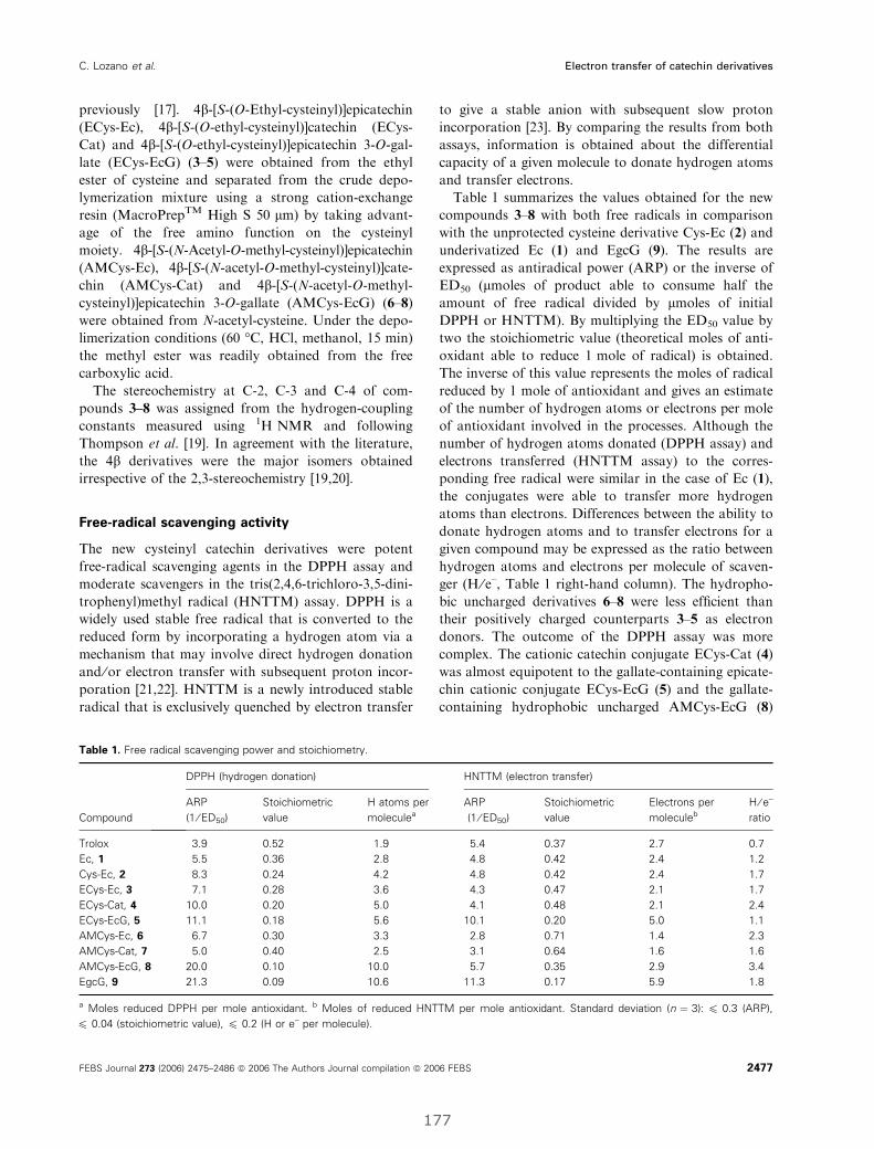
previously [17]. 4b-[S-(O-Ethyl-cysteinyl)]epicatechin
(ECys-Ec), 4b-[S-(O-ethyl-cysteinyl)]catechin (ECys-
Cat) and 4b-[S-(O-ethyl-cysteinyl)]epicatechin 3-O-gal-
late (ECys-EcG) (3–5) were obtained from the ethyl
ester of cysteine and separated from the crude depo-
lymerization mixture using a strong cation-exchange
resin (MacroPrepTM High S 50 lm) by taking advant-
age of the free amino function on the cysteinyl
moiety. 4b-[S-(N-Acetyl-O-methyl-cysteinyl)]epicatechin
(AMCys-Ec), 4b-[S-(N-acetyl-O-methyl-cysteinyl)]cate-
chin (AMCys-Cat) and 4b-[S-(N-acetyl-O-methyl-
cysteinyl)]epicatechin 3-O-gallate (AMCys-EcG) (6–8)
were obtained from N-acetyl-cysteine. Under the depo-
limerization conditions (60 �C, HCl, methanol, 15 min)
the methyl ester was readily obtained from the free
carboxylic acid.
The stereochemistry at C-2, C-3 and C-4 of com-
pounds 3–8 was assigned from the hydrogen-coupling
constants measured using 1H NMR and following
Thompson et al. [19]. In agreement with the literature,
the 4b derivatives were the major isomers obtained
irrespective of the 2,3-stereochemistry [19,20].
Free-radical scavenging activity
The new cysteinyl catechin derivatives were potent
free-radical scavenging agents in the DPPH assay and
moderate scavengers in the tris(2,4,6-trichloro-3,5-dini-
trophenyl)methyl radical (HNTTM) assay. DPPH is a
widely used stable free radical that is converted to the
reduced form by incorporating a hydrogen atom via a
mechanism that may involve direct hydrogen donation
and ⁄or electron transfer with subsequent proton incor-
poration [21,22]. HNTTM is a newly introduced stable
radical that is exclusively quenched by electron transfer
to give a stable anion with subsequent slow proton
incorporation [23]. By comparing the results from both
assays, information is obtained about the differential
capacity of a given molecule to donate hydrogen atoms
and transfer electrons.
Table 1 summarizes the values obtained for the new
compounds 3–8 with both free radicals in comparison
with the unprotected cysteine derivative Cys-Ec (2) and
underivatized Ec (1) and EgcG (9). The results are
expressed as antiradical power (ARP) or the inverse of
ED50 (lmoles of product able to consume half the
amount of free radical divided by lmoles of initial
DPPH or HNTTM). By multiplying the ED50 value by
two the stoichiometric value (theoretical moles of anti-
oxidant able to reduce 1 mole of radical) is obtained.
The inverse of this value represents the moles of radical
reduced by 1 mole of antioxidant and gives an estimate
of the number of hydrogen atoms or electrons per mole
of antioxidant involved in the processes. Although the
number of hydrogen atoms donated (DPPH assay) and
electrons transferred (HNTTM assay) to the corres-
ponding free radical were similar in the case of Ec (1),
the conjugates were able to transfer more hydrogen
atoms than electrons. Differences between the ability to
donate hydrogen atoms and to transfer electrons for a
given compound may be expressed as the ratio between
hydrogen atoms and electrons per molecule of scaven-
ger (H ⁄ e–, Table 1 right-hand column). The hydropho-
bic uncharged derivatives 6–8 were less efficient than
their positively charged counterparts 3–5 as electron
donors. The outcome of the DPPH assay was more
complex. The cationic catechin conjugate ECys-Cat (4)
was almost equipotent to the gallate-containing epicate-
chin cationic conjugate ECys-EcG (5) and the gallate-
containing hydrophobic uncharged AMCys-EcG (8)
Table 1. Free radical scavenging power and stoichiometry.
Compound
DPPH (hydrogen donation) HNTTM (electron transfer)
ARP
(1 ⁄ ED50)
Stoichiometric
value
H atoms per
moleculea
ARP
(1 ⁄ ED50)
Stoichiometric
value
Electrons per
moleculeb
H ⁄ e–
ratio
Trolox 3.9 0.52 1.9 5.4 0.37 2.7 0.7
Ec, 1 5.5 0.36 2.8 4.8 0.42 2.4 1.2
Cys-Ec, 2 8.3 0.24 4.2 4.8 0.42 2.4 1.7
ECys-Ec, 3 7.1 0.28 3.6 4.3 0.47 2.1 1.7
ECys-Cat, 4 10.0 0.20 5.0 4.1 0.48 2.1 2.4
ECys-EcG, 5 11.1 0.18 5.6 10.1 0.20 5.0 1.1
AMCys-Ec, 6 6.7 0.30 3.3 2.8 0.71 1.4 2.3
AMCys-Cat, 7 5.0 0.40 2.5 3.1 0.64 1.6 1.6
AMCys-EcG, 8 20.0 0.10 10.0 5.7 0.35 2.9 3.4
EgcG, 9 21.3 0.09 10.6 11.3 0.17 5.9 1.8
a Moles reduced DPPH per mole antioxidant. b Moles of reduced HNTTM per mole antioxidant. Standard deviation (n ¼ 3): 6 0.3 (ARP),
6 0.04 (stoichiometric value), 6 0.2 (H or e– per molecule).
C. Lozano et al. Electron transfer of catechin derivatives
FEBS Journal 273 (2006) 2475–2486 ª 2006 The Authors Journal compilation ª 2006 FEBS 2477
177
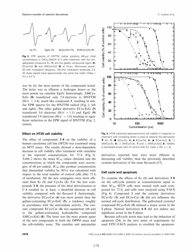
was by far the most potent of the compounds tested.
The latter was as efficient a hydrogen donor as the
most potent tea catechin EgcG. Interestingly, AMCys-
EcG (8) transferred only 2.9 electrons to HNTTM
(H ⁄ e– ¼ 3.4), much like compound 1, resulting in sim-
ilar EPR spectra for the HNTTM radical (Fig. 2, left
and right). The other gallate derivative ECys-EcG (5)
transferred 5.0 electrons (H ⁄ e– ¼ 1.1) and EgcG (9)
transferred 5.9 electrons (H ⁄ e– ¼ 1.8) resulting in signi-
ficant reduction in the EPR signal of HNTTM (Fig. 2,
centre).
Effect on HT29 cell viability
The effect of compounds 3–8 on the viability of a
human carcinoma cell line (HT29) was examined using
an MTT assay. The results showed a dose-dependent
decrease in cell viability after treatment with catechins
at the reported concentrations for 72 h (Fig. 3).
Table 2 shows the mean IC50 values obtained and the
concentrations at which the compounds were scaven-
gers of 60 lm radical. IC50 (the product concentration
that diminished viability by 50%) was calculated with
respect to the total number of control cells after 72 h
of incubation. All the new conjugates were more effi-
cient than Ec (1) and Cys-Ec (2). For the new com-
pounds 3–8, the presence of the thiol derivatization at
C-4 resulted in, at least, a threefold decrease in cell
viability compared with the flavanol 1. Interestingly,
the derivative ECys-Cat (4) was more potent than the
gallate-containing ECys-EcG (5), a tendency roughly
in accordance with the antioxidant activity. The cati-
onic compound ECys-Cat (4) was equipotent (68 lm)
to the gallate-containing hydrophobic compound
AMCys-EcG (8). The latter was the most potent agent
of the new compounds in both the DPPH assay and
the cell-viability assay. The catechin and epicatechin
derivatives reported here were more efficient in
decreasing cell viability than the previously described
cysteine derivatives of the same flavanols [17].
Cell cycle and apoptosis
To examine the effects of Ec (1) and derivatives 3–8
on the cell-cycle pattern at concentrations equal to
their IC50, HT29 cells were treated with each com-
pound for 72 h, and cells were analysed using FACS
(Fig. 4). Compound 1 and the cationic derivatives
ECys-Ec (3) and ECys-Cat (4) did not influence the
normal cell-cycle distribution. The galloylated cysteinyl
compound ECys-EcG (5) induced a major arrest in the
S phase. Neutral derivatives 6–8 did not induce any
significant arrest in the S phase.
Because cell-cycle arrest may lead to the induction of
apoptosis [24], in the next series of experiments we
used FITC-FACS analysis to establish the apoptosis-
Fig. 2. EPR spectra of HNTTM radical solutions (60 lM initial
concentration) in CHCl3 ⁄ MeOH (2:1) after treatment with the non-
galloylated compound Ec (1) and the gallate compounds EgcG (9),
ECys-EcG (5) and AMCys-EcG (8) at 10 lM. Microwave power,
20 mW; modulation frequency, 100 kHz; modulation amplitude, 3.2.
All these signals have approximately the same line width, AHpp ¼3.2 ± 0.2 G.
Fig. 3. HT29 colorectal adenocarcinoma cell viability in response to
treatment with increasing doses (x axis) of catechin thio-derivatives
. (Ec, 1), d (ECys-Ec, 3), n (ECys-Cat, 4), m (ECys-EcG, 5), h
(AMCys-Ec, 6), s (AMCys-Cat, 7) and n (AMCys-EcG, 8). Viability
is expressed as per cent of control cells (Ct, mean ± SD, n ¼ 4).
Electron transfer of catechin derivatives C. Lozano et al.
2478 FEBS Journal 273 (2006) 2475–2486 ª 2006 The Authors Journal compilation ª 2006 FEBS
178
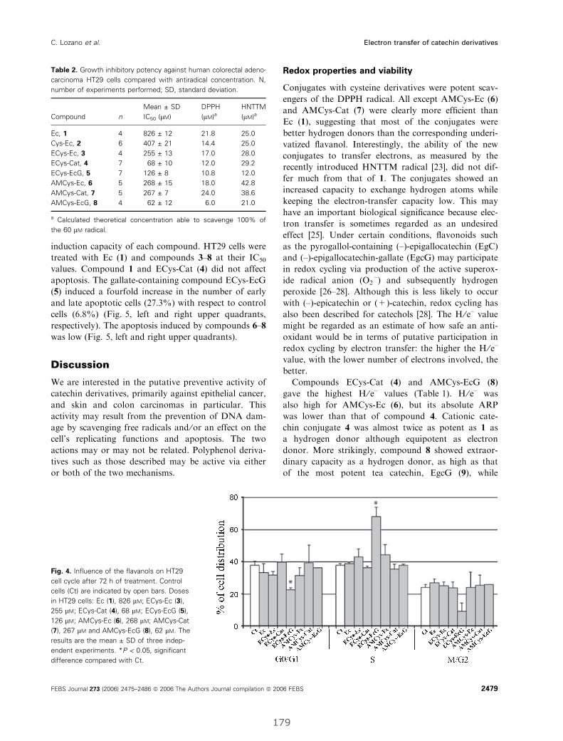
induction capacity of each compound. HT29 cells were
treated with Ec (1) and compounds 3–8 at their IC50
values. Compound 1 and ECys-Cat (4) did not affect
apoptosis. The gallate-containing compound ECys-EcG
(5) induced a fourfold increase in the number of early
and late apoptotic cells (27.3%) with respect to control
cells (6.8%) (Fig. 5, left and right upper quadrants,
respectively). The apoptosis induced by compounds 6–8
was low (Fig. 5, left and right upper quadrants).
Discussion
We are interested in the putative preventive activity of
catechin derivatives, primarily against epithelial cancer,
and skin and colon carcinomas in particular. This
activity may result from the prevention of DNA dam-
age by scavenging free radicals and ⁄or an effect on the
cell’s replicating functions and apoptosis. The two
actions may or may not be related. Polyphenol deriva-
tives such as those described may be active via either
or both of the two mechanisms.
Redox properties and viability
Conjugates with cysteine derivatives were potent scav-
engers of the DPPH radical. All except AMCys-Ec (6)
and AMCys-Cat (7) were clearly more efficient than
Ec (1), suggesting that most of the conjugates were
better hydrogen donors than the corresponding underi-
vatized flavanol. Interestingly, the ability of the new
conjugates to transfer electrons, as measured by the
recently introduced HNTTM radical [23], did not dif-
fer much from that of 1. The conjugates showed an
increased capacity to exchange hydrogen atoms while
keeping the electron-transfer capacity low. This may
have an important biological significance because elec-
tron transfer is sometimes regarded as an undesired
effect [25]. Under certain conditions, flavonoids such
as the pyrogallol-containing (–)-epigallocatechin (EgC)
and (–)-epigallocatechin-gallate (EgcG) may participate
in redox cycling via production of the active superox-
ide radical anion (O2Æ–) and subsequently hydrogen
peroxide [26–28]. Although this is less likely to occur
with (–)-epicatechin or (+)-catechin, redox cycling has
also been described for catechols [28]. The H ⁄ e– value
might be regarded as an estimate of how safe an anti-
oxidant would be in terms of putative participation in
redox cycling by electron transfer: the higher the H ⁄ e–
value, with the lower number of electrons involved, the
better.
Compounds ECys-Cat (4) and AMCys-EcG (8)
gave the highest H ⁄ e– values (Table 1). H ⁄ e– was
also high for AMCys-Ec (6), but its absolute ARP
was lower than that of compound 4. Cationic cate-
chin conjugate 4 was almost twice as potent as 1 as
a hydrogen donor although equipotent as electron
donor. More strikingly, compound 8 showed extraor-
dinary capacity as a hydrogen donor, as high as that
of the most potent tea catechin, EgcG (9), while
Table 2. Growth inhibitory potency against human colorectal adeno-
carcinoma HT29 cells compared with antiradical concentration. N,
number of experiments performed; SD, standard deviation.
Compound n
Mean ± SD
IC50 (lM)
DPPH
(lM)aHNTTM
(lM)a
Ec, 1 4 826 ± 12 21.8 25.0
Cys-Ec, 2 6 407 ± 21 14.4 25.0
ECys-Ec, 3 4 255 ± 13 17.0 28.0
ECys-Cat, 4 7 68 ± 10 12.0 29.2
ECys-EcG, 5 7 126 ± 8 10.8 12.0
AMCys-Ec, 6 5 268 ± 15 18.0 42.8
AMCys-Cat, 7 5 267 ± 7 24.0 38.6
AMCys-EcG, 8 4 62 ± 12 6.0 21.0
a Calculated theoretical concentration able to scavenge 100% of
the 60 lM radical.
Fig. 4. Influence of the flavanols on HT29
cell cycle after 72 h of treatment. Control
cells (Ct) are indicated by open bars. Doses
in HT29 cells: Ec (1), 826 lM; ECys-Ec (3),
255 lM; ECys-Cat (4), 68 lM; ECys-EcG (5),
126 lM; AMCys-Ec (6), 268 lM; AMCys-Cat
(7), 267 lM and AMCys-EcG (8), 62 lM. The
results are the mean ± SD of three indep-
endent experiments. *P < 0.05, significant
difference compared with Ct.
C. Lozano et al. Electron transfer of catechin derivatives
FEBS Journal 273 (2006) 2475–2486 ª 2006 The Authors Journal compilation ª 2006 FEBS 2479
179
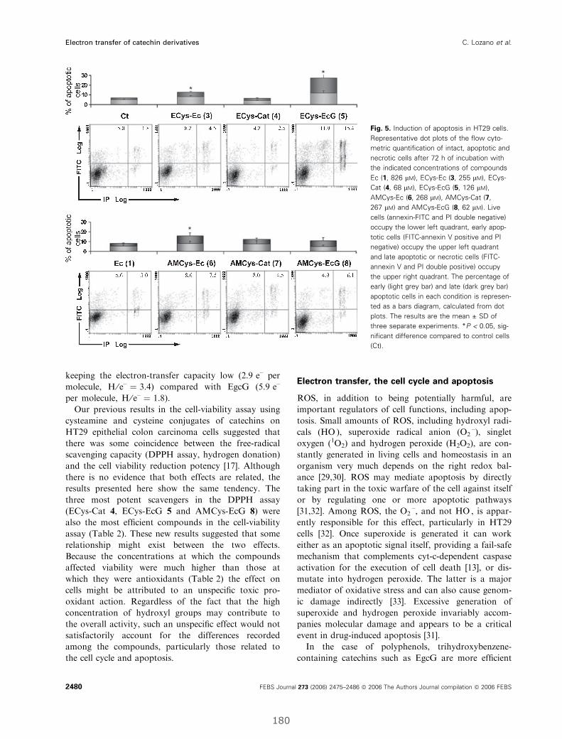
keeping the electron-transfer capacity low (2.9 e– per
molecule, H ⁄ e– ¼ 3.4) compared with EgcG (5.9 e–
per molecule, H ⁄ e– ¼ 1.8).
Our previous results in the cell-viability assay using
cysteamine and cysteine conjugates of catechins on
HT29 epithelial colon carcinoma cells suggested that
there was some coincidence between the free-radical
scavenging capacity (DPPH assay, hydrogen donation)
and the cell viability reduction potency [17]. Although
there is no evidence that both effects are related, the
results presented here show the same tendency. The
three most potent scavengers in the DPPH assay
(ECys-Cat 4, ECys-EcG 5 and AMCys-EcG 8) were
also the most efficient compounds in the cell-viability
assay (Table 2). These new results suggested that some
relationship might exist between the two effects.
Because the concentrations at which the compounds
affected viability were much higher than those at
which they were antioxidants (Table 2) the effect on
cells might be attributed to an unspecific toxic pro-
oxidant action. Regardless of the fact that the high
concentration of hydroxyl groups may contribute to
the overall activity, such an unspecific effect would not
satisfactorily account for the differences recorded
among the compounds, particularly those related to
the cell cycle and apoptosis.
Electron transfer, the cell cycle and apoptosis
ROS, in addition to being potentially harmful, are
important regulators of cell functions, including apop-
tosis. Small amounts of ROS, including hydroxyl radi-
cals (HOÆ), superoxide radical anion (O2Æ–), singlet
oxygen (1O2) and hydrogen peroxide (H2O2), are con-
stantly generated in living cells and homeostasis in an
organism very much depends on the right redox bal-
ance [29,30]. ROS may mediate apoptosis by directly
taking part in the toxic warfare of the cell against itself
or by regulating one or more apoptotic pathways
[31,32]. Among ROS, the O2Æ–, and not HOÆ, is appar-
ently responsible for this effect, particularly in HT29
cells [32]. Once superoxide is generated it can work
either as an apoptotic signal itself, providing a fail-safe
mechanism that complements cyt-c-dependent caspase
activation for the execution of cell death [13], or dis-
mutate into hydrogen peroxide. The latter is a major
mediator of oxidative stress and can also cause genom-
ic damage indirectly [33]. Excessive generation of
superoxide and hydrogen peroxide invariably accom-
panies molecular damage and appears to be a critical
event in drug-induced apoptosis [31].
In the case of polyphenols, trihydroxybenzene-
containing catechins such as EgcG are more efficient
Fig. 5. Induction of apoptosis in HT29 cells.
Representative dot plots of the flow cyto-
metric quantification of intact, apoptotic and
necrotic cells after 72 h of incubation with
the indicated concentrations of compounds
Ec (1, 826 lM), ECys-Ec (3, 255 lM), ECys-
Cat (4, 68 lM), ECys-EcG (5, 126 lM),
AMCys-Ec (6, 268 lM), AMCys-Cat (7,
267 lM) and AMCys-EcG (8, 62 lM). Live
cells (annexin-FITC and PI double negative)
occupy the lower left quadrant, early apop-
totic cells (FITC-annexin V positive and PI
negative) occupy the upper left quadrant
and late apoptotic or necrotic cells (FITC-
annexin V and PI double positive) occupy
the upper right quadrant. The percentage of
early (light grey bar) and late (dark grey bar)
apoptotic cells in each condition is represen-
ted as a bars diagram, calculated from dot
plots. The results are the mean ± SD of
three separate experiments. *P < 0.05, sig-
nificant difference compared to control cells
(Ct).
Electron transfer of catechin derivatives C. Lozano et al.
2480 FEBS Journal 273 (2006) 2475–2486 ª 2006 The Authors Journal compilation ª 2006 FEBS
180
producers of the superoxide radical anion than cate-
chol-containing catechins such as Ec (1) [14,29,34], and
they inhibit cancer cell growth via cell-cycle arrest and
apoptosis induction by mechanisms involving the gal-
late moiety [12,35–37]. In agreement with the litera-
ture, our results show that the gallate-containing
compound ECys-EcG (5) induced apoptosis (threefold
compared with control cells, see Fig. 5) and triggered a
significant arrest in the S phase of the cell cycle
(Fig. 4). Several authors have reported that the gallate
group decreases cyclin-dependent kinase 2 (Cdk2)
expression and activity, and upregulates the expression
of p21, a Cdk inhibitor [38–40] essential for progres-
sion from the G1 to the S phase of the cell cycle.
Alternatively, the catechin gallates, acting as pro-oxi-
dants, may damage the DNA directly through ROS
[41]. All these events lead to stoppage of the cell-cycle
progression at the S phase and may potentially result
in inhibition of proliferation, cytostasis and possibly
apoptosis in human tumours. It seems increasingly evi-
dent that the duality antioxidant ⁄pro-oxidant of cate-
chins may play a crucial role in their interactions with
the cell machinery, most probably via formation of the
superoxide anion radical by electron transfer.
ECys-EcG (5), together with EgcG (9), possessed a
high electron-transfer capacity (five and six electrons
per molecule, respectively). Compounds 1–4 and 6, 7,
all lacking the gallate moiety and showing low elec-
tron-transfer capacity, did not induce any significant
cell-cycle arrest or apoptosis. Surprisingly, the gallate-
containing compound AMCys-EcG (8) did not induce
cell-cycle arrest or apoptosis. Curiously, the electron-
transfer capacity of 8 (2.9 electrons per molecule) as
measured by the stable radical HNTTM was lower
than expected, similar to those of the nongalloylated
species (approximately 2 electrons per molecule, see
Fig. 2). This finding appears to corroborate the obser-
vation from other authors that links the induction of
apoptosis to the electron-transfer capacity and forma-
tion of the superoxide radical anion. The presence of
the gallate moiety does not appear to be a sufficient
condition for the induction of apoptosis in HT29 cells.
Whether cell-cycle arrest and apoptosis are due to
redox cycling with the production of hydrogen perox-
ide (pro-oxidant effect) or other ROS-mediated events
is something that must be explored further.
Conclusions
The conjugation of catechins with both cationic and
neutral cysteine derivatives produced compounds with
an improved capacity to donate hydrogen atoms while
keeping their capacity to participate in electron-trans-
fer reactions low. One of the new molecules, AMCys-
EcG (8), the most efficient DPPH scavenger of the
flavanol thio-conjugates described to date, was the
most effective derivative against colon carcinoma cell
viability. Despite including a gallate moiety, this com-
pound showed a low electron-transfer capacity and
neither arrested the cell cycle nor induced apoptosis.
This result, together with the observation that pro-
apoptotic ECys-EcG (5) and EgcG (9) possessed
higher electron-transfer capacity is suggesting that the
gallate moiety may not be a sufficient condition to
trigger apoptosis, which would be more directly related
to the ability of the flavanol derivatives to transfer
electrons. Our newly introduced scavenging assay using
the stable radical HNTTM, which is exclusively sensi-
tive to electron transfer, may be a valuable tool for
predicting the pro-apoptotic activity of polyphenols
and other putative drugs. The electron-transfer capa-
city of exogenous plant phenolics and its influence on
the delicate balance between the antioxidant and pro-
oxidant events governing cell functions may help to
explain the putative cancer-preventive properties of
catechins and their derivatives.
Experimental procedures
Materials
Analytical grade methanol (MeOH, Panreac, Montcada i
Reixac, Spain) was used for the acid cleavage reaction and
DPPH assay, deionized water and bulk ethanol (EtOH,
Momplet y Esteban, Barcelona, Spain) for semipreparative
and preparative cation-exchange chromatography, Milli-Q�
water and HPLC grade acetonitrile (CH3CN, Merck,
Darmstadt, Germany) for analytical RP-HPLC, and
deionized water and preparative grade CH3CN (Scharlau,
Barcelona, Spain) for preparative and semipreparative
RP-HPLC. Deuterated solvents for NMR were from SDS
(Peypin, France). Cysteine hydrochloride, l-cysteine ethyl
ester hydrochloride and N-acetyl-l-cysteine (Aldrich, Stein-
heim, Germany) were of synthesis grade. (–)-Epicatechin
(Ec, 1) (–)-epigallocatechin 3-O-gallate (EgcG, 9), MTT,
dimethylsulfoxide, Trypan Blue solution 0.4%, propidium
iodide (PI), the nonionic surfactant Igepal CA-630, a,a,a-tris(hydroxymethyl)aminomethane and NaCl ⁄Pi were from
Sigma (Steinheim, Germany). Acetic acid, 37% HCl
(Merck) and NaCl (Carlo Erba, Milan, Italy) were of ana-
lytical grade. Triethylamine (Merck) was of buffer grade.
Trifluoroacetic acid (Fluorochem, Glossop, UK) biotech
grade was distilled in-house. DPPH (95%) was from Ald-
rich (Gillingham, UK), 6-hydroxy-2,5,7,8-tetramethyl-chro-
man-2-carboxylic acid (Trolox) (97%) was from Aldrich
(Milwaukee, MN). Fetal bovine serum was purchased from
Gibco (Invitrogen, Carlsbad, CA). Trypsin–EDTA solution
C. Lozano et al. Electron transfer of catechin derivatives
FEBS Journal 273 (2006) 2475–2486 ª 2006 The Authors Journal compilation ª 2006 FEBS 2481
181
C (0.05% trypsin and EDTA 1:5000 in NaCl ⁄Pi) was from
Biological Industries (Beit Haemek, Israel). RNase was
from Roche Diagnostics (Mannheim, Germany). FITC–
annexin V kit and binding buffer 4· for apoptosis assay
were purchased from Bender MedSystems (MedSystems
Diagnostics GmbH, Vienna, Austria).
Chromatographic equipment and columns
Analytical RP-HPLC was performed on a Kontron Analyt-
ical system (Kontron Instruments, Basel, Switzerland) fitted
with a VYDACTM (The Separations Group, Hesperia,
USA) C18, 300 A pore size, 5 lm particle size,
250 · 4.6 mm i.d. column. Cation-exchange chromatogra-
phy was performed on a flash chromatography-type glass
column (21 · 2.5 cm i.d., � 105 mL bed volume) packed
in-house with MacroPrepTM High S 50 lm (Bio-Rad
Laboratories, Hercules, CA). Preparative RP-HPLC chro-
matography was performed on a Waters (Milford, USA)
Prep LC 4000 pumping system with a Waters PrepPack�
1000 module fitted with a PrepPack� Waters cartridge
(30 · 4.7 cm i.d) filled with VYDACTM (The Separations
Group) C18, 300 A pore size, 15–20 lm particle size station-
ary phase. Detection was carried out using an analytical
Merck-Hitachi (Darmstadt, Germany) L-4000 UV detector.
MS, NMR and EPR measurements
ES-MS analyses were recorded on a VG-Quattro� system
from Fisons Instruments (Altrincham, UK). The carrier
solution was Milli-Q water ⁄CH3CN (1:1) containing 1%
(v ⁄ v) formic acid. 1H NMR spectra were acquired on a
Varian (Palo Alto, CA) Unity 300 spectrometer in the deu-
terated solvents (CD3)2CO and D2O.
EPR measurements were performed on a Varian E-109
spectrometer working in the X-band (microwave power,
20 mW; modulation amplitude, 3.2 G).
Preparation of the conjugates
Conjugates were obtained by acid depolymerization of
plant procyanidins essentially as described previously [16].
To obtain the thio-conjugates 3–8 (Fig. 1) the solvent was
eliminated from an aqueous fraction (400 mL, 6 g estima-
ted polyphenols by mass, from 3.2 kg of grape byproduct)
of polymeric procyanidins. The pellet was then dissolved in
MeOH (400 mL) and dried. The resulting syrupy residue
was dissolved in MeOH (400 mL) and a solution of the
appropriate cysteine derivative (20 g) and 37% HCl
(10 mL) in MeOH (400 mL) was added. The mixture was
kept at 65 �C for 20 min under stirring. The reaction was
then quenched with cold water (3.2 L).
Conjugates were separated from the whole mixture using
the MacroPrepTM High S resin. The eluents were: (A)
20 mm sodium phosphate, pH 2.3 buffer ⁄EtOH (13:7, v ⁄ v)and (B) 20 mm sodium phosphate, pH 2.3 buffer ⁄EtOH
(3:2, v ⁄ v), 100 mm NaCl. The column was equilibrated with
eluent (A), loaded with the quenched depolymerized mix-
ture (500 mL) and washed with (A) (500 mL, 4.75 bed vol-
umes). The retained catechin derivatives were released with
500 mL (4.75 bed volumes) of eluent (B). The operation
was repeated until the whole mixture was consumed. The
separation process was monitored by analytical RP-HPLC
on a VYDACTM C18 column eluted with a binary system:
(C) 0.10% (v ⁄ v) aqueous trifluoroacetic acid, (D) 0.09%
(v ⁄ v) trifluoroacetic acid in water ⁄CH3CN (1:4, v ⁄ v) underisocratic conditions 19% (D) at a flow rate of 1.5 mLÆmin)1
and detection at 214 nm. The eluates containing the corres-
ponding conjugate were pooled (3.5 L).
The mixture containing the O-ethyl-cysteinyl conjugates
3–5 was fractionated on a preparative RP-HPLC cartridge
filled with VYDACTM C18 stationary phase by a CH3CN
gradient in 0.10% (v ⁄ v) aqueous trifluoroacetic acid (4–20%
CH3CN over 45 min). Fractions enriched in each of the three
compounds were obtained: fraction I, 9–11% CH3CN, com-
pound ECys-Cat (4); fraction II, 12–16% CH3CN, com-
pound ECys-Ec (3); fraction III, 17–19% CH3CN,
compound ECys-EcG (5).
4b-[S-(O-ethyl-cysteinyl)]epicatechin (ECys-Ec, 3)
Fraction II from reversed-phase fractionation was concen-
trated, loaded onto the preparative cartridge and the target
compound purified by CH3CN gradient in triethylamine
phosphate buffer and aqueous trifluoroacetic acid. Analysis
of the fractions was accomplished under isocratic condi-
tions in 0.10% (v ⁄ v) aqueous trifluoroacetic acid ⁄CH3CN
using the VYDACTM C18 column, solvent system, flow rate
and detection as described above. ECys-Ec (3) (354 mg)
was obtained as the trifluoroacetate by lyophilization.
dH(300 MHz; (CD3)2CO +3 drops D2O) 1.24 (3 H, t, J
7.2 Hz, O-CH2-CH3), 3.93 (1 H, d, J3,4 2.1 Hz, 4-H 3,4-
trans configuration), 4.06 (1 H, dd, J 2.4 and 0.9 Hz, 3-H),
4.26 (2 H, q, J 7.2 and 1.5 Hz, O-CH2-CH3), 4.71 (1 H, m,
S-CH2-CH <), 5.09 (1 H, s, 2-H 2,3-cis configuration),
5.90 (1 H, d, J 2.4 Hz, 8-H), 6.09 (1 H, d, J 2.4 Hz, 6-H),
6.80–6.81 (2 H, m, 5¢-H, 6¢-H), 7.04 (1 H, d, J 1.8 Hz,
2¢-H). m ⁄ z 438.1 (M + 1)+, calculated for C20H24N1O8S1(M + H)+ 438.1. Purity (> 95%) was ascertained by
RP-HPLC.
4b-[S-(O-ethyl-cysteinyl)]catechin (ECys-Cat, 4)
Fraction I from reversed-phase fractionation was concen-
trated, loaded onto the preparative cartridge, purified and
characterized as stated for compound 3. ECys-Cat (4)
(68 mg) was obtained as the trifluoroacetate. dH(300 MHz;
(CD3)2CO +3 drops D2O) 1.24 (3 H, t, J 7.0 Hz, O-CH2-
Electron transfer of catechin derivatives C. Lozano et al.
2482 FEBS Journal 273 (2006) 2475–2486 ª 2006 The Authors Journal compilation ª 2006 FEBS
182
CH3), 4.06 (1 H, 2d, J2,3 9.6 and 2.4 Hz, 3-H 2,3-trans con-
figuration), 4.23 (1 H, d, J3,4 2.4 Hz, 4-H 3,4-cis configur-
ation), 4.26 (2 H, q, J 7.0 and 2.4 Hz, O-CH2-CH3), 4.68–
4.72 (1 H, m, S-CH2-CH <), 4,78 (1 H, d, J 8.6 Hz; 2-H),
5.89 (1 H, d, J 2.4 Hz, 8-H), 6.10 (1 H, d, J 2.4 Hz, 6-H),
6.62 (2 H, m, 5¢-H, 6¢-H), 6.91 (1 H, s, 2¢-H). m ⁄ z 438.1
(M + 1)+, calculated for C20H24N1O8S1 (M + H)+ 438.1.
Purity (> 93%) was ascertained by RP-HPLC.
4b-[S-(O-ethyl-cysteinyl)]epicatechin3-O-gallate (ECys-EcG, 5)
Fraction III from reversed-phase fractionation was concen-
trated, loaded onto the preparative cartridge, purified and
characterized as stated for compound 3. ECys-EcG (5)
(33 mg) was obtained as the trifluoroacetate. dH(300 MHz;
(CD3)2CO +3 drops D2O) 1.28 (3 H, t, J 7.0 Hz, O-CH2-
CH3), 4.15 (1 H, d, J3,4 1.8 Hz, 4-H 3,4-trans configur-
ation), 4.29 (2H, q, J 7.0 and 1.8 Hz, O-CH2-CH3), 4.77
(1 H, m, S-CH2-CH <), 5.28 (1 H, m, 3-H), 5.36 (1 H, bs,
2-H 2,3-cis configuration), 6.01 (1 H, d, J 2.1 Hz, 6-H),
6.13 (1 H, d, J 2.1 Hz, 8-H), 6.79 (1 H, d, J 8.1 Hz, 5¢-H),
6.88 (1 H, dd, J 8.4 and 2.1 Hz, 6¢-H), 6.96 (2 H, s, galloyl-
H), 7.10 (1 H, d, J 1.8 Hz, 2¢-H). m ⁄ z 590.1 (M + 1)+ cal-
culated for C27H28N1O12S1 (M + H)+ 590.1. Purity
(> 96%) was ascertained by RP-HPLC.
The preparative RP-HPLC fractionation of the N-acetyl-
O-methyl-cysteinyl conjugates 6–8 was performed directly
from the depolymerized mixture under chromatographic con-
ditions (6–20% CH3CN over 54 min) similar to the condi-
tions described for the ethyl-cysteine conjugates. Fractions of
interest: fraction IV, 13–14% CH3CN, compound AMCys-
Cat (7); fraction V, 15–18% CH3CN, compound AMCys-Ec
(6); fraction VI, 18–19% CH3CN, compound AMCys-EcG
(8).
4b-[S-(N-Acetyl-O-methyl-cysteinyl)]epicatechin
(AMCys-Ec, 6)
Fraction V from reversed-phase fractionation was concen-
trated, loaded onto the preparative cartridge and the target
compound purified by CH3CN gradient in triethylamine
phosphate buffer and aqueous trifluoroacetic acid. Analysis
of the fractions was accomplished as described above.
AMCys-Ec (6) (818 mg) was obtained by lyophilization.
dH(300 MHz; (CD3)2CO +3 drops D2O) 2.05 (3 H, s,
CO-CH3), 3.69 (3 H, s, O-CH3), 4.02 (1 H, dd, J2,3 2.4 and
1.2 Hz, 3-H 2,3-cis configuration), 4.06 (1 H, d, J3,4 2.4 Hz,
4-H 3,4-trans configuration), 4.94 (1 H, m, S-CH2-CH <),
5.22 (1 H, s, 2-H), 5.89 (1 H, d, J 2.4 Hz, 8-H), 6.06 (1 H,
d, J 2.4 Hz, 6-H), 6.81–6.83 (2 H, m, 5¢-H, 6¢-H), 7.06
(1 H, d, J 2.1 Hz, 2¢-H). m ⁄ z 464.7 (M ) 1)–, calculated for
C21H23N1O9S1 (M ) H)– 464.5. Purity (> 99%) was ascer-
tained by RP-HPLC.
4b-[S-(N-Acetyl-O-methyl-cysteinyl)]catechin
(AMCys-Cat, 7)
Fraction IV from reversed-phase fractionation was concen-
trated, loaded onto the preparative cartridge, purified and
characterized as stated for compound 6. After lyophiliza-
tion, AMCys-Cat (7) (64 mg) was obtained. dH(300 MHz;
(CD3)2CO +3 drops D2O) 2.11 (3 H, s, CO-CH3), 3.65
(3 H, s, O-CH3), 4.15 (1 H, 2d, J 9.6 and 3.9 Hz, 3-H),
4.38 (1 H, d, J3,4 3.9 Hz, 4-H 3,4-cis configuration), 4.82
(1 H, m, S-CH2-CH <), 4.95 (1 H, d, J2,3 9.6 Hz, 2-H 2,3-
trans configuration), 5.78 (1 H, d, J 2.4 Hz, 8-H), 6.06
(1 H, d, J 2.4 Hz, 6-H), 6.78 (2 H, m, 5¢-H, 6¢-H), 6.92
(1 H, s, 2¢-H). m ⁄ z 464.9 (M ) 1)–, calculated for
C21H23N1O9S1 (M-H)– 464.5. Purity (99%) was ascertained
by RP-HPLC.
4b-[S-(N-Acetyl-O-methyl-cysteinyl)]epicatechin
3-O-gallate (AMCys-EcG, 8)
Fraction VI from reversed-phase fractionation was concen-
trated, loaded onto the preparative cartridge, purified and
characterized as stated for compound 6. After lyophiliza-
tion, AMCys-EcG (8) (88 mg) was obtained. dH (300 MHz;
(CD3)2CO +3 drops D2O) 2.09 (3H s, CO-CH3), 3.71
(3 H, s, O-CH3), 4.26 (1 H, d, J3,4 2.4 Hz, 4-H 3,4-trans
configuration), 5.01 (1 H, m, S-CH2-CH <), 5.21 (1 H, m,
3-H), 5.48 (1 H, bs, 2-H), 6.01 (1 H, d, J 2.4 Hz, 8-H), 6.07
(1 H, d, J 2.4 Hz, 6-H), 6.78 (1 H, d, J 8.1 Hz, 5¢-H), 6.89
(1 H, dd, J 8.1 and 2.1 Hz, 6¢-H), 6.96 (2 H, s, galloyl-H),
7.08 (1 H, d, J 2.1 Hz, 2¢-H). m ⁄ z (EI) 616.3 (M ) 1)– cal-
culated for C28H27N1O13S1 (M ) H)– 616.6. Purity (95%)
was ascertained by RP-HPLC.
Free-radical scavenging
Hydrogen atom donation capacity was evaluated using the
DPPH assay [42,43]. The samples (0.1 mL) were added to
aliquots (3.9 mL) of a solution made up with 4.8 mg DPPH
in 200 mL of MeOH and the mixture incubated for 1 h at
room temperature. The initial concentration of DPPH,
� 60 lm, was calculated for every experiment from a calib-
ration curve made by measuring the absorbance at 517 nm
of standard samples of DPPH at different concentrations.
The equation of the curve was A517 ¼ 11345 · CDPPH as
determined by linear regression. The electron-transfer capa-
city was evaluated using the HNTTM assay. The radical
scavengers were dissolved in CHCl3 ⁄MeOH (2:1, v ⁄ v) at
different concentrations. Aliquots (1 mL) were added to a
solution (1 mL) of HNTTM (120 lm in CHCl3 ⁄MeOH 2:1,
v ⁄ v) [23] and the mixture incubated for 30 min. The initial
concentration of radical, � 60 lm, was calculated for every
experiment from calibration curves made by measuring the
intensity (I0) of the EPR signal (peak-to-peak line distance)
C. Lozano et al. Electron transfer of catechin derivatives
FEBS Journal 273 (2006) 2475–2486 ª 2006 The Authors Journal compilation ª 2006 FEBS 2483
183

of standard samples of the radical at different concentra-
tions. The equations of the curves were I ¼ 1980 · Cradical
or I ¼ 2262 · Cradical depending on the experiment. For
both assays the initial concentration of the scavengers ran-
ged from 1 to 40 lm. The results were plotted as the degree
of disappearance of absorbance ([(1 ) A ⁄A0) · 100], DPPH
assay) or signal intensity ([(1 ) I ⁄ I0) · 100], HNTTM
assay) against lmoles of the sample divided by the initial
lmoles of the radical. Each point was acquired in triplicate.
A dose–response curve was obtained for every product. The
results were expressed as the efficient dose ED50 given as
lmoles of product able to consume half the amount of free
radical divided by lmoles of initial free radical.
Cell culture
HT29 cells (colorectal adenocarcinoma) were purchased
from American Type Culture Collection (ATCC) and cul-
tured in Dulbecco’s modified Eagle’s medium supplemented
with 10% (v ⁄ v) heat-inactivated fetal bovine serum, 2 mm
l-glutamine and antibiotics: 100 UÆmL)1 penicillin and
100 lgÆmL)1 streptomycin. Cells were grown in an isolated
37 �C)5% CO2 tissue incubator compartment and the med-
ium was changed every 3 days.
Cell-viability assay
Cell growth was determined using a variation of the MTT
assay described by Mosmann [44]. HT29 cells were counted
using Trypan Blue solution 4% in a Neubauer cell counter
chamber (Brand, Wertheim, Germany) by observing viable
(nonstained) and nonviable (stained) cells under a micro-
scope [45]. Cells were seeded into 96-well plates at
2.5 · 103 cells per well and incubated for 24 h prior to
addition of the compounds (dissolved in NaCl ⁄Pi). After
3 days of culture, the supernatant was aspirated and
100 lL of filtered MTT (0.5 mgÆmL)1 in cell culture med-
ium) was added. The cell plates were incubated during 1 h
and metabolically active cells reduced the dye to purple
formazan. The supernatant was removed, and the dark blue
MTT formazan precipitated was dissolved in dimethylsulf-
oxide (100 lL) and optical density (OD) measured at
550 nm on a multiwell reader (Merck ELISA System
MIOS�).
The IC50 or compound concentration causing a 50%
reduction in the mean OD value relative to the control was
estimated using grafit 3.00 (Data Analysis and Graphics
Program, Erithacus Software Ltd. Microsoft Corp., Surrey,
UK) curve option: IC50 curve – start at 0.
Cell-cycle analysis
Cell-cycle analysis and apoptosis quantification were con-
ducted by flow cytometry. HT29 cells were seeded into six-
well plates at 8 · 104 cells per well and incubated for 24 h
prior to addition of the compounds. The IC50 concentration
values of each sample were measured after 3 days of sub-
culturing. Both treated and untreated cells were resuspend-
ed in ice-cold TBS 1· buffer (1 mL of 10 mm Tris and
150 mm NaCl, pH 7.4). PI (50 lL, 50 lg) and 1 mL Vinde-
lov buffer at pH 7.4 containing 10 mm Tris, 10 mm NaCl,
PI (50 lL, 50 lg), RNase (1 lL, 10 lg) and Igepal CA-630
(1 lL) were added to each sample, and cells were incubated
for 1 h at 4 �C in the dark [46]. Cell-cycle distribution was
analysed by flow cytometry using a FACS system. DNA
histograms were collected with an Epics XL flow cytometer
(Coulter Corporation, Miami, FL) and analysed using
multicycle software (Phoenix Flow Systems, San Diego,
CA).
Assessment of apoptosis
After 3 days of subculturing the cell plates as described for
the cell-cycle treatment, cells were washed once in ice-cold
binding buffer (10 mm Hepes sodium hydroxide pH 7.4,
140 mm NaCl, 2.5 mm calcium chloride) and resuspended
in the same buffer (95 lL) at a maximum of 0.8 · 106 cell-
sÆmL)1 in the presence of FITC–annexin V binding (3 lL).After 30 min of incubation at room temperature in the
dark, PI (20 lL, 20 lg) was added [47]. Cells, double-
stained with PI and FITC–annexin V were processed by
flow cytometry and laser-scanning cytometry, which collec-
ted green (525 nm) fluorescence for FITC-conjugated anti-
body and red (675 nm) fluorescence for PI, under 488 nm
excitation.
Statistics
For statistical analysis Student’s t-test was used. For each
compound, a minimum of four duplicate experiments for
the growth inhibition test and a minimum of three experi-
ments for both cell-cycle analysis and assessment of apop-
tosis were conducted. Data are given as the mean ± SD.
P-values of <0.05 were considered significant.
Acknowledgements
Financial support (research grants PPQ2003-06602-
C04-01 and -04, BQU2002-0485-C02-01 and doctoral
fellowships to CL and ST from the Spanish Ministry
of Education and Science; grant 2001SGR00048 and
ITT program of the Work Community of Pyrenees
from Generalitat de Catalalunya) is acknowledged. We
thank Dr Irene Fernandez from the Servei d’Espec-
trometria de Masses of the University of Barcelona
for the mass spectrometry analyses, Ms Avencia Dıez
from the Servei d’Espectrometria de Ressonancia
Electron transfer of catechin derivatives C. Lozano et al.
2484 FEBS Journal 273 (2006) 2475–2486 ª 2006 The Authors Journal compilation ª 2006 FEBS
184

Paramagnetica Electronica at IIQAB-CSIC for the
EPR analyses and Dr Francisco Jose Sanchez-Baeza
and Ms Montserrat Sindreu from the Servei d’Espec-
trometria de Ressonancia Magnetica Nuclear at
IIQAB-CSIC for the NMR analyses.
References
1 Rice-Evans CA, Miller NJ & Paganga G (1996) Struc-
ture–antioxidant activity relationships of flavonoids and
phenolic acids. Free Radical Biol Med 20, 933–956.
2 Yokozawa T, Chen CP, Dong E, Tanaka T, Nonaka
GI & Nishioka I (1998) Study on the inhibitory effect
of tannins and flavonoids against the 1,1-diphenyl-2-
picrylhydrazyl radical. Biochem Pharmacol 56, 213–222.
3 Katiyar SK & Mukhtar H (1997) Tea antioxidants in
cancer chemoprevention. J Cell Biochem (Suppl) 27,
59–67.
4 Yang CS, Landau JM, Huang MT & Newmark HL
(2001) Inhibition of carcinogenesis by dietary polyphe-
nolic compounds. Ann Rev Nutr 21, 381–406.
5 Diplock AT, Charleux JL, Crozier-Willi G, Kok FJ,
Rice-Evans C, Roberfroid M, Stahl W & Vina-Ribes J
(1998) Functional food science and defence against reac-
tive oxidative species. Br J Nutr 80 (Suppl. 1), S77–
S112.
6 Mertens-Talcott SU, Talcott ST & Percival SS (2003)
Low concentrations of quercetin and ellagic acid syner-
gistically influence proliferation, cytotoxicity and apop-
tosis in MOLT-4 human leukemia cells. J Nutr 133,
2669–2674.
7 Chung FL, Schwartz J, Herzog CR & Yang YM (2003)
Tea and cancer prevention: studies in animals and
humans. J Nutr 133, 3268S–3274S.
8 Hsu S, Lewis J, Singh B, Schoenlein P, Osaki T, Athar
M, Porter AG & Schuster G (2003) Green tea polyphe-
nol targets the mitochondria in tumor cells inducing
caspase 3-dependent apoptosis. Anticancer Res 23,
1533–1539.
9 Gupta S, Hussain T & Mukhtar H (2003) Molecular
pathway for (–)-epigallocatechin-3-gallate-induced cell
cycle arrest and apoptosis of human prostate carcinoma
cells. Arch Biochem Biophys 410, 177–185.
10 Ahmad N, Gali H, Javed S & Agarwal R (1998) Skin
cancer chemopreventive effects of a flavonoid antioxi-
dant silymarin are mediated via impairment of receptor
tyrosine kinase signaling and perturbation in cell cycle
progression. Biochem Biophys Res Commun 247, 294–
301.
11 Liang YC, Lin-shiau SY, Chen CF & Lin JK (1997)
Suppression of extracellular signals and cell prolifera-
tion through EGF receptor binding by (–)-epigallocate-
chin gallate in human A431 epidermoid carcinoma cells.
J Cell Biochem 67, 55–65.
12 Alanko J, Riutta A, Holm P, Mucha I, Vapaatalo H &
Metsa-Ketela T (1999) Modulation of arachidonic acid
metabolism by phenols: relation to their structure and
antioxidant ⁄ prooxidant properties. Free Radical Biol
Med 26, 193–201.
13 Cai J & Jones DP (1998) Superoxide in apoptosis.
Mitochondrial generation triggered by cytochrome c
loss. J Biol Chem 273, 11401–11404.
14 Azam S, Hadi N, Khan NU & Hadi SM (2004) Prooxi-
dant property of green tea polyphenols epicatechin and
epigallocatechin-3-gallate: implications for anticancer
properties. Toxicol Vitro 18, 555–561.
15 Tanaka T, Kusano R & Kouno I (1998) Synthesis and
antioxidant activity of novel amphipathic derivatives of
tea polyphenol. Bioorg Med Chem Lett 8, 1801–1806.
16 Torres JL & Bobet R (2001) New flavanol derivatives
from grape (Vitis vinifera) byproducts. Antioxidant ami-
noethylthio-flavan-3-ol conjugates from a polymeric
waste fraction used as a source of flavanols. J Agric
Food Chem 49, 4627–4634.
17 Torres JL, Lozano C, Julia L, Sanchez-Baeza FJ,
Anglada JM, Centelles JJ & Cascante M (2002)
Cysteinyl-flavan-3-ol conjugates from grape procyani-
dins. Antioxidant and antiproliferative properties.
Bioorg Med Chem 10, 2497–2509.
18 Alonso C, Ramon E, Lozano C, Parra JL, Torres JL &
Coderch L (2004) Percutaneous absorption of flavan-
3-ol conjugates from plant procyanidins. Drugs Exp Clin
Res 30, 1–10.
19 Thompson RS, Jacques D & Haslam E (1972) Plant
proanthocyanidins. Part I. Introduction: the isolation,
structure, and distribution in nature of plant procyani-
dins. J Chem Soc Perkin Trans I, 1387–1399.
20 Kolodziej H (1990) Thiolysis of birch bark procyani-
dins: structural dependence in formation of 2,3-cis-
3,4-cis-flavan-4-benzylthioethers from procyanidins.
Phytochemistry 29, 1671–1674.
21 Dangles O, Fargeix G & Dufour C (2000) Antioxidant
properties of anthocyanins and tannins: a mechanistic
investigation with catechin and the 3’,4’,7-trihydroxy-
flavylium ion. J Chem Soc Perkin Trans 2, 1653–1663.
22 Foti MC, Daquino C & Geraci C (2004) Electron-trans-
fer reaction of cinnamic acids and their methyl esters
with the DPPH center dot radical in alcoholic solutions.
J Org Chem 69, 2309–2314.
23 Torres JL, Varela B, Brillas E & Julia L (2003)
Tris(2,4,6-trichloro-3,5-dinitrophenyl) methyl radical: a
new stable coloured magnetic species as a chemosensor
for natural polyphenols. Chem Commun 1, 74–75.
24 Pucci B, Kasten M & Giordano A (2000) Cell cycle and
apoptosis. Neoplasia 2, 291–299.
25 Kovacic P, Sacman A & Wu-Weis M (2002) Nephrotox-
ins: widespread role of oxidative stress and electron
transfer. Curr Med Chem 9, 823–847.
C. Lozano et al. Electron transfer of catechin derivatives
FEBS Journal 273 (2006) 2475–2486 ª 2006 The Authors Journal compilation ª 2006 FEBS 2485
185
26 Kondo K, Kurihara M, Miyata N, Suzuki T & Toyoda
M (1999) Scavenging mechanisms of (–)-epigallocatechin
gallate and (–)-epicatechin gallate on peroxyl radicals
and formation of superoxide during the inhibitory
action. Free Radical Biol Med 27, 855–863.
27 Kondo K, Kurihara M, Miyata N, Suzuki T & Toyoda
M (1999) Mechanistic studies of catechins as antioxi-
dants against radical oxidation. Arch Biochem Biophys
362, 79–86.
28 Long LH, Clement MV & Halliwell B (2000) Artifacts
in cell culture: rapid generation of hydrogen peroxide
on addition of (–)-epigallocatechin, (–)-epigallocatechin
gallate, (+)-catechin, and quercetin to commonly used
cell culture media. Biochem Biophys Res Commun 273,
50–53.
29 Kovacic P & Jacintho JD (2001) Mechanisms of carci-
nogenesis: focus on oxidative stress and electron trans-
fer. Curr Med Chem 8, 773–796.
30 Pelicano H, Feng L, Zhou Y, Carew JS, Hileman EO,
Plunkett W, Keating MJ & Huang P (2003) Inhibition
of mitochondrial respiration: a novel strategy to
enhance drug-induced apoptosis in human leukemia
cells by a reactive oxygen species-mediated mechanism.
J Biol Chem 278, 37832–37839.
31 Wardman P (2001) Electron transfer and oxidative
stress as key factors in the design of drugs selectively
active in hypoxia. Curr Med Chem 8, 739–761.
32 Wenzel U, Nickel A, Kuntz S & Daniel H (2004) Ascor-
bic acid suppresses drug-induced apoptosis in human
colon cancer cells by scavenging mitochondrial superox-
ide anions. Carcinogenesis 25, 703–712.
33 Konat GW & Cochrane CG (2003) H2O2-induced
higher order chromatin degradation: a novel mechanism
of oxidative genotoxicity. J Biosci 28, 57–60.
34 Khan AU & Wilson T (1995) Reactive oxygen species
as cellular messengers. Chem Biol 2, 437–445.
35 Ahmad N, Cheng P & Mukhtar H (2000) Cell cycle
dysregulation by green tea polyphenol epigallocatechin-
3-gallate. Biochem Biophys Res Commun 275, 328–334.
36 Jung YD & Ellis LM (2001) Inhibition of tumour inva-
sion and angiogenesis by epigallocatechin gallate
(EGCG), a major component of green tea. Int J Exp
Pathol 82, 309–316.
37 Valcic S, Timmermann BN, Alberts DS, Wachter GA,
Krutzsch M, Wymer J & Guillen JM (1996) Inhibitory
effect of six green tea catechins and caffeine on the
growth of four selected human tumor cell lines. Anti-
cancer Drugs 7, 461–468.
38 Kim CH & Moon SK (2005) Epigallocatechin-3-gallate
causes the p2l ⁄WAF1-mediated G(1)-phase arrest of cell
cycle and inhibits matrix metalloproteinase-9 expression
in TNF-alpha-induced vascular smooth muscle cells.
Arch Biochem Biophys 435, 264–272.
39 Ahmad N, Adhami VM, Gupta S, Cheng PY & Mukh-
tar H (2002) Role of the retinoblastoma (pRb)-E2F ⁄DP
pathway in cancer chemopreventive effects of green tea
polyphenol epigallocatechin-3-gallate. Arch Biochem
Biophys 398, 125–131.
40 Liberto M & Cobrinik D (2000) Growth factor-depen-
dent induction of p21 (CIP1) by the green tea polyphe-
nol, epigallocatechin gallate. Cancer Lett 154, 151–161.
41 Shackelford RE, Kaufmann WK & Paules RS (2000)
Oxidative stress and cell cycle checkpoint function. Free
Radical Biol Med 28, 1387–1404.
42 Blois MS (1958) Antioxidant determinations by the use
of a stable free radical. Nature 181, 1199–1200.
43 Brand-Williams W, Cuvelier ME & Berset C (1995) Use
of a free-radical method to evaluate antioxidant activity.
Lebensm-Wiss Technol 28, 25–30.
44 Mosmann T (1983) Rapid colorimetric assay for cellular
growth and survival: application to proliferation and
cytotoxicity assays. J Immunol Methods 65, 55–63.
45 Freshney R (1987) Culture of Animal Cells: A Manual
of Basic Technique. Liss, New York.
46 Comın-Anduix B, Agell N, Bachs O, Ovadi J &
Cascante M (2001) A new bis-indole, KARs, induces
selective M arrest with specific spindle aberration in
neuroblastoma cell line SH-SY5Y. Mol Pharmacol 60,
1235–1242.
47 Comın-Anduix B, Boros LG, Marın S, Boren J, Callol-
Massot C, Centelles JJ, Torres JL, Agell N, Bassilian S
& Cascante M (2002) Fermented wheat germ extract
inhibits glycolysis ⁄ pentose cycle enzymes and induces
apoptosis through poly(ADP-ribose) polymerase activa-
tion in Jurkat T-cell leukemia tumor cells. J Biol Chem
277, 46408–46414.
Electron transfer of catechin derivatives C. Lozano et al.
2486 FEBS Journal 273 (2006) 2475–2486 ª 2006 The Authors Journal compilation ª 2006 FEBS
186
II. Functional fatty fish supplemented with grape
procyanidins. Antioxidant and proapoptotic
properties on colon cell lines.
Medina, I.; Lois, S.; Lizárraga, D.; Pazos, M.; Touriño, S.;
Cascante, M. and Torres; J.L.; J Agric. Food Chem.; 54
(10): 3598 -3603; 2006

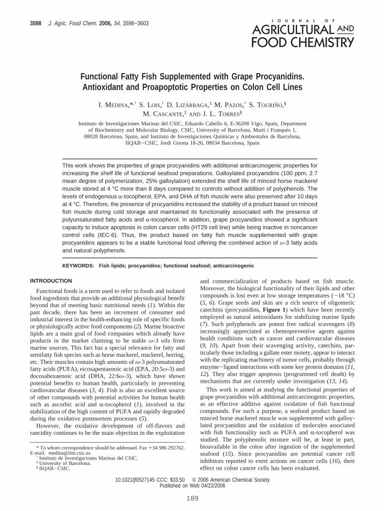
Functional Fatty Fish Supplemented with Grape Procyanidins.Antioxidant and Proapoptotic Properties on Colon Cell Lines
I. MEDINA,*,† S. LOIS,† D. LIZAÄ RRAGA,‡ M. PAZOS,† S. TOURINO,§
M. CASCANTE,‡ AND J. L. TORRES§
Instituto de Investigaciones Marinas del CSIC, Eduardo Cabello 6, E-36208 Vigo, Spain, Departmentof Biochemistry and Molecular Biology, CSIC, University of Barcelona, Martı i Franque`s 1,
08028 Barcelona, Spain, and Instituto de Investigaciones Quımicas y Ambientales de Barcelona,IIQAB-CSIC, Jordi Girona 18-26, 08034 Barcelona, Spain
This work shows the properties of grape procyanidins with additional anticarcinogenic properties forincreasing the shelf life of functional seafood preparations. Galloylated procyanidins (100 ppm, 2.7mean degree of polymerization, 25% galloylation) extended the shelf life of minced horse mackerelmuscle stored at 4 °C more than 8 days compared to controls without addition of polyphenols. Thelevels of endogenous R-tocopherol, EPA, and DHA of fish muscle were also preserved after 10 daysat 4 °C. Therefore, the presence of procyanidins increased the stability of a product based on mincedfish muscle during cold storage and maintained its functionality associated with the presence ofpolyunsaturated fatty acids and R-tocopherol. In addition, grape procyanidins showed a significantcapacity to induce apoptosis in colon cancer cells (HT29 cell line) while being inactive in noncancercontrol cells (IEC-6). Thus, the product based on fatty fish muscle supplemented with grapeprocyanidins appears to be a stable functional food offering the combined action of ω-3 fatty acidsand natural polyphenols.
KEYWORDS: Fish lipids; procyanidins; functional seafood; anticarcinogenic
INTRODUCTION
Functional foods is a term used to refer to foods and isolatedfood ingredients that provide an additional physiological benefitbeyond that of meeting basic nutritional needs (1). Within thepast decade, there has been an increment of consumer andindustrial interest in the health-enhancing role of specific foodsor physiologically active food components (2). Marine bioactivelipids are a main goal of food companies which already haveproducts in the market claiming to be stableω-3 oils frommarine sources. This fact has a special relevance for fatty andsemifatty fish species such as horse mackerel, mackerel, herring,etc. Their muscles contain high amounts ofω-3 polyunsaturatedfatty acids (PUFA), eicosapentaenoic acid (EPA, 20:5ω-3) anddocosahexaenoic acid (DHA, 22:6ω-3), which have shownpotential benefits to human health, particularly in preventingcardiovascular diseases (3,4). Fish is also an excellent sourceof other compounds with potential activities for human healthsuch as ascorbic acid andR-tocopherol (1), involved in thestabilization of the high content of PUFA and rapidly degradedduring the oxidative postmortem processes (5).
However, the oxidative development of off-flavors andrancidity continues to be the main objection in the exploitation
and commercialization of products based on fish muscle.Moreover, the biological functionality of their lipids and othercompounds is lost even at low storage temperatures (-18 °C)(5, 6). Grape seeds and skin are a rich source of oligomericcatechins (procyanidins,Figure 1) which have been recentlyemployed as natural antioxidants for stabilizing marine lipids(7). Such polyphenols are potent free radical scavengers (8)increasingly appreciated as chemopreventive agents againsthealth conditions such as cancer and cardiovascular diseases(9, 10). Apart from their scavenging activity, catechins, par-ticularly those including a gallate ester moiety, appear to interactwith the replicating machinery of tumor cells, probably throughenzyme-ligand interactions with some key protein domains (11,12). They also trigger apoptosis (programmed cell death) bymechanisms that are currently under investigation (13,14).
This work is aimed at studying the functional properties ofgrape procyanidins with additional anticarcinogenic properties,as an effective additive against oxidation of fish functionalcompounds. For such a purpose, a seafood product based onminced horse mackerel muscle was supplemented with galloy-lated procyanidins and the oxidation of molecules associatedwith fish functionality such as PUFA andR-tocopherol wasstudied. The polyphenolic mixture will be, at least in part,bioavailable in the colon after ingestion of the supplementedseafood (15). Since procyanidins are potential cancer cellinhibitors reported to exert actions on cancer cells (16), theireffect on colon cancer cells has been evaluated.
* To whom correspondence should be addressed. Fax+34 986 292762.E-mail: [email protected].
† Instituto de Investigaciones Marinas del CSIC.‡ University of Barcelona.§ IIQAB-CSIC.
3598 J. Agric. Food Chem. 2006, 54, 3598−3603
10.1021/jf0527145 CCC: $33.50 © 2006 American Chemical SocietyPublished on Web 04/22/2006
189
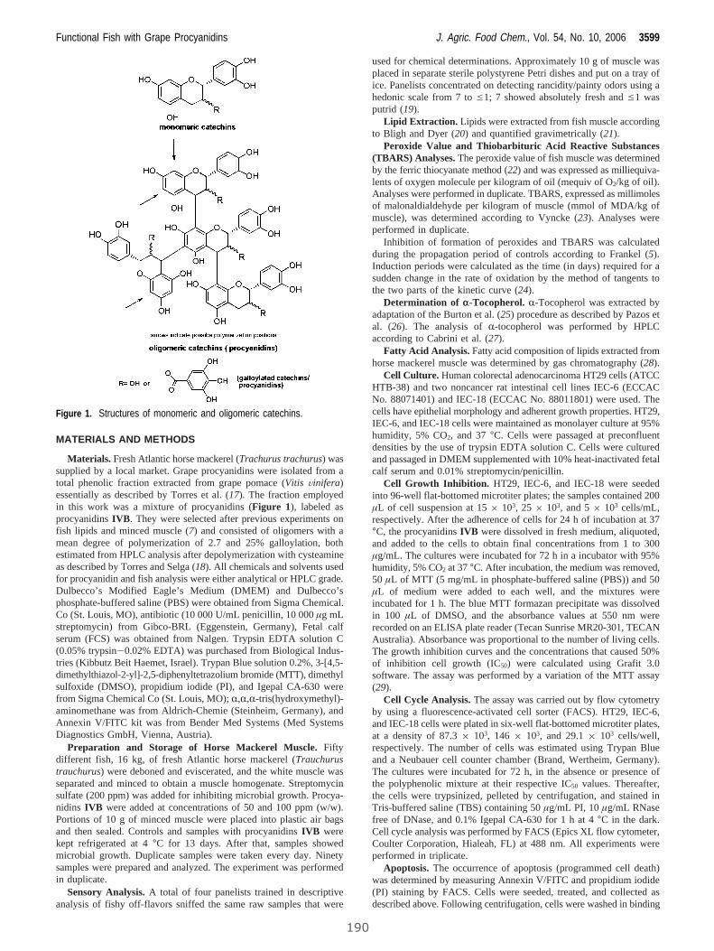
MATERIALS AND METHODS
Materials. Fresh Atlantic horse mackerel (Trachurus trachurus) wassupplied by a local market. Grape procyanidins were isolated from atotal phenolic fraction extracted from grape pomace (Vitis Vinifera)essentially as described by Torres et al. (17). The fraction employedin this work was a mixture of procyanidins (Figure 1), labeled asprocyanidinsIVB. They were selected after previous experiments onfish lipids and minced muscle (7) and consisted of oligomers with amean degree of polymerization of 2.7 and 25% galloylation, bothestimated from HPLC analysis after depolymerization with cysteamineas described by Torres and Selga (18). All chemicals and solvents usedfor procyanidin and fish analysis were either analytical or HPLC grade.Dulbecco’s Modified Eagle’s Medium (DMEM) and Dulbecco’sphosphate-buffered saline (PBS) were obtained from Sigma Chemical.Co (St. Louis, MO), antibiotic (10 000 U/mL penicillin, 10 000µg mLstreptomycin) from Gibco-BRL (Eggenstein, Germany), Fetal calfserum (FCS) was obtained from Nalgen. Trypsin EDTA solution C(0.05% trypsin-0.02% EDTA) was purchased from Biological Indus-tries (Kibbutz Beit Haemet, Israel). Trypan Blue solution 0.2%, 3-[4,5-dimethylthiazol-2-yl]-2,5-diphenyltetrazolium bromide (MTT), dimethylsulfoxide (DMSO), propidium iodide (PI), and Igepal CA-630 werefrom Sigma Chemical Co (St. Louis, MO);R,R,R-tris(hydroxymethyl)-aminomethane was from Aldrich-Chemie (Steinheim, Germany), andAnnexin V/FITC kit was from Bender Med Systems (Med SystemsDiagnostics GmbH, Vienna, Austria).
Preparation and Storage of Horse Mackerel Muscle.Fiftydifferent fish, 16 kg, of fresh Atlantic horse mackerel (Trauchurustrauchurus) were deboned and eviscerated, and the white muscle wasseparated and minced to obtain a muscle homogenate. Streptomycinsulfate (200 ppm) was added for inhibiting microbial growth. Procya-nidins IVB were added at concentrations of 50 and 100 ppm (w/w).Portions of 10 g of minced muscle were placed into plastic air bagsand then sealed. Controls and samples with procyanidinsIVB werekept refrigerated at 4°C for 13 days. After that, samples showedmicrobial growth. Duplicate samples were taken every day. Ninetysamples were prepared and analyzed. The experiment was performedin duplicate.
Sensory Analysis.A total of four panelists trained in descriptiveanalysis of fishy off-flavors sniffed the same raw samples that were
used for chemical determinations. Approximately 10 g of muscle wasplaced in separate sterile polystyrene Petri dishes and put on a tray ofice. Panelists concentrated on detecting rancidity/painty odors using ahedonic scale from 7 toe1; 7 showed absolutely fresh ande1 wasputrid (19).
Lipid Extraction. Lipids were extracted from fish muscle accordingto Bligh and Dyer (20) and quantified gravimetrically (21).
Peroxide Value and Thiobarbituric Acid Reactive Substances(TBARS) Analyses.The peroxide value of fish muscle was determinedby the ferric thiocyanate method (22) and was expressed as milliequiva-lents of oxygen molecule per kilogram of oil (mequiv of O2/kg of oil).Analyses were performed in duplicate. TBARS, expressed as millimolesof malonaldialdehyde per kilogram of muscle (mmol of MDA/kg ofmuscle), was determined according to Vyncke (23). Analyses wereperformed in duplicate.
Inhibition of formation of peroxides and TBARS was calculatedduring the propagation period of controls according to Frankel (5).Induction periods were calculated as the time (in days) required for asudden change in the rate of oxidation by the method of tangents tothe two parts of the kinetic curve (24).
Determination of r-Tocopherol. R-Tocopherol was extracted byadaptation of the Burton et al. (25) procedure as described by Pazos etal. (26). The analysis ofR-tocopherol was performed by HPLCaccording to Cabrini et al. (27).
Fatty Acid Analysis. Fatty acid composition of lipids extracted fromhorse mackerel muscle was determined by gas chromatography (28).
Cell Culture. Human colorectal adenocarcinoma HT29 cells (ATCCHTB-38) and two noncancer rat intestinal cell lines IEC-6 (ECCACNo. 88071401) and IEC-18 (ECCAC No. 88011801) were used. Thecells have epithelial morphology and adherent growth properties. HT29,IEC-6, and IEC-18 cells were maintained as monolayer culture at 95%humidity, 5% CO2, and 37°C. Cells were passaged at preconfluentdensities by the use of trypsin EDTA solution C. Cells were culturedand passaged in DMEM supplemented with 10% heat-inactivated fetalcalf serum and 0.01% streptomycin/penicillin.
Cell Growth Inhibition. HT29, IEC-6, and IEC-18 were seededinto 96-well flat-bottomed microtiter plates; the samples contained 200µL of cell suspension at 15× 103, 25 × 103, and 5× 10³ cells/mL,respectively. After the adherence of cells for 24 h of incubation at 37°C, the procyanidinsIVB were dissolved in fresh medium, aliquoted,and added to the cells to obtain final concentrations from 1 to 300µg/mL. The cultures were incubated for 72 h in a incubator with 95%humidity, 5% CO2 at 37°C. After incubation, the medium was removed,50 µL of MTT (5 mg/mL in phosphate-buffered saline (PBS)) and 50µL of medium were added to each well, and the mixtures wereincubated for 1 h. The blue MTT formazan precipitate was dissolvedin 100 µL of DMSO, and the absorbance values at 550 nm wererecorded on an ELISA plate reader (Tecan Sunrise MR20-301, TECANAustralia). Absorbance was proportional to the number of living cells.The growth inhibition curves and the concentrations that caused 50%of inhibition cell growth (IC50) were calculated using Grafit 3.0software. The assay was performed by a variation of the MTT assay(29).
Cell Cycle Analysis.The assay was carried out by flow cytometryby using a fluorescence-activated cell sorter (FACS). HT29, IEC-6,and IEC-18 cells were plated in six-well flat-bottomed microtiter plates,at a density of 87.3× 10³, 146 × 103, and 29.1× 10³ cells/well,respectively. The number of cells was estimated using Trypan Blueand a Neubauer cell counter chamber (Brand, Wertheim, Germany).The cultures were incubated for 72 h, in the absence or presence ofthe polyphenolic mixture at their respective IC50 values. Thereafter,the cells were trypsinized, pelleted by centrifugation, and stained inTris-buffered saline (TBS) containing 50µg/mL PI, 10µg/mL RNasefree of DNase, and 0.1% Igepal CA-630 for 1 h at 4°C in the dark.Cell cycle analysis was performed by FACS (Epics XL flow cytometer,Coulter Corporation, Hialeah, FL) at 488 nm. All experiments wereperformed in triplicate.
Apoptosis. The occurrence of apoptosis (programmed cell death)was determined by measuring Annexin V/FITC and propidium iodide(PI) staining by FACS. Cells were seeded, treated, and collected asdescribed above. Following centrifugation, cells were washed in binding
Figure 1. Structures of monomeric and oligomeric catechins.
Functional Fish with Grape Procyanidins J. Agric. Food Chem., Vol. 54, No. 10, 2006 3599
190
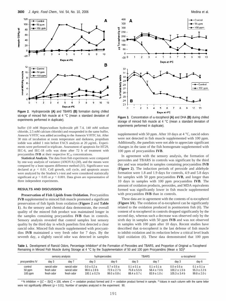
buffer (10 mM Hepes/sodium hydroxide pH 7.4, 140 mM sodiumchloride, 2.5 mM calcium chloride) and resuspended in the same buffer,Annexin V/FITC was added according to the Annexin V/FITC kit. After30 min of incubation at room temperature and darkness, propidiumiodide was added 1 min before FACS analysis at 20µg/mL. Experi-ments were performed in triplicate. Assessment of apoptosis for HT29,IEC-6, and IEC-18 cells was done after 72 h of treatment withprocyanidinsIVB at their respective IC50 concentrations.
Statistical Analysis. The data from fish experiments were comparedby one-way analysis of variance (ANOVA) (30), and the means werecompared by a least squares difference method (31). Significance wasdeclared atp < 0.01. Cell growth, cell cycle, and apoptosis assayswere analyzed by the Student’st-test and were considered statisticallysignificant atp < 0.05 orp < 0.001. Data given are representative ofthree independent experiments.
RESULTS AND DISCUSSION
Preservation of Fish Lipids from Oxidation. ProcyanidinsIVB supplemented to minced fish muscle promoted a significantpreservation of fish lipids from oxidation (Figure 2 andTable1). As the sensory and chemical data demonstrate, the overallquality of the minced fish product was maintained longer inthe samples containing procyanidinsIVB than in controls.Sensory analysis revealed that control samples lost sensoryquality by the third day in which panelists first detected a clearrancid odor. Minced fish muscle supplemented with procyani-dins IVB maintained a very fresh odor for 7 days. By theseventh day, a slightly rancid odor was detected in samples
supplemented with 50 ppm. After 10 days at 4°C, rancid odorswere not detected in fish muscle supplemented with 100 ppm.Additionally, the panelists were not able to appreciate significantchanges in the taste of the fish homogenate supplemented with100 ppm of procyanidinsIVB.
In agreement with the sensory analysis, the formation ofperoxides and TBARS in controls was significant by the thirdday and was retarded in samples containing procyanidinsIVB(Figure 2). The induction periods of peroxide and aldehydeformation were 1.8 and 1.9 days for controls, 4.9 and 5.8 daysfor samples with 50 ppm procyanidinsIVB, and longer than10 days in samples with 100 ppm procyanidinsIVB. Theamount of oxidation products, peroxides, and MDA equivalentsformed was significantly lower in fish muscle supplementedwith procyanidinsIVB than in controls.
These data are in agreement with the contents ofR-tocopherol(Figure 3A). The oxidation ofR-tocopherol can be significantlyrelated to the oxidation produced in postmortem fish (6). Thecontent ofR-tocopherol in controls dropped significantly by thesecond day, whereas such a decrease was observed only by thesixth day in samples with 50 ppmIVB and was not observedin samples with 100 ppm after 10 days. Recent studies havedescribed thatR-tocopherol is the last defense of fish muscleto inhibit oxidation and its reduction below a critical level leadslipid oxidation (6). These data demonstrated that 100 ppm
Figure 2. Hydroperoxide (A) and TBARS (B) formation during chilledstorage of minced fish muscle at 4 °C (mean ± standard deviation ofexperiments performed in duplicate).
Table 1. Development of Rancid Odors, Percentage Inhibitiona of the Formation of Peroxides and TBARS, and Proportion of Original R-TocopherolRemaining in Minced Fish Muscle during Storage at 4 °C by the Supplementation of 50 and 100 ppm Procyanidins (Mean ± SD)b
sensory analysis hydroperoxides TBARS R-tocopherol
procyanidins IV day 3 day 7 day 3 day 6 day 3 day 7 day 3 day 6
control rancid odor rancid odor 0.0 ± 0.2 a 0.7 ± 0.5 a 0.1 ± 0.1 a 0.1 ± 0.1 a 0.0 ± 0.5 a 0.1 ± 0.1 a50 ppm fresh odor rancid odor 98.6 ± 2.8 b 72.9 ± 2.7 b 75.8 ± 5.5 b 58.4 ± 7.0 b 100.2 ± 1.5 b 55.3 ± 2.2 b100 ppm fresh odor fresh odor 100.1 ± 0.2 b 99.0 ± 0.8 c 88.4 ± 0.7 c 83.9 ± 1.0 c 105.3 ± 3.4 b 99.8 ± 2.0 c
a % inhibition ) [(C − S)/C] × 100, where C ) oxidation product formed and S ) oxidation product formed in sample. b Values in each column with the same letterwere not significantly different (p < 0.01). Number of samples analyzed in the experiment: 90.
Figure 3. Concentration of R-tocopherol (A) and DHA (B) during chilledstorage of minced fish muscle at 4 °C (mean ± standard deviation ofexperiments performed in duplicate).
3600 J. Agric. Food Chem., Vol. 54, No. 10, 2006 Medina et al.
191
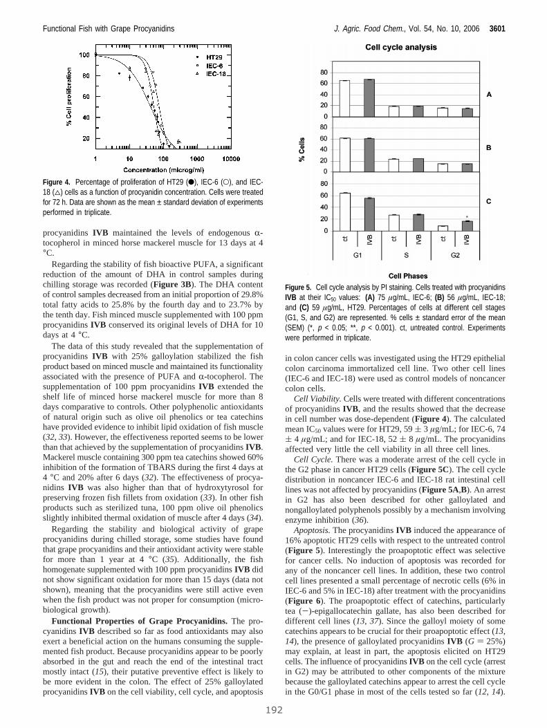
procyanidinsIVB maintained the levels of endogenousR-tocopherol in minced horse mackerel muscle for 13 days at 4°C.
Regarding the stability of fish bioactive PUFA, a significantreduction of the amount of DHA in control samples duringchilling storage was recorded (Figure 3B). The DHA contentof control samples decreased from an initial proportion of 29.8%total fatty acids to 25.8% by the fourth day and to 23.7% bythe tenth day. Fish minced muscle supplemented with 100 ppmprocyanidinsIVB conserved its original levels of DHA for 10days at 4°C.
The data of this study revealed that the supplementation ofprocyanidinsIVB with 25% galloylation stabilized the fishproduct based on minced muscle and maintained its functionalityassociated with the presence of PUFA andR-tocopherol. Thesupplementation of 100 ppm procyanidinsIVB extended theshelf life of minced horse mackerel muscle for more than 8days comparative to controls. Other polyphenolic antioxidantsof natural origin such as olive oil phenolics or tea catechinshave provided evidence to inhibit lipid oxidation of fish muscle(32,33). However, the effectiveness reported seems to be lowerthan that achieved by the supplementation of procyanidinsIVB.Mackerel muscle containing 300 ppm tea catechins showed 60%inhibition of the formation of TBARS during the first 4 days at4 °C and 20% after 6 days (32). The effectiveness of procya-nidins IVB was also higher than that of hydroxytyrosol forpreserving frozen fish fillets from oxidation (33). In other fishproducts such as sterilized tuna, 100 ppm olive oil phenolicsslightly inhibited thermal oxidation of muscle after 4 days (34).
Regarding the stability and biological activity of grapeprocyanidins during chilled storage, some studies have foundthat grape procyanidins and their antioxidant activity were stablefor more than 1 year at 4°C (35). Additionally, the fishhomogenate supplemented with 100 ppm procyanidinsIVB didnot show significant oxidation for more than 15 days (data notshown), meaning that the procyanidins were still active evenwhen the fish product was not proper for consumption (micro-biological growth).
Functional Properties of Grape Procyanidins.The pro-cyanidinsIVB described so far as food antioxidants may alsoexert a beneficial action on the humans consuming the supple-mented fish product. Because procyanidins appear to be poorlyabsorbed in the gut and reach the end of the intestinal tractmostly intact (15), their putative preventive effect is likely tobe more evident in the colon. The effect of 25% galloylatedprocyanidinsIVB on the cell viability, cell cycle, and apoptosis
in colon cancer cells was investigated using the HT29 epithelialcolon carcinoma immortalized cell line. Two other cell lines(IEC-6 and IEC-18) were used as control models of noncancercolon cells.
Cell Viability. Cells were treated with different concentrationsof procyanidinsIVB, and the results showed that the decreasein cell number was dose-dependent (Figure 4). The calculatedmean IC50 values were for HT29, 59( 3 µg/mL; for IEC-6, 74( 4 µg/mL; and for IEC-18, 52( 8 µg/mL. The procyanidinsaffected very little the cell viability in all three cell lines.
Cell Cycle.There was a moderate arrest of the cell cycle inthe G2 phase in cancer HT29 cells (Figure 5C). The cell cycledistribution in noncancer IEC-6 and IEC-18 rat intestinal celllines was not affected by procyanidins (Figure 5A,B). An arrestin G2 has also been described for other galloylated andnongalloylated polyphenols possibly by a mechanism involvingenzyme inhibition (36).
Apoptosis.The procyanidinsIVB induced the appearance of16% apoptotic HT29 cells with respect to the untreated control(Figure 5). Interestingly the proapoptotic effect was selectivefor cancer cells. No induction of apoptosis was recorded forany of the noncancer cell lines. In addition, these two controlcell lines presented a small percentage of necrotic cells (6% inIEC-6 and 5% in IEC-18) after treatment with the procyanidins(Figure 6). The proapoptotic effect of catechins, particularlytea (-)-epigallocatechin gallate, has also been described fordifferent cell lines (13,37). Since the galloyl moiety of somecatechins appears to be crucial for their proapoptotic effect (13,14), the presence of galloylated procyanidinsIVB (G ) 25%)may explain, at least in part, the apoptosis elicited on HT29cells. The influence of procyanidinsIVB on the cell cycle (arrestin G2) may be attributed to other components of the mixturebecause the galloylated catechins appear to arrest the cell cyclein the G0/G1 phase in most of the cells tested so far (12, 14).
Figure 4. Percentage of proliferation of HT29 (b), IEC-6 (O), and IEC-18 (4) cells as a function of procyanidin concentration. Cells were treatedfor 72 h. Data are shown as the mean ± standard deviation of experimentsperformed in triplicate.
Figure 5. Cell cycle analysis by PI staining. Cells treated with procyanidinsIVB at their IC50 values: (A) 75 µg/mL, IEC-6; (B) 56 µg/mL, IEC-18;and (C) 59 µg/mL, HT29. Percentages of cells at different cell stages(G1, S, and G2) are represented. % cells ± standard error of the mean(SEM) (*, p < 0.05; **, p < 0.001). ct, untreated control. Experimentswere performed in triplicate.
Functional Fish with Grape Procyanidins J. Agric. Food Chem., Vol. 54, No. 10, 2006 3601
192

In any case, and most interestingly, the effects recorded for theantioxidant mixture on cell cycle and apoptosis was selectivefor cancer cells.
Briefly, data from the cellular studies show that the procya-nidins IVB exerted little effect on the cell viability and cellcycle integrity of epithelial colon cells of both cancer andnoncancer phenotype. The procyanidins exert a mild andselective proapoptotic effect on HT29 colon carcinoma cells.The healthy noncancer cells are not significantly affected bythe procyanidins.
Conclusions.Functionalω-3 unsaturated fatty acids as wellasR-tocopherol in minced horse mackerel muscle were stabi-lized by addition of an antioxidant extract composed mainly ofprocyanidins. The results with cell lines suggest that grapeprocyanidinsIVB may also exert a direct preventive effect oncolon epithelial cells by acting as an antioxidant and a selectiveproapoptotic agent. Thus, the product based on fatty fish musclesupplemented with grape procyanidins appears to be an interest-ing and stable functional food offering the combined action ofω-3 fatty acids and natural polyphenols.
ACKNOWLEDGMENT
M. Jesus Gonzalez and Elsa Silva are gratefully acknowledgedfor their technical assistance.
LITERATURE CITED
(1) Mazza, G. Functional Food, Biochemical and Processingaspects; Mazza, G., Ed.; Technomic Publishing Co., Inc.:Lancaster, PA, 1998.
(2) Hasler, C. M. Functional foods: Their role in disease preventionand health promotion.Food Technol.1998,52, 63-70.
(3) Nilsen, D. W.; Albrektsen, G.; Landmark, K. Effects of a high-dose concentrate of n-3 fatty acids or corn oil introduced earlyafter an acute myocardial infarction on serum triacylglycerol andHDL cholesterol.Am. J. Clin. Nutr.2001,74 (1), 50-56.
(4) Lee, K. W.; Lip, G. Y. H. The role of omega-3 fatty acids inthe secondary prevention of cardiovascular disease.QJM 2003,96 (7), 465-480.
(5) Frankel, E. N.Lipid Oxidation; The Oily Press: Dundee,Scotland, 1998.
(6) Pazos, M.; Sanchez, L.; Medina, I. Alpha-tocopherol oxidationin fish muscle during chilling and frozen storage.J. Agric. FoodChem.2005,53, 4000-4005.
(7) Pazos, M.; Gallardo, J. M.; Torres, J. L.; Medina, I. Activity ofgrape polyphenols as inhibitors of the oxidation of fish lipidsand frozen fish muscle.Food Chem.2005,92, 547-557.
(8) Yokozawa, T.; Chen, C. P.; Dong, E.; Tanaka, T.; Nonaka, G.I.; Nishioka, I. Study on the inhibitory effect of tannins andflavonoids against the 1,1-diphenyl-2-picrylhydrazyl radical.Biochem. Pharmacol.1998,56, 213-222.
(9) Yang, C. S.; Landau, J. M.; Huang, M. T.; Newmark, H. L.Inhibition of carcinogenesis by dietary polyphenolic compounds.Annu. ReV. Nutr.2001,21, 381-406.
(10) Murphy, K. J.; Chronopoulos, A. K.; Singh, I.; Francis, M. A.;Moriarty, H.; Pike, M. J.; Turner, A. H.; Mann, N. J.; Sinclair,A. J. Dietary flavanols and procyanidin oligomers from cocoa(Theobroma cacao) inhibit platelet function.Am. J. Clin. Nutr.2003,77 (6), 1466-1473.
(11) Liang, Y. C.; Lin-Shiau, S. Y.; Chen, C. F.; Lin, J. K. Suppressionof extracellular signals and cell proliferation through EGFreceptor binding by (-)-epigallocatechin gallate in human A431epidermoid carcinoma cells.J. Cell Biochem. 1997, 67 (1), 55-65.
(12) Ahmad, N.; Cheng, P. Y.; Mukhtar, H. Cell cycle dysregulationby green tea polyphenol epigallocatechin-3-gallate.Biochem.Biophys. Res. Commun.2000,275 (2), 328-334.
(13) Tan, X. H.; Hu, D. R.; Li, S. R.; Han, Y.; Zhang, Y. L.; Zhou,D. Y. Differences of four catechins in cell cycle arrest andinduction of apoptosis in LoVo cells.Cancer Lett.2000,158(1), 1-6.
(14) Gupta, S.; Hussain, T.; Mukhtar, H. Molecular pathway for (-)-epigallocatechin-3-gallate-induced cell cycle arrest and apoptosisof human prostate carcinoma cells.ArchiV. Biochem. Biophys.2003,410 (1), 177-185.
(15) Gonthier, M. P.; Donovan, J. L.; Texier, O.; Felgines, C.;Remesy, C.; Scalbert, A. Metabolism of dietary procyanidins inrats.Free Radical Biol. Med.2003,35 (8), 837-844.
(16) Matito, C.; Mastorakou, F.; Centelles, J. J.; Torres, J. L.;Cascante, M. 2003. Antiproliferative effect of antioxidantpolyphenols from grape in murine Hepa-1c1c7.Eur. J. Nutr.2003,42 (1), 43-49.
(17) Torres, J. L.; Varela, B.; Garcıa, M. T.; Carilla, J.; Matito, C.;Centelles, J. J.; Cascante, C.; Sort, X.; Bobet, R. Valorizationof grape (VitisVinifera) byproducts. Antioxidant and biologicalproperties of polyphenolic fractions differing in procyanidincomposition and flavonol content.J. Agric. Food Chem. 2002,50, 7548-7555.
(18) Torres, J. L.; Selga, A. Procyanidin size and composition bythiolysis with cysteamine hydrochloride and chromatography.Chromatographia2003,57, 441-445.
(19) Kelleher, S. D.; Silva, L. A.; Hultin, H. O.; Wilhelm. K. A.Inhibition of lipid oxidation during processing of washed, mincedAtlantic mackerel.J. Food Sci.1992,57, 1103-1108.
(20) Bligh, E.; Dyer, W. A rapid method of total lipid extraction andpurification.Can. J. Biochem. Physiol.1959,37, 911-917.
(21) Herbes, S.; Allen, C. Lipid quantification of freshwater inver-tebrates: method modification for microquantitation.Can. J. FishAquat. Sci.1983,40, 1315-1317.
(22) Chapman, R. H.; McKay, J. The estimation of peroxides in fatsand oils by the ferric thiocyanate method.J. Am. Oil Chem. Soc.1949,26, 360-363.
Figure 6. Analysis of apoptosis by annexin V/FITC and PI staining. Cellstreated with procyanidins IVB at their IC50 values: (A) 75 µg/mL, IEC-6;(B) 56 µg/mL, IEC-18; and (C) 59 µg/mL, HT29. Percentages of cells atdifferent cell stages are represented. Cell stages: early apoptosis (AnnexinV+PI-); late apoptosis/necrotic (Annexin V+PI-); necrotic (Annexin V-PI+).% cells ± SEM (*, p < 0.05; **, p < 0.001). Experiments were performedin triplicate.
3602 J. Agric. Food Chem., Vol. 54, No. 10, 2006 Medina et al.
193
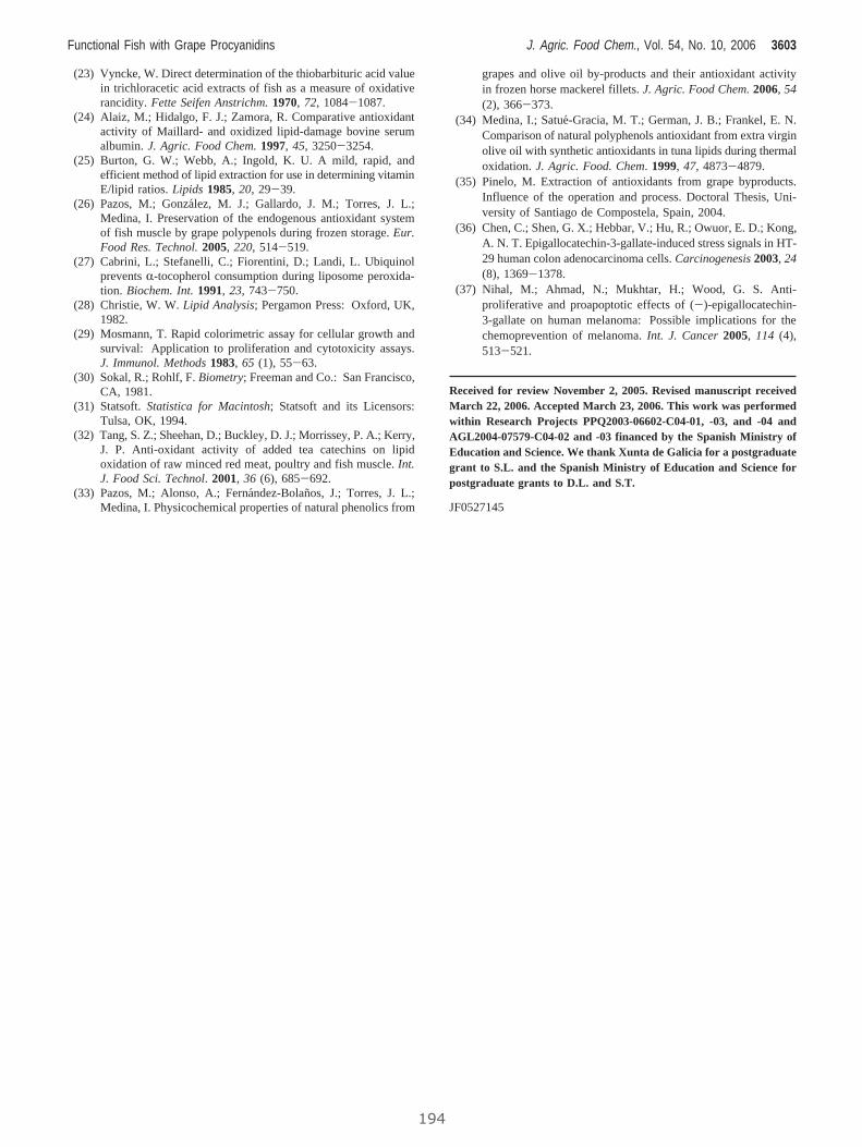
(23) Vyncke, W. Direct determination of the thiobarbituric acid valuein trichloracetic acid extracts of fish as a measure of oxidativerancidity.Fette Seifen Anstrichm.1970,72, 1084-1087.
(24) Alaiz, M.; Hidalgo, F. J.; Zamora, R. Comparative antioxidantactivity of Maillard- and oxidized lipid-damage bovine serumalbumin.J. Agric. Food Chem.1997,45, 3250-3254.
(25) Burton, G. W.; Webb, A.; Ingold, K. U. A mild, rapid, andefficient method of lipid extraction for use in determining vitaminE/lipid ratios.Lipids 1985,20, 29-39.
(26) Pazos, M.; Gonzalez, M. J.; Gallardo, J. M.; Torres, J. L.;Medina, I. Preservation of the endogenous antioxidant systemof fish muscle by grape polypenols during frozen storage.Eur.Food Res. Technol.2005,220, 514-519.
(27) Cabrini, L.; Stefanelli, C.; Fiorentini, D.; Landi, L. UbiquinolpreventsR-tocopherol consumption during liposome peroxida-tion. Biochem. Int.1991,23, 743-750.
(28) Christie, W. W.Lipid Analysis; Pergamon Press: Oxford, UK,1982.
(29) Mosmann, T. Rapid colorimetric assay for cellular growth andsurvival: Application to proliferation and cytotoxicity assays.J. Immunol. Methods1983,65 (1), 55-63.
(30) Sokal, R.; Rohlf, F.Biometry; Freeman and Co.: San Francisco,CA, 1981.
(31) Statsoft.Statistica for Macintosh; Statsoft and its Licensors:Tulsa, OK, 1994.
(32) Tang, S. Z.; Sheehan, D.; Buckley, D. J.; Morrissey, P. A.; Kerry,J. P. Anti-oxidant activity of added tea catechins on lipidoxidation of raw minced red meat, poultry and fish muscle.Int.J. Food Sci. Technol.2001,36 (6), 685-692.
(33) Pazos, M.; Alonso, A.; Fernandez-Bolanos, J.; Torres, J. L.;Medina, I. Physicochemical properties of natural phenolics from
grapes and olive oil by-products and their antioxidant activityin frozen horse mackerel fillets.J. Agric. Food Chem.2006,54(2), 366-373.
(34) Medina, I.; Satue-Gracia, M. T.; German, J. B.; Frankel, E. N.Comparison of natural polyphenols antioxidant from extra virginolive oil with synthetic antioxidants in tuna lipids during thermaloxidation.J. Agric. Food. Chem.1999,47, 4873-4879.
(35) Pinelo, M. Extraction of antioxidants from grape byproducts.Influence of the operation and process. Doctoral Thesis, Uni-versity of Santiago de Compostela, Spain, 2004.
(36) Chen, C.; Shen, G. X.; Hebbar, V.; Hu, R.; Owuor, E. D.; Kong,A. N. T. Epigallocatechin-3-gallate-induced stress signals in HT-29 human colon adenocarcinoma cells.Carcinogenesis2003, 24(8), 1369-1378.
(37) Nihal, M.; Ahmad, N.; Mukhtar, H.; Wood, G. S. Anti-proliferative and proapoptotic effects of (-)-epigallocatechin-3-gallate on human melanoma: Possible implications for thechemoprevention of melanoma.Int. J. Cancer2005, 114 (4),513-521.
Received for review November 2, 2005. Revised manuscript receivedMarch 22, 2006. Accepted March 23, 2006. This work was performedwithin Research Projects PPQ2003-06602-C04-01, -03, and -04 andAGL2004-07579-C04-02 and -03 financed by the Spanish Ministry ofEducation and Science. We thank Xunta de Galicia for a postgraduategrant to S.L. and the Spanish Ministry of Education and Science forpostgraduate grants to D.L. and S.T.
JF0527145
Functional Fish with Grape Procyanidins J. Agric. Food Chem., Vol. 54, No. 10, 2006 3603
194
III. Procyanidins from pine bark: relationships between
structure, composition and antiradical activity.
Jerez, M.; Touriño, S.; Sineiro, J.; Torres, J.L. and Núñez,
M.J.; Food Chem.; 104 (2): 518-527; 2007

www.elsevier.com/locate/foodchem
Food Chemistry 104 (2007) 518–527
FoodChemistry
Procyanidins from pine bark: Relationships betweenstructure, composition and antiradical activity
Marıa Jerez a, Sonia Tourino b, Jorge Sineiro a, Josep Lluıs Torres b, Marıa Jose Nunez a,*
a Escuela Tecnica Superior de Ingenierıa, Universidad de Santiago de Compostela, Lope Gomez de Marzoa, 15782 Santiago de Compostela, Spainb Instituto de Investigaciones Quımicas y Ambientales, CSIC, Jordi Girona 18-26, 08034 Barcelona, Spain
Received 16 June 2006; received in revised form 5 October 2006; accepted 28 November 2006
Abstract
Barks of Pinus pinaster and Pinus radiata were studied as source of procyanidins; these raw materials, considered a byproduct of for-estal industry, were extracted with ethanol. The extract was partially fractionated to obtain an aqueous fraction (FA) containing a greatpart of the procyanidins from barks and with potential application to both food and medical fields. FAs were rich in polyphenols, 65–87% of which were procyanidins; the mean degree of polymerization (mDP) was 7.9 for radiata (rFA) and 10.6 for pinaster (pFA)varieties.
The aqueous fractions were chromatographed on Sephadex LH-20 to obtain specific fractions differing in DP and composition. Thesefractions were analysed by thiolysis with cysteamine, followed by RP-HPLC. Results showed that (+)-catechin was the main terminalunit for both barks and also the main extension unit for radiata. In contrast (�)-epicatechin was predominant as extension unit in pin-aster and this could have implications for applications in oils, emulsions and biological systems.
In terms of antiradical activity, expressed as specific antiradical units, the entire rFA gave the best results, together with rF5. ForP. pinaster bark, the best results were achieved for fractions F5–F8, with DP 7–22. The whole fraction from radiata represents an eco-nomic alternative of great interest because the fractionation costs can be avoided.� 2006 Elsevier Ltd. All rights reserved.
Keywords: Pinus pinaster; Pinus radiata; Bark; Antiradical power; Procyanidins; Aqueous fraction; Fractionation
1. Introduction
Great interest is currently centred on potential benefitsof plant polyphenols as complements to the organism’santioxidant defence system. Polyphenols are potent freeradical-scavengers and preventive against cardiovasculardiseases, cancer and other disorders; they are increasinglyused as natural food additives, acting as flavouring, colour-ing and antioxidant agents (Plumb, de Pascual-Teresa,Santos-Buelga, Cheynier, & Williamson, 1998). Amongthe different phenolics, flavonoids, phenolic acids, stilbenesand tannins, especially condensed tannins (proanthocyani-dins), are particularly important (Hagerman et al., 1998).
0308-8146/$ - see front matter � 2006 Elsevier Ltd. All rights reserved.
doi:10.1016/j.foodchem.2006.11.071
* Corresponding author. Tel.: +34 981 563100x16792; fax: +34 981528050.
E-mail address: [email protected] (M.J. Nunez).
19
Proanthocyanidins are the most abundant polyphenols inplants after lignins, and they may represent up to 50% inseveral barks (Matthews et al., 1997); they can be dividedinto procyanidins and prodelphynidins. Pycnogenol, basi-cally a procyanidin mixture from maritime pine, is proba-bly the most studied phenolic from trees.
Procyanidins are composed of oligomers and polymers,consisting of (+)-catechin and/or (�)-epicatechin unitslinked mainly through C4 ? C8 and/or C4 ? C6 bonds(B-type). These flavan-3-ol units can be doubly linked bya C4 ? C8 bond and an additional ether bond fromO7 ? C2 (A-type). Most of the activities of procyanidins,including the free radical-scavenging capacity, largelydepend on their structure, particularly their degree of poly-merization (DP) (Gaulejac, Vivas, Freitas, & Bourgeois,1999; Tourino et al., 2005). Moreover, the presence of gal-late esters seems decisive for the regulation of the cell cycle
7
M. Jerez et al. / Food Chemistry 104 (2007) 518–527 519
by catechins (Liberto & Cobrinik, 2000). In food systems,partition phenomena of procyanidins play an importantrole in the preservant action (Frankel, 2001). The main dif-ficulty in studies on procyanidins is probably that ofobtaining them in an individual molecular form. The com-plete purification of a procyanidin with a DP above five isalmost impossible. Therefore, for studying their structuresand properties, mixtures more or less polymerized are oftenemployed (Guyot, Marnet, & Drilleau, 2001). Moreover,synergistic effects of active mixtures make plant extractsand fractions more interesting than pure compounds forfunctional food applications.
Many methods have been proposed for separatingprocyanidins according to their DP. For analytical pur-poses, a suitable separation can be achieved by normal-phase HPLC, TLC or reversed-phase HPLC. On a prepara-tive scale, gel chromatography with different packings, suchas Sephadex G-25, LH-20 and Toyopearl TSK HW 40, hasbeen used (Kantz & Singleton, 1990; Karonen, Loponen,Ossipov, & Pihlaja, 2004). Normal-phase HPLC can sepa-rate procyanidins according to their degree of polymeriza-tion up to decamers, but this method is specific formonomers and oligomers and results in a severe underesti-mation of procyanidins, considering the prevalence of thepolymers in nature (Czochanska, Foo, Newman, & Porter,1980). Reversed-phase HPLC is the method commonly usedfor the separation of flavan-3-ol monomers and some smalloligomers, especially dimers and trimers. However, there isdifficulty in determining the DP using reversed-phase meth-ods since oligomers elute in non-sequential order. Further-more, analysis of higher oligomeric procyanidins is notfeasible, since the number of isomers increases with increas-ing degrees of polymerization. This effect results in a reten-tion time overlap of isomers of differing DP, causing thehigher oligomers to co-elute as a large unresolved peak(Santos-Buelga & Williamson, 2003). The technique ofthin-layer chromatography (TLC), with a silica phase, per-mits the separation of oligomeric proanthocyanidins up toheptamers (Lea, 1978). This method can be used only forqualitative analysis. Sephadex LH-20 and Toyopearl TSKHW-40 or TSK HW-50 columns are classically used to frac-tionate proanthocyanidins on the basis of molecular size;these are eluted with a mobile phase of alcohol–water oracetone–water. Large molecular weight procyanidins arethen recovered with 60–70% acetone in water, with no fur-ther separation (Taylor, Barofsky, Kennedy, & Deinzer,2003; Tourino et al., 2005).
Depolymerization by thiolysis has been proved as anefficient method for determining the nature of the flavan-3-ol units within procyanidins and for determining theaverage degree of polymerization. Thiolysis is performedin the presence of acid and a nucleophilic reagent, suchas toluene-a-thiol (Rigaud, Perez-Ilzarbe, Da Silva, &Cheynier, 1991), cysteamine hydrochloride (Torres &Selga, 2003) or phloroglucinol (Kennedy & Jones, 2001).The extension subunits of procyanidins are attacked bythe nucleophilic reagent to form the corresponding thio-
198
derivatives and the terminal unit is released as the free fla-van-3-ol. Degradation products can be analysed byreversed-phase HPLC, and the results will provide infor-mation on the nature of the extension and terminal unitsand on the average degree of polymerization.
Pine is one of the plants with the highest content ofprocyanidins. The phenolics of pine bark are (+)-catechin,(�)-epicatechin, dihydroquercetin, phenolic acids and,most of all procyanidin dimers, trimers, oligomers andpolymers (Wood, Senthilmohan, & Peskin, 2002). Pinebark procyanidins have diverse biomedical applications(Packer, Rimbach, & Virgili, 1999; Rohdewald, 2002). Sev-eral constituents of pine bark extracts, such as gallic, pro-tocatechuic acids and catechin, are readily adsorbed byhuman skin and make the preparations useful for topicalapplication (Sarikaki et al., 2004). Besides, because pineprocyanidins are devoid of gallate esters, which appear tointerfere with crucial cell functions, they may be innocuouschemopreventive agents of choice for many applications(Tourino et al., 2005). The use of polyphenols as food anti-oxidants is also frequent, especially in fatty foods, such asfish, making it possible to prolong the storage time of sev-eral species both fresh and frozen products (Pazos, Alonso,Fernandez-Bolanos, Torres, & Medina, 2006). Anotherdietetic application has recently reported (in a patent) byDegre (2003) who found that polymeric procyanidins(DP > 10) were strong inhibitors of intestinal a-amylase,so they may be useful in dietetics to suppress a-amylaseactivities, and/or the caloric values of food.
Working with procyanidins from different pine species,we have detected structural variations (Jerez, Pinelo,Sineiro, & Nunez, 2006) which may be related to differentphysicochemical and biological properties. The aim of thiswork is to achieve the fractionation of structurally differentprocyanidins contained in the aqueous fractions fromcrude ethanolic extracts of bark of two varieties of pine,Pinus pinaster and Pinus radiata, and to characterize thedifferent fractions for procyanidin content, degree of poly-merization and antiradical activity. The information gainedabout the properties of the mixtures will help to define theirpossible applications as food antioxidants and/or as func-tional components .
2. Materials and methods
2.1. Plant material
Pine (P. pinaster and P. radiata) barks provided byM. Bouzas Garrido, S.A. (Vedra, A Coruna, Spain) weredried at room temperature for a week. They were thenground in an analytical mill MF 10 IKA-WERKE (Staufen,Germany) to less than 1 mm.
2.2. Reagents
Ethanol, methanol, acetone, toluene, formic acid,hydrochloric acid, sulphuric acid and vanillin were
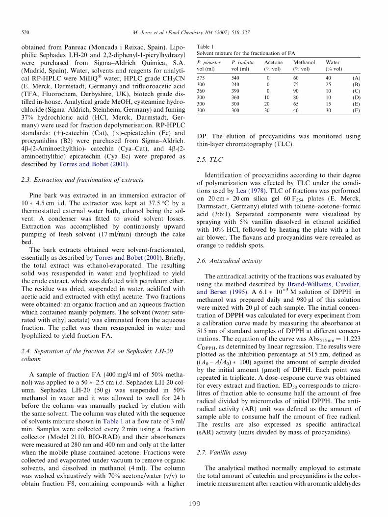
Table 1Solvent mixture for the fractionation of FA
P. pinaster
vol (ml)P. radiata
vol (ml)Acetone(% vol)
Methanol(% vol)
Water(% vol)
575 540 0 60 40 (A)300 240 0 75 25 (B)360 390 0 90 10 (C)300 360 10 80 10 (D)300 300 20 65 15 (E)300 300 30 40 30 (F)
520 M. Jerez et al. / Food Chemistry 104 (2007) 518–527
obtained from Panreac (Moncada i Reixac, Spain). Lipo-philic Sephadex LH-20 and 2,2-diphenyl-1-picrylhydrazylwere purchased from Sigma–Aldrich Quımica, S.A.(Madrid, Spain). Water, solvents and reagents for analyti-cal RP-HPLC were MilliQ� water, HPLC grade CH3CN(E. Merck, Darmstadt, Germany) and trifluoroacetic acid(TFA, Fluorochem, Derbyshire, UK), biotech grade dis-tilled in-house. Analytical grade MeOH, cysteamine hydro-chloride (Sigma–Aldrich, Steinheim, Germany) and fuming37% hydrochloric acid (HCl, Merck, Darmstadt, Ger-many) were used for fraction depolymerisation. RP-HPLCstandards: (+)-catechin (Cat), (�)-epicatechin (Ec) andprocyanidins (B2) were purchased from Sigma–Aldrich.4b-(2-Aminoethylthio)- catechin (Cya–Cat), and 4b-(2-aminoethylthio) epicatechin (Cya–Ec) were prepared asdescribed by Torres and Bobet (2001).
2.3. Extraction and fractionation of extracts
Pine bark was extracted in an immersion extractor of10 � 4.5 cm i.d. The extractor was kept at 37.5 �C by athermostatted external water bath, ethanol being the sol-vent. A condenser was fitted to avoid solvent losses.Extraction was accomplished by continuously upwardpumping of fresh solvent (17 ml/min) through the cakebed.
The bark extracts obtained were solvent-fractionated,essentially as described by Torres and Bobet (2001). Briefly,the total extract was ethanol-evaporated. The resultingsolid was resuspended in water and lyophilized to yieldthe crude extract, which was defatted with petroleum ether.The residue was dried, suspended in water, acidified withacetic acid and extracted with ethyl acetate. Two fractionswere obtained: an organic fraction and an aqueous fractionwhich contained mainly polymers. The solvent (water satu-rated with ethyl acetate) was eliminated from the aqueousfraction. The pellet was them resuspended in water andlyophilized to yield fraction FA.
2.4. Separation of the fraction FA on Sephadex LH-20
column
A sample of fraction FA (400 mg/4 ml of 50% metha-nol) was applied to a 50 � 2.5 cm i.d. Sephadex LH-20 col-umn. Sephadex LH-20 (50 g) was suspended in 50%methanol in water and it was allowed to swell for 24 hbefore the column was manually packed by elution withthe same solvent. The column was eluted with the sequenceof solvents mixture shown in Table 1 at a flow rate of 3 ml/min. Samples were collected every 2 min using a fractioncollector (Model 2110, BIO-RAD) and their absorbanceswere measured at 280 nm and 400 nm and only at the latterwhen the mobile phase contained acetone. Fractions werecollected and evaporated under vacuum to remove organicsolvents, and dissolved in methanol (4 ml). The columnwas washed exhaustively with 70% acetone/water (v/v) toobtain fraction F8, containing compounds with a higher
19
DP. The elution of procyanidins was monitored usingthin-layer chromatography (TLC).
2.5. TLC
Identification of procyanidins according to their degreeof polymerization was effected by TLC under the condi-tions used by Lea (1978). TLC of fractions was performedon 20 cm � 20 cm silica gel 60 F254 plates (E. Merck,Darmstadt, Germany) eluted with toluene–acetone–formicacid (3:6:1). Separated components were visualized byspraying with 5% vanillin dissolved in ethanol acidifiedwith 10% HCl, followed by heating the plate with a hotair blower. The flavans and procyanidins were revealed asorange to reddish spots.
2.6. Antiradical activity
The antiradical activity of the fractions was evaluated byusing the method described by Brand-Williams, Cuvelier,and Berset (1995). A 6.1 � 10�5 M solution of DPPH� inmethanol was prepared daily and 980 ll of this solutionwere mixed with 20 ll of each sample. The initial concen-tration of DPPH was calculated for every experiment froma calibration curve made by measuring the absorbance at515 nm of standard samples of DPPH at different concen-trations. The equation of the curve was Abs515 nm = 11,223CDPPH, as determined by linear regression. The results wereplotted as the inhibition percentage at 515 nm, defined as((A0 – A/A0) � 100) against the amount of sample dividedby the initial amount (lmol) of DPPH. Each point wasrepeated in triplicate. A dose–response curve was obtainedfor every extract and fraction. ED50 corresponds to micro-litres of fraction able to consume half the amount of freeradical divided by micromoles of initial DPPH. The anti-radical activity (AR) unit was defined as the amount ofsample able to consume half the amount of free radical.The results are also expressed as specific antiradical(sAR) activity (units divided by mass of procyanidins).
2.7. Vanillin assay
The analytical method normally employed to estimatethe total amount of catechin and procyanidins is the color-imetric measurement after reaction with aromatic aldehydes
9
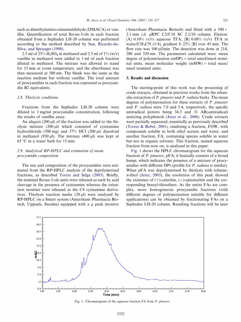
M. Jerez et al. / Food Chemistry 104 (2007) 518–527 521
such as dimethylamino-cinnamaldehyde (DMACA) or van-illin. Quantification of total flavan-3-ols in each fractionobtained from a Sephadex LH-20 column was performedaccording to the method described by Sun, Ricardo-da-Silva, and Spranger (1998).
2.5 ml of 25% H2SO4 in methanol and 2.5 ml of 1% (w/v)vanillin in methanol were added to 1 ml of each fractiondiluted in methanol. The mixture was allowed to standfor 15 min at room temperature, and the absorbance wasthen measured at 500 nm. The blank was the same as thereaction medium but without vanillin. The total amountof procyanidins in each fraction was expressed as procyani-din B2 equivalents.
2.8. Thiolysis conditions
Fractions from the Sephadex LH-20 column werediluted to 1 mg/ml procyanidin concentration, followingthe results of vanillin assay.
An aliquot (200 ll) of the fraction was added to the thi-olysis mixture (200 ll) which consisted of cysteaminehydrochloride (500 mg) and 37% HCl (200 ll) dissolvedin methanol (930 ll). The mixture (400 ll) was kept at65 �C in a water bath for 15 min.
2.9. Analytical RP-HPLC and estimation of mean
procyanidin composition
The size and composition of the procyanidins were esti-mated from the RP-HPLC analysis of the depolymerisedfractions, as described Torres and Selga (2003). Briefly,the terminal flavan-3-ols units were released as such by acidcleavage in the presence of cysteamine whereas the exten-sion moieties were released as the C4 cysteamine deriva-tives. Thiolysis reaction media (20 ll) were analysed byRP-HPLC on a Smart system (Amersham–Pharmacia Bio-tech, Uppsala, Sweden) equipped with a l peak monitor
Fig. 1. Chromatogram of the aqueo
200
(Amersham–Pharmacia Biotech) and fitted with a 100 �2.1 mm i.d. lRPC C2/C18 SC 2.1/10 column. Elution:[A] 0.10% (v/v) aqueous TFA, [B] 0.08% (v/v) TFA inwater/CH3CN (1:4), gradient 8–23% [B] over 45 min. Theflow rate was 100 ll/min. The detection was done at 214,280 and 320 nm. The parameters calculated were: meandegree of polymerization (mDP) = total nmol/nmol termi-nal units, mean molecular weight (mMW) = total mass/nmol terminal units.
3. Results and discussion
The starting-point of this work was the processing ofcrude extracts, obtained in previous works from the ethan-olic extraction of P. pinaster and P. radiata barks. The meandegrees of polymerisation for these extracts of P. pinaster
and P. radiata were 7.0 and 5.4, respectively, the specificantiradical powers being 34.3 and 31 AR(antiradical)units/mg polyphenols (Jerez et al., 2006). Crude extractswere partially separated, essentially as previously described(Torres & Bobet, 2001), rendering a fraction, FOW, withcompounds soluble in both ethyl acetate and water, andanother fraction, FA, containing species soluble in waterbut not in organic solvents. This fraction, named aqueousfraction from now on, is analysed in this paper.
Fig. 1 shows the HPLC chromatogram for the aqueousfraction of P. pinaster, pFA; it basically consists of a broadhump, which indicates the presence of a mixture of procy-anidins with different DPs (profile for P. radiata is similar).When pFA was depolymerised by thiolysis with toluene-a-thiol (Jerez, 2003), the resolution of this peak showedthe existence of (+)-catechin, (�)-epicatechin and the cor-responding benzyl-thioethers. As the entire FAs are com-plex, more homogeneous procyanidin fractions (withdifferent degrees of polymerisation suitable for differentapplications) can be obtained by fractionating FAs on aSephadex LH-20 column. Resulting fractions will be later
us fraction FA from P. pinaster.
522 M. Jerez et al. / Food Chemistry 104 (2007) 518–527
depolymerised for characterization. Table 1 summarisesthe elution conditions used. This mixture was previouslyused by other authors (Taylor et al., 2003) for fractioning
0
0.5
1
1.5
2
2.5
3
3.5
4
4.5
0 200 400
Time (mi
Ab
sorb
ance
280
nm
F0 F1 F2 F3 F4
(A) (B) (C)
0
0.5
1
1.5
2
2.5
3
0 200 400
Time (mi
Ab
sorb
ance
280
nm
F0 F1 F2 F3 F4
(A) (B) (C)
a
b
Fig. 2. Chromatographic profile obtained by fractionation of fraction
Fig. 3. TLC of fractions from Sephadex LH-20 column runs in one dimensiP. radiata.
20
hop procyanidins. Under these conditions, low molecularweight (MW) compounds are eluted first and bulkierprocyanidins are recovered last.
600 800
n)
0
0.02
0.04
0.06
0.08
0.1
0.12
0.14
Ab
sorb
ance
400
nm
F7F5 F6
(D) (E) (F)
600 800
n)
0
0.01
0.02
0.03
0.04
0.05
0.06
0.07
0.08
Ab
sorb
ance
400
nm
F5 F6 F7
(D) (E) (F)
FA on Sephadex LH-20 column: (a) P. pinaster and (b) P. radiata.
on with (toluene/acetone/formic acid) as solvent: (a) P. pinaster and (b)
1
M. Jerez et al. / Food Chemistry 104 (2007) 518–527 523
Fig. 2a and b shows the elution profiles which wereobtained by measuring the absorbance of the fractions col-lected at intervals of 2 min in an UV-V Jasco V-530 spec-trophotometer. Fractions under each peak were pooledinto 8 fractions, F0–F7. Procyanidins of higher MWremained tightly adsorbed at the top of the column.Because the chromatographic mode is adsorption ratherthan exclusion, the separation can be incomplete (Yanag-ida et al., 2000). Washing with 70% acetone resulted inthe recovery of high mDP procyanidins. This washingwas carried out, instigating the recovery of fraction F8.Acetone was chosen because the carbonyl oxygen servesas a strong H-bond acceptor and allows displacement ofbound polymeric phenols from Sephadex LH-20.
TLC of each fraction was performed to obtain prelimin-ary information concerning their procyanidin content(DP), following the vanillin assay (Fig. 3a and b). In the
Fig. 4. RP-HPLC chromatograms of fraction F3 from P. pinaster (pF3), ob
202
first fractions, pF0, pF2 for P. pinaster and rF0, rF1 forP. radiata, no spots were observed, indicating the absenceof flavan-3-ols, either as monomers or as their derivatives,and these fractions were not included in the subsequentanalyses. In any case these fractions may contain phenolicacids and taxifolin (dihydroquercetin, present basically infractions soluble in both ethyl acetate and water), frequentin pine bark (Wood et al., 2002). (+)-catechin was onlyobserved, as traces, in rF2. The developed spots in frac-tions pF3–pF4 and rF2–rF4 corresponded to procyanidinsof low molecular weight. For the other fractions, the spotsremained on the base-line, indicating that they includedbulky hydrophilic high molecular weight compounds.
Each one of the obtained fractions containing procyani-dins was thiolysed with cysteamine, which was preferred totoluene-a-thiol for being more user-friendly and much lesstoxic (Torres & Selga, 2003). Figs. 4a and b and 5a and b
tained by fractionating FA: (a) raw fraction and (b) thiolysed fraction.
524 M. Jerez et al. / Food Chemistry 104 (2007) 518–527
show the chromatogram of F3 of the barks from the twopine varieties. In agreement with our previous results, P.
radiata contained more procyanidins than P. pinaster.The profile after thiolysis indicated that (+)-catechin wasthe main terminal unit in both cases. Regarding the exten-sion units, the situation is different. P. radiata containedalmost exclusively catechin units whereas P. Pinaster
included both epicatechin and catechin extension units(Fig. 4b).
In Tables 2 and 3, the structural compositions of the dif-ferent fractions are indicated. For all P. pinaster fractionsand whole pFA, epicatechin was the main extension unit,with a ratio of 3–6 with respect to catechin, except forpF3, for which the ratio was 1.2.
Fig. 5. RP-HPLC chromatogram of fraction F3 from P. radiata (rF3), obt
20
P. radiata fractions were also consistent with wholerFA, with a proportion catechin/epicatechin extensionunits of 1.2. Again, rF3 was the exception to the rule, theratio being 10.8. In this case, the mean composition ofrF2 was also different, because epicatechin predominatedas the extension unit. The presence of catechin as an abun-dant extension unit in P. radiata may influence the physico-chemical properties of procyanidins, which will be morehydrophobic than those from P. pinaster. This fact canbe of interest for the possible antioxidant activity in oils,emulsions and biological systems.
Tables 4 and 5 summarise the mean structural parame-ters and activity of the entire FAs and eluted fractions(procyanidin content, antiradical power, mDP and
ained by fractionating FA: (a) raw fraction and (b) thiolysed fraction.
3

Table 2Structural composition of fractions from FA of P. pinaster, determined by HPLC following thiolysis degradation
Fraction Proportion of terminal units (%) Proportion of extension units (%)
Catechin Epicatechin Aminoethylthio catechin Aminoethylthio epicatechin
pFA 8.1 ± 0.1 1.4 ± 0.2 13.5 ± 0.4 77.0 ± 0pF3 35.9 ± 0.3 3.8 ± 0 26.9 ± 1.0 33.5 ± 0.7pF4 20.5 ± 0.6 1.6 ± 0.3 18.0 ± 0.3 60.0 ± 0pF5 13.1 ± 0.3 0.8 ± 0 14.5 ± 0.7 71.7 ± 0.9pF6 7.8 ± 0.4 nd 12.4 ± 0.2 79.6 ± 0.4pF7 5.5 ± 0.1 nd 12.6 ± 0.1 81.9 ± 0.1pF8 4.6 ± 0.2 nd 12.9 ± 0.5 82.7 ± 0.2
Table 3Structural composition of fractions from FA of P. radiata, determined by HPLC following thiolysis degradation
Fraction Proportion of terminal units (%) Proportion of extension units (%)
Catechin Epicatechin Aminoethylthio catechin Aminoethylthio epicatechin
FA 10.4 ± 0.6 2.3 ± 0.2 44.2 ± 0.4 43.4 ± 0.17F2 40.8 ± 3.2 7.9 ± 0.9 21.7 ± 1.2 29.8 ± 1.1F3 44.7 ± 1.5 3.4 ± 0.7 47.5 ± 0.4 4.4 ± 0.4F4 25.8 ± 0.9 2.4 ± 0.3 48.3 ± 1.0 23.6 ± 0.2F5 15.2 ± 1.2 0.4 ±0.5 45.8 ± 0.3 38.7 ± 0.4F6 9.9 ± 0.1 1.1 ± 0.1 49.1 ± 0.1 39.9 ± 0.2F7 8.0 ± 0.2 nd 50.4 ± 0.4 41.5 ± 0.4F8 6.3 ± 0.3 0.6 ± 0.1 53 ± 0 40 ± 0
M. Jerez et al. / Food Chemistry 104 (2007) 518–527 525
mMW), which were calculated from the thiolysis data.Procyanidins for FAs represent 65% of total polyphenolsin pinaster, increasing to 87% for radiata. Up to fraction4, eluted with water–methanol, mDP were similar for thetwo varieties; P. radiata was richer in procyanidins andwas more active. The antiradical activity per mass (specificantiradical activity, sAR) is more suitable for interpretingthe effect of polymerisation on the scavenging activity.According to this index, values are similar for pF3, pF4,and not very different from that of the total pFA. In con-trast, sAR for the whole rFA was much higher than sARfor rF2–rF4. From F5 on, fractions eluted with acetone,the situation is different; pF5 and rF5, with mDP about 7,showed the highest sAR, and the subsequent fractions fromP.radiata presented a gradual decrease in this parameter,down to values similar to those of the first fractions. InP. pinaster, the behaviour did not follow exactly the sametrend; pF6–pF8 showed similar sAR values, around 56.This different pattern may be related to the differencesdetected in the extension units for both varieties (Tables 2and 3). Fractions with mDP above 7 from P. pinaster con-
Table 4Characteristics of fractions from FA of P. pinaster
Fractions mDP mMW Procyanidins (mg)
pFA (400 mg) 10.6 ± 0.4 3067 ± 119 139 ± 3.0pF3 2.5 ± 0 731 ± 6 1.1 ± 0.1pF4 4.6 ± 0.1 1313 ± 18 2.9 ± 0.3pF5 7.2 ± 0.1 2090 ± 44 8.8 ± 0.2pF6 12.8 ± 0.4 3713 ± 110 24 ± 0.4pF7 18.2 ± 0.3 5258 ± 91 10.5 ± 0.2pF8 22.2 ± 1.1 6425 ± 303 22.4 ± 0.5
204
tained mostly epicatechin as extension unit while P. radiata
oligomers contained both catechin and epicatechin in simi-lar proportions. This fact may influence the antiradicaleffectiveness, because the stereoposition of the phenolicOH group in C3 is involved in the oxidation mechanism,especially by radicals.
The increase of antiradical power of flavanols with mDPup to 7 can be explained by the electron delocalisationthrough intramolecular links among monomers, becauseC4–C8 confers a very tight conformation but, at a givenpoint, with new units the steric hindrance can be decisive(Gaulejac et al., 1999). Gaulejac et al. (1999) found anincrease in activity for procyanidins from 1 to 4 units. Inthe present work, we have recorded an increase up to 6–7units and from there, either a stabilisation (P. pinaster
12–22 mDP) or a drop in sAR (P. radiata 9–15 mDP).When procyanidins incorporate gallate, tendencies are
diverse. The trend is an increase of antiradical power untilDP = 3; if we compare values for F3 (Tables 3, 4) with ananalogous fraction of grape pomace with 25% galloylation(Pazos, 2005), antiradical power for grape is 1.5 times
Total AR activity units AR activity units/mg procyanidins (sAR)
5488 39.541.9 38.2
106.7 36.3561.8 63.5
1360.5 56.7580.6 55.2
1261.8 56.3
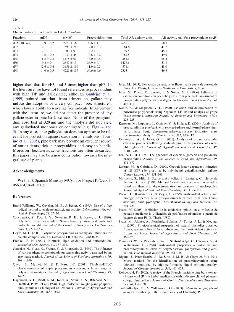
Table 5Characteristics of fractions from FA of P. radiata
Fractions mDP mMW Procyanidins (mg) Total AR activity units AR activity units/mg procyanidins (sAR)
rFA (400 mg) 7.9 ± 0.2 2278 ± 56 166 ± 4 9650 58.1rF2 2.1 ± 0.1 598 ± 28 2.0 ± 0.3 84.6 41.2rF3 2.1 ± 0.1 603 ± 9 2.2 ± 0.1 99.5 45.8rF4 3.6 ± 0.2 1033 ± 45 3.8 ± 0.2 187.8 49.9rF5 6.5 ± 0.3 1875 ±88 13.0 ± 0.4 851.1 65.4rF6 9.2 ± 0.1 2647 ± 13 26.9 ± 0.1 1428.6 53.1rF7 12.6 ± 0.4 3651 ± 110 11.9 ± 0.3 571.4 48.0rF8 14.6 ± 0.5 4226 ± 137 50.6 ± 0.6 2353 46.5
526 M. Jerez et al. / Food Chemistry 104 (2007) 518–527
higher than that for rF3, and 3 times higher than pF3. Inthe literature, we have not found references to procyanidinswith high DP and galloylated, although Gaulejac et al.(1999) pointed out that, from trimers on, gallate mayinduce the adoption of a very compact ‘‘box structure”,which lowers ability to scavenge free radicals. In agreementwith the literature, we did not detect the presence of anygallate ester in pine bark extracts. None of the procyani-dins absorbed at 320 nm and the thiolysis did not yieldany galloylated monomer or conjugate (e.g. Figs. 4 and5). In any case, since galloylation does not appear to be rel-evant for protection against oxidation in emulsions (Tour-ino et al., 2005), pine bark may become an excellent sourceof antioxidants, rich in procyanidins and easy to handle.Moreover, because aqueous fractions are often discarded,this paper may also be a new contribution towards the inte-gral use of plants.
Acknowledgements
We thank Spanish Ministry MCyT for Project PPQ2003-06602-C04-01 y 02.
References
Brand-Williams, W., Cuvelier, M. E., & Berset, C. (1995). Use of a free
radical method to evaluate antioxidant activity. Lebensmittel-Wissens-
chaft & Technologie, 28, 25–30.Czochanska, Z., Foo, L. Y., Newman, R. H., & Porter, L. J. (1980).
Polymeric proanthocyanidins. Stereochemistry, structural units and
molecular weight. Journal of the Chemical Society – Perkin Transac-
tions, I, 2278–2286.Degre, M. F. (2003). Polymeric procyanidins as a-amylase inhibitors for
dietetic composition. Fr. Demande FR 2002-2571 20020228.Frankel, E. N. (2001). Interfacial lipid oxidation and antioxidation.
Journal of Oleo Science, 50, 387–391.Gaulejac, N., Vivas, N., Freitas, V., & Bourgeois, G. (1999). The influence
of various phenolic compounds on scavenging activity assessed by an
enzymatic method. Journal of the Science of Food and Agriculture, 79,1081–1090.
Guyot, S., Marnet, N., & Drilleau, J-F. (2001). Thiolysis-HPLC
characterization of apple procyanidins covering a large range of
polymerization states. Journal of Agricultural and Food Chemistry, 49,14–20.
Hagerman, A. E., Riedl, K. M., Jones, A., Sovik, K. N., Ritchard, N. T.,Hartfeld, P. W., et al. (1998). High molecular weight plant poliphen-
olics (tannins) as biological antioxidants. Journal of Agricultural and
Food Chemistry, 46, 1887–1892.
20
Jerez, M. (2003). Extraccion de sustancias Bioactivas a partir de corteza dePino. Ms. Thesis, University Santiago de Compostela, Spain.
Jerez, M., Pinelo, M., Sineiro, J., & Nunez, M. J. (2006). Influence of
extraction conditions on phenolic yields from pine bark: assessment of
procyanidins polymerization degree by thiolysis. Food Chemistry, 94,406–414.
Kantz, K., & Singleton, V. L. (1990). Isolation and determination of
polymeric polyphenols using Sephadex LH-20 and analysis of grape
tissue extracts. American Journal of Enology and Viticulture, 41(3),223–228.
Karonen, M., Loponen, J., Ossipov, V., & Pihlaja, K. (2004). Analysis of
procyanidins in pine bark with reversed-phase and normal-phase high-
performance liquid chromatography-electrospray ionization mass
spectrometry. Analytica Chimica Acta, 522, 105–112.Kennedy, J. A., & Jones, G. P. (2001). Analysis of proanthocyanidin
cleavage products following acid-catalysis in the presence of excess
phloroglucinol. Journal of Agricultural and Food Chemistry, 49,1740–1746.
Lea, A. G. H. (1978). The phenolics of ciders: oligomeric and polymeric
procyanidins. Journal of the Science of Food and Agriculture, 29,471–477.
Liberto, M., & Cobrinik, D. (2000). Growth factor-dependent induction
of p21 (CIP1) by green tea by polyphenol, epigallocatechin gallate.Cancer Letters, 154, 151–161.
Matthews, S., Mila, I., Scalbert, A., Pollet, B., Lapierre, C., Herve duPenhoat, C., et al. (1997). Method for estimation of proanthocyanidins
based on their acid depolymerization in presence of nucleophiles.Journal of Agricultural and Food Chemistry, 45, 1195–1201.
Packer, L., Rimbach, G., & Virgili, F. (1999). Antioxidant activity and
biologic properties of a procyanidin-rich extract from pine (Pinus
maritima) bark, pycnogenol. Free Radical Biology and Medicine, 27,704–724.
Pazos, M. (2005). Inhibicion de la oxidacion lipıdica en el musculo depescado mediante la utilizacion de polifenoles obtenidos a partir debagazo de uva Ph.D. Thesis, USC.
Pazos, M., Alonso, A., Fernandez-Bolanos, J., Torres, J. L., & Medina,I. (2006). Physicochemical properties of natural phenolics obtained
from grape and olive oil by-products and their antioxidant activity in
frozen fish fillets. Journal of Agricultural and Food Chemistry, 54,366–373.
Plumb, G. W., de Pascual-Teresa, S., Santos-Buelga, C., Cheynier, V., &Williamson, G. (1998). Antioxidant properties of catechins and
proanthocyanidins: effect of polymerisation, galloylation and glycos-
ilation. Free Radical Research, 29, 351–358.Rigaud, J., Perez-Ilzarbe, J., Da Silva, J. M. R., & Cheynier, V. (1991).
Micro method for the identification of proanthocyanidin using
thiolysis monitored by high-performance liquid chromatography.Journal of Chromatography A, 540, 401–405.
Rohdewald, P. (2002). A review of the French maritime pine bark extract
(Pycnogenol (R)), a herbal medication with a diverse clinical pharma-
cology. International Journal of Clinical Pharmacology and Therapeu-
tics, 40, 158–168.Santos-Buelga, C., & Williamson, G. (2003). Methods in polyphenol
analysis. Cambridge, UK: Royal Society of Chemistry Pub..
5

M. Jerez et al. / Food Chemistry 104 (2007) 518–527 527
Sarikaki, V., Rallis, M., Tanojo, H., Panten, I., Dotsiva, Y., Laukas, Y.L., et al. (2004). In vitro percutaneous absorption of pine bark extract
(PYC) in human skin. Journal of Toxicology – Cutaneous and Ocular
Toxicology, 23, 149–158.Sun, B., Ricardo-da-Silva, J. M., & Spranger, I. (1998). Critical factors of
vanillin assay for catechins and proanthocyanidins. Journal of Agri-
cultural and Food Chemistry, 46, 4267–4274.Taylor, A. W., Barofsky, E., Kennedy, J. A., & Deinzer, M. L. (2003).
Hop (Humulus lupulus L.) Proanthocyanidins characterized by massspectrometry, acid catalysis, and gel permeation chromatography.Journal of Agricultural and Food Chemistry, 51, 4101–4110.
Torres, J. L., & Bobet, R. (2001). New flavanol-derivatives from grape
(Vitis vinifera) byproducts. Antioxidant aminoethylthio-flavan-3-olconjugates from a polymeric waste fraction used as a source offlavanols. Journal of Agricultural and Food Chemistry, 49, 4627–4634.
206
Torres, J. L., & Selga, A. (2003). Procyanidin size and composition by
thiolysis with cysteamine hydrochloride and chromatography. Chro-
matographia, 57, 441–445.Tourino, S., Selga, A., Jimenez, A., Julia, L., Lozano, C., Lizarraga, D.,
et al. (2005). Procyanidin fractions from Pinus pinaster bark: radicalscavenging power in solution, antioxidant activity in emulsion andantiproliferative effect in mellanoma cells. Journal of Agricultural and
Food Chemistry, 53, 4728–4735.Wood, J. E., Senthilmohan, S. T., & Peskin, A. V. (2002). Antioxidant
activity of procyanidin-containing plant extracts at different pHs. Food
Chemistry, 77, 155–161.Yanagida, A., Kanda, T., Takahashi, T., Kamimura, A., Hamazono,
T., & Honda, S. (2000). Fractionation of apple procyanidins
according to their degree of polymerization by normal-phase high-
performance liquid chromatography. Journal of Chromatography A,
890, 251–259.
IV. The importance of polymerization and galloylation
for the antiproliferative properties of procyanidin-
rich natural extracts.
Lizárraga, D.; Lozano, C.; Briede´, J. J.; Van Delft, J. H.;
Touriño, S.; Centelles, J. J.; Torres J.L. and Cascante,
M.; FEBS Journal; 274: 4802–4811; 2007

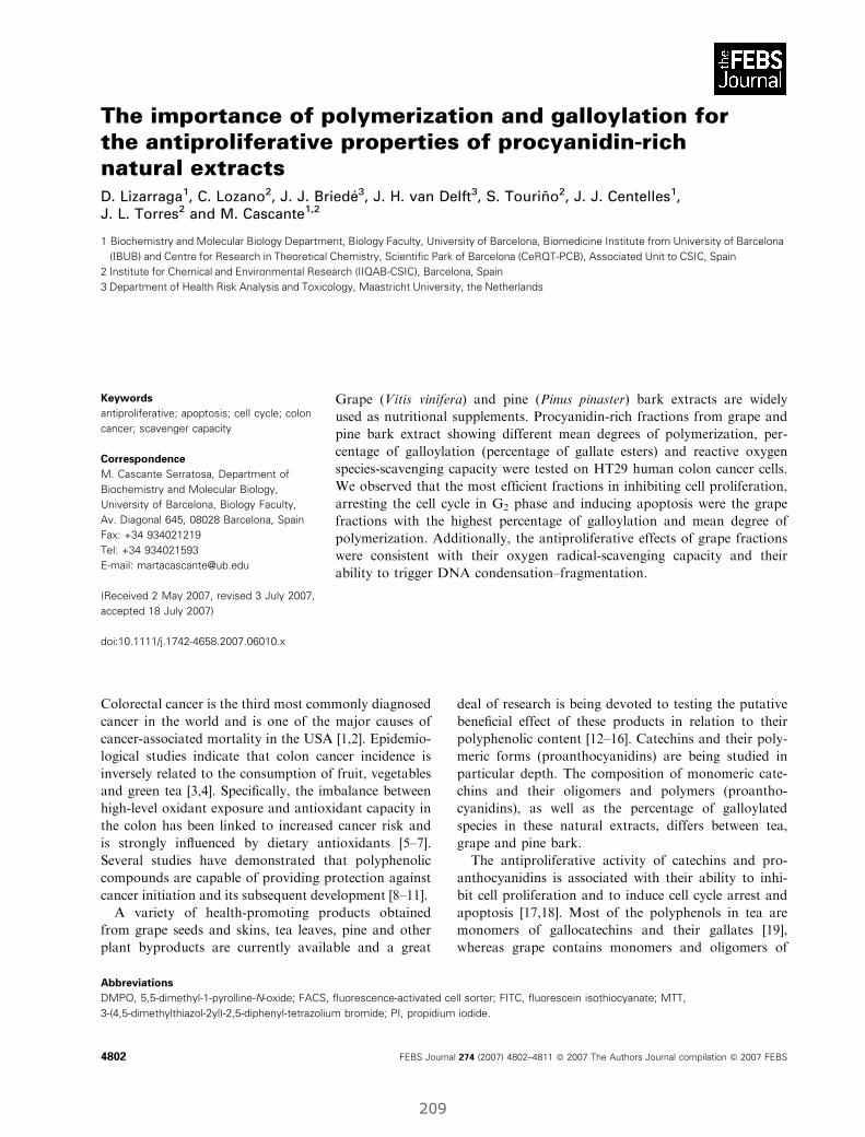
The importance of polymerization and galloylation forthe antiproliferative properties of procyanidin-richnatural extractsD. Lizarraga1, C. Lozano2, J. J. Briede3, J. H. van Delft3, S. Tourino2, J. J. Centelles1,J. L. Torres2 and M. Cascante1,2
1 Biochemistry and Molecular Biology Department, Biology Faculty, University of Barcelona, Biomedicine Institute from University of Barcelona
(IBUB) and Centre for Research in Theoretical Chemistry, Scientific Park of Barcelona (CeRQT-PCB), Associated Unit to CSIC, Spain
2 Institute for Chemical and Environmental Research (IIQAB-CSIC), Barcelona, Spain
3 Department of Health Risk Analysis and Toxicology, Maastricht University, the Netherlands
Colorectal cancer is the third most commonly diagnosed
cancer in the world and is one of the major causes of
cancer-associated mortality in the USA [1,2]. Epidemio-
logical studies indicate that colon cancer incidence is
inversely related to the consumption of fruit, vegetables
and green tea [3,4]. Specifically, the imbalance between
high-level oxidant exposure and antioxidant capacity in
the colon has been linked to increased cancer risk and
is strongly influenced by dietary antioxidants [5–7].
Several studies have demonstrated that polyphenolic
compounds are capable of providing protection against
cancer initiation and its subsequent development [8–11].
A variety of health-promoting products obtained
from grape seeds and skins, tea leaves, pine and other
plant byproducts are currently available and a great
deal of research is being devoted to testing the putative
beneficial effect of these products in relation to their
polyphenolic content [12–16]. Catechins and their poly-
meric forms (proanthocyanidins) are being studied in
particular depth. The composition of monomeric cate-
chins and their oligomers and polymers (proantho-
cyanidins), as well as the percentage of galloylated
species in these natural extracts, differs between tea,
grape and pine bark.
The antiproliferative activity of catechins and pro-
anthocyanidins is associated with their ability to inhi-
bit cell proliferation and to induce cell cycle arrest and
apoptosis [17,18]. Most of the polyphenols in tea are
monomers of gallocatechins and their gallates [19],
whereas grape contains monomers and oligomers of
Keywords
antiproliferative; apoptosis; cell cycle; colon
cancer; scavenger capacity
Correspondence
M. Cascante Serratosa, Department of
Biochemistry and Molecular Biology,
University of Barcelona, Biology Faculty,
Av. Diagonal 645, 08028 Barcelona, Spain
Fax: +34 934021219
Tel: +34 934021593
E-mail: [email protected]
(Received 2 May 2007, revised 3 July 2007,
accepted 18 July 2007)
doi:10.1111/j.1742-4658.2007.06010.x
Grape (Vitis vinifera) and pine (Pinus pinaster) bark extracts are widely
used as nutritional supplements. Procyanidin-rich fractions from grape and
pine bark extract showing different mean degrees of polymerization, per-
centage of galloylation (percentage of gallate esters) and reactive oxygen
species-scavenging capacity were tested on HT29 human colon cancer cells.
We observed that the most efficient fractions in inhibiting cell proliferation,
arresting the cell cycle in G2 phase and inducing apoptosis were the grape
fractions with the highest percentage of galloylation and mean degree of
polymerization. Additionally, the antiproliferative effects of grape fractions
were consistent with their oxygen radical-scavenging capacity and their
ability to trigger DNA condensation–fragmentation.
Abbreviations
DMPO, 5,5-dimethyl-1-pyrolline-N-oxide; FACS, fluorescence-activated cell sorter; FITC, fluorescein isothiocyanate; MTT,
3-(4,5-dimethylthiazol-2yl)-2,5-diphenyl-tetrazolium bromide; PI, propidium iodide.
4802 FEBS Journal 274 (2007) 4802–4811 ª 2007 The Authors Journal compilation ª 2007 FEBS
209

catechins with some galloylation and mainly poly-
merized procyanidins [20]. In contrast, procyanidin
fractions from pine bark extracts do not contain gallo-
catechins or gallates.
The influence of polyphenolic structure on antioxi-
dant activity, protective capacity and, particularly, on
the mechanism of action remains open to debate and
further study is required. Research with different cell
lines has shown that the most widely studied natural
polyphenol, epigallocatechin-3-gallate from green tea, is
a potent antioxidant and chemopreventive agent [21,22].
These and other results suggest that the galloylation of
catechins and the presence of gallocatechin moieties in
natural extracts could be important chemical character-
istics. They may be useful indicators in evaluating the
potential of natural plant extracts for colon cancer pre-
vention or treatment and the degree of polymerization
related to the bioavailability in the colon.
Procyanidins and monomeric catechins (Fig. 1) are
the main active polyphenols in grape and pine bark.
The difference between grape and pine catechins and
procyanidins is found in the presence of gallate esters
in position 3 (galloylation). Whereas grape flavanols
are galloylated to some extent [23,24], pine bark
appears to be devoid of gallate esters [25,26]. It has
been reported that oligomeric procyanidins are not sig-
nificantly absorbed in the intestinal tract, and reach
the colon mainly intact [27]. They are therefore bio-
available to the epithelial cells in the intestinal wall,
where procyanidins and other phenolics are extensively
degraded, metabolized and absorbed. In a first stage,
the oligomers are depolymerized and the constitutive
catechin units are partially absorbed as glucuronates,
sulfates and methyl esthers, as described for the small
intestine [28]. They are also, in part, extensively metab-
olized to phenolic acids such as 3-hydroxyphenylvaleric
acid and 3-hydroxyphenylpropionic acid, which are
then absorbed as glucuronates and sulfates [27,29].
The gallate esters are more stable than the simple cate-
chins upon being metabolized [30] and may be more
bioavailable in the colon. Gallates have been reported
to inhibit cell growth, trigger cell cycle arrest in tumor
cell lines and induce apoptosis [31,32]. Furthermore,
studies have shown that they also offer protection by
scavenging reactive oxygen species such as superoxide
anion, hydrogen peroxide and hydroxyl radicals, which
cause destruction of biochemical components that are
important in physiological metabolism [33,34]. This
capacity to prevent the imbalance between high-level
oxidant exposure and antioxidant capacity, which
leads to several pathological processes, may contribute
to the chemopreventive effect of the gallic acid deriva-
tives. Because grape is a rich source of procyanidins
and contains some galloylation, procyanidin fractions
from grape could be potential antiproliferative com-
pounds of interest in the prevention of colon cancer.
In the present study, we investigated the relationship
of different structural factors of procyanidins, such as
the mean degree of polymerization and percentage of
galloylation, with their antiproliferative potential and
their scavenging capacity for hydroxyl and superoxide
anion radicals.
Results and Discussion
Growth inhibition capacity
Table 1 shows that pine bark extracts containing
oligomers (XIP, VIIIP, IVP, VIP and OWP) reduced
proliferation of the carcinoma cell line HT29 dose-
dependently with IC50 values between 100 and 200 lm
and IC80 values between 200 and 300 lm, whereas the
IC50 and IC80 values of fraction VP containing mono-
mers were almost one order of magnitude higher (1551
and 2335 lm, respectively). If we consider that the pine
Fig. 1. Structure of the major polyphenols found in white grape
pomace.
D. Lizarraga et al. Antiproliferative properties of natural extracts
FEBS Journal 274 (2007) 4802–4811 ª 2007 The Authors Journal compilation ª 2007 FEBS 4803
210

fractions are not galloylated, it can clearly be con-
cluded that oligomers are much more efficient than
monomers at inhibiting colon carcinoma cell prolifera-
tion.
Under the same experimental conditions, the grape
polyphenolic fractions with an equivalent degree of
polymerization but also with a percentage of galloyla-
tion ‡ 15% (VIIIG, IVG, VIG and OWG) produced
IC50 and IC80 values that were approximately half
those of the homologous pine fractions. Moreover, as
was observed for pine fractions, the grape oligomers
were much more efficient than the monomers.
These results clearly show that both polymerization
and galloylation enhance the antiproliferative capacity
of polyphenolic fractions, which suggests that natural
polyphenolic extracts with a high degree of galloyla-
tion and containing oligomers are more suitable as
potential antiproliferative agents than those containing
monomers.
Cell cycle analysis
To examine the effects of grape and pine fractions on
the cell cycle pattern at concentrations equal to their
IC50 and IC80 values (Table 1), HT29 cells were treated
with each fraction for 72 h and then analyzed with a
fluorescence-activated cell sorter (FACS) (Fig. 2). The
cell cycle distribution pattern induced after grape poly-
phenolic treatments showed that, at IC50, the fractions
with the highest mean degree of polymerization and
percentage of galloylation (VIIIG and IVG) induced a
G2-phase cell cycle arrest, whereas the rest of the frac-
tions did not have a significant effect on the cell cycle
distribution. At IC80, the G2-phase arrest induced by
fractions VIIIG and IVG was enhanced, and fraction
VIG displayed a significant effect (Fig. 2A). Fraction
VIG is chemically classified in Table 1 as having the
third highest mean degree of polymerization and
galloylation, situated below fractions VIIIG and IVG,
respectively.
To determine whether galloylation was required to
induce the G2-phase arrest, we also examined the non-
galloylated pine fractions with high mean degrees of
polymerization (VIIIP and IVP) and observed that
they also induced a G2-phase arrest at their respective
IC50 values (Fig. 2B). These results showed that pro-
cyanidin polymerization plays a more important role
than galloylation in cell cycle arrest.
Apoptosis induction
HT29 cell incubations with polyphenolic fractions
were performed at the concentrations described in
Experimental procedures. As show in Fig. 3A, at
IC50, the grape polyphenolic fractions VIIIG and
IVG induced significant percentages of apoptosis in
HT29 cells (approximately 25% and 17%, respec-
tively) as measured by FACS analysis. Fraction VI-
IIG also induced a significant percentage of necrosis
(approximately 5%), which could be due to a pro-
oxidant effect at high concentration [35,36]. More-
over, this percentage is negligible in comparison to
the apoptotic effect induced by fraction VIIIG on
HT29 cells. At a concentration equal to IC80, frac-
tions VIIIG and IVG induced significant percentages
of apoptosis in HT29 cells (approximately 24% and
18%, respectively) and fraction VIG also displayed
a significant effect (approximately 22%) (Fig. 3A).
Fraction VIG is chemically classified in Table 1 as
having the third highest mean degree of polymeriza-
tion and galloylation, situated below fractions VIIIG
and IVG, respectively.
The pine fractions VIIIP and IVP were analyzed
to determine whether galloylation enhanced the apop-
totic induction observed; a significant percentage of
apoptosis was induced, but the percentages were
Table 1. Comparative chemical characteristics and HT29 cell growth inhibition of grape and pine fractions. Percentage of galloylation (%G),
mean degree of polymerization (mDP) and mean relative molecular mass (mMr) from Torres et al. [50] and Tourino et al. [26].
Fraction %G mDP mMr IC50 (lM) IC80 (lM)
Grape VIIIG 34 3.4 1160 55 ± 3 76 ± 3
IVG 25 2.7 880 67 ± 3 100 ± 3
VIG 16 2.4 751 56 ± 7 113 ± 7
OWG 15 1.7 552 99 ± 18 134 ± 18
VG 0 1 290 410 ± 10 483 ± 10
Pine XIP 0 3.4 999 108 ± 4 308 ± 4
VIIIP 0 3 876 123 ± 6 199 ± 6
IVP 0 2.9 833 127 ± 6 204 ± 6
VIP 0 2.7 777 143 ± 7 230 ± 7
OWP 0 2.1 601 190 ± 5 305 ± 5
VP 0 1 290 1551 ± 14 2335 ± 14
Antiproliferative properties of natural extracts D. Lizarraga et al.
4804 FEBS Journal 274 (2007) 4802–4811 ª 2007 The Authors Journal compilation ª 2007 FEBS
211
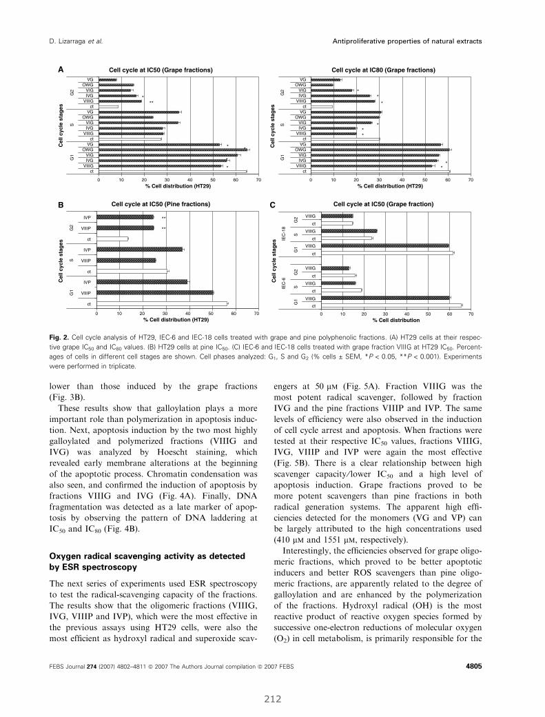
lower than those induced by the grape fractions
(Fig. 3B).
These results show that galloylation plays a more
important role than polymerization in apoptosis induc-
tion. Next, apoptosis induction by the two most highly
galloylated and polymerized fractions (VIIIG and
IVG) was analyzed by Hoescht staining, which
revealed early membrane alterations at the beginning
of the apoptotic process. Chromatin condensation was
also seen, and confirmed the induction of apoptosis by
fractions VIIIG and IVG (Fig. 4A). Finally, DNA
fragmentation was detected as a late marker of apop-
tosis by observing the pattern of DNA laddering at
IC50 and IC80 (Fig. 4B).
Oxygen radical scavenging activity as detected
by ESR spectroscopy
The next series of experiments used ESR spectroscopy
to test the radical-scavenging capacity of the fractions.
The results show that the oligomeric fractions (VIIIG,
IVG, VIIIP and IVP), which were the most effective in
the previous assays using HT29 cells, were also the
most efficient as hydroxyl radical and superoxide scav-
engers at 50 lm (Fig. 5A). Fraction VIIIG was the
most potent radical scavenger, followed by fraction
IVG and the pine fractions VIIIP and IVP. The same
levels of efficiency were also observed in the induction
of cell cycle arrest and apoptosis. When fractions were
tested at their respective IC50 values, fractions VIIIG,
IVG, VIIIP and IVP were again the most effective
(Fig. 5B). There is a clear relationship between high
scavenger capacity ⁄ lower IC50 and a high level of
apoptosis induction. Grape fractions proved to be
more potent scavengers than pine fractions in both
radical generation systems. The apparent high effi-
ciencies detected for the monomers (VG and VP) can
be largely attributed to the high concentrations used
(410 lm and 1551 lm, respectively).
Interestingly, the efficiencies observed for grape oligo-
meric fractions, which proved to be better apoptotic
inducers and better ROS scavengers than pine oligo-
meric fractions, are apparently related to the degree of
galloylation and are enhanced by the polymerization
of the fractions. Hydroxyl radical (OH) is the most
reactive product of reactive oxygen species formed by
successive one-electron reductions of molecular oxygen
(O2) in cell metabolism, is primarily responsible for the
Cell cycle at IC50 (Grape fraction)
ct
ct
ct
ct
ct
ct
VIIIG
VIIIG
VIIIG
VIIIG
VIIIG
VIIIGG
1S
G2
G1
SG
2
IEC
-6IE
C-1
8
Cell cycle at IC50 (Grape fractions)A
B C
0 10 20 30 40 50 60 70
ctVIIIG
IVGVIG
OWGVG
ctVIIIG
IVGVIG
OWGVG
ctVIIIG
IVGVIG
OWGVG
G1
SG
2
G1
SG
2
Cel
l cyc
le s
tag
es
Cel
l cyc
le s
tag
es
Cel
l cyc
le s
tag
es
Cel
l cyc
le s
tag
es
% Cell distribution (HT29) 0 10 20 30 40 50 60 70
% Cell distribution (HT29)
0 10 20 30 40 50 60 70
% Cell distribution (HT29) 0 10 20 30 40 50 60 70
% Cell distribution
*
*
***
*
*
Cell cycle at IC50 (Pine fractions)
ct
ct
IVP
ct
VIIIP
VIIIP
VIIIP
IVP
IVP
G1
SG
2
**
**
Cell cycle at IC80 (Grape fractions)
ctVIIIG
IVGVIG
OWGVG
ctVIIIG
IVGVIG
OWGVG
ctVIIIG
IVGVIG
OWGVG
**
*
***
*
*
Fig. 2. Cell cycle analysis of HT29, IEC-6 and IEC-18 cells treated with grape and pine polyphenolic fractions. (A) HT29 cells at their respec-
tive grape IC50 and IC80 values. (B) HT29 cells at pine IC50. (C) IEC-6 and IEC-18 cells treated with grape fraction VIIIG at HT29 IC50. Percent-
ages of cells in different cell stages are shown. Cell phases analyzed: G1, S and G2 (% cells ± SEM, *P < 0.05, **P < 0.001). Experiments
were performed in triplicate.
D. Lizarraga et al. Antiproliferative properties of natural extracts
FEBS Journal 274 (2007) 4802–4811 ª 2007 The Authors Journal compilation ª 2007 FEBS 4805
212
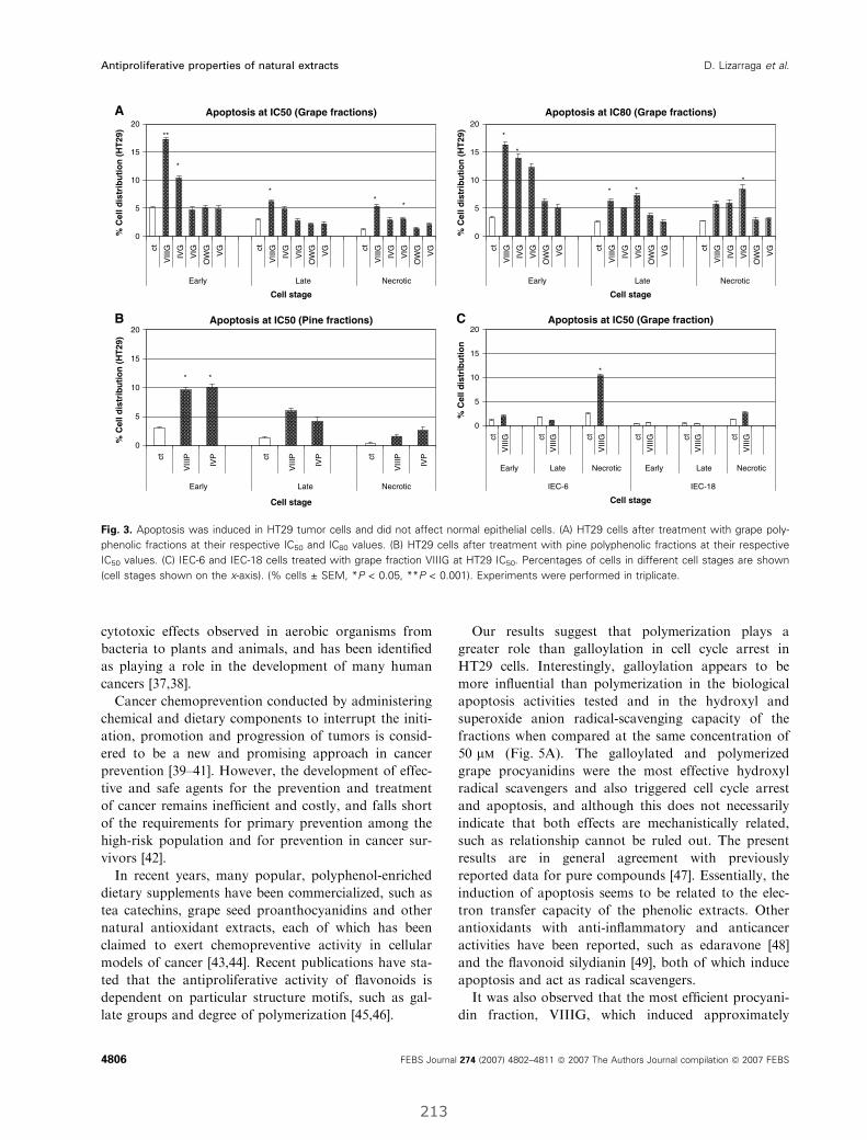
cytotoxic effects observed in aerobic organisms from
bacteria to plants and animals, and has been identified
as playing a role in the development of many human
cancers [37,38].
Cancer chemoprevention conducted by administering
chemical and dietary components to interrupt the initi-
ation, promotion and progression of tumors is consid-
ered to be a new and promising approach in cancer
prevention [39–41]. However, the development of effec-
tive and safe agents for the prevention and treatment
of cancer remains inefficient and costly, and falls short
of the requirements for primary prevention among the
high-risk population and for prevention in cancer sur-
vivors [42].
In recent years, many popular, polyphenol-enriched
dietary supplements have been commercialized, such as
tea catechins, grape seed proanthocyanidins and other
natural antioxidant extracts, each of which has been
claimed to exert chemopreventive activity in cellular
models of cancer [43,44]. Recent publications have sta-
ted that the antiproliferative activity of flavonoids is
dependent on particular structure motifs, such as gal-
late groups and degree of polymerization [45,46].
Our results suggest that polymerization plays a
greater role than galloylation in cell cycle arrest in
HT29 cells. Interestingly, galloylation appears to be
more influential than polymerization in the biological
apoptosis activities tested and in the hydroxyl and
superoxide anion radical-scavenging capacity of the
fractions when compared at the same concentration of
50 lm (Fig. 5A). The galloylated and polymerized
grape procyanidins were the most effective hydroxyl
radical scavengers and also triggered cell cycle arrest
and apoptosis, and although this does not necessarily
indicate that both effects are mechanistically related,
such as relationship cannot be ruled out. The present
results are in general agreement with previously
reported data for pure compounds [47]. Essentially, the
induction of apoptosis seems to be related to the elec-
tron transfer capacity of the phenolic extracts. Other
antioxidants with anti-inflammatory and anticancer
activities have been reported, such as edaravone [48]
and the flavonoid silydianin [49], both of which induce
apoptosis and act as radical scavengers.
It was also observed that the most efficient procyani-
din fraction, VIIIG, which induced approximately
Apoptosis at IC50 (Pine fractions)
ct
VIII
P
IVP ct
VIII
P
IVP ct
VIII
P
IVP
Early Late Necrotic
Cell stage
* *
Apoptosis at IC50 (Grape fractions)A
B C
0
5
10
15
20
ct
VIII
G
IVG
VIG
OW
G
VG ct
VIII
G
IVG
VIG
OW
G
VG ct
VIII
G
IVG
VIG
OW
G
VG
Early Late Necrotic
Cell stage
% C
ell d
istr
ibu
tio
n (
HT
29)
0
5
10
15
20
% C
ell d
istr
ibu
tio
n (
HT
29)
0
5
10
15
20
% C
ell d
istr
ibu
tio
n (
HT
29)
0
5
10
15
20
% C
ell d
istr
ibu
tio
n
**
* *
*
*
Apoptosis at IC80 (Grape fractions)
ct
VIII
G
IVG
VIG
OW
G
VG ct
VIII
G
IVG
VIG
OW
G
VG ct
VIII
G
IVG
VIG
OW
G
VG
Early Late Necrotic
Cell stage
*
** *
*
Apoptosis at IC50 (Grape fraction)
ct
VIII
G ct
VIII
G ct
VIII
G ct
VIII
G ct
VIII
G ct
VIII
G
Early Late Necrotic Early Late Necrotic
IEC-6 IEC-18
Cell stage
*
Fig. 3. Apoptosis was induced in HT29 tumor cells and did not affect normal epithelial cells. (A) HT29 cells after treatment with grape poly-
phenolic fractions at their respective IC50 and IC80 values. (B) HT29 cells after treatment with pine polyphenolic fractions at their respective
IC50 values. (C) IEC-6 and IEC-18 cells treated with grape fraction VIIIG at HT29 IC50. Percentages of cells in different cell stages are shown
(cell stages shown on the x-axis). (% cells ± SEM, *P < 0.05, **P < 0.001). Experiments were performed in triplicate.
Antiproliferative properties of natural extracts D. Lizarraga et al.
4806 FEBS Journal 274 (2007) 4802–4811 ª 2007 The Authors Journal compilation ª 2007 FEBS
213
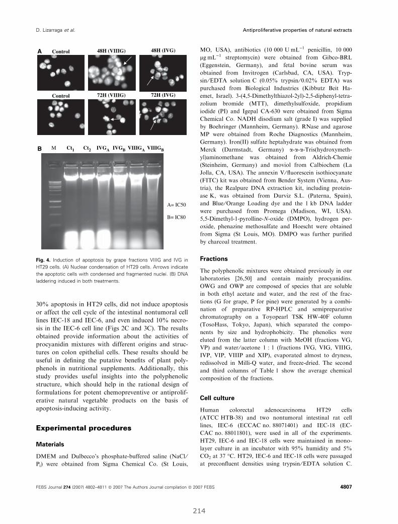
30% apoptosis in HT29 cells, did not induce apoptosis
or affect the cell cycle of the intestinal nontumoral cell
lines IEC-18 and IEC-6, and even induced 10% necro-
sis in the IEC-6 cell line (Figs 2C and 3C). The results
obtained provide information about the activities of
procyanidin mixtures with different origins and struc-
tures on colon epithelial cells. These results should be
useful in defining the putative benefits of plant poly-
phenols in nutritional supplements. Additionally, this
study provides useful insights into the polyphenolic
structure, which should help in the rational design of
formulations for potent chemopreventive or antiprolif-
erative natural vegetable products on the basis of
apoptosis-inducing activity.
Experimental procedures
Materials
DMEM and Dulbecco’s phosphate-buffered saline (NaCl ⁄Pi) were obtained from Sigma Chemical Co. (St Louis,
MO, USA), antibiotics (10 000 UÆmL)1 penicillin, 10 000
lgÆmL)1 streptomycin) were obtained from Gibco-BRL
(Eggenstein, Germany), and fetal bovine serum was
obtained from Invitrogen (Carlsbad, CA, USA). Tryp-
sin ⁄EDTA solution C (0.05% trypsin ⁄ 0.02% EDTA) was
purchased from Biological Industries (Kibbutz Beit Ha-
emet, Israel). 3-(4,5-Dimethylthiazol-2yl)-2,5-diphenyl-tetra-
zolium bromide (MTT), dimethylsulfoxide, propidium
iodide (PI) and Igepal CA-630 were obtained from Sigma
Chemical Co. NADH disodium salt (grade I) was supplied
by Boehringer (Mannheim, Germany). RNase and agarose
MP were obtained from Roche Diagnostics (Mannheim,
Germany). Iron(II) sulfate heptahydrate was obtained from
Merck (Darmstadt, Germany) a-a-a-Tris(hydroxymeth-
yl)aminomethane was obtained from Aldrich-Chemie
(Steinheim, Germany) and moviol from Calbiochem (La
Jolla, CA, USA). The annexin V ⁄ fluorescein isothiocyanate
(FITC) kit was obtained from Bender System (Vienna, Aus-
tria), the Realpure DNA extraction kit, including protein-
ase K, was obtained from Durviz S.L. (Paterna, Spain),
and Blue ⁄Orange Loading dye and the 1 kb DNA ladder
were purchased from Promega (Madison, WI, USA).
5,5-Dimethyl-1-pyrolline-N-oxide (DMPO), hydrogen per-
oxide, phenazine methosulfate and Hoescht were obtained
from Sigma (St Louis, MO). DMPO was further purified
by charcoal treatment.
Fractions
The polyphenolic mixtures were obtained previously in our
laboratories [26,50] and contain mainly procyanidins.
OWG and OWP are composed of species that are soluble
in both ethyl acetate and water, and the rest of the frac-
tions (G for grape, P for pine) were generated by a combi-
nation of preparative RP-HPLC and semipreparative
chromatography on a Toyopearl TSK HW-40F column
(TosoHass, Tokyo, Japan), which separated the compo-
nents by size and hydrophobicity. The phenolics were
eluted from the latter column with MeOH (fractions VG,
VP) and water ⁄ acetone 1 : 1 (fractions IVG, VIG, VIIIG,
IVP, VIP, VIIIP and XIP), evaporated almost to dryness,
redissolved in Milli-Q water, and freeze-dried. The second
and third columns of Table 1 show the average chemical
composition of the fractions.
Cell culture
Human colorectal adenocarcinoma HT29 cells
(ATCC HTB-38) and two nontumoral intestinal rat cell
lines, IEC-6 (ECCAC no. 88071401) and IEC-18 (EC-
CAC no. 88011801), were used in all of the experiments.
HT29, IEC-6 and IEC-18 cells were maintained in mono-
layer culture in an incubator with 95% humidity and 5%
CO2 at 37 �C. HT29, IEC-6 and IEC-18 cells were passaged
at preconfluent densities using trypsin ⁄EDTA solution C.
M Ct1 Ct2 IVGA IVGB VIIIGA VIIIGB
Control 48H (VIIIG)
72H (VIIIG)
48H (IVG)
72H (IVG)Control
A
B
A= IC50
B= IC80
Fig. 4. Induction of apoptosis by grape fractions VIIIG and IVG in
HT29 cells. (A) Nuclear condensation of HT29 cells. Arrows indicate
the apoptotic cells with condensed and fragmented nuclei. (B) DNA
laddering induced in both treatments.
D. Lizarraga et al. Antiproliferative properties of natural extracts
FEBS Journal 274 (2007) 4802–4811 ª 2007 The Authors Journal compilation ª 2007 FEBS 4807
214
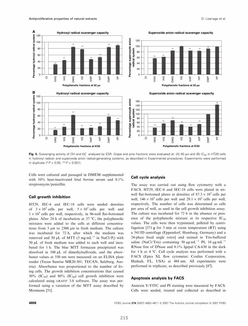
Cells were cultured and passaged in DMEM supplemented
with 10% heat-inactivated fetal bovine serum and 0.1%
streptomycin ⁄penicillin.
Cell growth inhibition
HT29, IEC-6 and IEC-18 cells were seeded densities
of 3 · 103 cells per well, 5 · 103 cells per well and
1 · 103 cells per well, respectively, in 96-well flat-bottomed
plates. After 24 h of incubation at 37 �C, the polyphenolic
mixtures were added to the cells at different concentra-
tions from 5 lm to 2300 lm in fresh medium. The culture
was incubated for 72 h, after which the medium was
removed and 50 lL of MTT (5 mgÆmL)1 in NaCl ⁄Pi) with
50 lL of fresh medium was added to each well and incu-
bated for 1 h. The blue MTT formazan precipitated was
dissolved in 100 lL of dimethylsulfoxide, and the absor-
bance values at 550 nm were measured on an ELISA plate
reader (Tecan Sunrise MR20-301; TECAN, Salzburg, Aus-
tria). Absorbance was proportional to the number of liv-
ing cells. The growth inhibition concentrations that caused
50% (IC50) and 80% (IC80) cell growth inhibition were
calculated using grafit 3.0 software. The assay was per-
formed using a variation of the MTT assay described by
Mosmann [51].
Cell cycle analysis
The assay was carried out using flow cytometry with a
FACS. HT29, IEC-6 and IEC-18 cells were plated in six-
well flat-bottomed plates at densities of 87.3 · 103 cells per
well, 146 · 103 cells per well and 29.1 · 103 cells per well,
respectively. The number of cells was determined as cells
per area of well, as used in the cell growth inhibition assay.
The culture was incubated for 72 h in the absence or pres-
ence of the polyphenolic mixture at its respective IC50
values. The cells were then trypsinized, pelleted by centri-
fugation [371 g for 3 min at room temperature (RT) using
a 5415D centrifuge (Eppendorf, Hamburg, Germany) and a
24-place fixed angle rotor] and stained in Tris-buffered
saline (NaCl ⁄Tris) containing 50 lgÆmL)1 PI, 10 lgÆmL)1
RNase free of DNase and 0.1% Igepal CA-630 in the dark
for 1 h at 4 �C. Cell cycle analysis was performed with a
FACS (Epics XL flow cytometer; Coulter Corporation,
Hialeah, FL, USA) at 488 nm. All experiments were
performed in triplicate, as described previously [47].
Apoptosis analysis by FACS
Annexin V ⁄FITC and PI staining were measured by FACS.
Cells were seeded, treated and collected as described in
Superoxide anion radical scavenger capacity
** **
**
**
**
**
**
Hydroxyl radical scavenger capacityA
B
0
20
40
60
80
100
120
Ct
VIII
G
IVG
OW
G
VG
VIII
P
IVP
OW
P
VP
Polyphenolic fractions at 50 µM
0
20
40
60
80
100
120
Ct
VIII
G
IVG
OW
G
VG
VIII
P
IVP
OW
P
VP
Polyphenolic fractions at 50 µM
Per
cen
tag
e h
ydro
xyl r
adic
al s
yste
m
0
20
40
60
80
100
120
Ct
VIII
G
IVG
OW
G
VG
VIII
P
IVP
OW
P
VPP
erce
nta
ge
hyd
roxy
l rad
ical
sys
tem
** **
**
**
**
** ** *
Superoxide anion radical scavenger capacity
Per
cen
tag
e su
per
oxy
de
anio
nra
dic
al s
yste
m0
20
40
60
80
100
120
140
160
Per
cen
tag
e su
per
oxy
de
anio
nra
dic
al s
yste
m
**
**
**
** ** **
Hydroxyl radical scavenger capacity
Polyphenolic fractions at IC50 C
t
VIII
G
IVG
OW
G
VG
VIII
P
IVP
OW
P
VP
Polyphenolic fractions at IC50
** ****
**
**
**
**
**
Fig. 5. Scavenging activity of OH and O��2 analyzed by ESR. Grape and pine fractions were evaluated at: (A) 50 lM and (B) IC50 in HT29 cells
in hydroxyl radical- and superoxide anion radical-generating systems, as described in Experimental procedures. Experiments were performed
in duplicate (*P < 0.05, **P < 0.001).
Antiproliferative properties of natural extracts D. Lizarraga et al.
4808 FEBS Journal 274 (2007) 4802–4811 ª 2007 The Authors Journal compilation ª 2007 FEBS
215
the previous section. Following centrifugation [371 g for
3 min at RT using a 5415D centrifuge (Eppendorf) with
24-place fixed angle rotor], cells were washed in binding
buffer (10 mm Hepes, pH 7.4, 140 mm sodium chloride,
2.5 mm calcium chloride) and resuspended in the same
buffer. Annexin V ⁄FITC was added using the annex-
in V ⁄FITC kit. Following 30 min of incubation at room
temperature and in the dark, PI was added 1 min before
the FACS analysis at 20 lgÆmL)1. Experiments were per-
formed in triplicate.
Apoptosis detection by DNA laddering
DNA isolation and purification were performed after 72 h
in the presence and absence of grape fractions VIIIG and
IVG. The fractions were assayed at their respective IC50
and IC80 values. After treatment, cells were scraped off
slides and collected by centrifugation at 14 000 g for 10 s
at RT using a 5415D centrifuge (Eppendorf) and 24-place
fixed angle rotor. Cells were then lysed by adding 600 lLof Realpure kit lysis buffer and 10 lL of proteinase K,
and incubated for 1 h at 55 �C. RNA digestion was per-
formed with 1.5 lL of RNase for 1 h at 37 �C, and this
was followed by protein precipitation with 360 lL of
Realpure kit buffer and centrifugation at 14 000 g for
10 min at RT using a 5415D centrifuge (Eppendorf) and
24-place fixed angle rotor. The DNA sample was
extracted with isopropanol ⁄ ethanol, dried, and eluted in
100 lL of Realpure kit DNA hydration solution. Equal
amounts of DNA (20 lg), estimated by measuring absorp-
tion at 260 ⁄ 280 nm, were electrophoretically separated on
1% TAE agarose gel and viewed under a UV transillumi-
nator (Vilber Lourmat, Marne-la-Vallee, France).
Apoptosis detection by Hoescht staining
Apoptotic induction was also studied using Hoescht stain-
ing. Samples were incubated with grape fractions VIIIG
and IVG at 0, 48 and 72 h. After incubation, cells were
trypsinized and fixed with cold methanol for 1 h at ) 20 �C.After being rinsed with NaCl ⁄Pi three times, cells were
stained in the dark with Hoescht (50 ngÆmL)1 in NaCl ⁄Pi)
for 50 min. Finally, cells were rinsed, suspended in NaCl ⁄Pi
and diluted 1 : 2 with moviol. The samples were mounted
on a slide and observed with a fluorescent microscope at an
excitation wavelength of 334 nm and an emission wave-
length of 365 nm.
ESR spectroscopy
ESR measurements were performed at concentrations that
caused 50% cell growth inhibition (IC50) and 50 lm grape
and pine fractions (VIIIG, IVG, OWG, VG, VIIIP, IVP,
OWP and VP). Molar concentrations were calculated from
the mean molecular mass of the fractions estimated by thiol-
ysis with cysteamine, as described in [52]. OH and O2– forma-
tion were detected by ESR spectroscopy using DMPO
(100 mm) as a spin trap. ESR spectra were recorded at room
temperature in glass capillaries (100 lL; Brand AG,
Wertheim, Germany) on a Bruker EMX 1273 spectrometer
(Bruker, Karlsruhe, Germany) equipped with an ER 4119HS
high-sensitivity cavity and a 12 kW power supply operating
at X-band frequencies. The modulation frequency of the
spectrometer was 100 kHz. Instrumental conditions for the
recorded spectra were: magnetic field, 3490 G; scan range,
60 G; modulation amplitude, 1 G; receiver gain, 1 · 105;
microwave frequency, 9.85 GHz; power, 50 mW; time
constant, 40.96 ms; scan time, 20.97 s; number of scans, 25.
Spectra were quantified by peak surface measurements using
the WIN-EPR spectrum manipulation program (Bruker).
All incubations were done at room temperature; the
hydroxyl radical generation system used 500 lm FeSO4 and
550 lm H2O2, and hydroxyl radicals generated in this system
were trapped by DMPO, forming a spin adduct detected by
the ESR spectrometer. The typical 1 : 2 : 2 : 1 ESR signal
of DMPO-OH was observed. The superoxide radical genera-
tion system used performed using 50 lm of the reduced form
of b-NADH and 3.3 lm phenazine methosulfate, and the
superoxide radicals generated in this system were trapped by
DMPO, forming a spin adduct detected by the ESR spec-
trometer. The typical ESR signal of DMPO-OOH ⁄DMPO-
OH was observed. The OH and O2-scavenging activity was
calculated on the basis of decreases in the DMPO-OH or
DMPO-OOH ⁄DMPO-OH signals, respectively, in which the
coupling constant for DMPO-OH was 14.9 G.
Data presentation and statistical analysis
Assays were analyzed using the Student’s t-test and
were considered statistically significant at P < 0.05 and
P < 0.001. The data shown are representative of three
independent experiments, with the exception of ESR experi-
ments, which were performed in duplicate. ESR experi-
ments were analyzed separately by radicals, Two-way
anova was applied (day was a block factor; due to the
nonsignificant effect of the day factor, we reanalyzed with a
one-way anova), and finally, a multicomparison between
compounds with respect to the control was performed.
anova with Bonferroni and Scheffe post hoc test was per-
formed in ESR experiments.
Acknowledgements
This work was supported by grants PPQ 2003-06602-
C04-01, PPQ 2003-06602-C04-04, AGL2004-07579-
C04-02 and AGL2004-07579-C04-03 from the Spanish
Ministry of Education and Science, and ISCIII-RTICC
(RD06 ⁄0020 ⁄ 0046) from the Spanish government and
D. Lizarraga et al. Antiproliferative properties of natural extracts
FEBS Journal 274 (2007) 4802–4811 ª 2007 The Authors Journal compilation ª 2007 FEBS 4809
216

the European Union FEDER funds. We thank Profes-
sor Francesc Oliva (Department of Statistics at the
University of Barcelona) for his assistance with statisti-
cal analysis.
References
1 Parkin DM (2004) International variation. Oncogene 23,
6329–6340.
2 Potter JD, Slattery ML, Bostick RM & Gapstur SM
(1993) Colon cancer: a review of the epidemiology.
Epidemiol Rev 15, 499–545.
3 Steinmetz KA & Potter JD (1991) Vegetables, fruit, and
cancer. I. Epidemiology. Cancer Causes Control 2, 325–
357.
4 Park OJ & Surh YJ (2004) Chemopreventive potential
of epigallocatechin gallate and genistein: evidence from
epidemiological and laboratory studies. Toxicol Lett
150, 43–56.
5 Bruce WR, Giacca A & Medline A (2000) Possible
mechanisms relating diet and risk of colon cancer.
Cancer Epidemiol Biomarkers Prev 9, 1271–1279.
6 Hietanen E, Bartsch H, Bereziat JC, Camus AM, McCl-
inton S, Eremin O, Davidson L & Boyle P (1994) Diet
and oxidative stress in breast, colon and prostate cancer
patients: a case-control study. Eur J Clin Nutr 48, 575–
586.
7 Theodoratou E, Kyle J, Cetnarskyj R, Farrington SM,
Tenesa A, Barnetson R, Porteous M, Dunlop M & Camp-
bell H (2007) Dietary flavonoids and the risk of colorectal
cancer. Cancer Epidemiol Biomarkers Prev 16, 684–693.
8 Mukhtar H & Ahmad N (1999) Green tea in chemopre-
vention of cancer. Toxicol Sci 52, 111–117.
9 Lee KW, Lee HJ & Lee CY (2004) Vitamins, phyto-
chemicals, diets, and their implementation in cancer
chemoprevention. Crit Rev Food Sci Nutr 44, 437–452.
10 Witschi H, Espiritu I, Ly M, Uyeminami D, Morin D
& Raabe OG (2004) Chemoprevention of tobacco
smoke-induced lung tumors by inhalation of an epigal-
locatechin gallate (EGCG) aerosol: a pilot study. Inhal
Toxicol 16, 763–770.
11 Delmas D, Lancon A, Colin D, Jannin B & Latruffe N
(2006) Resveratrol as a chemopreventive agent: a prom-
ising molecule for fighting cancer. Curr Drug Targets 7,
423–442.
12 Hakimuddin F, Paliyath G & Meckling K (2006) Treat-
ment of mcf-7 breast cancer cells with a red grape wine
polyphenol fraction results in disruption of calcium
homeostasis and cell cycle arrest causing selective cyto-
toxicity. J Agric Food Chem 54, 7912–7923.
13 Sime S & Reeve VE (2004) Protection from inflamma-
tion, immunosuppression and carcinogenesis induced by
UV radiation in mice by topical Pycnogenol. Photochem
Photobiol 79, 193–198.
14 Kumar N, Shibata D, Helm J, Coppola D & Malafa M
(2007) Green tea polyphenols in the prevention of colon
cancer. Front Biosci 12, 2309–2315.
15 McKay DL & Blumberg JB (2007) A review of the bio-
activity of south African herbal teas: rooibos (Aspala-
thus linearis) and honeybush (Cyclopia intermedia).
Phytother Res 21, 1–16.
16 Wright TI, Spencer JM & Flowers FP (2006) Chemo-
prevention of nonmelanoma skin cancer. J Am Acad
Dermatol 54, 933–946; quiz 947–950.
17 Tan XHD, Li S, Han Y, Zhang Y & Zhou D (2000)
Differences of four catechins in cell cycle arrest and
induction of apoptosis in LoVo cells. Cancer Lett 158,
1–6.
18 Kozikowski AP, Tuckmantel W, Bottcher G &
Romanczyk LJ Jr (2003) Studies in polyphenol
chemistry and bioactivity. 4 (1) Synthesis of trimeric,
tetrameric, pentameric, and higher oligomeric
epicatechin-derived procyanidins having all-4beta,
8-interflavan connectivity and their inhibition of cancer
cell growth through cell cycle arrest. J Org Chem 68,
1641–1658.
19 Nakamuta M, Higashi N, Kohjima M, Fukushima M,
Ohta S, Kotoh K, Kobayashi N & Enjoji M (2005)
Epigallocatechin-3-gallate, a polyphenol component of
green tea, suppresses both collagen production and col-
lagenase activity in hepatic stellate cells. Int J Mol Med
16, 677–681.
20 Shi J, Yu J, Pohorly JE & Kakuda Y (2003) Polypheno-
lics in grape seeds ) biochemistry and functionality.
J Med Food 6, 291–299.
21 Siddiqui IA, Adhami VM, SaleemM &Mukhtar H
(2006) Beneficial effects of tea and its polyphenols against
prostate cancer.Mol Nutr Food Res 50, 130–143.
22 Zhang Q, Tang X, Lu Q, Zhang Z, Rao J & Le AD
(2006) Green tea extract and (–)-epigallocatechin-3-gal-
late inhibit hypoxia- and serum-induced HIF-1alpha
protein accumulation and VEGF expression in human
cervical carcinoma and hepatoma cells. Mol Cancer
Ther 5, 1227–1238.
23 Prieur CRJ, Cheynier V & Moutounet M (1994) Oligo-
meric and polymeric procyanidins from grape seeds.
Phytochemistry 36, 781–784.
24 Souquet J-MCV, Brossaud F & Moutounet M (1996)
Polymeric proanthocyanidins from grape skins. Phyto-
chemistry 43, 509–512.
25 Rohdewald P (2002) A review of the French maritime
pine bark extract (Pycnogenol), a herbal medication
with a diverse clinical pharmacology. Int J Clin
Pharmacol Ther 40, 158–168.
26 Tourino S, Selga A, Jimenez A, Julia L, Lozano C,
Lizarraga D, Cascante M & Torres JL (2005)
Procyanidin fractions from pine (Pinus pinaster) bark:
radical scavenging power in solution, antioxidant
Antiproliferative properties of natural extracts D. Lizarraga et al.
4810 FEBS Journal 274 (2007) 4802–4811 ª 2007 The Authors Journal compilation ª 2007 FEBS
217
activity in emulsion, and antiproliferative effect in
melanoma cells. J Agric Food Chem 53, 4728–4735.
27 Gonthier MP, Donovan JL, Texier O, Felgines C,
Remesy C & Scalbert A (2003) Metabolism of dietary
procyanidins in rats. Free Radic Biol Med 35, 837–
844.
28 Kuhnle G, Spencer JP, Schroeter H, Shenoy B, Debnam
ES, Srai SK, Rice-Evans C & Hahn U (2000) Epicate-
chin and catechin are O-methylated and glucuronidated
in the small intestine. Biochem Biophys Res Commun
277, 507–512.
29 Rechner AR, Smith MA, Kuhnle G, Gibson GR, Deb-
nam ES, Srai SK, Moore KP & Rice-Evans CA (2004)
Colonic metabolism of dietary polyphenols: influence of
structure on microbial fermentation products. Free
Radic Biol Med 36, 212–225.
30 Meselhy MR, Nakamura N & Hattori M (1997) Biotrans-
formation of (–)-epicatechin 3-O-gallate by human intes-
tinal bacteria. Chem Pharm Bull (Tokyo) 45, 888–893.
31 Salucci M, Stivala LA, Maiani G, Bugianesi R & Van-
nini V (2002) Flavonoids uptake and their effect on cell
cycle of human colon adenocarcinoma cells (Caco2).
Br J Cancer 86, 1645–1651.
32 Stagos D, Kazantzoglou G, Magiatis P, Mitaku S,
Anagnostopoulos K & Kouretas D (2005) Effects of plant
phenolics and grape extracts from Greek varieties of Vitis
vinifera on mitomycin C and topoisomerase I-induced
nicking of DNA. Int J Mol Med 15, 1013–1022.
33 Subirade I, Fernandez Y, Periquet A & Mitjavila S
(1995) Catechin protection of 3T3 Swiss fibroblasts in
culture under oxidative stress. Biol Trace Elem Res 47,
313–319.
34 Cao Z & Li Y (2004) Potent induction of cellular anti-
oxidants and phase 2 enzymes by resveratrol in cardio-
myocytes: protection against oxidative and electrophilic
injury. Eur J Pharmacol 489, 39–48.
35 Alanko J, Riutta A, Holm P, Mucha I, Vapaatalo H &
Metsa-Ketela T (1999) Modulation of arachidonic acid
metabolism by phenols: relation to their structure and
antioxidant ⁄ prooxidant properties. Free Radic Biol Med
26, 193–201.
36 Azam S, Hadi N, Khan NU & Hadi SM (2004) Prooxi-
dant property of green tea polyphenols epicatechin and
epigallocatechin-3-gallate: implications for anticancer
properties. Toxicol In Vitro 18, 555–561.
37 Halliwell B & Gutteridge JM (1992) Biologically rele-
vant metal ion-dependent hydroxyl radical generation.
An update. FEBS Lett 307, 108–112.
38 Valko M, Leibfritz D, Moncol J, Cronin MT, Mazur M
& Telser J (2007) Free radicals and antioxidants in nor-
mal physiological functions and human disease. Int J
Biochem Cell Biol 39, 44–84.
39 Mathers JC (2002) Pulses and carcinogenesis: potential
for the prevention of colon, breast and other cancers.
Br J Nutr 88 (Suppl. 3), S273–S279.
40 Witte JS, Longnecker MP, Bird CL, Lee ER, Frankl
HD & Haile RW (1996) Relation of vegetable, fruit,
and grain consumption to colorectal adenomatous pol-
yps. Am J Epidemiol 144, 1015–1025.
41 Manju V & Nalini N (2005) Chemopreventive efficacy of
ginger, a naturally occurring anticarcinogen during the
initiation, post-initiation stages of 1,2-dimethylhydrazine-
induced colon cancer. Clin Chim Acta 358, 60–67.
42 Zou DM, Brewer M, Garcia F, Feugang JM, Wang J,
Zang R, Liu H & Zou C (2005) Cactus pear: a natural
product in cancer chemoprevention. Nutr J 4, 25–37.
43 Joshi SS, Kuszynski CA & Bagchi D (2001) The cellular
and molecular basis of health benefits of grape seed
proanthocyanidin extract. Curr Pharm Biotechnol 2,
187–200.
44 Depeint F, Gee JM, Williamson G & Johnson IT (2002)
Evidence for consistent patterns between flavonoid struc-
tures and cellular activities. Proc Nutr Soc 61, 97–103.
45 Fiuza SM, Gomes C, Teixeira LJ, Girao da Cruz MT,
Cordeiro MN, Milhazes N, Borges F & Marques MP
(2004) Phenolic acid derivatives with potential antican-
cer properties ) a structure–activity relationship study.
Part 1: methyl, propyl and octyl esters of caffeic and
gallic acids. Bioorg Med Chem 12, 3581–3589.
46 Brusselmans K, Vrolix R, Verhoeven G & Swinnen JV
(2005) Induction of cancer cell apoptosis by flavonoids
is associated with their ability to inhibit fatty acid syn-
thase activity. J Biol Chem 280, 5636–5645.
47 Lozano C, Torres JL, Julia L, Jimenez A, Centelles JJ
& Cascante M (2005) Effect of new antioxidant cyste-
inyl-flavanol conjugates on skin cancer cells. FEBS Lett
579, 4219–4225.
48 Kokura S, Yoshida N, Sakamoto N, Ishikawa T, Tak-
agi T, Higashihara H, Nakabe N, Handa O, Naito Y &
Yoshikawa T (2005) The radical scavenger edaravone
enhances the anti-tumor effects of CPT-11 in murine
colon cancer by increasing apoptosis via inhibition of
NF-kappaB. Cancer Lett 229, 223–233.
49 Zielinska-Przyjemska M & Wiktorowicz K (2006) An
in vitro study of the protective effect of the flavonoid
silydianin against reactive oxygen species. Phytother Res
20, 115–119.
50 Torres JL, Varela B, Garcia MT, Carilla J, Matito C,
Centelles JJ, Cascante M, Sort X & Bobet R (2002) Val-
orization of grape (Vitis vinifera) byproducts. Antioxi-
dant and biological properties of polyphenolic fractions
differing in procyanidin composition and flavonol con-
tent. J Agric Food Chem 50, 7548–7555.
51 Mosmann T (1983) Rapid colorimetric assay for cellular
growth and survival: application to proliferation and
cytotoxicity assays. J Immunol Methods 65, 55–63.
52 Selga A & Torres JL (2005) Efficient preparation of
catechin thio conjugates by one step extraction ⁄depolymerization of pine (Pinus pinaster) bark
procyanidins. J Agric Food Chem 53, 7760–7765.
D. Lizarraga et al. Antiproliferative properties of natural extracts
FEBS Journal 274 (2007) 4802–4811 ª 2007 The Authors Journal compilation ª 2007 FEBS 4811
218

V. Comparative antioxidant and cytotoxic effect of
procyanidin fractions from grape and pine.
Ugartondo, V.; Mitjans, M.; Touriño, S.; Torres, J.L. and
Vinardell M.P.; Chem. Res. Toxicol.; 20: 1543–1548;
2007

Comparative Antioxidant and Cytotoxic Effect of ProcyanidinFractions from Grape and Pine
Vanessa Ugartondo,† Montserrat Mitjans,† Sonia Touriño,‡ Josep Lluis Torres,‡ andMaría Pilar Vinardell*,†
Department of Physiology, Faculty of Pharmacy, UniVersitat de Barcelona, AV. Joan XXIII s/n,08028 Barcelona, Spain, and Institute for Chemical and EnVironmental Research, CSIC, Jordi Girona 18-26,
08034 Barcelona, Spain
ReceiVed July 11, 2007
There is a great interest in characterizing the biological properties of natural compounds obtainedfrom plants, especially polyphenols. We studied the structure–activity–cytotoxicity relationships ofpolyphenolic fractions obtained from grape pomace and pine bark. These fractions contained similarpolymerised flavonoids but different percentages of pyrogallol groups that confer on them differentbiological properties. The human keratinocyte cell line HaCaT and the mouse fibroblast cell line 3T3were used to study the cytotoxicity of the different fractions after 24, 48, and 72 h of exposure. Antioxidantactivity of the fractions was evaluated by measuring the inhibition of hemolysis mediated by AAPH.Our results demonstrate that the polyphenolic fractions studied show high antioxidant capacity in aconcentration range that is not harmful to normal human cells. Pine fractions presented slightly lowerantioxidant activity than grape fractions but are less cytotoxic. This data provides useful information tohelp design safe antioxidant products that act without altering critical cell functions.
Introduction
The efficient use of natural resources is currently the focusof many efforts in both science and technology. The manage-ment of agriculture and forestry must be sustainable from botheconomic and environmental viewpoints. The agrifood industriesproduce a large volume of waste each year (1). Because of thehigh economic cost of disposal and the potential environmentalrisk associated with an excess of biomass, the possibility ofrecycling by finding new applications for these wastes has greatpotential.
Plant residues from food and forestry industries containconsiderable amounts of potentially interesting compounds, butthe value of the products obtained must compensate for the costof their recovery. For this reason, it is essential both to improvethe extraction processes and to substantiate the activity andsafety claims (2) of the new products.
Among the biologically active species present in agriculturalby-products, polyphenols and particularly flavonoids are widelyappreciated for their putative health-promoting properties. Thebest-described property of flavonoids is their capacity to act asfree radical scavengers (3). They also show other properties thatmay or may not be related to their scavenging potential. Theseinclude, but are not limited to, antiproliferation of carcinogeniccells, cell cycle regulation, induction of apoptosis, inhibitionof platelet aggregation, and antibacterial and antiallergic proper-ties (4–8). Therefore, it is assumed that flavonoids play arelevant role in the prevention of degenerative diseases such ascancer and cardiovascular diseases and that it may be wise toinclude in our diet vegetables, fruits, and moderate amounts ofplant-derived products such as tea, wine, and chocolate, whichare rich in polyphenols (9, 10).
Polyphenolic mixtures have already been proposed as foodantioxidants and preventive agents against skin irritation andcancer (4, 11). In accordance with the scientific and marketinterest in polyphenols as chemopreventive agents, our groupis investigating the possible applications of plant proanthocya-nidins in the fields of food preservation, skin protection, andcancer with particular emphasis on their structure–activityrelationships and safety profiles.
From white grape (Vitis Vinifera) pomace and pine (Pinuspinaster) bark polyphenolic extracts, we generated fractionscontaining different amounts of monomeric catechins andoligomeric procyanidins (12–14). Figure 1 summarizes thestructure of the procyanidins found in both sources. Wepreviously carried out several assays to assess the structure/function relationships of these fractions. We determined theirefficiency as antioxidants under different experimental setups,including free radical scavenging in solution and inhibition oflipid peroxidation in both pure oil and oil-in-water emulsion.Furthermore, we investigated the influence of these compoundson the proliferation of different tumoral cell lines and theircapacity to induce apoptosis (13, 14).
The fractions from the two sources are highly homologousin terms of mean molecular size; they are mainly differentiatedby their galloylation (presence of gallate esters). Grape pomance(skin, seeds, and a small amount of stems) are galloylated tosome extent, but pine bark appears to contain only procyanidinswith no measurable galloylation (4, 15). Because the gallategroup both provides high antiradical power and appears tointerfere with crucial cell functions, galloylation appears to bea crucial structural feature defining the activity and toxicity ofphenolic mixtures.
The aim of this study is to take a step forward in thecharacterization of the biological properties of procyanidins byusing a set of grape and pine fractions with different mean sizeand galloylation. We report the protective antioxidant potential
* Corresponding author: Phone: +34 934024505. Fax: +34934035901.E-mail: [email protected].
† Universitat de Barcelona.‡ Institute for Chemical and Environmental Research.
Chem. Res. Toxicol. 2007, 20, 1543–1548 1543
10.1021/tx700253y CCC: $37.00 2007 American Chemical SocietyPublished on Web 09/08/2007
221
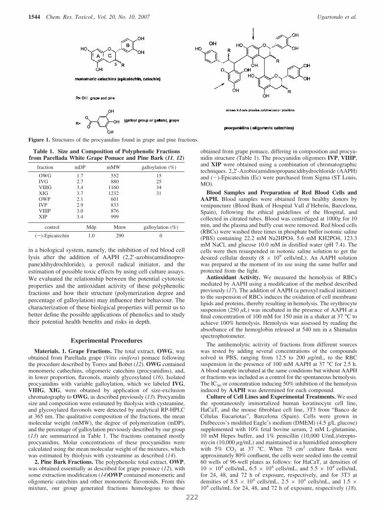
in a biological system, namely, the inhibition of red blood celllysis after the addition of AAPH (2,2′-azobis(amidinopro-pane)dihydrochloride), a peroxil radical initiator, and theestimation of possible toxic effects by using cell culture assays.We evaluated the relationship between the potential cytotoxicproperties and the antioxidant activity of these polyphenolicfractions and how their structure (polymerization degree andpercentage of galloylation) may influence their behaviour. Thecharacterization of these biological properties will permit us tobetter define the possible applications of phenolics and to studytheir potential health benefits and risks in depth.
Experimental Procedures
Materials. 1. Grape Fractions. The total extract, OWG, wasobtained from Parellada grape (Vitis Vinifera) pomace followingthe procedure described by Torres and Bobet (12). OWG containedmonomeric cathechins, oligomeric catechins (procyanidins), and,in lower proportion, flavonols, mainly glycosylated (16). Isolatedprocyanidins with variable galloylation, which we labeled IVG,VIIIG, XIG, were obtained by application of size-exclusionchromatography to OWG, as described previously (13). Procyanidinsize and composition were estimated by thiolysis with cysteamine,and glycosylated flavonols were detected by analytical RP-HPLCat 365 nm. The qualitative composition of the fractions, the meanmolecular weight (mMW), the degree of polymerization (mDP),and the percentage of galloylation previously described by our group(13) are summarized in Table 1. The fractions contained mostlyprocyanidins. Molar concentrations of these procyanidins werecalculated using the mean molecular weight of the mixtures, whichwas estimated by thiolysis with cysteamine as described (14).
2. Pine Bark Fractions. The polyphenolic total extract, OWP,was obtained essentially as described for grape pomace (12), withsome extraction modification (14)OWP contained monomeric andoligomeric catechins and other monomeric flavonoids. From thismixture, our group generated fractions homologous to those
obtained from grape pomace, differing in composition and procya-nidin structure (Table 1). The procyanidin oligomers IVP, VIIIP,and XIP were obtained using a combination of chromatographictechniques. 2,2′-Azobis(amidinopropane)dihydrochloride (AAPH)and (-)-Epicatechin (Ec) were purchased from Sigma (ST Louis,MO).
Blood Samples and Preparation of Red Blood Cells andAAPH. Blood samples were obtained from healthy donors byvenipuncture (Blood Bank of Hospital Vall d’Hebrón, Barcelona,Spain), following the ethical guidelines of the Hospital, andcollected in citrated tubes. Blood was centrifuged at 1000g for 10min, and the plasma and buffy coat were removed. Red blood cells(RBCs) were washed three times in phosphate buffer isotonic saline(PBS) containing 22.2 mM Na2HPO4, 5.6 mM KH2PO4, 123.3mM NaCl, and glucose 10.0 mM in distilled water (pH 7.4). Thecells were then resuspended in isotonic saline solution to get thedesired cellular density (8 × 10⁹ cells/mL). An AAPH solutionwas prepared at the moment of its use using the same buffer andprotected from the light.
Antioxidant Activity. We measured the hemolysis of RBCsmediated by AAPH using a modification of the method describedpreviously (17). The addition of AAPH (a peroxyl radical initiator)to the suspension of RBCs induces the oxidation of cell membranelipids and proteins, thereby resulting in hemolysis. The erythrocytesuspension (250 µL) was incubated in the presence of AAPH at afinal concentration of 100 mM for 150 min in a shaker at 37 °C toachieve 100% hemolysis. Hemolysis was assessed by reading theabsorbance of the hemoglobin released at 540 nm in a Shimadzuspectrophotometer.
The antihemolytic activity of fractions from different sourceswas tested by adding several concentrations of the compoundssolved in PBS, ranging from 12.5 to 200 µg/mL, to the RBCsuspension in the presence of 100 mM AAPH at 37 °C for 2.5 h.A blood sample incubated at the same conditions but without AAPHor fractions was included as a control for the spontaneous hemolysis.The IC50 or concentration inducing 50% inhibition of the hemolysisinduced by AAPH was determined for each compound.
Culture of Cell Lines and Experimental Treatments. We usedthe spontaneously immortalized human keratinocyte cell line,HaCaT, and the mouse fibroblast cell line, 3T3 from “Banco deCélulas Eucariotas”, Barcelona (Spain). Cells were grown inDulbeccos’s modified Eagle’s medium (DMEM) (4.5 g/L glucose)supplemented with 10% fetal bovine serum, 2 mM L-glutamine,10 mM Hepes buffer, and 1% penicillin (10,000 U/mL)/strepto-mycin (10,000 µg/mL) and maintained in a humidified atmospherewith 5% CO2 at 37 °C. When 75 cm2 culture flasks wereapproximately 80% confluent, the cells were seeded into the central60 wells of 96-well plates as follows: for HaCaT, at densities of10 × 104 cells/mL, 6.5 × 10⁴ cells/mL, and 5.5 × 10⁴ cells/mLfor 24, 48, and 72 h of exposure, respectively, and for 3T3 atdensities of 8.5 × 10⁴ cells/mL, 2.5 × 10⁴ cells/mL, and 1.5 ×10⁴ cells/mL for 24, 48, and 72 h of exposure, respectively (18).
Figure 1. Structures of the procyanidins found in grape and pine fractions.
Table 1. Size and Composition of Polyphenolic Fractionsfrom Parellada White Grape Pomace and Pine Bark (11, 12)
fraction mDP mMW galloylation (%)
OWG 1.7 552 15IVG 2.7 880 25VIIIG 3.4 1160 34XIG 3.7 1232 31OWP 2.1 601IVP 2.9 833VIIIP 3.0 876XIP 3.4 999
control Mdp Mmw galloylation (%)
(-)-Epicatechin 1.0 290 0
1544 Chem. Res. Toxicol., Vol. 20, No. 10, 2007 Ugartondo et al.
222

Plates were incubated at 37 °C, 5% CO2 for 24 h. Triplicate runswere performed with different passage cells.
After 1 day of incubation, the growth medium was removed andreplaced with exposure medium (DMEM medium supplementedwith 5% FBS, 2 mM L-glutamine, 10 mM Hepes buffer, and 1%antibiotic mixture), with or without the polyphenolic fractions atconcentrations ranging from 500 µg/mL to 7 µg/mL previouslysterilized by filtration. Controls, containing culture medium only,were included in each plate. Cells were then incubated at 37 °Cand 5% CO2 for 24, 48, or 72 h.
NRU Assay. The NRU assay was performed as described byBorenfreund and Puerner (19) and modified to remove the use offormaldehyde (20). After the treatments, the medium was aspiratedand replaced with 100 µL per well of NR solution (50 µg/mL inRPMI medium without phenol red and serum). After 3 h ofincubation at 37 °C and 5% CO2, the medium was aspirated, thecells were washed twice with PBS, and a solution containing 50%ethanol and absolute 1% acetic acid in distilled water was added(100 µL per well) to release into the supernatant the dye that hadbeen absorbed into the viable cells. After 10 min on a microtitre-plate shaker, the absorbance of neutral red was measured at awavelength of 550 nm in a Bio-Rad 550 microplate reader.
Statistical Analysis. Each experiment was performed at leastthree times using three replicates for each concentration assayed.Results were expressed as the mean ( SE.
The cytotoxicity of each fraction was expressed as the percentageof viability compared to control wells (the mean optical density ofuntreated cells was set to 100% viability) in terms of its IC50
(concentration of product that causes 50% inhibition of growth ordeath of the cell population); IC50 was calculated from thedose–response curves by linear regression analysis. NRU assayresults were expressed as the percentage of uptake of neutral reddye by the lysosomes.
Statistical significance was determined by Student’s t-test andone-way analysis of variance (ANOVA) using the SPSS software(SPSS Inc., Chicago, IL, USA). Statistical significance wasconsidered P < 0.05.
Results and Discussion
Antioxidant Activity. Because of the basic chemical structureof their components (monomeric and oligomeric catechins), themost obvious feature of polyphenolic fraction mixtures is theirstrong antioxidant activity (4). By means of chemical methods(DPPH, HNTTM, and ABTS Assays), previous studies havedemonstrated that extracts from pine and grape appear to beefficient antioxidant agents (13, 14, 21). It is known thatflavonoids can display antioxidant activity in numerous biologi-cal systems; therefore, we considered it appropriate to evaluatethe antioxidant potential of these fractions using a biologicalmethod. Because of their susceptibility to peroxidation, red bloodcells (RBCs) have been used as a model to investigate oxidativedamage in biomembranes. We therefore chose to investigatethe oxidation of RBCs induced by AAPH, a well-known peroxylradical initiator that causes hemolysis by means of membranelipid and protein oxidation, and the extent of protection offeredby the polyphenolic fractions in order to compare their efficacyas antioxidants.
Dose–response curves were analyzed, and IC50 values wereobtained (concentration inducing 50% inhibition of hemolysisinduced by AAPH). These values are represented in Figure 2,together with that for (-)-Epicatechin, a known antioxidantflavonoid present in grapes and tea (22, 23).
All of the fractions tested showed an inhibition of the in vitroAAPH-induced red blood cell hemolysis in a dose-dependentmanner (data not shown), and all of them were more effectivethan (-)-Epicatechin, showing significant differences in allcases.
Among grape fractions, the highest antioxidant power cor-responded to the mixtures of compounds with the highest degreeof polymerization and galloylation and no glycosylated fla-vonols. The presence of glycosylated flavonols, which are lessefficient scavengers than the aglycons, lowered the overallantiradical power of fractions such as the total fraction OWG(13).
The most efficient grape fraction was IVG, although VIIIGand XIG gave similar results. (No statistical differences werenoted for IC50 values.) At equal galloylation (VIIIG and XIG)antioxidant capacity was proportional to mDP. These observa-tions corroborate other studies in which it is described thatantioxidant activity depends on polymerization and increaseswith galloylation (24).
Pine bark fractions also showed good antioxidant activityagainst oxidation of RBCs. In this case, the most potentantioxidant was fraction XIP with an antioxidant efficiency 3times higher than that obtained for (-)-Epicatechin. We alsofound a very strong correlation between antioxidant activity andthe degree of polymerization (r ) 0.967) of pine bark fractions,i.e., the higher the mDP, the better the capacity to inhibit AAPH-induced oxidation. Total fraction OWP was the least effective,possibly because of its higher levels of monomeric catechins,which reduce antioxidant capacity.
When comparing homologous fractions, pine polyphenolswere slightly less potent antioxidants than those from grape(although no statistically significant differences were recorded).These data agree with the results previously obtained by ourgroup (14). This less effective antioxidant activity may beattributed to the absence of galloyl esters in their structure, whichconfer extra antioxidant capacity as reported (25, 26).
Studies have suggested that prehemolytic damage caused byAAPH is mediated mainly through lipid peroxidation and to alesser extent by the oxidation of proteins located in thehydrophobic region of the membrane (27). Then, fractionsaccording to their antihemolytic effect should prevent lipidperoxidation and protein oxidation.
Several studies have tried to discover a structure–activityrelationship responsible for the biological activity of catechinsand other flavonoids, but no conclusive evidence has been foundso far (28). Several investigations have shown that flavonoidssuch as (-)-Epicatechin, (+)-catechin, and their related pro-cyanidins can adsorb to membranes through associations withthe polar headgroups of phospholipids, generating an environ-ment rich in flavonoids. Such a flavonoid coat would provideprotection against oxidants as well as other external aggressorsby limiting the access of oxidants to the bilayer and/orcontrolling the rate of propagation of free radical chain reactions
Figure 2. Antioxidant activity of the fractions from different sourcesand (-)-Epicatechin by the AAPH assay in red blood cells. Resultsare expressed as IC50 or the concentration inducing 50% inhibition ofthe hemolysis induced by AAPH (mean ( SE). (*) Marked compoundsare statistically different to the rest. P < 0.05 was considered to denotestatistically significant differences.
Cytotoxicity of Procyanidin Fractions Chem. Res. Toxicol., Vol. 20, No. 10, 2007 1545
223

occurring in the hydrophobic core membranes (29). Particularly,galloylated catechins could affect the membrane configurationby forming more compact structures that limit the access ofpro-oxidants (16). This could be one of the reasons why thegrape fractions were in general more active antioxidants.However, it is known that gallate groups influence intracellularevents (cell cycle, apoptosis) as reported elsewhere (13, 30–32);therefore, it may be preferable, in some cases, to use fractionscomposed of nongalloylated catechins for applications relatedto food and skin protection.
In conclusion, all of these polyphenolic fractions are effectiveantioxidants that can protect human red blood cells from freeradical induced oxidative hemolysis (33). We have demonstratedthat pine fractions although slightly less potent than grapefractions showed effective antioxidant properties (especiallythose with high mDP (XIP and IVP)) and that for this reasonthey are an interesting option for the design of safe productsthat exert antioxidant protection without influencing normal cellfunctions.
Cytotoxicity Evaluation. The natural antioxidant propertiesshown by the polyphenolic fractions suggested that potentialapplications in different areas can be explored, but we have toguarantee that these new fractions are safe, that is, that thepossible concentration range employed does not result inunacceptable damage to normal body cells (34). We think it isreasonable to use, as a primary screening stage, in vitro toxicityassays to select the least toxic compounds from among the mostactives ones. Use of simple and reproducible in vitro testsconsisting of cultures of submerged monolayers of epidermalkeratinocytes and dermal fibroblast will allow us to predictadverse effects including potential toxicity and to define safeapplication concentrations for future formulations (35). In thisstudy, we determined cytotoxicity through the neutral red uptakeassay in human keratinocyte HaCaT and murine fibroblast 3T3cell lines and clarified the in vitro cell toxicity effects of ournew polyphenolic fractions. We selected the 3T3 cell linebecause 3T3 neutral red uptake assay is recommended by theU.S. National Institute of Environmental Health Science (NIEHS)Interagency Coordinating Committee on the Validation ofAlternative Methods (ICCVAM). The use of the HaCaT,nontumorigenic, spontaneously immortalized keratinocyte cellline provides an almost unlimited supply of identical cells,ensuring high intra and interlaboratory reproducibility (34).
We exposed cell cultures to the test compounds for 24, 48,and 72 h, and typical concentration–response curves wererecorded to calculate the IC50 or dose of compound that inhibitsviability to 50%. These IC50 values are represented in Figure 3and Figure 4 for HaCaT and 3T3.
All the fractions showed a certain degree of toxicity asindicated by the decrease in the rate of neutral red uptake. After48 h of exposure, there was an increase in the cytotoxicityinduced by all the fractions, but no significant differences wererecorded as compared to the cytotoxicity after 72 h.
The responses of fibroblast and keratinocytes to the givenfractions were different. The 3T3 cell line was in general themost sensitive to both pine and grape fractions, although grapefractions showed more significant effects. This different sensi-tivity of the cells has been previously reported (36), and isrelated to morphologic and physiologic differences between thecell types, especially the differing ability to resist oxidativestress.
Cytotoxicity of grape fractions showed a strong correlationwith the degree of polymerization (r ) 0.968 and 0.978 for72 h to HaCaT and 3T3, respectively) and the percentage of
galloylation (r ) 0.973 and 0.966 for 72 h to HaCaT and 3T3,respectively) i.e., the fractions with the highest degree ofpolymerization and galloylation (XIG and VIIIG) exerted themost toxic effect on the cell cultures. This result is in agreementwith those of other authors who also attribute the greater levelof cytotoxicity to polyphenolic compounds with these charac-teristics (32, 37).
Pine fractions exhibited lower cytotoxicity, but toxicityincreased with the degree of polymerization (r ) 0.897 and
Figure 3. Comparative cytotoxicity of pine fractions (a) and grapefractions (b) toward proliferation of HaCaTcells after 24, 48, and 72 hof exposure. Data are presented as IC50 or dose inhibiting viability to50% (mean ( SE). (a) a, statistically different from OWP; b, statisticallydifferent from IVP. (b) a, statistically different from OWG; b, sta-tistically different from IVG; c, statistically different from VIIIG. P <0.05 was considered to denote statistically significant differences.
Figure 4. Comparative cytotoxicity of pine fractions (a) and grapefractions (b) toward proliferation of 3T3cells after 24, 48, and 72 h ofexposure. The data are presented as IC50 or dose inhibiting viability to50% (mean ( SE). (a) a, statistically different from OWP; b, statisticallydifferent from IVP. (b) a, statistically different from OWG; b, sta-tistically different from IVG; c, statistically different from VIIIG. P <0.05 was considered to denote statistically significant differences.
1546 Chem. Res. Toxicol., Vol. 20, No. 10, 2007 Ugartondo et al.
224
r ) 0.932 for 72 h to HaCaT and 3T3, respectively). The lowertoxic effect of these fractions was not an unexpected resultbecause their composition is devoid of galloyl esters.
When comparing homologous fractions, those from grapewere more cytotoxic in all cases, except for the pair IVG–IVP,for which we did not find significant differences. As galloylationis the main difference between grape and pine fractions, theseresults confirmed the influence of gallate groups in cell viability,and their role in cell cycle regulation. Previous studies inmelanoma cells have detected higher antiproliferative andapoptotic effects of galloylated catechins than nongalloylatedones (26, 38), but in some cases, especially for applicationsnot related to anticancer drugs such as food or skin protection,it is preferable to use compounds that do not alter normal cellfunctions.
Although all the fractions tested in this study have shownmore cytotoxicity than (-)-Epicatechin (data not shown) (34),they exhibited antioxidant activities at concentrations nontoxicto cells. We found a strong correlation between antioxidant andcytotoxic activities for all fractions and for all exposureconditions. The best antioxidant fraction was also the most toxicto cells. To find out if we can work in a safe range ofconcentrations with these fractions, we calculated the relation-ship between the cytotoxicity index (IC50) at 72 h in 3T3 andthe antioxidant potential. We found that while antioxidantconcentration of the more effective pine fractions, XIP andVIIIP, was approximately 2.5-fold lower than the cytotoxicconcentration, in the case of homologous grape fractions, XIGand VIIIG, it was only 1.3-fold lower. From all of this, we canconclude that an effective antioxidant activity of procyanidinmixtures can be obtained at a concentration range not toxic forthe cell lines studied. This is especially true in the case of pinefractions, which present an effective antioxidant capacity withlow cytotoxicity due to their lack of gallate groups.
Summary and Conclusions
Plant phenolics from agrifood byproducts are being increas-ingly used as nutraceuticals. To explore the structure–activity–toxicity relationships of antioxidant procyanidins present in plantextracts, we used a collection of polyphenolic fractions fromtwo different sources (grape and pine). The results obtained inthis study show that we can get effective antioxidant activityfrom these compounds in a concentration range that is safe fornormal cells.
Although grape fractions presented slightly higher antioxidantcapacity, the observation that pine fractions such as VIIIP andXIP that are rich in nongalloylated procyanidins with moderatemDP showed an efficient antihemolysis activity with relativelylow cytotoxicity provides useful information for the design ofsafe antioxidant products that exert their protection withoutaltering crucial cell functions.
Acknowledgment. This work was supported by grantAGL2006-12210-C03-02/ALI from Ministerio de Ciencia yTecnología, Spain. Vanessa Ugartondo holds a doctoral grantfrom Generalitat de Catalunya, Spain. We are grateful to RobinRycroft for language assistance.
References
(1) Laufenberg, G., Kunz, B., and Nystroem, M. (2003) Transformationof vegetable waste into value added products: (A) the upgradingconcept; (B) practical implementations. Bioresour. Technol. 87, 167–198.
(2) Torres, J. L., Selga, A., and Cascante, M. (2003) Bioactive productsfrom by-products and wastes. Electron. J. EnViron. Agric. Food. Chem.2, 211–214.
(3) Rice-Evans, C. (2001) Flavonoid antioxidants. Curr. Med. Chem. 8,797–807.
(4) Packer, L., Rimbach, G., and Virgili, F. (1999) Antioxidant activityand biologic properties of a procyanidin-rich extract from pine (Pinusmaritima) bark, pycnogenol. Free Radical Biol. Med. 27, 704–724.
(5) Hara-Kudo, Y., Yamasaki, A., Sasaki, M., Okubo, T., Minai, Y., Haga,M., Kondo, K., and Sugita-Konishi, Y. (2005) Antibacterial actionon pathogenic bacterial spore by green tea catechins. J. Sci. Food Agric.85, 2354–2361.
(6) Toda, M., Okubo, S., Hiyoshi, R., and Shimamura, T. (1989) TheBactericidal Activity of Tea and Coffee. Lett. Appl. Microbiol. 8, 123–125.
(7) Hayakawa, S., Kimura, T., Saeki, K., Koyama, Y., Aoyagi, Y., Noro,T., Nakamura, Y., and Isemura, M. (2001) Apoptosis-inducing activityof high molecular weight fractions of tea extracts. Biosci. Biotechnol.Biochem. 65, 459–462.
(8) Ruf, J. C. (1999) Wine and polyphenols related to platelet aggregationand atherothrombosis. Drugs Exp. Clin. Res. 25, 125–131.
(9) Katiyar, S. K., and Mukhtar, H. (1997) Tea antioxidants in cancerchemoprevention. J. Cell Biochem. Suppl. 27, 59–67.
(10) Middlenton, E, Jr., Kandaswami, C., and Theoharis, C. (2000) Theeffects of plant flavonoids on mammalian cells: implications forinflammation, heart disease, and cancer. Pharmacol. ReV. 52, 673–751.
(11) Medina, I., Lois, S., Lizarraga, D., Pazos, M., Touriño, S., Cascante,M., and Torres, J. L. (2006) Functional fatty fish supplemented withgrape procyanidins. Antioxidant and proapoptotic properties on coloncell lines. J. Agric. Food Chem. 54, 3598–3603.
(12) Torres, J. L., and Bobet, R. (2001) New flavanol derivatives fromgrape (Vitis Vinifera) byproducts. Antioxidant aminoethylthio-flavan-3-ol conjugates from a polymeric waste fraction used as a source offlavanols. J. Agric. Food Chem. 49, 4627–4634.
(13) Torres, J. L., Varela, B., García, M. T., Carilla, J., Matito, C., Centelles,J. J., Cascante, M., Sort, X., and Bobet, R. (2002) Valorization ofgrape (Vitis Vinifera) byproducts. Antioxidant and biological propertiesof polyphenolic fractions differing in procyanidin composition andflavonol content. J. Agric. Food. Chem. 50, 7548–7555.
(14) Touriño, S., Selga, A., Jiménez, A., Julià, L., Lozano, C., Lizarraga,D., Cascante, M., and Torres, J. L. (2005) Procyanidin fractions fromPine (Pinus pinaster) bark: radical scavenging power in solution,antioxidant activity in emulsion, and antiproliferative effect inmelanoma cells. J. Agric. Food Chem. 53, 4728–4735.
(15) Selga, A., Sort, X., Bobet, R., and Torres, J. L. (2004) Efficient onepot extraction and depolymerization of grape (Vitis Vinifera) pomaceprocyanidins for the preparation of antioxidant thio-conjugates. J.Agric. Food Chem. 52, 467–473.
(16) Pazos, M., Lois, S., Torres, J. L., and Medina, I. (2006) Inhibition ofhemoglobin- and iron-promoted oxidation in fish microsomes bynatural phenolics. J. Agric. Food. Chem. 54, 4417–4423.
(17) Miki, M., Tamai, H., Mino, M., Yamamoto, Y., and Niké, E. (1987)Free radical chain oxidation of rar red blood cells by molecular oxygenand its inhibition by R-toxopherol. Arch. Biochem. Biophys. 258, 373–80.
(18) Babich, H., Krupka, M. E., Nissim, H. A., and Zuckerbraun, H. L.(2005) Differential in vitro cytotoxicity of (-)-epicatechin gallate(ECG) to cancer and normal cells from the human oral cavity. Toxicol.In Vitro, 19, 231–42.
(19) Borenfreund, E. P. J. A. (1983) Rapid colorimetric assay to cellulargrowth and survival: Application to proliferation and cytotoxicityassay. Toxicol. Lett. 24, 119–124.
(20) Riddell, R. J., Clothier, R. H., and Balls, M. (1986) An evaluation ofthree in vitro cytotoxicity assays. Food Chem. Toxicol. 24, 469–471.
(21) Vuorela, S., Kreander, K., Karonen, M., Nieminem, R., Hämäläinen,M., Galkin, A., Laitinen, L., Salminen, J., Moilanen, E., Pihlaja, K.,Vuorela, H., Vuorela, P., and Heinonen, M. (2005) Preclinicalevaluation of rapeseed, raspberry, and pine bark phenolics for healthrelated effects. J. Agric. Food Chem. 53, 5922–5931.
(22) Mitjans, M., Del Campo, J., Abajo, C., Martinez, V., Selga, A., Lozano,C., Torres, J. L., and Vinardell, M. P. (2004) Immunomodulatoryactivity of a new family of antioxidants obtained from grapepolyphenols. J. Agric. Food Chem. 52, 7297–7299.
(23) Kitawa, S., Sakamoto, H., and Tano, H. (2004) Inhibitory effects offlavonoids on free-radical-induced hemolysis and their oxidative effectson hemoglobin. Chem. Pharm. Bull. 52, 999–1001.
(24) Plumb, G. W., De Pascual-Teresa, S., Santos-Buelga, C., Cheynier,V., and Williamson, G. (1998) Antioxidant properties of catechinsand proanthocyanidins: effect of polymerisation, galloylation andglycosylation. Free Radical Res. 29, 351–358.
(25) Sroka, Z. (2005) Antioxidative and antiradical properties of plantphenolics. Z. Naturforsch., C: Biosci. 60, 833–843.
Cytotoxicity of Procyanidin Fractions Chem. Res. Toxicol., Vol. 20, No. 10, 2007 1547
225

(26) Torres, J. L., Lozano, C., Julià, L., Sánchez, F. J., Anglada, J.,Centelles, J. J., and Cascante, M. (2002) Cysteiniyl-flavan-3-olconjugates from grape procyanidins. Antioxidant and antiproliferativeproperties. Bioorg. Med. Chem. 10, 2497–2509.
(27) Simao, A., Suzukawa, A., Casado, M. F., Oliveira, R., Guarnier, F.,and Cecchini, R. (2006) Genistein abrogates pre-hemolytic andoxidative stress damage induced by 2,2′-azobis(amidinopropane). LifeSci. 78, 1202–1210.
(28) Caturla, N., Vera-Samper, E., Villalaín, J., Mateo, C., and Micol, V.(2003) The relationship between the antioxidant and the antibacterialpreperties of galloylated catechins and the structure of phospholipidmodel membranes. Free Radical Biol. Med. 34, 648–662.
(29) Erlejman, A.G., Verstraeten, S. V., Fraga, C. G., and Oteiza, P. I.(2004) The interaction of flavonoids with membranes: Potentialdetermination of flavonoid antioxidant effects. Free Radical Res. 38,1311–1320.
(30) Na, H. K., and Surh, Y. J. (2006) Intracellular signaling network as aprime chemopreventive target of (-)-epigallocatechin gallate. Mol.Nutr. Food Res. 50, 152–159.
(31) Tan, X. H., Hu, D. R., Li, S. R., Han, Y., Zhanc, Y. L., and Zhou,D. Y. (2000) Differences of four cathechins in cell cycle arrest andinduction of apoptosis in LoVo cells. Cancer Lett. 158, 1–6.
(32) Matito, C., Mastorakou, F., Centelles, J. J., Torres, J. L., and Cascante,M. (2003) Antiproliferative effect of antioxidant polyphenols fromgrape in murine Hepa-1c1c7. Eur. J. Nutr. 42, 43–49.
(33) Dai, F., Miao, Q., Zhou, B., Yang, L., and Liu, Z. (2006) Protectiveeffects of flavonols and theirglycosides against free radical-inducedoxidative hemolysis of red blood cells. Life Sci. 78, 2488–2493.
(34) Ugartondo, V., Mitjans, M., Lozano, C., Torres, J. L., and Vinardell,M. P. (2006) Comparative study of the cytotoxicity induced byantioxidant epicatechin conjugates obtained from grape. J. Agric. FoodChem. 54, 6945–6950.
(35) Benavides, T., Mitjans, M., Martínez, V., Clapés, P., Infante, M. R.,Clothier, R. H., and Vinardell, M. P. (2004) Assessment of primaryeye and skin irritants by in vitro cytotoxicity and phototoxicity models:an in vitro approach of nex arginine-based surfactants-inducedirritation. Toxicology 197, 229–237.
(36) Clothier, R., Willshaw, A., and Cox, H. (1999) The use of humankeratynocytes in the EU/COLIPA international in vitro phototoxicitytest validation study and the ECVAM/COLIPA study on UV filterchemicals. ATLA. Altern. Lab. Anim. 27, 247–259.
(37) Schmidt, M., Schmitz, H. J., Baumgart, A., Guédon, D., Netsch, M. I.,Kreuter, M. H., Schmidlin, C. B., and Schrenk, D. (2005) Toxicity ofgreen tea extracts and their constituents in rat hepatocytes in primaryculture. Food Chem. Toxicol, 43, 307–314.
(38) Lozano, C., Torres, J., Julià, L., Jiménez, A., Centelles, J. J., andCascante, M. (2005) Effect of new antioxidant cysteinyl-flavanolconjugates on skin cancer cells. FEBS Lett. 579, 4219–4225.
TX700253Y
1548 Chem. Res. Toxicol., Vol. 20, No. 10, 2007 Ugartondo et al.
226
VI. Witch Hazel (Hamamelis virginiana) fractions and the
importance of gallate moieties electron transfer
capacities in their antitumoral properties.
Lizárraga, D.; Touriño, S.; Reyes-Zurita, F.J.; De Kok
T.M.; Van Delft J.H.; Maas, L.M.; Briedé J.J.; Centelles J.J.;
Torres J.L. and Cascante M.; J. Agric. Food Chem.;
56(24):11675–11682, 2008

Witch Hazel (Hamamelis virginiana) Fractions andthe Importance of Gallate MoietiessElectron Transfer
Capacities in Their Antitumoral Properties
DANEIDA LIZARRAGA,†,§ SONIA TOURINO,# FERNANDO J. REYES-ZURITA,⊥
THEO M. DE KOK,§ JOOST H. VAN DELFT,§ LOU M. MAAS,§ JACCO J. BRIEDE,§
JOSEP J. CENTELLES,† JOSEP L. TORRES,# AND MARTA CASCANTE*,†,#
Department of Biochemistry and Molecular Biology, unit associated with CSIC, Faculty of Biology,University of Barcelona, Biomedicine Institute of the University of Barcelona (IBUB), Diagonal 645,E-08028 Barcelona, Spain; Institute for Chemical and Environmental Research (IIQAB-CSIC), JordiGirona 18-26, 08034 Barcelona, Spain; Biochemistry and Molecular Biology Department, Science
Faculty, University of Granada, Campus Fuentenueva s/n, 18071 Granada, Spain; and Department ofHealth Risk Analysis and Toxicology, Maastricht University, P.O. Box 616,
6200 MD, Maastricht, The Netherlands
Witch hazel (Hamamelis virginia) extracts are used in traditional medicine. They are particularly richin gallate esters included in proanthocyanidins, hydrolyzable tannins (galloylated sugars), and methylgallate. This study examines the response of human colon cancer cells to treatment with fractionsobtained from a witch hazel polyphenolic extract. The results are compared with those obtainedpreviously with homologous fractions from grape (less galloylated) and pine (nongalloylated). Witchhazel fractions were the most efficient in inhibiting cell proliferation in HT29 and HCT116 humancolon cancer cell lines, which clearly shows that the more galloylated the fractions, the more effectivethey were at inhibiting proliferation of colon cancer cells. Witch hazel fractions were, in addition,more potent in arresting the cell cycle at the S phase and inducing apoptosis; they also induced asignificant percentage of necrosis. Interestingly, the apoptosis and cell cycle arrest effects inducedwere proportional to their galloylation. Moreover, witch hazel fractions with a high degree of galloylationwere also the most effective as scavengers of both hydroxyl and superoxide radicals and in protectingagainst DNA damage triggered by the hydroxyl radical system. These findings provide a betterunderstanding of the structure-bioactivity relationships of polyphenolics, which should be of assistancein choosing an appropriate source and preparing a rational design for formulations of plant polyphenolsin nutritional supplements.
KEYWORDS: Antitumoral; DNA protection; gallate; scavenger; colon cancer; apoptosis; cell cycle
INTRODUCTION
Several phytochemicals (e.g., polyphenols) found in plantsexert antioxidant and anticancer activities, including cell cyclearrest and induction of apoptosis in cancer cells (1, 2). Interestin plants as a source of bioactive compounds such as (-)-epigallocatechin-3-gallate (EGCG), (-)-epigallocatechin (EGC),(-)-epicatechin (EC), and (+)-catechin (C) has recently in-creased. Some of these polyphenols can be found in green tea
(Camellia sinensis), grape (Vitis Vinifera), pine (Pinus pinaster),and witch hazel (Hamamelis Virginiana) (3-6).
Plant polyphenols are natural antioxidants, and most of theirpharmacological properties are understood to be based on theirantioxidant capacity (7). This capacity is also generally con-sidered to be linked to the scavenging of endogenously generatedoxygen radicals or exogenous radicals produced by radiationor exposure to certain xenobiotics (8). A particularly valuableeffect attributed to polyphenolics is their capacity to preventoxidative DNA damage.
It has been suggested that EGCG, which is a galloylatedcompound and the major biologically active component of greentea, is associated with reduced risk of cancer through its pro-oxidant property (9). This anticancer effect has been linked toinhibition of cell growth, deregulation of cell cycle, and
* Author to whom correspondence should be addressed (telephone0034934021593; fax 0034934021219; e-mail [email protected]).
† University of Barcelona.§ Maastricht University.# Institute for Chemical and Environmental Research (IIQAB-CSIC).⊥ University of Granada.
J. Agric. Food Chem. 2008, 56, 11675–11682 11675
10.1021/jf802345x CCC: $40.75 2008 American Chemical SocietyPublished on Web 11/26/2008
229
apoptosis induction (10-12). Moreover, other related com-pounds such as epicatechin, trans-resveratrol, and gallic acidshave been described as antioxidant protectors in intestinal modelsystems (13). The influence of the polyphenolic structure onantioxidant activity, protective capacity, and, particularly, themechanism of action remains open to debate, and further studiesare required. It has been observed that polymerization andgalloylation may render polyphenolics more or less reactive andbioavailable (14, 15).
It is usually argued that the extent of the potency ofpolyphenols in vivo is dependent on their metabolization andtherefore their absorption (16). Some but not all polyphenolscan be extensively degraded, metabolized, and absorbed throughthe gastrointestinal tract by several chemical reactions, but theirfinal uptake is sometimes incomplete and their plasma and bodylevels are low (16). Indeed, there are several studies thatdemonstrate the presence of intact polyphenols in the gas-trointestinal tract and suggest their importance as antioxidants,producing health-promoting activities in the colon (17, 18). Thegallate esters are more stable than the simple catechins uponmetabolization (19) and may be more bioavailable in the colon.Gallates have been reported to inhibit cell growth, trigger cellcycle arrest in tumor cell lines, and induce apoptosis (20). Thesechemical properties may be useful indicators for evaluating thepotential of polyphenolic fractions for colon cancer preventionor treatment and the degree of polymerization related to thebioavailability in the colon.
For the present study we evaluated the inhibition of cellgrowth by witch hazel fractions on colorectal adenocarcinomaHT29 cells and the effect of those fractions on the cell cycleand induction of apoptosis through FACS analysis. Furthermore,we analyzed the properties of witch hazel fractions as scavengersof hydroxyl and superoxide radicals by ESR. We also comparedthe results obtained for the highly galloylated fractions fromwitch hazel with those from grape and pine. All of the fractionsfrom the three sources (witch hazel, grape, and pine) wereextracted using the same chemical procedures but differ in termsof gallate ester content (high, medium, and nil, respectively)and polymerization. All of the fractions were evaluated asprotective agents against DNA damage through the measurementof 8-oxo-dG by HPLC. The results may help to clarify the roleplayed by polyphenols from different sources and the relation-ship between structure and inhibition of cell proliferation,scavenger capacity, and DNA protection processes.
EXPERIMENTAL PROCEDURES
Materials. Dubelcco’s Modified Eagle’s Medium (DMEM) andDubelcco’s phosphate buffer saline (PBS) were obtained from Sigma
Chemical Co (St. Louis, MO), antibiotics (10000 units/mL penicillin,10000 µg/mL streptomycin) were from Gibco-BRL (Eggenstein,Germany), and fetal calf serum (FCS) was from Invitrogen (Carlsbad,CA). Trypsin EDTA solution C (0.05% trypsin-0.02% EDTA) waspurchased from Biological Industries (Kibbutz Beit Haemet, Israel).3-[4,5-Dimethylthiazol-2-yl]-2,5-diphenyltetrazolium bromide (MTT),dimethyl sulfoxide (DMSO), propidium iodide (PI), and Igepal CA-630 were obtained from Sigma Chemical Co. NADH disodium salt(grade I) was supplied by Boehringer (Mannheim, Germany). RNaseand agarose MP were obtained from Roche Diagnostics (Mannheim,Germany). Iron(II) sulfate heptahydrated was obtained from Merck(Darmstadt, Germany), R,R,R-Tris(hydroxymethyl)aminomethane wasfrom Aldrich-Chemie (Steinheim, Germany) and moviol from Calbio-chem (La Jolla, CA). Annexin V/FITC kit was obtained from BenderSystem (Vienna, Austria). The Realpure DNA extraction kit, includingproteinase K, was obtained from Durviz S.L (Paterna, Spain). Blue/Orange loading dye and 1 kb DNA ladder were purchased fromPromega (Madison, WI). 5,5-Dimethyl-1-pyrolline N-oxide (DMPO),hydrogen peroxide, phenazine methosulfate (PMS), and Hoescht wereobtained from Sigma. DMPO was further purified by charcoal treatment.Salmon sperm DNA 2′-deoxyguanosine (dG) was obtained fromSigma.
Fractions. The polyphenolic fractions were obtained previously inour laboratories (4-6) and contain mainly monomeric catechins andprocyanidins (OWG and OWP from grape and pine, respectively),which are soluble in both ethyl acetate and water. OWH from witchhazel contains mixtures of monomeric catechins, proanthocyanidins(procyanidins + prodelphinidins), and gallotannins (hydrolyzabletannins). The fractions derived from the extracts OWH, OWG, andOWP were generated by a combination of preparative reversed-phasehigh-performance liquid chromatography and semipreparative chro-matography on Toyopearl TSK HW-40F (TosoHass, Tokyo, Japan),which separated the components by size and hydrophobicity. Thephenolics were eluted from the latter column with MeOH (fractionsVG and VP) and water/acetone 1:1 (fractions VIIIH, IVH, VH, VIIIG,IVG, VG, VIIIP, IVP, and V), evaporated almost to dryness, redissolvedin Milli-Q water, and freeze-dried. The third to sixth columns in Table1 show the average chemical compositions of the fractions. Witch hazelfractions contain mainly small proanthocyanidins (monomers anddimers) so their mDPs are around 1 or 2. The composition of thefractions was estimated as previously described (4-6). Condensedtannins represented in Figure 1 (monomeric catechins and proantho-cyanidins) were estimated by thioacidolytic depolymerization and HPLCanalysis of the cleaved units, and hydrolyzable tannins represented inFigure 2 (HT, hamamelitannin, gallic acid, methyl gallate, andpentagalloylglucose) were determined directly from the fractions byHPLC and standards.
Cell Culture. Human colorectal adenocarcinoma HT29 cells (ATCCHTB-38) and HCT116 cells (ATCC CCL-247) were used in allexperiments. HT29 and HCT116 cells were maintained in monolayerculture in an incubator with 95% humidity and 5% CO2 at 37 °C. HT29
Table 1. Polyphenolic Composition of Witch Hazel, Grape, and Pine Extracts and Growth Inhibition Potency on HT29 and HCT116 Cells
plant source fraction % CTa mDPb % HTc % Gb HT29 IC50d (µg/mL) HCT116 IC50
d (µg/mL)
witch hazel VIIIH 4 1.1 96 97 35 ( 2 22 ( 2IVH 63 1.6 37 52 21 ( 2 36 ( 2VH 79 1 21 16 25 ( 2 27 ( 3
grape VIIIG >99 3.4 0 34 64 ( 4a 73 ( 4IVG >99 2.7 0 25 59 ( 3a 82 ( 5VG >99 1 0 0 119 ( 6a 122 ( 5
pine VIIIP >99 3 0 0 108 ( 5a 128 ( 6IVP >99 2.9 0 0 106 ( 5a 128 ( 4VP >99 1 0 0 422 ( 4a 447 ( 4
a CT, condensed tannins: monomeric catechins and proanthocyanidins; molar percentage. b mDP (mean degree of polymerization) refers only to condensed tannins. %G (percentage of galloylation) adds up the contributions of both condensed and hydrolyzable tannins. mDP and %G from refs 4-6. c HT, hydrolyzable tannins (hamamelitannin,gallic acid, methyl gallate and pentagalloylglucose); molar percentage estimated by HPLC and standards. d IC50 of grape and pine in HT29 from ref 14.
11676 J. Agric. Food Chem., Vol. 56, No. 24, 2008 Lizarraga et al.
230
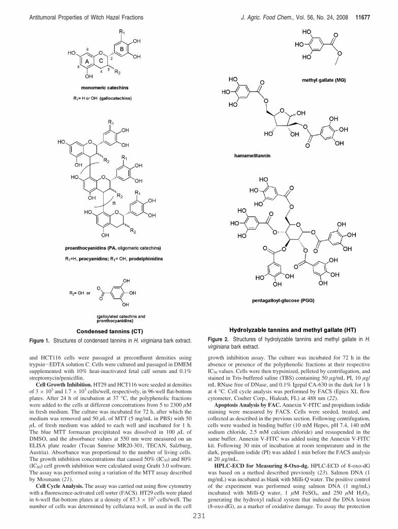
and HCT116 cells were passaged at preconfluent densities usingtrypsin-EDTA solution C. Cells were cultured and passaged in DMEMsupplemented with 10% heat-inactivated fetal calf serum and 0.1%streptomycin/penicillin.
Cell Growth Inhibition. HT29 and HCT116 were seeded at densitiesof 3 × 10³ and 1.7 × 10³ cells/well, respectively, in 96-well flat-bottomplates. After 24 h of incubation at 37 °C, the polyphenolic fractionswere added to the cells at different concentrations from 5 to 2300 µMin fresh medium. The culture was incubated for 72 h, after which themedium was removed and 50 µL of MTT (5 mg/mL in PBS) with 50µL of fresh medium was added to each well and incubated for 1 h.The blue MTT formazan precipitated was dissolved in 100 µL ofDMSO, and the absorbance values at 550 nm were measured on anELISA plate reader (Tecan Sunrise MR20-301, TECAN, Salzburg,Austria). Absorbance was proportional to the number of living cells.The growth inhibition concentrations that caused 50% (IC50) and 80%(IC80) cell growth inhibition were calculated using Grafit 3.0 software.The assay was performed using a variation of the MTT assay describedby Mosmann (21).
Cell Cycle Analysis. The assay was carried out using flow cytometrywith a fluorescence-activated cell sorter (FACS). HT29 cells were platedin 6-well flat-bottom plates at a density of 87.3 × 10³ cells/well. Thenumber of cells was determined by cells/area well, as used in the cell
growth inhibition assay. The culture was incubated for 72 h in theabsence or presence of the polyphenolic fractions at their respectiveIC50 values. Cells were then trypsinized, pelleted by centrifugation, andstained in Tris-buffered saline (TBS) containing 50 µg/mL PI, 10 µg/mL RNase free of DNase, and 0.1% Igepal CA-630 in the dark for 1 hat 4 °C. Cell cycle analysis was performed by FACS (Epics XL flowcytometer, Coulter Corp., Hialeah, FL) at 488 nm (22).
Apoptosis Analysis by FAC. Annexin V-FITC and propidium iodidestaining were measured by FACS. Cells were seeded, treated, andcollected as described in the previous section. Following centrifugation,cells were washed in binding buffer (10 mM Hepes, pH 7.4, 140 mMsodium chloride, 2.5 mM calcium chloride) and resuspended in thesame buffer. Annexin V-FITC was added using the Annexin V-FITCkit. Following 30 min of incubation at room temperature and in thedark, propidium iodide (PI) was added 1 min before the FACS analysisat 20 µg/mL.
HPLC-ECD for Measuring 8-Oxo-dg. HPLC-ECD of 8-oxo-dGwas based on a method described previously (23). Salmon DNA (1mg/mL) was incubated as blank with Milli-Q water. The positive controlof the experiment was performed using salmon DNA (1 mg/mL)incubated with Milli-Q water, 1 µM FeSO4, and 250 µM H2O2,generating the hydroxyl radical system that induced the DNA lesion(8-oxo-dG), as a marker of oxidative damage. To assay the protection
Figure 1. Structures of condensed tannins in H. virginiana bark extract. Figure 2. Structures of hydrolyzable tannins and methyl gallate in H.virginiana bark extract.
Antitumoral Properties of Witch Hazel Fractions J. Agric. Food Chem., Vol. 56, No. 24, 2008 11677
231
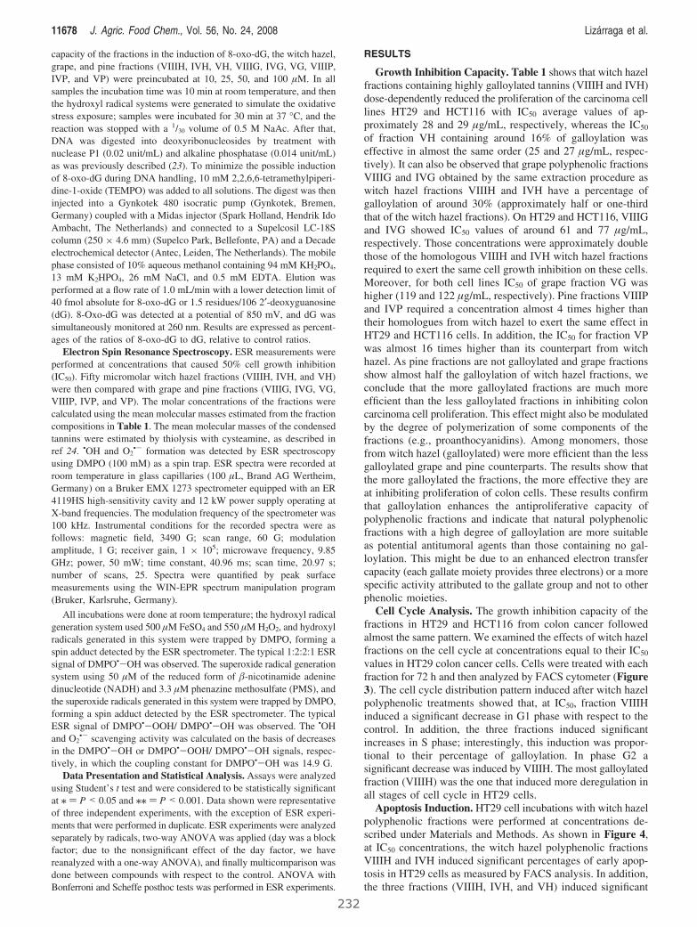
capacity of the fractions in the induction of 8-oxo-dG, the witch hazel,grape, and pine fractions (VIIIH, IVH, VH, VIIIG, IVG, VG, VIIIP,IVP, and VP) were preincubated at 10, 25, 50, and 100 µM. In allsamples the incubation time was 10 min at room temperature, and thenthe hydroxyl radical systems were generated to simulate the oxidativestress exposure; samples were incubated for 30 min at 37 °C, and thereaction was stopped with a 1/30 volume of 0.5 M NaAc. After that,DNA was digested into deoxyribonucleosides by treatment withnuclease P1 (0.02 unit/mL) and alkaline phosphatase (0.014 unit/mL)as was previously described (23). To minimize the possible inductionof 8-oxo-dG during DNA handling, 10 mM 2,2,6,6-tetramethylpiperi-dine-1-oxide (TEMPO) was added to all solutions. The digest was theninjected into a Gynkotek 480 isocratic pump (Gynkotek, Bremen,Germany) coupled with a Midas injector (Spark Holland, Hendrik IdoAmbacht, The Netherlands) and connected to a Supelcosil LC-18Scolumn (250 × 4.6 mm) (Supelco Park, Bellefonte, PA) and a Decadeelectrochemical detector (Antec, Leiden, The Netherlands). The mobilephase consisted of 10% aqueous methanol containing 94 mM KH2PO4,13 mM K2HPO4, 26 mM NaCl, and 0.5 mM EDTA. Elution wasperformed at a flow rate of 1.0 mL/min with a lower detection limit of40 fmol absolute for 8-oxo-dG or 1.5 residues/106 2′-deoxyguanosine(dG). 8-Oxo-dG was detected at a potential of 850 mV, and dG wassimultaneously monitored at 260 nm. Results are expressed as percent-ages of the ratios of 8-oxo-dG to dG, relative to control ratios.
Electron Spin Resonance Spectroscopy. ESR measurements wereperformed at concentrations that caused 50% cell growth inhibition(IC50). Fifty micromolar witch hazel fractions (VIIIH, IVH, and VH)were then compared with grape and pine fractions (VIIIG, IVG, VG,VIIIP, IVP, and VP). The molar concentrations of the fractions werecalculated using the mean molecular masses estimated from the fractioncompositions in Table 1. The mean molecular masses of the condensedtannins were estimated by thiolysis with cysteamine, as described inref 24. •OH and O2
•- formation was detected by ESR spectroscopyusing DMPO (100 mM) as a spin trap. ESR spectra were recorded atroom temperature in glass capillaries (100 µL, Brand AG Wertheim,Germany) on a Bruker EMX 1273 spectrometer equipped with an ER4119HS high-sensitivity cavity and 12 kW power supply operating atX-band frequencies. The modulation frequency of the spectrometer was100 kHz. Instrumental conditions for the recorded spectra were asfollows: magnetic field, 3490 G; scan range, 60 G; modulationamplitude, 1 G; receiver gain, 1 × 10⁵; microwave frequency, 9.85GHz; power, 50 mW; time constant, 40.96 ms; scan time, 20.97 s;number of scans, 25. Spectra were quantified by peak surfacemeasurements using the WIN-EPR spectrum manipulation program(Bruker, Karlsruhe, Germany).
All incubations were done at room temperature; the hydroxyl radicalgeneration system used 500 µM FeSO4 and 550 µM H2O2, and hydroxylradicals generated in this system were trapped by DMPO, forming aspin adduct detected by the ESR spectrometer. The typical 1:2:2:1 ESRsignal of DMPO•-OH was observed. The superoxide radical generationsystem using 50 µM of the reduced form of �-nicotinamide adeninedinucleotide (NADH) and 3.3 µM phenazine methosulfate (PMS), andthe superoxide radicals generated in this system were trapped by DMPO,forming a spin adduct detected by the ESR spectrometer. The typicalESR signal of DMPO•-OOH/ DMPO•-OH was observed. The •OHand O2
•- scavenging activity was calculated on the basis of decreasesin the DMPO•-OH or DMPO•-OOH/ DMPO•-OH signals, respec-tively, in which the coupling constant for DMPO•-OH was 14.9 G.
Data Presentation and Statistical Analysis. Assays were analyzedusing Student’s t test and were considered to be statistically significantat / ) P < 0.05 and // ) P < 0.001. Data shown were representativeof three independent experiments, with the exception of ESR experi-ments that were performed in duplicate. ESR experiments were analyzedseparately by radicals, two-way ANOVA was applied (day was a blockfactor; due to the nonsignificant effect of the day factor, we havereanalyzed with a one-way ANOVA), and finally multicomparison wasdone between compounds with respect to the control. ANOVA withBonferroni and Scheffe posthoc tests was performed in ESR experiments.
RESULTS
Growth Inhibition Capacity. Table 1 shows that witch hazelfractions containing highly galloylated tannins (VIIIH and IVH)dose-dependently reduced the proliferation of the carcinoma celllines HT29 and HCT116 with IC50 average values of ap-proximately 28 and 29 µg/mL, respectively, whereas the IC50
of fraction VH containing around 16% of galloylation waseffective in almost the same order (25 and 27 µg/mL, respec-tively). It can also be observed that grape polyphenolic fractionsVIIIG and IVG obtained by the same extraction procedure aswitch hazel fractions VIIIH and IVH have a percentage ofgalloylation of around 30% (approximately half or one-thirdthat of the witch hazel fractions). On HT29 and HCT116, VIIIGand IVG showed IC50 values of around 61 and 77 µg/mL,respectively. Those concentrations were approximately doublethose of the homologous VIIIH and IVH witch hazel fractionsrequired to exert the same cell growth inhibition on these cells.Moreover, for both cell lines IC50 of grape fraction VG washigher (119 and 122 µg/mL, respectively). Pine fractions VIIIPand IVP required a concentration almost 4 times higher thantheir homologues from witch hazel to exert the same effect inHT29 and HCT116 cells. In addition, the IC50 for fraction VPwas almost 16 times higher than its counterpart from witchhazel. As pine fractions are not galloylated and grape fractionsshow almost half the galloylation of witch hazel fractions, weconclude that the more galloylated fractions are much moreefficient than the less galloylated fractions in inhibiting coloncarcinoma cell proliferation. This effect might also be modulatedby the degree of polymerization of some components of thefractions (e.g., proanthocyanidins). Among monomers, thosefrom witch hazel (galloylated) were more efficient than the lessgalloylated grape and pine counterparts. The results show thatthe more galloylated the fractions, the more effective they areat inhibiting proliferation of colon cells. These results confirmthat galloylation enhances the antiproliferative capacity ofpolyphenolic fractions and indicate that natural polyphenolicfractions with a high degree of galloylation are more suitableas potential antitumoral agents than those containing no gal-loylation. This might be due to an enhanced electron transfercapacity (each gallate moiety provides three electrons) or a morespecific activity attributed to the gallate group and not to otherphenolic moieties.
Cell Cycle Analysis. The growth inhibition capacity of thefractions in HT29 and HCT116 from colon cancer followedalmost the same pattern. We examined the effects of witch hazelfractions on the cell cycle at concentrations equal to their IC50
values in HT29 colon cancer cells. Cells were treated with eachfraction for 72 h and then analyzed by FACS cytometer (Figure3). The cell cycle distribution pattern induced after witch hazelpolyphenolic treatments showed that, at IC50, fraction VIIIHinduced a significant decrease in G1 phase with respect to thecontrol. In addition, the three fractions induced significantincreases in S phase; interestingly, this induction was propor-tional to their percentage of galloylation. In phase G2 asignificant decrease was induced by VIIIH. The most galloylatedfraction (VIIIH) was the one that induced more deregulation inall stages of cell cycle in HT29 cells.
Apoptosis Induction. HT29 cell incubations with witch hazelpolyphenolic fractions were performed at concentrations de-scribed under Materials and Methods. As shown in Figure 4,at IC50 concentrations, the witch hazel polyphenolic fractionsVIIIH and IVH induced significant percentages of early apop-tosis in HT29 cells as measured by FACS analysis. In addition,the three fractions (VIIIH, IVH, and VH) induced significant
11678 J. Agric. Food Chem., Vol. 56, No. 24, 2008 Lizarraga et al.
232
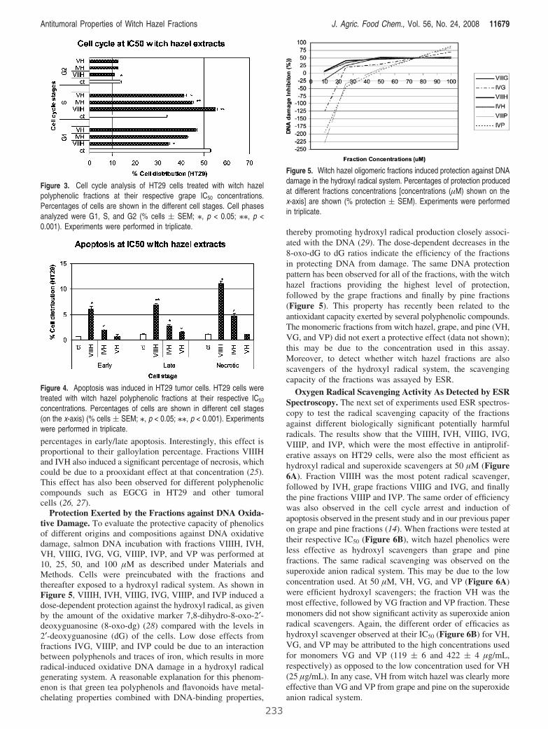
percentages in early/late apoptosis. Interestingly, this effect isproportional to their galloylation percentage. Fractions VIIIHand IVH also induced a significant percentage of necrosis, whichcould be due to a prooxidant effect at that concentration (25).This effect has also been observed for different polyphenoliccompounds such as EGCG in HT29 and other tumoralcells (26, 27).
Protection Exerted by the Fractions against DNA Oxida-tive Damage. To evaluate the protective capacity of phenolicsof different origins and compositions against DNA oxidativedamage, salmon DNA incubation with fractions VIIIH, IVH,VH, VIIIG, IVG, VG, VIIIP, IVP, and VP was performed at10, 25, 50, and 100 µM as described under Materials andMethods. Cells were preincubated with the fractions andthereafter exposed to a hydroxyl radical system. As shown inFigure 5, VIIIH, IVH, VIIIG, IVG, VIIIP, and IVP induced adose-dependent protection against the hydroxyl radical, as givenby the amount of the oxidative marker 7,8-dihydro-8-oxo-2′-deoxyguanosine (8-oxo-dg) (28) compared with the levels in2′-deoxyguanosine (dG) of the cells. Low dose effects fromfractions IVG, VIIIP, and IVP could be due to an interactionbetween polyphenols and traces of iron, which results in moreradical-induced oxidative DNA damage in a hydroxyl radicalgenerating system. A reasonable explanation for this phenom-enon is that green tea polyphenols and flavonoids have metal-chelating properties combined with DNA-binding properties,
thereby promoting hydroxyl radical production closely associ-ated with the DNA (29). The dose-dependent decreases in the8-oxo-dG to dG ratios indicate the efficiency of the fractionsin protecting DNA from damage. The same DNA protectionpattern has been observed for all of the fractions, with the witchhazel fractions providing the highest level of protection,followed by the grape fractions and finally by pine fractions(Figure 5). This property has recently been related to theantioxidant capacity exerted by several polyphenolic compounds.The monomeric fractions from witch hazel, grape, and pine (VH,VG, and VP) did not exert a protective effect (data not shown);this may be due to the concentration used in this assay.Moreover, to detect whether witch hazel fractions are alsoscavengers of the hydroxyl radical system, the scavengingcapacity of the fractions was assayed by ESR.
Oxygen Radical Scavenging Activity As Detected by ESRSpectroscopy. The next set of experiments used ESR spectros-copy to test the radical scavenging capacity of the fractionsagainst different biologically significant potentially harmfulradicals. The results show that the VIIIH, IVH, VIIIG, IVG,VIIIP, and IVP, which were the most effective in antiprolif-erative assays on HT29 cells, were also the most efficient ashydroxyl radical and superoxide scavengers at 50 µM (Figure6A). Fraction VIIIH was the most potent radical scavenger,followed by IVH, grape fractions VIIIG and IVG, and finallythe pine fractions VIIIP and IVP. The same order of efficiencywas also observed in the cell cycle arrest and induction ofapoptosis observed in the present study and in our previous paperon grape and pine fractions (14). When fractions were tested attheir respective IC50 (Figure 6B), witch hazel phenolics wereless effective as hydroxyl scavengers than grape and pinefractions. The same radical scavenging was observed on thesuperoxide anion radical system. This may be due to the lowconcentration used. At 50 µM, VH, VG, and VP (Figure 6A)were efficient hydroxyl scavengers; the fraction VH was themost effective, followed by VG fraction and VP fraction. Thesemonomers did not show significant activity as superoxide anionradical scavengers. Again, the different order of efficacies ashydroxyl scavenger observed at their IC50 (Figure 6B) for VH,VG, and VP may be attributed to the high concentrations usedfor monomers VG and VP (119 ( 6 and 422 ( 4 µg/mL,respectively) as opposed to the low concentration used for VH(25 µg/mL). In any case, VH from witch hazel was clearly moreeffective than VG and VP from grape and pine on the superoxideanion radical system.
Figure 3. Cell cycle analysis of HT29 cells treated with witch hazelpolyphenolic fractions at their respective grape IC50 concentrations.Percentages of cells are shown in the different cell stages. Cell phasesanalyzed were G1, S, and G2 (% cells ( SEM; /, p < 0.05; //, p <0.001). Experiments were performed in triplicate.
Figure 4. Apoptosis was induced in HT29 tumor cells. HT29 cells weretreated with witch hazel polyphenolic fractions at their respective IC50
concentrations. Percentages of cells are shown in different cell stages(on the x-axis) (% cells ( SEM; /, p < 0.05; //, p < 0.001). Experimentswere performed in triplicate.
Figure 5. Witch hazel oligomeric fractions induced protection against DNAdamage in the hydroxyl radical system. Percentages of protection producedat different fractions concentrations [concentrations (µM) shown on thex-axis] are shown (% protection ( SEM). Experiments were performedin triplicate.
Antitumoral Properties of Witch Hazel Fractions J. Agric. Food Chem., Vol. 56, No. 24, 2008 11679
233

DISCUSSION
The present study shows that significant differences in theinhibition of cell growth in HT29 and HCT116 cells broughtabout by phenolics of different origins (fractions VIIIH, IVH,VH, VIIIG, IVG, VG, VIIIP, IVP, and VP) may be related tothe structural properties of their components, mainly thepercentage of galloylation and perhaps the degree of polymer-ization. Witch hazel fractions VIIIH, IVH, and VH were moreeffective than VIIIG, IVG, VG, VIIIP, IVP, and VP from grapeand pine (Table 1). The same differences were observed in cellcycle deregulation and apoptosis induction on HT29 cells, theprotective activity against DNA damage by oxidative stress, andthe free radical scavenging capacities at the same concentration(50 µM). Galloylation appears to play an important role in theinhibition of cell growth. Even with monomers, galloylated VHwas more potent than nongalloylated VG and VP. FractionVIIIH was the most potent cell cycle deregulator, inducingsignificant changes in cell percentages of all the phases of thecell cycle. This fraction was also the most effective scavengeramong those tested. Because the increases of cell percentageson the S phase of the cell cycle were proportional to theirgalloylation, this structural feature appears to be clearlysignificant for the explanation of the activity of phenolics, inaccordance with our previous studies (14, 15). This highlygalloylated fraction was also the most active apoptotic inducerand best free radical scavenger. The cell cycle arrest in the Sphase observed for the three witch hazel fractions tested mightbe triggered by a change in the intracellular redox balance.Perturbations of the intracellular redox signals may blockproliferation of tumor cells. It has been observed that cellmembrane potentials change in response to H2O2 depending onthe cell cycle phase. Intracellular ROS are particularly high inthe S phase (30). Therefore, the cell cycle arrest induced bywitch hazel fractions may be explained by their ROS scavengercapacity. Witch hazel fractions may block the cell cycle of HT29
cells in the S phase by scavenging ROS, with subsequentinhibition of DNA synthesis.
Because each gallate moiety provides three hydroxyl groups,galloylation clearly enhances the scavenger capacity of thefractions. Interestingly, galloylation also enhanced the inductionof apoptosis and necrosis on HT29 cells. Fractions VIIIH andIVH induced both effects, whereas VH induced only someapoptosis. In this assay VIIIH also was the most active fraction,inducing around 13% of apoptosis and 11% of necrosis. Thelatter may be due to a pro-oxidant effect. This pro-oxidant effectof some polyphenolic fractions has been extensively discussedin the literature, where it has been described as a protectivemechanism by up-regulation of genes implicated in the intra-cellular defense and biotransformation of xenobiotics such asenzymes of phases I and II (31).
There is a clear relationship between high galloylationpercentages with low IC50, cell cycle deregulation, apoptosis-necrosis induction, and scavenging capacity. Witch hazelfractions, which are heavily galloylated (Table 1), were morepotent scavengers than grape and pine fractions in the tworadical generation systems (hydroxyl and superoxide) at 50 µM.This is due to the presence of pyrogallol moieties (three adjacentphenolic groups) in the form of gallates from both condensedand hydrolyzable tannins and gallocatechins (pyrogallol moietiesin the condensed ring of proanthocyanidins). The main hydro-lyzable tannins in the fractions are hamamelitannin (2′,5′-di-O-galloyl hamamelose) and pentagalloyl glucose (6), includedin Table 1 under HT. Moreover, witch hazel is a source ofgallocatechins such as pyrogallol-containing epigallocatechingallate (EGC), whereas grape and pine are mainly composedof epicatechin (EC) units. These structural features make witchhazel fractions effective electron donors (6). This could bebehind the alleged pro-oxidant effect of some polyphenols. Infact, ROS may be formed by phenolics through one-electronreductions of molecular oxygen (O2) to form the superoxideradical, which is primarily responsible for the cytotoxic effects
Figure 6. Scavenging activity of OH• and O2•- analyzed by ESR. Witch hazel, grape, and pine fractions were evaluated at (A) 50 µM and (B) IC50
concentration in HT29 cells in hydroxyl radical and superoxide anion radical generating systems, described under Experimental Procedures. Experimentswere performed in duplicate (/, p < 0.05; //, p < 0.001). Data from grape and pine are from ref 18.
11680 J. Agric. Food Chem., Vol. 56, No. 24, 2008 Lizarraga et al.
234
observed in aerobic organisms (32, 33). It has been observedthat several pyrogallol-containing polyphenols, such as EGCGfrom green tea, exert their chemotherapeutical properties bymodulating the cellular redox system on cells, including theproduction of reactive oxygen species, and influence glutathionemetabolism and lipid peroxidation in different subcellularcompartments (9). Moreover, it has been observed thatantioxidant-prooxidant properties of ECGC are dose-responsedependent (34). The redox states of polyphenolic flavonoidsappear to influence their ability to induce oxidative moleculardamage or antioxidant protective action (35). Nowadays, manynatural polyphenolic extracts are commercialized, so it is crucialto increase knowledge of the polyphenolic structures within theextracts, the relationship with their putative antioxidant protec-tive activity, and their mechanisms of action. The resultspresented here should be useful for a better understanding ofstructure-bioactivity relationships of polyphenolics, whichshould be of assistance in choosing an adequate source and arational design for formulations of plant polyphenols in nutri-tional supplements and as chemotherapy in cancer.
LITERATURE CITED
(1) Seeram, N. P.; Adams, L. S.; Zhang, Y.; Lee, R.; Sand, D.;Scheuller, H. S.; Heber, D. Blackberry, black raspberry, blueberry,cranberry, red raspberry, and strawberry extracts inhibit growthand stimulate apoptosis of human cancer cells in vitro. J. Agric.Food Chem. 2006, 54, 9329–9339.
(2) Lee, S. Y.; Munerol, B.; Pollard, S.; Youdim, K. A.; Pannala,A. S.; Kuhnle, G. G.; Debnam, E. S.; Rice-Evans, C.; Spencer,J. P. The reaction of flavanols with nitrous acid protects againstN-nitrosamine formation and leads to the formation of nitrosoderivatives which inhibit cancer cell growth. Free Radical Biol.Med. 2006, 40, 323–334.
(3) Reto, M.; Figueira, M. E.; Filipe, H. M.; Almeida, C. M. Chemicalcomposition of green tea (Camellia sinensis) infusions com-mercialized in Portugal. Plant Foods Hum. Nutr. 2007, 62, 139–144.
(4) Torres, J. L.; Varela, B.; Garcıa, M. T.; Carilla, J.; Matito, C.;Centelles, J. J.; Cascante, M.; Sort, X.; Bobet, R. Valorization ofgrape (Vitis Vinifera) byproducts. Antioxidant and biologicalproperties of polyphenolic fractions differing in procyanidincomposition and flavonol content. J. Agric. Food Chem. 2002,50, 7548–7555.
(5) Tourino, S.; Selga, A.; Jimenez, A.; Julia, L.; Lozano, C.;Lizarraga, D.; Cascante, M.; Torres, J. L. Procyanidin fractionsfrom pine (Pinus pinaster) bark: radical scavenging power insolution, antioxidant activity in emulsion, and antiproliferativeeffect in melanoma cells. J. Agric. Food Chem. 2005, 53, 4728–4735.
(6) Tourino, S.; Lizarraga, D.; Carreras, A.; Lorenzo, S.; Ugartondo,V.; Mitjans, M.; Vinardell, M. P.; Julia, Ll.; Cascante, M.; Torres,J. L. Highly galloylated tannin fractions from witch hazel(Hamamelis Virginiana) bark: electron transfer capacity, in vitroantioxidant activity and effects on skin related cells. Chem. Res.Toxicol. 2008, 21, 696–704.
(7) Adhami, V. M.; Mukhtar, H. Anti-oxidants from green tea andpomegranate for chemoprevention of prostate cancer. Mol. Bio-technol. 2007, 37, 52–57.
(8) Hofmann, T.; Liegibel, U.; Winterhalter, P.; Bub, A.; Rech-kemmer, G.; Pool-Zobel, B. L. Intervention with polyphenol-richfruit juices results in an elevation of glutathione S-transferase P1(hGSTP1) protein expression in human leucocytes of healthyvolunteers. Mol. Nutr. Food Res. 2006, 50, 1191–1200.
(9) Raza, H.; John, A. Green tea polyphenol epigallocatechin-3-gallatedifferentially modulates oxidative stress in PC12 cell compart-ments. Toxicol. Appl. Pharmacol. 2005, 207, 212–220.
(10) Yang, G. Y.; Liao, J.; Kim, K.; Yurkow, E. J.; Yang, C. S.Inhibition of growth and induction of apoptosis in human cancercell lines by tea polyphenols. Carcinogenesis 1998, 19, 611–6.
(11) Jung, Y. D.; Kim, M. S.; Shin, B. A.; Chay, K. O.; Ahn, B. W.;Liu, W.; Bucana, C. D.; Gallick, G. E.; Ellis, L. M. EGCG, amajor component of green tea, inhibits tumour growth byinhibiting VEGF induction in human colon carcinoma cells. Br. J.Cancer 2001, 84, 844–850.
(12) Shimizu, M.; Deguchi, A.; Lim, J. T.; Moriwaki, H.; Kopelovich,L.; Weinstein, I. B. (-)-Epigallocatechin gallate and polyphenonE inhibit growth and activation of the epidermal growth factorreceptor and human epidermal growth factor receptor-2 signalingpathways in human colon cancer cells. Clin. Cancer Res. 2005,11, 2735–2746.
(13) Kerem, Z.; Chetrit, D.; Shoseyov, O.; Regev-Shoshani, G.Protection of lipids from oxidation by epicatechin, trans-resvera-trol, and gallic and caffeic acids in intestinal model systems. J.Agric. Food Chem. 2006, 54, 10288–10293.
(14) Lizarraga, D.; Lozano, C.; Briede, J. J.; van Delft, J. H.; Tourino,S.; Centelles, J. J.; Torres, J. L.; Cascante, M. The importance ofpolymerization and galloylation for the antiproliferative propertiesof procyanidin-rich natural extracts. FEBS J. 2007, 274, 4802–4811.
(15) Lozano, C.; Julia, L.; Jimenez, A.; Tourino, S.; Centelles, J. J.;Cascante, M.; Torres, J. L. Electron-transfer capacity of catechinderivatives and influence on the cell cycle and apoptosis in HT29cells. FEBS J. 2006, 273, 2475–2486.
(16) Rice-Evans, C. Flavonoids and isoflavones: absorption, metabo-lism, and bioactivity. Free Radical Biol. Med. 2004, 36, 827–828.
(17) Jenner, A. M.; Rafter, J.; Halliwell, B. Human fecal water contentof phenolics: the extent of colonic exposure to aromatic com-pounds. Free Radical Biol. Med. 2005, 38, 763–772.
(18) Halliwell, B.; Zhao, K.; Whiteman, M. The gastrointestinal tract:a major site of antioxidant action. Free Radical Res. 2000, 33,819–830.
(19) Meselhy, M. R.; Nakamura, N.; Hattori, M. Biotransformationof (-)-epicatechin 3-O-gallate by human intestinal bacteria. Chem.Pharm. Bull. (Tokyo) 1997, 45, 888–893.
(20) Salucci, M.; Stivala, L. A.; Maiani, G.; Bugianesi, R.; Vannini,V. Flavonoids uptake and their effect on cell cycle of human colonadenocarcinoma cells (Caco2). Br. J. Cancer 2002, 86, 1645–1651.
(21) Mosmann, T. Rapid colorimetric assay for cellular growth andsurvival: application to proliferation and cytotoxicity assays.J. Immunol. Methods 1983, 65, 55–63.
(22) Lozano, C.; Torres, J. L.; Julia, L.; Jimenez, A.; Centelles, J. J.;Cascante, M. Effect of new antioxidant cysteinyl-flavanol con-jugates on skin cancer cells. FEBS Lett. 2005, 579, 4219–4225.
(23) de Kok, T. M.; ten Vaarwerk, F.; Zwingman, I.; van Maanen,J. M.; Kleinjans, J. C. Peroxidation of linoleic, arachidonic andoleic acid in relation to the induction of oxidative DNA damageand cytogenetic effects. Carcinogenesis 1994, 15, 1399–1404.
(24) Selga, A.; Torres, J. L. Efficient preparation of catechin thioconjugates by one step extraction/depolymerization of pine (Pinuspinaster) bark procyanidins. J. Agric. Food Chem. 2005, 53, 7760–7765.
(25) Azam, S.; Hadi, N.; Khan, N. U.; Hadi, S. M. Prooxidant propertyof green tea polyphenols epicatechin and epigallocatechin-3-gallate: implications for anticancer properties. Toxicol In Vitro2004, 18, 555–561.
(26) Hwang, J. T.; Ha, J.; Park, I. J.; Lee, S. K.; Baik, H. W.; Kim,Y. M.; Park, O. J. Apoptotic effect of EGCG in HT-29 coloncancer cells via AMPK signal pathway. Cancer Lett. 2007, 247,115–121.
(27) Weisburg, J. H.; Weissman, D. B.; Sedaghat, T.; Babich, H. Invitro cytotoxicity of epigallocatechin gallate and tea extracts tocancerous and normal cells from the human oral cavity. BasicClin. Pharmacol. Toxicol. 2004, 95, 191–200.
Antitumoral Properties of Witch Hazel Fractions J. Agric. Food Chem., Vol. 56, No. 24, 2008 11681
235

(28) Zinov’eva, V. N.; Ostrovskii, O. V. Free radical oxidation of DNAand its biomarker oxidized guanosine(8-oxodG). Vopr. Med. Khim.2002, 48, 419–431.
(29) Leanderson, P.; Faresjo, A. O.; Tagesson, C. Green tea polyphe-nols inhibit oxidant-induced DNA strand breakage in cultured lungcells. Free Radical Biol. Med. 1997, 23, 235–242.
(30) Gamaley, I. A.; Klyubin, I. V. Roles of reactive oxygen species:signaling and regulation of cellular functions. Int. ReV. Cytol. 1999,188, 203–255.
(31) Dhakshinamoorthy, S.; Long, D. J., 2nd; Jaiswal, A. K. Antioxi-dant regulation of genes encoding enzymes that detoxify xeno-biotics and carcinogens. Curr. Top. Cell Regul. 2000, 36, 201–216.
(32) Halliwell, B.; Gutteridge, J. M. Biologically relevant metal ion-dependent hydroxyl radical generation. An update. FEBS Lett.1992, 307, 108–112.
(33) Valko, M.; Leibfritz, D.; Moncol, J.; Cronin, M. T.; Mazur, M.;Telser, J. Free radicals and antioxidants in normal physiological
functions and human disease. Int. J. Biochem. Cell Biol. 2007,39, 44–84.
(34) Tian, B.; Sun, Z.; Xu, Z.; Hua, Y. Chemiluminescence analysisof the prooxidant and antioxidant effects of epigallocatechin-3-gallate. Asia Pac. J. Clin. Nutr 2007, 16, 153–157.
(35) Kim, B. H.; Cho, S. M.; Chang, Y. S.; Han, S. B.; Kim, Y. Effectof quercitrin gallate on zymosan A-induced peroxynitrite produc-tion in macrophages. Arch. Pharm. Res. 2007, 30, 733–738.
Received for review July 29, 2008. Revised manuscript received October1, 2008. Accepted October 16, 2008. This work was supported by GrantsPPQ2003-06602-C04-01 and -04, AGL2006-12210-C03-02/ALI, SAF2008-00164, doctoral fellowships to S.T. and D.L. from the Spanish Ministryof Education and Science, ISCIII-RTICC (RD06/0020/0046) from theSpanish government and the European Union FEDER funds andGeneralitat de Catalunya (2005SGR00204; 2006ITT-10007).
JF802345X
11682 J. Agric. Food Chem., Vol. 56, No. 24, 2008 Lizarraga et al.
236

VII. The maize ZmMYB42 represses the phenylpropanoid
pathway and affects the cell wall structure,
composition and degradability in Arabidopsis
thaliana
Sonbol, F-M.; Capellades, M.; Fornalé, S.; Ruel, K.;
Touriño, S.; Torres, J.L.; Rovira, P.; Encina, A.;
Puigdomènech, P.; Rigau, J. and Caparrós-Ruiz, D. Plant
Mol. Biol.; 70(3): 283-296, 2009

The maize ZmMYB42 represses the phenylpropanoid pathwayand affects the cell wall structure, composition and degradabilityin Arabidopsis thaliana
Fathi-Mohamed Sonbol Æ Silvia Fornale Æ Montserrat Capellades ÆAntonio Encina Æ Sonia Tourino Æ Josep-Lluıs Torres Æ Pere Rovira ÆKatia Ruel Æ Pere Puigdomenech Æ Joan Rigau Æ David Caparros-Ruiz
Received: 4 December 2008 / Accepted: 12 February 2009
� Springer Science+Business Media B.V. 2009
Abstract The involvement of the maize ZmMYB42 R2R3-
MYB factor in the phenylpropanoid pathway and cell wall
structure and composition was investigated by overexpres-
sion in Arabidopsis thaliana. ZmMYB42 down-regulates
several genes of the lignin pathway and this effect reduces
the lignin content in all lignified tissues. In addition,
ZmMYB42 plants generate a lignin polymer with a
decreased S to G ratio through the enrichment in H and G
subunits and depletion in S subunits. This transcription
factor also regulates other genes involved in the synthesis of
sinapate esters and flavonoids. Furthermore, ZmMYB42
affects the cell wall structure and degradability, and its
polysaccharide composition. Together, these results suggest
that ZmMYB42 may be part of the regulatory network
controlling the phenylpropanoid biosynthetic pathway.
Keywords Arabidopsis thaliana � Maize �Phenylpropanoids � Lignin regulation � R2R3-MYB factors
Introduction
Lignin is, after cellulose the most abundant component of
biomass (Boerjan et al. 2003). This polymer is synthesised
through the phenylpropanoid pathway, a metabolic grid that
synthesises other secondary metabolites such as flavonoids
and sinapate esters (Fig. 1). Lignin is deposited in the
secondary cell wall of vascular plants and its presence
increases the efficiency of water transport, the stiffness of
mechanical tissues and constitutes by itself a physical bar-
rier against microbial attacks. However, the covalent
interaction of lignin with cell wall polysaccharides makes
Fathi-Mohamed Sonbol and Silvia Fornale contributed equally to this
work.
Electronic supplementary material The online version of thisarticle (doi:10.1007/s11103-009-9473-2) contains supplementarymaterial, which is available to authorized users.
F.-M. Sonbol � S. Fornale � M. Capellades � P. Puigdomenech �J. Rigau (&) � D. Caparros-Ruiz
Consorci CSIC-IRTA-UAB, Centre de Recerca en
AgriGenomica (CRAG), Jordi Girona 18-26, 08034 Barcelona,
Spain
e-mail: [email protected]
A. Encina
Departamento de Ingenierıa y Ciencias Agrarias, Universidad de
Leon, 24071 Leon, Spain
S. Tourino � J.-L. Torres
Institute for Chemical and Environmental Research, IIQAB-
CSIC, Barcelona, Spain
P. Rovira
Departament de Biologia Vegetal, Facultat de Biologia,
Universitat de Barcelona, 08028 Barcelona, Spain
K. Ruel
Centre de Recherche sur les Macromolecules Vegetales,
UPR-CNRS-5301, 38041 Grenoble Cedex 09, France
Present Address:F.-M. Sonbol
Department of Biology, Faculty of Dentistry, Sinai University,
P.O. Box 7, El Massaid-El Arish, North Sinai, Egypt
Present Address:P. Rovira
Centre Tecnologic Forestal Catalunya, 25280 Solsona, Spain
123
Plant Mol Biol
DOI 10.1007/s11103-009-9473-2
239
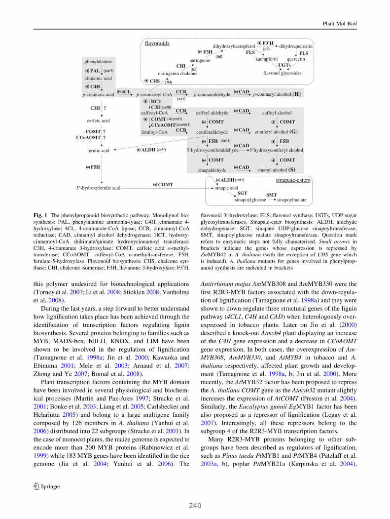
this polymer undesired for biotechnological applications
(Torney et al. 2007; Li et al. 2008; Sticklen 2008; Vanholme
et al. 2008).
During the last years, a step forward to better understand
how lignification takes place has been achieved through the
identification of transcription factors regulating lignin
biosynthesis. Several proteins belonging to families such as
MYB, MADS-box, bHLH, KNOX, and LIM have been
shown to be involved in the regulation of lignification
(Tamagnone et al. 1998a; Jin et al. 2000; Kawaoka and
Ebinuma 2001; Mele et al. 2003; Arnaud et al. 2007;
Zhong and Ye 2007; Bomal et al. 2008).
Plant transcription factors containing the MYB domain
have been involved in several physiological and biochem-
ical processes (Martin and Paz-Ares 1997; Stracke et al.
2001; Bonke et al. 2003; Liang et al. 2005; Carlsbecker and
Helariutta 2005) and belong to a large multigene family
composed by 126 members in A. thaliana (Yanhui et al.
2006) distributed into 22 subgroups (Stracke et al. 2001). In
the case of monocot plants, the maize genome is expected to
encode more than 200 MYB proteins (Rabinowicz et al.
1999) while 183 MYB genes have been identified in the rice
genome (Jia et al. 2004; Yanhui et al. 2006). The
Antirrhinum majus AmMYB308 and AmMYB330 were the
first R2R3-MYB factors associated with the down-regula-
tion of lignification (Tamagnone et al. 1998a) and they were
shown to down-regulate three structural genes of the lignin
pathway (4CL1, C4H and CAD) when heterologously over-
expressed in tobacco plants. Later on Jin et al. (2000)
described a knock-out Atmyb4 plant displaying an increase
of the C4H gene expression and a decrease in CCoAOMT
gene expression. In both cases, the overexpression of Am-
MYB308, AmMYB330, and AtMYB4 in tobacco and A.
thaliana respectively, affected plant growth and develop-
ment (Tamagnone et al. 1998a, b; Jin et al. 2000). More
recently, the AtMYB32 factor has been proposed to repress
the A. thaliana COMT gene as the Atmyb32 mutant slightly
increases the expression of AtCOMT (Preston et al. 2004).
Similarly, the Eucalyptus gunnii EgMYB1 factor has been
also proposed as a repressor of lignification (Legay et al.
2007). Interestingly, all these repressors belong to the
subgroup 4 of the R2R3-MYB transcription factors.
Many R2R3-MYB proteins belonging to other sub-
groups have been described as regulators of lignification,
such as Pinus taeda PtMYB1 and PtMYB4 (Patzlaff et al.
2003a, b), poplar PttMYB21a (Karpinska et al. 2004),
phenylalanine
cinnamic acid
p-coumaric acid p-coumaroyl-CoA p-coumaraldehyde p-coumaryl alcohol (H)
caffeoyl-CoA
feruloyl-CoA coniferaldehyde coniferyl alcohol (G)
5’hydroxyconiferaldehyde
sinapaldehyde sinapyl alcohol (S)
5’hydroxyconiferyl alcohol
PAL
C4H4CL
HCTC3H
CCR
F5H
COMTCCoAOMT
sinapic acid
sinapoylglucose sinapoylmalate
ALDH
SGT SMT
sinapate-esters
naringenin chalcone
naringenin
dihydroxykaempferol
kaempferol
dihydroquercetin
quercetin
flavonol glycosides
CHS
CHI
F3H
F3’H
FLS FLS
UGTs
ferulic acid
caffeyl aldehyde caffeyl alcoholCCR
CCR
flavonoids
COMT
F5H
COMT COMT
COMT(Atomt1)
(fah1)
(ref8)
(ccomt1)
(tt4)
(tt5)
(tt6)
(tt7)
(ref1)
(pal1)
(irx4)
(ref1)ALDH
caffeic acid
5’-hydroxyferulic acid
( )
( )( )
( )
( )
( )
( )
( )
( )
CAD( )
( )
CAD( )
CAD( )
CAD( )
CAD( )
( )
( )
( )
( )
( )
( )
COMT ?CCoAOMT ?
C3H ?
F5H
COMT( )
( )
Fig. 1 The phenylpropanoid biosynthetic pathway. Monolignol bio-
synthesis: PAL, phenylalanine ammonia-lyase; C4H, cinnamate 4-
hydroxylase; 4CL, 4-coumarate-CoA ligase; CCR, cinnamoyl-CoA
reductase; CAD, cinnamyl alcohol dehydrogenase; HCT, hydroxy-
cinnamoyl-CoA shikimate/quinate hydroxycinnamoyl transferase;
C3H, 4-coumarate 3-hydroxylase; COMT, caffeic acid o-methyl-
transferase; CCoAOMT, caffeoyl-CoA o-methyltransferase; F5H,
ferulate-5-hydroxylase. Flavonoid biosynthesis: CHS, chalcone syn-
thase; CHI, chalcone isomerase; F3H, flavanone 3-hydroxylase; F30H,
flavonoid 30-hydroxylase; FLS, flavonol synthase; UGTs, UDP sugar
glycosyltransferases. Sinapate-ester biosynthesis: ALDH, aldehyde
dehydrogenase; SGT, sinapate UDP-glucose sinapoyltransferase;
SMT, sinapoylglucose malate sinapoyltransferase. Question mark
refers to enzymatic steps not fully characterised. Small arrows in
brackets indicate the genes whose expression is repressed by
ZmMYB42 in A. thaliana (with the exception of CHS gene which
is induced). A. thaliana mutants for genes involved in phenylprop-
anoid synthesis are indicated in brackets
Plant Mol Biol
123
240

AtMYB61 (Newman et al. 2004), EgMYB2 (Goicoechea
et al. 2005), Vitis vinifera VvMYB5a (Deluc et al. 2006)
and tobacco NtMYBJS1 (Galis et al. 2006). These factors
have been associated with the regulation of lignin biosyn-
thesis through their in vitro interaction with ACI, ACII, and
ACIII cis-elements typically recognised by these tran-
scription factors (Romero et al. 1998).
In maize, few members of the R2R3-MYB family have
been characterised so far. This is the case for C1 and PL
(Paz-Ares et al. 1987; Marocco et al. 1989; Cone et al.
1993; Pilu et al. 2003), P (Grotewold et al. 1991, 1994),
Zm1 and Zm38 (Franken et al. 1989; Marocco et al. 1989),
all of which are involved in the regulation of flavonoid
biosynthesis. Another R2R3-MYB factor has been cloned
and proposed to participate in maize heterosis (Ju et al.
2006) while other maize factors, such as ZmMYB-IF35,
have been associated with secondary metabolic pathways
(Heine et al. 2007). Recently five new maize R2R3-MYB
factors, ZmMYB2, ZmMYB8, ZmMYB31, ZmMYB39, and
ZmMYB42, have been identified and we have shown that
two of them, ZmMYB31 and ZmMYB42, act as repressors
of lignin biosynthesis (Fornale et al. 2006).
In this work, we have investigated the role of ZmMYB42
in the biosynthesis of the lignin polymer and studied its role
in the regulation of other branches of the phenylpropanoid
pathway, such as the biosynthesis of sinapate esters and
flavonoids. In addition, we have also investigated the effect
of ZmMYB42 on cell wall structure, polysaccharides con-
tent and composition, and cell wall degradability.
Material and methods
Plant material
Arabidopsis thaliana (ecotype WS) plants were grown under
standard greenhouse conditions (25�C day and 22�C night
with 60% humidity) and a 16/8 hr photoperiod. For the in
vitro culture, plants were grown in solid MS medium (Mu-
rashige and Skoog 1962) supplemented with 1% sucrose in
Petri dishes and kept in growth chamber at 22 ± 2�C with a
16 h light period. The transgenic plants used in this work
have been already published (Fornale et al. 2006) and several
independent transgenic lines were studied.
Isolation of total RNA, RT-PCR and PCR
Total RNA was extracted with Trizol Reagent (Invitrogen)
and 4 lg of total RNA were reverse-transcribed using M-
MLV Reverse Transcriptase (Invitrogen). First-strand
cDNA was generated using an oligo(dT)15 primer, and 2 ll
of the first-strand cDNA used as a template in subsequent
PCR reactions. Gene-specific primers were used to amplify
ZmMYB42, and ‘‘no-RT’’ PCR assays were performed to
confirm the absence of genomic DNA contamination. For
each assay, several numbers of cycles were tested to ensure
that the amplification was in the exponential range. The
gene-specific primers used in this work are reported in the
Supplementary Table S1 online.
Histology
Cross-sections (150 lm thick) of the basal part of inflores-
cence stems were obtained using a vibratome (Vibratome
Series 1000, TPI Inc., St. Louis, MO, USA) and stained as
follow. For the Wiesner staining, sections were incubated
for 1 min in 2% Phloroglucinol (w/v) in 95% ethanol, then in
50% HCl previous to the observation under light micro-
scope. For the Maule staining, sections were stained for
10 min in 0.5% KMnO4 (w/v) solution. After a brief wash in
distilled water, samples were incubated for 5 min in 10%
HCl, washed with distilled water and mounted onto micro-
scope slides using a concentrated NH4OH solution for
observation under light microscopy.
Lignin auto-florescence was detected using UV-excita-
tion under DAPI-filter and the thickness measurement of
lignified tissues was performed using the ImageJ 1.38x
program (Rasband 2007).
Assay of Klason lignin
Lignin content from Arabidopsis thaliana mature stems was
quantitatively measured using the Klason method (Kirk and
Obst 1988). Briefly, plant material was extracted four times
in methanol and vacuum dried. 100 mg of the samples were
hydrolysed in 2 ml of 72% (v/v) H2SO4 at 30�C for 1 h. The
hydrolysate was diluted with 56 ml of water and autoclaved
for 1 h. The sample solution was filtered through a fritted
glass-crucible and lignin was measured and expressed as
mg of Klason lignin per gram of cell wall residue.
Analysis of lignin monomer composition
The analysis was carried out by the CuO oxidation method
(Kogel and Bochter 1985; Heddges and Mann 1979).
Briefly, mature stems from 30 wt and ZmMYB42 plants
respectively were collected and immediately frozen and
grounded in liquid nitrogen. After extraction in 20 volumes
of methanol, the extract-free samples were dried and ali-
quots of 50 mg of dried plant material were placed in a
sealable Pyrex tube, together with 100 mg of CuO, 200 mg
of Fe2SO4 and 10 ml of 2 M NaOH were added and a
stream of N2 was bubbled through the solution. The tube
was sealed under a N2 stream and placed at 170�C for 2 h,
with occasional shaking. Tubes were cooled to room tem-
perature and their content was transferred to polypropylene
Plant Mol Biol
123
241
tubes and centrifuged at 3,000 rpm. The pellet was washed
with 20 ml of distilled water, centrifuged, and the new
supernatant pooled with the previous one. The extract was
then acidified with HCl to pH 1, centrifuged again and the
pellet washed with 0.1 M HCl. The acidified extract was
then filtered through a Waters sep-pack cartridge (Waters)
and dried under a N2 stream, and eluted, first with 2 ml ethyl
acetate, then with 1 ml acetonitrile. Eluates were collected
in Pyrex vials, dried under N2 stream and re-dissolved with
1 ml methanol for the HPLC analysis.
The separation and quantitation of the phenolic units
was performed with a chromatograph under the following
conditions. Injected volume: 20 ll. Column: Teknokroma
Tracer Extrasil ODS2 column of 5 mm, 25 9 0.46 cm.
Eluents: (a) 0.05% H3PO4, (b) acetonitrile. Flux:
1 ml min-1. Temperature: 40�C. Gradient: 10% A at the
start, increasing slowly up to 17% in 40 min. The column
was washed with 100% acetonitrile between two samples,
following the sequence: 17% to 100% in 10 min, 100%
during 5 min, dropping to 10% in 10 min, 5 minutes in
10% A to recover a total equilibrium. Phenolic compounds
were detected at 280 nm.
Organic extraction of soluble phenolics and high-
performance liquid chromatograph with diode array
detection (HPLC/DAD)
Leaves or stems (ca. 500 mg) from wt and ZmMYB42
plants were frozen in liquid N2 and extracted as previously
reported (Perez-Jimenez and Saura-Calixto 2008). Samples
were analysed by HPLC/DAD on a Hitachi (San Jose, CA,
USA) Lachrom Elite HPLC system equipped with a qua-
ternary pump, temperature control unit and photo-diode
array UV detector (DAD) fitted with a Kromasil C-18
(Tecknokroma, Madrid, Spain) column (25 9 0.4 cm i.d.,
100 A, 5 l particle size). Acquisition was made using
EZChrom Elite version 3.1.3 from Scientific Software Inc
(Pleasanton, CA, USA). Loads: 50 ll. Elution with a bin-
ary system of solvents [A] 0.1% (v/v) aqueous TFA, [B]
0.05% TFA in CH3CN under gradient conditions percent-
age [B] from 0 to 8 over 5 min; 8 to 10 over 10 min and 10
to 50 over 30 min followed by washing 100% CH3CN for
10 min and re-equilibration of the column to the initial
gradient conditions.
High-performance liquid chromatography–electrospray
mass spectrometry (HPLC/ESI/MS)
Analyses were performed on a HPLC/MS system, con-
sisting of a LC200 pump, PE Nelson 1050S integrator
(Perkin Elmer, USA) fitted with a reversed phase Phe-
nomenex (Torrance, CA, USA) Luna C18 (2) column
(150 9 2.0 mm i.d, 5 lm particle size) coupled to a
API3000 triple quadrupole mass spectrometer PE Sciex
(Concord, Canada) with a turbo ion spray source that was
used to obtain the MS/MS data. HPLC elution conditions:
[A] 0,1% aqueous formic acid, [B] CH3CN gradient from 5
to 23 over 30 min followed washing and equilibration of
the column to the initial conditions. The flow rate was
0.4 ml min-1. Ionisation (negative mode) conditions:
capillary voltage -3500 V, nebulizer gas (N2) 10 (arbitrary
units), curtain gas (N2) 12 (arbitrary units), focusing
potential -200 V, entrance potential 10 V, drying gas (N2)
heated to 400�C. Analysis of the ions was carried out using
full scan (FS) data acquisition from m/z 100 to 800 l in
profile mode and using a cycle time of 2 s with a step size
of 0.1 l and a pause between each of 2 ms. To confirm the
identity of some of the compounds, neutral loss (NL)
experiments by tandem mass spectrometry (HPLC/ESI/
MS/MS) were performed.
HPLC/DAD/MS analysis of sinapate esters
The HPLC/DAD profile after injection of the polyphenolic
extract from leaves included a major peak corresponding to
sinapoylmalate as ascertained by UV spectrophotometry
and LC/ESI/MS. The UV spectrum (190–600 nm) of this
peak on HPLC-DAD was compatible with sinapate. Two
major MS signals recorded in the negative mode, namely
the molecular ion (m/z 339.2, MW sinapoylmalate 340) and
a fragment corresponding to the sinapate moiety (m/z
223.1) revealed the structure of this major phenolic.
Auxin transport assay
Auxin transport was measured according to Besseau et al.
(2007). A 2.5-cm segment of flowering stems was excised
5 cm above the base of the stem. Segments were placed in
a 1.5-ml microcentrifuge tube and the apical ends sub-
merged in 30 ll of MES buffer (5 mM MES and 1%
sucrose, pH 5.5) containing 1 lM indole-3-acetic acid and
66 nM of tritiated indole-3-acetic acid. After 4 h of incu-
bation in the dark, segments were removed and the last
5 mm of the non-submerged ends were excised and placed
in 2.5 ml of liquid scintillation cocktail (Optiphase-high-
safe 2, Perkin-Elmer). Samples were slowly shaken
overnight before measuring radioactivity in a scintillation
counter (Beckman LS6000 SC).
Cell wall analysis
Dried plant material (1 g) was added to Poly-Prep tubes
and extracted with 10 volumes of 70% EtOH for 5 days at
room temperature with wheel-shaking, then washed
6 times with 70% EtOH, 6 times with acetone, and air-
dried to obtain the alcohol insoluble residue (AIR). AIR
Plant Mol Biol
123
242
was then de-starched, treated with acidified phenol and
washed with organic solvents to obtain the cell wall residue
as previously described (Encina et al. 2002).
Neutral sugar analysis was performed according to
Albersheim et al. (1967). Dried cell walls were hydrolysed
with 2 M TFA (trifluoroacetic acid) for 1 h at 121�C and
the resulting sugars were derivatised to alditol acetates and
analysed by gas chromatography (GC) on a Supelco SP-
2330 column. Uronic acid contents were determined by the
m-hydroxybiphenyl method (Blumenkrantz and Asboe-
Hansen 1973), with galacturonic acid as a standard. Cel-
lulose was quantified in crude cell walls by the Updegraff
method (Updegraff 1969) with the hydrolytic conditions
described by Saeman et al. (1963) and quantification of the
glucose released by the anthrone method (Dische 1962)
with glucose as a standard.
For the cell wall degradability assays, cell walls were
hydrolysed (20 mg/1.5 ml) in a mixture of Cellulase R10
(1%); Macerozyme R-10 (0.5%) and purified Driselase
(0.1%) dissolved in sodium acetate 20 mM (pH 4.8). Ali-
quots were taken at 6, 48 and 72 h, clarified by
centrifugation and assayed for total sugars (Dubois et al.
1956).
UV treatment
UV-B treatment was performed according to Jin et al.
(2000). Plants were grown for 10 days onto solid MS
medium. The lids of Petri dishes were removed and plants
were irradiated for 10 min with a short wave transillumi-
nator. After exposure, plants were grown for one week
more and then photographed.
Measurement of soluble phenolics
Total phenolic quantitation was performed according to
Cliff et al. (2007). Stems were extracted with 10% EtOH
(0.1 mg FW/ll). A 10 ll aliquot of each sample was added
to 10 ll of 0.1% HCl in 95% ethanol and 182 ll of 2%
HCl. Each sample was vortexed and allowed to stand for
15 min. Absorbance was measured at 280, 360, and
520 nm using a Shimadzu UV-1630 spectrophotometer.
Phenolic content was determined from standard curves
obtained using dilutions of gallic acid, rutin and cyanidin
chloride at 280, 360, and 520 nm, respectively.
Electron microscopy assays
Hand-cut transverse sections of the basal region of stems of
5-week-old plants were processed for TEM as previously
described (Day et al. 2005). Wall polysaccharides of ultra-
thin transverse sections (50 nm) were PATAg (Periodic
acid-thio carbohydrazide silver proteinate) stained
according to Ruel et al. (1977). Observations were per-
formed at 80 kV with a Philips CM 200 Cryo-electron
microscope.
Results
Phenotype of A. thaliana plants overexpressing
ZmMYB42
Transgenic plants overexpressing ZmMYB42 show several
phenotypic alterations (Fig. 2a) at the macroscopic level:
they are dwarf with smaller leaves that present a severe
adaxial curvature and an altered vascular network charac-
terised by a reduced number of tertiary veins. Previous
studies have shown that the overexpression of R2R3-MYB
factors acting as repressors of lignification produces alter-
ations of the leaf morphology, with the appearance of white
lesions on the older leaves, and reduction of the growth rate
when overexpressed in tobacco and A. thaliana (Tamag-
none et al. 1998a; Jin et al. 2000). In the case of
ZmMYB42, transgenic plants do not present these white
lesions.
ZmMYB42 affects lignin biosynthesis
The lignification pattern in different tissues of wt and
ZmMYB42 plants was analysed by Wiesner staining
(Figs. 2b, 3). In stem cross-sections of wild type and
transgenic plants, xylem vessels (X) and interfascicular
fibres (IF) regions are stained with a red colour, indicative
of lignin deposition. However, transgenic stems retain less
colour intensity than wild type (Fig. 2b).
Transgenic stems are thinner than the wild type ones
(about 65%) and while lignified tissues contain 5-6 cell
layers in wild type, transgenic plants display a reduction of
approximately 3 cell layers and do not develop secondary
xylem at least in the interfascicular region (Fig. 2b).
Despite the reduction of lignified layers, the thickness of
vascular bundles is not affected in transgenic plants when
compared to the stem size (Fig. 2c). In contrast, the
thickness of the area of interfascicular fibres decreases by
about 30% (Fig. 2c).
Mature siliques of plants expressing ZmMYB42 show
reduced colour intensity both in the replum and pedicel,
indicative of reduced lignin content (Fig. 3). Both the lig-
nified valve marginal cell and endocarp b regions of the
silique are also reduced in the transgenic lines (Fig. 3).
We determined the absolute lignin content by the Klason
method. This result shows that ZmMYB42 plants have 60%
reduction of total lignin content compared to wt plants
(Fig. 4a). We also analysed the lignin monomer composi-
tion by staining stem cross sections with the Maule reagent
Plant Mol Biol
123
243
that stains the S subunits of the lignin polymer specifically.
Wild type plants exhibit two staining patterns (Fig. 3): dark
red staining is detected in the IF region, indicative of S-
enriched lignin, while the fascicular region stain slightly
yellow-red, reflecting the deposition of G-enriched lignin.
The transgenic ZmMYB42 stem show the same pattern,
(A)
(B) Wild type 35S::MYB42
0
0.02
0.04
0.06
0.08
0.10
0.12
fibres
xylem
Wt MYB42 Wt MYB42
thic
knes
s(l
igni
vfied
tiss
ue v
sst
em d
iam
eter
)
*sX
100 m 100 m 100 m
Wild type 35S::MYB42
35S::MYB42Wild type
35S::MYB42
Wild type
Cauline leavesRosette leaves
(C)
IF
X
IFX IF
X
Fig. 2 ZmMYB42 affects
Arabidopsis growth. aPhenotype of wt and two
independent transgenic plants
and leaves at the end of the
inflorescence stage. b Wiesner
staining of cross sections of
lignified stalks. (X, xylem; sX,
secondary xylem; IF,
interfascicular fibres). cQuantification of thickness of
xylem and fibres in wt and
transgenic plants. Values are
means of n = 10 independent
measurements. (*) indicates
significant differences
(P B 0.01). The cell thickness
was measured using the ImageJ
1.38x program (Rasband 2007)
Replum
Replum
Pedicel
Pedicel
SiliqueVMCVMC
VMCVMC
enbenb
200 m
500 m 500 m
500 m 500 m
500 m 500 m 200 m 200 m
VMCVMC
VMCVMC
enbenb
200 m
500 m
200 m
VMCVMC
VMCVMC
enbenb
200 m 200 m
200 m
VMCVMC
VMCVMC
enbenb
500 m
Stalk
Wild type 35S::MYB42
Maüle
XX
XFXF
500 m
XX XFXF
500 m
Wild type 35S::MYB42
Wiesner
Fig. 3 Histochemical analyses of ZmMYB42 transgenic plants. Wiesner (left) and Maule (right) staining of cross sections of lignified tissues
(VMC, valve marginal cell; enb, endocarp-b)
Plant Mol Biol
123
244
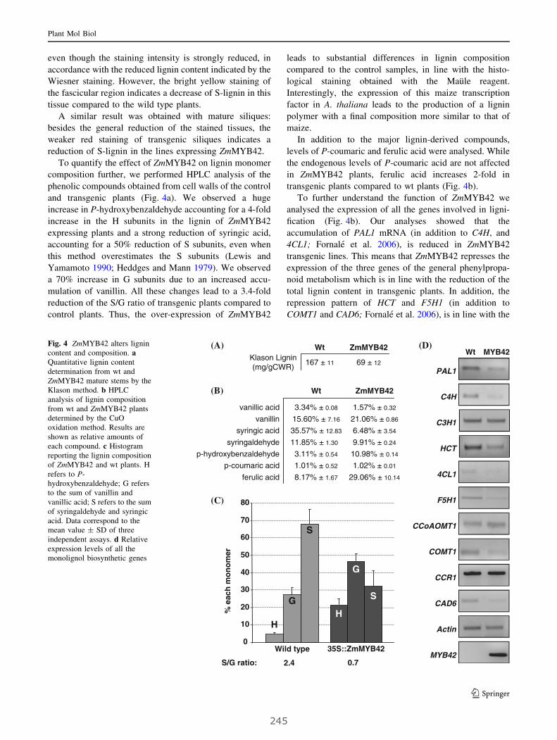
even though the staining intensity is strongly reduced, in
accordance with the reduced lignin content indicated by the
Wiesner staining. However, the bright yellow staining of
the fascicular region indicates a decrease of S-lignin in this
tissue compared to the wild type plants.
A similar result was obtained with mature siliques:
besides the general reduction of the stained tissues, the
weaker red staining of transgenic siliques indicates a
reduction of S-lignin in the lines expressing ZmMYB42.
To quantify the effect of ZmMYB42 on lignin monomer
composition further, we performed HPLC analysis of the
phenolic compounds obtained from cell walls of the control
and transgenic plants (Fig. 4a). We observed a huge
increase in P-hydroxybenzaldehyde accounting for a 4-fold
increase in the H subunits in the lignin of ZmMYB42
expressing plants and a strong reduction of syringic acid,
accounting for a 50% reduction of S subunits, even when
this method overestimates the S subunits (Lewis and
Yamamoto 1990; Heddges and Mann 1979). We observed
a 70% increase in G subunits due to an increased accu-
mulation of vanillin. All these changes lead to a 3.4-fold
reduction of the S/G ratio of transgenic plants compared to
control plants. Thus, the over-expression of ZmMYB42
leads to substantial differences in lignin composition
compared to the control samples, in line with the histo-
logical staining obtained with the Maule reagent.
Interestingly, the expression of this maize transcription
factor in A. thaliana leads to the production of a lignin
polymer with a final composition more similar to that of
maize.
In addition to the major lignin-derived compounds,
levels of P-coumaric and ferulic acid were analysed. While
the endogenous levels of P-coumaric acid are not affected
in ZmMYB42 plants, ferulic acid increases 2-fold in
transgenic plants compared to wt plants (Fig. 4b).
To further understand the function of ZmMYB42 we
analysed the expression of all the genes involved in ligni-
fication (Fig. 4b). Our analyses showed that the
accumulation of PAL1 mRNA (in addition to C4H, and
4CL1; Fornale et al. 2006), is reduced in ZmMYB42
transgenic lines. This means that ZmMYB42 represses the
expression of the three genes of the general phenylpropa-
noid metabolism which is in line with the reduction of the
total lignin content in transgenic plants. In addition, the
repression pattern of HCT and F5H1 (in addition to
COMT1 and CAD6; Fornale et al. 2006), is in line with the
MYB42
CCR1
PAL1
C4H
4CL1
COMT1
CAD6
F5H1
CCoAOMT1
Actin
C3H1
HCT
MYB42Wt
(B)
(C)
0
10
20
30
40
50
60
70
80
Wild type 35S::ZmMYB42
G
S
H
G
S
H% e
ach
mo
no
mer
S/G ratio: 2.4 0.7
(A)Klason Lignin(mg/gCWR)
Wt ZmMYB42
167 ± 11 69 ± 12
Wt ZmMYB42
vanillic acid 3.34% ± 0.08 1.57% ± 0.32
vanillin 15.60% ± 7.16 21.06% ± 0.86
syringic acid 35.57% ± 12.83 6.48% ± 3.54
syringaldehyde 11.85% ± 1.30 9.91% ± 0.24
p-hydroxybenzaldehyde 3.11% ± 0.54 10.98% ± 0.14
p-coumaric acid 1.01% ± 0.52 1.02% ± 0.01
ferulic acid 8.17% ± 1.67 29.06% ± 10.14
(D)Fig. 4 ZmMYB42 alters lignin
content and composition. aQuantitative lignin content
determination from wt and
ZmMYB42 mature stems by the
Klason method. b HPLC
analysis of lignin composition
from wt and ZmMYB42 plants
determined by the CuO
oxidation method. Results are
shown as relative amounts of
each compound. c Histogram
reporting the lignin composition
of ZmMYB42 and wt plants. H
refers to P-
hydroxybenzaldehyde; G refers
to the sum of vanillin and
vanillic acid; S refers to the sum
of syringaldehyde and syringic
acid. Data correspond to the
mean value ± SD of three
independent assays. d Relative
expression levels of all the
monolignol biosynthetic genes
Plant Mol Biol
123
245

alteration in the final lignin composition of the transgenic
plants. On the other hand, the expression of C3H,
CCoAOMT and CCR genes is not affected by ZmMYB42.
ZmMYB42 affects cell wall structure, polysaccharide
content, composition and degradability
The huge reduction of lignin and its altered S/G ratio led us
to investigate whether these changes affected the structure
of the cell wall of ZmMYB42 plants. Therefore, we ana-
lysed by TEM (transmission electron microscopy) the cell
wall structure of wild type and transgenic plants (Fig. 5a).
The modifications induced by ZmMYB42 essentially affect
the interfascicular fibres. In ZmMYB42 plants, the sec-
ondary cell wall thickenings are interrupted by a greater
number of wide ‘‘pits’’ in which the primary wall is clearly
visible and sometimes extended between two cell corners.
In addition, the interrupted secondary wall of the transgenic
plants exhibits a weaker delimitation between S1 and S2
sub-layers. It is worth noting that the reactivity to PATAg
of ZmMYB42 fibre walls appears enhanced. This may be
explained by an enhanced accessibility of polysaccharides
to the periodic acid treatment due to the reduced lignin
content.
As lignin interacts with polysaccharides within the cell
wall and putatively constitutes a competing carbon sink,
we also investigated whether ZmMYB42 affected the cell
wall polysaccharide content and composition by GC
analysis. The cell wall yield does not vary between wild
type (0.59 ± 0.02) and plants overexpressing ZmMYB42
(0.60 ± 0.02). As shown in Fig. 5b, while cellulose
content is not significatively affected by the overexpres-
sion of ZmMYB42, a broad alteration in cell wall
composition is detected. In particular, a general increase
(A)
(B) (C)
carbohydrates released(mg g-1 dry cell wall)
Wild type 35S::ZmMYB42
6h 249.7 ± 11.5 406.2 ± 35.5
48h 270.9 ± 16.8 475.5 ± 27.9
72h 326.6 ± 14.9 545.2 ± 26.9
1.68 1degradability
ratio
10 µm 10 µm
1 µm1 µm
Wild type 35S::ZmMYB42
PWPW
SWSW
PWPW
SWSW
PWPW
SWSW
S1S1
S2S2
SWSW
PMPM
CC
g m
g-1
dry
cell
wal
l
Wild type 35S::ZmMYB42
9.50 ± 3.63.78 ± 0.1Rhamnose
0.32 ± 0.0 1.81 ± 0.3Fucose
4.63 ± 0.1 25.82 ± 2.6Arabinose
248.58 ± 0.5 219.27 ± 6.2Xylose
2.99 ± 0.2 13.05 ± 1.4Galactose
5.36 ± 0.2 6.33 ± 3.0Glucose
151.32 ± 9.7 233.73 ± 26.4Uronic acids
366.90 ± 38.2 354.21 ± 24.6Cellulose
Pec
tin
sH
emic
ellu
lose
s
2.59 ± 0.1 2.49 ± 0.6Mannose
***
**
*
419.50 ± 37.9 512.00 ± 83.7Matrix poly.
786.40 ± 65.2 866.21 ± 103.4Total sugars
Fig. 5 ZmMYB42 affects cell
wall structure and composition.
a Transmission electron
microscopy after PATAg
staining of interfascicular fibres
of wt and transgenic plants (PM,
Pit-Membrane; PW, primary
wall; SW, secondary wall; S1
and S2, secondary cell wall sub-
layers; CC, cell corner). Broken
line circle indicates the
appearance of wide ‘‘pits’’ in
transgenic cell walls. b Sugar
analyses of cell walls from
stems of wt and transgenic
plants. Data are expressed as
mean value of six independent
assays ± SD. (*) indicate
significant differences by
Student’s t test (P \ 0.05). cResistance of cell walls to
digestion with polysaccharide
hydrolases. A time-course of
cell wall degradability was
performed using stems of wt
and transgenic plants. Data
correspond to the mean
value ± SD of three
independent assays
Plant Mol Biol
123
246
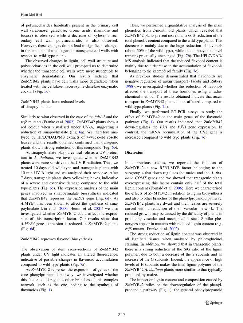
of polysaccharides habitually present in the primary cell
wall (arabinose, galactose, uronic acids, rhamnose and
fucose) is observed while a decrease of xylose, a sec-
ondary cell wall polysaccharide, is also observed.
However, these changes do not lead to significant changes
in the amounts of total sugars in transgenic cell walls with
respect to wild type plants.
The observed changes in lignin, cell wall structure and
polysaccharides in the cell wall prompted us to determine
whether the transgenic cell walls were more susceptible to
enzymatic degradability. Our results indicate that
ZmMYB42 plants have cell walls more degradable when
treated with the cellulase-macerozyme-driselase enzymatic
cocktail (Fig. 5c).
ZmMYB42 plants have reduced levels
of sinapoylmalate
Similarly to what observed in the case of the fah1-2 and the
ref8 mutants (Franke et al. 2002), ZmMYB42 plants show a
red colour when visualised under UV-A, suggesting a
reduction of sinapoylmalate (Fig. 6a). We therefore ana-
lysed by HPLC/DAD/MS extracts of 4-week-old rosette
leaves and the results obtained confirmed that transgenic
plants show a strong reduction of this compound (Fig. 6b).
As sinapoylmalate plays a central role as a UV-protec-
tant in A. thaliana, we investigated whether ZmMYB42
plants were more sensitive to the UV-B radiation. Thus, we
treated 10-days old wild type and transgenic plants with
10 min UV-B light and we analysed their response. After
7 days, transgenic plants show yellowing leaves, indicative
of a severe and extensive damage compared to the wild
type plants (Fig. 6c). The expression analysis of the main
genes involved in sinapoylmalate biosynthesis indicated
that ZmMYB42 represses the ALDH gene (Fig. 6d). As
AtMYB4 has been shown to affect the synthesis of sina-
poylmalate (Jin et al. 2000; Hemm et al. 2001) we also
investigated whether ZmMYB42 could affect the expres-
sion of this transcription factor. Our results show that
AtMYB4 gene expression is reduced in ZmMYB42 plants
(Fig. 6d).
ZmMYB42 represses flavonol biosynthesis
The observation of stem cross-sections of ZmMYB42
plants under UV light indicates an altered fluorescence,
indicative of possible changes in flavonoid accumulation
compared to wild type plants (Fig. 7a).
As ZmMYB42 represses the expression of genes of the
core phenylpropanoid pathway, we investigated whether
this factor could regulate other branches of this complex
network, such as the one leading to the synthesis of
flavonoids (Fig. 1).
Thus, we performed a quantitative analysis of the main
phenolics from 2-month old plants, which revealed that
ZmMYB42 plants present more than a 66% reduction of the
total phenolic content compared to the wild type plants. This
decrease is mainly due to the huge reduction of flavonols
(about 50% of the wild type), while the anthocyanins level
remains practically unchanged (Fig. 7b). The HPLC/DAD/
MS analysis indicated that the reduced flavonol content is
mainly due to a decrease in the accumulation of flavonols
belonging to the kaempferol family (Fig. 7c).
As previous studies demonstrated that flavonoids are
negative regulators of auxin transport (Jacobs and Rubery
1988), we investigated whether this reduction of flavonols
affected the transport of these hormones using a radio-
chemical method. The results obtained indicate that auxin
transport in ZmMYB42 plants is not affected compared to
wild type plants (Fig. 7d).
Finally, we performed RT-PCR assays to study the
effect of ZmMYB42 on the main genes of the flavonoid
pathway (Fig. 1). Our results indicated that ZmMYB42
down-regulates the F3H and F30H gene expression. In
contrast, the mRNA accumulation of the CHS gene is
increased compared to wild type plants (Fig. 7e).
Discussion
In a previous studies, we reported the isolation of
ZmMYB42, a new R2R3-MYB factor belonging to the
subgroup 4 that down-regulates the maize and the A. tha-
liana COMT genes and we showed that transgenic plants
overexpressing this factor contain only half of the total
lignin content (Fornale et al. 2006). Here we characterised
the effects of ZmMYB42 in relation to lignin biosynthesis
and also to other branches of the phenylpropanoid pathway.
ZmMYB42 plants are dwarf and their leaves are severely
curved with a reduction of their vascular network. The
reduced growth may be caused by the difficulty of plants in
producing vascular and mechanical tissues. Similar phe-
notypes appear in mutants with reduced lignin content (e.g.
ref8 mutant; Franke et al. 2002).
The strong reduction of lignin content was observed in
all lignified tissues when analysed by phloroglucinol
staining. In addition, we showed that in transgenic plants,
there is a strong reduction of the S/G ratio of the lignin
polymer, due to both a decrease of the S subunits and an
increase of the G subunits. Indeed, the appearance of high
levels of H subunits makes the final lignin polymer of the
ZmMYB42 A. thaliana plants more similar to that typically
produced by maize.
The impact on lignin content and composition caused by
ZmMYB42 relies on the downregulation of the phenyl-
propanoid pathway (Fig. 1); the general phenylpropanoid
Plant Mol Biol
123
247

genes (PAL, C4H, and 4CL), several genes involved in the
conversion from esters to aldehydes and alcohols (HCT and
CAD) and genes belonging to the branches leading to the
production of the three main monolignols (F5H and
COMT). In contrast, C3H, CCoAOMT and CCR genes are
not regulated by this transcription factor (Figs. 1, 4).
Together, and in accordance with previous observations
(Ralph et al. 2008) these results show once more the high
level of flexibility of the phenylpropanoid pathway.
Several reports have shown that reduction of CAD
(Sibout et al. 2003), F5H (Marita et al. 1999) and COMT
(Goujon et al. 2003; Do et al. 2007) results in a decrease of
the S units in the final lignin polymer. In agreement with
these results, the repression of F5H, COMT and CAD genes
produces a lignin polymer strongly depleted in S units. In
addition, the increased levels of ferulic acid are not
expected if this compound is produced by COMT, as this
gene is repressed in ZmMYB42 plants. Thus, this finding is
in line with other works indicating that COMT does not
catalyse the in vivo methylation of caffeic acid to produce
ferulic acid (Do et al. 2007). Together, the overall pattern
of repression of the lignin genes accounts for the reduced
capacity of transgenic plants to produce S units.
Cell walls of transgenic fibres show an altered structure
characterised by a higher number of pit-membranes and a
reduced thickness of secondary cell walls. The increase of
Wild type 35S::ZmMYB42
UV-A
35S::ZmMYB42
Wild type
10 min UV-BNot irradiated
(A)
(C)
ALDH
SGT
SMT
MYB42
Actin
MYB4
MYB42Wt(D)
20 21 22 23 24 25 26 27 28 29 30 31 32 33 34 35 36 37 38 39 40m
AU
-5
0
5
10
15
20
25
30
35
40
Retention time (min)
Wild type
ZmMYB42
Sinapoylmalate
Sinapoylmalate
(B)
Fig. 6 ZmMYB42 represses sinapoylmalate biosynthesis. a Pheno-
type of wt and transgenic plantlets exposed to UV light. b HPLC-
DAD analysis of sinapoylmalate content in leaves of wt and
transgenic plants. c Phenotype of 20-days old wt and transgenic
plantlets submitted to 10 min UV irradiation. d Relative expression of
the genes involved in sinapoylmalate synthesis and of AtMYB4 gene
Plant Mol Biol
123
248
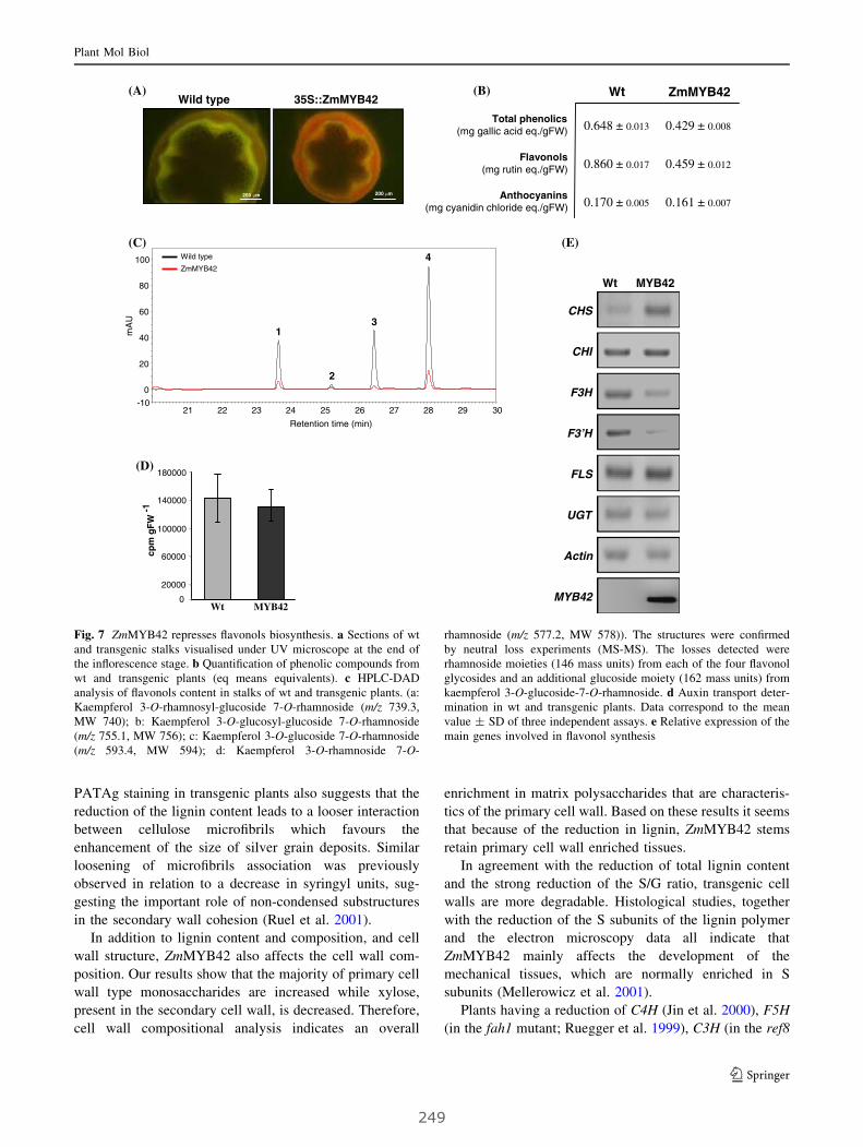
PATAg staining in transgenic plants also suggests that the
reduction of the lignin content leads to a looser interaction
between cellulose microfibrils which favours the
enhancement of the size of silver grain deposits. Similar
loosening of microfibrils association was previously
observed in relation to a decrease in syringyl units, sug-
gesting the important role of non-condensed substructures
in the secondary wall cohesion (Ruel et al. 2001).
In addition to lignin content and composition, and cell
wall structure, ZmMYB42 also affects the cell wall com-
position. Our results show that the majority of primary cell
wall type monosaccharides are increased while xylose,
present in the secondary cell wall, is decreased. Therefore,
cell wall compositional analysis indicates an overall
enrichment in matrix polysaccharides that are characteris-
tics of the primary cell wall. Based on these results it seems
that because of the reduction in lignin, ZmMYB42 stems
retain primary cell wall enriched tissues.
In agreement with the reduction of total lignin content
and the strong reduction of the S/G ratio, transgenic cell
walls are more degradable. Histological studies, together
with the reduction of the S subunits of the lignin polymer
and the electron microscopy data all indicate that
ZmMYB42 mainly affects the development of the
mechanical tissues, which are normally enriched in S
subunits (Mellerowicz et al. 2001).
Plants having a reduction of C4H (Jin et al. 2000), F5H
(in the fah1 mutant; Ruegger et al. 1999), C3H (in the ref8
(A) (B)
Wt MYB420
20000
60000
100000
140000
180000
cpm
gF
W-1
(D)
(C)
Retention time (min)
21 22 23 24 25 26 27 28 29 30
mA
U
-10
0
20
40
60
80
100
1
2
3
4Wild type
ZmMYB42
MYB42
MYB42Wt
CHS
CHI
FLS
UGT
F3H
F3’H
Actin
(E)
35S::ZmMYB42
200 m
Wild type
200 m
Wt ZmMYB42
Total phenolics(mg gallic acid eq./gFW)
Flavonols(mg rutin eq./gFW)
Anthocyanins(mg cyanidin chloride eq./gFW)
0.648 ± 0.013
0.860 ± 0.017
0.170 ± 0.005
0.429 ± 0.008
0.459 ± 0.012
0.161 ± 0.007
Fig. 7 ZmMYB42 represses flavonols biosynthesis. a Sections of wt
and transgenic stalks visualised under UV microscope at the end of
the inflorescence stage. b Quantification of phenolic compounds from
wt and transgenic plants (eq means equivalents). c HPLC-DAD
analysis of flavonols content in stalks of wt and transgenic plants. (a:
Kaempferol 3-O-rhamnosyl-glucoside 7-O-rhamnoside (m/z 739.3,
MW 740); b: Kaempferol 3-O-glucosyl-glucoside 7-O-rhamnoside
(m/z 755.1, MW 756); c: Kaempferol 3-O-glucoside 7-O-rhamnoside
(m/z 593.4, MW 594); d: Kaempferol 3-O-rhamnoside 7-O-
rhamnoside (m/z 577.2, MW 578)). The structures were confirmed
by neutral loss experiments (MS-MS). The losses detected were
rhamnoside moieties (146 mass units) from each of the four flavonol
glycosides and an additional glucoside moiety (162 mass units) from
kaempferol 3-O-glucoside-7-O-rhamnoside. d Auxin transport deter-
mination in wt and transgenic plants. Data correspond to the mean
value ± SD of three independent assays. e Relative expression of the
main genes involved in flavonol synthesis
Plant Mol Biol
123
249
mutant; Franke et al. 2002) and COMT gene expression (in
the Atomt1 mutant; Goujon et al. 2003; Do et al. 2007)
show a reduction in the synthesis of sinapoylmalate and
display red fluorescence when visualised under UV light.
Accordingly, ZmMYB42 represses C4H, F5H, and COMT
gene expression, and thus transgenic plants have a strong
reduction in sinapoylmalate as demonstrated by HPLC/
DAD/MS, the observation under UV light and the
increased sensitivity to UV-B irradiation. In addition,
ZmMYB42 also represses the expression of ALDH gene,
which is the first specific enzyme for the synthesis of si-
napoylmalate (Fig. 1). This result is in line with what
observed in the Arabidopsis ref1 mutant in which the
mutation of the ALDH gene results in a strong reduction of
the sinapate esters (Nair et al. 2004).
The involvement of C4H repression in the synthesis of
sinapoylmalate was described through the study of AtMYB4
gene (Jin et al. 2000), a repressor of sinapoylmalate syn-
thesis. In ZmMYB42 plants, the expression of AtMYB4
gene is downregulated. This behaviour could be the con-
sequence of a homeostatic response by which the
transgenic plants tend to counteract the effect produced by
the overexpression of ZmMYB42.
As mentioned above, ZmMYB42 also represses the
synthesis of PAL1, C4H, and 4CL genes. These genes are
involved in the core-phenylpropanoid pathway and cata-
lyse the synthesis of hydroxycinnamic acid intermediates,
which are precursors for the route-specific pathways lead-
ing to the synthesis of a wide range of secondary
metabolites such as flavonoids (Fig. 1). Transgenic plants
overexpressing ZmMYB42 have stems in which the cortex
region appears orange under UV light, suggesting an
alteration in the synthesis of some flavonoids. A deeper
characterisation revealed that transgenic plants have
decreased levels of total soluble phenolics and in particu-
lar, a strong reduction of flavonols belonging to the family
of kaempferol. Although ZmMYB42 induces the expres-
sion of CHS gene, the entry point to flavonoid biosynthesis,
no increased levels of naringenin chalcone were detected
(data not shown). However, ZmMYB42 represses the
expression of F3H and F30H genes, which could explain
the reduced levels of flavonols in transgenic plants.
Therefore, in addition to the strong reduction of sina-
poylmalate, the fact that transgenic plants are more
sensitive to UV irradiation could be, at least partially, also
caused by the reduction of the flavonol content as described
in the case of the transparent testa 4 (tt4) mutant (Li et al.
1993).
It has been reported that flavonoids are negative regu-
lators of the auxin transport (Brown et al. 2001; Besseau
et al. 2007). In our case, although ZmMYB42 have reduced
levels of flavonoids, the auxin transport is not induced in
transgenic plants, suggesting that a 50% reduction of
flavonols is not sufficient to increase the levels of auxin
transport.
Together, these results show that ZmMYB42 is a
general repressor of the phenylpropanoid pathway
affecting cell wall structure and composition when over-
expressed in A. thaliana. The reduction of the total lignin
content, the strong reduction of its S/G ratio, and the
increased cell wall degradability, makes this factor a good
candidate to control lignin metabolism in maize for bio-
technological applications.
Acknowledgements This work received financial support from the
CONSOLIDER-INGENIO programme (CSD2007-00036) from the
Spanish Ministerio de Ciencia e Innovacion. F.M. Sonbol was
financed by the European Project INCO-II (ICA4-CT-2000-30017),
by the CRAG laboratory, and by the ‘‘Instituto de Cooperacion con el
Mundo Arabe, Agencia Espanola de Cooperacion Internacional’’. S.
Fornale was financed by a post-doctoral grant from the ‘‘Generalitat
de Catalunya’’ (2003PIV-A-00033) and by an I3P contract from the
‘‘Consejo Superior de Investigaciones Cientıficas’’. D. Caparros-Ruiz
was financed by the Spanish ‘‘Ministerio de Educacion y Ciencia’’
(‘‘Ramon y Cajal’’ Program). This work was carried out within the
framework of the ‘‘Xarxa de Referencia de Biotecnologia’’ (XarBa)
from the Autonomous Government of Catalonia. We are indebted to
Dr. Jean-Paul Joseleau (CERMAV-CNRS Grenoble) and Dr. Cathie
R. Martin (John Innes Center) for critical reading of the manuscript.
We also thank the sequencing and greenhouse teams of IBMB-CSIC
as well as Dr. R.S. Joshi for the English revision of the manuscript.
References
Albersheim P, Nevins DS, English PD (1967) A method for the
analysis of sugars in plant cell wall polysaccharides by gas liquid
chromatography. Carbohydr Res 5:340–345. doi:10.1016/S0008-
6215(00)80510-8
Arnaud D, Dejardin A, Leple JC, Lesage-Descauses MC, Pilate G
(2007) Genome-wide analysis of LIM gene family in Populustrichocarpa, Arabidopsis thaliana, and Oryza sativa. DNA Res
14:103–116. doi:10.1093/dnares/dsm013
Besseau S, Hoffmann L, Geoffroy P, Lapierre C, Pollet B, Legrand M
(2007) Flavonoid accumulation in Arabidopsis repressed in
lignin synthesis affects auxin transport and plant growth. Plant
Cell 19:148–162. doi:10.1105/tpc.106.044495
Blumenkrantz N, Asboe-Hansen G (1973) New method for quanti-
tative determination of uronic acids. Anal Biochem 54:484–489.
doi:10.1016/0003-2697(73)90377-1
Boerjan W, Ralph J, Baucher M (2003) Lignin biosynthesis. Annu
Rev Plant Biol 54:519–546. doi:10.1146/annurev.arplant.54.
031902.134938
Bomal C, Bedon F, Caron S, Mansfield SD, Levasseur C, Cooke JE,
Blais S, Tremblay L, Morency MJ, Pavy N, Grima-Pettenati J,
Seguin A, Mackay J (2008) Involvement of Pinus taeda MYB1
and MYB8 in phenylpropanoid metabolism and secondary cell
wall biogenesis: a comparative in planta analysis. J Exp Bot
59:3925–3939. doi:10.1093/jxb/ern234
Bonke M, Thitamadee S, Mahonen AP, Hauser MT, Helariutta Y
(2003) APL regulates vascular tissue identity in Arabidopsis.
Nature 426:181–186. doi:10.1038/nature02100
Brown DE, Rashotte AM, Murphy AS, Normanly J, Tague BW, Peer
WA, Taiz L, Muday GK (2001) Flavonoids act as negative
Plant Mol Biol
123
250

regulators of auxin transport in vivo in Arabidopsis. Plant
Physiol 126:524–535. doi:10.1104/pp.126.2.524
Carlsbecker A, Helariutta Y (2005) Phloem and xylem specification:
pieces of the puzzle emerge. Curr Opin Plant Biol 8:512–517.
doi:10.1016/j.pbi.2005.07.001
Cliff MA, King MC, Schlosser J (2007) Anthocyanin, phenolic
composition, colour measurement and sensory analysis of BC
commercial red wines. Food Res Int 40:92–100. doi:10.1016/
j.foodres.2006.08.002
Cone KC, Cocciolone SM, Burr FA, Burr B (1993) Maize anthocy-
anin regulatory gene pI is a duplicate of c1 that functions in the
plant. Plant Cell 5:1795–1805
Day A, Ruel K, Neutelings G, Cronier D, David H, Hawkins S,
Chabbert B (2005) Lignification in the flax stem: evidence for an
unusual lignin in bast fibers. Planta 222:234–245. doi:10.1007/
s00425-005-1537-1
Deluc L, Barrieu F, Marchive C, Lauvergeat V, Decendit A, Richard
T, Carde JP, Merillon JM, Hamdi S (2006) Characterization of a
grapevine R2R3-MYB transcription factor that regulates the
phenylpropanoid pathway. Plant Physiol 140:499–511. doi:
10.1104/pp.105.067231
Dische J (1962) Color reactions of carbohydrates. In: Whistler RL,
Wolfrom ML (eds) Methods in carbohydrate chemistry, vol 1.
Academic Press, New York, pp 475–514
Do CT, Pollet B, Thevenin J, Sibout R, Denoue D, Barriere Y,
Lapierre C, Jouanin L (2007) Both caffeoyl Coenzyme A 3-O-
methyltransferase 1 and caffeic acid O-methyltransferase 1 are
involved in redundant functions for lignin, flavonoids and
sinapoyl malate biosynthesis in Arabidopsis. Planta 226:1117–
1129. doi:10.1007/s00425-007-0558-3
Dubois M, Giles KA, Hamilton JK, Rebers PA, Smith F (1956)
Colorimetric method of determination of sugars and related
substances. Anal Chem 28:350–356. doi:10.1021/ac60111a017
Encina A, Sevillano JM, Acebes JL, Alvarez J (2002) Cell wall
modifications of bean (Phaseolus vulgaris) cell suspensions
during habituation and dehabituation to dichlobenil. Physiol
Plant 114:182–191. doi:10.1034/j.1399-3054.2002.1140204.x
Fornale S, Sonbol FM, Maes T, Capellades M, Puigdomenech P,
Rigau J, Caparros-Ruiz D (2006) Down-regulation of the maize
and Arabidopsis thaliana caffeic acid O-methyl-transferase
genes by two new maize R2R3-MYB transcription factors. Plant
Mol Biol 62:809–823. doi:10.1007/s11103-006-9058-2
Franke R, Humphreys JM, Hemm MR, Denault JW, Ruegger MO,
Cusumano JC, Chapple C (2002) The Arabidopsis REF8 gene
encodes the 3-hydroxylase of phenylpropanoid metabolism.
Plant J 30:33–45. doi:10.1046/j.1365-313X.2002.01266.x
Franken P, Schrell S, Peterson PA, Saedler H, Wienand U (1989)
Molecular analysis of protein domain function encoded by the
myb-homologous maize genes C1, Zm 1 and Zm 38. Plant J
6:21–30. doi:10.1046/j.1365-313X.1994.6010021.x
Galis I, Simek P, Narisawa T, Sasaki M, Horiguchi T, Fukuda H,
Matsuoka K (2006) A novel R2R3-MYB transcription factor
NtMYBJS1 is a methyl Jasmonate-dependent regulator of
phenylpropanoid-conjugate biosynthesis in tobacco. Plant J
46:573–592. doi:10.1111/j.1365-313X.2006.02719.x
Goicoechea M, Lacombe E, Legay S, Mihaljevic S, Rech P, Jauneau
A, Lapierre C, Pollet B, Verhaegen D, Chaubet-Gigot N, Grima-
Pettenati G (2005) EgMYB2, a new transcriptional activator
from Eucalyptus xylem, regulates secondary cell wall formation
and lignin biosynthesis. Plant J 43:553–567. doi:10.1111/
j.1365-313X.2005.02480.x
Goujon T, Sibout R, Pollet B, Maba B, Nussaume L, Bechtold N, Lu
F, Ralph J, Mila I, Barriere Y, Lapierre C, Jouanin L (2003) A
new Arabidopsis thaliana mutant deficient in the expression of
O-methyltransferase impacts lignins and sinapoyl esters. Plant
Mol Biol 51:973–989. doi:10.1023/A:1023022825098
Grotewold E, Athma P, Peterson T (1991) Alternatively spliced
products of the maize P gene encode proteins with homology to
the DNA-binding domain of myb-like transcription factors. Proc
Natl Acad Sci USA 88:4587–4591. doi:10.1073/pnas.88.11.4587
Grotewold E, Drummond BJ, Bowen B, Peterson T (1994) The myb-
homologous P gene controls phlobaphene pigmentation in maize
floral organs by directly activating a flavonoid biosynthetic gene
subset. Cell 76:543–553. doi:10.1016/0092-8674(94)90117-1
Heddges JI, Mann DC (1979) The characterization of plant tissues by
their lignin oxidation products. Geochim Cosmochim Acta
43:1803–1807. doi:10.1016/0016-7037(79)90028-0
Heine GF, Malik V, Dias AP, Grotewold E (2007) Expression and
molecular characterization of ZmMYB-IF35 and related R2R3-
MYB transcription factors. Mol Biotechnol 37:155–164. doi:
10.1007/s12033-007-0061-x
Hemm MR, Herrmann KM, Chapple C (2001) AtMYB4: a transcrip-
tion factor general in the battle against UV. Trends Plant Sci
6:135–136. doi:10.1016/S1360-1385(01)01915-X
Jacobs M, Rubery PH (1988) Naturally occurring auxin transport
regulators. Science 241:346–349. doi:10.1126/science.241.
4863.346
Jia L, Clegg MT, Jiang T (2004) Evolutionary dynamics of the DNA-
binding domains in putative R2R3-MYB genes identified from
rice subspecies indica and japonica genomes. Plant Physiol
134:575–585. doi:10.1104/pp.103.027201
Jin H, Cominelli E, Bailey P, Parr A, Mehrtens F, Jones J, Tonelli C,
Weisshaar B, Martin C (2000) Transcriptional repression by
AtMYB4 controls production of UV-protecting sunscreens in
Arabidopsis. EMBO J 15:6150–6161. doi:10.1093/emboj/19.
22.6150
Ju C, Zhang F, Gao Y, Zhang W, Yan J, Dai J, Li J (2006) Cloning,
chromosome mapping and expression analysis of an R2R3-MYBgene under-expressed in maize hybrid. Mol Biol Rep 33:103–
110. doi:10.1007/s11033-006-0016-5
Karpinska B, Karlsson M, Srivastava M, Stenberg A, Schrader J,
Sterky F, Bhalerao R, Wingsle G (2004) MYB transcription
factors are differentially expressed and regulated during second-
ary vascular tissue development in hybrid aspen. Plant Mol Biol
56:255–270. doi:10.1007/s11103-004-3354-5
Kawaoka A, Ebinuma H (2001) Transcriptional control of lignin
biosynthesis by tobacco LIM protein. Phytochemistry 57:1149–
1157. doi:10.1016/S0031-9422(01)00054-1
Kirk TK, Obst JR (1988) Lignin determination. Methods Enzymol
161:87–100. doi:10.1016/0076-6879(88)61014-7
Kogel I, Bochter R (1985) Characterization of lignin in forest humus
layers by high-performance liquid chromatography of cupric
oxide oxidation products. Soil Biol Biochem 17:637–640. doi:
10.1016/0038-0717(85)90040-9
Legay S, Lacombe E, Goicoechea M, Briere C, Seguin A, Mackay J,
Grima-Pettenati J (2007) Molecular characterization of Eg-
MYB1, a putative transcriptional repressor of the lignin
biosynthetic pathway. Plant Sci 173:542–549. doi:10.1016/
j.plantsci.2007.08.007
Lewis NG, Yamamoto E (1990) Lignin: occurrence, biogenesis and
biodegradation. Annu Rev Plant Physiol Plant Mol Biol 41:455–
496. doi:10.1146/annurev.pp.41.060190.002323
Li J, Ou-Lee TM, Raba R, Amundson RG, Last RL (1993)
Arabidopsis flavonoid mutants are hypersensitive to UV-B
irradiation. Plant Cell 5:171–179
Li X, Weng JK, Chapple C (2008) Improvement of biomass through
lignin modification. Plant J 54:569–581. doi:10.1111/j.1365-
313X.2008.03457.x
Liang YK, Dubos C, Dodd IC, Holroyd GH, Hetherington AM,
Campbell MM (2005) AtMYB61, an R2R3-MYB transcription
factor controlling stomatal aperture in Arabidopsis thaliana.
Curr Biol 15:1201–1206. doi:10.1016/j.cub.2005.06.041
Plant Mol Biol
123
251
Marita JM, Ralph J, Hatfield RD, Chapple C (1999) NMR charac-
terization of lignins in Arabidopsis altered in the activity of
ferulate 5-hydroxylase. Proc Natl Acad Sci USA 96:12328–
12332. doi:10.1073/pnas.96.22.12328
Marocco A, Wissenbach M, Becker D, Paz-Ares J, Saedler H,
Salamini F, Rohde W (1989) Multiple genes are transcribed in
Hordeum vulgare and Zea mays that carry the DNA binding
domain of the myb oncoproteins. Mol Gen Genet 216:183–187.
doi:10.1007/BF00334354
Martin C, Paz-Ares J (1997) MYB transcription factors in plants.
Trends Genet 13:67–73. doi:10.1016/S0168-9525(96)10049-4
Mele G, Ori N, Sato Y, Hake S (2003) The knotted1-like homeobox
gene BREVIPEDICELLUS regulates cell differentiation by
modulating metabolic pathways. Genes Dev 17:2088–2093. doi:
10.1101/gad.1120003
Mellerowicz EJ, Baucher M, Sundberg B, Boerjan W (2001)
Unravelling cell wall formation in the woody dicot stem. Plant
Mol Biol 47:239–274. doi:10.1023/A:1010699919325
Murashige T, Skoog F (1962) A revised medium for rapid growth and
bioassays with tobacco tissue cultures. Physiol Plant 15:473–
497. doi:10.1111/j.1399-3054.1962.tb08052.x
Nair RB, Bastress KL, Ruegger MO, Denault JW, Chapple C (2004)
The Arabidopsis thaliana REDUCED EPIDERMAL FLUORES-
CENCE1 gene encodes an aldehyde dehydrogenase involved in
ferulic acid and sinapic acid biosynthesis. Plant Cell 16:544–
554. doi:10.1105/tpc.017509
Newman LJ, Perazza DE, Juda L, Campbell MM (2004) Involvement
of the R2R3-MYB, AtMYB61, in the ectopic lignification and
dark-photomorphogenic components of the det3 mutant pheno-
type. Plant J 37:239–250
Patzlaff A, Newman LJ, Dubos C, Whetten RW, Smith C, McInnis S,
Bevan MW, Sederoff RR, Campbell MM (2003a) Characterisa-
tion of Pt MYB1, an R2R3-MYB from pine xylem. Plant Mol
Biol 53:597–608. doi:10.1023/B:PLAN.0000019066.07933.d6
Patzlaff A, McInnis S, Courtenay A, Surman C, Newman LJ, Smith
C, Bevan MW, Mansfield S, Whetten RW, Sederoff RR,
Campbell MM (2003b) Characterisation of a pine MYB that
regulates lignification. Plant J 36:743–754. doi:10.1046/j.1365-
313X.2003.01916.x
Paz-Ares J, Ghosal D, Wienand U, Peterson PA, Saedler H (1987)
The regulatory c1 locus of Zea mays encodes a protein with
homology to myb proto-oncogene products and with structural
similarities to transcriptional activators. EMBO J 6:3553–3558
Perez-Jimenez J, Saura-Calixto F (2008) Anti-oxidant capacity of
dietary polyphenols determined by ABTS assay: a kinetic
expression of the results. Int J Food Sci Technol 43:185–191
Pilu R, Piazza P, Petroni K, Ronchi A, Martin C, Tonelli C (2003) pl-
bol3, a complex allele of the anthocyanin regulatory pl1 locus
that arose in a naturally occurring maize population. Plant J
36:510–521. doi:10.1046/j.1365-313X.2003.01898.x
Preston J, Wheeler J, Heazlewood J, Li SF, Parish RW (2004)
AtMYB32 is required for normal pollen development in
Arabidopsis thaliana. Plant J 40:979–995. doi:10.1111/j.1365-
313X.2004.02280.x
Rabinowicz PD, Braun EL, Wolfe AD, Bowen B, Grotewold E (1999)
Maize R2R3 Myb genes: sequence analysis reveals amplification
in the higher plants. Genetics 153:427–444
Ralph J, Kim H, Lu F, Grabber JH, Leple JC, Berrio-Sierra J,
Derikvand MM, Jouanin L, Boerjan W, Lapierre C (2008)
Identification of the structure and origin of a thioacidolysis
marker compound for ferulic acid incorporation into angiosperm
lignins (and an indicator for cinnamoyl CoA reductase
deficiency). Plant J 53:368–379. doi:10.1111/j.1365-313X.2007.
03345.x
Rasband WS (2007) ImageJ, U. S. National Institutes of Health,
Bethesda, Maryland, USA. http://rsb.info.nih.gov/ij/, 1997–2004
Romero I, Fuertes A, Benito MJ, Malpica JM, Leyva A, Paz-Ares J
(1998) More than 80 R2R3-MYB regulatory genes in the
genome of Arabidopsis thaliana. Plant J 14:273–284. doi:
10.1046/j.1365-313X.1998.00113.x
Ruegger M, Meyer K, Cusumano JC, Chapple C (1999) Regulation of
ferulate-5-hydroxylase expression in Arabidopsis in the context
of sinapate ester biosynthesis. Plant Physiol 119:101–110. doi:
10.1104/pp.119.1.101
Ruel K, Comtat J, Barnoud F (1977) Localisation histologique et
ultrastructurale des xylanes dans les parois primaires des tissus
d’ Arundo donax. C R Acad Sci Gen 284:1421–1424
Ruel K, Chabannes M, Boudet AM, Legrand M, Joseleau JP (2001)
Reassessment of qualitative changes in lignification of transgenic
tobacco plants and their impact on cell wall assembly. Phyto-
chemistry 57:1–9. doi:10.1016/S0031-9422(01)00118-2
Saeman JF, Moore WE, Millet MA (1963) Sugar units present. In:
Whistler RL (ed) Methods in carbohydrate chemistry, vol 3.
Academic Press, New York, pp 54–69
Sibout R, Eudes A, Pollet B, Goujon T, Mila I, Granier F, Seguin A,
Lapierre C, Jouanin L (2003) Expression pattern of two paralogs
encoding cinnamyl alcohol dehydrogenases in Arabidopsis.
Isolation and characterization of the corresponding mutants.
Plant Physiol 132:848–860. doi:10.1104/pp.103.021048
Sticklen MB (2008) Plant genetic engineering for biofuel production:
towards affordable cellulosic ethanol. Nat Rev Genet 9:433–443.
doi:10.1038/nrg2336
Stracke R, Werber M, Weisshaar B (2001) The R2R3-MYB gene
family in Arabidopsis thaliana. Curr Opin Plant Biol 4:447–456.
doi:10.1016/S1369-5266(00)00199-0
Tamagnone L, Merida A, Parr A, Mackay S, Culianez-Macia FA,
Roberts K, Martin C (1998a) The AmMYB308 and AmMYB330
transcription factors from antirrhinum regulate phenylpropanoid
and lignin biosynthesis in transgenic tobacco. Plant Cell 10:135–
154
Tamagnone L, Merida A, Stacey N, Plaskitt K, Parr A, Chang CF,
Lynn D, Dow JM, Roberts K, Martin C (1998b) Inhibition of
phenolic acid metabolism results in precocious cell death and
altered cell morphology in leaves of transgenic tobacco plants.
Plant Cell 10:1801–1816
Torney F, Moeller L, Scarpa A, Wang K (2007) Genetic engineering
approaches to improve bioethanol production from maize. Curr
Opin Biotechnol 18:193–199. doi:10.1016/j.copbio.2007.03.006
Updegraff DM (1969) Semi-micro determination of cellulose in
biological materials. Anal Biochem 32:420–424. doi:10.1016/
S0003-2697(69)80009-6
Vanholme R, Morreel K, Ralph J, Boerjan W (2008) Lignin
engineering. Curr Opin Plant Biol 11:1–8. doi:10.1016/j.pbi.
2008.03.005
Yanhui C, Xiaoyuan Y, Kun H, Meihua L, Jigang L, Zhaofeng G,
Zhiqiang L, Yunfei Z, Xiaoxiao W, Xiaoming Q, Yunping S, Li
Z, Xiaohui D, Jingchu L, Xing-Wang D, Zhangliang C, Hongya
G, Li-Jia Q (2006) The MYB transcription factor superfamily of
Arabidopsis: expression analysis and phylogenetic comparison
with the rice MYB family. Plant Mol Biol 60:107–124. doi:
10.1007/s11103-005-2910-y
Zhong R, Ye ZH (2007) Regulation of cell wall biosynthesis. Curr
Opin Plant Biol 10:564–572. doi:10.1016/j.pbi.2007.09.001
Plant Mol Biol
123
252
8.2 Material suplementario

8.2.1 Material suplementario de la publicación 1
(Publicación mostrada en el apartado 4.1.1)


8. Anexos
257
a
b
RP-HPLC chromatograms, column µRPC C2/C18 SC 2.1/10 (100 x 2.1mmi.d). Elution : [A] 0.10% (v/v) aqueous TFA, [B] 0.08 % (v/v) TFA in water /CH3CN 1:4. Flow rate 200µL/min. Detection at 214nm. a) Load 5 µg crude fraction, gradient elution 8 to 23 % [B] over 38 min; b) Load 2.5 µg fraction after thioacidolysis, gradient elution 8 to 23 % [B] over 45 min.

8.2 Material suplementario (publicación 1; apartado 4.1.1, ver pág. 73)
258
a
b
RP-HPLC chromatograms, column µRPC C2/C18 SC 2.1/10 (100 x 2.1mmi.d). Elution : [A] 0.10% (v/v) aqueous TFA, [B] 0.08 % (v/v) TFA in water /CH3CN 1:4. Flow rate 200µL/min. Detection at 214nm. a) Load 5 µg crude fraction, gradient elution 8 to 23 % [B] over 38 min; b) Load 2.5 µg fraction after thioacidolysis, gradient elution 8 to 23 % [B] over 45 min.

8. Anexos
259
a
b
RP-HPLC chromatograms, column µRPC C2/C18 SC 2.1/10 (100 x 2.1mmi.d). Elution : [A] 0.10% (v/v) aqueous TFA, [B] 0.08 % (v/v) TFA in water /CH3CN 1:4. Flow rate 200µL/min. Detection at 214nm. a) Load 5 µg crude fraction, gradient elution 8 to 23 % [B] over 38 min; b) Load 2.5 µg fraction after thioacidolysis, gradient elution 8 to 23 % [B] over 45 min.
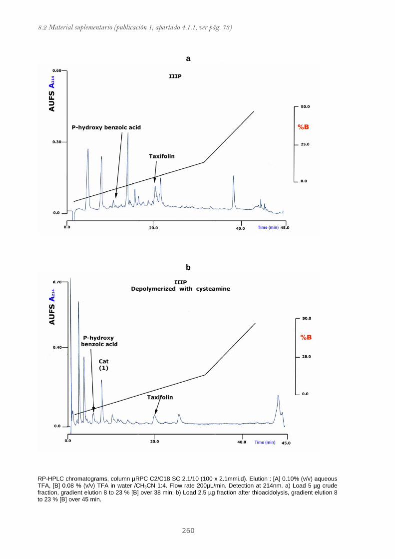
8.2 Material suplementario (publicación 1; apartado 4.1.1, ver pág. 73)
260
a
b
RP-HPLC chromatograms, column µRPC C2/C18 SC 2.1/10 (100 x 2.1mmi.d). Elution : [A] 0.10% (v/v) aqueous TFA, [B] 0.08 % (v/v) TFA in water /CH3CN 1:4. Flow rate 200µL/min. Detection at 214nm. a) Load 5 µg crude fraction, gradient elution 8 to 23 % [B] over 38 min; b) Load 2.5 µg fraction after thioacidolysis, gradient elution 8 to 23 % [B] over 45 min.

8. Anexos
261
a
b
RP-HPLC chromatograms, column µRPC C2/C18 SC 2.1/10 (100 x 2.1mmi.d). Elution : [A] 0.10% (v/v) aqueous TFA, [B] 0.08 % (v/v) TFA in water /CH3CN 1:4. Flow rate 200µL/min. Detection at 214nm. a) Load 5 µg crude fraction, gradient elution 8 to 23 % [B] over 38 min; b) Load 2.5 µg fraction after thioacidolysis, gradient elution 8 to 23 % [B] over 45 min.
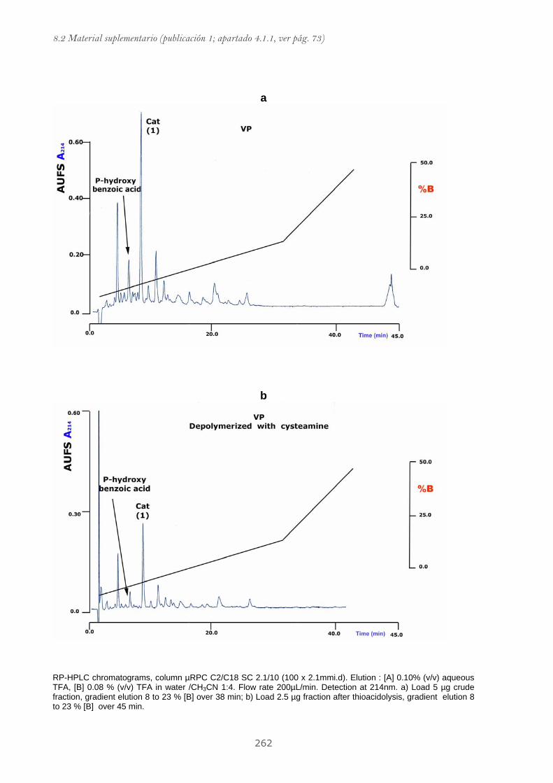
8.2 Material suplementario (publicación 1; apartado 4.1.1, ver pág. 73)
262
a
b
RP-HPLC chromatograms, column µRPC C2/C18 SC 2.1/10 (100 x 2.1mmi.d). Elution : [A] 0.10% (v/v) aqueous TFA, [B] 0.08 % (v/v) TFA in water /CH3CN 1:4. Flow rate 200µL/min. Detection at 214nm. a) Load 5 µg crude fraction, gradient elution 8 to 23 % [B] over 38 min; b) Load 2.5 µg fraction after thioacidolysis, gradient elution 8 to 23 % [B] over 45 min.
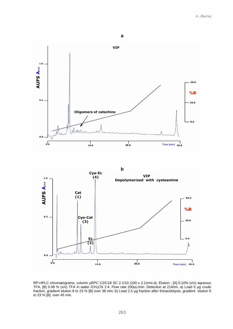
8. Anexos
263
a
b
RP-HPLC chromatograms, column µRPC C2/C18 SC 2.1/10 (100 x 2.1mmi.d). Elution : [A] 0.10% (v/v) aqueous TFA, [B] 0.08 % (v/v) TFA in water /CH3CN 1:4. Flow rate 200µL/min. Detection at 214nm. a) Load 5 µg crude fraction, gradient elution 8 to 23 % [B] over 38 min; b) Load 2.5 µg fraction after thioacidolysis, gradient elution 8 to 23 % [B] over 45 min.
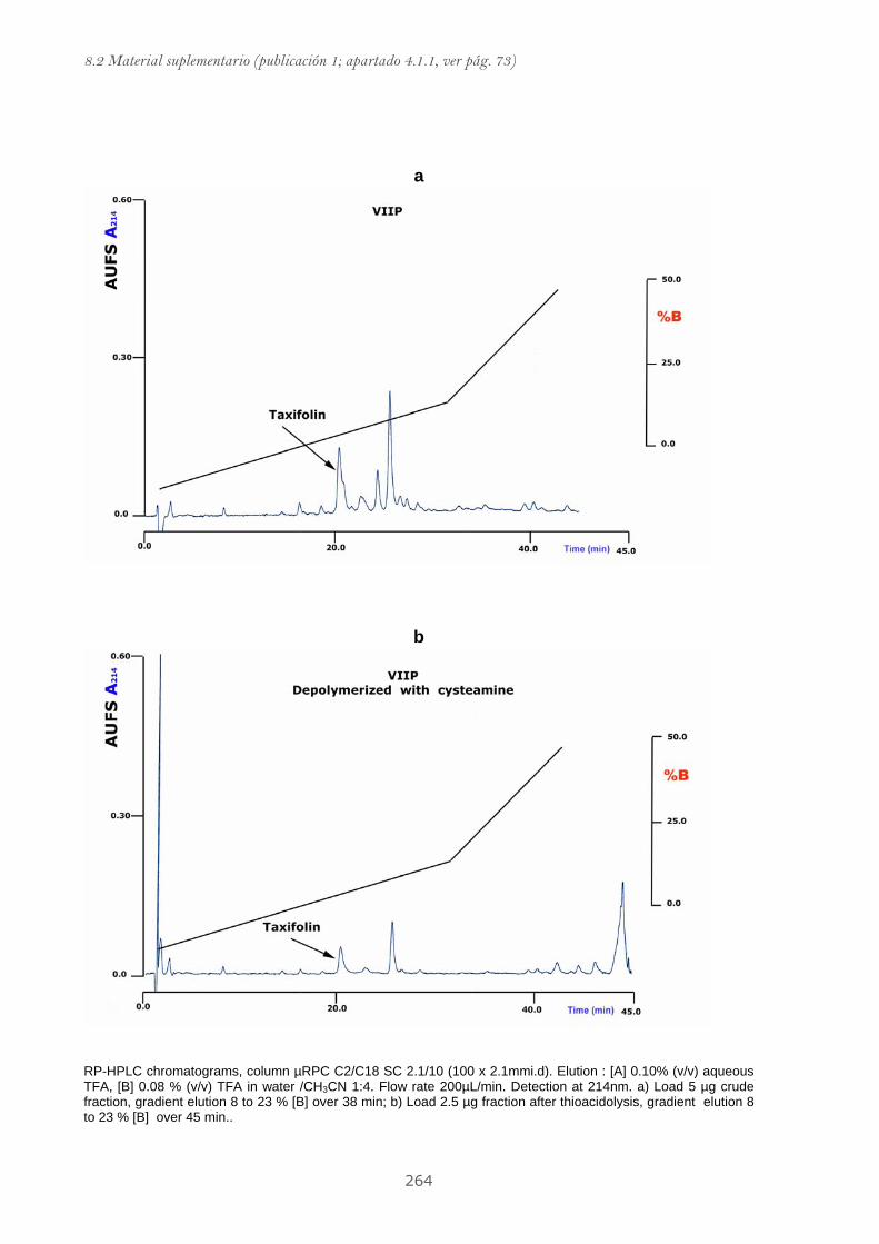
8.2 Material suplementario (publicación 1; apartado 4.1.1, ver pág. 73)
264
a
b
RP-HPLC chromatograms, column µRPC C2/C18 SC 2.1/10 (100 x 2.1mmi.d). Elution : [A] 0.10% (v/v) aqueous TFA, [B] 0.08 % (v/v) TFA in water /CH3CN 1:4. Flow rate 200µL/min. Detection at 214nm. a) Load 5 µg crude fraction, gradient elution 8 to 23 % [B] over 38 min; b) Load 2.5 µg fraction after thioacidolysis, gradient elution 8 to 23 % [B] over 45 min..

8. Anexos
265
a
b
RP-HPLC chromatograms, column µRPC C2/C18 SC 2.1/10 (100 x 2.1mmi.d). Elution : [A] 0.10% (v/v) aqueous TFA, [B] 0.08 % (v/v) TFA in water /CH3CN 1:4. Flow rate 200µL/min. Detection at 214nm. a) Load 5 µg crude fraction, gradient elution 8 to 23 % [B] over 38 min; b) Load 2.5 µg fraction after thioacidolysis, gradient elution 8 to 23 % [B] over 45 min..

8.2 Material suplementario (publicación 1; apartado 4.1.1, ver pág. 73)
266
a
b
RP-HPLC chromatograms, column µRPC C2/C18 SC 2.1/10 (100 x 2.1mmi.d). Elution : [A] 0.10% (v/v) aqueous TFA, [B] 0.08 % (v/v) TFA in water /CH3CN 1:4. Flow rate 200µL/min. Detection at 214nm. a) Load 5 µg crude fraction, gradient elution 8 to 23 % [B] over 38 min; b) Load 2.5 µg fraction after thioacidolysis, gradient elution 8 to 23 % [B] over 45 min.
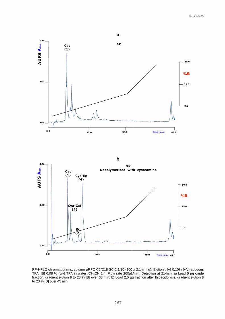
8. Anexos
267
a
b
RP-HPLC chromatograms, column µRPC C2/C18 SC 2.1/10 (100 x 2.1mmi.d). Elution : [A] 0.10% (v/v) aqueous TFA, [B] 0.08 % (v/v) TFA in water /CH3CN 1:4. Flow rate 200µL/min. Detection at 214nm. a) Load 5 µg crude fraction, gradient elution 8 to 23 % [B] over 38 min; b) Load 2.5 µg fraction after thioacidolysis, gradient elution 8 to 23 % [B] over 45 min.
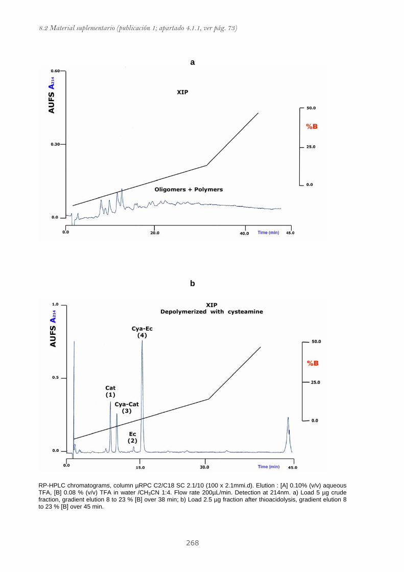
8.2 Material suplementario (publicación 1; apartado 4.1.1, ver pág. 73)
268
a
b
RP-HPLC chromatograms, column µRPC C2/C18 SC 2.1/10 (100 x 2.1mmi.d). Elution : [A] 0.10% (v/v) aqueous TFA, [B] 0.08 % (v/v) TFA in water /CH3CN 1:4. Flow rate 200µL/min. Detection at 214nm. a) Load 5 µg crude fraction, gradient elution 8 to 23 % [B] over 38 min; b) Load 2.5 µg fraction after thioacidolysis, gradient elution 8 to 23 % [B] over 45 min.

8. Anexos
269
OWP
0 1 2 3 40
25
50
75
100Blank100µg/mL50µg/mL10µg/mL3µg/mL1µg/mL
oxidation time (days)
mm
oles
lino
leat
ehy
drop
erox
ides
/kg
oil
IVP
0 1 2 3 40
25
50
75
100Blank50µg/mL10µg/mL2µg/mL
oxidation time (days)
mm
oles
lino
leat
ehy
drop
erox
ides
/kg
oil
VIIIP
0 1 2 3 40
25
50
75
100 Blank50µg/mL10µg/mL2µg/mL
mm
oles
lino
leat
ehy
drop
erox
ides
/kg
oil
oxidation time (days)
IXP
0 1 2 3 40
25
50
75
100Blank50µg/mL10µg/mL2µg/mL
oxidation time (days)
mm
oles
lino
leat
ehy
drop
erox
ides
/kg
oil
XP
0 1 2 3 40
25
50
75
100Blank50µg/mL10µg/mL2µg/mL
oxidation time (days)
mm
oles
lino
leat
ehy
drop
erox
ides
/kg
oil
XIP
0 1 2 3 40
25
50
75
100Blank50µg/mL10µg/mL2µg/mL
oxidation time (days)
mm
oles
lino
leat
ehy
drop
erox
ides
/kg
oil
Corn oil air oxidation in an oil-in-water emulsion in the presence of pine bark polyphenolic fractions.
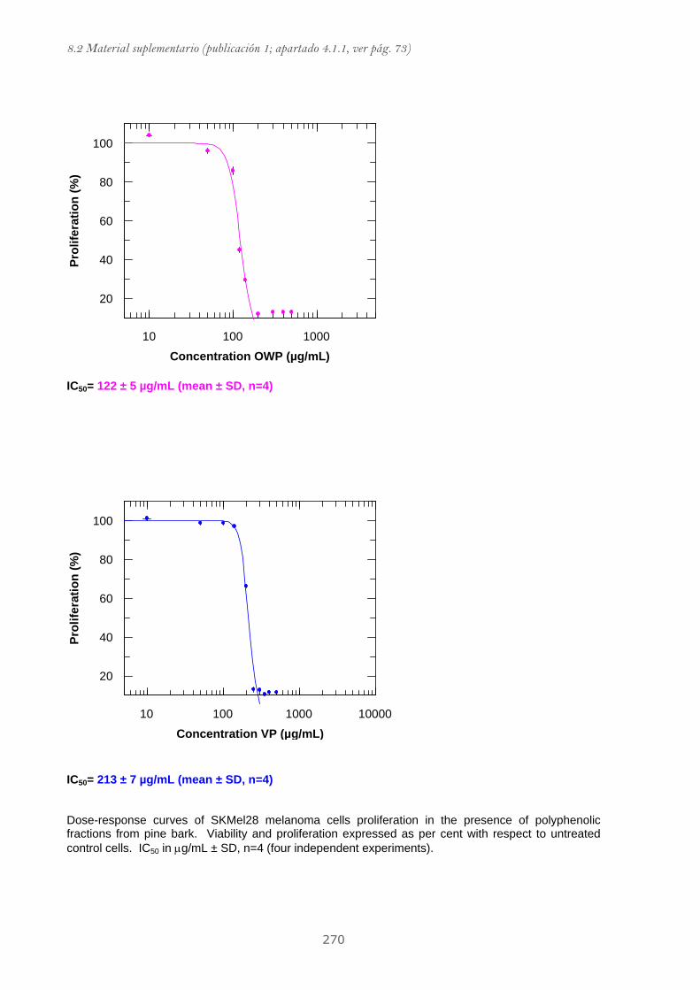
8.2 Material suplementario (publicación 1; apartado 4.1.1, ver pág. 73)
270
100010010
100
80
60
40
20
Concentration OWP (µg/mL)
Prol
ifera
tion
(%)
IC50= 122 ± 5 µg/mL (mean ± SD, n=4)
10000100010010
100
80
60
40
20
Concentration VP (µg/mL)
Prol
ifera
tion
(%)
IC50= 213 ± 7 µg/mL (mean ± SD, n=4) Dose-response curves of SKMel28 melanoma cells proliferation in the presence of polyphenolic fractions from pine bark. Viability and proliferation expressed as per cent with respect to untreated control cells. IC50 in μg/mL ± SD, n=4 (four independent experiments).
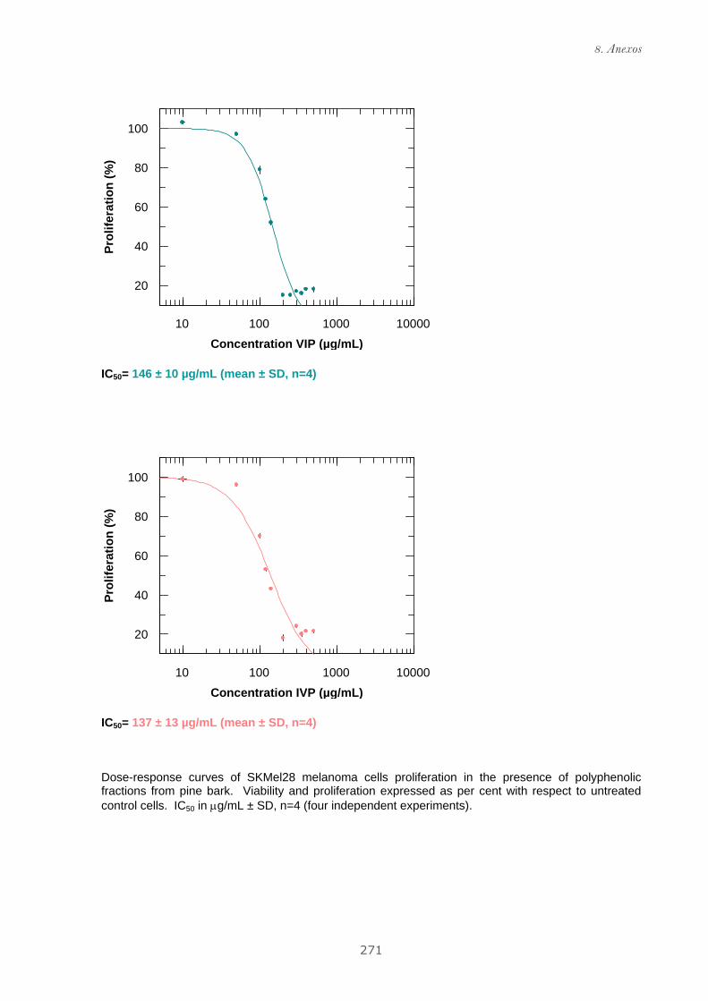
8. Anexos
271
10000100010010
100
80
60
40
20
Concentration VIP (µg/mL)
Prol
ifera
tion
(%)
IC50= 146 ± 10 µg/mL (mean ± SD, n=4)
10000100010010
100
80
60
40
20
Concentration IVP (µg/mL)
Prol
ifera
tion
(%)
IC50= 137 ± 13 µg/mL (mean ± SD, n=4) Dose-response curves of SKMel28 melanoma cells proliferation in the presence of polyphenolic fractions from pine bark. Viability and proliferation expressed as per cent with respect to untreated control cells. IC50 in μg/mL ± SD, n=4 (four independent experiments).

8.2 Material suplementario (publicación 1; apartado 4.1.1, ver pág. 73)
272
10000100010010
100
80
60
40
20
Concentration VIIIP (µg/mL)
Prol
ifera
tion
(%)
IC50= 134 ± 21 µg/mL (mean ± SD, n=4) Dose-response curves of SKMel28 melanoma cells proliferation in the presence of polyphenolic fractions from pine bark. Viability and proliferation expressed as per cent with respect to untreated control cells. IC50 in μg/mL ± SD, n=4 (four independent experiments).
8.2.2 Material suplementario de la publicación 2
(Publicación mostrada en el apartado 4.1.2)


8. Anexos
275
RP-HPLC chromatograms, column Kromasil 100 C18 (25 x 0,4cm i.d., 5 µm particle size) Elution: [A] 0.10% (v/v) aqueous TFA, [B] 0.08 % (v/v) TFA in water /CH3CN 1:4. Flow rate 1mL/min. DAD dectetion and intregation were perfomed to 214nm. a) Load 10 µg crude fraction, gradient elution 12 to 30 % [B] over 30 min; b) Load 10 µg fraction after thioacidolysis, gradient elution 12 to 30 % [B] over 30 min.
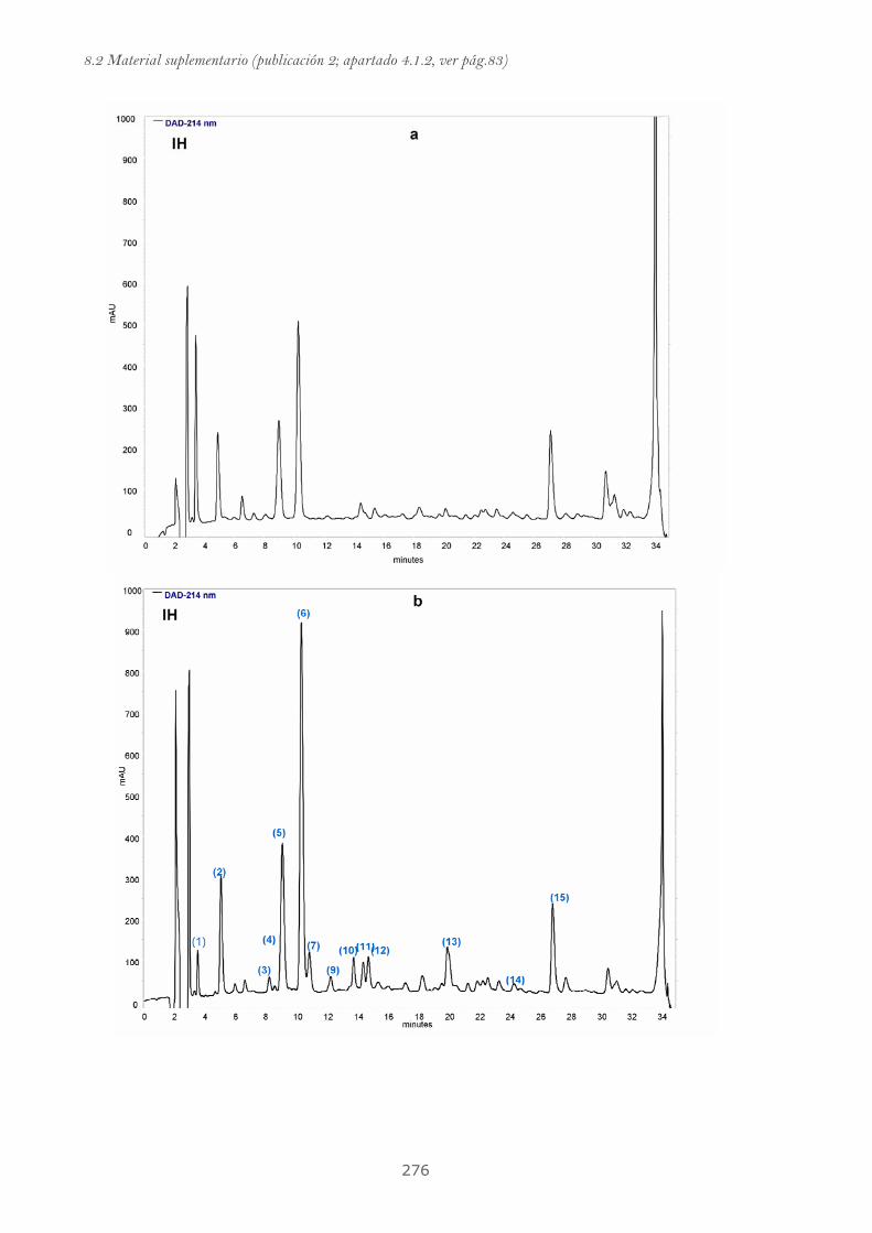
8.2 Material suplementario (publicación 2; apartado 4.1.2, ver pág.83)
276

8. Anexos
277
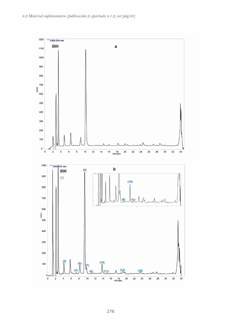
8.2 Material suplementario (publicación 2; apartado 4.1.2, ver pág.83)
278
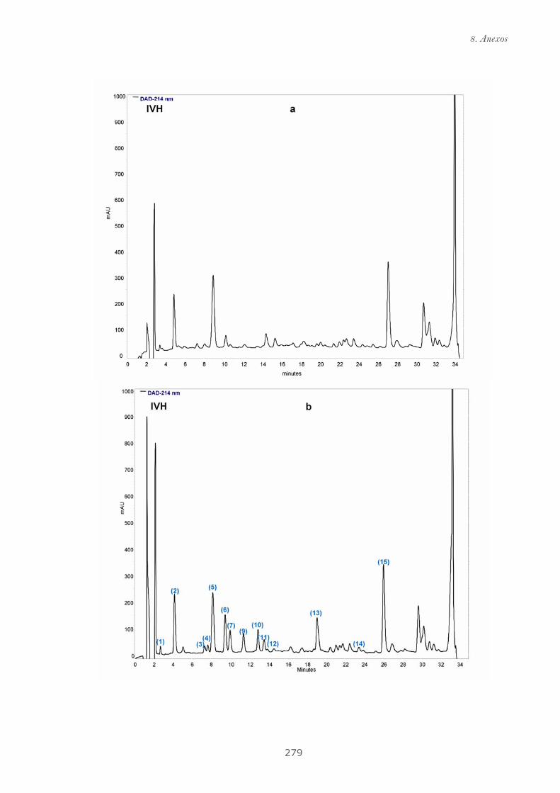
8. Anexos
279
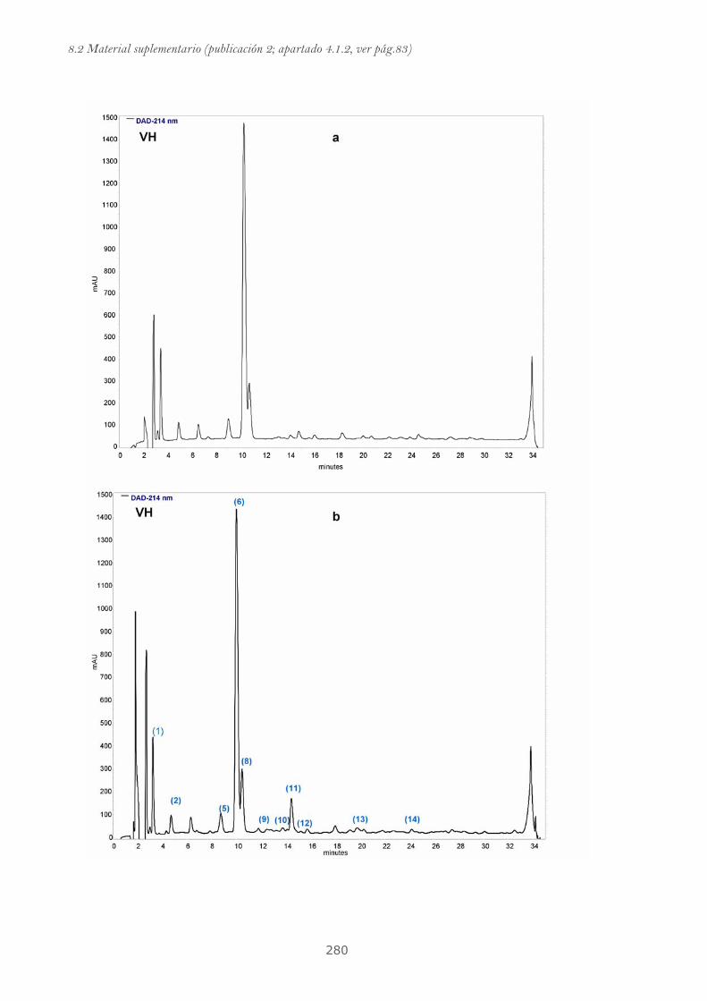
8.2 Material suplementario (publicación 2; apartado 4.1.2, ver pág.83)
280
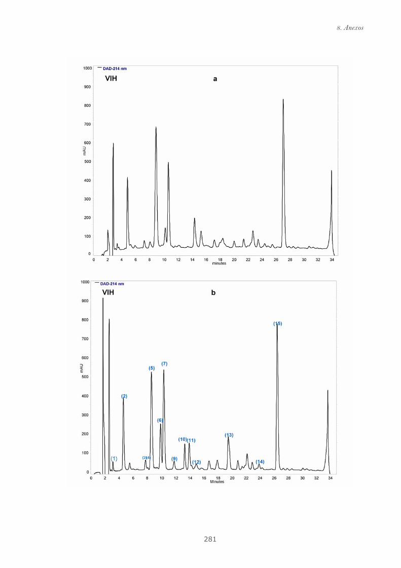
8. Anexos
281

8.2 Material suplementario (publicación 2; apartado 4.1.2, ver pág.83)
282
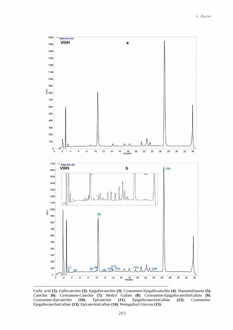
8. Anexos
283
Gallic acid (1); Gallocatechin (2); Epigallocatechin (3); Cysteamine-Epigallocatechin (4); Hamamelitannin (5); Catechin (6); Cysteamine-Catechin (7); Methyl Gallate (8); Cysteamine-EpigallocatechinGallate (9); Cysteamine-Epicatechin (10); Epicatechin (11); EpigallocatechinGallate (12); Cysteamine-EpigallocatechinGallate (13); EpicatechinGallate (14); Pentagalloyl Glucose (15).
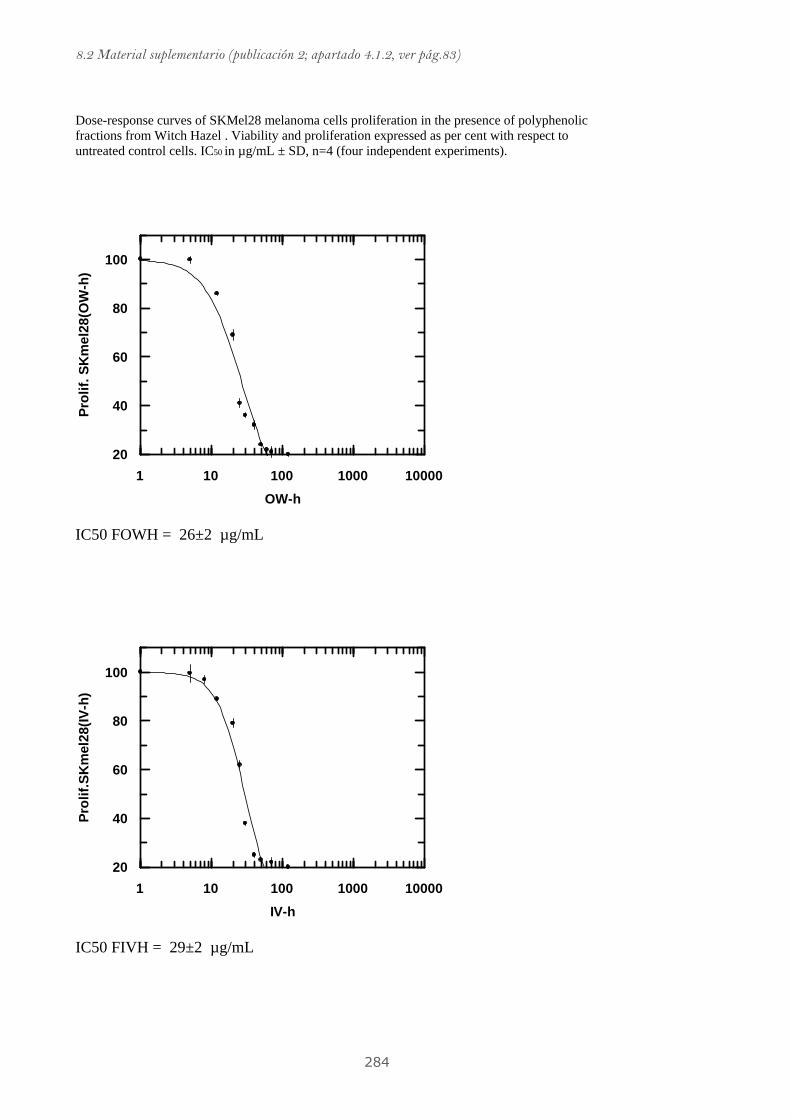
8.2 Material suplementario (publicación 2; apartado 4.1.2, ver pág.83)
284
Dose-response curves of SKMel28 melanoma cells proliferation in the presence of polyphenolic fractions from Witch Hazel . Viability and proliferation expressed as per cent with respect to untreated control cells. IC50 in µg/mL ± SD, n=4 (four independent experiments).
100001000100101
100
80
60
40
20
OW-h
Prol
if. S
Km
el28
(OW
-h)
IC50 FOWH = 26±2 µg/mL
100001000100101
100
80
60
40
20
IV-h
Prol
if.SK
mel
28(IV
-h)
IC50 FIVH = 29±2 µg/mL
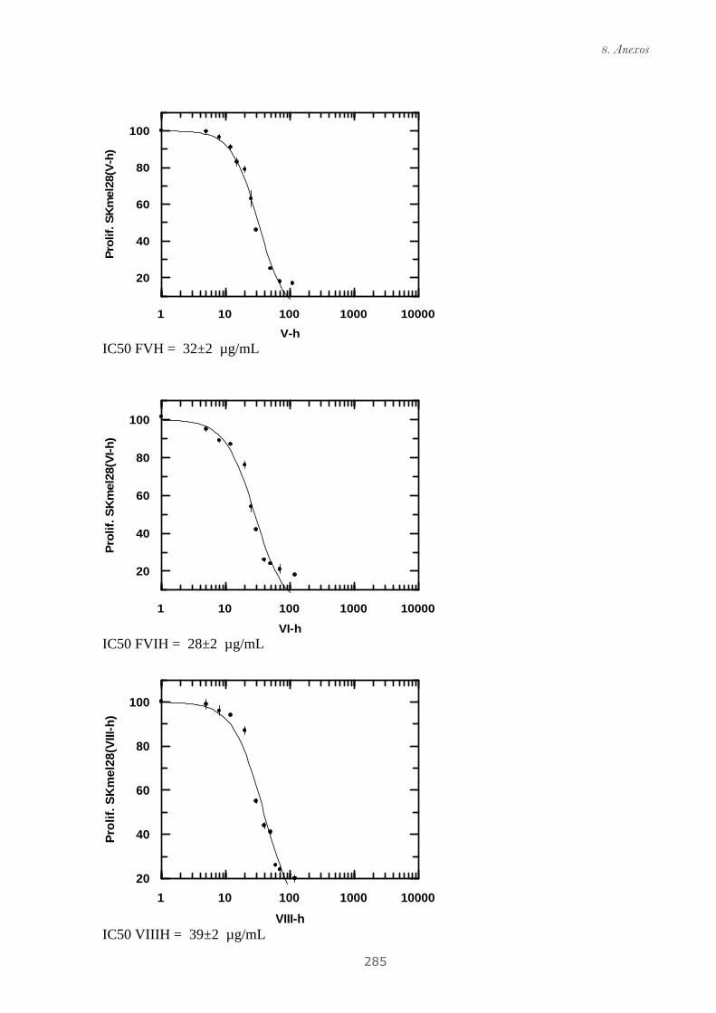
8. Anexos
285
100001000100101
100
80
60
40
20
V-h
Prol
if. S
Km
el28
(V-h
)
IC50 FVH = 32±2 µg/mL
100001000100101
100
80
60
40
20
VI-h
Prol
if. S
Km
el28
(VI-h
)
IC50 FVIH = 28±2 µg/mL
100001000100101
100
80
60
40
20
VIII-h
Prol
if. S
Km
el28
(VIII
-h)
IC50 VIIIH = 39±2 µg/mL

8.2.3 Material suplementario de la publicación 4
(Publicación mostrada en el apartado 4.2.2)

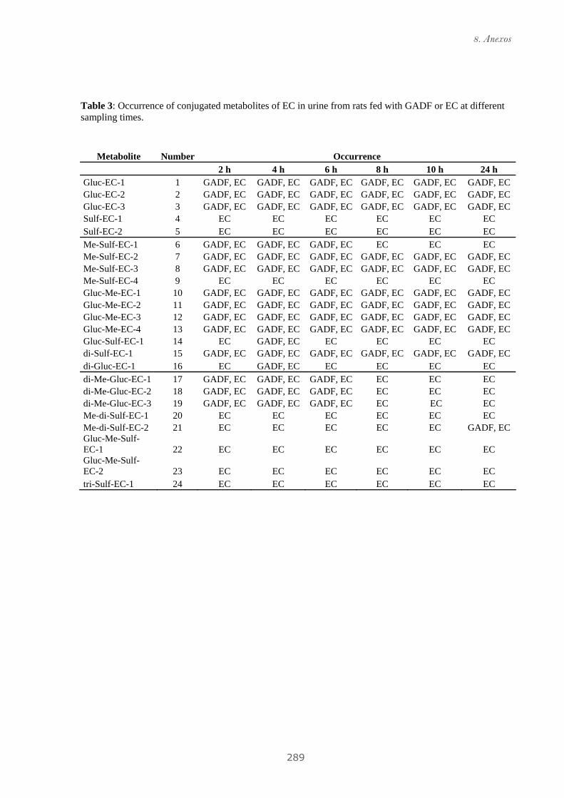
8. Anexos
289
Table 3: Occurrence of conjugated metabolites of EC in urine from rats fed with GADF or EC at different sampling times.
Metabolite Number Occurrence 2 h 4 h 6 h 8 h 10 h 24 h Gluc-EC-1 1 GADF, EC GADF, EC GADF, EC GADF, EC GADF, EC GADF, EC Gluc-EC-2 2 GADF, EC GADF, EC GADF, EC GADF, EC GADF, EC GADF, EC Gluc-EC-3 3 GADF, EC GADF, EC GADF, EC GADF, EC GADF, EC GADF, EC Sulf-EC-1 4 EC EC EC EC EC EC Sulf-EC-2 5 EC EC EC EC EC EC Me-Sulf-EC-1 6 GADF, EC GADF, EC GADF, EC EC EC EC Me-Sulf-EC-2 7 GADF, EC GADF, EC GADF, EC GADF, EC GADF, EC GADF, EC Me-Sulf-EC-3 8 GADF, EC GADF, EC GADF, EC GADF, EC GADF, EC GADF, EC Me-Sulf-EC-4 9 EC EC EC EC EC EC Gluc-Me-EC-1 10 GADF, EC GADF, EC GADF, EC GADF, EC GADF, EC GADF, EC Gluc-Me-EC-2 11 GADF, EC GADF, EC GADF, EC GADF, EC GADF, EC GADF, EC Gluc-Me-EC-3 12 GADF, EC GADF, EC GADF, EC GADF, EC GADF, EC GADF, EC Gluc-Me-EC-4 13 GADF, EC GADF, EC GADF, EC GADF, EC GADF, EC GADF, EC Gluc-Sulf-EC-1 14 EC GADF, EC EC EC EC EC di-Sulf-EC-1 15 GADF, EC GADF, EC GADF, EC GADF, EC GADF, EC GADF, EC di-Gluc-EC-1 16 EC GADF, EC EC EC EC EC di-Me-Gluc-EC-1 17 GADF, EC GADF, EC GADF, EC EC EC EC di-Me-Gluc-EC-2 18 GADF, EC GADF, EC GADF, EC EC EC EC di-Me-Gluc-EC-3 19 GADF, EC GADF, EC GADF, EC EC EC EC Me-di-Sulf-EC-1 20 EC EC EC EC EC EC Me-di-Sulf-EC-2 21 EC EC EC EC EC GADF, EC Gluc-Me-Sulf-EC-1 22 EC EC EC EC EC EC Gluc-Me-Sulf-EC-2 23 EC EC EC EC EC EC tri-Sulf-EC-1 24 EC EC EC EC EC EC
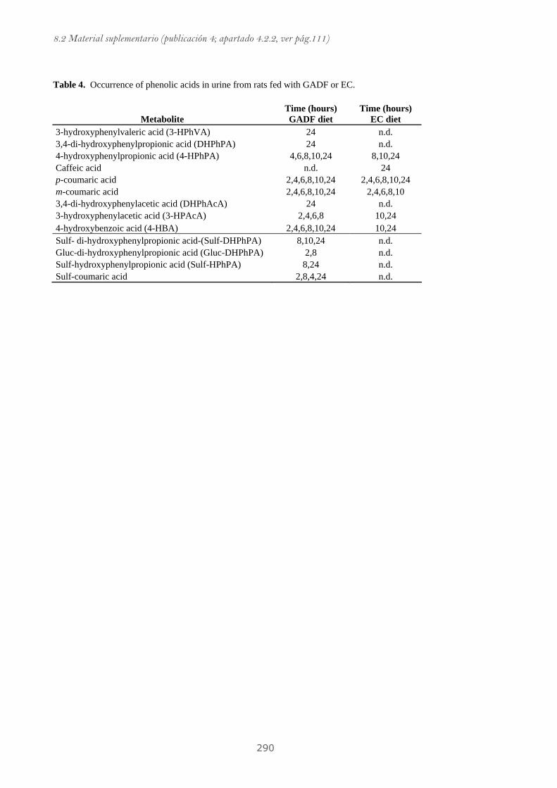
8.2 Material suplementario (publicación 4; apartado 4.2.2, ver pág.111)
290
Table 4. Occurrence of phenolic acids in urine from rats fed with GADF or EC.
Metabolite Time (hours) GADF diet
Time (hours) EC diet
3-hydroxyphenylvaleric acid (3-HPhVA) 24 n.d. 3,4-di-hydroxyphenylpropionic acid (DHPhPA) 24 n.d. 4-hydroxyphenylpropionic acid (4-HPhPA) 4,6,8,10,24 8,10,24 Caffeic acid n.d. 24 p-coumaric acid 2,4,6,8,10,24 2,4,6,8,10,24 m-coumaric acid 2,4,6,8,10,24 2,4,6,8,10 3,4-di-hydroxyphenylacetic acid (DHPhAcA) 24 n.d. 3-hydroxyphenylacetic acid (3-HPAcA) 2,4,6,8 10,24 4-hydroxybenzoic acid (4-HBA) 2,4,6,8,10,24 10,24 Sulf- di-hydroxyphenylpropionic acid-(Sulf-DHPhPA) 8,10,24 n.d. Gluc-di-hydroxyphenylpropionic acid (Gluc-DHPhPA) 2,8 n.d. Sulf-hydroxyphenylpropionic acid (Sulf-HPhPA) 8,24 n.d. Sulf-coumaric acid 2,8,4,24 n.d.
